Cell types and structures involved in tench, Tinca tinca (L.), defence mechanisms against a systemic digenean infection B S Dezfuli 1 , A Lui 1 , F Pironi 1 , M Manera 2 , A P Shinn 3 and M Lorenzoni 4 1 Department of Biology and Evolution, University of Ferrara, Ferrara, Italy 2 Department of Food Science, University of Teramo, Teramo, Italy 3 Institute of Aquaculture, University of Stirling, Stirling, UK 4 Department of Cellular and Environmental Biology, University of Perugia, Perugia, Italy Abstract Histopathological and ultrastructural investigations were conducted on 36 tench, Tinca tinca (L.), from Lake Trasimeno (Italy). The gills, intestine, liver, spleen, kidney and heart of 21 individuals were found to harbour an extensive infection of larvae of an unidentified digenean trematode. The eyes, gonads, swim bladder and muscles were uninfected. The para- sites in each tissue type were embedded in a granu- lomatous proliferation of tissue, forming a reactive fibroconnective capsule around each larva. Most of the encysted larvae were metacercariae, in a degenera- tive state, but on occasion some cercariae were found. Many of the granulomas were either necrotic or had a calcified core. Within the granuloma of each, the occurrence of granulocytes, macrophages, rodlet cells and pigment-bearing macrophage aggregates was observed. Hearts bore the highest parasitic infection. Whilst the presence of metacercariae within the intes- tine was found positioned between the submucosa and muscle layers, metacercariae in the liver were commonly found encysted on its surface where the hepatocytes in close contact with the granuloma were observed to have electron-lucent vesicles within their cytoplasm. Metacercariae encysting adjacent to the cartilaginous rods of gill filaments were seen to elicit a proliferation of the cartilage from the perichondrium. Rodlet cells, neutrophils and mast cells were frequently observed in close proximity to, and within, infected gill capillaries. Given the degenerated state of most granulomas, a morphology-based identification of the enclosed digeneans was not possible. Keywords: fish infection, granuloma, helminth lar- vae, immune cells, inflammation. Introduction Despite the popularity of tench, Tinca tinca (L.), as trash fish for angling and its’ wide geographic range, the pathological effects their helminth para- sites exert have received very little attention. The life cycles of several digeneans are complex, with those of most species requiring two intermediate hosts and a definitive host (Poulin & Cribb 2002). In fish, which act as the second intermedi- ate host for several digenean families, the metac- ercariae are found to encyst in a range of different body organs (see Olson & Pierce 1997; Wang, Yao & Nie 2001; Ogawa, Nakatsugawa & Yasu- zaki 2004). Following infection, the extent of the subse- quent host reaction can vary considerably. Typi- cally, a fibrous capsule of collagen, associated with host fibroblasts, is produced surrounding the invading parasite with, according to some authors, little, if any, host immune response (Stein & Lumsden 1971; Galaktionov et al. 1997). There is a widely held belief that the main function of the host immune system is to protect the organism against infection in order to minimize the fitness costs of being infected (Rohlenova ´ et al. 2011). The current study sets out to detail the cellular inflammatory reaction launched by tench in Correspondence B S Dezfuli, Department of Biology and Evo- lution, University of Ferrara, Borsari St. 46, 44121 Ferrara, Italy (e-mail: [email protected]) 1 Ó 2013 Blackwell Publishing Ltd Journal of Fish Diseases 2013 doi:10.1111/jfd.12049

Welcome message from author
This document is posted to help you gain knowledge. Please leave a comment to let me know what you think about it! Share it to your friends and learn new things together.
Transcript

Cell types and structures involved in tench, Tinca tinca(L.), defence mechanisms against a systemic digeneaninfection
B S Dezfuli1, A Lui1, F Pironi1, M Manera2, A P Shinn3 and M Lorenzoni4
1 Department of Biology and Evolution, University of Ferrara, Ferrara, Italy
2 Department of Food Science, University of Teramo, Teramo, Italy
3 Institute of Aquaculture, University of Stirling, Stirling, UK
4 Department of Cellular and Environmental Biology, University of Perugia, Perugia, Italy
Abstract
Histopathological and ultrastructural investigationswere conducted on 36 tench, Tinca tinca (L.), fromLake Trasimeno (Italy). The gills, intestine, liver,spleen, kidney and heart of 21 individuals were foundto harbour an extensive infection of larvae of anunidentified digenean trematode. The eyes, gonads,swim bladder and muscles were uninfected. The para-sites in each tissue type were embedded in a granu-lomatous proliferation of tissue, forming a reactivefibroconnective capsule around each larva. Most ofthe encysted larvae were metacercariae, in a degenera-tive state, but on occasion some cercariae were found.Many of the granulomas were either necrotic or had acalcified core. Within the granuloma of each, theoccurrence of granulocytes, macrophages, rodlet cellsand pigment-bearing macrophage aggregates wasobserved. Hearts bore the highest parasitic infection.Whilst the presence of metacercariae within the intes-tine was found positioned between the submucosaand muscle layers, metacercariae in the liver werecommonly found encysted on its surface where thehepatocytes in close contact with the granuloma wereobserved to have electron-lucent vesicles within theircytoplasm. Metacercariae encysting adjacent to thecartilaginous rods of gill filaments were seen to elicit aproliferation of the cartilage from the perichondrium.Rodlet cells, neutrophils and mast cells were frequentlyobserved in close proximity to, and within, infectedgill capillaries. Given the degenerated state of most
granulomas, a morphology-based identification ofthe enclosed digeneans was not possible.
Keywords: fish infection, granuloma, helminth lar-vae, immune cells, inflammation.
Introduction
Despite the popularity of tench, Tinca tinca (L.),as trash fish for angling and its’ wide geographicrange, the pathological effects their helminth para-sites exert have received very little attention. Thelife cycles of several digeneans are complex, withthose of most species requiring two intermediatehosts and a definitive host (Poulin & Cribb2002). In fish, which act as the second intermedi-ate host for several digenean families, the metac-ercariae are found to encyst in a range of differentbody organs (see Olson & Pierce 1997; Wang,Yao & Nie 2001; Ogawa, Nakatsugawa & Yasu-zaki 2004).Following infection, the extent of the subse-
quent host reaction can vary considerably. Typi-cally, a fibrous capsule of collagen, associated withhost fibroblasts, is produced surrounding theinvading parasite with, according to some authors,little, if any, host immune response (Stein &Lumsden 1971; Galaktionov et al. 1997). There isa widely held belief that the main function of thehost immune system is to protect the organismagainst infection in order to minimize the fitnesscosts of being infected (Rohlenova et al. 2011).The current study sets out to detail the cellular
inflammatory reaction launched by tench in
Correspondence B S Dezfuli, Department of Biology and Evo-
lution, University of Ferrara, Borsari St. 46, 44121 Ferrara, Italy
(e-mail: [email protected])
1� 2013
Blackwell Publishing Ltd
Journal of Fish Diseases 2013 doi:10.1111/jfd.12049

response to an infection of digenean larvae withinthe gills and several visceral organs. The intenseinflammatory response documented involves mastcells (MCs), neutrophils, macrophage aggregates(MAs), macrophages, epithelioid cells, collagenfibres and rodlet cells (RCs).Mast cells, also referred to as eosinophilic granular
cells (Reite & Evensen 2006), in fish are reported tooccur at the sites of inflammation caused by parasiticinfection (Reite & Evensen 2006; Dezfuli et al.2009). MCs degranulate when exposed to a varietyof known degranulation agents (Manera et al. 2011)and pathogens (Dezfuli & Giari 2008).Neutrophils are the first cell type recruited to the
site of an acute inflammatory response (Mollinedo,Borregaard & Boxer 1999), and their chemotaxis,phagocytosis and destruction of intracellular andextracellular pathogens demonstrate their importantrole in innate immunity against pathogens and par-asitic infection (Secombes 1996; Stakauskas et al.2007).MAs or melanomacrophage centres may also be
found within tissue responses encapsulating manyforeign bodies and parasites (Ferguson 2006).MAs form from aggregations of pigment-contain-ing cells (for a review, see Agius & Roberts2003), and their functions have been reported toinclude the focal destruction, detoxification andrecycling of endogenous and exogenous materials(Ellis 1980; Vigliano et al. 2006).Rodlet cells are characterized by a thick subplas-
malemmar fibrillar capsule, by a basal nucleus andby the presence of conspicuous rodlets (for areview, see Manera & Dezfuli 2004). Evidence forthe possible function of these cells, as immunecells, results from their reported increase in fishthat are infected with parasites (Reite & Evensen2006; Matisz, Goater & Bray 2010).Certain cells, which are involved in the chronic
inflammatory response of teleost fish, have manyfeatures that are typical of mammalian epithelioidcells (Noga, Dykstra & Wright 1989; Ferguson2006). In the presence of a foreign antigen, thiscell type becomes activated and avidly phagocytic(Johnson 1988).
Materials and methods
In September 2011, 36 specimens of tench fromLake Trasimeno (Province of Perugia, UmbriaRegion, Central Italy) were provided by the Trasi-meno Fish Consortium. The fish were transferred
alive to the Consortium’s facility and killed using125 mg L�1 MS222 (tricaine methanesulfonate,Sandoz), and their spinal cords severed beforebeing lengthed, 41.85 ± 12.24 cm (mean totallength ± SD) and weighed, 1463.6 ± 816.2 g(mean weight ± SD). The tench were dissected andsexed before the internal organs and gill filamentsfrom each were removed and screened for para-sitic infection. From each infected organ, a15 9 15 mm piece of tissue was excised and thenfixed in either chilled (4 °C) Bouin’s or in 10%neutral buffered formalin for 24 h. The sampleswere then processed routinely for paraffin embed-ding, sectioned (5 lm thick) and then stained withhaematoxylin and eosin and/or Alcian Blue 8 GXpH 2.5 and periodic acid Schiff’s (AB/PAS)stains. Multiple histological sections were takenfrom each tissue block, examined and photo-graphed using an optical microscope (NikonEclipse 80i).For transmission electron microscopy (TEM),
7 9 7 mm pieces of tissue from infected organswere fixed in chilled 2.5% glutaraldehyde in 0.1 M
sodium cacodylate buffer for 3 h. The fixed tissueswere then post-fixed in 1% osmium tetroxide for2 h and then rinsed and stored in 0.1 M sodium cac-odylate buffer containing 6% sucrose for 12 h.Then, the samples were dehydrated through a gradedacetone series and then embedded in epoxy resin(Durcupan ACM, Fluka). Semi-thin sections(1.5 lm) were cut on a Reichert Om U 2 ultramicrotome (Reichert-Jung) and stained with tolui-dine blue. Ultrathin sections (90 nm) were stainedwith a 4% uranyl acetate solution in 50% ethanoland Reynold’s lead citrate and observed using a Hit-achi H-800 transmission electron microscope. Forboth light and transmission electron microscopy, cor-responding pieces of organs from uninfected tenchwere also processed for comparison with the parasit-ized organ.In addition, 5 9 5 mm pieces of tissue were excised
from the infected organs and then fixed in 90% etha-nol for DNA extraction, PCR and sequencing.
Results
Light microscopy
Of the 36 tench that were examined, the gills andviscera (i.e. intestine, liver, spleen, kidney, heart) of21 fish (58.3%) were found to be infected with lar-vae of an unidentified digenean trematode. No dige-nean larvae were encountered in the eyes, gonads,
2
Journal of Fish Diseases 2013 B S Dezfuli et al. Cellular defence mechanisms in infected tench
� 2013
Blackwell Publishing Ltd

swim bladder and muscles of tench. In all theinfected organs, a granulomatous inflammatorytissue surrounded the parasite larvae which variedmarkedly in size and structural organization (seebelow for the electron microscopy description). Thevast majority of granulomas contained necroticmaterial or a calcified core; as such, the enclosedparasite stages were in a degenerated state with nodiscernible taxonomic features upon which to base adefinitive identification. In a small proportion ofgranulomas, general metacercarian features were evi-dent and in a few granulomas, cercarial-like stageswere seen. Whilst suggestions as to the possible dig-enean species involved (e.g. Asymphylodora tincae(Modeer, 1790)) can be made by cross-referencinghost and geographic location details contained inopen-source, online parasite databases (i.e. http://www.nhm.ac.uk/research-curation/research/projects/host-parasites/database/index.jsp), a definitive identi-fication, as determined by base sequencing of infor-mative genomic regions, is preferred. Although aparallel molecular study is ongoing, no definitiveidentification has, as yet, been possible.In the following sections, the host reaction
within each tissue to the parasitic infection isdescribed.
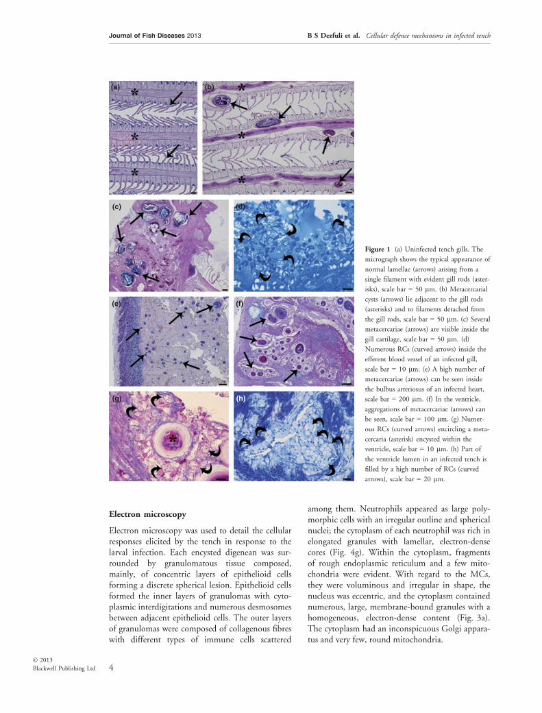
Gills. Figure 1a shows the gill filaments of anuninfected tench, whilst those presented inFig. 1(b) shows that the presence of encystedmetacercariae adjacent to the cartilaginous rods ofthe gill filaments elicits a proliferation of the carti-lage from the perichondrium. In all the infectedtench that were examined, the gill cartilage har-boured the highest number of larvae that werefound in the gills (Fig. 1c). In a few cases, hyperpla-sia of the gill epithelium and fusion of the lamellaewere seen in filaments that were close to the sites ofmetacercarial infection. Frequently, the metacerca-riae induced detachment of the gill filaments fromthe cartilaginous rods (Fig. 1b). Numerous RCswere commonly observed inside the efferent bloodvessels of infected gills (Fig. 1d).
Heart. This was the most heavily parasitizedorgan. Of the 21 parasitized hearts, both the bulbusarteriosus (Fig. 1e) and the ventricle (Fig. 1f) har-boured single or aggregations of larvae. RCs werethe dominant cells found in the heart, and thoseencountered in the ventricle either partially or com-pletely surround the larvae (Fig. 1g). Some of the
granulomas seen within the heart contained necro-tic tissue and/or a calcified core (Fig. 1g). RCs werealso found in abundance in tissues located at a dis-tance away from the sites of infection (Fig. 1h).RCs were found to co-occur with MCs and neu-trophils, whilst the number of the granulocytesrecorded was remarkably low. Macrophages werealso commonly encountered within the vicinity ofmetacercariae; these cells, which possessed indefin-able inclusions, appeared large in comparison withthe other cell types that were observed (see thedescriptions provided in the electron microscopysection).
Intestine. Larvae were observed in the submucosaland muscle layers (Fig. 2a) and within the thick-ness of the serosa where they induced a hyperplas-tic response (not shown). MAs, some RCs, MCsand neutrophils were seen in close proximity tothe metacercariae; most notably, numerous MAswere seen in the muscle layer (Fig. 2a). Again,granulomas with necrotic and/or calcified centreswere seen (Fig. 2a, right side of the photograph).
Liver. Metacercariae were observed throughoutthe liver (Fig. 2b), and not only on the surface(Fig. 2c). MAs were frequently observed in inti-mate contact with parasites (Fig. 2d). MCs andRCs were observed in infected tissues, and thehepatocytes close to the metacercariae had electron-lucent vesicles within their cytoplasm (see below).
Spleen. The spleen had a lower number of larvaecompared to those observed in the other visceralorgans. The larvae were seen in both the deepertissues (Fig. 2e) and on the surface of the organ(Fig. 2f). MAs were occasionally observed in con-tact with larvae (Fig. 2f), and RCs (Fig. 2f), neu-trophils and MCs were all found to co-occurwithin the vessels of infected splenic tissue.
Kidney. The pathology and cellular responses dis-played by infected kidneys were similar to thoseof the spleen. Parasite larvae were found through-out the kidney (Fig. 2g), as well as close to(Fig. 2h) or on the surface. MAs were commonlyobserved in contact with the metacercariae(Fig. 2h); MCs and neutrophils were notablyabundant throughout infected kidneys (see below).Again, granulomas containing necrotic or calcifiedmaterial were seen (Fig. 2g).
3
Journal of Fish Diseases 2013 B S Dezfuli et al. Cellular defence mechanisms in infected tench
� 2013
Blackwell Publishing Ltd
Electron microscopy
Electron microscopy was used to detail the cellularresponses elicited by the tench in response to thelarval infection. Each encysted digenean was sur-rounded by granulomatous tissue composed,mainly, of concentric layers of epithelioid cellsforming a discrete spherical lesion. Epithelioid cellsformed the inner layers of granulomas with cyto-plasmic interdigitations and numerous desmosomesbetween adjacent epithelioid cells. The outer layersof granulomas were composed of collagenous fibreswith different types of immune cells scattered
among them. Neutrophils appeared as large poly-morphic cells with an irregular outline and sphericalnuclei; the cytoplasm of each neutrophil was rich inelongated granules with lamellar, electron-densecores (Fig. 4g). Within the cytoplasm, fragmentsof rough endoplasmic reticulum and a few mito-chondria were evident. With regard to the MCs,they were voluminous and irregular in shape, thenucleus was eccentric, and the cytoplasm containednumerous, large, membrane-bound granules with ahomogeneous, electron-dense content (Fig. 3a).The cytoplasm had an inconspicuous Golgi appara-tus and very few, round mitochondria.
(a)
(c) (d)
(e) (f)
(g) (h)
(b)
Figure 1 (a) Uninfected tench gills. The
micrograph shows the typical appearance of
normal lamellae (arrows) arising from a
single filament with evident gill rods (aster-
isks), scale bar = 50 lm. (b) Metacercarial
cysts (arrows) lie adjacent to the gill rods
(asterisks) and to filaments detached from
the gill rods, scale bar = 50 lm. (c) Several
metacercariae (arrows) are visible inside the
gill cartilage, scale bar = 50 lm. (d)
Numerous RCs (curved arrows) inside the
efferent blood vessel of an infected gill,
scale bar = 10 lm. (e) A high number of
metacercariae (arrows) can be seen inside
the bulbus arteriosus of an infected heart,
scale bar = 200 lm. (f) In the ventricle,
aggregations of metacercariae (arrows) can
be seen, scale bar = 100 lm. (g) Numer-
ous RCs (curved arrows) encircling a meta-
cercaria (asterisk) encysted within the
ventricle, scale bar = 10 lm. (h) Part of
the ventricle lumen in an infected tench is
filled by a high number of RCs (curved
arrows), scale bar = 20 lm.
4
Journal of Fish Diseases 2013 B S Dezfuli et al. Cellular defence mechanisms in infected tench
� 2013
Blackwell Publishing Ltd

The RCs that were seen were large in size, ovaland contained several typical club-shaped forma-tions (‘rodlets’), part of which had a crystallinecore. The cytoplasm of the RCs was vesicularand foamy (Fig. 3b,c), with no recognizableorganelles within. The macrophages, by compari-son, appeared large (Fig. 4d) and contained vesic-ular structures with electron-opaque contents(dense bodies) and electron-lucent vesicles.
Gills. Digenean larvae within the gills were sur-rounded by host neutrophils, MCs (Fig. 3a) andRCs (Fig. 3b). Numerous RCs, some of whichwere discharging their contents, were seen withinthe lumen of the efferent vessels of infected fish(Fig. 3b).
Heart. RCs were the dominant cells seen in asso-ciation with larvae in the ventricle (Figure 3c),although RCs were also seen further away withinthe lumen of the ventricle where they were foundto co-occur with MCs (Fig. 3d). Several RCs wereobserved in the process of discharging their con-tents. Macrophages were also present, frequentlyin close proximity to the metacercariae or, as seenin several instances, in contact with the externalsurface of a granuloma. These cells appeared largein size with indefinable electron-opaque inclusions(not shown).
Intestine. Metacercariae within the submucosallayer were seen surrounded by MCs, neutrophilsand RCs (Fig. 3e), where interestingly in some
(a)
(c) (d)
(e) (f)
(g) (h)
(b)
Figure 2 (a) Metacercariae (arrows) ency-
sted in the muscle layer of the tench intestine;
MAs (curved arrows) in close vicinity to the
parasite can be seen, scale bar = 100 lm.
(b) Histological section through an infected
liver showing metacercariae (arrows) embed-
ded within the tissue, scale bar = 200 lm.
(c) Metacercariae (arrows) encysted on the
surface of the liver (asterisk); MAs (curved
arrows) in close proximity to the parasites
are evident, scale bar = 50 lm. (d) Three
metacercariae (arrows) in close contact
with a hepatic MA (curved arrow), scale
bar = 20 lm. (e) Histological section
through an infected spleen showing the deep
position of a metacercaria (arrow) encysted
within. It can be seen that the parasite is in
close contact with a MA (curved arrow),
scale bar = 20 lm. (f) A parasite (arrow)
embedded on the surface of the spleen with
a MA (curved arrow) in contact with the
external surface of the parasite. Several RCs
(circle) inside the vessel can be seen, scale
bar = 20 lm. (g) Histological section of an
infected kidney with encysted metacercariae
(arrows), scale bar = 50 lm. (h) A metacer-
caria (arrow) and two MAs (curved arrows)
in close proximity to one another, scale
bar = 20 lm.
5
Journal of Fish Diseases 2013 B S Dezfuli et al. Cellular defence mechanisms in infected tench
� 2013
Blackwell Publishing Ltd
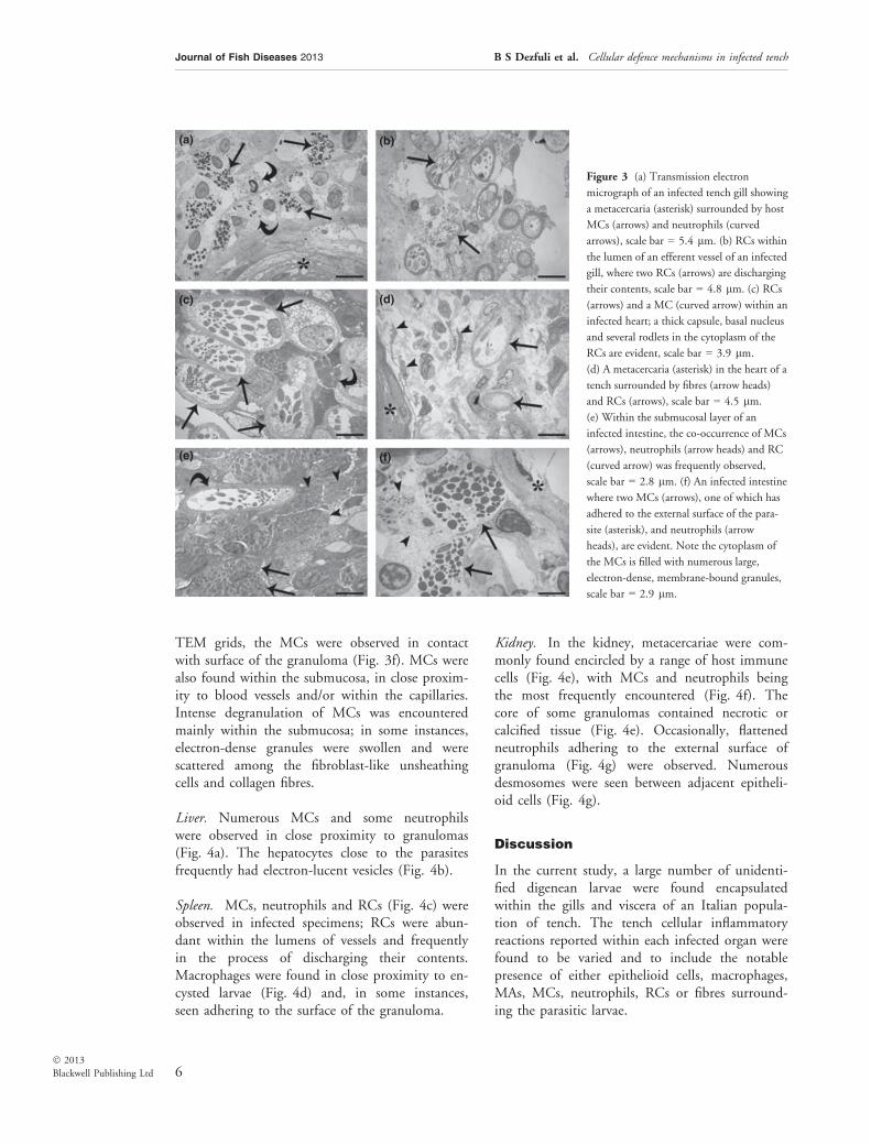
TEM grids, the MCs were observed in contactwith surface of the granuloma (Fig. 3f). MCs werealso found within the submucosa, in close proxim-ity to blood vessels and/or within the capillaries.Intense degranulation of MCs was encounteredmainly within the submucosa; in some instances,electron-dense granules were swollen and werescattered among the fibroblast-like unsheathingcells and collagen fibres.
Liver. Numerous MCs and some neutrophilswere observed in close proximity to granulomas(Fig. 4a). The hepatocytes close to the parasitesfrequently had electron-lucent vesicles (Fig. 4b).
Spleen. MCs, neutrophils and RCs (Fig. 4c) wereobserved in infected specimens; RCs were abun-dant within the lumens of vessels and frequentlyin the process of discharging their contents.Macrophages were found in close proximity to en-cysted larvae (Fig. 4d) and, in some instances,seen adhering to the surface of the granuloma.
Kidney. In the kidney, metacercariae were com-monly found encircled by a range of host immunecells (Fig. 4e), with MCs and neutrophils beingthe most frequently encountered (Fig. 4f). Thecore of some granulomas contained necrotic orcalcified tissue (Fig. 4e). Occasionally, flattenedneutrophils adhering to the external surface ofgranuloma (Fig. 4g) were observed. Numerousdesmosomes were seen between adjacent epitheli-oid cells (Fig. 4g).
Discussion
In the current study, a large number of unidenti-fied digenean larvae were found encapsulatedwithin the gills and viscera of an Italian popula-tion of tench. The tench cellular inflammatoryreactions reported within each infected organ werefound to be varied and to include the notablepresence of either epithelioid cells, macrophages,MAs, MCs, neutrophils, RCs or fibres surround-ing the parasitic larvae.
(a)
(c) (d)
(e) (f)
(b)
Figure 3 (a) Transmission electron
micrograph of an infected tench gill showing
a metacercaria (asterisk) surrounded by host
MCs (arrows) and neutrophils (curved
arrows), scale bar = 5.4 lm. (b) RCs within
the lumen of an efferent vessel of an infected
gill, where two RCs (arrows) are discharging
their contents, scale bar = 4.8 lm. (c) RCs
(arrows) and a MC (curved arrow) within an
infected heart; a thick capsule, basal nucleus
and several rodlets in the cytoplasm of the
RCs are evident, scale bar = 3.9 lm.
(d) A metacercaria (asterisk) in the heart of a
tench surrounded by fibres (arrow heads)
and RCs (arrows), scale bar = 4.5 lm.
(e) Within the submucosal layer of an
infected intestine, the co-occurrence of MCs
(arrows), neutrophils (arrow heads) and RC
(curved arrow) was frequently observed,
scale bar = 2.8 lm. (f) An infected intestine
where two MCs (arrows), one of which has
adhered to the external surface of the para-
site (asterisk), and neutrophils (arrow
heads), are evident. Note the cytoplasm of
the MCs is filled with numerous large,
electron-dense, membrane-bound granules,
scale bar = 2.9 lm.
6
Journal of Fish Diseases 2013 B S Dezfuli et al. Cellular defence mechanisms in infected tench
� 2013
Blackwell Publishing Ltd

Larval migration of digeneans and encapsula-tion within the viscera and body tissues of fish(Olson & Pierce 1997; Sitja-Bobadilla 2008; Ma-tisz et al. 2010) generally induces the develop-ment of fibrogranulomatous lesions (Balouet &Baudin-Laurencin 1986). Histologically, fishgranulomas are inflammatory foci composed ofconcentric layers of epithelioid cells, and are verysimilar to mammalian granulomas (Noga et al.1989; Roberts 2012). Epithelioid cells are so-called because of their morphological similarityto epithelial cells (Cotran, Kumar & Collins
1999). It is assumed that these epithelioid cellsare typically transformed macrophages, whichhave the principal role of engulfing foreignagents (Ferguson 2006). In each metacercariae-infected organ examined in the current study,the parasites were found encircled by epithelioidcells. Numerous macrophages were found withinthe hearts of infected tench and, in someinstances, were seen adhering to the external sur-face of the larvae. Almost all the macrophagesthat were encountered possessed vesicular struc-tures with electron-opaque contents (dense
(a)
(c) (d)
(e) (f)
(g)
(b)
Figure 4 (a) Metacercaria (asterisk) encir-
cled by MCs (arrows) in the infected liver of
a tench, scale bar = 3.4 lm. (b) Electron-
lucent vesicles (arrows) within the cytoplasm
of hepatocytes of an infected liver were
commonly seen, scale bar = 2.7 lm.
(c) Micrograph shows the lumen of a vessel
within a parasitized spleen, in which RCs
(curved arrows) were abundant, scale
bar = 4.4 lm. (d) Within an infected
spleen, a macrophage (arrow) can be seen in
close vicinity to the tegument of the parasite
(asterisk), scale bar = 1.4 lm. (e) Trans-
verse section through a metacercaria (aster-
isk) within the kidney; host cells (arrow
heads) can be seen encircling the larva, scale
bar = 5.6 lm. (f) Numerous MCs (arrows)
and neutrophils (curved arrows) encircling a
metacercaria (asterisk), scale bar = 5.1 lm.
(g) High magnification of a section taken
through an infected kidney where a flattened
neutrophil (arrows) attached to the external
surface of a granuloma (asterisk) can be seen,
note numerous desmosomes (curved arrows)
between adjacent epithelioid cells, scale
bar = 0.6 lm.
7
Journal of Fish Diseases 2013 B S Dezfuli et al. Cellular defence mechanisms in infected tench
� 2013
Blackwell Publishing Ltd

bodies) and electron-lucent vesicles, suggestingthat they were actively engulfing particles.A prominent feature of chronic inflammation is
the presence of melanin and/or other pigment-containing macrophages, also known as MAs(Agius & Roberts 2003). In the present study,MAs were predominantly seen in close proximityto metacercariae in the liver, spleen and kidney;these results are in agreement with the active roleof these centres in retaining resistant pathogenssuch as parasitic spores or bacteria, and in antigenprocessing during the immune response (Agius &Roberts 2003).Mast cells in fish are reported to occur at sites
of inflammation that are induced by parasiticinfection (Reite & Evensen 2006; Dezfuli et al.2009). The presence of MCs is closely linked tothe ability to rapidly recruit neutrophils andmacrophages to the site of infection (Lamas et al.1991; Secombes 1996). Several records docu-ment the degranulation of MCs following theirexposure to a variety of known degranulationagents (Manera et al. 2011) and pathogens(Dezfuli & Giari 2008). In infected tench, thedegranulation of MCs was frequently observedwithin the connective tissue surrounding capillar-ies and within the blood vessels of the spleen,intestine and gills.According to Roberts (2012), when a common
acute inflammatory response is elicited, it is char-acterized by the presence of neutrophils andmonocytes in the blood and by an accumulationof neutrophils and macrophages at the site ofinfection. Several researchers have reported thatinstead of degranulating, neutrophils have beenseen tightly bound to the surface of the parasite(Butterworth 1984). The latter was seen in thecurrent study, with the neutrophils present incertain infected organs binding tightly to the sur-face of the granulomas with no evidentdegranulation.Numerous RCs were observed within the gills,
spleens and hearts of infected tench, where inter-estingly, an intense migration of RCs was seenwithin the circulatory system of each organ. Thefinding of a high number and recruitment ofRCs provides further evidence regarding theirpotential function and lends additional supportto the findings from the observational studies onhelminth–fish interactions (Reite 2005; Matiszet al. 2010).
Acknowledgements
We thank L. Giari and S. Squerzanti from theUniversity of Ferrara for their technical assistancewith aspects of this study. Thanks are also due toF. Bisonni for his assistance in collecting fish. Thisstudy was supported by grants from the ItalianMinistry of the University and from ScientificResearch and Technology.
References
Agius C. & Roberts R.J. (2003) Melano-macrophage centres
and their role in fish pathology. Journal of Fish Diseases 26,499–509.
Balouet G. & Baudin-Laurencin F. (1986) Granulomatous
nodules in fish: an experimental assessment in rainbow
trout, Salmo gairdneri Richardson, and turbot, Scophthalmusmaximus (L.). Journal of Fish Diseases 9, 417–429.
Butterworth A.E. (1984) Cell-mediated damage to helminths.
Advances in Parasitology 23, 143–235.
Cotran R.S., Kumar V. & Collins Y. (eds) (1999) PathologicBasis of Disease. WB Saunders, Philadelphia.
Dezfuli B.S. & Giari L. (2008) Mast cells in the gills and
intestines of naturally infected fish: evidence of
migration and degranulation. Journal of Fish Diseases31, 845–852.
Dezfuli B.S., Giovinazzo G., Lui A., Boldrini P. & Giari L.
(2009) Intestinal inflammatory response of powan Coregonuslavaretus (Pisces) to the presence of acanthocephalan
infections. Parasitology 136, 929–937.
Ellis A.E. (1980) Antigen-trapping in the spleen and kidney of
the plaice Pleuronectes platessa L. Journal of Fish Diseases 3,413–426.
Ferguson H.W. (2006) A text and atlas of normal tissues in
teleosts and their responses in disease. In: Systemic Pathologyof Fish (ed. by H.W. Ferguson), pp. 10–23. Scotian Press,
London.
Galaktionov K.V., Malkova I.I., Irwin S.W., Saville D.H.
& Maguire J.G. (1997) The structure and formation of
metacercarial cysts in the trematode family
Microphallidae Travassos 1920. Journal of Helminthology71, 13–20.
Johnson R.B. (1988) Monocytes and macrophages. The NewEngland Journal of Medicine 318, 747–752.
Lamas J., Bruno D.W., Santos Y., Anadon R. & Ellis A.E.
(1991) Eosinophilic granular cell response to intraperitoneal
injection with Vibrio anguillarum and its extracellular
products in rainbow trout, Oncorhynchus mykiss. Fish andShellfish Immunology 1, 187–194.
Manera M. & Dezfuli B.S. (2004) Rodlet cells in teleosts: a
new insight into their nature and functions. Journal of FishBiology 65, 597–619.
Manera M., Giammarino A., Borreca C., Giari L. & Dezfuli
B.S. (2011) Degranulation of mast cells due to compound
48/80 induces concentration-dependent intestinal
8
Journal of Fish Diseases 2013 B S Dezfuli et al. Cellular defence mechanisms in infected tench
� 2013
Blackwell Publishing Ltd
contraction in rainbow trout (Oncorhynchus mykissWalbaum) ex vivo. Journal of Experimental Zoology Part A315A, 447–457.
Matisz C.E., Goater C.P. & Bray D. (2010) Density and
maturation of rodlet cells in brain tissue of fathead
minnows (Pimephales promelas) exposed to trematode
cercariae. International Journal for Parasitology 40,307–312.
Mollinedo F., Borregaard N. & Boxer L.A. (1999) Novel
trends in neutrophil structure, function and development.
Immunology Today 20, 535–537.
Noga E.J., Dykstra M.J. & Wright J.F. (1989) Chronic
inflammatory cell with epithelial cell characteristics in teleost
fishes. Veterinary Pathology 26, 429–437.
Ogawa K., Nakatsugawa T. & Yasuzaki M. (2004) Heavy
metacercarial infections of cyprinid fishes in Uji River.
Fisheries Science 70, 132–140.
Olson R.E. & Pierce J.R. (1997) A trematode metacercaria
causing gill cartilage proliferation in steelhead trout from
Oregon. Journal of Wildlife Diseases 33, 886–890.
Poulin R. & Cribb T.H. (2002) Trematode life cycles: short is
sweet? Trends in Parasitology 18, 176–183.
Reite O.B. (2005) The rodlet cells of teleostean fish: their
potential role in host defence in relation to the role of mast
cells/eosinophilic granule cells. Fish and Shellfish Immunology19, 253–267.
Reite O.B. & Evensen Ø. (2006) Inflammatory cells of
teleostean fish: a review focusing on mast cells ⁄ eosinophilicgranule cells and rodlet cells. Fish and Shellfish Immunology20, 192–208.
Roberts R.J. (ed) (2012) Fish Pathology, 4th edn. Wiley-
Blackwell, Chicester.
Rohlenova K., Morand S., Hyrsl P., Tolarova S., Flajshans M.
& Simkova A. (2011) Are fish immune systems really
affected by parasites? An immunoecological study of
common carp (Cyprinus carpio). Parasites & Vectors 4, 120.
Secombes C.J. (1996) The nonspecific immune system: cellular
defences. In: The Fish Immune System: Organism, Pathogen,and Environment (ed. by G. Iwama & T. Nakanishi),
pp. 63–105. San Diego Academic Press, San Diego.
Sitja-Bobadilla A. (2008) Living off a fish: a trade-off between
parasites and the immune system. Fish and ShellfishImmunology 25, 358–372.
Stakauskas R., Schuberth H.J., Leibold W. & Steinhagen D.
(2007) Modulation of carp (Cyprinus carpio) neutrophilfunctions during an infection with the haemoparasite
Trypanoplasma borreli. Fish and Shellfish Immunology 23,446–458.
Stein P.C. & Lumsden R.D. (1971) The ultrastructure of
developing metacercarial cysts of Ascocotyle leighi Burton,1956 (Heterophyidae). Proceedings of the HelminthologicalSociety of Washington 38, 1–10.
Vigliano F.A., Bermudez R., Quiroga M.I. & Nieto J.M.
(2006) Evidence for melano-macrophage centres of teleost as
evolutionary precursors of germinal centres of higher
vertebrates: an immunohistochemical study. Fish andShellfish Immunology 21, 467–471.
Wang G.T., Yao W.J. & Nie P. (2001) Seasonal occurrence of
Dollfustrema vaneyi (Digenea: Bucephalidae) metacercariae in
the bullhead catfish Pseudobagrus fulvidraco in a reservoir in
China. Diseases of Aquatic Organisms 44, 127–131.
Received: 26 September 2012Revision received: 13 October 2012Accepted: 13 October 2012
9
Journal of Fish Diseases 2013 B S Dezfuli et al. Cellular defence mechanisms in infected tench
� 2013
Blackwell Publishing Ltd
Related Documents