Liao Agatha Zawadzka, Kristie Wetzel, Douglas B. Sawyer, Wilson S. Colucci, Carl S. Apstein and Ronglih Mohit Jain, Harout DerSimonian, Daniel A. Brenner, Soeun Ngoy, Paige Teller, Albert S. B. Edge, After Myocardial Infarction Cell Therapy Attenuates Deleterious Ventricular Remodeling and Improves Cardiac Performance Print ISSN: 0009-7322. Online ISSN: 1524-4539 Copyright © 2001 American Heart Association, Inc. All rights reserved. is published by the American Heart Association, 7272 Greenville Avenue, Dallas, TX 75231 Circulation doi: 10.1161/01.CIR.103.14.1920 2001;103:1920-1927 Circulation. http://circ.ahajournals.org/content/103/14/1920 Wide Web at: The online version of this article, along with updated information and services, is located on the World http://circ.ahajournals.org//subscriptions/ is online at: Circulation Information about subscribing to Subscriptions: http://www.lww.com/reprints Information about reprints can be found online at: Reprints: document. and Rights Question and Answer Permissions the middle column of the Web page under Services. Further information about this process is available in the the online version of the published article for which permission is being requested is located, click Request Permissions in can be obtained via RightsLink, a service of the Copyright Clearance Center, not the Editorial Office. Once Circulation Requests for permissions to reproduce figures, tables, or portions of articles originally published in Permissions: by guest on May 21, 2014 http://circ.ahajournals.org/ Downloaded from by guest on May 21, 2014 http://circ.ahajournals.org/ Downloaded from

Welcome message from author
This document is posted to help you gain knowledge. Please leave a comment to let me know what you think about it! Share it to your friends and learn new things together.
Transcript
LiaoAgatha Zawadzka, Kristie Wetzel, Douglas B. Sawyer, Wilson S. Colucci, Carl S. Apstein and Ronglih
Mohit Jain, Harout DerSimonian, Daniel A. Brenner, Soeun Ngoy, Paige Teller, Albert S. B. Edge,After Myocardial Infarction
Cell Therapy Attenuates Deleterious Ventricular Remodeling and Improves Cardiac Performance
Print ISSN: 0009-7322. Online ISSN: 1524-4539 Copyright © 2001 American Heart Association, Inc. All rights reserved.
is published by the American Heart Association, 7272 Greenville Avenue, Dallas, TX 75231Circulation doi: 10.1161/01.CIR.103.14.1920
2001;103:1920-1927Circulation.
http://circ.ahajournals.org/content/103/14/1920Wide Web at:
The online version of this article, along with updated information and services, is located on the World
http://circ.ahajournals.org//subscriptions/
is online at: Circulation Information about subscribing to Subscriptions:
http://www.lww.com/reprints Information about reprints can be found online at: Reprints:
document. and Rights Question and Answer
Permissionsthe middle column of the Web page under Services. Further information about this process is available in thethe online version of the published article for which permission is being requested is located, click Request Permissions in
can be obtained via RightsLink, a service of the Copyright Clearance Center, not the Editorial Office. OnceCirculation Requests for permissions to reproduce figures, tables, or portions of articles originally published inPermissions:
by guest on May 21, 2014http://circ.ahajournals.org/Downloaded from by guest on May 21, 2014http://circ.ahajournals.org/Downloaded from
Cell Therapy Attenuates Deleterious VentricularRemodeling and Improves Cardiac Performance After
Myocardial Infarction
Mohit Jain*; Harout DerSimonian, PhD*; Daniel A. Brenner, MA; Soeun Ngoy; Paige Teller, MA;Albert S.B. Edge, PhD; Agatha Zawadzka; Kristie Wetzel; Douglas B. Sawyer, MD, PhD;
Wilson S. Colucci, MD; Carl S. Apstein, MD; Ronglih Liao, PhD
Background—Myocardial infarction (MI) promotes deleterious remodeling of the myocardium, resulting in ventriculardilation and pump dysfunction. We examined whether supplementing infarcted myocardium with skeletal myoblastswould (1) result in viable myoblast implants, (2) attenuate deleterious remodeling, and (3) enhance in vivo and ex vivocontractile performance.
Methods and Results—Experimental MI was induced by 1-hour coronary ligation followed by reperfusion in adult maleLewis rats. One week after MI, 106 myoblasts were injected directly into the infarct region. Three groups of animalswere studied at 3 and 6 weeks after cell therapy: noninfarcted control (control), MI plus sham injection (MI), and MIplus cell injection (MI1cell). In vivo cardiac function was assessed by maximum exercise capacity testing and ex vivofunction was determined by pressure-volume curves obtained from isolated, red cell–perfused, balloon–in–left ventricle(LV) hearts. MI and MI1cell hearts had indistinguishable infarct sizes of'30% of the LV. At 3 and 6 weeks after celltherapy, 92% (13 of 14) of MI1cell hearts showed evidence of myoblast graft survival. MI1cell hearts exhibitedattenuation of global ventricular dilation and reduced septum–to–free wall diameter compared with MI hearts notreceiving cell therapy. Furthermore, cell therapy improved both post-MI in vivo exercise capacity and ex vivo LVsystolic pressures.
Conclusions—Implanted skeletal myoblasts form viable grafts in infarcted myocardium, resulting in enhanced post-MIexercise capacity and contractile function and attenuated ventricular dilation. These data illustrate that syngeneicmyoblast implantation after MI improves both in vivo and ex vivo indexes of global ventricular dysfunction anddeleterious remodeling and suggests that cellular implantation may be beneficial after MI.(Circulation. 2001;103:1920-1927.)
Key Words myocardial infarctionn remodelingn exercisen myocardial contraction
Despite advances in the treatment of myocardial infarc-tion (MI), congestive heart failure secondary to infarc-
tion continues to be a major complication. MI promotes acuteand chronic transformation of both the necrotic infarct zoneand the nonnecrotic, peri-infarct tissue, leading to globalalterations that have collectively been termed “ventricularremodeling.”1–3The cardiomyocytes lost during an MI cannotbe regenerated, and the extent of the loss is inversely relatedto cardiac output, pressure-generating capacity, and, ulti-mately, survival.4,5 Cell therapy, or the supplementation oftissue with exogenous cells, has previously been used in thetreatment of disease in which terminally differentiated cellsare irreparably damaged.6 Recently, it has been suggested thatcell therapy with skeletal myoblasts may be effective in thetreatment of MI.7,8
Myoblasts maintain the regenerative potential of skeletalmuscle and, during periods of stress, proliferate and differ-entiate into myotubes, eventually forming new muscle fiberscapable of contraction. Previous studies have shown thatmyoblasts implanted into myocardium undergo myotubeformation, withdraw from the cell cycle, and remain via-ble.9,10 Furthermore, myoblasts implanted into cryoinfarctedmyocardium have yielded similar results, with differentiationinto slow-twitch skeletal myocytes expressingb-MHC andcapable of contraction on stimulation.11 Functional studieshave also shown an improvement in regional contractility andcompliance in cryoinfarcted myocardium after myoblast im-plantation.12 We therefore hypothesized that supplementinginfarcted myocardium with syngeneic skeletal myoblastswould result in the formation of viable muscle grafts capable
Received September 13, 2000; revision received November 1, 2000; accepted November 2, 2000.From the Cardiac Muscle Research Laboratory (M.J., D.A.B., S.N., P.T., C.S.A., R.L.) and the Myocardial Biology Unit (D.B.S., W.S.C.), Boston
University School of Medicine, Boston, Mass; and Diacrin, Inc (H.D., A.S.B.E., A.Z., K.W.), Charlestown, Mass.*These authors contributed equally to this work.Correspondence to Dr Ronglih Liao, Boston University School of Medicine, 650 Albany St, X-726, Boston, MA 02118. E-mail [email protected]© 2001 American Heart Association, Inc.
Circulation is available at http://www.circulationaha.org
1920
by guest on May 21, 2014http://circ.ahajournals.org/Downloaded from
of attenuating deleterious post-MI remodeling and improvingglobal cardiac performance. With the use of a rat coronaryligation model of MI,1,2,13 we demonstrate the physiologicalefficacy of myoblast implantation on both in vivo and ex vivoindexes of global cardiac remodeling and contractile failure.
Methods
Animal ModelMale inbred adult Lewis rats were obtained from Charles RiverLaboratories at 8 weeks of age, placed on a rat chow diet and waterad libitum, and housed under an alternating 12-hour light-dark cycle.Experimental MI was induced by coronary ligation of the mainbranch of the left marginal artery, as previously described.14 After 1hour of coronary occlusion, the suture was removed, the myocardiumreperfused, and the chest closed. Noninfarcted control animals
received an identical procedure with the exception of tying of thecoronary suture. All animal handling and procedures strictly adheredto the regulations of Boston University Animal Care and the NationalSociety for Medical Research.
Myoblast Generation and Cell ImplantationMyoblasts were isolated from skeletal hind leg muscle of neonatalLewis rats. Neonatal tissue allowed for generation of a greaternumber of myoblast cells, with less fibroblast contamination, in ashorter time frame. We have previously isolated competent skeletalmyoblasts from adult animals and humans with similar results.
Neonatal tissue was minced and digested (incubated at 37°C for10 minutes) with a mixture of trypsin (0.5 mg/mL; GibcoBRL) andcollagenase (0.5 mg/mL; GibcoBRL) to release satellite cells. Cellrelease was repeated 10 times for a given tissue isolation tomaximize satellite cell recovery relative to contaminating fibroblasts.Cells from each isolation were seeded on poly-L-lysine/laminin(Sigma)–coated plates for expansion in myoblast growth basal
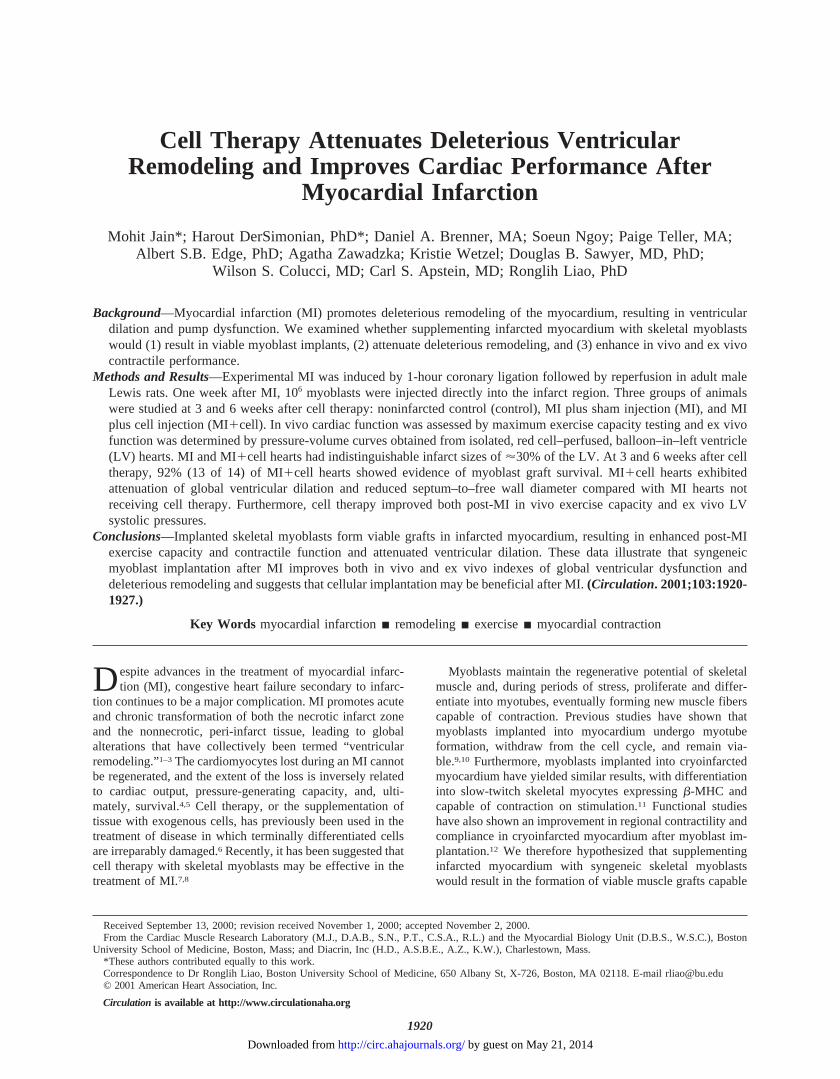
Figure 1. Myoblast survival in infarctedmyocardium at 9 days after implantation.Infarcted LV free wall of rat is shownunder increasing magnification withtrichrome staining (A, B, and C) andimmunohistochemical staining for myo-genin (nuclear transcription factor uniqueto skeletal myoblasts) (D, E, and F).Under trichrome staining, replacementfibrosis appears blue and remaining via-ble myocardium red. Myogenin stainingdemonstrates presence of myoblastswithin infarct region. Encircled area (Aand D) identifies one area of graft sur-vival within infarct region. Areas markedby arrows in B and E are shown underhigher magnification in C and F, respec-tively. Bars represent distances of 1 mmin A and D; 200 mm in B and E; and 100mm in C and F.
TABLE 1. Animal Characteristics
Time Group nBody
Weight, gHeart
Weight, g
HeartWeight/Body
WeightLung
Wet/DryLiver
Wet/Dry
3 Wk after Control 9 35867 1.1660.06 3.2360.15 4.8860.05 3.3160.01therapy MI 6 34765 1.4460.03* 4.1560.12* 4.8360.08 3.2860.07
MI1cell 7 34064 1.4360.06* 4.1160.08* 4.8660.04 3.2460.04
6 Wk after Control 9 39167 1.1860.06 3.0160.13 4.9460.04 3.2660.01therapy MI 7 39868 1.5460.05* 3.8360.11* 4.9760.02 3.2560.01
MI1cell 7 38367 1.4260.04* 3.7060.17* 4.8860.07 3.2760.01
*P,0.05 vs control.
Jain et al Post-MI Cell Therapy 1921
by guest on May 21, 2014http://circ.ahajournals.org/Downloaded from
medium (SkBM; Clonetics) containing 20% fetal bovine serum(Hyclone), recombinant human epidermal growth factor (rhEGF: 10ng/mL), and dexamethasone (0.39mg/mL). Myoblast-enrichedplates were identified after 48 hours of expansion and harvested with0.05% trypsin-EDTA (GibcoBRL). For a given experiment,'107
cells were harvested from 6 to 10 plates, seeded by satellite cellsisolated from the limb muscle of 2 neonate equivalents. Similarresults have been reported with adult skeletal muscle.11,12Cells werewashed and suspended in cold HBSS at 107 cells/mL and kept up to4 hours on ice before injection. A total of 100mL (106 cells) wasinjected into each animal. Under these conditions, cells weredetermined to be'50% myoblasts by flow cytometry with themonoclonal antibody H36 (anti-rata-7 integrin).15 The remainingcells were fibroblast-like, as determined by cell morphology. Theability for myoblasts to fuse into multinucleated myotubes in vitrowas also confirmed.
Seven days after MI, infarcted animals were randomized toreceive cell or sham implantation. Rats underwent a second thora-cotomy, and the left ventricle was visualized. Each rat received 6 to10 injections (total of 106 cells/heart) of 10 to 16mL of myoblastsuspension in HBSS directly into the infarct and peri-infarct regions,'1 to 2 mm apart, with a 30-gauge Hamilton needle. Infarctedanimals not receiving cells underwent an identical injection proce-
dure with HBSS alone. Noninfarcted control animals were subjectedto the same surgical procedures without injection.
Animal GroupsThree groups of animals were studied: control animals receivingneither infarction nor implantation (control), infarcted animals with-out cell therapy (MI), and infarcted animals receiving myoblast celltherapy (MI1cell). In vivo and ex vivo cardiac physiology andmyoblast cell survival were studied at 3 and 6 weeks after celltherapy. Cell grafts were also examined at 9 days and 12 weeks afterimplantation in several animals to determine the time course ofsurvival of implanted cells.
In Vivo Maximum Exercise CapacityMaximum exercise capacity is often used as a measure of in vivoventricular function and overall cardiac performance and recentlyhas been demonstrated to be a valuable tool in the assessment ofcardiac performance in smaller animal models.16,17 Maximum exer-cise capacity was assessed before implantation (1 week after MI) aswell as at 3 and 6 weeks after implantation. Maximum exercisecapacity was measured as the distance run on a modified rodenttreadmill (Columbus Instruments) until exhaustion.18 Exhaustion
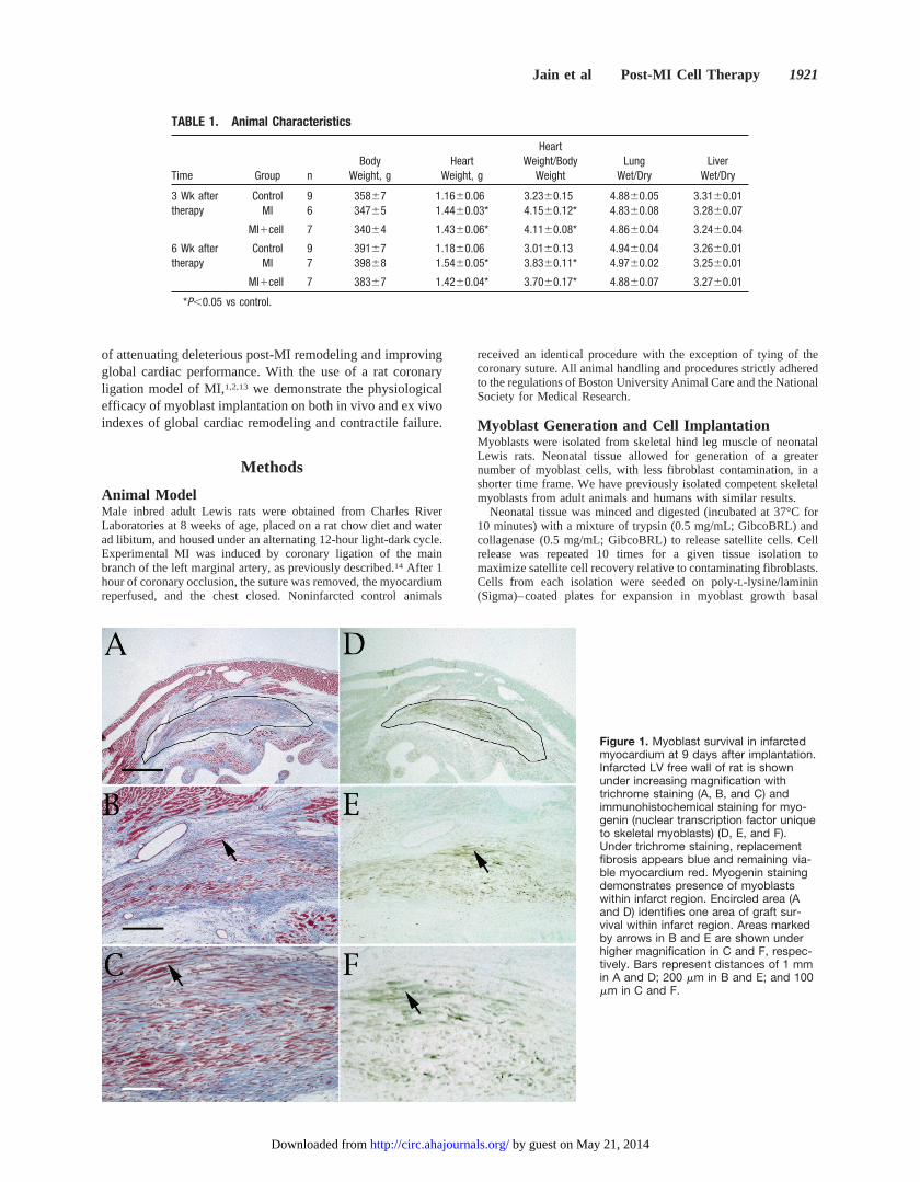
Figure 2. Myoblast survival in infarct and peri-infarct regions at 3 (A, C, E, and G) and 6 weeks(B, D, F, and H) after implantation. Correspond-ing tissue sections are shown with trichromestaining (A through D) and immunohistochemicalstaining for skeletal-specific myosin heavy chain(E through H). Viable cell grafts are demon-strated within infarct zone (upper 4 panels) andperi-infarct zone (lower 4 panels). Bar representsdistance of 200 mm in all panels.
1922 Circulation April 10, 2001
by guest on May 21, 2014http://circ.ahajournals.org/Downloaded from
was defined as the inability to run for 15 consecutive seconds despiteminor electric shock. Initial treadmill speed was set at 15 m/min ata 15 degree grade and increased by 1-m/min increments everyminute.
Ex Vivo Ventricular FunctionTo characterize myocardial remodeling and ex vivo cardiac functionat 3 and 6 weeks after implantation, whole-heart Langendorffperfusion studies were performed in isolated isovolumically beating(balloon–in–left ventricle [LV]) hearts as previously described.19,20
Briefly, isolated hearts were retrogradely perfused with a perfusateconsisting of bovine red blood cells suspended in modified Krebs-Henseleit buffer at a hematocrit of 40%. A fluid-filled cling-filmballoon connected to a Statham P23Db pressure transducer (StathamInstruments) was placed into the left ventricle to monitor ventricularpressures. Coronary perfusion pressure was set to 80 mm Hg, and
active pressure-volume relations were then generated. From a bal-loon volume of zero, the balloon was filled in increments of 0.05 mL,and subsequent peak systolic and end-diastolic pressures wererecorded. Systolic and diastolic pressure-volume relations werederived as previously described.19,20
Tissue Histology and MorphometryAfter pressure-volume experiments, hearts were arrested in diastoleand fixed with 4% buffered paraformaldehyde at a final ventriculardistending pressure of 5 mm Hg. Hearts were then weighed, paraffin-embedded, and sectioned (5 to 7mm thick) from each of 4 equallyspread levels (atrium through apex).
Six-micron–thick sections were cut, mounted, and stained withtrichrome. Myogenin immunohistochemistry was performed foridentification of implanted skeletal myoblasts, whereas skeletal-specific myosin heavy chain immunohistochemistry was performedfor identification of differentiated myotubes.9 For detection ofmyogenin, deparaffinized sections were blocked for endogenousperoxidase activity and subjected to antigen retrieval by boiling for10 minutes in citrate buffer. Sections were blocked for endogenousbiotin before adding primary polyclonal rabbit anti-rat myogeninantibody (Santa Cruz Biotechnology) followed by biotinylated goatanti-rabbit secondary antibody. For detection of myosin heavy chain,deparaffinized sections were incubated directly with alkaline phos-phatase–conjugated MY-32 mAb (Sigma), specific for skeletalmuscle myosin heavy chain, overnight at 4°C. Sections weredeveloped with diaminobenzidine (DAB Substrate Kit; Vector) formyogenin or with BCIP-NBT (Zymed) for MY-32 mAb and coun-terstained with nuclear red or fast green.
In addition, trichrome sections were used for morphometricanalysis. Stained sections were digitally imaged, and infarct size wasdetermined as the mean percentage of epicardial and endocardialcircumference occupied by scar tissue.20 LV endocardial chamberdiameter, septal wall thickness, and infarct wall thickness werederived from an average of 5 measurements taken throughout therespective regions.
StatisticsData were analyzed by 1-factor ANOVA or pairedt test whereappropriate. Pressure-volume relations were analyzed by a 2-factorrepeated-measures ANOVA and a least-significant-difference posthoc test. All data are presented as mean6SEM. A value ofP,0.05was considered statistically significant.
ResultsAnimal CharacteristicsTable 1 outlines the animal characteristics of control, MI, andMI1cell animals at both 3 and 6 weeks after therapy. Allgroups had a comparable increase in body weight over time.In addition, at all time points, hearts from MI and MI1cellgroups displayed comparable degrees of LV hypertrophy of'20% relative to control animals, as indicated by increasedheart weights and heart–to–body weight ratios. Neither MInor cell therapy altered lung or liver wet-to-dry ratios,suggesting the absence of pulmonary or hepatic congestion.
Animal Survival, Infarct Size, andMyoblast ImplantationExperimental MI resulted in'15% acute mortality ratewithin 24 hours of operation, whereas the cell implantationprocedure caused no additional animal deaths. Comparableinfarct sizes of 3161% and 3261% of the LV were observedin MI and MI1cell hearts, respectively. Hearts receiving
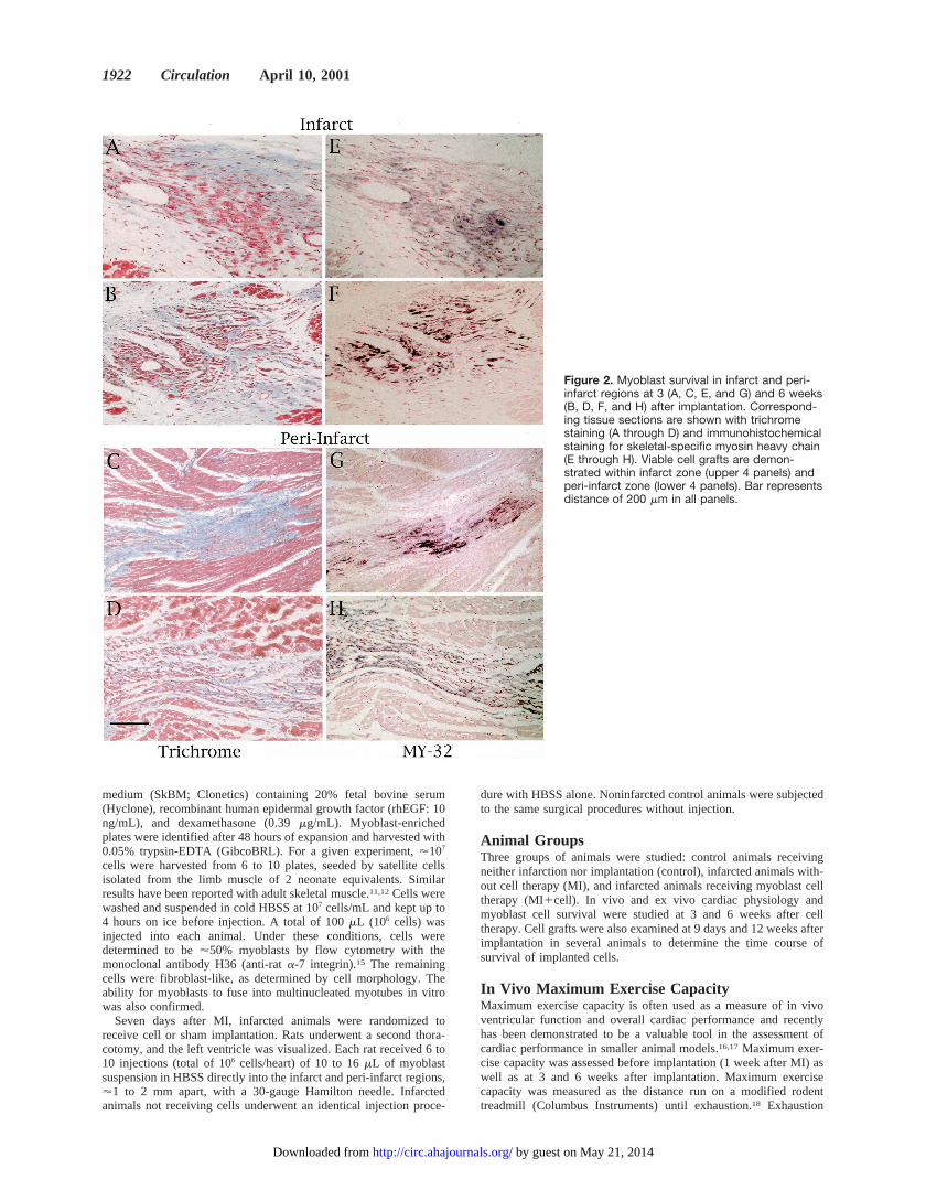
Figure 3. Myoblast survival in infarcted myocardium at 12weeks after implantation. Infarcted LV free wall is shown withtrichrome staining (A) and immunohistochemical staining formyogenin (B) or skeletal-specific myosin heavy chain (C). Myo-genin and skeletal myosin heavy chain expression demonstratespresence of viable myoblasts and myotubes at 12 weeks afterimplantation. Bar represents distance of 200 mm in A, B, and C.
Jain et al Post-MI Cell Therapy 1923
by guest on May 21, 2014http://circ.ahajournals.org/Downloaded from
infarction displayed areas of concentrated fibrosis, whereasnoninfarcted control hearts appeared as continuous viablemyocardium with homogenous thickness. Mortality rate wasalso similar in MI and MI1cell animals over the observationperiod, with no deaths at 3 weeks after implantation and 2deaths in each group before assessment of cardiac function at6 weeks after implantation.
Animals undergoing syngeneic cell therapy displayed noevidence for cell rejection as determined by excessive mac-rophage accumulation in tissue sections. Graft survival wasidentified at 9 days (Figure 1), 3 weeks (Figure 2, A, C, E,and G), 6 weeks (Figure 2, B, D, F, and H), and 12 weeks(Figure 3) after implantation, by immunohistochemical stain-ing for myogenin (skeletal myoblasts) and for skeletal-specific myosin heavy chain (skeletal myotubes). Myogenin-positive staining was observed as early as 9 days (Figure 1, D,E, and F) and as late as 12 weeks after implantation (Figure3B). Skeletal myosin heavy chain expression was not de-tected at 9 days after implantation (data not shown) and wasfirst observed at 3 weeks after implantation (Figure 2, E andG). Continued skeletal myosin heavy chain staining wasevident at 6 weeks (Figure 2, F and H) and 12 weeks (Figure3C) after implantation.
Cell survival was confirmed in 6 of 7 animals at 3 weeksafter therapy and in 7 of 7 animals at 6 weeks after therapy.At all examined time points, implanted cell grafts ranged insize from large patches of myoblasts and myotubes touniformly dispersed single cells within both the infarct andadjacent peri-infarct regions. Examination of cell grafts athigher magnification indicated that after 3 weeks, implantedcells developed the elongated morphology characteristic offused polynucleated myotubes. Implanted cells occasionallyappeared to orient parallel to the endocardium and epicar-dium in similar alignment to cardiomyocytes. Furthermore,vascular structures were present within or adjacent to the
engrafted areas, suggesting that blood supply was availablefor implanted cells. In the nonnecrotic, peri-infarct region,implanted cells formed regions of myoblasts and myotubessurrounded by fibrosis (Figure 2, C and D).
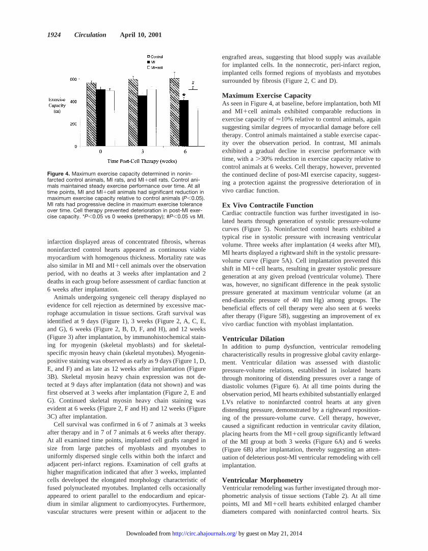
Maximum Exercise CapacityAs seen in Figure 4, at baseline, before implantation, both MIand MI1cell animals exhibited comparable reductions inexercise capacity of'10% relative to control animals, againsuggesting similar degrees of myocardial damage before celltherapy. Control animals maintained a stable exercise capac-ity over the observation period. In contrast, MI animalsexhibited a gradual decline in exercise performance withtime, with a.30% reduction in exercise capacity relative tocontrol animals at 6 weeks. Cell therapy, however, preventedthe continued decline of post-MI exercise capacity, suggest-ing a protection against the progressive deterioration of invivo cardiac function.
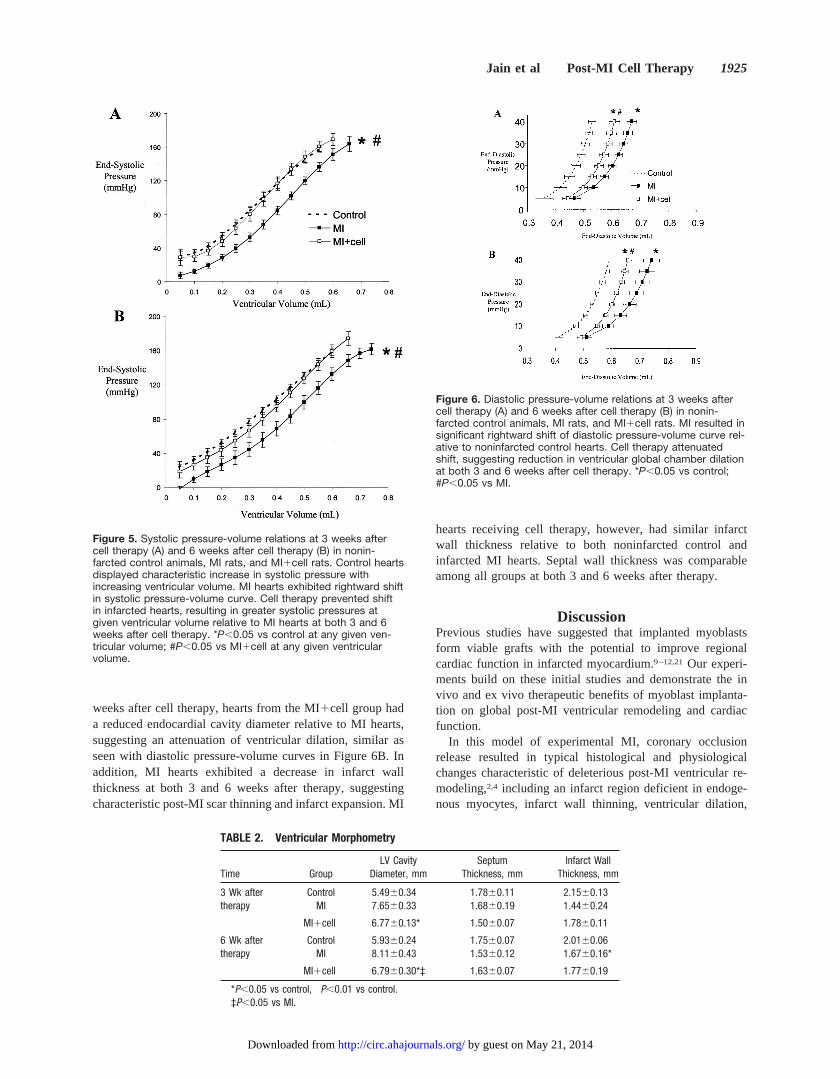
Ex Vivo Contractile FunctionCardiac contractile function was further investigated in iso-lated hearts through generation of systolic pressure-volumecurves (Figure 5). Noninfarcted control hearts exhibited atypical rise in systolic pressure with increasing ventricularvolume. Three weeks after implantation (4 weeks after MI),MI hearts displayed a rightward shift in the systolic pressure-volume curve (Figure 5A). Cell implantation prevented thisshift in MI1cell hearts, resulting in greater systolic pressuregeneration at any given preload (ventricular volume). Therewas, however, no significant difference in the peak systolicpressure generated at maximum ventricular volume (at anend-diastolic pressure of 40 mm Hg) among groups. Thebeneficial effects of cell therapy were also seen at 6 weeksafter therapy (Figure 5B), suggesting an improvement of exvivo cardiac function with myoblast implantation.
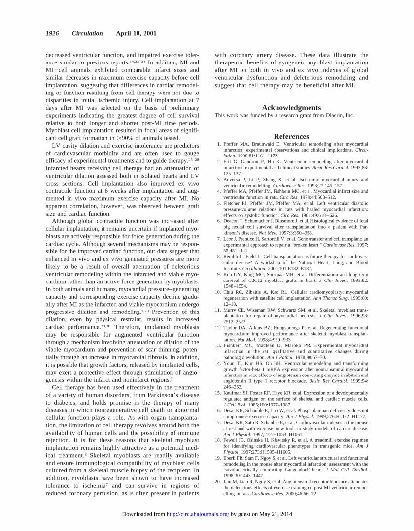
Ventricular DilationIn addition to pump dysfunction, ventricular remodelingcharacteristically results in progressive global cavity enlarge-ment. Ventricular dilation was assessed with diastolicpressure-volume relations, established in isolated heartsthrough monitoring of distending pressures over a range ofdiastolic volumes (Figure 6). At all time points during theobservation period, MI hearts exhibited substantially enlargedLVs relative to noninfarcted control hearts at any givendistending pressure, demonstrated by a rightward reposition-ing of the pressure-volume curve. Cell therapy, however,caused a significant reduction in ventricular cavity dilation,placing hearts from the MI1cell group significantly leftwardof the MI group at both 3 weeks (Figure 6A) and 6 weeks(Figure 6B) after implantation, thereby suggesting an atten-uation of deleterious post-MI ventricular remodeling with cellimplantation.
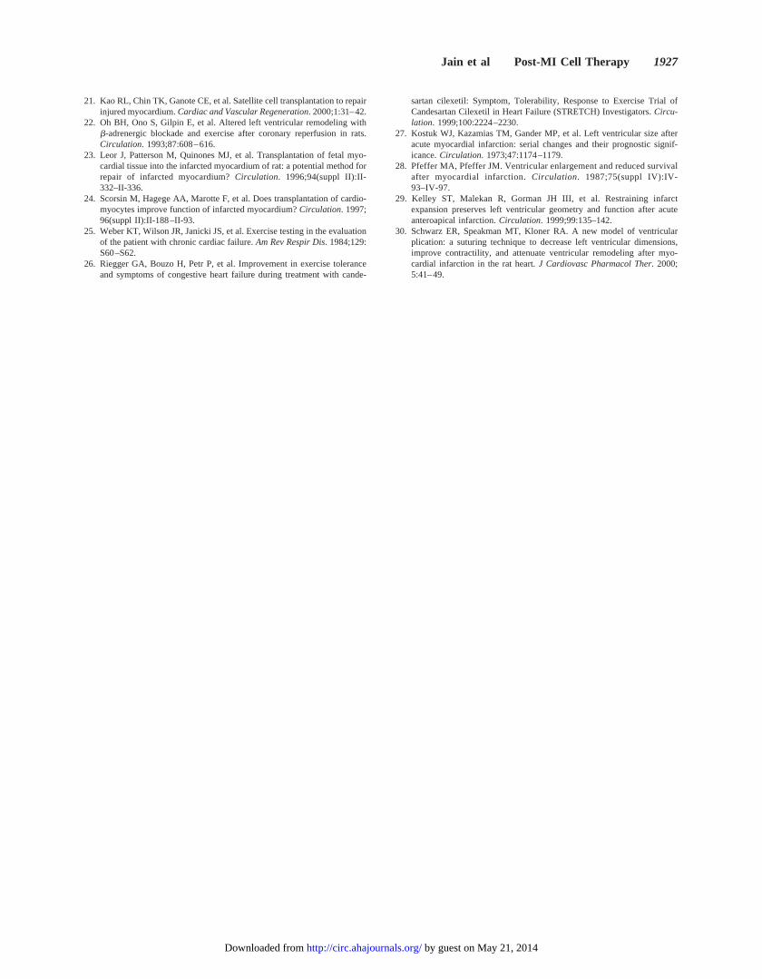
Ventricular MorphometryVentricular remodeling was further investigated through mor-phometric analysis of tissue sections (Table 2). At all timepoints, MI and MI1cell hearts exhibited enlarged chamberdiameters compared with noninfarcted control hearts. Six
Figure 4. Maximum exercise capacity determined in nonin-farcted control animals, MI rats, and MI1cell rats. Control ani-mals maintained steady exercise performance over time. At alltime points, MI and MI1cell animals had significant reduction inmaximum exercise capacity relative to control animals (P,0.05).MI rats had progressive decline in maximum exercise toleranceover time. Cell therapy prevented deterioration in post-MI exer-cise capacity. *P,0.05 vs 0 weeks (pretherapy); #P,0.05 vs MI.
1924 Circulation April 10, 2001
by guest on May 21, 2014http://circ.ahajournals.org/Downloaded from
weeks after cell therapy, hearts from the MI1cell group hada reduced endocardial cavity diameter relative to MI hearts,suggesting an attenuation of ventricular dilation, similar asseen with diastolic pressure-volume curves in Figure 6B. Inaddition, MI hearts exhibited a decrease in infarct wallthickness at both 3 and 6 weeks after therapy, suggestingcharacteristic post-MI scar thinning and infarct expansion. MI
hearts receiving cell therapy, however, had similar infarctwall thickness relative to both noninfarcted control andinfarcted MI hearts. Septal wall thickness was comparableamong all groups at both 3 and 6 weeks after therapy.
DiscussionPrevious studies have suggested that implanted myoblastsform viable grafts with the potential to improve regionalcardiac function in infarcted myocardium.9–12,21Our experi-ments build on these initial studies and demonstrate the invivo and ex vivo therapeutic benefits of myoblast implanta-tion on global post-MI ventricular remodeling and cardiacfunction.
In this model of experimental MI, coronary occlusionrelease resulted in typical histological and physiologicalchanges characteristic of deleterious post-MI ventricular re-modeling,2,4 including an infarct region deficient in endoge-nous myocytes, infarct wall thinning, ventricular dilation,
Figure 5. Systolic pressure-volume relations at 3 weeks aftercell therapy (A) and 6 weeks after cell therapy (B) in nonin-farcted control animals, MI rats, and MI1cell rats. Control heartsdisplayed characteristic increase in systolic pressure withincreasing ventricular volume. MI hearts exhibited rightward shiftin systolic pressure-volume curve. Cell therapy prevented shiftin infarcted hearts, resulting in greater systolic pressures atgiven ventricular volume relative to MI hearts at both 3 and 6weeks after cell therapy. *P,0.05 vs control at any given ven-tricular volume; #P,0.05 vs MI1cell at any given ventricularvolume.
TABLE 2. Ventricular Morphometry
Time GroupLV Cavity
Diameter, mmSeptum
Thickness, mmInfarct Wall
Thickness, mm
3 Wk after Control 5.4960.34 1.7860.11 2.1560.13therapy MI 7.6560.33† 1.6860.19 1.4460.24†
MI1cell 6.7760.13* 1.5060.07 1.7860.11
6 Wk after Control 5.9360.24 1.7560.07 2.0160.06therapy MI 8.1160.43† 1.5360.12 1.6760.16*
MI1cell 6.7960.30*‡ 1.6360.07 1.7760.19
*P,0.05 vs control, †P,0.01 vs control.‡P,0.05 vs MI.
Figure 6. Diastolic pressure-volume relations at 3 weeks aftercell therapy (A) and 6 weeks after cell therapy (B) in nonin-farcted control animals, MI rats, and MI1cell rats. MI resulted insignificant rightward shift of diastolic pressure-volume curve rel-ative to noninfarcted control hearts. Cell therapy attenuatedshift, suggesting reduction in ventricular global chamber dilationat both 3 and 6 weeks after cell therapy. *P,0.05 vs control;#P,0.05 vs MI.
Jain et al Post-MI Cell Therapy 1925
by guest on May 21, 2014http://circ.ahajournals.org/Downloaded from
decreased ventricular function, and impaired exercise toler-ance similar to previous reports.14,22–24 In addition, MI andMI1cell animals exhibited comparable infarct sizes andsimilar decreases in maximum exercise capacity before cellimplantation, suggesting that differences in cardiac remodel-ing or function resulting from cell therapy were not due todisparities in initial ischemic injury. Cell implantation at 7days after MI was selected on the basis of preliminaryexperiments indicating the greatest degree of cell survivalrelative to both longer and shorter post-MI time periods.Myoblast cell implantation resulted in focal areas of signifi-cant cell graft formation in.90% of animals tested.
LV cavity dilation and exercise intolerance are predictorsof cardiovascular morbidity and are often used to gaugeefficacy of experimental treatments and to guide therapy.25–28
Infarcted hearts receiving cell therapy had an attenuation ofventricular dilation assessed both in isolated hearts and LVcross sections. Cell implantation also improved ex vivocontractile function at 6 weeks after implantation and aug-mented in vivo maximum exercise capacity after MI. Noapparent correlation, however, was observed between graftsize and cardiac function.
Although global contractile function was increased aftercellular implantation, it remains uncertain if implanted myo-blasts are actively responsible for force generation during thecardiac cycle. Although several mechanisms may be respon-sible for the improved cardiac function, our data suggest thatenhanced in vivo and ex vivo generated pressures are morelikely to be a result of overall attenuation of deleteriousventricular remodeling within the infarcted and viable myo-cardium rather than an active force generation by myoblasts.In both animals and humans, myocardial pressure–generatingcapacity and corresponding exercise capacity decline gradu-ally after MI as the infarcted and viable myocardium undergoprogressive dilation and remodeling.2,28 Prevention of thisdilation, even by physical restraint, results in increasedcardiac performance.29,30 Therefore, implanted myoblastsmay be responsible for augmented ventricular functionthrough a mechanism involving attenuation of dilation of theviable myocardium and prevention of scar thinning, poten-tially through an increase in myocardial fibrosis. In addition,it is possible that growth factors, released by implanted cells,may exert a protective effect through stimulation of angio-genesis within the infarct and noninfarct regions.7
Cell therapy has been used effectively in the treatmentof a variety of human disorders, from Parkinson’s diseaseto diabetes, and holds promise in the therapy of manydiseases in which nonregenerative cell death or abnormalcellular function plays a role. As with organ transplanta-tion, the limitation of cell therapy revolves around both theavailability of human cells and the possibility of immunerejection. It is for these reasons that skeletal myoblastimplantation remains highly attractive as a potential med-ical treatment.8 Skeletal myoblasts are readily availableand ensure immunological compatibility of myoblast cellscultured from a skeletal muscle biopsy of the recipient. Inaddition, myoblasts have been shown to have increasedtolerance to ischemia7 and can survive in regions ofreduced coronary perfusion, as is often present in patients
with coronary artery disease. These data illustrate thetherapeutic benefits of syngeneic myoblast implantationafter MI on both in vivo and ex vivo indexes of globalventricular dysfunction and deleterious remodeling andsuggest that cell therapy may be beneficial after MI.
AcknowledgmentsThis work was funded by a research grant from Diacrin, Inc.
References1. Pfeffer MA, Braunwald E. Ventricular remodeling after myocardial
infarction: experimental observations and clinical implications.Circu-lation. 1990;81:1161–1172.
2. Ertl G, Gaudron P, Hu K. Ventricular remodeling after myocardialinfarction: experimental and clinical studies.Basic Res Cardiol. 1993;88:125–137.
3. Anversa P, Li P, Zhang X, et al. Ischaemic myocardial injury andventricular remodelling.Cardiovasc Res. 1993;27:145–157.
4. Pfeffer MA, Pfeffer JM, Fishbein MC, et al. Myocardial infarct size andventricular function in rats.Circ Res. 1979;44:503–512.
5. Fletcher PJ, Pfeffer JM, Pfeffer MA, et al. Left ventricular diastolicpressure-volume relations in rats with healed myocardial infarction:effects on systolic function.Circ Res. 1981;49:618–626.
6. Deacon T, Schumacher J, Dinsmore J, et al. Histological evidence of fetalpig neural cell survival after transplantation into a patient with Par-kinson’s disease.Nat Med. 1997;3:350–353.
7. Leor J, Prentice H, Sartorelli V, et al. Gene transfer and cell transplant: anexperimental approach to repair a “broken heart.”Cardiovasc Res. 1997;35:431–441.
8. Reinlib L, Field L. Cell transplantation as future therapy for cardiovas-cular disease? A workshop of the National Heart, Lung, and BloodInstitute.Circulation. 2000;101:E182–E187.
9. Koh GY, Klug MG, Soonpaa MH, et al. Differentiation and long-termsurvival of C2C12 myoblast grafts in heart.J Clin Invest. 1993;92:1548–1554.
10. Chiu RC, Zibaitis A, Kao RL. Cellular cardiomyoplasty: myocardialregeneration with satellite cell implantation.Ann Thorac Surg. 1995;60:12–18.
11. Murry CE, Wiseman RW, Schwartz SM, et al. Skeletal myoblast trans-plantation for repair of myocardial necrosis.J Clin Invest. 1996;98:2512–2523.
12. Taylor DA, Atkins BZ, Hungspreugs P, et al. Regenerating functionalmyocardium: improved performance after skeletal myoblast transplan-tation.Nat Med. 1998;4:929–933.
13. Fishbein MC, Maclean D, Maroko PR. Experimental myocardialinfarction in the rat: qualitative and quantitative changes duringpathologic evolution.Am J Pathol. 1978;90:57–70.
14. Youn TJ, Kim HS, Oh BH. Ventricular remodeling and transforminggrowth factor-beta 1 mRNA expression after nontransmural myocardialinfarction in rats: effects of angiotensin converting enzyme inhibition andangiotensin II type 1 receptor blockade.Basic Res Cardiol. 1999;94:246–253.
15. Kaufman SJ, Foster RF, Haye KR, et al. Expression of a developmentallyregulated antigen on the surface of skeletal and cardiac muscle cells.J Cell Biol. 1985;100:1977–1987.
16. Desai KH, Schauble E, Luo W, et al. Phospholamban deficiency does notcompromise exercise capacity.Am J Physiol. 1999;276:H1172–H1177.
17. Desai KH, Sato R, Schauble E, et al. Cardiovascular indexes in the mouseat rest and with exercise: new tools to study models of cardiac disease.Am J Physiol. 1997;272:H1053–H1061.
18. Fewell JG, Osinska H, Klevitsky R, et al. A treadmill exercise regimenfor identifying cardiovascular phenotypes in transgenic mice.Am JPhysiol. 1997;273:H1595–H1605.
19. Eberli FR, Sam F, Ngoy S, et al. Left ventricular structural and functionalremodeling in the mouse after myocardial infarction: assessment with theisovolumetrically contracting Langendorff heart.J Mol Cell Cardiol.1998;30:1443–1447.
20. Jain M, Liao R, Ngoy S, et al. Angiotensin II receptor blockade attenuatesthe deleterious effects of exercise training on post-MI ventricular remod-elling in rats.Cardiovasc Res. 2000;46:66–72.
1926 Circulation April 10, 2001
by guest on May 21, 2014http://circ.ahajournals.org/Downloaded from
21. Kao RL, Chin TK, Ganote CE, et al. Satellite cell transplantation to repairinjured myocardium.Cardiac and Vascular Regeneration. 2000;1:31–42.
22. Oh BH, Ono S, Gilpin E, et al. Altered left ventricular remodeling withb-adrenergic blockade and exercise after coronary reperfusion in rats.Circulation. 1993;87:608–616.
23. Leor J, Patterson M, Quinones MJ, et al. Transplantation of fetal myo-cardial tissue into the infarcted myocardium of rat: a potential method forrepair of infarcted myocardium?Circulation. 1996;94(suppl II):II-332–II-336.
24. Scorsin M, Hagege AA, Marotte F, et al. Does transplantation of cardio-myocytes improve function of infarcted myocardium?Circulation. 1997;96(suppl II):II-188–II-93.
25. Weber KT, Wilson JR, Janicki JS, et al. Exercise testing in the evaluationof the patient with chronic cardiac failure.Am Rev Respir Dis. 1984;129:S60–S62.
26. Riegger GA, Bouzo H, Petr P, et al. Improvement in exercise toleranceand symptoms of congestive heart failure during treatment with cande-
sartan cilexetil: Symptom, Tolerability, Response to Exercise Trial ofCandesartan Cilexetil in Heart Failure (STRETCH) Investigators.Circu-lation. 1999;100:2224–2230.
27. Kostuk WJ, Kazamias TM, Gander MP, et al. Left ventricular size afteracute myocardial infarction: serial changes and their prognostic signif-icance.Circulation. 1973;47:1174–1179.
28. Pfeffer MA, Pfeffer JM. Ventricular enlargement and reduced survivalafter myocardial infarction.Circulation. 1987;75(suppl IV):IV-93–IV-97.
29. Kelley ST, Malekan R, Gorman JH III, et al. Restraining infarctexpansion preserves left ventricular geometry and function after acuteanteroapical infarction.Circulation. 1999;99:135–142.
30. Schwarz ER, Speakman MT, Kloner RA. A new model of ventricularplication: a suturing technique to decrease left ventricular dimensions,improve contractility, and attenuate ventricular remodeling after myo-cardial infarction in the rat heart.J Cardiovasc Pharmacol Ther. 2000;5:41–49.
Jain et al Post-MI Cell Therapy 1927
by guest on May 21, 2014http://circ.ahajournals.org/Downloaded from
Related Documents