3188 Research Article Introduction Tissue transglutaminase (tTG or TG2, EC 2.3.2.13) belongs to a multigene family of Ca 2+ -dependent protein crosslinking enzymes (Lorand and Graham, 2003). tTG is a ubiquitously expressed multifunctional protein that possesses transglutaminase (Folk and Cole, 1966), GTPase (Nakaoka et al., 1994) and protein disulfide isomerase (Hasegawa et al., 2003) activities. tTG is localized in the cytoplasm and is also present on the cell surface and in the extracellular matrix (ECM), where it plays an important role in cell-matrix interactions (Fesus and Piacentini, 2002; Lorand and Graham, 2003). Cell-surface tTG associates with the major ECM protein fibronectin (Fellin et al., 1988; Turner and Lorand, 1989) and 1 and 3 integrins (Akimov et al., 2000). The high-affinity interaction of tTG with fibronectin enhances cell-matrix adhesion and other adhesion-dependent phenomena, including cell migration, ECM assembly and remodeling and outside-in signaling (reviewed in Zemskov et al., 2006). The non-covalent association of tTG with the 1 and 3 integrin subunits leads to the formation of ternary fibronectin-tTG-integrin complexes and stabilizes the interaction of integrins with fibronectin (Akimov et al., 2000). Importantly, the transamidating- and GTPase-deficient mutants of the protein retain these properties, indicating that this recently defined adhesive function of tTG is independent of its enzymatic activities (Akimov et al., 2000; Akimov and Belkin, 2001a; Janiak et al., 2006). Moreover, the interaction of tTG with integrins promotes their clustering, causing activation of RhoA and its downstream target ROCK by means of inhibition of the Src-p190RhoGAP signaling pathway (Janiak et al., 2006). Taking into account the numerous functions of cell-surface tTG both as a crosslinking enzyme and as an adhesion co-receptor involved in signal transduction, it could be predicted that excessive levels of tTG on the surface would impair cellular functions. To prevent a disproportionate crosslinking of the ECM and to downregulate tTG-dependent cell-matrix adhesion and outside-in signaling, cells should employ an efficient mechanism(s) for its removal from their surfaces. LRP1 is a member of the LDL receptor superfamily which consists of six structurally related and widely expressed proteins [LRP1, LRP1B, LRP2 (also known as gp330), LDL receptor, very low-density lipoprotein (VLDL) receptor and LRP8 (also known as apolipoprotein E receptor 2)] (Lillis et al., 2005). This large endocytic receptor functions in lipoprotein metabolism, degradation of proteases, activation of Tissue transglutaminase functions as a protein crosslinking enzyme and an integrin-binding adhesion co-receptor for fibronectin on the cell surface. These activities of transglutaminase and the involvement of this protein in cell-matrix adhesion, integrin-mediated signaling, cell migration and matrix organization suggest a precise and efficient control of its cell-surface expression. We report a novel mechanism of regulation of surface transglutaminase through internalization and subsequent lysosomal degradation. Constitutive endocytosis of cell-surface transglutaminase depends on plasma membrane cholesterol and the activity of dynamin-2, and involves both clathrin-coated pits and lipid rafts or caveolae. Furthermore, the key matrix ligands of transglutaminase, fibronectin and platelet-derived growth factor, promote its endocytosis from the cell surface. Our results also indicate that transglutaminase interacts in vitro and on the cell surface with the major endocytic receptor, low-density lipoprotein receptor-related protein 1, and demonstrate the requirement for this receptor in the endocytosis of transglutaminase. Finally, a deficiency of this endocytic receptor or blockade of endo-lysosomal function upregulate transglutaminase expression on the cell surface, leading to increased cell adhesion and matrix crosslinking. These findings characterize a previously unknown pathway of transglutaminase internalization and degradation that might be crucial for regulation of its adhesive and signaling functions on the cell surface and reveal a novel functional link between cell-matrix adhesion and endocytosis. Supplementary material available online at http://jcs.biologists.org/cgi/content/full/119/18/3188/DC1 Key words: Transglutaminase, Integrin, Fibronectin, Cell-matrix adhesion, LRP1, Endocytosis, Lysosomal degradation Summary Cell-surface transglutaminase undergoes internalization and lysosomal degradation: an essentialrole for LRP1 Evgeny A. Zemskov 1,2 , Irina Mikhailenko 2,3 , Dudley K. Strickland 2,4 and Alexey M. Belkin 1,2,5, * 1 Department of Biochemistry and Molecular Biology, 2 Center for Vascular and Inflammatory Diseases, Departments of 3 Physiology and 4 Surgery and 5 Marlene and Stewart Greenebaum Cancer Center, University of Maryland School of Medicine, Baltimore, MD 21201, USA *Author for correspondence (e-mail: [email protected]) Accepted 6 July 2007 Journal of Cell Science 120, 3188-3199 Published by The Company of Biologists 2007 doi:10.1242/jcs.010397 Journal of Cell Science

Welcome message from author
This document is posted to help you gain knowledge. Please leave a comment to let me know what you think about it! Share it to your friends and learn new things together.
Transcript
3188 Research Article
IntroductionTissue transglutaminase (tTG or TG2, EC 2.3.2.13) belongs toa multigene family of Ca2+-dependent protein crosslinkingenzymes (Lorand and Graham, 2003). tTG is a ubiquitouslyexpressed multifunctional protein that possessestransglutaminase (Folk and Cole, 1966), GTPase (Nakaoka etal., 1994) and protein disulfide isomerase (Hasegawa et al.,2003) activities. tTG is localized in the cytoplasm and is alsopresent on the cell surface and in the extracellular matrix(ECM), where it plays an important role in cell-matrixinteractions (Fesus and Piacentini, 2002; Lorand and Graham,2003).
Cell-surface tTG associates with the major ECM proteinfibronectin (Fellin et al., 1988; Turner and Lorand, 1989) and�1 and �3 integrins (Akimov et al., 2000). The high-affinityinteraction of tTG with fibronectin enhances cell-matrixadhesion and other adhesion-dependent phenomena, includingcell migration, ECM assembly and remodeling and outside-insignaling (reviewed in Zemskov et al., 2006). The non-covalentassociation of tTG with the �1 and �3 integrin subunits leadsto the formation of ternary fibronectin-tTG-integrin complexesand stabilizes the interaction of integrins with fibronectin(Akimov et al., 2000). Importantly, the transamidating- and
GTPase-deficient mutants of the protein retain these properties,indicating that this recently defined adhesive function of tTGis independent of its enzymatic activities (Akimov et al., 2000;Akimov and Belkin, 2001a; Janiak et al., 2006). Moreover, theinteraction of tTG with integrins promotes their clustering,causing activation of RhoA and its downstream target ROCKby means of inhibition of the Src-p190RhoGAP signalingpathway (Janiak et al., 2006). Taking into account thenumerous functions of cell-surface tTG both as a crosslinkingenzyme and as an adhesion co-receptor involved in signaltransduction, it could be predicted that excessive levels of tTGon the surface would impair cellular functions. To prevent adisproportionate crosslinking of the ECM and to downregulatetTG-dependent cell-matrix adhesion and outside-in signaling,cells should employ an efficient mechanism(s) for its removalfrom their surfaces.
LRP1 is a member of the LDL receptor superfamily whichconsists of six structurally related and widely expressedproteins [LRP1, LRP1B, LRP2 (also known as gp330), LDLreceptor, very low-density lipoprotein (VLDL) receptor andLRP8 (also known as apolipoprotein E receptor 2)] (Lillis etal., 2005). This large endocytic receptor functions inlipoprotein metabolism, degradation of proteases, activation of
Tissue transglutaminase functions as a protein crosslinkingenzyme and an integrin-binding adhesion co-receptor forfibronectin on the cell surface. These activities oftransglutaminase and the involvement of this protein incell-matrix adhesion, integrin-mediated signaling, cellmigration and matrix organization suggest a precise andefficient control of its cell-surface expression. We report anovel mechanism of regulation of surface transglutaminasethrough internalization and subsequent lysosomaldegradation. Constitutive endocytosis of cell-surfacetransglutaminase depends on plasma membranecholesterol and the activity of dynamin-2, and involves bothclathrin-coated pits and lipid rafts or caveolae.Furthermore, the key matrix ligands of transglutaminase,fibronectin and platelet-derived growth factor, promote itsendocytosis from the cell surface. Our results also indicatethat transglutaminase interacts in vitro and on the cellsurface with the major endocytic receptor, low-density
lipoprotein receptor-related protein 1, and demonstrate therequirement for this receptor in the endocytosis oftransglutaminase. Finally, a deficiency of this endocyticreceptor or blockade of endo-lysosomal functionupregulate transglutaminase expression on the cell surface,leading to increased cell adhesion and matrix crosslinking.These findings characterize a previously unknown pathwayof transglutaminase internalization and degradation thatmight be crucial for regulation of its adhesive and signalingfunctions on the cell surface and reveal a novel functionallink between cell-matrix adhesion and endocytosis.
Supplementary material available online athttp://jcs.biologists.org/cgi/content/full/119/18/3188/DC1
Key words: Transglutaminase, Integrin, Fibronectin, Cell-matrixadhesion, LRP1, Endocytosis, Lysosomal degradation
Summary
Cell-surface transglutaminase undergoesinternalization andlysosomal degradation:an essentialrole for LRP1Evgeny A. Zemskov1,2, Irina Mikhailenko2,3, Dudley K. Strickland2,4 and Alexey M. Belkin1,2,5,*1Department of Biochemistry and Molecular Biology, 2Center for Vascular and Inflammatory Diseases, Departments of 3Physiology and 4Surgeryand 5Marlene and Stewart Greenebaum Cancer Center, University of Maryland School of Medicine, Baltimore, MD 21201, USA*Author for correspondence (e-mail: [email protected])
Accepted 6 July 2007Journal of Cell Science 120, 3188-3199 Published by The Company of Biologists 2007doi:10.1242/jcs.010397
Jour
nal o
f Cel
l Sci
ence

3189LRP1-mediated endocytosis of tTG
lysosomal enzymes and cellular entry of bacterial toxins andviruses (Herz and Strickland, 2001). Furthermore, recentpapers demonstrate the involvement of this protein in severalsignal-transduction cascades (see Lillis et al., 2005).Importantly, LRP1 has also been shown to function in theturnover of extracellular tTG-binding proteins, �1 and �3integrins and fibronectin (Czekay et al., 2003; Salicioni et al.,2002; Salicioni et al., 2004).
Here, we demonstrate that downregulation of cell surfacetTG is mediated by constitutive endocytosis. Cell-surface tTGinteracts directly with LRP1 and promotes the association ofLRP1 with �1 integrins and the ECM. The efficientinternalization of tTG from the cell surface occurs by meansof a dynamin-dependent process that involves clathrin- andcaveolin-dependent endocytic pathways and requires LRP1.Finally, we show that accumulation of surface tTG in LRP1-deficient cells or in cells with blocked endo-lysosomal functionincreases extracellular transglutaminase activity and cell-matrix adhesion. Our findings underscore the crucial role ofthis endocytic receptor in the internalization and degradationof tTG and in the regulation of its adhesive and signalingfunctions on the cell surface.
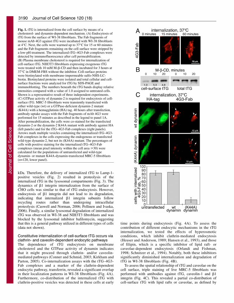
ResultstTG is internalized from the cell surface by a cholesterol-dependent, dynamin-mediated mechanismEndocytosis is a specific cellular mechanism ofinternalization of macromolecules and particles by means ofvesicles derived from the plasma membrane. To examinewhether cell-surface tTG undergoes endocytosis, antibody-uptake experiments were performed with WI-38 fibroblaststhat express high surface levels of tTG (Akimov and Belkin,2001a). As bivalent antibodies cause aggregation of cell-surface tTG and might artificially stimulate its endocytosis,we employed monovalent antigen-binding fragments (Fab) ofthe 4G3 monoclonal antibody (mAb) against tTG (Akimovand Belkin, 2001b). After binding the 4G3-Fab fragments tothe cell surfaces at 4°C, the cells were allowed to internalizethe tTG–4G3-Fab complexes at 37°C for 15 or 60 minutes(Fig. 1A). The remaining surface-bound tTG–4G3-Fabcomplexes were stripped off with a low-pH treatmentand the internalized complexes were detected byimmunofluorescence after cell permeabilization. Theinternalized tTG was detected initially in intracellularperipheral vesicles, whereas prolonged incubation increasedthe amounts of the internalized tTG that resided in numerousperinuclear vesicles. No surface binding or internalizationwas observed with the 4G3-Fab fragments in cells lackingsurface tTG (data not shown).
To confirm that tTG undergoes internalization, the levels ofcell-surface tTG were determined in NIH3T3-tTG cells treatedwith the cholesterol-binding agent M-�-CD, which acutelyinhibits endocytosis (Rodal et al., 1999; Subtil et al., 1999).The cells treated with M-�-CD for short intervals displayedsharply increased amounts of tTG on the cell surface, althoughtotal tTG expression remained unaffected (Fig. 1B). Thisincrease in cell-surface presentation of tTG caused by theendocytosis-arresting drug indicates that the levels of surfacetTG are efficiently controlled by cholesterol-dependentinternalization.
The function of the large GTPase dynamin-2 (Altschuler et
al., 1998) is required for fission of vesicles from the plasmamembrane during endocytic processes (Damke et al., 1994;Gaborik et al., 2001; Henley et al., 1999; Torgersen et al.,2001). Thus, we next compared the internalization of thetTG–4G3-Fab complexes in MRC-5 fibroblasts transientlyexpressing either wild-type dynamin-2 or a dominant-negativemutant dynamin-2 (K44A), both containing a hemagglutinin(HA) tag. Double immunostaining for the HA tag and thetTG–4G3-Fab complexes following a standard internalizationprocedure revealed efficient endocytosis of tTG andaccumulation of tTG in the vesicular compartments of the cellstransfected with wild-type dynamin-2, whereas tTG could notbe detected inside the cells expressing the dominant-negativemutant (Fig. 1C). Hence, tTG is internalized from the cellsurface, and this process is dependent upon membranecholesterol and the GTPase activity of dynamin-2.
Internalized tTG is transported through early and lateendocytic endosomes to lysosomes for degradationTo determine the endocytic route(s) and destination(s) ofinternalized tTG, we defined the endosomal compartmentswhere tTG was localized after internalization (Fig. 2). Theintracellular localization of internalized tTG–4G3-Fabcomplexes in WI-38 fibroblasts was compared with those ofmarkers of endocytic compartments. Double immunostainingrevealed the internalized tTG co-distributed with EEA1, amarker of early endosomes (Mu et al., 1995) after 5 minutesof internalization, whereas 30 minutes later it appearedcolocalized with Rab7-positive compartments, indicative oflate endosomes (Chavrier et al., 1990). Co-staining withantibody against Arf1 did not reveal translocation of theinternalized tTG to the Golgi complex. However, we observedoccasional colocalization of internalized tTG with peripheralArf1-positive membrane structures, likely to be the endosomecarrier vesicles (ECVs) transporting proteins from the early tothe late endosomes (Donaldson and Honda, 2005). Importantly,extended (>90 minute) incubations resulted in the appearanceof the internalized tTG in Lamp-1-positive lysosomes (Chen etal., 1985).
Double immunofluorescence staining demonstratedcolocalization of the internalized tTG and �1 integrins in theWI-38 fibroblasts (Fig. 3A), suggesting that tTG and �1integrins, interacting on the cell surface (Akimov et al., 2000;Janiak et al., 2006), also undergo endocytosis as a complexand/or follow the same endocytic route(s). To assess the fateof internalized tTG and �1 integrins, the following experimentswere performed. First, CHO cells were chilled to 4°C to stopinternalization and cell-surface proteins were biotinylated withmembrane-impermeable SH-cleavable sulfo-NHS-SS-biotin.Then, the cells were incubated for varying periods of time (Fig.3B) at 37°C to allow internalization, and the remaining biotinon the cell-surface proteins was removed by reduction, whileinternalized biotinylated proteins retained their biotin label. Asexpected, significant levels of biotinylated tTG was detected incells incubated at 37°C, demonstrating the efficientinternalization of tTG from the cell surface (Fig. 3B). At earlytime points of endocytosis, the intact internalized tTG wasdetected inside the CHO cells, whereas longer incubationsrevealed a gradual decline in the amounts of intact tTG,coinciding with the appearance and subsequent accumulationof two proteolytic tTG fragments of molecular mass of ~42-45
Jour
nal o
f Cel
l Sci
ence
3190
kDa. Therefore, the delivery of internalized tTG to Lamp-1-positive vesicles (Fig. 2) resulted in proteolysis of theinternalized tTG in the lysosomal compartments (Fig. 3). Thedynamics of �1 integrin internalization from the surface ofCHO cells was similar to that of tTG endocytosis. However,endocytosis of �1 integrin did not lead to its degradation,indicating that internalized �1 integrin subunits followrecycling routes rather than undergoing intracellularproteolysis (Caswell and Norman, 2006; Pellinen and Ivaska,2006). Finally, a similar lysosomal degradation of internalizedtTG was observed in WI-38 and NIH3T3 fibroblasts and wasblocked by the lysosomal inhibitor bafilomycin, suggestingthat this is a general pathway utilized in different types of cells(data not shown).
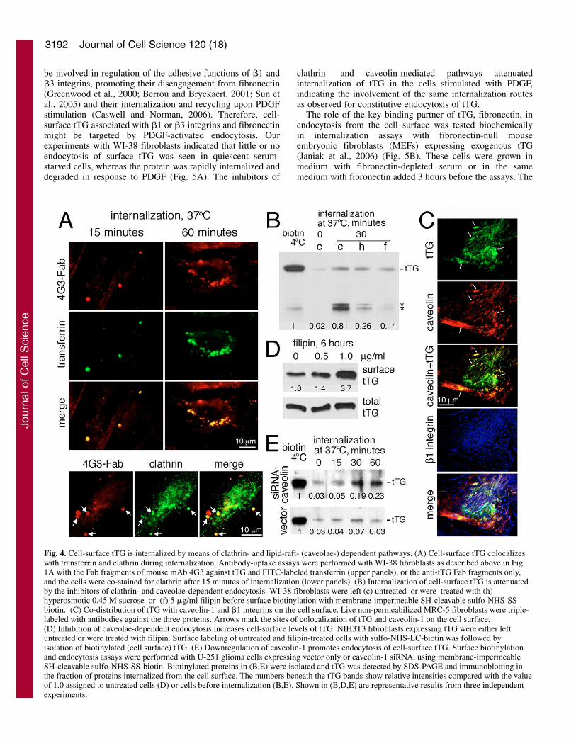
Constitutive internalization of cell-surface tTG occurs viaclathrin- and caveolin-dependent endocytic pathwaysThe dependence of tTG endocytosis on membranecholesterol and the GTPase activity of dynamin indicatesthat it might proceed through clathrin- and/or caveolin-mediated pathways (Conner and Schmid, 2003; Kirkham andParton, 2005). Co-internalization assays with the tTG–4G3-Fab complexes and a marker of the clathrin-dependentendocytic pathway, transferrin, revealed a significant overlapin their localization patterns in WI-38 fibroblasts (Fig. 4A).Furthermore, co-distribution of the internalized tTG withclathrin-positive vesicles was detected in these cells at early
Journal of Cell Science 120 (18)
time points during endocytosis (Fig. 4A). To assess thecontribution of different endocytic mechanisms in the tTGinternalization, we tested the effects of hyperosmoticconditions, which inhibit clathrin-mediated endocytosis(Heuser and Anderson, 1989; Hansen et al., 1993), and thoseof filipin, which is a specific inhibitor of lipid raft- orcaveolae-dependent endocytosis (Orlandi and Fishman,1998; Schnitzer et al., 1994). Notably, both these inhibitorssignificantly diminished internalization and degradation oftTG in WI-38 fibroblasts (Fig. 4B).
To assess the spatial relationship of tTG and caveolae on thecell surface, triple staining of live MRC-5 fibroblasts wasperformed with antibodies against tTG, caveolin-1 and �1integrin (Fig. 4C). This revealed a partial co-distribution ofcell-surface tTG with lipid rafts or caveolae, as defined by
Fig. 1. tTG is internalized from the cell surface by means of acholesterol- and dynamin-dependent mechanism. (A) Endocytosis oftTG from the surface of WI-38 fibroblasts. The Fab fragments ofmouse mAb 4G3 against tTG were incubated with WI-38 fibroblastsat 4°C. Next, the cells were warmed up to 37°C for 15 or 60 minutesand the Fab fragments remaining on the cell surface were stripped bya low-pH treatment. The internalized tTG–4G3-Fab complexes weredetected by immunofluorescence after cell permeabilization.(B) Plasma membrane cholesterol is required for internalization ofcell-surface tTG. NIH3T3 fibroblasts expressing exogenous tTGwere treated with 10 mM M-�-CD and then incubated for 3 hours at37°C in DMEM-FBS without the inhibitor. Cell-surface proteinswere biotinylated with membrane-impermeable sulfo-NHS-LC-biotin. Biotinylated proteins were isolated and total cellular and cell-surface fractions were analyzed for tTG by SDS-PAGE andimmunoblotting. The numbers beneath the tTG bands display relativeintensities compared with a value of 1.0 assigned to untreated cells.Shown is a representative result of three independent experiments.(C) GTPase activity of dynamin-2 is required for endocytosis of cell-surface tTG. MRC-5 fibroblasts were transiently transfected witheither wild-type (wt) or a GTPase-deficient dynamin-2 mutant(K44A) with a hemagglutinin (HA) tag. 40 hours after transfection,antibody-uptake assays with the Fab fragments of mAb 4G3 wereperformed for 15 minutes as described in the legend to panel 1A.After permeabilization, the cells were co-stained for the transfecteddynamin-2 or the dynamin-2 K44A mutant with antibody against HA(left panels) and for the tTG–4G3-Fab complexes (right panels).Arrows mark multiple vesicles containing the internalized tTG–4G3-Fab complexes in the cells expressing the endogenous or transfectedwild-type dynamin-2, but not its (K44A) mutant. The percentages ofcells with positive staining for the internalized tTG–4G3-Fabcomplexes (mean pixel intensity within the cell area �30) werecalculated for the populations of untransfected and wild-typedynamin- or mutant K44A-dynamin-transfected MRC-5 fibroblasts(n=120, lower panel).
Jour
nal o
f Cel
l Sci
ence

3191LRP1-mediated endocytosis of tTG
colocalization of tTG with caveolin-1. In turn,these membrane structures containing tTG oftenoverlapped with, or were adjacent to, cell-matrixcontacts on the dorsal cell surfaces, as defined bylocalization of �1 integrins.
We further defined the role of lipid rafts orcaveolae in the endocytosis of cell-surface tTG byusing either a pharmacological inhibitor (filipin)or siRNA approach (downregulation of caveolin-1) to block caveolae-dependent endocytosis (Fig.4D,E). Treatment with filipin showed a robustelevation of cell-surface tTG in the NIH3T3-tTGfibroblasts without any significant effect on totaltTG expression (Fig. 4D). Finally, biochemicalendocytosis assays with U251 glioma cells stablytransfected with siRNA against caveolin-1 or a control vectorshowed an increased rate of tTG internalization in the cells thathad decreased expression of caveolin-1 (Fig. 4E). As recentstudies have shown that caveolin-1 negatively regulatescaveolae-dependent endocytosis by stabilizing these structures(Le et al., 2002; Thomsen et al., 2002), our data indicate asignificant role for these membrane microdomains in theinternalization of tTG from the cell surface. Together, theyimply that endocytosis of cell-surface tTG proceeds throughboth clathrin- and caveolae-mediated pathways.
Platelet-derived growth factor and fibronectin promoteendocytosis of cell-surface tTGWe investigated whether endocytosis of cell-surface tTG is
regulated by growth factors such as platelet-derived growthfactor (PDGF), an important physiological modulator ofanchorage-dependent cells (Heldin and Westermark, 1999).Importantly, the PDGF receptor (PDGFR�) has been shown to
Fig. 2. The internalized cell-surface tTG is transportedthrough endosomal compartments to lysosomes.Antibody-uptake experiments were performed asdescribed above in Fig. 1A. Double-immunofluorescence staining of WI-38 fibroblasts forinternalized tTG–4G3-Fab complexes (central panels)and endocytic markers (left panels), including EEA1(early endosomes, 10 minutes of endocytosis), Rab7(late endosomes, 30 minutes of endocytosis), Arf1(ECV/Golgi, 30 minutes of endocytosis) or Lamp-1(lysosomes, 120 minutes of endocytosis). Mergedimages are shown in the right panels. Arrows indicatecolocalization of the internalized tTG–4G3-Fabcomplexes with various organelle markers.
Fig. 3. The internalized cell-surface tTG undergoes proteolyticdegradation. (A) tTG colocalizes with its binding partner, the �1integrin subunit, early after endocytosis from the cell surface.Antibody-uptake experiments were performed for 15 minutes, asdescribed above in Fig. 1A, with WI-38 fibroblasts and the Fabfragments of mouse mAb 4G3 against tTG and rat mAb 9EG7 against�1 integrins. (B) Cell-surface tTG, but not the �1 integrin subunit, isdegraded after internalization. Surface biotinylation and endocytosisassays were performed with CHO cells and membrane-impermeableSH-cleavable sulfo-NHS-SS-biotin. Biotinylated proteins wereisolated and tTG and �1 integrin were detected by SDS-PAGE andimmunoblotting in the fraction of proteins internalized from the cellsurface. The intensities of protein bands at various time points ofinternalization were quantified by densitometry and compared withthose of cell-surface tTG and �1 integrins before endocytosis. Shownare the means ± s.d. for three independent experiments. Asterisksindicate proteolytic fragments of tTG.
Jour
nal o
f Cel
l Sci
ence
3192
be involved in regulation of the adhesive functions of �1 and�3 integrins, promoting their disengagement from fibronectin(Greenwood et al., 2000; Berrou and Bryckaert, 2001; Sun etal., 2005) and their internalization and recycling upon PDGFstimulation (Caswell and Norman, 2006). Therefore, cell-surface tTG associated with �1 or �3 integrins and fibronectinmight be targeted by PDGF-activated endocytosis. Ourexperiments with WI-38 fibroblasts indicated that little or noendocytosis of surface tTG was seen in quiescent serum-starved cells, whereas the protein was rapidly internalized anddegraded in response to PDGF (Fig. 5A). The inhibitors of
Journal of Cell Science 120 (18)
clathrin- and caveolin-mediated pathways attenuatedinternalization of tTG in the cells stimulated with PDGF,indicating the involvement of the same internalization routesas observed for constitutive endocytosis of tTG.
The role of the key binding partner of tTG, fibronectin, inendocytosis from the cell surface was tested biochemicallyin internalization assays with fibronectin-null mouseembryonic fibroblasts (MEFs) expressing exogenous tTG(Janiak et al., 2006) (Fig. 5B). These cells were grown inmedium with fibronectin-depleted serum or in the samemedium with fibronectin added 3 hours before the assays. The
Fig. 4. Cell-surface tTG is internalized by means of clathrin- and lipid-raft- (caveolae-) dependent pathways. (A) Cell-surface tTG colocalizeswith transferrin and clathrin during internalization. Antibody-uptake assays were performed with WI-38 fibroblasts as described above in Fig.1A with the Fab fragments of mouse mAb 4G3 against tTG and FITC-labeled transferrin (upper panels), or the anti-tTG Fab fragments only,and the cells were co-stained for clathrin after 15 minutes of internalization (lower panels). (B) Internalization of cell-surface tTG is attenuatedby the inhibitors of clathrin- and caveolae-dependent endocytosis. WI-38 fibroblasts were left (c) untreated or were treated with (h)hyperosmotic 0.45 M sucrose or (f) 5 �g/ml filipin before surface biotinylation with membrane-impermeable SH-cleavable sulfo-NHS-SS-biotin. (C) Co-distribution of tTG with caveolin-1 and �1 integrins on the cell surface. Live non-permeabilized MRC-5 fibroblasts were triple-labeled with antibodies against the three proteins. Arrows mark the sites of colocalization of tTG and caveolin-1 on the cell surface.(D) Inhibition of caveolae-dependent endocytosis increases cell-surface levels of tTG. NIH3T3 fibroblasts expressing tTG were either leftuntreated or were treated with filipin. Surface labeling of untreated and filipin-treated cells with sulfo-NHS-LC-biotin was followed byisolation of biotinylated (cell surface) tTG. (E) Downregulation of caveolin-1 promotes endocytosis of cell-surface tTG. Surface biotinylationand endocytosis assays were performed with U-251 glioma cells expressing vector only or caveolin-1 siRNA, using membrane-impermeableSH-cleavable sulfo-NHS-SS-biotin. Biotinylated proteins in (B,E) were isolated and tTG was detected by SDS-PAGE and immunoblotting inthe fraction of proteins internalized from the cell surface. The numbers beneath the tTG bands show relative intensities compared with the valueof 1.0 assigned to untreated cells (D) or cells before internalization (B,E). Shown in (B,D,E) are representative results from three independentexperiments.
Jour
nal o
f Cel
l Sci
ence
3193LRP1-mediated endocytosis of tTG
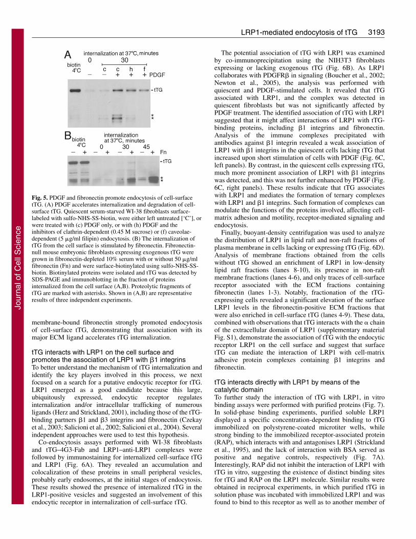
membrane-bound fibronectin strongly promoted endocytosisof cell-surface tTG, demonstrating that association with itsmajor ECM ligand accelerates tTG internalization.
tTG interacts with LRP1 on the cell surface andpromotes the association of LRP1 with �1 integrinsTo better understand the mechanism of tTG internalization andidentify the key players involved in this process, we nextfocused on a search for a putative endocytic receptor for tTG.LRP1 emerged as a good candidate because this large,ubiquitously expressed, endocytic receptor regulatesinternalization and/or intracellular trafficking of numerousligands (Herz and Strickland, 2001), including those of the tTG-binding partners �1 and �3 integrins and fibronectin (Czekayet al., 2003; Salicioni et al., 2002; Salicioni et al., 2004). Severalindependent approaches were used to test this hypothesis.
Co-endocytosis assays performed with WI-38 fibroblastsand tTG–4G3-Fab and LRP1–anti-LRP1 complexes werefollowed by immunostaining for internalized cell-surface tTGand LRP1 (Fig. 6A). They revealed an accumulation andcolocalization of these proteins in small peripheral vesicles,probably early endosomes, at the initial stages of endocytosis.These results showed the presence of internalized tTG in theLRP1-positive vesicles and suggested an involvement of thisendocytic receptor in internalization of cell-surface tTG.
The potential association of tTG with LRP1 was examinedby co-immunoprecipitation using the NIH3T3 fibroblastsexpressing or lacking exogenous tTG (Fig. 6B). As LRP1collaborates with PDGFR� in signaling (Boucher et al., 2002;Newton et al., 2005), the analysis was performed withquiescent and PDGF-stimulated cells. It revealed that tTGassociated with LRP1, and the complex was detected inquiescent fibroblasts but was not significantly affected byPDGF treatment. The identified association of tTG with LRP1suggested that it might affect interactions of LRP1 with tTG-binding proteins, including �1 integrins and fibronectin.Analysis of the immune complexes precipitated withantibodies against �1 integrin revealed a weak association ofLRP1 with �1 integrins in the quiescent cells lacking tTG thatincreased upon short stimulation of cells with PDGF (Fig. 6C,left panels). By contrast, in the quiescent cells expressing tTG,much more prominent association of LRP1 with �1 integrinswas detected, and this was not further enhanced by PDGF (Fig.6C, right panels). These results indicate that tTG associateswith LRP1 and mediates the formation of ternary complexeswith LRP1 and �1 integrins. Such formation of complexes canmodulate the functions of the proteins involved, affecting cell-matrix adhesion and motility, receptor-mediated signaling andendocytosis.
Finally, buoyant-density centrifugation was used to analyzethe distribution of LRP1 in lipid raft and non-raft fractions ofplasma membrane in cells lacking or expressing tTG (Fig. 6D).Analysis of membrane fractions obtained from the cellswithout tTG showed an enrichment of LRP1 in low-densitylipid raft fractions (lanes 8-10), its presence in non-raftmembrane fractions (lanes 4-6), and only traces of cell-surfacereceptor associated with the ECM fractions containingfibronectin (lanes 1-3). Notably, fractionation of the tTG-expressing cells revealed a significant elevation of the surfaceLRP1 levels in the fibronectin-positive ECM fractions thatwere also enriched in cell-surface tTG (lanes 4-9). These data,combined with observations that tTG interacts with the � chainof the extracellular domain of LRP1 (supplementary materialFig. S1), demonstrate the association of tTG with the endocyticreceptor LRP1 on the cell surface and suggest that surfacetTG can mediate the interaction of LRP1 with cell-matrixadhesive protein complexes containing �1 integrins andfibronectin.
tTG interacts directly with LRP1 by means of thecatalytic domainTo further study the interaction of tTG with LRP1, in vitrobinding assays were performed with purified proteins (Fig. 7).In solid-phase binding experiments, purified soluble LRP1displayed a specific concentration-dependent binding to tTGimmobilized on polystyrene-coated microtiter wells, whilestrong binding to the immobilized receptor-associated protein(RAP), which interacts with and antagonises LRP1 (Stricklandet al., 1995), and the lack of interaction with BSA served aspositive and negative controls, respectively (Fig. 7A).Interestingly, RAP did not inhibit the interaction of LRP1 withtTG in vitro, suggesting the existence of distinct binding sitesfor tTG and RAP on the LRP1 molecule. Similar results wereobtained in reciprocal experiments, in which purified tTG insolution phase was incubated with immobilized LRP1 and wasfound to bind to this receptor as well as to another member of
Fig. 5. PDGF and fibronectin promote endocytosis of cell-surfacetTG. (A) PDGF accelerates internalization and degradation of cell-surface tTG. Quiescent serum-starved WI-38 fibroblasts surface-labeled with sulfo-NHS-SS-biotin, were either left untreated [‘C’], orwere treated with (c) PDGF only, or with (h) PDGF and theinhibitors of clathrin-dependent (0.45 M sucrose) or (f) caveolae-dependent (5 �g/ml filipin) endocytosis. (B) The internalization oftTG from the cell surface is stimulated by fibronectin. Fibronectin-null mouse embryonic fibroblasts expressing exogenous tTG weregrown in fibronectin-depleted 10% serum with or without 50 �g/mlfibronectin (Fn) and were surface-biotinylated using sulfo-NHS-SS-biotin. Biotinylated proteins were isolated and tTG was detected bySDS-PAGE and immunoblotting in the fraction of proteinsinternalized from the cell surface (A,B). Proteolytic fragments oftTG are marked with asterisks. Shown in (A,B) are representativeresults of three independent experiments.
Jour
nal o
f Cel
l Sci
ence

3194 Journal of Cell Science 120 (18)
Fig. 6. tTG is colocalized with LRP1 during internalization and the two interact on the cell surface. (A) tTG colocalizes with LRP1 early afterendocytosis from the cell surface. Antibody-uptake experiments were performed for 5 or 15 minutes with WI-38 fibroblasts and the Fabfragments of mouse mAb 4G3 against tTG and rabbit antibody Rb2629 against LRP1, as described above in Fig. 1A. The internalizedtTG–4G3-Fab and LRP1–anti-LRP1 complexes were detected by immunofluorescence after cell permeabilization. Note the significantcolocalization of the internalized proteins in the numerous peripheral endocytic vesicles. (B) tTG is associated with LRP1. Extracts ofquiescent (–) and PDGF-treated (+) NIH3T3 cells expressing exogenous tTG were subjected to immunoprecipitation with non-immune IgGor antibody 2629 against LRP1. The resulting immune complexes were examined by SDS-PAGE and immunoblotting for tTG and LRP1.(C) tTG enhances the association of LRP1 with �1 integrins. �1 integrins were immunoprecipitated from the extracts of quiescent (–) orPDGF-treated (+) NIH3T3 cells lacking (vector) or expressing tTG. The resulting immune complexes were analyzed for LRP1, tTG and �1integrins by SDS-PAGE and immunoblotting. (D) Cell-surface tTG mediates a shift of LRP1 towards high-density membrane or ECMfractions enriched in tTG and fibronectin. NIH3T3 fibroblasts lacking (vector) or expressing tTG were surface-biotinylated with membrane-impermeable sulfo-NHS-LC-biotin. Carbonate extracts at pH 11 of the cells were subjected to ultracentrifugation in a discontinuous (45%-35%-5%) sucrose gradient. Biotinylated (cell surface) proteins from each gradient fraction were isolated and analyzed by SDS-PAGE andimmunoblotting with antibodies against LRP1, tTG and fibronectin. The high-density fractions enriched in tTG and fibronectin andexpressing increased amounts of LRP1 in the tTG-expressing cells are marked with asterisks. The numbers beneath the LRP1 bands showrelative intensities compared with the value of 1.0 assigned to cells lacking tTG in the absence of PDGF. Shown are representative results ofthree independent experiments.
Fig. 7. tTG interacts directly with LRP1 by means of the catalyticdomain. (A) The interaction of purified LRP1 with tTGimmobilized on plastic wells. Binding assays were performed asdescribed in Materials and Methods in the presence or absence ofthe LRP1 interactor RAP. Immobilized BSA and RAP were used asnegative and positive binding controls, respectively. (B) Theinteraction of purified tTG with LRP1 and soluble VLDL receptor(sVLDLR) immobilized on plastic wells. Binding assays wereperformed as described in Materials and Methods with BSA usedas a negative binding control. Shown in (A,B) are the means ± s.d.for two independent experiments performed in triplicate. (C) LRP1interacts with the second (catalytic) domain of tTG. Full-lengthtTG and its deletion mutants (Hang et al., 2005), all containing theC-terminal c-Myc tag, were expressed in NIH3T3 fibroblasts. Cellextracts were analyzed by SDS-PAGE and immunoblotting withantibody against c-Myc for total expression levels of full-lengthtTG and its deletion mutants (upper panel). LRP1 wasimmunoprecipitated from cell extracts with the rabbit antibodyRb2629, and the resulting LRP1 immune complexes were analyzedby SDS-PAGE and immunoblotting with antibody against c-Myc(lower panel). Molecular mass markers (kDa) are shown to theright of the gels.
Jour
nal o
f Cel
l Sci
ence

3195LRP1-mediated endocytosis of tTG
the LDL receptor superfamily, VLDLR, which is structurallyrelated to LRP1 (Fig. 7B). Together, these in vitro experimentsspecify a direct interaction between tTG and LRP1.
To define the domain(s) of tTG involved in this interaction,the association of LRP1 with previously characterized tTGdeletion mutants (Hang et al., 2005) was studied by co-immunoprecipitation (Fig. 7C). These experiments revealedthat, in addition to full-length tTG, only deletion mutantscontaining the second (catalytic) domain of the protein interactwith LRP1. Therefore, the catalytic domain of tTG is involvedin the interaction with LRP1.
LRP1 is required for endocytosis of cell-surface tTGThe above observations of co-internalization of tTG and LRP1(Fig. 6A) and the interaction between these proteins (Figs 6,7) prompted us to test the role of LRP1 in endocytosis of tTGfrom the cell surface (Fig. 8). To evaluate whether LRP1 isrequired for tTG internalization in cells endogenouslyexpressing this protein, we used the original CHO cells thatproduce significant amounts of LRP1 and their derivativeslacking LRP1 expression (CHO 13-5-1 cell line; Fig. 8A)(Fitzgerald et al., 1995). Cell-surface biotinylation andinternalization assays demonstrated a robust endocytosis andintracellular degradation of tTG in the original CHO cells butshowed a strong impairment of tTG internalization in theirderivatives lacking LRP1.
To prove that the crucial role of LRP1 in endocytosis ofsurface tTG is not limited to certain cell lines, exogenoushuman tTG was stably expressed in normal MEFs and theircounterparts with a genetically knocked-out LRP1 gene[Lrp1–/– PEA-13 MEFs (Willnow and Herz, 1994)].Internalization assays were performed with surface-biotinylated MEF and PEA-13 cells expressing exogenous tTG(Fig. 8B). Biochemical analysis with surface biotinylation andendocytosis of tTG revealed that, although cell-surface tTGwas efficiently internalized and intracellularly degraded in theLRP1-expressing MEFs, its endocytosis was abolished in theLRP1-deficient PEA-13 cells. These findings show that, in twodifferent cell systems, the endocytic receptor LRP1 is requiredfor internalization of cell-surface tTG.
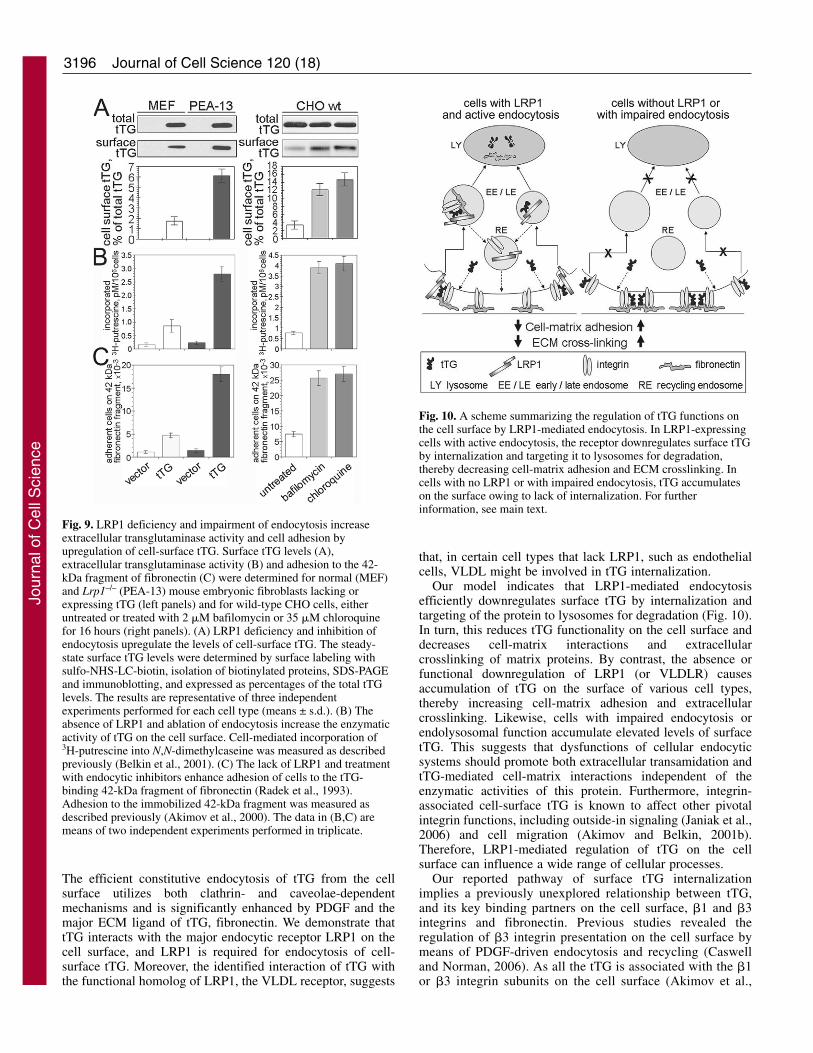
LRP1 deficiency and blockade of endocytosisupregulate transglutaminase activity and cell-matrixadhesion owing to accumulation of cell-surface tTGFinally, we tested the effects of LRP1 deficiency and inhibitionof endocytosis on steady-state levels of surface tTG andenzymatic and adhesive functions of this protein on the cellsurface (Fig. 9). With similar expression levels of exogenoustTG in the MEF and PEA-13 transfectants, increased levels ofsurface tTG were detected in the latter cells deficient in LRP1(Fig. 9A, left panels). Likewise, prolonged inhibition ofendolysosomal function in wild-type CHO cells bybafilomycin or chloroquine resulted in greater accumulation ofcell-surface tTG compared with untreated cells (Fig. 9A, rightpanels). In turn, the increased levels of surface tTG in the PEA-13 transfectants and CHO cells treated with bafilomycin orchloroquine resulted in elevated transamidation on the cellsurface (Fig. 9B). Finally, accumulation of tTG on the surfaceof these cells led to upregulation of its adhesive function, asjudged by the increased adhesion of these cells on the 42-kDatTG-binding fragment of fibronectin (Fig. 9C). Therefore,
LRP1 deficiency, or inhibition of endolysosomal function,elevates the expression and transglutaminase activity of cell-surface tTG and promotes cell-matrix interactions.
DiscussionPrevious studies demonstrated a dual function for cell-surfacetTG as a protein crosslinking enzyme and high-affinityintegrin-binding co-receptor for fibronectin (reviewed inZemskov et al., 2006). Therefore, precise regulation of cell-matrix adhesion and adhesion-mediated signaling requires aversatile and efficient regulation of cell-surface tTG. Previouswork delineated two major pathways of surface tTG regulationby means of the Ras-Raf-MEK1-ERK1 (ERK2) signalingmodule (Akimov and Belkin, 2003) and pericellularproteolysis by membrane-anchored and solublemetalloproteases (Belkin et al., 2001; Belkin et al., 2004).Here, we describe a novel means of regulation of tTGfunctioning on the cell surface that involves its internalizationfollowed by intracellular degradation in lysosomes. We foundthat internalization of tTG proceeds in a dynamin-mediatedcholesterol-dependent manner in a wide range of cell types.
Fig. 8. Internalization of cell-surface tTG requires the endocyticreceptor LRP1. LRP1 deficiency inhibits internalization of cell-surface tTG. Wild-type (CHO wt) and LRP1-deficient (CHO 13-5-1)CHO cells (A) or normal (MEF) and Lrp1–/– (PEA-13) mouseembryonic fibroblasts expressing exogenous tTG (B) were surface-biotinylated with sulfo-NHS-SS-biotin and subjected tointernalization assays. (A,B) Biotinylated proteins were isolated andtTG was detected by SDS-PAGE and immunoblotting in the fractionof proteins internalized from the cell surface. Proteolytic fragmentsof tTG are marked with asterisks. Graphs on the right show the levelsof internalized tTG for each cell type at different time points ofinternalization. The values presented are expressed as percentages ofthose obtained for cell-surface tTG before internalization. The resultsare representative of three independent experiments performed foreach cell type (means ± s.d.).
Jour
nal o
f Cel
l Sci
ence
3196
The efficient constitutive endocytosis of tTG from the cellsurface utilizes both clathrin- and caveolae-dependentmechanisms and is significantly enhanced by PDGF and themajor ECM ligand of tTG, fibronectin. We demonstrate thattTG interacts with the major endocytic receptor LRP1 on thecell surface, and LRP1 is required for endocytosis of cell-surface tTG. Moreover, the identified interaction of tTG withthe functional homolog of LRP1, the VLDL receptor, suggests
Journal of Cell Science 120 (18)
that, in certain cell types that lack LRP1, such as endothelialcells, VLDL might be involved in tTG internalization.
Our model indicates that LRP1-mediated endocytosisefficiently downregulates surface tTG by internalization andtargeting of the protein to lysosomes for degradation (Fig. 10).In turn, this reduces tTG functionality on the cell surface anddecreases cell-matrix interactions and extracellularcrosslinking of matrix proteins. By contrast, the absence orfunctional downregulation of LRP1 (or VLDLR) causesaccumulation of tTG on the surface of various cell types,thereby increasing cell-matrix adhesion and extracellularcrosslinking. Likewise, cells with impaired endocytosis orendolysosomal function accumulate elevated levels of surfacetTG. This suggests that dysfunctions of cellular endocyticsystems should promote both extracellular transamidation andtTG-mediated cell-matrix interactions independent of theenzymatic activities of this protein. Furthermore, integrin-associated cell-surface tTG is known to affect other pivotalintegrin functions, including outside-in signaling (Janiak et al.,2006) and cell migration (Akimov and Belkin, 2001b).Therefore, LRP1-mediated regulation of tTG on the cellsurface can influence a wide range of cellular processes.
Our reported pathway of surface tTG internalizationimplies a previously unexplored relationship between tTG,and its key binding partners on the cell surface, �1 and �3integrins and fibronectin. Previous studies revealed theregulation of �3 integrin presentation on the cell surface bymeans of PDGF-driven endocytosis and recycling (Caswelland Norman, 2006). As all the tTG is associated with the �1or �3 integrin subunits on the cell surface (Akimov et al.,
Fig. 9. LRP1 deficiency and impairment of endocytosis increaseextracellular transglutaminase activity and cell adhesion byupregulation of cell-surface tTG. Surface tTG levels (A),extracellular transglutaminase activity (B) and adhesion to the 42-kDa fragment of fibronectin (C) were determined for normal (MEF)and Lrp1–/– (PEA-13) mouse embryonic fibroblasts lacking orexpressing tTG (left panels) and for wild-type CHO cells, eitheruntreated or treated with 2 �M bafilomycin or 35 �M chloroquinefor 16 hours (right panels). (A) LRP1 deficiency and inhibition ofendocytosis upregulate the levels of cell-surface tTG. The steady-state surface tTG levels were determined by surface labeling withsulfo-NHS-LC-biotin, isolation of biotinylated proteins, SDS-PAGEand immunoblotting, and expressed as percentages of the total tTGlevels. The results are representative of three independentexperiments performed for each cell type (means ± s.d.). (B) Theabsence of LRP1 and ablation of endocytosis increase the enzymaticactivity of tTG on the cell surface. Cell-mediated incorporation of3H-putrescine into N,N-dimethylcaseine was measured as describedpreviously (Belkin et al., 2001). (C) The lack of LRP1 and treatmentwith endocytic inhibitors enhance adhesion of cells to the tTG-binding 42-kDa fragment of fibronectin (Radek et al., 1993).Adhesion to the immobilized 42-kDa fragment was measured asdescribed previously (Akimov et al., 2000). The data in (B,C) aremeans of two independent experiments performed in triplicate.
Fig. 10. A scheme summarizing the regulation of tTG functions onthe cell surface by LRP1-mediated endocytosis. In LRP1-expressingcells with active endocytosis, the receptor downregulates surface tTGby internalization and targeting it to lysosomes for degradation,thereby decreasing cell-matrix adhesion and ECM crosslinking. Incells with no LRP1 or with impaired endocytosis, tTG accumulateson the surface owing to lack of internalization. For furtherinformation, see main text.
Jour
nal o
f Cel
l Sci
ence

3197LRP1-mediated endocytosis of tTG
2000), it is plausible that tTG is internalized as a complexwith integrins and that this process is accelerated by certaingrowth factors (Fig. 10). Indeed, our experiments revealedsimilar dynamics of endocytosis for cell-surface tTG and �1integrins in CHO cells. However, unlike the �1 or �3integrins that typically are recycled back to the cell surfaceby means of several distinctive pathways (Powelka et al.,2004; Pellinen and Ivaska, 2006), internalized tTG has adifferent fate and is targeted to lysosomes for degradation.These findings indicate that the putative internalized integrin-tTG adhesive complexes are probably dissociated in theendocytic compartments.
Notably, previous work showed that fibronectin isinternalized by an LRP1-dependent mechanism in CHO cellsand MEFs, whereas our findings revealed a significantenhancement of surface tTG endocytosis by membrane-associated fibronectin. Therefore, in the absence of fibronectin,tTG is mostly retained on the cell surface, whereas theformation of tTG-fibronectin complexes promotes their LRP1-mediated endocytosis. As fibronectin interacts directly withLRP1, it might bridge surface tTG to this endocytic receptorand facilitate tTG internalization (Fig. 10). In future work, itwill be important to compare the effects of soluble and matrixforms of fibronectin on endocytosis of cell-surface tTG.Although membrane-bound monomeric fibronectin was foundto promote this process, polymeric fibronectin (matrix fibrils)might prevent endocytosis of tTG by anchoring the integrin-tTG complexes on the cell surface, thus stabilizing cell-matrixadhesion and promoting fibronectin assembly.
Our findings indicate that a key binding partner andfunctional antagonist of LRP1, RAP, does not perturb theinteraction of tTG with LRP1 or interfere with the endocytosisof cell-surface tTG (data not shown). Therefore, although tTGinteracts with LRP1 directly, it might not represent a typicalligand, but instead function as a mediator involved in LRP1-dependent endocytosis through the association with LRP1ligands such as fibronectin. Furthermore, initial analysisindicates that tTG does not bind to the � chain of LRP1(supplementary material Fig. S1). Future studies should help todelineate the tTG-binding site(s) on the � chain of the LRP1molecule. They also should define the relationships betweenintegrins, tTG and fibronectin in the LRP1-dependent endocyticprocess and determine the role of the identified interactionsbetween cell-surface tTG and LRP1 in internalization ofintegrin- and fibronectin-containing adhesive complexes.
An emerging general theme in the field highlights theintricate functional relationship between cell-matrix adhesionand endocytosis. A recent study showed that nascent adhesivestructures, the focal-adhesion complexes, might be targeted forendocytosis from inside the cell through the interaction ofdynamin-2 with the key regulatory component of thesecomplexes, focal adhesion kinase, to modulate cell-matrixadhesion and migration (Ezratti et al., 2005). The present workdescribes yet-another functional link between a constituent ofcell-matrix adhesive structures, cell-surface tTG, and a majorendocytic receptor, LRP1. Regulation of surface tTG by LRP1-mediated internalization endows cells with the means toefficiently adjust their interactions with the ECM in responseto outside cues. Future investigations will determine theimportance of this regulatory mechanism in endocytosis, cell-matrix adhesion, migration and signaling.
Materials and MethodsCells and plasmidsHuman (MRC-5, WI-38) and mouse (NIH3T3) fibroblasts and human U-251 gliomacells were maintained in Dulbecco’s modified Eagle’s medium (DMEM)supplemented with 10% fetal bovine serum (FBS) and 1% antibiotic antimycotic(100�) (Invitrogen, Carlsbad, CA). Chinese hamster ovarian cells (CHO), wild-typeand LRP1-deficient derivatives (13-5-1 CHO) (Fitzgerald et al., 1995), weremaintained in DMEM/F-12 (1:1) medium supplemented with 5% FBS. LRP1-deficient mouse embryonic fibroblasts (Lrp1–/– MEF, clone PEA-13) (Willnow andHerz, 1994), fibronectin-null mouse embryonic fibroblasts (Fn–/– MEF, clone 7E)(Tomasini-Johansson et al., 2001) and their wild-type counterparts were maintainedin DMEM supplemented with 10% FBS. U251 glioma cells and their stabletransfectants expressing caveolin-1 siRNA were provided by A. Strongin (TheBurnham Institute, La Jolla, CA). All the cells were grown at 37°C in an atmosphereof 5% CO2.
Transfections of cells were performed with lipofectamine 2000 (Invitrogen),following the manufacturer’s instructions. Wild-type dynamin-2 and a GTPase-deficient (K44A) mutant dynamin-2, both containing an HA tag, were a gift fromS. L. Schmid (The Scripps Institute, La Jolla, CA). The plasmids were transientlyexpressed in MRC-5 cells. Mifepristone (Mfp)-inducible expression of full-lengthhuman tTG and deletion mutants in NIH3T3 cells and in Fn–/– MEFs has beendescribed previously (Hang et al., 2005; Janiak et al., 2006).
The cDNA of human LRP1 (Ulery et al., 2000) was used as a template to generateexpression vectors for the LRP� essentially as described previously (Mikhailenkoet al., 2001). Briefly, the fragment of cDNA that encodes amino acids 3844-4525of LRP1 (GenBankTM accession number X13916) was generated by PCRamplification and subcloned into the pSecTag expression vector (Invitrogen)modified to produce a protein with two copies of the Myc epitope at its aminoterminus. The mini-receptor contained a portion of the LRP1 extracellular domain(including membrane-proximal YWTD �-propeller and EGF-like repeats),transmembrane domain and cytoplasmic tail. Deletion of a cytoplasmic domain wasachieved by mutation of the codon encoding Ala4432 to a termination codon usingthe TransformerTM site-directed mutagenesis kit (Clontech) and the constructauthenticity confirmed by sequencing.
Antibodies, proteins and reagentsRabbit polyclonal antibodies against EEA1, Rab7, Arf1, Lamp-1, caveolin-1,clathrin heavy chain and rat anti-mouse �1 integrin mAb 9EG7 were all obtainedfrom BD Biosciences (San Jose, CA). Rabbit antibody against the cytoplasmicdomain of �1 integrin, anti-rabbit and anti-mouse IgG conjugated with peroxidasewere all sourced from Chemicon International (Temecula, CA). Mouse mAbsTG100 and CUB7402 against human tTG were obtained from NeoMarkers(Freemont, CA). Mouse anti-tTG mAb 4G3 has been described previously (Akimovand Belkin, 2001b). Fab fragments of mAb 4G3 were prepared by limitedproteolysis using papain immobilized on agarose (Pierce, Rockford, IL). Rabbitaffinity-purified antibody Rb2629 and mAbs 11E4 and 5A6 against LRP1 weregenerated in the laboratory of D.K.S. The polyclonal affinity-purified antibody tofibronectin has been described previously (Akimov and Belkin, 2001a). Mouse mAb9E10 against a c-Myc tag and a rabbit polyclonal antibody against an HA tag wereobtained from Abgent (San Diego, CA).
tTG was purified from human red blood cells as described previously (Radek etal., 1993). LRP1 was purified from bovine placenta as previously reported (Ashcomet al., 1990). The LRP1 antagonist, receptor-associated protein (RAP) was preparedas described earlier (Williams et al., 1992). The soluble VLDL receptor fragmentcontaining ligand-binding repeats was prepared and characterized as reportedpreviously (Ruiz et al., 2005). Purified human plasma fibronectin was obtained fromChemicon.
FITC-labeled transferrin was acquired from Molecular Probes (Eugene, OR).Human recombinant PDGF-BB was sourced from R&D Systems (Minneapolis,MN). Sulfo-NHS-LC-biotin, sulfo-NHS-SS-biotin, NeutrAvidin-agarose andSuperSignal West Pico chemiluminescent substrate were obtained from Pierce.Filipin, methyl-�-cyclodextrin (M-�-CD), bafilomycin and chloroquine wereobtained from Sigma (St Louis, MO).
Cell-surface biotinylation and endocytosis assaysTo compare the overall levels of cell-surface tTG in Mfp-induced NIH3T3fibroblasts expressing tTG in the absence or presence of endocytosis inhibitors, thecells were labeled using cell-impermeable sulfo-NHS-LC-biotin as describedpreviously (Janiak et al., 2006).
For endocytosis assays, cell monolayers were labeled with cell-impermeable SH-cleavable sulfo-NHS-SS-biotin. After biotinylation and quenching, the cells wereincubated in cell-culture medium at 37°C with or without inhibitors to allowinternalization of biotinylated proteins. To remove the remaining biotin residuesfrom the cell-surface proteins, the cells were washed with ice-cold PBS andincubated three times for 10 minutes on ice with 50 mM cell-impermeable reducingreagent MESNA (sodium 2-mercaptoethane sulfonate) in stripping buffer (50 mMTris-HCl, pH 8.4, 150 mM NaCl). Preparation of cell extracts and isolation ofbiotinylated (internalized) proteins were performed as described previously (Janiak
Jour
nal o
f Cel
l Sci
ence

3198
et al., 2006). Proteins isolated on NeutrAvidin-agarose were subjected to SDS-PAGE in Novex 4-12% polyacrylamide Bis-Tris gels (Invitrogen) followed byelectrotransfer onto Immobilon-P membrane (Millipore, Billerica, MA) andimmunoblotting.
To study internalization of cell-surface tTG in fibronectin-null mouse embryonicfibroblasts (Fn–/– MEF, clone 7E), the cells growing in DMEM supplemented with10% fibronectin-depleted FBS were treated with Mfp for 24 hours. Purifiedfibronectin (50 �g/ml) was added to some dishes 3 hours before cell biotinylation.The cells were labeled with sulfo-NHS-SS-biotin and used in the endocytosis assayas described above. Protein fractions isolated on NeutrAvidin-agarose wereanalyzed by SDS-PAGE and immunoblotting.
To inhibit endocytosis, the cell monolayers were treated with the general inhibitorof endocytosis, M-�-CD, or the specific inhibitor of lipid rafts or caveolae, filipin.To block endo-lysosomal function, cells were treated with bafilomycin orchloroquine. To specifically inhibit clathrin-dependent endocytosis by hyperosmoticshock, cells were incubated in 0.45 M sucrose. To block LRP1-mediatedendocytosis, cells were incubated with 100 ng/ml RAP.
Ultracentrifugation in discontinuous sucrose gradientsTo analyze the membrane distribution of various proteins, cell homogenates weresubjected to detergent-free fractionation by ultracentrifugation in a discontinuoussucrose gradient, essentially as described previously (Song et al., 1996). Briefly,NIH3T3 fibroblasts lacking or expressing tTG were labeled with sulfo-NHS-LC-biotin, scrapped in 0.5 M carbonate buffer (pH 11.0) on ice and sonicated. Theresulting homogenates were mixed with 80% sucrose in MBS (25 mM MES, pH6.5, 150 mM NaCl) to a final concentration of 45% sucrose and placed on the bottomof the centrifuge tube. A 5-35% discontinuous sucrose gradient was formed above(4 ml of 35% sucrose and 4 ml of 5% sucrose; both in MBS) and centrifuged at39,000 rpm for 18 hours in an SW41 rotor (Beckman Instruments, Palo Alto, CA).After centrifugation, 1 ml gradient fractions were collected, adjusted to 1% SDS byaddition of 50 �l of 20% SDS, boiled and incubated with NeutrAvidin-agarose toisolate biotinylated proteins. The isolated membrane protein fractions were analyzedby SDS-PAGE and immunoblotting.
Analysis of the interaction of tTG and LRP1 in vitroTo study the interaction between tTG and LRP1 in vitro, purified human tTG inTBS (10 �g/ml) was immobilized in polystyrene-coated microtiter plates byincubation for 3 hours at room temperature. As positive and negative bindingcontrols, RAP and BSA were immobilized in parallel wells. The wells were blockedwith 3% BSA in TBS for 1 hour at room temperature, washed and the immobilizedproteins incubated with purified LRP1 (0-300 nM) in the binding buffer (1% BSAin TBS) for 1 hour at 37°C. LRP1 bound to the proteins was detected with 0.1 �g/mlmouse mAb 11E4 to human LRP1 and 0.2 �g/ml secondary anti-mouse IgGconjugated with peroxidase. The reaction was developed with a SureBlue TMBmicrowell peroxidase chromogenic substrate (KPL, Gaithersburg, MD), stoppedwith 1 M HCl and measured by spectrophotometry at 450 nm. In reciprocalexperiments, purified LRP1 and soluble VLDL receptor were immobilized inmicrotiter plates, and human tTG (0-1500 nM) in the binding buffer (1% BSA inTBS) was incubated with immobilized proteins. Bound tTG was detected with 0.1�g/ml mouse mAb TG100.
Immunofluorescence microscopyTo analyze the localization of internalized Fab fragments of anti-tTG–4G3-mAb inthe antibody-uptake experiments, WI-38 fibroblasts grown on fibronectin-coatedglass coverslips were washed with ice-cold PBS and incubated with 10 �g/ml Fabfragments in PBS with 0.1% BSA for 1 hour on ice. Cells with surface-bound Fabfragments were washed with cold PBS-BSA and warmed up for the indicated timeperiods at 37°C in DMEM-FBS. Next, the cells were rinsed with cold PBS andincubated in cold 0.1 M glycine-HCl (pH 2.5) twice for 5 minutes to strip theremaining Fab fragments from the cell surface. The cells were fixed with 3%paraformaldehyde, permeabilized with 0.5% Triton X-100 in PBS and incubatedwith antibodies against the endocytic markers or clathrin. The internalizedtTG–4G3-Fab complexes were detected with Alexa Fluor 594 goat anti-mouse IgGthat reacts with Fab fragments of mouse IgG, whereas clathrin and the endocyticmarkers were visualized with Alexa Fluor 488 goat anti-rabbit IgG.
To study internalization of anti-tTG–4G3-Fab in MRC-5 fibroblasts expressingHA-tagged dynamin-2 or its inactive K44A mutant, the cells with internalizedtTG–4G3-Fab complexes were labeled with a rabbit polyclonal antibody to the HAtag, followed by Alexa Fluor 594 goat anti-mouse IgG and Alexa Fluor 488 goatanti-rabbit IgG.
In double-antibody-uptake experiments, co-internalization of anti-tTG–4G3-Faband either rat mAb 9EG7 against �1 integrin or rabbit anti-LRP1 antibody Rb2629was examined. In this case, the latter internalized protein-antibody complexes weredetected with either Alexa Fluor 488 goat anti-rat IgG or Alexa Fluor 488 goat anti-rabbit IgG. Also, co-internalization assays were performed with anti-tTG–4G3-Faband 5 �g/ml FITC-labeled transferrin.
To examine the relationship of cell-surface tTG, �1 integrins and caveolae, triplestaining of live non-permeabilized MRC-5 fibroblasts was performed with mouse
Journal of Cell Science 120 (18)
anti-tTG–4G3-mAb, rat anti-�1 integrin mAb 9EG7 and rabbit anti-caveolin-1antibody. The cell-surface proteins were visualized, respectively, with Alexa Fluor488 goat anti-mouse IgG, Alexa Fluor 350 goat anti-rat IgG and Alexa Fluor 594goat anti-rabbit IgG.
Cells were viewed and photographed with 63� and 100� objectives using aNikon Eclipse E800 microscope (Nikon, Melville, NY) and SPOT RT digitalcamera. Images were acquired and digitally merged with Advance Spot software(Diagnostic Instruments, Sterling Heights, MI).
Immunoprecipitation, SDS-PAGE and immunoblottingTo study the interaction of �1 integrins, tTG and LRP1-tTG, �1 integrins or LRP1were immunoprecipitated from the RIPA extracts of NIH3T3 fibroblasts expressingor lacking tTG (Janiak et al., 2006). To examine an effect of PDGFR� activationon the LRP1–�1-integrin complexes, quiescent cells were stimulated with PDGF-BB (20 ng/ml) for 5 minutes. The cells were lysed in ice-cold lysis buffer (1% NP-40 in TBS with protease and phosphatase inhibitor cocktails), cell lysates werecleared by centrifugation (15,000 g, 30 minutes, 4°C) and used forimmunoprecipitation with 2 �g of rabbit polyclonal antibody against thecytoplasmic domain of �1 integrin, or mouse mAb 11E4 against LRP1, and protein-G–agarose beads. The immune complexes were analyzed by SDS-PAGE andimmunoblotting with antibodies against LRP1, tTG and �1 integrin. SDS-PAGEunder denaturing conditions was performed in Novex 4-12% gradientpolyacrylamide Bis-Tris gels using MOPS running buffer (Invitrogen). Separatedproteins were electrotransferred onto PVDF membranes in a mini trans-blotelectrophoretic transfer cell. Immunoblots were developed with SuperSignal WestPico chemiluminescent substrate.
Other methodsTransglutaminase activity on the surface of live cells was measured by determiningincorporation of 3H-putrescine in N,N-dimethylcaseine (Belkin et al., 2001). tTG-dependent adhesion of cells on the 42-kDa gelatin-binding domain of fibronectinwas determined as described previously (Akimov et al., 2000).
We are grateful to Alex Strongin (The Burnham Institute) for thegenerous gift of U251 glioma cells expressing caveolin-1 siRNA. Wealso thank Sandra L. Schmid (The Scripps Institute) for providing theplasmids encoding dynamin-2 and its dominant-negative mutantK44A. This work was supported by the NIH grants GM62895 toA.M.B. and HL50784 to D.K.S.
ReferencesAkimov, S. S. and Belkin, A. M. (2001a). Cell-surface transglutaminase promotes
fibronectin assembly via interaction with the gelatin-binding domain of fibronectin: arole in TGF�-dependent matrix deposition. J. Cell Sci. 114, 2989-3000.
Akimov, S. S. and Belkin, A. M. (2001b). Cell surface tissue transglutaminase isinvolved in adhesion and migration of monocytic cells on fibronectin. Blood 98, 1567-1576.
Akimov, S. S. and Belkin, A. M. (2003). Opposing roles of Ras/Raf oncogenes and theMEK1/ERK signaling module in regulation of expression and adhesive function oftissue transglutaminase. J. Biol. Chem. 278, 35609-35619.
Akimov, S. S., Krylov, D., Fleischman, L. F. and Belkin, A. M. (2000). Tissuetransglutaminase is an integrin-binding adhesion coreceptor for fibronectin. J. CellBiol. 148, 825-838.
Altschuler, Y., Barbas, S. M., Terlecky, L. J., Tang, K., Hardy, S., Mostov, K. E. andSchmid, S. L. (1998). Redundant and distinct functions for dynamin-1 and dynamin-2 isoforms. J. Cell Biol. 143, 1871-1881.
Ashcom, J. D., Tiller, S. E., Dickerson, K., Cravens, J. L., Argraves, W. S. andStrickland, D. K. (1990). The human alpha 2-macroglobulin receptor: identificationof a 420-kD cell surface glycoprotein specific for the activated conformation of alpha2-macroglobulin. J. Cell Biol. 110, 1041-1048.
Belkin, A. M., Akimov, S. S., Zaritskaya, L. S., Ratnikov, B. I., Deryugina, E. I. andStrongin. A. Y. (2001). Matrix-dependent proteolysis of surface transglutaminase bymembrane-type metalloproteinase regulates cancer cell adhesion and locomotion. J.Biol. Chem. 276, 18415-18422.
Belkin, A. M., Zemskov, E. A., Hang, J., Akimov, S. S., Sikora, S. and Strongin, A.Y. (2004). Cell-surface-associated tissue transglutaminase is a target of MMP-2proteolysis. Biochemistry 43, 11760-11769.
Berrou, E. and Bryckaert, M. (2001). Platelet-derived growth factor inhibits smoothmuscle cell adhesion to fibronectin by ERK-dependent and ERK-independentpathways. J. Biol. Chem. 276, 39303-39309.
Boucher, P., Liu, P., Gotthardt, M., Hiesberger, T., Anderson, R. G. and Herz, J.(2002). Platelet-derived growth factor mediates tyrosine phosphorylation of thecytoplasmic domain of the low density lipoprotein receptor-related protein in caveolae.J. Biol. Chem. 277, 15507-15513.
Caswell, P. T. and Norman, J. C. (2006). Integrin trafficking and the control of cellmigration. Traffic 7, 14-21.
Chavrier, P., Parton, R. G., Hauri, H. P., Simons, K. and Zerial, M. (1990).Localization of low molecular weight GTP binding proteins to exocytic and endocyticcompartments. Cell 62, 317-329.
Jour
nal o
f Cel
l Sci
ence

3199LRP1-mediated endocytosis of tTG
Chen, J. W., Murphy, T. L., Willingham, M. C., Pastan, I. and August, J. T. (1985).Identification of two lysosomal membrane glycoproteins. J. Cell Biol. 101, 85-95.
Conner, S. D. and Schmid, S. L. (2003). Regulated portals of entry into the cell. Nature422, 37-44.
Czekay, R. P., Aertgeerts, K., Curriden, S. A. and Loskutoff, D. J. (2003). Plasminogenactivator inhibitor-1 detaches cells from extracellular matrices by inactivating integrins.J. Cell Biol. 160, 781-791.
Damke, H., Baba, T., Warnock, D. E. and Schmid, S. L. (1994). Induction of mutantdynamin specifically blocks endocytic coated vesicle formation. J. Cell Biol. 127, 915-934.
Donaldson, J. G. and Honda, A. (2005). Localization and function of Arf familyGTPases. Biochem. Soc. Trans. 33, 639-642.
Ezratti, E. J., Partridge, M. A. and Gundersen, G. G. (2005). Microtubule-inducedfocal adhesion disassembly is mediated by dynamin and focal adhesion kinase. Nat.Cell Biol. 7, 581-590.
Fellin, F. M., Barsigian, C., Rich, E. and Martinez, J. (1988). Binding and cross-linkingof rabbit fibronectin by rabbit hepatocytes in suspension. J. Biol. Chem. 263, 1791-1797.
Fesus, L. and Piacentini, M. (2002). Transglutaminase 2, an enigmatic enzyme withdiverse functions. Trends Biochem. Sci. 27, 534-539.
Fitzgerald, D. J., Fryling, C. M., Zdanovsky, A., Saelinger, C. B., Kounnas, M.,Winkles, J. A., Strickland, D. K. and Leppla, S. (1995). Pseudomonas exotoxin-mediated selection yields cells with altered expression of low-density lipoproteinreceptor-related protein. J. Cell Biol. 129, 1533-1541.
Folk, J. E. and Cole, P. W. (1966). Identification of a functional cysteine essential forthe activity of guinea pig liver transglutaminase. J. Biol. Chem. 241, 3238-3240.
Gaborik, Z., Szaszak, M., Szidonya, L., Balla, B., Paku, S., Catt, K. J., Clark, A. J.L. and Hunyady, L. (2001). �-Arrestin- and dynamin-dependent endocytosis of theAT1 angiotensin receptor. Mol. Pharmacol. 59, 239-247.
Greenwood, J. A., Theibert, A. B., Prestwich, G. D. and Murphy-Ullrich, J. E. (2000).Restructuring of focal adhesion plaques by PI-3 kinase: regulation by PtdIns (3,4,5)-P3 binding to �-actinin. J. Cell Biol. 150, 673-685.
Hang, J., Zemskov, E. A., Lorand, L. and Belkin, A. M. (2005). Identification of anovel recognition sequence for fibronectin within the NH2-terminal beta-sandwichdomain of tissue transglutaminase. J. Biol. Chem. 280, 23675-23683.
Hansen, S. H., Sandvig, K. and van Deurs, B. (1993). Clathrin and HA2 adaptors:effects of potassium depletion, hypertonic medium, and cytosol acidification. J. CellBiol. 121, 61-72.
Hasegawa, G., Suwa, M., Ichikawa, Y., Ohtsuka, T., Kumagai, S., Kikuchi, M., Sato,Y. and Saito, Y. (2003). A novel function of tissue-type transglutaminase: proteindisulfide isomerase. Biochem. J. 373, 793-803.
Heldin, C.-H. and Westermark, B. (1999). Mechanism of action and in vivo role ofplatelet-derived growth factor. Physiol. Rev. 79, 1283-1316.
Henley, J. R., Cao, H. and McNiven, M. A. (1999). Participation of dynamin in thebiogenesis of cytoplasmic vesicles. FASEB J. 2, S243-S247.
Herz, J. and Strickland, D. K. (2001). LRP: a multifunctional scavenger and signalingreceptor. J. Clin. Invest. 108, 779-784.
Heuser, J. E. and Anderson, R. G. W. (1989). Hypertonic media inhibit receptor-mediated endocytosis by blocking clathrin-coated pit formation. J. Cell Biol. 108, 389-400.
Janiak, A., Zemskov, E. A. and Belkin, A. M. (2006). Cell surface transglutaminasepromotes RhoA activation via integrin clustering and suppression of the Src-p190RhoGAP signaling pathway. Mol. Biol. Cell 17, 1606-1619.
Kirkham, M. and Parton, R. G. (2005). Clathrin-independent endocytosis: new insightsinto caveolae and non-caveolar lipid raft carriers. Biochim. Biophys. Acta 1746, 349-363.
Le, P. U., Guay, G., Altschuler, Y. and Nabi, I. R. (2002). Caveolin-1 is a negativeregulator of caveolae-mediated endocytosis to the endoplasmic reticulum. J. Biol.Chem. 277, 3371-3379.
Lillis, A. P., Mikhailenko, I. and Strickland, D. K. (2005). Beyond endocytosis: LRPfunction in cell migration, proliferation and vascular permeability. J. Thromb. Haemost.3, 1884-1893.
Lorand, L. and Graham, R. M. (2003). Transglutaminases: crosslinking enzymes withpleiotropic functions. Nat. Rev. Mol. Cell Biol. 4, 140-156.
Mikhailenko, I., Battey, F., Migliorini, M., Ruiz, J. F., Argraves, K., Moayeri, M. andStrickland, D. K. (2001). Recognition of alpha 2-macroglobulin by the low densitylipoprotein receptor-related protein requires the cooperation of two ligand bindingcluster regions. J. Biol. Chem. 276, 39484-39491.
Mu, F. T., Callaghan, J. M., Steele-Mortimer, O., Stenmark, H., Parton, R. G.,Campbell, P. L., McCluskey, J., Yeo, J. P., Tock, E. P. and Toh, B. H. (1995). EEA1,an early endosome-associated protein. J. Biol. Chem. 270, 13503-13511.
Nakaoka, H., Perez, D. M., Baek, K. J., Das, T., Husain, A., Misono, K., Im, M. J.
and Graham, R. M. (1994). Gh: a GTP-binding protein with transglutaminase activityand receptor signaling function. Science 264, 1593-1596.
Newton, C. S., Loukinova, E., Mikhailenko, I., Ranganathan, S., Gao, Y.,Haudenschild, C. and Strickland, D. K. (2005). Platelet-derived growth factorreceptor-� (PDGFR-�) activation promotes its association with the low densitylipoprotein receptor-related protein (LRP). Evidence for co-receptor function. J. Biol.Chem. 280, 27872-27878.
Orlandi, P. A. and Fishman, P. H. (1998). Filipin-dependent inhibition of cholera toxin:evidence for toxin internalization and activation through caveolae-like domains. J. CellBiol. 141, 905-915.
Pellinen, T and Ivaska, J. (2006). Integrin traffic. J. Cell Sci. 119, 3723-3731.Powelka, A. M., Sun, J., Li, J., Gao, M., Shaw, L. M., Sonnenberg, A. and Hsu, V.
W. (2004). Stimulation-dependent recycling of integrin beta1 regulated by ARF6 andRab11. Traffic 5, 20-36.
Radek, J. T., Jeong, J. M., Murthy, S. N., Ingham, K. C. and Lorand, L. (1993).Affinity of human erythrocyte transglutaminase for a 42-kDa gelatin-binding fragmentof human plasma fibronectin. Proc. Natl. Acad. Sci. USA 90, 3152-3156.
Rodal, S. K., Skretting, G., Garred, O., Vilhardt, F., van Deurs, B. and Sandvig, K.(1999). Extraction of cholesterol with methyl-�-cyclodextrin perturbs formation ofclathrin-coated endocytic vesicles. Mol. Biol. Cell 10, 961-974.
Ruiz, J., Kouiavskaia, D., Migliorini, M., Robinson, S., Saenko, E. L., Gorlatova, N.,Li, D., Lawrence, D., Hyman, B. T., Weisgraber, K. H. et al. (2005). The apoEisoform binding properties of the VLDL receptor reveal marked differences from LRPand the LDL receptor. J. Lipid Res. 46, 1721-1731.
Salicioni, A. M., Mizelle, K. S., Loukinova, E., Mikhailenko, I., Strickland, D. K. andGonias, S. L. (2002). The low density lipoprotein receptor-related protein mediatesfibronectin catabolism and inhibits fibronectin accumulation on cell surfaces. J. Biol.Chem. 277, 16160-16166.
Salicioni, A. M., Gaultier, A., Brownlee, C., Cheezum, M. K. and Gonias, S. L. (2004).Low density lipoprotein receptor-related protein-1 promotes beta1 integrin maturationand transport to the cell surface. J. Biol. Chem. 279, 10005-10012.
Schnitzer, J. E., Oh, P., Pinney, E. and Allard, J. (1994). Filipin-sensitive caveolae-mediated transport in endothelium: reduced transcytosis, scavenger endocytosis, andcapillary permeability of select macromolecules. J. Cell Biol. 127, 1217-1232.
Song, K. S., Li, S., Okamoto, T., Quilliam, L. A., Sargiacomo, M. and Lisanti, M. P.(1996). Co-purification and direct interaction of Ras with caveolin, an integralmembrane protein of caveolae microdomains. Detergent-free purification of caveolaemicrodomains. J. Biol. Chem. 271, 9690-9697.
Strickland, D. K., Kounnas, M. Z. and Argraves, W. S. (1995). LDL receptor-relatedprotein: multiligand receptor for lipoprotein and proteinase catabolism. FASEB J. 9,890-898.
Subtil, A., Gaidarov, I., Kobylarz, K., Lampson, M. A., Keen, J. H. and McGraw, T.E. (1999). Acute cholesterol depletion inhibits clathrin-coated pit budding. Proc. Natl.Acad. Sci. USA 96, 6775-6780.
Sun, Z., Martinez-Lemus, L. A., Trache, A., Trzeciakowski, J. P., Davis, G. E., Pohl,U. and Meininger, G. A. (2005). Mechanical properties of the interaction betweenfibronectin and �5�1-integrin on vascular smooth muscle cells studied using atomicforce microscopy. Am. J. Physiol. 289, H2526-H2535.
Thomsen, P., Roepstorff, K., Stahlhut, M. and van Deurs, B. (2002). Caveolae arehighly immobile plasma membrane microdomains, which are not involved inconstitutive endocytic trafficking. Mol. Biol. Cell 13, 238-250.
Tomasini-Johansson, B. R., Kaufman, N. R., Ensenberger, M. G., Ozeri, V., Hanski,E. and Mosher, D. F. (2001). A 49-residue peptide from adhesion F1 of Streptococcuspyogenes inhibits fibronectin matrix assembly. J. Biol. Chem. 276, 23430-23439.
Torgersen, M. L., Skretting, G., van Deurs, B. and Sandvig, K. (2001). Internalizationof cholera toxin by different endocytic mechanisms. J. Cell Sci. 114, 3737-3747.
Turner, P. M. and Lorand, L. (1989). Complexation of fibronectin with tissuetransglutaminase. Biochemistry 28, 628-635.
Ulery, P. G., Beers, J., Mikhailenko, I., Tanzi, R. E., Rebeck, G. W., Hyman, B. T. andStrickland, D. K. (2000). Modulation of beta-amyloid precursor protein processing bythe low density lipoprotein receptor-related protein (LRP). Evidence that LRPcontributes to the pathogenesis of Alzheimer’s disease. J. Biol. Chem. 275, 7410-7415.
Williams, S. E., Ashcom, J. D., Argraves, W. S. and Strickland, D. K. (1992). A novelmechanism for controlling the activity of alpha 2-macroglobulin receptor/low densitylipoprotein receptor-related protein. Multiple regulatory sites for 39-kDa receptor-associated protein. J. Biol. Chem. 267, 9035-9040.
Willnow, T. E. and Herz, J. (1994). Genetic deficiency in low density lipoproteinreceptor-related protein confers cellular resistance to Pseudomonas exotoxin A.Evidence that this protein is required for uptake and degradation of multiple ligands.J. Cell Sci. 107, 719-726.
Zemskov, E. A., Janiak, A., Hang, J., Waghray, A. and Belkin, A. M. (2006). The roleof tissue transglutaminase in cell-matrix interactions. Front. Biosci. 11, 1057-1076.
Jour
nal o
f Cel
l Sci
ence
Related Documents