Project Number: JXR-502I Cell Surface Phenotype of CNS Infiltrate and Resident Microglia During the Course of Relapsing Experimental Autoimmune Encephalomyelitis in Mice A Major Qualifying Project Report Submitted to the Faculty of WORCESTER POLYTECHNIC INSTITUTE In partial fulfillment of the requirements for the Degree of Bachelor of Science In Biology & Biotechnology and Biochemistry by ________________________________ Morgan Oexner April 30, 2009 APPROVED: ______________________ Dr. Jill Rulfs WPI Project Advisor

Welcome message from author
This document is posted to help you gain knowledge. Please leave a comment to let me know what you think about it! Share it to your friends and learn new things together.
Transcript

Project Number: JXR-502I
Cell Surface Phenotype of CNS Infiltrate and Resident
Microglia During the Course of Relapsing Experimental Autoimmune Encephalomyelitis in Mice
A Major Qualifying Project Report
Submitted to the Faculty of
WORCESTER POLYTECHNIC INSTITUTE
In partial fulfillment of the requirements for the
Degree of Bachelor of Science
In Biology & Biotechnology and Biochemistry
by
________________________________ Morgan Oexner April 30, 2009
APPROVED: ______________________ Dr. Jill Rulfs WPI Project Advisor
1
Abstract
Multiple Sclerosis (MS) is the most common cause of neurological disability in
young adults. In this study, the animal model of MS, experimental autoimmune
encephalomyelitis (EAE) was used in conjunction with tissue harvest techniques, flow
cytometry and an ex vivo assay system in order to immunophenotype cellular infiltrate
over the course of disease, study microglial activation, evaluate the ability of microglia
to stimulate T-cells and to determine the effects of small molecule inhibitors on certain
biochemical pathways. This study demonstrates that CNS infiltrate kinetics mirror
disease course and suggests that microglia be involved in antigen presentation and
effector function during disease course.

2
Acknowledgements
I would like to thank Jill Rulfs and Bradford McRae for all of their patience and
guidance throughout the project. I would also like to thank Craig Wallace for his help
and Kimberly Black for all of her help, encouragement, and unwavering support.

3
Table of Contents
ABSTRACT ................................................................................................................... 1
ACKNOWLEDGEMENTS ............................................................................................... 2
CLINCAL COURSE AND DIAGNOSIS ........................................................................................... 4
CAUSES AND RISK FACTORS .................................................................................................... 5
PATHOLOGICAL PHYSIOLOGY AND DISEASE MECHANISMS ............................................................ 6
MANAGEMENT AND TREATMENT ............................................................................................ 7
THE EAE MODEL ................................................................................................................. 7
OBJECTIVE OF PROJECT .......................................................................................................... 7
METHODS ................................................................................................................... 9
ANIMALS ............................................................................................................................ 9
INDUCTION OF EAE AND DISEASE PROGRESSION ........................................................................ 9
ISOLATION OF INFILTRATING LEUKOCYTES AND MICROGLIA FROM THE CNS ...................................... 9
CELL SORTING AND EX VIVO ASSAY PROTOCOL .......................................................................... 11
RESULTS.................................................................................................................... 12
CNS INFILTRATE KINETICS .................................................................................................... 12
ACTIVATION STATES OF MICROGLIA ....................................................................................... 12
MICROGLIA AND T-CELL ASSAY ............................................................................................. 13
DISCUSSION .............................................................................................................. 14
INTERPRETING EXPERIMENTAL RESULTS .................................................................................. 14
FUTURE DIRECTIONS ........................................................................................................... 14
TABLES AND FIGURES ................................................................................................ 16
TABLE 1: LIST OF CELL MARKERS ........................................................................................... 16
FIGURE 1: CNS HARVESTING TIME POINTS THROUGH TYPICAL PLP-INDUCED EAE DISEASE COURSE
(SEM ERROR BARS SHOWN) ................................................................................................ 16
FIGURE 2: CELL COUNTS IN THE CNS AND THEIR CORRELATION WITH CLINICAL SIGNS OF EAE (SEM
ERROR BARS SHOWN) N=5/TIME POINT ................................................................................. 17
FIGURE 3: FACS PLOTS OF NAÏVE AND DAY 13 DISEASED CNS (CD45 STAINS HEMATOPOIETIC CELL, CD11B STAINS MONOCYTES AND MACROPHAGES) .................................................................... 18
FIGURE 5: ACTIVATION STATES OF MICROGLIA IN EAE DISEASE COURSE (CD45 STAINS ALL
HEMATOPOIETIC CELLS AND CD80 STAINS ACTIVATED MONOCYTES AND MACROPHAGES) ................. 19
FIGURE 6: MICROGLIA AND T-CELL ASSAY ............................................................................... 19
REFERENCES ...................................................................................................................... 20
4
Introduction
Multiple Sclerosis is an immune-mediated inflammatory demyelinating disease
of the central nervous system (CNS) charactized by mononuclear cellular infiltrate into
brain parenchyma, gliosis, axonal loss and neurodegeneration. The illness most
commonly presents itself in young adults aged 20-40 and is most often characterized by
relapses and remissions of neurological disturbance which are attributable to the acute
development of plaques (Greenstein, 2006). The relapsing and remitting phase of the
disease is followed by a phase of continuous progression of disability in most patients
(McDonald and Ron, 1999). While clinical and pathological research have contributed
much to the understanding of the disease process of MS, much more remains unknown.
Studies into the mechanisms of disease will inform successful strategies for the target-
based treatment of MS to limit and repair damage (Compston and Coles, 2008).
Clincal Course and Diagnosis
The onset of MS is either optic neuritis, transverse myelitis or a brain-stem
presentation in 85% of cases (Greenstein, 2006) which is known as the clinically isolated
syndrome. If white-matter abnormalities are detected by MRI at clinically unaffected
sites in addition to one of these episodes, the chance of a second attack of
demyelination occurring to fulfill the criteria for relapsing-remitting MS increases from
50% at 2 years to 82% at 20 years (Compston and Coles, 2008). Recovery from each
episode, occurring erratically between 1-5 times per year, is only partial. Over time,
persistent symptoms accumulate. Eventually, 65% of patients reach the secondary
progressive stage, which typically begins around 40 years of age (Compston and Coles,
2008). Cases of children with MS have also occurred, usually in girls presenting with
encephalopathy. In these cases, the secondary progressive stage takes longer to reach
from disease onset than in adults although children with MS usually reach the secondary
progressive stage at a younger age than most adults. In 65% of cases, death can be
attributed to the disease (Compston and Coles, 2008).

5
There is no one test for MS and diagnosis is primarily clinical (McDonald and Ron,
1999). Few clinical symptoms are specifically linked to MS, but Lhermitte’s symptom (a
sensation of electricity running down the limbs or spine when the neck is flexed) and the
Uhthoff phenomenon (transient worsening of disease signs when core body
temperature is elevated) are characteristic of the disease (Compston and Coles, 2008).
Other symptoms include vertigo, diplopia, weakness in the limbs, cognitive impairment,
bladder or bowel disturbances and fatigue unrelated to weakness (McDonald and Ron,
1999). In most cases, clinical evidence is sufficient for diagnosis, but when diagnosis is
ambiguous, MRIs can be used to indicate paraclinical features of disease including
abnormalities in white matter and radiological lesions. In 90% of patients, oligoclonal
bands after protein electrophoresis of cerebrospinal fluid can also be detected
(Compston and Coles, 1999).
Causes and Risk Factors
The cause of MS involves environmental factors and genetic susceptibility. MS
occurs more frequently in temperate climates and is more common in regions
populated by northern Europeans (Greenstein, 2006). Moving from a high-risk area to a
low-risk area during childhood is associated with a reduced risk-factor for MS, whereas a
migration towards a high-risk area from a low-risk area is associated with an increased
risk when compared to the population of origin (Compston and Coles, 2008). A
significant number of relapses are preceded by viral infections. Patients with MS report
being infected with measles, mumps, rubella and Epstien-Barr virus at later ages than do
HLA-DR2 matched controls (Compston and Coles, 2008). Multiple sclerosis is more
common in females than in males (~2:1) (Greenstein, 2006) and has a familial
recurrence rate of about 20% (Compston and Coles, 2008).

6
Pathological Physiology and Disease Mechanisms
Many clinical and laboratory features of MS can be explained by the effects of
demyelination on saltatory conduction (Compston and Coles, 2002). The
oligodendrocyte, a principal target of immune attack in MS, synthesizes and maintains
the myelin sheath of up to 40 neighboring nerve axons in the CNS. Axons are insulated
by this myelin sheath needed for salutatory conduction. From the nodes of Ranvier, the
unmyelinated segments, voltage-gated sodium channels cluster and action potentials
are propagated down the axons.
It is believed that the disease process begins with an increased migration of T-cells
across the blood-brain barrier (BBB). This defect arises from regulatory pathogenesis
which allows the cells to set up an immune response in the brain. Particular sites of
inflammation are dominated by CD8+ T-cells, causing plaques. These cells secrete
interleukins 17 and 22 which disrupt the human BBB, allowing penetration of these
cells. As T and B lymphocytes and macrophages accumulate in the CNS, pro-
inflammatory cytokines amplify the immune response through recruitment of naïve
microglia. Microglial cells are resident CNS leukocytes that take part in innate immunity
and adaptive immune responses in the CNS tissue. When alerted to injury or disease,
microglial cells are thought to assume an activated phenotype. This activated phenotype
allows the cells to respond to tissue damage through proliferation, migration to site of
injury, phagocytosis of cellular debris, or the release of cytokines or reactive oxygen
species. Contact is established between activated microglia and oligodendrocyte-myelin
units and a lethal signal is delivered through cell surface bound tumour necrosis factor,
TNF. Lesions form and grow radially as focal brain inflammation fades into diffuse
parenchymal microglial activation resulting in abnormalities in white matter.
Shadow plaques are formed as remyelination occurs as oligodendrocyte precursors
migrate in response to myelin loss to act as a source of cells with the potential to
remyelinate naked axons (Williams et al, 2007).
7
Management and Treatment
In many situations, the priority is to improve the quality of life by masking
individual symptoms, though temporary improvement can be achieved at times with
high-dose methyl prednisolone (Miller et al, 2000). The efficacy of treatment varies
with the stage of disease course. Presently least contentious is the use of β interferons
and copaxone (glatiramir acetate) in relapsing-remitting disease where the frequency of
new episodes is reduced by about 30% (Ebers et al, 1998) with effects extending beyond
two years of treatment (Duquette et al, 1995). Mitoxantrone (an anthracenedione
antineoplastic drug) could be more efficacious than the interferons, however toxic
effects of the drug limit it to use only in more aggressive cases of MS (Le Page, 2008).
Natalizumab (humanisec anti-α4 integrin) has been shown to reduce relapse rate by
68% when given by monthly infusion. This drug is licensed for monotherapy for severe
relapsing-remitting disease (Polman, 2006).
The EAE Model
EAE has proven its value since its development in the 1930’s at Rockefeller
University. With a pathology including demyelination, axonal damage and clinical
events of relapsing and remitting paralysis, several models and variations of EAE have
mimicked common features of MS. The use of experimental autoimmune
encephalomyelitis led to the development of copaxone, mitoxantrone and natalizumab.
Clues to the pathogenesis of MS and new potential biomarkers have also been
discovered through the use of EAE-based research. Continuing research with this animal
model has much potential for the discovery of new treatments and the greater
understanding of disease mechanisms (Steinman and Zamvil, 2006).
Objective of Project
Despite the high potential for microglial cells to play an important role in CNS
autoimmunity, very little is known about their contributions during autoimmune

8
disease, such as MS. In addition, it is not clear how microglial cells become activated or
how they interact with T lymphocytes. In order to immunophenotype cellular infiltrate
over the course of disease, study microglial activation, evaluate the ability of microglia
to stimulate T cells, and evaluate the effects of small molecule inhibitors on certain
biochemical pathways, the animal model of MS, experimental autoimmune
encephalomyelitis (EAE) was used in conjunction with tissue harvest techniques, flow
cytometry and an ex vivo assay system.

9
Methods
Animals
Inbred SJL/J mice, aged four weeks and older, were obtained from The Jackson
Laboratory, Bar Harbor, ME. SJL/J mice were used for their susceptibility to
experimental autoimmune encephalomyelitis. All animals were given food and water ad
libitum and maintained on a twelve-hour light/dark cycle in the Abbott Bioresearch
Center’s Department of Bioresources, Association for Assessment and Accreditation of
Laboratory Animal Care (AAALAC) approved facility. All animal care was in accordance
with the National Institutes of Health guidelines.
Induction of EAE and Disease Progression
SJL/J mice were immunized subcutaneously with an emulsion containing 100g
of PLP 139-151 (New England Peptide, Inc., Gardner, MA) and complete Freund’s
adjuvant (CFA) containing 100g of heat-killed Mycobacterium tuberculosis H37Ra
(Difco, Detroit, MI) over two hind limb sites and one scruff of neck site on Day 0. The
mice were given an intraperitoneal injection with 60ng of Bordatella pertussis toxin (List
Biological Laboratories, Campbell,CA) on the same day as immunization. The mice were
weighed and assessed daily beginning on Day 7 for signs of EAE according to the
following scale; 0, no signs of disease; 1, loss of tail tone; 2, moderate hindlimb
weakness or irregular gait; 3, partial hind limb paralysis; 4, complete hind limb paralysis;
5, moribund condition.
Isolation of infiltrating leukocytes and microglia from the CNS
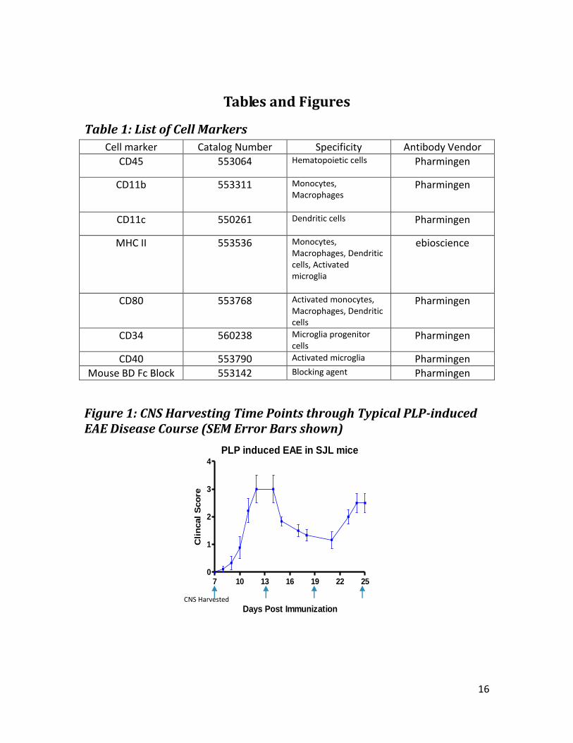
On Days 0, 7, 13, 19 and 25 (Figure 1), five animals were anesthetized with
isofluorane, terminally bled via cardiac puncture, and perfused with 10-20ml of

10
phosphate buffered solution (PBS) at pH 7.4 by insertion of a 25g needle into the left
ventricle of the heart until effluent ran clear. Brain and spinal cord were harvested in
cold PBS on ice and finely minced before being covered with a digestion mixture
containing Liberase RI (Roche, Germany) at 0.2 Wunsch Units/10ml, 50g/ml Dnase I
(Roche, Germany), 25mM Hepes (Invitrogen, Carlsbad, CA) in Hanks’ Balanced Salt
Solution without Magnesium Chloride or Calcium Chloride (Invitrogen, Carlsbad, CA) for
30mins at 37C. A single cell suspension was generated by pushing CNS through a BD
Falcon cell strainer in a cold rinsing buffer containing Hanks’ Balanced Salt Solution
without Magnesium Chloride or Calcium Chloride (Invitrogen, Carlsbad, CA) and 2mM
EDTA. Cells were pooled and collected in a 50ml tube then centrifuged at 313xg for ten
minutes. The pellet was re-suspended in 16.6ml of 1X PIPES Buffer (Sigma, St. Louis,
MO) and 7.2mls of 100% Percoll (Sigma, St.Louis, MO) was added. Cells were layered
carefully over 2.55ml of 65% Percoll in 1X PIPES in two 15ml conical tubes which were
centrifuged at 514xg at room temperature for thirty minutes, no brake. Before
collecting the cells of interest, the top 1ml containing CNS matrix tissue was removed
and 9mls of Percoll were removed. The cells at the 65%/30% interface in each 15ml tube
were pooled into another 15ml tube. The tube was then filled with Hanks’ Balanced Salt
Solution without Magnesium Chloride or Calcium Chloride to dilute and wash out the
Percoll and tubes were centrifuged at 313xg for 10 minutes. The supernatant was
removed and the pellet was re-suspended again in Hanks’ Balanced Salt Solution
without Magnesium Chloride or Calcium Chloride and centrifuged at 313xg for 10
minutes again to wash the cells. Cells were then counted and re-suspended in 150l
FACS Buffer (FB, 1x PBS + 2% v/v FBS + 0.2% w/v Na azide). Cells were placed in 96 well
plates at 30l per well and non-specific Fc binding was blocked with 30l Mouse BD Fc
Block (Pharmingen, San Diego, CA). After incubating with the blocking agent for 30
minutes on ice, the cells were stained with cell surface markers for CD45, CD11b, CD11c,
MHC II, CD80, CD40, CD34 for 30 minutes, covered, on ice (Table 1). Appropriate
isotype controls were used to define staining above background. The cells were then
washed three times with FB and analyzed on the FACSCaliber (BD, San Diego, CA).

11
Cell sorting and ex vivo assay protocol
Brain and spinal cord tissues were harvested from diseased animals on Day 13.
Tissues were processed and layered to collect a single cell suspension as described in
the Isolation of infiltrating leukocytes and microglia from the CNS protocol. Cells were
then counted and resuspended in 1ml FB, blocked with Mouse BD Fc Block (Pharmingen,
San Diego, CA) and stained with CD11b following a 30 minute incubation on ice. After
staining, the cells were washed and re-suspended in 1ml of Robosep Buffer. Cells were
separated using the EasySep (StemCell Technologies, Vancouver, BC, Canada) procedure
for phycoerythrin- (PE) selection.
Lymph nodes were harvested from PLP-immunized animals. The lymph nodes
were processed to single cell suspensions by grinding them between the frosted ends of
slides. CD4+ T-cells were separated with Robosep Selection (StemCell Technologies,
Vancouver, BC, Canada).
CD11b+ microglia and monocytes were plated at 2x10⁵ cells per well. CD4+ T-
cells were plated at 3x10⁵ cells per well. CD11b+ cells were plated alone, with PLP-
primed T-cells, T-cells and PLP, T-cells, PLP and Compound A at 1µM and T-cells, PLP,
and FTY at 100nM. Supernatant was collected for TNF, IFN-γ, and IL-17 levels.
12
Results
CNS Infiltrate Kinetics
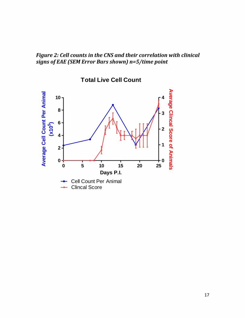
Immunized animals used for CNS harvesting on Days 0, 7, 13, and 25 had average
clinical scores throughout disease course and a typical relapsing-remitting trend of
disease scores (Figure 2). Figure 2 shows the average total CNS cell count per animal
determined by pooling five animals at each time point.
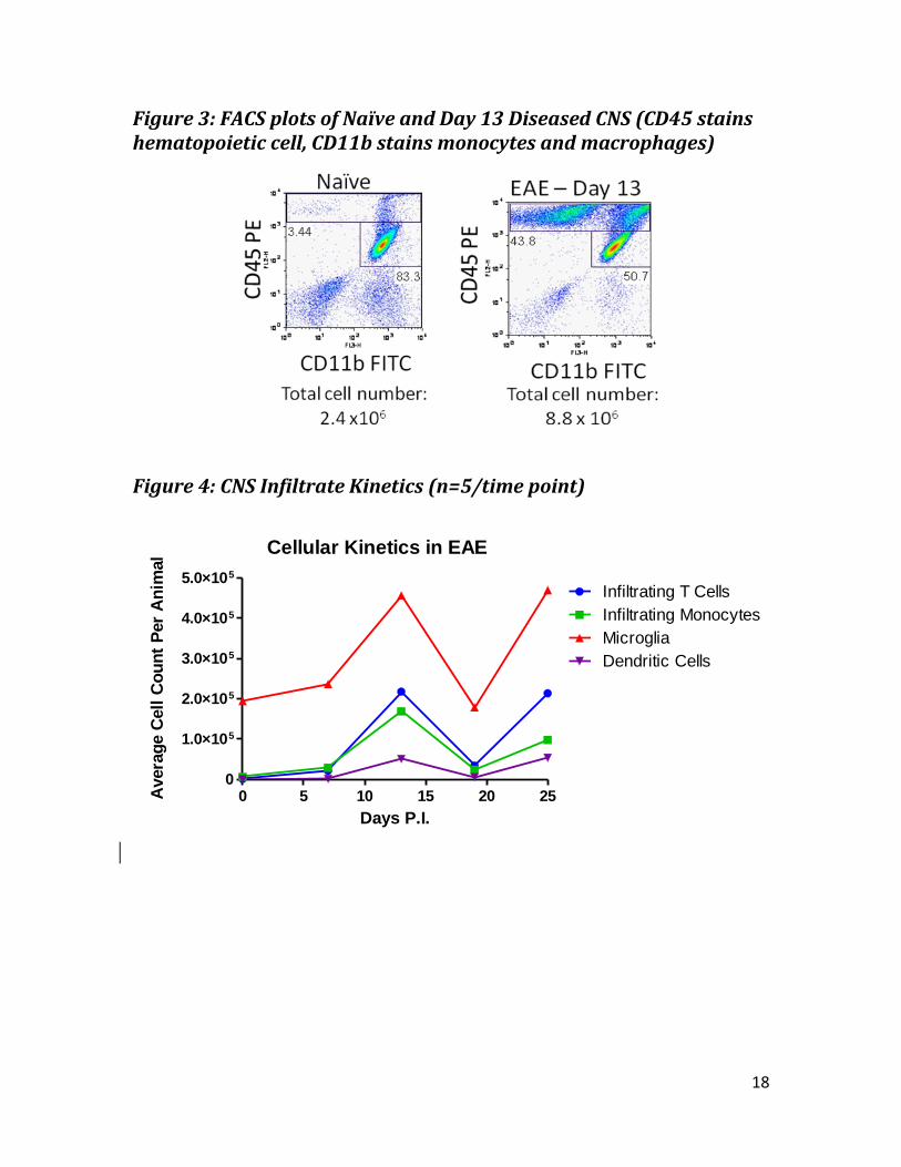
FACS plots of naïve animals (Day 0) and Day 13 animals, as seen in Figure 3, show
a forward and side scatter of CD45 and CD11b. CD45 med, CD11b+ cells are microglia.
CD45hi, CD11b- cells are infiltrating T-cells. CD45hi, CD11b+ cells are monocytes.
Although the percentage of resident microglia decreases from 83.3% to 50.7% between
naïve and diseased animals, the total cell count rises substantially from 2.4x10⁶ to
8.8x10⁶, and so the total number of cells expressing a microglial phenotype doubles
from 2.0x10⁶ cells to 4.5x10⁶. This data can be more clearly seen in Figure 4, which
shows the average cell count per animal at each time point (n=5 pooled) for microglia,
infiltrating T-cells, infiltrating monocytes and dendritic cells. All cell types follow the
same general trend over disease course, but the baseline microglial cell number is much
higher due to resident microglial cells in naïve CNS.
Activation States of Microglia
Though CD40 and MHC II cell markers were used to identify activated microglia,
no staining was found with either of these markers. CD34 also did not show staining for
microglial progenitor cells.
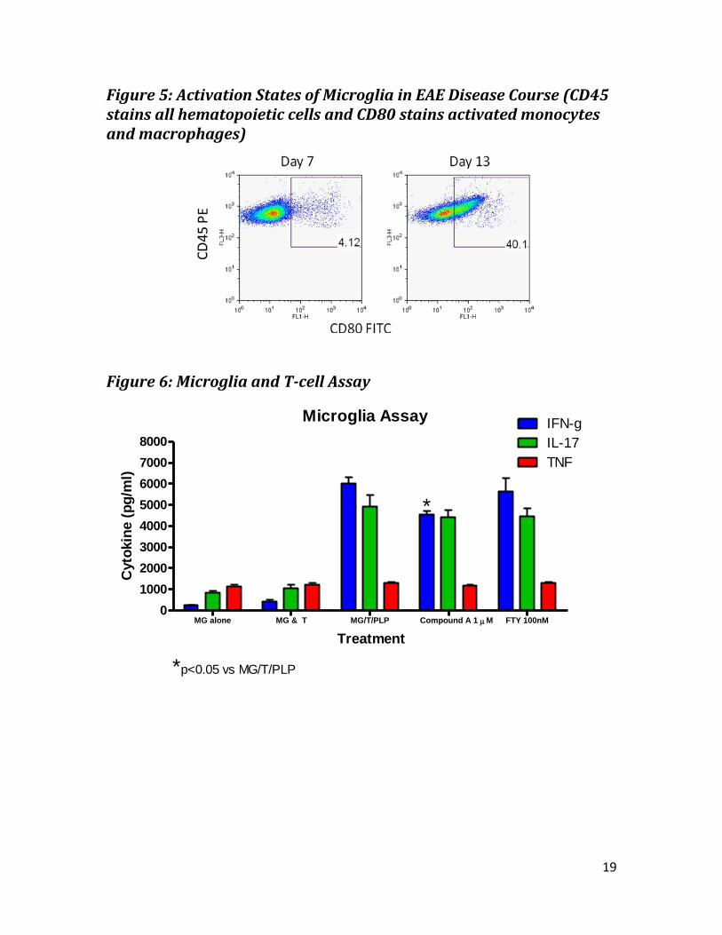
CD80 was also used to identify activated microglia, as seen in Figure 5. Figure 5
shows the FACS plots of Day 7 and Day 13 diseased animals with CD45 and CD80
staining. CD45med, CD80+ cells are activated microglia. This population percentage
increased from Day 7, 4.12%, to Day 13, 40.1%. These percentages are of the total
number of microglia at each of these days (n=5 pooled).

13
Microglia and T-cell Assay
Figure 6 shows the cytokine levels resulting from the assay with microglia from
diseased animals and PLP-primed T-cells. Microglia were plated alone, with T-cells, with
T-cells and PLP, with T-cells, PLP, and Compound A at 1µM, and with T-cells, PLP, and
FTY at 100nM. There is no change in cytokine levels resulting from microglia plated
alone and microglia plated with T-cells. When microglia are plated with T-cells and PLP,
IFN-γ and IL-17 levels rise while TNF levels remain consistent. When Compound A is
added, IFN-γ levels decrease significantly.

14
Discussion
Interpreting Experimental Results
Immunophenotyping of CNS in diseased animals showed CNS infiltrate kinetics
mirror disease course. T cell, dendritic cell, and macrophage numbers increased
through peak disease (Day 13), decreased during remitting phase of disease (Day 19)
and increased during relapse (Day 25). Microglial counts also increased from baseline
resident numbers during peak disease and relapse due to proliferation of resident
progenitor cells or infiltrating monocytes differentiating into microglial phenotype.
When microglia become alerted to tissue damage in the CNS, the cells take on an
activated phenotype which allows for proliferation, migration to site of injury or the
release of cytokines. In the course of EAE, microglia in the CNS take on an activated
phenotype between Day 7 (disease onset) and Day 13 (peak disease).
Microglia and T-cell assays show that microglia may act as APCs during
autoimmune disease. Treatment with Compound A significantly reduced IFN-γ
production after PLP stimulation, indicating that this assay may be a viable tool to
evaluate the effects of small molecule inhibitors on various biochemical pathways
through their effects on microglial function.
The increase in microglial cell numbers, the activation of microglia at peak
disease and the ability of microglia to act as APCs in disease signify that microglia may
have a substantial role in a variety of pathological conditions in the CNS, including MS.
Though disease mechanisms are still not fully understood, microglia may be a target for
drug development to limit and repair CNS damage in MS.
Future Directions
Further investigation into the roles that microglia play in disease are crucial in
understanding disease mechanisms. Repeat CNS infiltrate kinetics studies should be
conducted to confirm results and specific work with different cell markers, including

15
CD40 and MHC II in CNS infiltrate kinetics should be conducted to verify the activation of
microglia over disease course.
The ex vivo microglia and T-cell assay system should be further optimized and
utilized to study small molecule inhibitors through their effects on microglial function.
Dose response studies should be conducted with these compounds to detect any
significant changes in cytokine production levels.
16
Tables and Figures
Table 1: List of Cell Markers
Cell marker Catalog Number Specificity Antibody Vendor
CD45 553064 Hematopoietic cells
Pharmingen
CD11b 553311 Monocytes, Macrophages
Pharmingen
CD11c 550261 Dendritic cells
Pharmingen
MHC II 553536 Monocytes, Macrophages, Dendritic cells, Activated microglia
ebioscience
CD80 553768 Activated monocytes, Macrophages, Dendritic cells
Pharmingen
CD34 560238 Microglia progenitor cells
Pharmingen
CD40 553790 Activated microglia Pharmingen
Mouse BD Fc Block 553142 Blocking agent Pharmingen
Figure 1: CNS Harvesting Time Points through Typical PLP-induced EAE Disease Course (SEM Error Bars shown)
PLP induced EAE in SJL mice
7 10 13 16 19 22 250
1
2
3
4
Days Post Immunization
Cli
ncal
Sco
re
CNS Harvested
17
Figure 2: Cell counts in the CNS and their correlation with clinical signs of EAE (SEM Error Bars shown) n=5/time point
Total Live Cell Count
0 5 10 15 20 250
2
4
6
8
10
0
1
2
3
4
Cell Count Per AnimalClincal Score
Days P.I.
Avera
ge C
ell
Co
un
t P
er
An
imal
(x10
5)
Avera
ge C
lincal S
co
re o
f An
imals
18
Figure 3: FACS plots of Naïve and Day 13 Diseased CNS (CD45 stains hematopoietic cell, CD11b stains monocytes and macrophages)
Figure 4: CNS Infiltrate Kinetics (n=5/time point)
Cellular Kinetics in EAE
0 5 10 15 20 250
1.0×10⁵
2.0×10⁵
3.0×10⁵
4.0×10⁵
5.0×10⁵
Infiltrating T Cells
Infiltrating Monocytes
Microglia
Dendritic Cells
Days P.I.
Avera
ge C
ell
Co
un
t P
er
An
imal
19
Figure 5: Activation States of Microglia in EAE Disease Course (CD45 stains all hematopoietic cells and CD80 stains activated monocytes and macrophages)
Figure 6: Microglia and T-cell Assay
Microglia Assay
MG alone MG & T MG/T/PLP Compound A 1 M FTY 100nM0
1000
2000
3000
4000
5000
6000
7000
8000 IL-17
TNF
IFN-g
*
*p<0.05 vs MG/T/PLP
Treatment
Cyto
kin
e (
pg
/ml)

20
References
Compston A, Coles A, Multiple Sclerosis. Lancet 2002; 359; 1221-31. Compston A, Coles A, Multiple Sclerosis. Lancet 2008; 372; 1502-17. Ebers GC, PRISMS (Prevention of Relapses and Disability by Interferon beta-1a Subcutaneously in Multiple Sclerosis) Study Group. Randomised double-blind placebo-controlled study of interferon beta-1a in relapsing/remitting
sclerosis. Lancet 1998; 352; 1498-504.
Duquette P, Despault L, Knobler RL, et al. Interferon beta-1b in the treatment of multiple sclerosis: final outcome of the randomized controlled trial. Neurology 1995; 45; 1277-85. Greenstein JI, Current Concepts of the Cellular and Molecular Pathophysiology of Multiple Sclerosis. Develop Neurobio 2006; 67; 1248–1265. Le Page E, Leray E, Taurin G, et al. Mitoxantrone as induction treatment in aggressive relapsing remitting multiple sclerosis treatment response factors in a 5 year follow-up observational study of 100 consecutive patients. J Neurol Neurosurg
Psychiatry 2008; 79; 52-56.
McDonald WI, Ron MA, Mutiple Sclerosis: The Disease and its Manifestations. Bio Sciences 1999; 354; 1615-22.
Miller DM, Weinstock-Guttman B, Bethoux F, et al. A meta-analysis of methylprednisolone in recovery from multiple sclerosis exacerbations. Mult Scler 2000; 6; 878-86. Polman, CH, O’Connor PW, Havrdova E, et al. A randomized, placebo-controlled trial of natalizumab for relapsing multiple sclerosis. N Engl J Med 2006; 354; 911-23. Steinman L, Zamvil SS, How to successfully apply animals studies in experimental allergic encephalomyelitis to research on MS. Ann Neurol 2006; 60; 12-21. Williams A, Piaton G, Aigrot MS, et al. Semaphorin 3A and 3F: key players in myelin repair in multiple sclerosis? Brain 2007; 130; 2554-65.
Related Documents