CELL JUNCTIONS IN AMPHIBIAN SKIN MARILYN G. FARQUHAR, Ph.D., and GEORGE E. PALADE, M.D. From the Department of Pathology, University of California School of Medicine, San Francisco, and The Rockefeller Institute, New York ABSTRACT Cell junctions have been investigated in the amphibian epidermis, a stratified squamous epithelium, and compared to those described previously in simple columnar epithelia of mammalian cavitary organs. In adult frogs and toads, and in larvae approaching metamorphosis, belts of membrane fusion or zonulae occludentes of considerable depth are regularly found between adjoining cells of the outermost layer of the stratum corneum, binding the cells together into a continuous, uninterrupted sheet. Another set of occluding zonules appears in the second cornified layer (when such a layer is present), and a third set usually occurs in the outermost layer of the stratum granulosum. Specialized elements described as "modified" and "composite" desmo- somes are encountered along the lateral and basal aspects, respectively, of the cornified cells; ordinary desmosomes and maculae occludentes (i.e., spots of membrane fusion) are found in all other strata. The usual 200 A intercellular gap is generally maintained between the cells of the stratum germinativum at the basal ends of the intercellular spaces. Hence, the inter- cellular spaces of the epidermis form a largely continuous network, closed to the external medium and open to the dermal interstitia. The situation is comparable to that found in columnar epithelia, except that the intercellular spaces are much more extensive, and an extracellular subcompartment (or two) apparently exists in the stratum corneum and between the latter and the stratum granulosum. The last subcompartment is usually filled with a dense substance, probably derived from discharged secretory granules. The tripartite junctional complex characteristic of lumen-lining epithelia (i.e., a zonula occludens followed by a zonula adhaerens, and desmosome) is seen only in early larvae; in adults and in larvae approaching metamorphosis, the occluding zonule is followed directly by a series of modified desmosomes. Interpreted in the light of current physiological data, these findings suggest that the dif- fusion of water, ions, and small, water-soluble molecules is impeded along the intercellular spaces of the epidermis by zonulae occludentes while it is facilitated from cell to cell within the epidermis by zonulae and maculae occludentes. In a previous paper (1), we surveyed the epithelia lining a number of glands and cavitary organs of the rat and guinea pig and, in all cases investi- gated, found a characteristic tripartite junctional complex between adjacent epithelial cells. The complexes are located at the luminal end of the intercellular spaces and consist of three morpho- logically distinct junctional elements identified as zonula occludens (tight junction), zonula adhaerens, and macula adhaerens (desmosome). Along the occluding zonule, the intercellular space is obliter- ated by the fusion of the outer leaflets of the adja- cent cell membranes; by contrast, a distinct inter- cellular gap is maintained along the other elements of the complex. Since the occluding zonules are apparently continuous around each cell and 263 on January 24, 2015 jcb.rupress.org Downloaded from Published July 1, 1965

Welcome message from author
This document is posted to help you gain knowledge. Please leave a comment to let me know what you think about it! Share it to your friends and learn new things together.
Transcript
C E L L J U N C T I O N S I N A M P H I B I A N S K I N
M A R I L Y N G. F A R Q U H A R , Ph.D., and G E O R G E E. P A L A D E , M.D.
From the Department of Pathology, University of California School of Medicine, San Francisco, and The Rockefeller Institute, New York
ABSTRACT
Cell junctions have been investigated in the amphibian epidermis, a stratified squamous epithelium, and compared to those described previously in simple columnar epithelia of mammalian cavitary organs.
In adult frogs and toads, and in larvae approaching metamorphosis, belts of membrane fusion or zonulae occludentes of considerable depth are regularly found between adjoining cells of the outermost layer of the stratum corneum, binding the cells together into a continuous, uninterrupted sheet. Another set of occluding zonules appears in the second cornified layer (when such a layer is present), and a third set usually occurs in the outermost layer of the stratum granulosum. Specialized elements described as "modified" and "composite" desmo- somes are encountered along the lateral and basal aspects, respectively, of the cornified cells; ordinary desmosomes and maculae occludentes (i.e., spots of membrane fusion) are found in all other strata. The usual 200 A intercellular gap is generally maintained between the cells of the stratum germinativum at the basal ends of the intercellular spaces. Hence, the inter- cellular spaces of the epidermis form a largely continuous network, closed to the external medium and open to the dermal interstitia. The situation is comparable to that found in columnar epithelia, except that the intercellular spaces are much more extensive, and an extracellular subcompartment (or two) apparently exists in the stratum corneum and between the latter and the stratum granulosum. The last subcompartment is usually filled with a dense substance, probably derived from discharged secretory granules. The tripartite junctional complex characteristic of lumen-lining epithelia (i.e., a zonula occludens followed by a zonula adhaerens, and desmosome) is seen only in early larvae; in adults and in larvae approaching metamorphosis, the occluding zonule is followed directly by a series of modified desmosomes.
Interpreted in the light of current physiological data, these findings suggest that the dif- fusion of water, ions, and small, water-soluble molecules is impeded along the intercellular spaces of the epidermis by zonulae occludentes while it is facilitated from cell to cell within the epidermis by zonulae and maculae occludentes.
In a previous paper (1), we surveyed the epithelia lining a number of glands and cavitary organs of the rat and guinea pig and, in all cases investi- gated, found a characteristic tripartite junctional complex between adjacent epithelial cells. The complexes are located at the luminal end of the intercellular spaces and consist of three morpho- logically distinct junctional elements identified
as zonula occludens (tight junction), zonula adhaerens, and macula adhaerens (desmosome). Along the occluding zonule, the intercellular space is obliter- ated by the fusion of the outer leaflets of the adja- cent cell membranes; by contrast, a distinct inter- cellular gap is maintained along the other elements of the complex. Since the occluding zonules are apparently continuous around each cell and
263
on January 24, 2015jcb.rupress.org
Dow
nloaded from
Published July 1, 1965
throughout the epithelium, we suggested that they serve as "closing belts" which seal off the inter- cellular spaces from the lumen, and we postulated that they play a passive role in the maintenance of chemical and electrochemical potential grad- ients across epithelia by impeding back diffusion (leaks) along the intercellular spaces. This as- sumption was tested and confirmed for large molecules by using concentrated protein solu- tions (hemoglobin and zymogen) as mass tracers• The solutions filled the lumina but did not pene- trate the intercellular spaces beyond the occluding zonules (1). The impermeabil i ty of these zonules to water, ions, and small water-soluble molecules remained, however, to be demonstrated. Since no direct means of investigating this problem is available, we approached it indirectly by examin- ing the frog epidermis, an epithelium known to be practically impermeable to certain small mole- cules. The frog skin is an extensively studied biological membrane, frequently used in studies of t ransport functions of epithelia (2). I t is known to exhibit a unilateral osmotic response (3) and to maintain marked electrochemical potential gradients, and both properties have been localized to the epidermis (2). If occluding zonules are impermeable to water, ions, and small water- soluble molecules, they should be present in the frog epidermis, binding together the cells close to its outer surface. The observations to be pre- sented I fully confirm these expectations.
M A T E R I A L S A N D M E T H O D S
Skin taken from the abdomen and back of adult frogs (Rana pipiens and Rana catesbiana) were the principal tissues studied. For comparison, more limited observa- tions were carried out on abdominal skin from adult toads (Bufo marinus), tails of young Xenopus laeds larvae (stage 55), and tail fins of salamander larvae (Amblystoma punctatum) approaching metamorphosis.
Pieces of skin taken from pithed animals, or ampu- tated tails, were fixed for la/~ to 2 hours at 0 ° in 1 per cent OsO4 in either acetate-Veronal or phosphate buffer (pH 7.6). Other pieces were fixed in 2 per cent glutaraldehyde (6) in either 0.1 M phosphate or 0.1 M cacodylate buffer (pH 7.4) for 4 to 16 hours, fol- lowed by "postfixation" for 2 hours in OsO4. Araldite or Epon was used for embedding. Some specimens were stained in block with phosphotungstic acid (PTA) or KMnO4 (cf. 1), during dehydration, to en- hance the contrast of fibrillar and membranous struc- tures, respectively. More recently we have exten-
1 Preliminary reports of these findings have already been published (4, 5).
sively used staining in block, before dehydration, in aqueous uranyl acetate solutions. 2
Sections from all these blocks were stained for 30 minutes with lead alone (8) or doubly stained with the latter preceded by 1}~ hours in 5 per cent uranyl acetate. They were examined in a Siemens Elmiskop I, operating at 80 kv with a double condenser. Fur- ther details of the embedding and staining pro- cedures can be found elsewhere (1). For light micros- copy, sections of 1 to 2 # were cut from the same blocks and stained with azure II and methylene blue (9).
O B S E R V A T I O N S
The observations presented are limited primarily to cell junctions and associated structures (i.e., cell membranes, intercellular spaces, etc.) of the frog epidermis. Other aspects, such as differentia- tion and keratinizafion of the epidermis and the structure of the dermis, are not treated in any detail. However, for the sake of orientation, a brief description of the general organization of the skin is included. The bulk of the observations pertain to the skin of the frog; observations on toad and larval skin are mentioned where they are appropriate or instructive.
General Histology and Cytology of Frog
Epidermis
The frog skin consists of an epidermis composed of 5 to 7 layers of epithelial cells, and an underly- ing dermis containing blood vessels, glands, and
2 This is an adaptation, for animal tissue, of the procedure of Kellenberger et al. (7). Specimens fixed in OsO4 or glutaraldehyde followed by OsO4 are treated for 2 hours at room temperature with 0.5 per cent uranyl acetate in Michaelis buffer. The final pH of the solution is 5.0. Since neither calcium nor salt is necessary for staining, the procedure can be simplified by using acetate-Veronal buffer without any additives as a diluent for both the OsO4 and uranyl acetate. If the tissue is fixed in OsO4 or glutaraldehyde followed by OsO4 in phosphate buffer, the blocks must be washed (2 to 3 changes each of 15 minutes) before transfer to the staining solution in order to avoid precipitation of uranyl as uranyl phosphate. This technique is particularly effective in demonstrating the stratification of all cellular and intracellular membranes. A certain amount of extraction seems to be incurred by the specimens in the uranyl solution; it is more noticeable after fixation in OsO4 in acetate-Veronal than after the other procedures mentioned. Contrast in sections prepared from blocks processed according to this technique can be further increased by staining.
264 ThE JOVaNAL OF CELL BIOLOGY • VOLVME 26, 1965
on January 24, 2015jcb.rupress.org
Dow
nloaded from
Published July 1, 1965
General Abbreviations
SC: stratum corneum SG: stratum granulosum SS: stratum spinosum SGe: stratum germinativum B: basement membrane Co: collagen fibril D: dermis Is: intercellular space Zo: zonula ocdudens (occluding zonule) cm: cell membrane d." desmosome cd: composite desmosome rod: modified desmosome
din: dense material in intercellular space er: endoplasmic reticulum (rough surfaced) f: cytoplasmic filaments fl: fusion line g: Golgi complex i: intermediate line in desmosome il: inner leaflet of the cell membrane ol: outer leaflet of the cell membrane m: mitochondria mc: mucous coat n: nucleus r: ribosomes sg: small secretion granule lg: large secretion granule
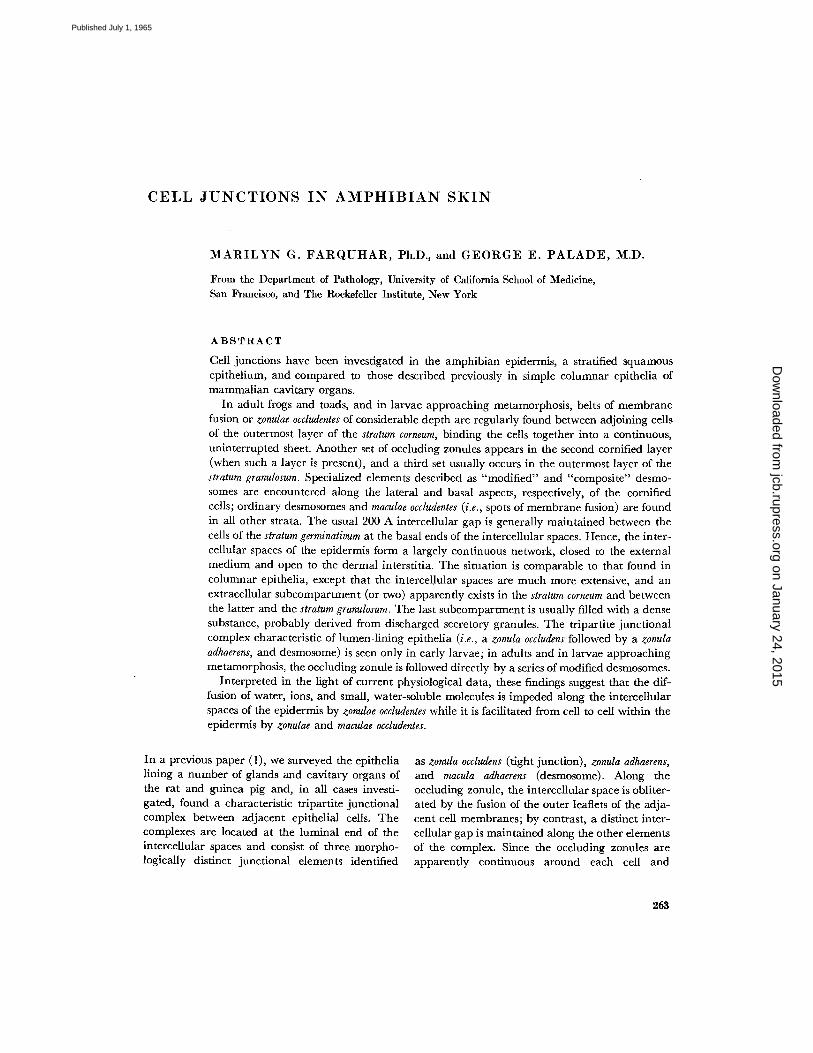
FIGURE 1 Photomicrograph showing the general organization of the abdominal skin of the frog (Rana p/p/ens). The epidermis (between arrows 1 and 2) is a stratified epithelium with 1 to ~ layers of partially cornified, squamous cells in the stratum corneum (SC), 1 to 3 layers of cuboidal or polyhedral cells in the stratum granulosum (SG) and stratum spinosum (SS), and a basal layer of cuboidal or columnar cells in the stratum germinativum (SGe). The dermis is divided into a loose layer, the stratum spongiosum (Ssp) (between arrows 2 and 3) which adjoins the epidermis, and a deeper more compact layer, the stratum compactum (SCo) (arrow 3 to bottom of field). The s. spongiosum contains blood vessels (V), bodies of glands (G), pigment cells (p), and various fibrillar and cellular connective tissue elements. The s. com- pactum (SCo) consists of a series of undulating layers, each composed of bundles of collagen fibrils in parallel array. From layer to layer the direction of the bundles changes by ~90 °. A discontinuous layer of extracellular, material, which stains red with this procedure, forms a series of arches (unnumbered arrows) between the two dermal strata.
Near the top of the epidermis at either edge of the field are two mitochondria-rich cells (M) which have a fusiform shape and extend from the middle layers to the s. corneum. The one on the right has a relatively pale cytoplasm.
Specimen fixed in 1 per cent OsOa in phosphate buffer (pH ' ~nd embedded in Araldite. One-micron section stained with azure II-methylene blue. X 900. ~"
on January 24, 2015jcb.rupress.org
Dow
nloaded from
Published July 1, 1965
various fibrillar or cellular connective tissue ele- ments (Fig. 1). The epidermis contains one or two outer layers of partially cornified, squamous cells (stratum corneum), 3 to 4 intermediate layers of cuboidal or polyhedral cells (stratum granulosum and stratum spinosum), and a basal layer of cuboidal or columnar cells (stratum basale or germinatioum) (Fig. 1).
Stra tum germinat ivum
The cells of the s. germinativum (Fig. 2) 3 have the usual set of subcellular components in rela- tively small quantities: a few mitochondria and rough-surfaced elements of the endoplasmic reticu- lure, some free ribosomes, a relatively rudimentary Golgi complex with one or two small flattened cisternae and associated vesicles, and variable numbers of small (pinocytic?) vesicles found es- pecially near the lateral cell membranes. In addi- tion, there is a well developed system of cytoplas- mic filaments (formerly called tonofilaments (cf. 10)) grouped in bundles and anchored in either desmosomal or "basal" plates (11, 12) (Fig. 18). The basal surface of these cells rests on a basement membrane which forms a continuous layer, 500 to 700 A thick, at the dermal-epidermal boundary (Figs. 2 and 18).
Stra tum sp inosum and S t ra tum granulosum
A s the cells of the epidermis differentiate and move from the germinal layer out to the s. spinosum and s. granulosum, they flatten and their bundles
3 Fig. 2 is from the skin of the toad, but the basic organization is the same as in the frog.
of cytoplasmic f laments become more abundant and more tightly packed (Figs. 3 and 6). At the same time, their rough-surfaced endoplasmic reticulum becomes more voluminous and their Golgi complexes more elaborate, with the number and size of the cisternae increasing. Multivesicular bodies also increase in number, and the cells acquire dense bodies of irregular size and shape with a heterogeneous content. Morphologically, these elements represent lysosomal derivatives, i.e., residual bodies, and lytic and autolytic vac- uoles (13). Moreover, they give a positive reaction for acid phosphatase in specimens fixed in glu- taraldehyde, incubated in Gomori medium and examined in the electron microscope following the general procedures given in reference 6. The same applies to similar bodies found in the s. corneum. The frequency of all these bodies increases de- cidedly in the s. granulosum (Fig. 6) in which many autolytic vacuoles contain small vesicles, of the type seen in muhivesicular bodies, as well as recog- nizable remnants of cell components, mostly ribo- somes (Fig. 6, inset). This finding suggests that there is extensive fusion of multivesicular bodies with autolytic vacuoles as cell differentiation progresses.
The cells of these strata contain cytoplasmic granules of two distinct types: one type is relatively small (100 to 150 m/z) in size; is spherical, ovoid or tear-like in shape; has a finely particulate,
moderately dense content; and has a distinct
limiting membrane. Granules of this type seem
to be formed within Golgi cisternae (Fig. 4).
Mature granules first appear randomly distributed
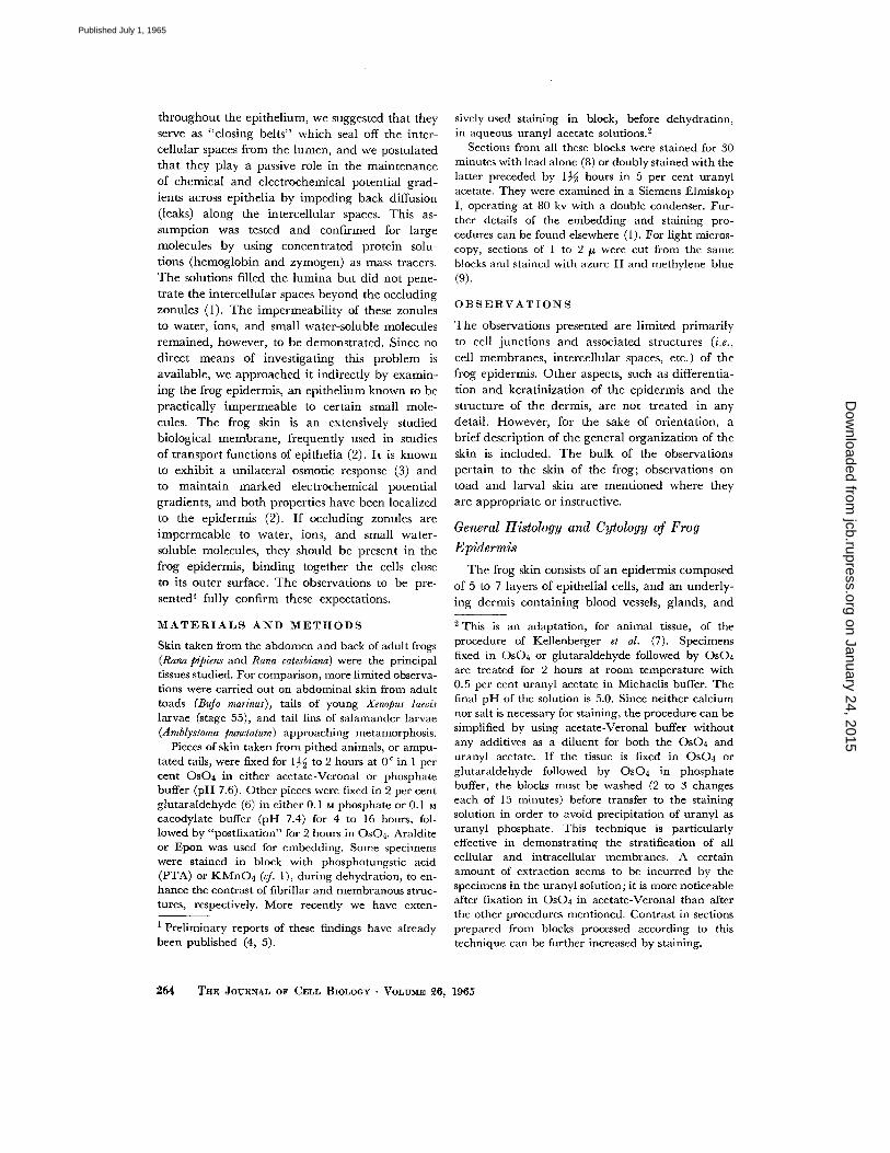
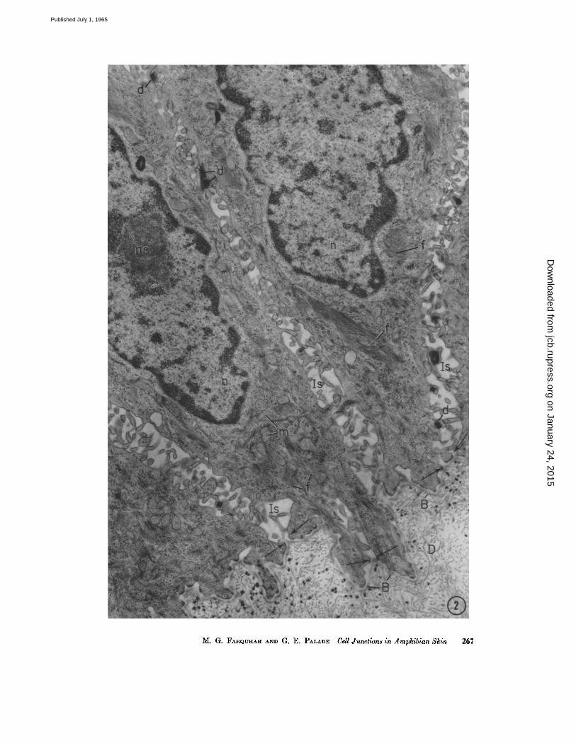
FIGURE ~ Base of the epidermis in toad skin, showing portions of four large cells of the s. germinativum, a highly indented dermal-epidermal junction, and part of the dermis (D) which forms a series of micropapillae containing mostly collagen fibrils. The base- ment membrane (B) forms a continuous (~500 A) layer which closely parallels the basal cell membranes of the germinative cells and separates dermal and epidermal elements.
The intercellular spaces (Is) between the basal cells are expanded and of complicated geometry due to extensive interdigitation between microvilli and other processes of ad- joining cells. The spaces are open toward the basement membrane since the apposed cell membranes remain separated by a distinct (~00 to 400 A) gap (arrows). Note the low frequency of the desmosomes (d) on the lateral cell aspects. The cytoplasm of the basal cell contains prominent bundles of filaments (f) as well as mitochondria (m) and numerous free ribosomes. The nuclei (n) show large nucleoli (no) and dense, peripheral chromatin masses.
Specimen fixed in 1 per cent Os04 in phosphate buffer (pH 7.6) and embedded in ArM- dire. Section doubly stained with uranyl and lead. X 14,000.
266 T R ~ JOURNAL OF CELL BIOLOGY • VOLUME ~6, 1965
on January 24, 2015jcb.rupress.org
Dow
nloaded from
Published July 1, 1965
M. G. FARQUHAR AND G. E. PALADE Cell Junctions in Amphibian Skin 267
on January 24, 2015jcb.rupress.org
Dow
nloaded from
Published July 1, 1965
throughout the cytoplasm of cells immediately distal 4 to the s. germinativum; they are particularly numerous in the cells just beneath the outermost granular layer, where they accumulate along the distal front of the cell (Figs. 3 and 4); and are usually absent from the outermost granular layer. ~ In this connection, it should be mentioned that whereas the intercellular spaces in the s. germi- nativum, spinosum, and granulosum appear " empty" (i.e., they are occupied by a material of low den- sity), a substance similar in density and texture to the granule content fills the intercellular spaces between the s. granulosum and s. corneum and within the latter (Figs. 3, 5, and 6). The orientation of the granules along the distal cell surface, and their disappearance from the cytoplasm at a level where a similar material appears in the intercellular spaces suggest that the granule content is dis- charged into, and fills the intercellular spaces. Images suggesting granule discharge by membrane fusion have been occasionally encountered.
A second type of cytoplasmic granule, also membrane-limited, is much larger in size (300 to 900 m~), more variable in shape, has a content of higher density and finer texture, and occurs pri- marily in the perinuclear cytoplasm (Figs. 3 and 4). These larger granules are found predominantly
4The terms "distal" and "proximal" are used throughout the text to refer to the position of epi- dermal structures in relation to the basement mem- brane. 5 They occasionally occur in small numbers along the distal front of these ceils.
in the two layers located just beneath the s. corneum, and, in contrast to the granules of the first type, are most numerous in the outer granular layer. There is some suggestive evidence that they are formed within Golgi vacuoles (Fig. 4), but their ultimate fate is not altogether clear. They decrease in number as the cells keratinize, and some, at least, are retained in such cells and finally included and apparently destroyed in autolytic vacuoles.
S t r a t u m c o r n e u m
Cornified elements occur only distal to the intercellular spaces filled with electron-opaque material. By implication, keratinization of a cell moving into the s. corneum begins between the discharge of its own small granules and the dis- charge of the same granules from the cell moving behind it into the s. granulosum. Cornification occurs gradually, for all stages from partially to fully keratinized elements can be found in this layer. Fully keratinized cells appear very dense (Figs. 3, 5 to 7), are completely occupied by cytoplasmic filaments, and contain only remnants of cell organelles which are sometimes included in autolytic vacuoles. Partially cornified cells have a cytoplasmic matrix of a variable but generally lower density, and their organelles are more" numerous and less altered (Fig. 6). Some of these cells still have recognizable ribosomes, usually clumped in the remaining spaces between fila- ments and cell organelle remnants (Fig. 6). Ribo- somal particles are absent or not recognizable in
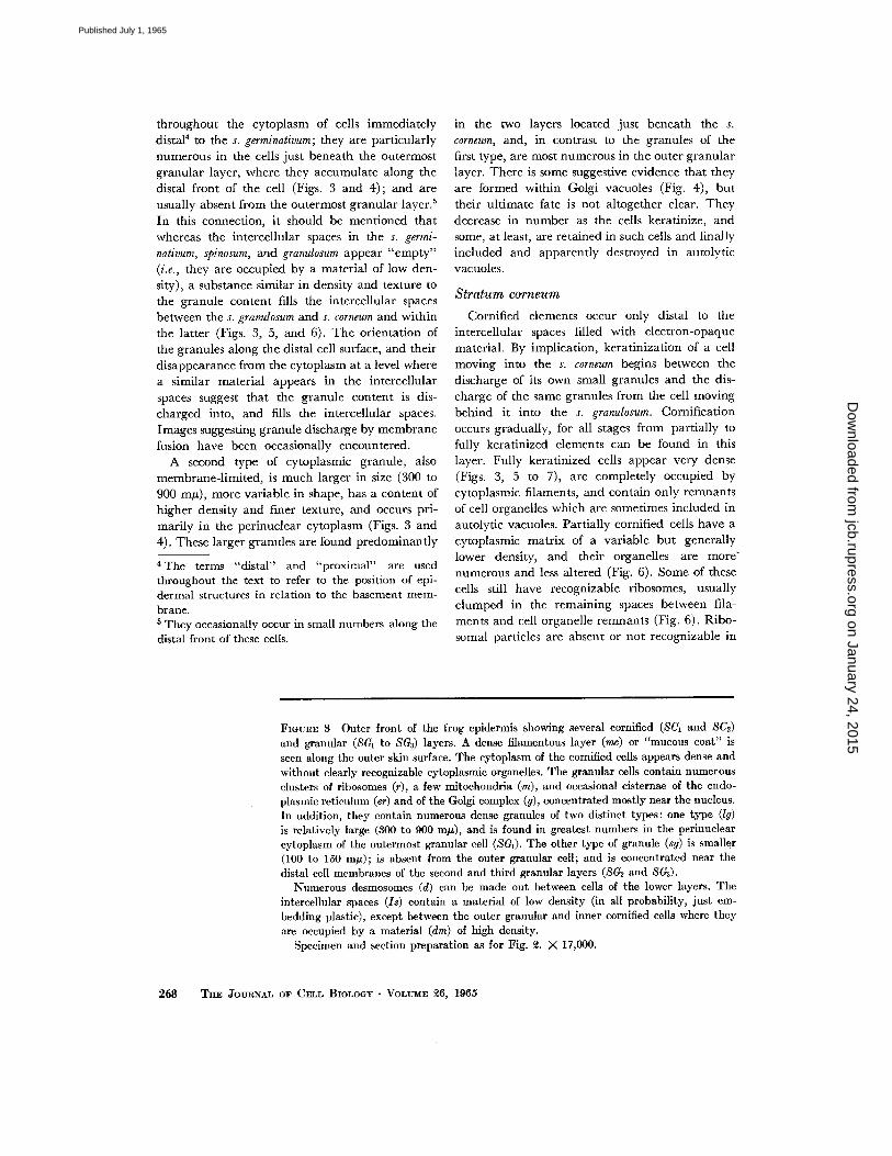
FIGURE 3 Outer front of the frog epidermis showing several cornified (8Ct and SC~) and granular (SG1 to SGs) layers. A dense filamentous layer (mc) or "mucous coat" is seen along the outer skin surface. The cytoplasm of the cornified cells appears dense and without clearly recognizable cytoplasmic organelles. The granular cells contain numerous clusters of ribosomes (r), a few mitochondria (m), and occasional cisternae of the endo- plasmic reticulum (er) and of the Golgi complex (g), concentrated mostly near the nucleus. In addition, they contain numerous dense granules of two distinct types: one type (lg) is relatively large (300 to 900 m#), and is found in greatest numbers in the perinuclear cytoplasm of the outermost granular cell (SG1). The other type of granule (sg) is smaller (100 to 150 m/~); is absent from the outer granular cell; and is concentrated near the distal cell membranes of the second and third granular layers (SG2 and 8Ga).
Numerous desmosomes (d) can be made out between cells of the lower layers. The intercellular spaces (Is) contain a material of low density (in all probability, just em- bedding plastic), except between the outer granular and inner cornified cells where they are occupied by a material (din) of high density.
Specimen and section preparation as for Fig. ~. X 17,000.
2 6 8 THE ~*OURNAL OF CELL BIOLOGY • VOLUME ~6, 1965
on January 24, 2015jcb.rupress.org
Dow
nloaded from
Published July 1, 1965
M. G. FARQUfIAR AND G. E. PAI,ADE Cell Junctions in Amphibian Skin 269
on January 24, 2015jcb.rupress.org
Dow
nloaded from
Published July 1, 1965

fully keratinized cells. When cells move from the s. granulosum to the s. corneum, they acquire a shell of dense cytoplasmic material along the cell mem- brane (vide infra) and the latter becomes thicker, as already noted by Rhodin and Reith (14) in mammal ian keratinizing epithelia.
Specia l Cell T y p e s wi th in the Ep idermi s
( M itoehondria- R ich Cells)
As is well known, various specialized cell Wpes occur scattered throughout the epidermis. In the frog, occasional lymphocytcs, macrophages, and pigment cells can be recognized, particularly in the deep strata. In addition, we have regularly encountered another type of cell, located pre- dominantly in the s. spinosum and s. granulosum. Such cells are typically shaped like an elongated
flask with a perikaryon in the s. spinosum and a
thick main process extending up to the s. corneum where it sometimes branches into a number of
secondary processes. Cytologically, the most dis-
tinctive features of these cells are a relatively light
matrix and the presence of large numbers of
mitochondria, especially in the main process.
We refer to these elements as "mitochondria-rich
cells," as done by Choi (15) for similar cells found
in the toad bladder (15, 16). They contain the
usual set of cell organelles, but usually no secretion
granules, and are attached to neighboring epider-
mal cells by desmosomes which are similar to, but
noticeably less frequent than, those between usual
epidermal cells at this level.
Cell M e m b r a n e s
The cell membranes of all except the cornified elements are of the thin, asymmetrical type, with a total thickness of about 70 to 80 A, and an outer leaflet thinner and less dense than the inner one. As pointed out elsewhere (1), the outer leaflets of membranes of this type are difficult to see in OsO4-fixed specimens, but can be regu- larly demonstrated after fixation or staining in KMnO4 (compare Figs. 13 and 18, which were not stained in KMnO4, with Figs. 14, 15, and 17 which were). The outer leaflets are especially well demonstrated after staining in block with uranyl acetate (Figs. 16, 19, and 20). In tissues fixed in glutaraldehyde followed by OsO4, the usual asymmetry is reversed by this procedure (see Figs. 16 and 19).
The entire cell membrane of the cornified cell
is of the thick (100 A), nearly symmetrical type
(cf. 1). The dense outer leaflet is clearly visible in
OsO4- or glutaraldehyde-fixed tissue stained only
with lead (Figs. 7 to 9 and 11). The light layer
stands out sharply because of the generally high
density of the cornified cell cytoplasm (Figs. 8
and 9). This thick membrane of the cornified
cells is backed all around the cell surface by a
continuous (200 to 300 A thick) shell of dense
material located in the cytoplasm immediately
adjacent to the inner leaflet (Figs. 7, l0 to 13).
The surface membrane of the cells of the outer
cornified layer is covered by a dense fibrillar
coating (Figs. 5, 7 to 9, and 1 l) which varies in
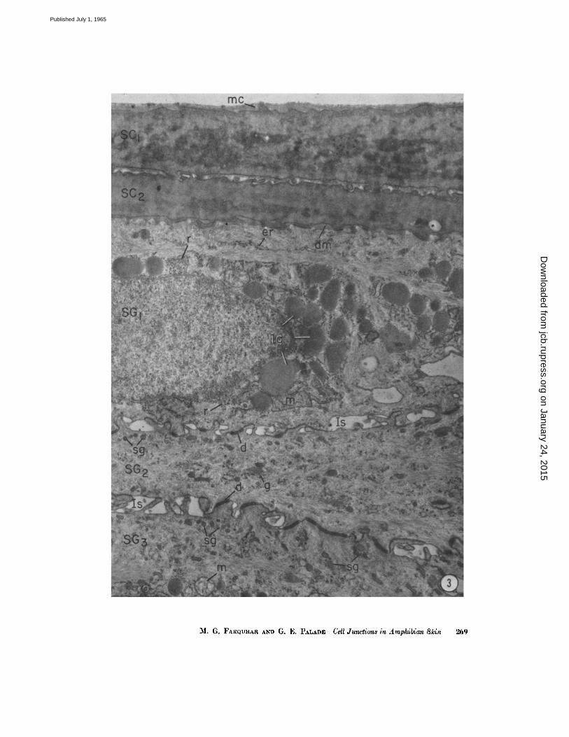
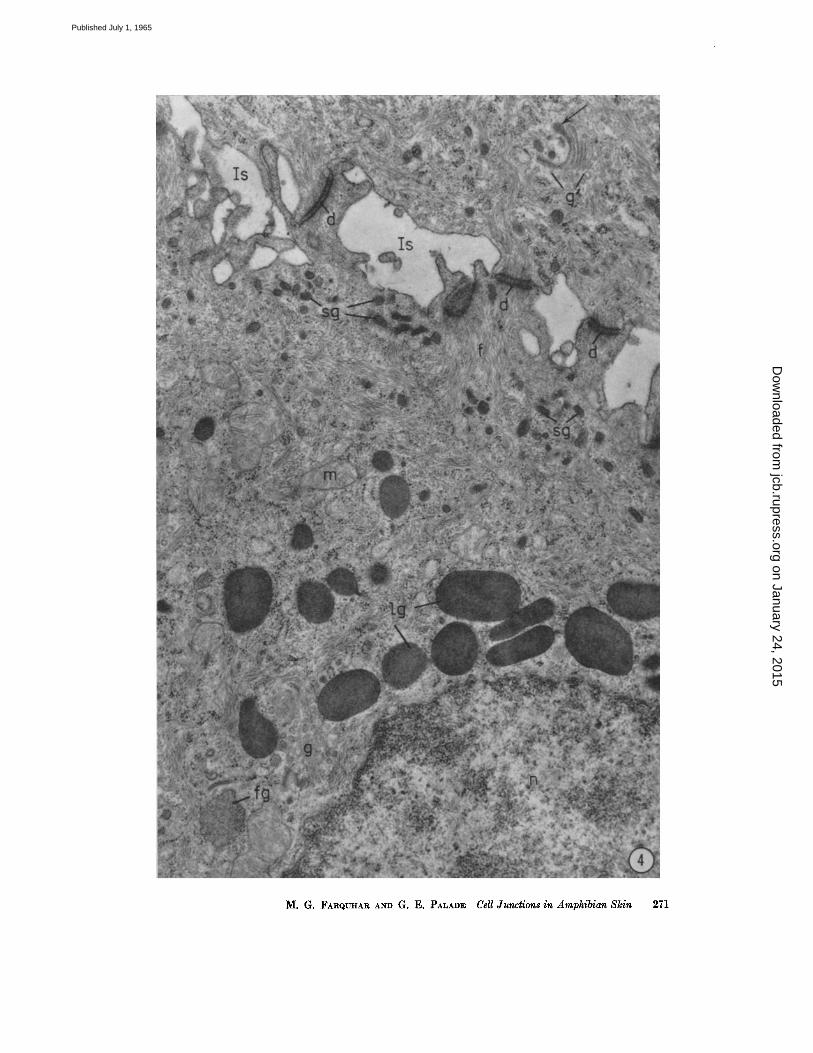
FIGURE 4 Cells from the s. granulosum of the frog epidermis, to illustrate the two dif- ferent granule populations found in the cells of the middle strata. One type (lg) is large in size, membrane-bonnded, variable in shape, and occurs primarily in the perinuclear cytoplasm. The other type (sg), also membrane-limlted, is much smaller in size, ovoid or tear-shaped, and accumulates along the cell membrane at the distal cell front, facing the expanded intercellular spaces (Is).
Numerous free ribosomes, bundles of cytoplasmic filaments, and mitochondria (m) occupy the remaining cytoplasm. Some of the filaments (f) converge on desmosomes (d) at the cell junctions. Part of a Golgi complex is seen at g on the lower left. Near it there is an irregularly shaped body (fg) with a moderately dense content which is believed to represent a "condensing vacuole," i.e., a granule in formation within the Golgi complex. Part of another Golgi complex (g~), consisting of a pile of 5 to 6 flattened cisternae, is seen in an adjoining cell on the upper right. A small granule appears to be forming by "budding" from the tip of the innermost Golgi cisterna (arrow).
Specimen and section preparation as for Fig. ~. )< ~8,000.
270 THE JOURNAL OF CELL BIOLOOY • VOLUME ~6, 1965
on January 24, 2015jcb.rupress.org
Dow
nloaded from
Published July 1, 1965
M. G. :FARQUHAR AND G. E. PALADE Cell Junctions in Amphibian Skin 271
on January 24, 2015jcb.rupress.org
Dow
nloaded from
Published July 1, 1965
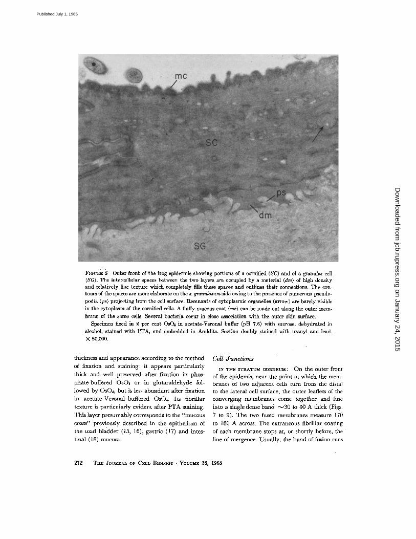
Fmmm 5 Outer front of the frog epidermis showing portions of a cornified (8C) and of a granular cell (SG). The intercellular spaces between the two layers are occupied by a material (din) of high dcasity aud rclativcly finc tcx-turc whi¢~ completely fills t h e ~ spaces and outlines their connections. The cort- tours of thc spathes arc more elaborate on thc s. granzdosum sidc owing to the presence of numerous pseudo- podia (ps) projecting from the cell surface. Remmqnt_s of cytoplasmic organelles (arrow) arc barely visiblc in the cytopla.qm of the cornified cells. A fl~lffy mucous coat (me) can be made out along the outer mem- brane of the same t~lls. Several bacteria oc~cur in close aaso~iation with the outer skin surface.
Specimca fixcd in 2 pc rccn t OsO4 in acetate-Veronal buffer (pH 7.6) with sucrose, dehydrated in alcohol, stained with PTA, and embedded in Araldite. Section doubly stained with uranyl and lead. X 9.0,000.
thickness and appearance according to the method
of fixation and staining: it appears par t icular ly
thick and well preserved after fixation in phos-
phate-buffered OsO, or in g lutara ldehyde fol-
lowed by ()sOu, bu t is less a b u n d a n t after fixation
in ace ta te-Veronal -buffered OsO(. Its fibrillar
texture is par t icular ly evident after P T A staining.
This layer presumably corresponds to the "mucous
coats" previously described in the epi the l ium of
the toad b ladder (15, 16), gastric (17) and intes-
t inal (18) mucosa.
CeU Junctions
IN THE STRATUM CORNED-M: On the outer front of the epidemis, near the point a t which the mem- brmlcs of two adjacent ceils turn from thc dista] to the lateral cell surface, the outer leaflets of the converging membranes come together and. fuse
into a single dense b a n d , '~30 to 40 A thick (Fig s.
7 to 9). T h e two fused membranes measure 170
to 180 A across. T h e extraneous fibrillar coating
of each m e m b r a n e stops at, or shortly before, the
l ine of mergence. .Usual ly , the b a n d of fusion runs
272 Tim ,lotrm~_~.l~ OF C~.r.r, BIOLOOY • VOLVhta~ 26, 1965
on January 24, 2015jcb.rupress.org
Dow
nloaded from
Published July 1, 1965
uninterrupted for 0.1 to 0.3# in the distal to proximal direction, although occasionally one or several focal splittings occur wi th in the junct ion (.Fig. 8). At the proximal end of the junction, the outer leaflets separate again, leaving in between an intercelltflar space of 250 to 300 A which is re- tained, except for focal dilatations, aJong most of the remaining parallel, zig-zagging course of the lateral cell membranes (Fig. 7). • Such areas of membrmm fusion are found in the loc~tfi0n de- scribed wherever the plane of ~ct ioning ap- proaches normal to the plane of the intercellular boundary, and, in favorable sections, are seen to run for considerable distances along the apical perimeter of the cells. Hence, it can be assumed that they are continuous around each cell and throughoutthe s. corneum, like their counterparts already described in .various columnar epithelia (l). AS such they can be properly described, according to the nomenclature we introduced, as zonulae ocdud~ntes, i.e., closing belts.
The occluding zonule is followed by a whole series of elements (Figs. 7, I(), and 12) which resemble dcsmosomes in that they are character- ized by strict parallelism of the apposing cell membranes, the presence of laminar densities in the intercellular space, and the occurrence of con- ten trations of dense amorphous and fibrillar ma- terial in the subjacent cytoplasmic matrix. They differ, however, in several respects: the plate of intel~cellular material is usually more dense; there is no distinct cytoplasmic plaque, but rather a continuous shell (described above) of condensed cytoplasmic matrix around the entire cell; and the bundles of filaments associated with this shell are rather indistinct. (For further details see legend for Fig. 12). These junctional elements are commonly more curved than desmosomes (Figs. 7 and 12) and occasionally show sharp bends. In addition, the fraction of the lateral cell surface occupied by such elements is much greater than that occupied by ordinary desmosomes else- where in the epidermis (see Fig. 7).
These modified desmosomal elements are inter- rupted at intervals by focally expanded inter- cellular spaces and occasionally by small arc.as of membrane fusion; the latter are irregularly dis- ~buted and shaflow in depth and hence are con- sidered to be discontinuous structures, i.e., maculae or fasciae (spots. or bands),-, rather than zonulae (belts).
When more than .one layer of cornified cells is
present (generally there are two such layers), occluding zonules are found in a similar (distal) location along the lateral aspects of the deeper layer (Fig. 10).
Along the proximal surface of the innermost cornified layer, across the intercellular spaces characterized by a dense content, there are bipartite or composite desmosomes (Fig. 13). Their distal half has the structure of the modified desmosomes of the cornified cells (i.e., thicker cell membrane, continuous cytoplasmic shell in- stead of a distinct plaque) and their proximal haft that of the ordinary desmosomes of the deeper layers. 1]aese composite junctional elements thus reflect the distinctive features of the involved cell surfaces.
IN 77~ STI~ATU~ a ~ U L O S ~ : Occluding zonules are found in yet another location, i.e., between ceils of the outer layer of the s. granulosum
• which face the intercellular spaces filled with an electron-opaque substance (Fig. 14). Like those in the keratinizing layers, these zonules are lo- cated where the cell membranes turn from the distal to the lateral cell surface and are found in most normal sections. In this case, however, the total thickness of the junct ion is less (only 120 to 140 A), as expected from the smaller dimensions of t h e component cell membranes. Typical desmosomes (vide infra) are found along the remainder of the perimeter of these cells, except for their distal aspect which, as described above, is provided with bipartite or composite desmosomes.
I~N TH3ff~ O T H E R S T R A T A : Typical desmosumes or adhering maculae form the predominant type of junctional element found in the remaining strata. They are least numerous and complex in the s. germinativum and reach their fullest development and highest frequency in the s. granulosum. The
s t ruc tu re of desmosomes is by now well known 6 (Fig. I6) : they consist of areas of strict parallelism of the cell membranes; an intercellular space occupied by a plug or disc of dense material bi- sected by a denser central layer; dense cytoplasmic
e There is still some lingering confusion (see reference 19) concerning the interpretation of the "inter- mediate dense layer" (20) or "lateral dense line" (19). According to Odland (20) and more recently to Roth and (]lark (19), this layer is an extracellular structure, whereas our cvidence indicates that in the epidermis, as ha many other epithelia studied (21, 1), it is the outer leaflet of the cell membrane (see Fig. 16).
M. G. FxmaL-r~.,,a A,~t) G. E. P a z , . ~ Cell J.uneKon8 in Amphibian Skin 273
on January 24, 2015jcb.rupress.org
Dow
nloaded from
Published July 1, 1965
plaques backing the inner m e m b r a n e leaflets; and profuse bundles of cytoplasmic fi laments converg- ing on the inner aspect of each plaque.
Small areas of m e m b r a n e fusion also occur th roughou t the epidermis (Figs. 15 and 17). Based on their l imited dimensions and irregular distr ibution, they are believed to be discontinu- ous structures, i.e., maculae or fasciae ra ther than zonulae.
In terce l lu lar Spaces
Except for the spaces between the s. corneum and s. granulosum, which conta in the electron- opaque amorphous mater ia l described above (see Figs. 3 and 5), intercel lular spaces th roughout the epidermis are occupied by a mater ia l of low density with only occasional clumps of flocculent precipi tate (Figs. 2 to 4 and 6). T h e geometry and volume of these spaces vary considerably from layer to layer: they reach thei r greatest volume in the s. spinosum in which they are frequently ex- panded into so called " in terce l lu lar lakes." In- deed it is the firm adherence of the cells a t the desmosomes, combined with the marked dilat ion of the intercel lular spaces in between, tha t creates
the typical "prickle-cell" appearance of this s tratum. T h e spaces are least voluminous in the s. granulosum and are most tortuous and complex in the s. germinativum owing to extensive interdigi ta- t ion between cells (Figs. 2 and 18).
Ar rangements at the basal ends of the inter- cellular spaces, where germinal cells meet the basement membrane , are somewhat var iable : most often there is the usual 200-A gap between adjoining cells (Figs. 2 and 18); sometimes the gap is wider or is bisected by an in te rmedia te line similar to tha t present in the ur inary slits of g lomerular capillaries; occasionally i t is br idged by a desmosome (Fig. 19); and only rarely areas of m e m b r a n e apposit ion ra ther than fusion (Fig. 20) are encountered in this location. Hence, the intercel lular spaces at this level are apparen t ly pa ten t or open towards the dermis. This si tuation contrasts markedly with tha t described above at the surface of the epidermis.
D e r m o - e p i d e r m a l J u n c t i o n
At the base of the s. germinativum, where the epidermis meets the dermis, there are two dist inct extracellular layers: (1) a thin (500 to 700 A)
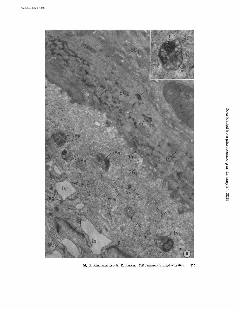
FIGURE 6 Field near the outer front of frog epidermis showing parts of cells from two cornified (SC1, SC:) and two granular (SGI, SG2) layers. The cytoplasm of the granular cells contains numerous cytoplasmic filaments (some of which 0 e) are seen to insert on the desmosomes (d)), a few profiles of rough-surfaced endoplasmic reticulum (er), free ribosomes (r), mitochondria (m), and a number of bodies (lvl to lye) which represent iysosomal derivatives. Several of the latter (lvl and lye) contain small vesicles embedded in a matrix of high density, and correspond to multivesicular bodies. Others (lv3 to lv6), which also contain ribosomal aggregates, correspond to autolytic vacuoles. One of these is enlarged in tile inset. The presence of vesicles of the type found in multivesicular bodies along with recognizable remnants of cell organelles suggests that multivesicular bodies and autolytic vacuoles merge.
As tile cells keratinize and move outward, they become increasingly more dense and compact, more devoid of recognizable cell organelles, and nmre filled with cytoplasmic fibrils. The cell of the second cornified layer (SC2) is only partially keratinized and still contains recognizable organelles: large clusters of ribosomes (r), several large granules (/g), and elements of the rough-surfaced endoplasmic reticulum (er). The outermost cornified cell is fully keratinized; both its nucleus (n) and cytoplasm appear very dense, with the latter occupied ahnost completely by tightly packed bundles of filaments.
Note that the intercellular spaces between the granular and cornified cells are filled with dense material (din), whereas those between granular ceils (Is) appear "empty." Several granules of the small variety (sg) are seen in the cytoplasm near one of these spaces.
The inset shows an autolytic vacuole with its distinct limiting membrane, dense ma- trix, and content of ribosonae aggregates (r) and nlembrane-limited vesicles (v) with light content.
Specimen and sections prepared as for Fig. 2. X 80,000. Inset, X 66,000.
274 T~E JOURNAL OF CELL BIOLOGY " VOLUME 26, 1965
on January 24, 2015jcb.rupress.org
Dow
nloaded from
Published July 1, 1965
M. G. FAIlQUItAR AND G. E. PALADE Cell Junctions in Amphibian Skin 275
on January 24, 2015jcb.rupress.org
Dow
nloaded from
Published July 1, 1965
finely fibrillar layer, which closely parallels the basal cell membranes of the s. germinativum and corresponds to basement membranes found else- where wherever epi thel ium adjoins connect ive tissue; and (2) a thicker (5 to 7 ~) frequently dis- cont inuous layer, which consists of plies of col- lagen fibrils whose or ienta t ion changes by ~ 9 0 ° f rom one ply to another . This last layer has an organizat ion similar to tha t of the basement lamella of larval amph ib i an epidermis (11, 12, 22). I t is visible by l ight microscopy, and has been called the "basemen t m e m b r a n e " or glassy layer of the skin (of. 23, 24). In this study, however, we shall use the t e rm "basemen t m e m b r a n e " to refer to the first, th in (500 to 700 A) layer which lies in close association wi th the germinal cells, since this is the type of structure to which the t e rm is generally applied in the electron micro- scope l i terature, including tha t concerning the epidermis (of. 25). ~
7 For other reasons than possible confusion in the description of skin layers, "basement membrane" has been criticized by Fawcett (27) and Coggeshall and Fawcett (26) who proposed "basement lamina" and "basal lamina," respectively, as more adequate terms. Admittedly, "basement membrane" is not accurately descriptive in many situations, but we prefer to re- tain it because it is widely used and generally under- stood to designate the structure with which we are concerned. Attempts to establish a new nomencla- ture based on the chemistry rather than morphology of this structure (see Bennett (28) "glycocalyx") have inherent merits, but may be premature (cf. 29, 30).
The dermal front of the epidermis and the basement m e m b r a n e which courses strictly par- allel to it have a highly irregular, scalloped surface (Fig. 18). Between the scallops are numerous dermal micropapil lae, 0.3 to 0.5 # wide, which are par t of a relatively wide space (1 to 2.5 #), located between the basement m e m b r a n e and the plies of collagen fibrils. ~h is space is occupied by a variety of fibrillar elements disposed mostly at r a n d o m (Fig. 18). Some of these elements are typical collagen fibrils. Others are finer fibrils, ~-~100 A in diameter , of a type found in many other locations in the connective tissue (cf. 31). Finally, others appear to be a characterist ic com- ponen t of this par t of the dermis (Fig. 18) and are described in detail in a subsequent paper (32).
Junc t ions in Toad S k i n
In the n u m b e r and a r r angemen t of its layers, the toad epidermis is quite similar to tha t of the frog, except tha t cell stratification is less regular and transit ion between non-cornified (granular) and cornified cells is less abrupt . As far as junc- t ional a r rangements are concerned, the situation is identical: the cells of the 1 or 2 outermost cornified layers are joined at the distal end of the corresponding intercel lular spaces by occluding zonules (Fig. 9); a second (or third) network of
similar zonules occurs in the outer g ranu la r layer;
modified, composite, and regular desmosomes
are found in the usual locations; occluding macu-
lae or fasciae (Figs. 15 and 17) occur scattered
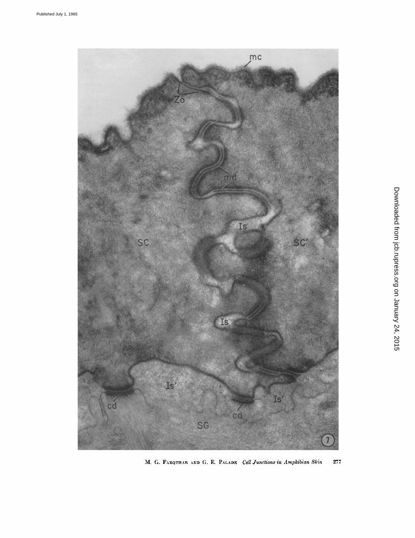
FIGURE 7 Junction line between two cornified cells (SC and SC') on the outer front of the frog epidermis, showing the parallel zig-zigging course of the closely apposed, lateral cell membranes. Near tile point where the cell membranes turn from tile superficial to the lateral cell surface, there is a zonula occludens (Zo) which measures about 1700 A in depth. I t is formed by the fusion of the outer leaflets of the converging membranes with resultant obliteration of the intercellular space at this level. At the proximal end of the occluding zonule, the outer leaflets separate again leaving between an intercellular space of 250 to 300 A which is retained, except for focal dilatations (Is), along most of the remaining tortuous course of the lateral cell membranes. Below the occluding zonule, there is a whole series of modified desmosomal elements (rod) with their characteristic dense plugs in the intercellular spaces. (See also Figs. 10 and IS.) Bends occur along some of these elements. Two of the composite desmosomes (cd), characteristically found along this cell surface, appear at the base of the cornified cells facing the s. granulosum (SG). (See Fig. 13.) In this location the intercellular spaces (Is') are occupied by a granular material of moderate density. Note that junctional elements occupy a greater proportion of the lateral than of the proximal cell surface. A fluffy, finely filamentous layer or "mucous coat" (me) covers the outer surface of the epidermis.
Specimen and section preparation as for Fig. 5. X 55,000.
276 T a E JOURNAL OF CELL BIOLOGY " VOLUME ~6, 1965
on January 24, 2015jcb.rupress.org
Dow
nloaded from
Published July 1, 1965
M. G. FARQUHAR AND G. E. PALADE Cell Junctions in Amphibian Skin 277
on January 24, 2015jcb.rupress.org
Dow
nloaded from
Published July 1, 1965
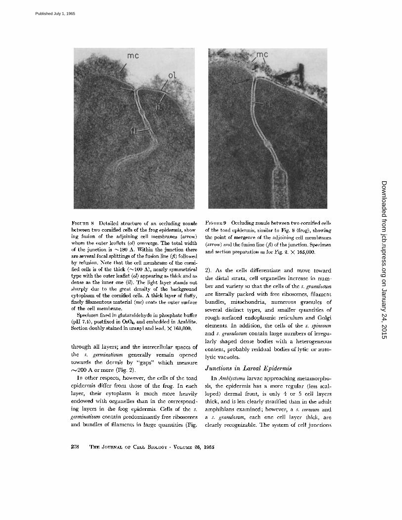
FIOVRE 8 Detailed structure of an occluding zonule between two cornified cells of the frog epidermis, show- ing fusion of the adjoining cell membranes (arrow) where the outer leaflets (ol) converge. The total width of the junction is ~180 A. Within the junction there are several focal splittings of the fusion line (fl) followed by refusion. Note that the cell membrane of the corni- fled cells is of the thick (~100 A), nearly symmetrical type with the outer leaflet (ol) appearing as thick and as dense as the immr one (i/). The light layer stands out sharply due to the great density of the background cytoplasm of the eornified cells. A thick layer of fluffy, finely filamentous material (mc) coats the outer sulface of the cell membrane.
Specimen fixed in glutaraldehyde in phosphate buffer (pH 7.4), postfixed in OsO4, and embedded in Araldite. Section doubly stained in uranyl and lead. )< 165,000.
through all layers; and the intercellular spaces of
the s. germinativum generally remain opened
towards the dermis by "gaps" which measure ~-~200 A or more (Fig. 2).
In other respects, however, the cells of the toad
epidermis differ from those of the frog. In each
layer, their cytoplasm is much more heavily
endowed with organelles than in the correspond-
ing layers in the frog epidermis. Cells of the s.
germinativum contain predominantly free ribosomes
and bundles of filaments in large quantities (Fig.
FIGURE 9 Occluding zonule between two eornified cells of the toad epidermis, similar to Fig. 8 (frog), showing the point of mergence of the adjoining cell membranes (arrow) and the fusion line (fl) of the junction. Specimen and section preparation as for Fig. ~2. )< 165,000.
2). As the cells differentiate and move toward
the distal strata, cell organelles increase in num-
ber and variety so that the cells of the s. granulosum
are literally packed with free ribosomes, filament
bundles, mitochondria, numerous granules of
several distinct types, and smaller quantities of
rough-surfaced endoplasmic reticulum and Golgi
elements. In addition, the cells of the s. spinosum
and s. granulosum contain large numbers of irregu-
larly shaped dense bodies with a heterogeneous
content, probably residual bodies of lytic or auto-
lyric vacuoles.
J u n c t i o n s i n L a r v a l E p i d e r m i s
In Amblystoma larvae approaching metamorpho-
sis, the epidermis has a more regular (less scal-
loped) dermal front, is only 4 or 5 cell layers
thick, and is less clearly stratified than in the adult
amphibians examined; however, a s. corneum and
a s. granutosum, each one cell layer thick, are
clearly recognizable. The system of cell junctions
278 THE JOURNAL OF CELL BIOLOGY • VOLUME ~6, 1965
on January 24, 2015jcb.rupress.org
Dow
nloaded from
Published July 1, 1965
is generally similar s to that described in the adult frog. It consists of typical occluding zonules and modified desmosomes in the s. corneum, composite desmosomes between the s. corneum and s. granu- losum, a more or less complete set of occluding zonules immediately behind the outer front of the s. granulosum, and regular desmosomes throughout the rest of the epidermis. As in the frog, the inter- cellular spaces are closed towards the outer front of the epidermis and open, frequently through gaps larger than 200 A, towards the dermis; an intercellular subcompartment, filled with dis- charged secretion, is found between the s. granu- losum and s. corneum.
In young Xenopus larvae (stage 55) the epidermis is only two cell layers thick. The cells of the outer layer are joined to one another by typical junc- tional complexes composed of an occluding zonule in the usual location, followed proximally by an adhering zonule of variable depth, and, in places, by one or more small desmosomes. The complex is, therefore, similar to that described previously in columnar epithelia (1). The cells of the inner layer are joined by usual desmosomes.
No frog larvae have been examined in this study, but a note recently published by Lanza- vecchia (33) clearly demonstrates the existence of typical occluding zonules in the outer cell layer of the epidermis in Rana esculenta larvae.
D I S C U S S I O N
G e n e r a l O r g a n i z a t i o n o f the E p i d e r m i s
Our findings demonstrate the regular occur- rence of occluding zonules, i.e., belts of membrane fusion, between the cells of the outermost layer of the frog epidermis. These zonules bind together the superficial cells into an uninterrupted sheet which constitutes a structurally continuous barrier throughout the entire epidermis.
Although the amphibian epidermis is a strati- fied squamous epithelium, the junctional arrange- ment is basically similar to that encountered in the simple columnar epithelia studied previously,
s In some places, however, where keratinization is less advanced, the occluding zonule is followed by an element reminiscent of an adhering zonule and by one or more desmosomes within the cornified layer. In such cases, composite desmosomes are absent and the existence of a distinct intercellular subcompart- ment between the s. ¢orneum and s. granulosum is questionable.
in that the intercellular spaces are closed near the surface facing the external medium and open toward the subepithelial spaces.
The arrangement differs, however, in other respects: the complete junctional complex charac- teristic of lumen-lining epithelia (i.e., a zonula occludens, followed by a zonula adhaerens, and a macula adhaerens or desmosome) is found only in the distal (outermost) epidermal layer of early larvae. In the corresponding layer of adults and larvae approaching metamorphosis, the occluding zonule is followed directly by a series of modified desmosomes. A second set of occluding zonules occurs in the proximal comified layer (when such a layer is present), and a second or third set is usually encountered in the distal layer of the s. granulosum. Throughout the rest of the epidermis, junctional elements--irrespective of type (i.e., maculae occludentes and conventional desmosomes) - -are discontinuous structures. As a result of this arrangement, the epidermal cells are firmly bound together; yet most intercellular spaces form a continuous network, a common labyrinthine compartment, which is closed towards the external medium and opened toward the interstitia of the dermis through the numerous, patent intercellular gaps of the s. germinativum. This main compart- ment may not include the intercellular spaces of the s. corneum, which apparently form one or two isolated subcompartments: one located within the s. corneum, and the other between the latter and the s. granulosum. Both seem to be closed by occluding zonules on each side, and usually con- tain a dense material, presumably discharged secretion.
The cells of the epidermis, joined by numerous junctional elements, form a network comple- mentary to that of the extracellular spaces. Struc- turally, this network is divided into distinct cell territories, but functionally it may behave as a continuous compartment (vide infra).
S t r u c t u r a l A s p e c t s
JUNCTIONAL ELEMENTS: A rather extensive literature already exists on various aspects of the fine structure of the integument in a variety of species (cf. 25, 34, 35). As far as intercellular junctions are concerned, previous studies have dealt primarily with the structure of desmosomes (20, 36), the predominant junctional element of the epidermis. Several investigators (37, 38) have noted that the desmosomes found along the
M. G. FARQUnAR ANn G. E; PALADE CellJimbtion~ in Amphlblan Skin ~ 279
on January 24, 2015jcb.rupress.org
Dow
nloaded from
Published July 1, 1965
lateral aspects of fully or partially cornified cells differ from those in the other strata. These modi- fied desmosomes appear in micrographs published in many studies of the epidermis (e.g., 39, 25), but their special features have not been com- mented upon. Our observations show that such junctions occupy a much larger proportion of the lateral cell surface in the s. corneum than do con- ventional desmosomes in the deeper layers of the epidermis. I t follows that prior to, or during keratinization interdesmosomal membrane must be withdrawn into the cell, or that the cell must produce and discharge into the extracellular space the material needed for the formation of additional intercellular plates.
The composite desmosomes along the proximal aspect of the innermost cornified layer have not been described before.
"Quintuple- layered cell interconnections," probably corresponding to our maculae occludentes,
were found by Karrer (21) in the human cervical epithelium, and a similar junctional element was mentioned by Brody (40) in psoriatic human epi- dermis. Recently Dewey and Barr (41) described, under the name of "nexus," the same type of junction in the deeper strata of the frog epidermis. They indicate that such junctional elements arc more frequent after fixation in KMnO~ than in Os04. In our experience, the difficulty in recog- nizing maculae ocdudentes in OsO~-fixed tissues is due primarily to inadequate contrast in the outer leaflets of the cell membranes. With our staining
procedures, we have found such junctions through- out all the strata of the frog epidermis after OsO4- fixation.
SECRETORY GRANULES: O u r study demonstrates the occurrence of two distinct types of granules in amphibian epidermal cells; both are membrane limited, have a relatively homogeneous, dense content, and seem to originate in the Golgi complex.
One type, the smaller (100 to 150 m#) and coarser in texture, appears to be discharged into the intercellular subcompartment located between the s. corneum and the s. granulosum. Granules which correspond in size and location to this first type have already been described in the mammal ian epidermis (human (42) and mouse (43, 14)), as well as in the epithelia of the oral (14, 44) and esophageal (14) mucosae. Some of these granules
differ in the organization of their content (cf. 43,
44), but all seem to have a similar fate: discharge
into tile intercellular space between the s. corneum
and the s. granulosum, as originally suggested by
Frei and Sheldon (43).
The granules of the second type are much
larger (300 to 900 m#). 9 Their exact fate is un-
9 The large granules are visible with the light microscope (of. Fig. 1); hence their presence justifies the recognition of a s. granulosum in amphibian epi- dermis. It should be pointed out, however, that they do not consist of keratohyalin (cf. 46) like the granules found in a typical s. granulosum of mammalian skin.
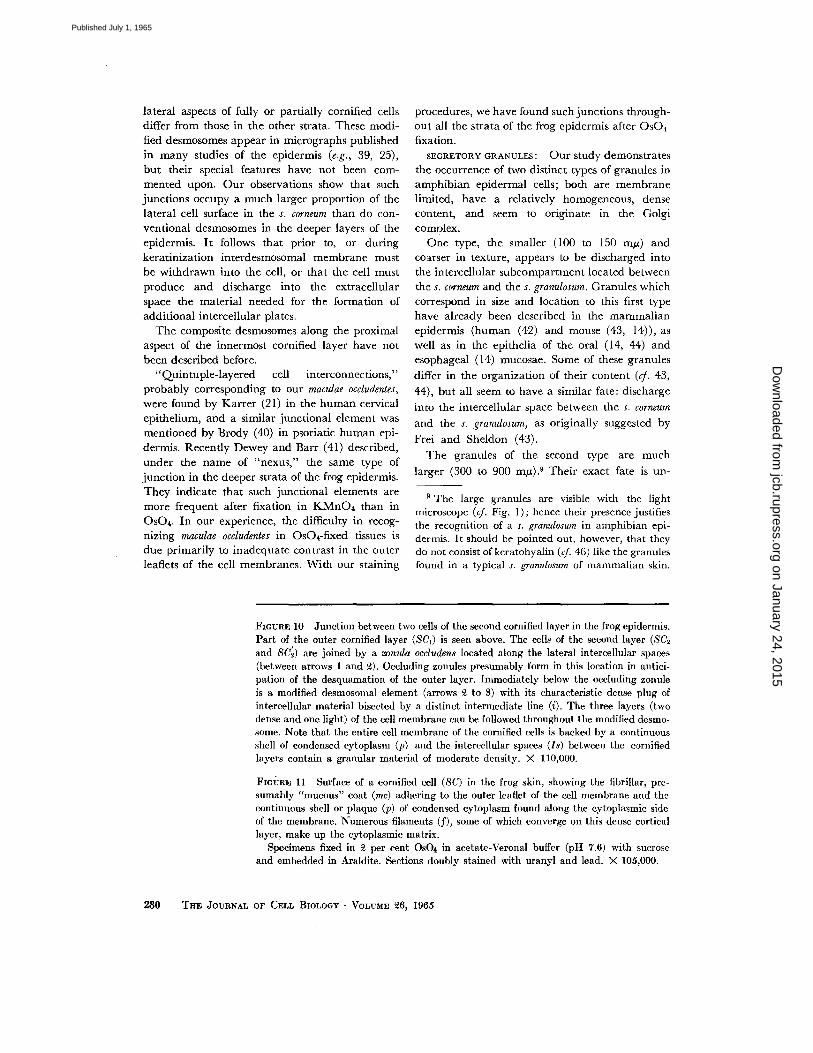
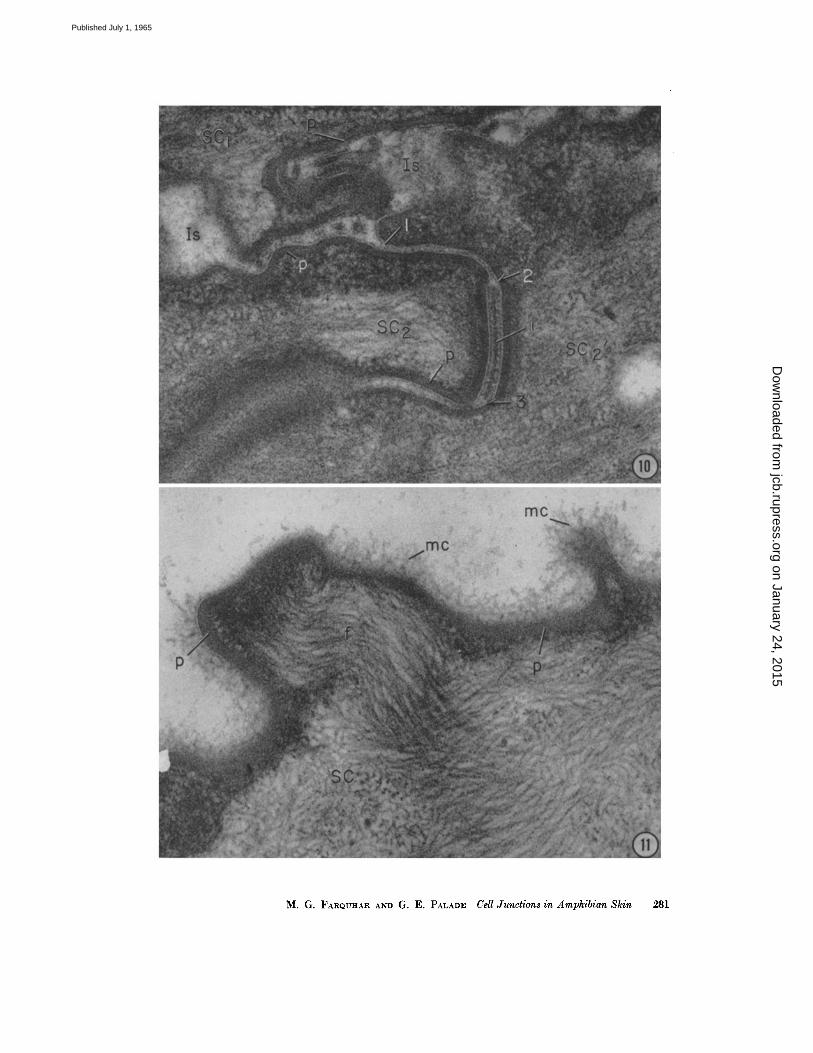
FiounE 10 Junction between two cells of the second cornified layer in the frog epidermis. Part of the outer cornified layer (SCt) is seen above. The ceils of the second layer (SC2 and SC'~) are joined by a zvnula occludens located along the lateral intercellular spaces (between arrows 1 and ~). Occluding zonules presumably form in this location in antici- pation of the desquamation of the outer layer. Immediately below the occluding zonule is a modified desmosomal element (arrows ~ to 3) with its characteristic dense plug of intercellular material bisected by a distinct intermediate line (i). The three layers (two dense and one light) of the cell membrane can be followed throughout the modified desmo- some. Note that the entire cell membrane of the cornified cells is backed by a continuous shell of condensed cytoplasm (p) and the intercellular spaces (Is) between the cornified layers contain a granular material of moderate density. )< 110,000.
FI(~yR~ 11 Surface of a cornified cell (SC) in the frog skin, showing the fibrillar, pre- sumably "mucous" coat (me) adhering to the outer leaflet of the cell membrane and the continuous shell or plaque (p) of condensed cytoplasm found along the cytoplasmic side of the membrane. Numerous filaments (f), some of which converge on this dense cortical layer, make up the cytoplasmic matrix.
Specimens fixed in ~ per cent Os04 in acetate-Veronal buffer (pH 7.6) with sucrose and embedded in Araldite. Sections doubly stained with uranyl and lead. )< 105,000.
280 T~E ffOV~NAL OF CELT. BIOLOGY • VOLVM~ ~6, 1965
on January 24, 2015jcb.rupress.org
Dow
nloaded from
Published July 1, 1965
M. G. FARQUItAR AND G. E. PALADE Cell Junctions in Amphibian Skin 281
on January 24, 2015jcb.rupress.org
Dow
nloaded from
Published July 1, 1965
known, bu t they persist in the outer g ranular layer. Al though they decrease in n u m b e r as the ceils keratinize, some remain in tac t in the cyto- plasm while the cells move from t he s. granulosum
to t he s. corneum and eventual ly appear to undergo degrada t ion wi th in autolytic vacuoles. These large granules apparen t ly have no counte rpar t in
most o ther kerat inizing epithelia, bu t have been
il lustrated previously in the frog epidermis by
Voute (45) and by Parakkal and Matol tsy (46).
T h e lat ter have pointed out tha t mucus is formed
in the a m p h i b i a n epidermis at the stage at which
m a m m a l i a n epidermis produces keratohyal in
granules. They did not distinguish two different
granule populations, and apparent ly assume tha t
in the frog all epidermal granules contain mucus.
Our findings demonst ra te the presence of two
dist inct g ranu la r types in this same species; raise
the possibility tha t their content is chemically
different; and suggest tha t one type, the smallest,
is concerned wi th the product ion of the dense
mater ia l tha t fills the intercel lular spaces between
the s. granulosum and s. corneum.
F u n c t i o n a l I m p l i c a t i o n s
OSMOTm RESPONSE: Cell membranes fused by zonulae occludentes on a succession of fronts form the only structurally cont inuous barriers in the frog epidermis. T h e first bar r ie r is located at the outer front of t he s. corneum and has a well estab- lished continuity. I t is followed at a distance of 3 to 4 ~ by one or two addi t ional cont inuous barriers: one of these is present only in places in which the s. corneum is composed of more t han one cell layer; the other is usually recognized at the distal front of the s. granulosum. These addi t ional barriers are probably formed in ant ic ipat ion of desquamation. The i r existence creates the extra- cellular subcompar tments already ment ioned, which could function as a buffer space between the external and the internal med ium of the frog, and as a closed space in which the mucous coat mater ia l is accumulated and the extensive plugs of the modified desmosomes characterist ic of the s. corneum are elaborated.
T h e fact tha t the intercel lular spaces of the frog epidermis are closed toward the external med ium and open toward the dermis provides the neces-
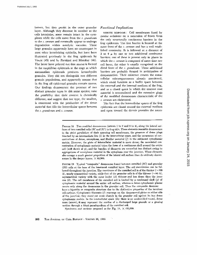
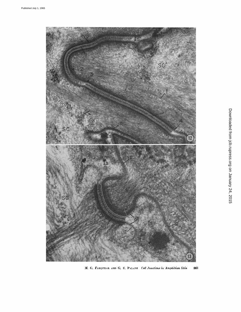
FIGURE 1£ Two modified desmosomes (arrows 1 to ~ and 3 to 4), along the lateral sur- faces of two cornified cells (SC and SC') in frog skin. These elements resemble desmosomes in the strict parallelism of their apposiug cell membranes, the presence of dense plugs bisected by an intermediate line (i) in the intercellular space, and the occurrence of con- centrations of dense, amorphous, and fibrillar material (f) in the subjacent cytoplasmic matrix. However, the plate of intercellular material is more dense; the associated con- centration of cytoplasmic material takes the form of a continuous shell around the entire cell (well shown at p); and the bundles of filaments are somewhat less distinct owing to aggregations of amorphous material in the cytoplasm near the junction. These elements also occupy a much greater proportion of the lateral cell surface than do ordinary desmo- somes in the deeper layers. X 85,000.
FIGVRE 13 Typical "composite" desmosome found between cornifled (SC) and granular (SG) cells at the base of the innermost cornified layer. The cell membranes can be fol- lowed throughout the junction. The membrane of the cornified cell is of the thicker (~100 A), nearly symmetrical variety, while that of tile granular cells is of the thinner (~80 A), asymmetrical variety with the outer leaflet (ol) thinner and less dense than the inner one (il). The cell membrane of the cornified cell is backed by a condensed shell (p) of cytoplasmic material around the entire cell surface, whereas a dense cytoplasmic plaoue occurs only along the desmosome in the granular cell. Thus the composite desmoso have a bipartite or composite structure due to the distinctive properties of the involved cell surfaces. Cytoplasmic filaments (f) converge on the desmosomal plates on either side of the junction; they stand out more sharply in the granular cell against its less dense cytoplasmic matrix. In the intercellular space (Is) there is an unidentified round, dense mass (arrow); it may represent the residue of a discharged large granule or a grazing section through a blunt pseudopodium of the cornified cell.
Specimens and secLions prepared as for Fig. l l . X 135,000.
282 THE JovRNx[~ OF CELL BIOLOaY " VOLVMB ~6, 1965
on January 24, 2015jcb.rupress.org
Dow
nloaded from
Published July 1, 1965
M. G. FARQU-I:IAR AND G. E. PALAD~ Cell Junctions in ;4mphibian Skin 283
on January 24, 2015jcb.rupress.org
Dow
nloaded from
Published July 1, 1965
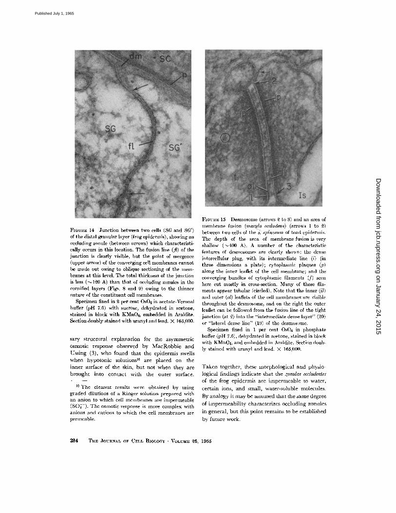
FIGURE 14 Junction between two cells (SG and 8G') of the distal granular layer (frog epidermis), showing an occluding zonule (between arrows) which characteristi- cally occurs in this location. The fusion line (fl) of the junction is clearly visible, but the point of mergence (upper arrow) of the converging cell membranes cannot be made out owing to oblique sectioning of the mem- branes at this level. The total thickness of the junction is less (--q~0 A) than that of occluding zonules in the cornified layers (Figs. 8 and 9) owing to the thinner nature of the constituent cell membranes.
Specimen fixed in ~ per cent OsO4 in acetate-Veronal buffer (pH 7.6) with sucrose, dehydrated in acetone, stained in block with KMnO4, embedded in Araldite. Section doubly stained with uranyl and lead. )< 165,000.
sa W structural explanat ion for the asymmetr ic osmotic response observed by M a c R o b b i e and Ussing (3), who found tha t the epidermis swells when hypotonic solutions 1° are placed on the inner surface of the skin, bu t not when they are b rough t into contact with the outer surface.
10 The clearest results were obtained by using graded dilutions of a Ringer solution prepared with an anion to which cell membranes are impermeable (SO~--). The osmotic response is more complex with anions and cations to which the cell membranes are permeable.
FIGURE 15 Desmosome (arrows ~ to 8) and an area of membrane fusion (mac.u.la occludens) (arrows 1 to ~2) between two cells of the s: sldnosurn of toad epidermis. The depth of the area of membrane fusion is very shallow ('-~400 A). A number of the characteristic features of desmosomes are clearly shown: the dense intercellular plug, with its intermediate line (i) (in three dimensions a plate); cytoplasmic plaques (p) along the inner leaflet of the cell membrane; and the converging bundles of cytoplasmic filaments (f) seen here cut mostly in cross-section. Many of these fila- ments appear tubular (circled). Note that the inner (il) and outer (ol) leaflets of the cell nlembranes are visible throughout the dcsmosome, and on the right the outer leaflet can be followed from the fusion line of the tight junction (at 2) into the "intermediate dense layer" (~0) or "lateral dense line" (19) of the desmosame.
Specimen fixed in 1 per cent OsO4 in phosphate buffer (pH 7.6), dehydrated in acetone, stained in block with KMnO4, and embedded in Araldite. Section doub- ly stained with uranyl and lead. X 165,000.
Taken together, these morphological and physio- logical findings indicate tha t the zonulae occludentes of the frog epidermis are impermeable to water,
cer ta in ions, and small, water-soluble molecules.
By analogy it may be assumed tha t the same degree
of impermeabi l i ty characterizes occluding zonules
in general, bu t this point remains to be established
by future work.
284 TH• JOVRNAL OF CaLL BIOLOGY • VOLUME ~6, 1965
on January 24, 2015jcb.rupress.org
Dow
nloaded from
Published July 1, 1965
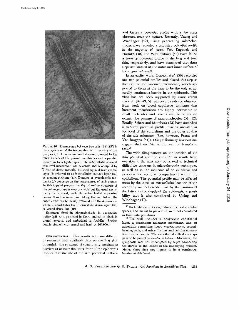
Fmurm 16 Desmosome between two cells (88, SS') in the s. spinosum 3f the frog epidermis. I t consists of two plaques (p! of dense material disposed parallel to the inner le.-~ets of the plasma membranes and separated theu.ff0m by a lighter space. The intercellular space at tlai~ level measures ~£50 A across and is occupied by
disc of dense material bisected by a denser central layer (i) referred to as intercellular contact layer (~0) or median stratum (~1). Bundles of cytoplasmic fila- ments (f) converge on the inner aspect of each plaque. In this type of preparation the trilaminar structure of the cell membrane is clearly visible but the usual asym- metry is reversed, with the outer leaflet appearing denser than the inner one. Along the cell below, the outer leaflet can be clearly followed into the desmosome where it constitutes the intermediate dense layer (~0) or lateral dense line (19).
Specimen fixed in glutaraldehyde in eaeodylate buffer (pH 7.4), postfixed in OsO4, stained in block in uranyl acetate, and embedded in Araldite. Section doubly stained with uranyl and lead. )< 165,000.
SKIN POTENTIAL: Our results are more difficult to reconcile with avai lable da ta on the frog skin potential. The existence of structurally cont inuous barriers at or near the outer front of the epidermis implies tha t the site of the skin potential is there
and favors a potent ial profile with a few steps clustered near the surface. Recently, Ussing and Windhage r (47), using penet ra t ing microelec- trodes, have recorded a mult is tep potential profile in the majori ty of cases. Yet, Engbaek and Hoshiko (48) and W h i t t e m b u r y (49) have found a two-step potential profile in the frog and toad skin, respectively, and have concluded tha t these steps are located at the outer and inner surface of the s. germinativum, n
In an earlier work, Ot toson et al. (50) recorded one-step potential profiles and placed this step at the level of the basement membrane , which ap- peared to t hem at the t ime to be the only struc- tural ly cont inuous barr ier in the epidermis. This view has not been supported by more recent research (47-49, 5); moreover, evidence ob ta ined from work on blood capillaries indicates tha t basement membranes are highly permeable to small molecules and also allow, to a cer tain extent, the passage of macromolecules (51, 52). Finally, Scheer and M u m b a c k (53) have described a two-step potent ial profile, placing one-step at the level of the epi thel ium and the other at t ha t of the tela subcutanea. (See, however, Franz and V an Bruggen (54)). O u r prel iminary observations suggest tha t the tela is the wall of lymphat ic s a c s . 12
T h e wide disagreement on the location of the skin potent ial and the var ia t ion in results from one skin to the next may be related to technical difficulties inheren t in microelcctrode techniques, as well as to the existence of an extensive and pervasive extraccllular co mp a r t men t within the epithelium. T h e potent ial profile may be affected more by the intra- or extracellular location of the recording microclectrode than by the position of the lat ter in the dep th of the epidermis, a possi- bility tha t is also considered by Ussing and Windhage r (47).
11 Back diffusion (leaks) along the intercellular spaces, and means to prevent it, were not considered in these interpretations.
12 The wall includes a phagocytic endothelial layer, a continuous basement membrane, and an adventitia containing blood vessels, nerves, crystal- hearing ceils, and other fibrillar and cellular connec- tive tissue elements. The endothelial cells do not ap- pear to be joined by zonulae occludentes. Moreover, the lymphatic sacs are interrupted by septa connecting the dermis to the fasciae of the underlying muscles. Hence there does not appear to be a continuous barrier at this level.
M. G. FARQUHAR AND G. E. PALADE Cell Junctions in Amphibian Skin 285
on January 24, 2015jcb.rupress.org
Dow
nloaded from
Published July 1, 1965
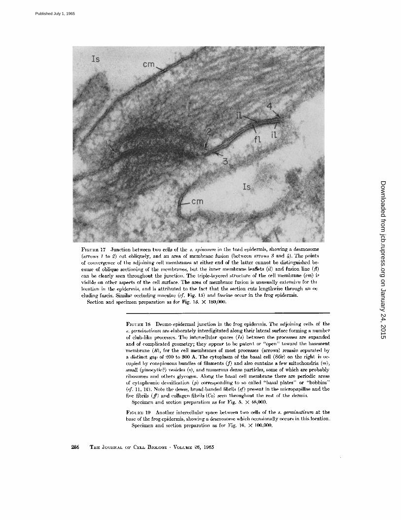
FIGURE 17 Junction between two cells of the s. spinosum in the toad epidermis, showing a desmosome (arrows 1 to 2) cut obliquely, and an area of membrane fusion (between arrows 3 and 4). The points of convergence of the adjoining cell membranes at either end of the latter cannot be distinguished be- cause of oblique sectioning of the nlcmbrancs, but the inner membrane leaflets (il) and fusion line (fl) can be clearly seen throughout the junction. The triple-layered structure of the cell membrane (cm) i.~ visible on other aspects of the cell surface. The area of membrane fusion is unusually extensive for thl location in the epidermis, and is attributed to the fact that the section cuts lengthwise through an oc- cluding fascia. Similar occluding maculae (of. Fig. 15) and fasciae occur in the frog epidermis.
Section and specimen preparation as for Fig. 15. )< 180,000.
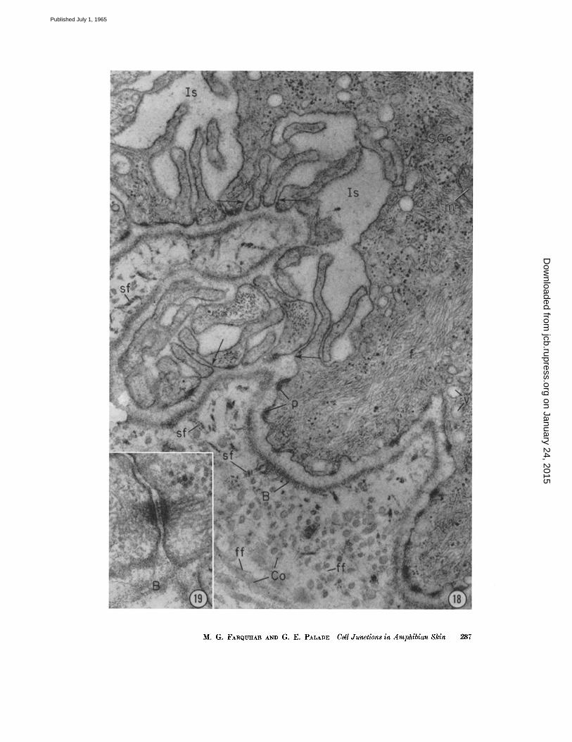
FIGURE 18 Dermo epidermal junction in the frog epidermis. The adjoining cells of the s. germinativum are elaborately interdigitated along their lateral surface forming a number of club-like processes. The intercellular spaces (Is) between the processes are expanded and of complicated geometry; they appear to be patent or "open" toward the basement membrane (B), for the cell membranes of most processes (arrows) remain separated by a distinct gap of ~00 to 300 A. The cytoplasm of the basal cell (SGe) on the right is oc- cupied by conspicuous bundles of filaments (f) and also contains a few mitochondria (m), small (pinocytic?) vesicles (v), and numerous dense particles, some of which are probably ribosomes and others glycogen. Along the basal cell membrane there are periodic areas of cytoplasmic densification (p) corresponding to so called "basal plates" or "bobbins" (cf. 11, 1~). Note the dense, broad-banded fibrils (sf) present in the micropapillae and the fine fibrils (if) and collagen fibrils (Co) seen throughout the rest of the dermis.
Specimen and section preparation as for Fig. 5. )< 48,000.
FIGURE 19 Another intercellular space between two cells of the s. germinatlvum at the base of the frog epidermis, showing a desmosome which occasionally occurs in this location.
Specimen and section preparation as for Fig. 16. × 100,000.
286 THE JOURNAL OF CELL BIOLOGY • VOLUME ~6, 1965
on January 24, 2015jcb.rupress.org
Dow
nloaded from
Published July 1, 1965
M. G. FARQtrlIArt AND G. E. PALADE Cell Junctions in Amphibian Skin 287
on January 24, 2015jcb.rupress.org
Dow
nloaded from
Published July 1, 1965
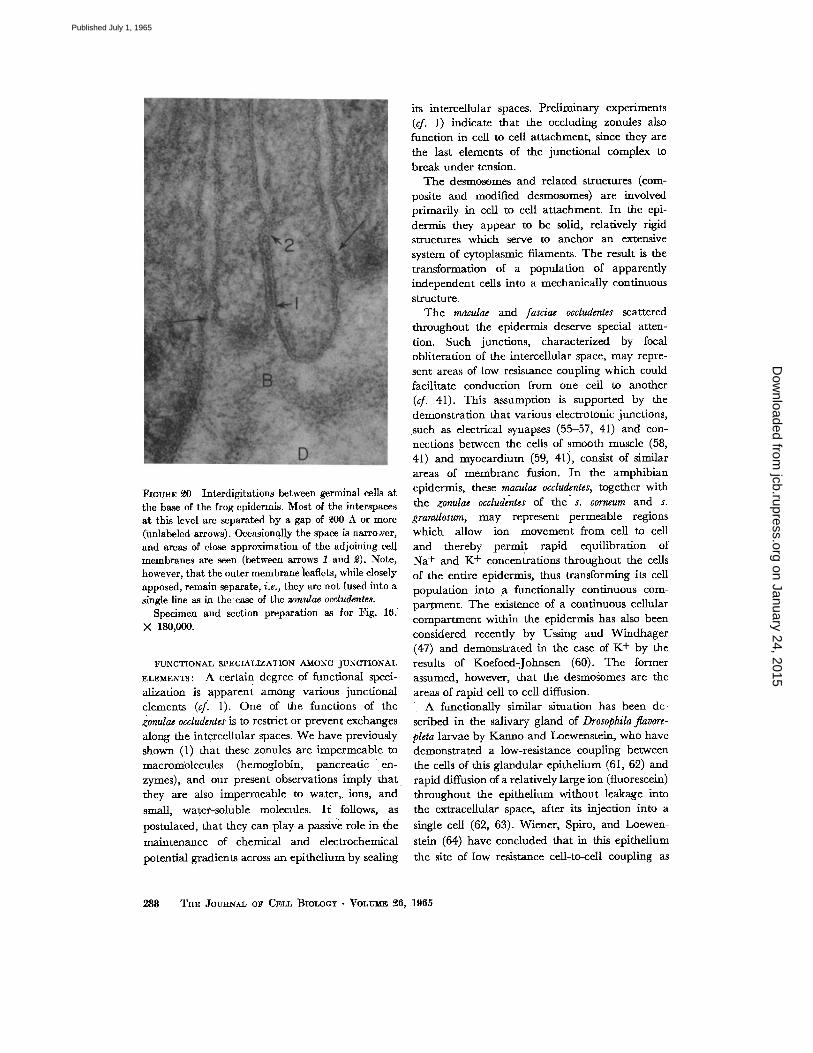
l~mmCE ~ Interdigitations between germinal cells at the ba.~e of the frog epidermis. Most of the interspaces at this level are separated by a gap of 9.00 A or more (unlabeled arrows). O~.asionally the space is nazro^,er, and areas of close approximation of the adjoining t~ll membranes are .~en (between arrows 1 and ~). Note, however, that the outer membrane ieafleks, while closely apposed, remain separate, i.e., they are noL fused into a single line as in theease of the zomdae occludentes.
Specimen and section preparation as for Fig. 16. X 180,000.
FUNCTIONAL SPEGIAI,IZATION AMONG JUNCTIONAl,
ELEMENTS: A certain degree of functional speci-. allzation is apparent among various junctional elements (cfl l). One of the functions of the zonulae occludentes is to restrict or prevent exchanges along the intercellular spaces. We have previously shown (1) that these zonules are impermeable to macromolecules (hemoglobin, pancreatic en- zymes),, and our present• observations imply that they are also impermeable to water,, ions, and
small, water-soluble molecules. I t follows, as
postulated, that .they can play., a passive role in the
mMntenancc of chemical and electrochemical
potential gradients across an epithelium by sealing
its intercellular spaces. Preliminary experiments (cf. 1) indicate that the occluding zontrles also function in cell to cell attachment, since they are the last elements of the junctional complex to break under tension.
The desmosomes and related structm'es (com- posite and modified desmosomes) are involved primarily in cell to cell attachment. In the epi- dermis they appear to be solid, relatively rigid structures which serve to anchor an extensive system of cytoplasmic filaments. The re.~lt is the transformation of a population of apparently independent ceils into a mechanically continuous structure.
The macu/ae and fasciae occludentes scattered throughout the elfidermis deserve special atten- tion. Such junctions, characterized by focal obliteration of the intercellular space, may repre- sent areas of low resistance coupling, which could facilitate conduction from one cell to another (cf. 41). This assumption is ~ tppo r t ed by the demonstration that various electrotonic junctions, • such as electrical synapses (55-57, 41) and con- nections between the cells of smooth muscle (58, 41) and myocardium (59, 41), consist of similar areas of membrane fusion. In the amphibian epidermis, these maculae occludentes,, together with the zonulae occludentes of the" s. corneum and s. gratmlosum, may represent permeable regions which allow ion movement from cell to cell and thereby .permit rapid equilibration of Na + and K + concentrations throughout the cells of the entire epidermis, thus transforming its cell population into .a functionally continuous com- partment. The existence of a continuous cellular compmXment within the epidermis has also been considered recently by Ussing and ~Vindhager (47) and demonsu'ated in the case of K + by the results of Koefoed-Johnsen (60). The former assumed, however, that the desmosomes are the areas of rapid cell to cell diffusion.
A functionally similar situation has been de- scribed in the salivary gland of Drosophila flavore- pleta lm~ae by Kanno and Loewenstein, who have demonstrated a low-resistance coupling between the cells of this glandular epithelium .(61, 62) and rapid diffusion of a relatively large ion (fluorescein) throughout the epithelium ~i thout leakage into
• the extracellular space, after its injection into a
single cell .(62, 63). XCriener, Spiro, and Loewen-
stein (64) have concluded that in this epithelium
the site of low resistance cell-to-cell coupling as
288 T'im .lomm~..L oF C~a, BioLooY • Vor~-m~ ~6, 1965
on January 24, 2015jcb.rupress.org
Dow
nloaded from
Published July 1, 1965
well as of h igh resistance along the intercel lular spaces is a ' :septate junc t ion . " T h e latter, like the "septate desmommes" ( 6 5 - 6 6 ) , appears to be a characteristic j uncdonM element of epithelia in invertebrates.
The bear ing of otlr findings and intel-pretations on current physiological models of the frog skin were briefly t reated in a previous paper (5) and ~11 be discussed in dctail in a paper to follow (67).
:\rote Added in Proof: In a recently published paper (J. Cell Biol., 1965, 24, 297), Matoltsy and Parakkal mggest that the small grmmles present in the middle layers of the epidermis contain a special membrane- coating substance which, upon discharge into the intercellular spaces, renders the cell membranc~ re.-
R E F E R E N C E S
1. FAgQmIAg, M. G., and PALADE, G. E., Junc- tional complexes in various epithelia, J. Cell Biol., 1963, 17, 375.
2. UssmG, II. II., ] 'he frog skin potential, or. Gem Physiol., 1960, 43,135.
3. MAnRonsm, E. A. C., and UsmNc, H. H., Os- motic behaviour of the epithelial ceils of frog skin, Acta Physiol. Stand., 1961, 53, 348.
4. FARQtmAR, M. G., and PAr.ADF., G. E., Cell junctions in •amphibian skin, J. Cell Biol., 1963, 19, 22A.
5. FARQIrHZm, M. G., and P~mAD~, G. E., Func- tional organization of amphibian skin, Proc. Nat. Aead. So., 1964, 51,569.
6. SA~ATr~r, I). I)., BENSCH, K., and BARRNETT, R. J., Cytoehemistry and electron microscopy. The preservation of cellular ultrastrucmre and enzymatic activity by "aldehyde fixation, or. Cell Biol., 1963, 17, 19.
7. K-ELLENBERGER, E., RYTI~.R, A., and S~.CHAUD, J., Electron microscope study of DNA-con- raining plasma. If. Vegetative and mature phage DNA a~ compared with normal bac- terial nucleoids in different physiological states, d. Biophysic. and Biochem. CytoL, 1958, 4, 671.
8. KAm~ovsxY, M. J., Simple methods for "staining with lead" at high pH in electron microscopy, .L Cell Biol., 1961, 11, 729.
9. RaCa-I~mDSON, K., J ~ . e r r , L., a n d Fucg~, E., Embedding in epoxy resins for ultrathin sec- tioning in electron microscopy, Stain Techn., 1960, 35, 313.
10. M~RCER, E. H., MUNGER, B. L., RO~ERS, G. E., and RorH, S. I., A suggested nomenclature for fine-structural components of keratin and keratin-like products of cells, Nature, 1964, 201, 367.
sistant to keratinolytie agents (0.1 ~ NaOH). This interpretation diifers from their previous Views quoted on page 282 of this paper. It is not clear, however, from their new e~ddence, whether the resistant ma- terial represents a transformed cell membrane, the subjacent shell of dense cytoplasm characteristic of cornified cells, or a combination of these structures.
This research was supported by research grants AM- 09090, and IIE-05648, from the United States Public Health Service, and by Public Health Ser~4ce research career program award 1-K3-GM-25, 109.
We gratefully acknowledge the technicM assistance of Mrs. Kar in Tay]or, Miss Agneta Stromblad, Mrs. I,ee Adinolfi, and Mrs. Sarah Benton.
Recdved for publication, November 6, 1964.
11. PORa'~R, K. R., Observations on the line struc- ture of animal epidermis, in Proceedings of the Third International Conference on Electron Microscopy, London, 1954, (R. Ross, editor), London, Royal Microscopical Society, 1956, 539.
12. WEISS, P., and F ~ . t s , W., Electron micrograms of larval amphibian epidermis, Exp. Cell Re- search, 1954, 6, 546.
13. DE Duv~., C., "]?he lysosome concept, in Ciba Foundation Symposium on Lysosomes, (A. V. S. de Reuck and M. P. Cameron, editors), Boston, Little, Brown and Co., 1963, 1.
14. RHODm, J. A. G., and R~n'u, E. J. , Ultrastruc- turc of keratin in oral mucosa, skin, e~ophagu.% claw, and hair, in Fundamentals of Keratiniza- tion, (E. O. Butcher, and R. F. Sogrmaes, editors), Wastfington D. C., A.A.A.S. Publica- tion No. 70, 1962, 61.
15. CHob J. K., The fine structure of the urinary bladder of the toad, Bufo marinus, J. Cell Biol., 1963, 16, 53.
16. PEACI-J:EY, L. D., and II~sMUss~, H., Structure of the toad's urinary bladder as related to its physiology, J. Biophysic. at~ Biochvm. Cytol., 1961, 10, 529.
]7. ITO, S., and WINCTrFaa~R, R. J. , The fine struc- ture of the gastric mucosa in the bat, J. Cell Biol., 1963, 16, .541.
18. ITO, S., The surface coathlg of enteric microvilli, Anat. Rec., 1964, 148, 294.
19. Roa~, S. I., and Cr.aax, W. H., JR., Ultrastruc- tural evidence related to the mechanism of keratin synthesis, in The Epidermis, (W. Montagna and W. C. Lobitz, Jr. , editors), New York and l.ondon, Acadenfic Press, Inc., 1964, .303.
20. ODLAI~D, G. F., The fine structure of the inter-
M. G. FXRQrn.~R A~U G. E. PALADE Cell Junctions in Amphibian Skin '289
on January 24, 2015jcb.rupress.org
Dow
nloaded from
Published July 1, 1965
relationship of cells in the human epidermis, J. Biophysic. and Biochem. Cytol., 1958, 4,529.
21. KARRER, H. E., Cell interconnections in normal human cervical epithelium, J. Biophysic. and Biochem. Cytol., 1960, 7, 181.
22. EDDS, M. V., JR., The basement lamella of de- veloping amphibian skin, in Small Blood Vessel Involvement in Diabetes Mellitus, (M. D. Siperstein, A. R. Colwell, Sr., and K. Meyer. editors), Washington D. C., American Insti- tute of Biological Sciences, 1964, 245.
23. MONTAGNA, W., The Structure and Function of Skin, New York, Academic Press, Inc., 1962, 2nd edition, 28.
24. ZELICKSON, A. S., Electron Microscopy of Skin and Mucous Membrane, Springfield, Illinois, Charles C Thomas, 1963.
25. MONTAONA, W., and LOmTZ, W. C., JR., The Epidermis, New York and London, Academic Press, Inc., 1964.
26. COOOESHALL, R. E., and FAWCETT, D. W., The fine structure of the central nervous system of the leech, Hirudo medicinalis, J. Neu~ophysiol., 1964, 27, 229.
27. FAWCETT, D. W., Comparative observations on the fine structure of blood capillaries, in The Peripheral Blood Vessels, (L. Orbison and D. E. Smith, editors), Baltimore, Williams and Wilkins Co., 1963, 17.
28. BENNETT, H. S., Morphological aspects of extra- cellular polysaccharides, J. Histochem. and Cytochem., 1963, 11,14.
29. LAZAROW, A., and SPEIDEL, E., The chemical composition of the glomerular basement mem- brane and its relationship to the production of diabetic complications, in Small Blood Vessel Involvement in Diabetes Mellitus, (M. D. Siperstein, A. R. Colwell, Sr., and K. Meyer, editors), Washington, D. C., American Insti- tute of Biological Sciences, 1964, 127.
30. DlSCHE, Z., The glycans of the mammalian lens capsule--a model of basement membranes, in Small Blood Vessel Involvement in Diabetes Mellitus, (M. D. Siperstein, A. R. Colwell, Sr., and K. Meyer, editors), Washington, D. C., American Institute of Biological Sciences, 1964, 201.
31. BRUNS, R. R., and PALADE, G. E., Studies on blood capillaries, J. Cell Biol., 1965, in press.
32. PALADE, G. E., and FARQUHAR, M. G., A special fibril of the dermis, or. Cell Biol., 1965, in press.
33. LANZAVECCHIA, G., Osservazioni sulla membrana cellulare nelli epidermide di embrioni di Rana esculenta L., Atti del IV Congresso Italiano di Microscopia Elettronica, 1963, 24, Tipo- grafia del Seminario, Padova.
34. RHODIN, J. A. G., An Atlas of Ultrastructure, Philadelphia, W. B. Saunders Co., 1963, 132.
35. MERCER, E. H., Keratin and Keratinization, New York, Pergamon Press, 1961.
36. HmBs, R. G., and CLARK, W. H., JR., Electron microscope studies of the human epidermis. The cell boundaries and topography of the stratum Malpighii, J. Biophysic. and Biochem. Cytol., 1959, 6, 71.
37. HORSTMANN, E., and KNOOP, A., Elektronen- mikroskopische Studien an der Epidermis. I. Rattenpfote, Z. Zellforsch. u. Mike. Anat., 1958, 47, 348.
38. LISTGARTEN, M. A., The ultrastructure of human gingival epithelium, Am. J. Anat., 1964, 114, 49.
39. BRODY, I., The ultrastructure of the tonofibrils in the keratinization process of normal human epidermis, J. Ultrastruct. Research, 1960, 4, 264.
40. BRODY, I., The ultrastructure of the epidermis in Psoriasis Vulgaris as revealed by electron microscopy. 1. The dermo-epidermal junction and the stratum basale in parakeratosis with- out keratohyalin, J. Ultrastruct. Research, 1962, 6, 304.
41. DEWEY, M. M., and BARR, L., A study of the structure and distribution of the nexus, J. Cell Biol., 1964, 23,553.
42. ODLAND, G. F., A submicroscopic granular com- ponent in human epidermis, J. Invest. Dermatol., 1960, 34, 11.
43. FREI, J. g . , and SHELDON, H., A small granular component of the cytoplasm of keratinizing epithelia, J. Biophysic. and Biochem. Cytol., 1961, 11, 719.
44. FARBMAN, A. I., Electron microscope study of a small cytoplasmic structure in rat oral epi- thelium, J. Cell Biol., 1964, 21, 491.
45. VOUTE, C. L., An electron microscopic study of the skin of the frog (Rana pipiens), J. Ultra- struct. Research, 1963, 9,497.
46. PARAKKAL, P. F., and MATOLTSY, A. G., A study of the fine structure of the epidermis of Rana pipiens, J. Cell. Biol., 1964, 20, 85.
47. USSlNG, H. H., and WINDHAGER, E. E., Active sodium transport at the cellular level, in Water and Electrolyte Metabolism (II), (J. de Graeff and B. Leijnse, editors), West European Sym- posia on Clinical Chemistry, 3, Amsterdam, Elsevier Publishing Co., 1964, 3.
48. ENGBAEK, L., and HOSHIKO, T., Electrical poten- tial gradients through frog skin, Acta Physiol. Scand., 1957, 39,348.
49. WHITTEMBURY, G., Electrical potential profile of the toad skin epithelium, J. Gen. Physiol., 1964, 47, 795.
50. OTTOSON, D., SJSSTRAND, F., STENSTROM, S., and SVAETICHIN, G., Microelectrode studies on the EMF of the frog skin related to electron
290 THE JOURNAL OF CELL BIOLOGY • VOLUME ~6, 1965
on January 24, 2015jcb.rupress.org
Dow
nloaded from
Published July 1, 1965
microscopy of the dermo-epidermal junction, Acta Physiol. Scand., 1953, 29, 611.
51. FARQUHAR, M. G., and PALADE, G. E., Glomeru- lar permeability. I. Ferritin transfer across the normal glomerular capillary wall, J. Exp. Med., 1961, 113, 47..
52. PALADE, G. E., and BRUNS, R. R., Structure and function in normal muscle capillaries, in Small Blood Vessel Involvement in Diabetes Mellitus, (M. D. Siperstein, A. R. Colwell, Sr., and K. Meyer, editors), Washington, D. C., American Institute of Biological Sci- ences, 1964, 39.
53. SCHEER, B. T., and MUMBACH, M. W., The locus of the EMF in the frog skin, J. Cell. and Comp. Physiol., 1960, 55, 259.
54. FRANZ, T. J. , and VAN BRUGGEN, J. T., Active sodium transport in frog skin. II. The passive role of the basal cell layer and its cholinesterase, J. Cell. and Comp. Physiol., 1964, 64, 193.
55. ROBERTSON, J. D., Ultrastructure of excitable membranes and the crayfish median-giant synapse, Ann. New York Acad. Sc., 1961, 94, 339.
56. BENNETT, M. V. L., ALJURE, E., NAKAJIMA, Y., and PAPPAS, G. D., Electrotonic junctions be- tween teleost spinal neurons: electrophysiology and ultrastructure, Science, 1963, 141,262.
57. ROBERTSON, J. D., BODENHEIMER, T. S., and STAGE, D. E., The ultrastructure of Mauthner cell synapses and nodes in goldfish brains, J. Cell Biol., 1963, 19, 159.
58. DEWEY, M. M., and BARR, L., Intercellular con- nections between smooth muscle cells: the nexus, Science, 1963, 137, 670.
59. KARRER, H. E., The striated musculature of blood vessels. II. Cell interconnections and cell surface, J. Biophysi¢. and Biochem. Cytol., 1960, 8, 135.
60. KOEEOED-JOHNSEN, V., in preparation, (quoted by Ussing, H., and Windhager, E. E., in refer- ence 47, p. 8.)
61. KANNO, Y., and LOEWENSTEIN, W. R., Low- resistance coupling between gland cells. Some observations on intercellular contact mem- branes and intercellular space, Nature, 1964, 201, 194.
62. LOEWENSTEIN, W. R., and KANNO, Y., Studies on an epithelial (gland) cell junction. I. Modifications of surface membrane perme- ability, J. Cell Biol., 1964, 22, 565.
63. KANNO, Y., and LOEWENSTEm, W. R., Intercellu- lar diffusion, Science, 1964, 143, 959.
64. WIENER, J. , SPmo, D., and LOEWENSTEIN, W. R., Studies on an epithelial (gland) cell junction. II. Surface structure, J. Cell Biol., 1964, 22, 587.
65. WooD, R. L., Intercellular a t tachment in the epithelium of Hydra as revealed by electron microscopy, J. Biophysic. and Biochem. Cytol., 1959, 6, 343.
66. OVERTON, J. , Intercellular connections in the outgrowing stolon of Cordylophora, J. Cell Biol., 1963, 17,661.
67. FARQOHAR, M. G., and PALADE, G. E., Adeno- sine triphosphatase activity in the epidermal cell membranes of frog and toad skin, 1965, in preparation.
M. G. FARQITHAR AND G. E. PALADI~ Cell Junctions in Amphibian Sldn 291
on January 24, 2015jcb.rupress.org
Dow
nloaded from
Published July 1, 1965
Related Documents


