Cell division Chapter 10 Genes and Development

Cell division Chapter 10 Genes and Development. Fig. 10.1-1 Copyright © The McGraw-Hill Companies, Inc. Permission required for reproduction or display.
Dec 18, 2015
Welcome message from author
This document is posted to help you gain knowledge. Please leave a comment to let me know what you think about it! Share it to your friends and learn new things together.
Transcript

Cell division
Chapter 10Genes and Development

Fig. 10.1-1
Copyright © The McGraw-Hill Companies, Inc. Permission required for reproduction or display.
Bacterial cell
Bacterial chromosome:Double-stranded DNA
Origin ofreplication

Fig. 10.1Copyright © The McGraw-Hill Companies, Inc. Permission required for reproduction or display.
Bacterial cell
Septum
Bacterial chromosome:Double-stranded DNA
Origin ofreplication

Fig. 10.2

Fig. 10.3a
Copyright © The McGraw-Hill Companies, Inc. Permission required for reproduction or display.
Septum
FtsZ protein
Chromosome
Prokaryotes
No nucleus, usuallyhave single circularchromosome. After DNA is replicated, it ispartitioned in the cell.After cell elongation,FtsZ protein assemblesinto a ring and facilitatesseptation and celldivision.

Fig. 10.3
Copyright © The McGraw-Hill Companies, Inc. Permission required for reproduction or display.
Septum
FtsZ protein
Chromosome
ChromosomeMicrotubule
Nucleus
Kinetochore microtubule
Centrioles Kinetochore
Polar microtubule
Spindle pole body
Kinetochore microtubule
Centriole
Prokaryotes Some Protists Other Protists Animals
Kinetochore microtubule
Polar microtubule
No nucleus, usually have single circularchromosome. After DNA is replicated, it ispartitioned in the cell.After cell elongation,FtsZ protein assemblesinto a ring and facilitatesseptation and celldivision.
Nucleus present andnuclear enveloperemains intact duringcell division.Chromosomes line up.Microtubule fibers passthrough tunnels in thenuclear membrane andset up an axis forseparation ofreplicatedchromosomes, and celldivision.
A spindle of micro-tubules forms betweentwo pairs of centrioles atopposite ends of thecell. The spindle passes through one tunnel inthe intact nuclearenvelope. Kinetochoremicrotubules formbetween kinetochoreson the chromosomesand the spindle polesand pull the chromo-somes to each pole.
Nuclear enveloperemains intact; spindlemicrotubules form insidethe nucleus betweenspindle pole bodies. Asingle kinetochoremicrotubule attaches toeach chromosome andpulls each to a pole.
Spindle microtubulesbegin to form betweencentrioles outside ofnucleus. Centrioles moveto the poles and thenuclear envelope breaksdown. Kinetochoremicrotubules attachkinetochores ofchromosomes to spindlepoles. Polar microtubulesextend toward the centerof the cell and overlap.
Yeasts
Central spindleof microtubules
Fragmentsof nuclearenvelope

Table 10.1

Fig. 10.5-1
Copyright © The McGraw-Hill Companies, Inc. Permission required for reproduction or display.
Chromosome

Fig. 10.5-2
Copyright © The McGraw-Hill Companies, Inc. Permission required for reproduction or display.
Chromosome Rosettes of Chromatin Loops
Scaffold protein

Fig. 10.5-3
Copyright © The McGraw-Hill Companies, Inc. Permission required for reproduction or display.
Chromosome Rosettes of Chromatin Loops Chromatin Loop
Chromatin loop
Scaffold protein Scaffoldprotein

Fig. 10.5-4
Copyright © The McGraw-Hill Companies, Inc. Permission required for reproduction or display.
Chromosome Rosettes of Chromatin Loops Chromatin Loop Solenoid
Chromatin loop
Scaffold protein Scaffoldprotein

Fig. 10.5
Copyright © The McGraw-Hill Companies, Inc. Permission required for reproduction or display.
Chromosome Rosettes of Chromatin Loops Chromatin Loop Solenoid
Nucleosome
Histone core
Chromatin loop
Scaffold protein Scaffoldprotein
DNA
DNA Double Helix (duplex)

Animation of DNA coiling
DNA coiling and cells dividing

Fig. 10.6

Fig. 10.7
Copyright © The McGraw-Hill Companies, Inc. Permission required for reproduction or display.
Homologous chromosomes Homologous chromosomes
Sister chromatids
Sister chromatids
Centromere
Replication
Kinetochore
Kinetochores
Cohesin proteinsCentromere

Fig. 10.8
Copyright © The McGraw-Hill Companies, Inc. Permission required for reproduction or display.
G1
G2
S Interphase
MitosisM Phase
Cytokinesis
M Phase
G2
S G1
Metaphase
Prophase
Anaphase
Telophase
Prometaphase
Cell cycle

Fig. 10.9
Copyright © The McGraw-Hill Companies, Inc. Permission required for reproduction or display.
Chromatid
Kinetochore
Cohesinproteins
Centromereregion ofchromosome
Metaphasechromosome
Kinetochoremicrotubules

Fig. 10.10
Red = CohesinGreen = KinetochoreBlue = Chromosome

Fig. 10.11aCopyright © The McGraw-Hill Companies, Inc. Permission required for reproduction or display.
© Andrew S. Bajer, University of Oregon
INTERPHASE G2
Nucleus
Nucleolus
Aster
80 µmCentrioles(replicated;animalcells only)
Nuclearmembrane
• DN A has been replicated• Centrioles replicate (animal cells)• Cell prepares for division
Chromatin(replicated)

Fig. 10.11bCopyright © The McGraw-Hill Companies, Inc. Permission required for reproduction or display.
Prophase
80 µm
• Chromosomes condense and become visible• Chromosomes appear as two sister chromatids held together at the centromere• Cytoskeleton is disassembled: spindle begins to form• Golgi and ER are dispersed• Nuclear envelope breaks down
Mitotic spindlebeginning to form
Condensedchromosomes
© Andrew S. Bajer, University of Oregon
MITOSIS

Prometaphase
80 µm
• Chromosomes attach to microtubules at the kinetochores• Each chromosome is oriented such that the kinetochores of sister chromatids are attached to microtubules from opposite poles.• Chromosomes move to equator of the cell
Centromere andkinetochore
Mitoticspindle
Fig. 10.11c Copyright © The McGraw-Hill Companies, Inc. Permission required for reproduction or display.
© Andrew S. Bajer, University of Oregon
MITOSIS
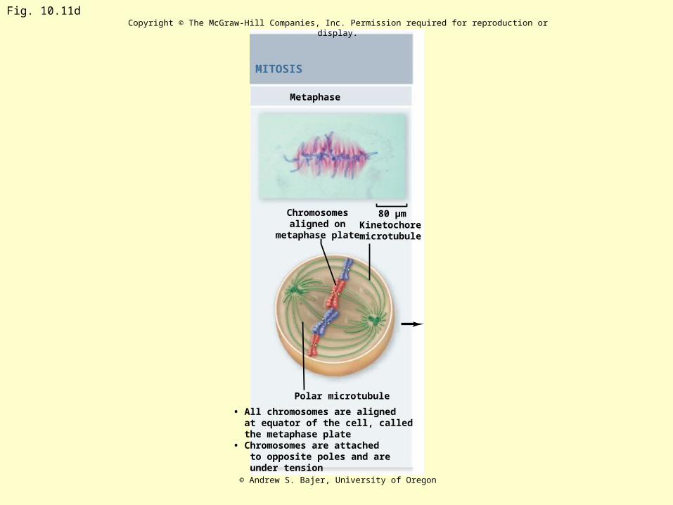
Metaphase
• All chromosomes are aligned at equator of the cell, called the metaphase plate• Chromosomes are attached to opposite poles and are under tension
Polar microtubule
80 µmChromosomesaligned on
metaphase plateKinetochoremicrotubule
Fig. 10.11dCopyright © The McGraw-Hill Companies, Inc. Permission required for reproduction or display.
© Andrew S. Bajer, University of Oregon
MITOSIS

Fig. 10.12Copyright © The McGraw-Hill Companies, Inc. Permission required for reproduction or display.
Centrioles
Sister chromatids
Aster
57 µm
Kinetochoremicrotubule
Metaphaseplate
Polarmicrotubule
© Andrew S. Bajer, University of Oregon

Anaphase
Chromosomes
• Proteins holding centromeres of sister chromatids are degraded, freeing individual chromosomes• Chromosomes are pulled to opposite poles (anaphase A)• Spindle poles move apart (anaphase B)
Kinetochoremicrotubule
80 µmPolar
microtubule
Fig. 10.11eCopyright © The McGraw-Hill Companies, Inc. Permission required for reproduction or display.
© Andrew S. Bajer, University of Oregon
MITOSIS

Fig. 10.13 Copyright © The McGraw-Hill Companies, Inc. Permission required for reproduction or display.
Metaphase
Pole Pole
Pole Pole 2 µm
Overlappingmicrotubules
Late Anaphase
Overlappingmicrotubules
© Dr. Jeremy Pickett-Heaps

Telophase
Polar microtubule
Nucleus reforming
• Chromosomes are clustered at opposite poles and decondense• Nuclear envelopes re-form around chromosomes• Golgi complex and ER re-form
80 µm
Kinetochoremicrotubule
Fig. 10.11f Copyright © The McGraw-Hill Companies, Inc. Permission required for reproduction or display.
© Andrew S. Bajer, University of Oregon
MITOSIS

Fig. 10.11g Copyright © The McGraw-Hill Companies, Inc. Permission required for reproduction or display.
Cleavage furrow
CYTOKINESIS
• In animal cells, cleavage furrow forms to divide the cells• In plant cells, cell plate forms to divide the cells
80 µm
© Andrew S. Bajer, University of Oregon

Fig. 10.11
Copyright © The McGraw-Hill Companies, Inc. Permission required for reproduction or display.
INTERPHASE G2
Prophase Prometaphase Metaphase Anaphase Telophase
Nucleus
Nucleolus
Aster
Chromosomes
Polar microtubule
Nucleus reforming
Cleavage furrow
80 µm80 µm80 µm
CYTOKINESISMITOSIS
Centrioles(replicated;animalcells only)
Nuclearmembrane
• DN A has been replicated• Centrioles replicate (animal cells)• Cell prepares for division
• Chromosomes condense and become visible• Chromosomes appear as two sister chromatids held together at the centromere• Cytoskeleton is disassembled: spindle begins to form• Golgi and ER are dispersed• Nuclear envelope breaks down
• Chromosomes attach to microtubules at the kinetochores• Each chromosome is oriented such that the kinetochores of sister chromatids are attached to microtubules from opposite poles.• Chromosomes move to equator of the cell
• All chromosomes are aligned at equator of the cell, called the metaphase plate• Chromosomes are attached to opposite poles and are under tension
• Proteins holding centromeres of sister chromatids are degraded, freeing individual chromosomes• Chromosomes are pulled to opposite poles (anaphase A)• Spindle poles move apart (anaphase B)
• Chromosomes are clustered at opposite poles and decondense• Nuclear envelopes re-form around chromosomes• Golgi complex and ER re-form
• In animal cells, cleavage furrow forms to divide the cells• In plant cells, cell plate forms to divide the cells
Polar microtubule
Kinetochoremicrotubule
Chromatin(replicated)
80 µm 80 µm 80 µm80 µm
Mitotic spindlebeginning to form
Condensedchromosomes
Centromere andkinetochore
Mitoticspindle
Chromosomesaligned on
metaphase plateKinetochoremicrotubule
Polarmicrotubule
Kinetochoremicrotubule
© Andrew S. Bajer, University of Oregon

Fig. 10.14

Fig. 10.15Copyright © The McGraw-Hill Companies, Inc. Permission required for reproduction or display.
Cell wall
Nucleus
0.7 µm
Vesicles containingmembrane componentsfusing to form cell plate
© B.A. Palevits & E.H. Newcomb/BPS/Tom Stack & AssociatesPlants

Fig. 10.16-1
Copyright © The McGraw-Hill Companies, Inc. Permission required for reproduction or display.
SCIENTIFIC THINKING
Hypothesis: There are positive regulators of mitosis.

Fig. 10.16-2
Copyright © The McGraw-Hill Companies, Inc. Permission required for reproduction or display.
SCIENTIFIC THINKING
Hypothesis: There are positive regulators of mitosis.
Prediction: Frog oocytes are arrested in G2 of meiosis I. They canbe induced to mature (undergo meiosis) by progesteronetreatment. If maturing oocytes contain a positive regulator of celldivision, injection of cytoplasm should induce an immatureoocyte to undergo meiosis.

Fig. 10.16-4Copyright © The McGraw-Hill Companies, Inc. Permission required for reproduction or display.
Removecytoplasm
Arrested oocyte Oocyte in meiosis I
SCIENTIFIC THINKING
Hypothesis: There are positive regulators of mitosis.
Prediction: Frog oocytes are arrested in G2 of meiosis I. They canbe induced to mature (undergo meiosis) by progesteronetreatment. If maturing oocytes contain a positive regulator of celldivision, injection of cytoplasm should induce an immatureoocyte to undergo meiosis.
Test: Oocytes are induced with progesterone, then cytoplasmfrom these maturing cells is injected into immature oocytes.
Result: Injected oocytes progress G2 from into meiosis I.
Injectcytoplasm
Progesterone-treated oocyte

Fig. 10.16-5Copyright © The McGraw-Hill Companies, Inc. Permission required for reproduction or display.
Removecytoplasm
Arrested oocyte Oocyte in meiosis I
SCIENTIFIC THINKING
Hypothesis: There are positive regulators of mitosis.
Prediction: Frog oocytes are arrested in G2 of meiosis I. They canbe induced to mature (undergo meiosis) by progesteronetreatment. If maturing oocytes contain a positive regulator of celldivision, injection of cytoplasm should induce an immatureoocyte to undergo meiosis.
Test: Oocytes are induced with progesterone, then cytoplasmfrom these maturing cells is injected into immature oocytes.
Result: Injected oocytes progress G2 from into meiosis I.
Conclusion: The progesterone treatment causes production of apositive regulator of maturation: Maturation Promoting Factor (MPF).
Injectcytoplasm
Progesterone-treated oocyte

Fig. 10.16-7 Copyright © The McGraw-Hill Companies, Inc. Permission required for reproduction or display.
Removecytoplasm
Arrested oocyte Oocyte in meiosis I
M phase cell G1 phase cell Fused cells
SCIENTIFIC THINKING
Hypothesis: There are positive regulators of mitosis.
Prediction: Frog oocytes are arrested in G2 of meiosis I. They canbe induced to mature (undergo meiosis) by progesteronetreatment. If maturing oocytes contain a positive regulator of celldivision, injection of cytoplasm should induce an immatureoocyte to undergo meiosis.
Test: Oocytes are induced with progesterone, then cytoplasmfrom these maturing cells is injected into immature oocytes.
Result: Injected oocytes progress G2 from into meiosis I.
Conclusion: The progesterone treatment causes production of apositive regulator of maturation: Maturation Promoting Factor (MPF).
Prediction: If mitosis is driven by positive regulators, thencytoplasm from a mitotic cell should cause a G1 cell to entermitosis.
Test: M phase cells are fused with G1 phase cells, then thenucleus from the G1 phase cell is monitored microscopically.
Conclusion: Cytoplasm from M phase cells contains a positiveregulator that causes a cell to enter mitosis.
Injectcytoplasm
Progesterone-treated oocyte

Fig. 10.17
Copyright © The McGraw-Hill Companies, Inc. Permission required for reproduction or display.
G1 S G2 MG2 M G1 S G2 M
Co
nc
entr
ati
on
Low
HighMPF activityCyclin

Fig. 10.18
Copyright © The McGraw-Hill Companies, Inc. Permission required for reproduction or display.
G2
M
S G1
Spindle checkpointG2/M checkpoint
G1/S checkpoint(Start or restriction point)

Fig. 10.19
Copyright © The McGraw-Hill Companies, Inc. Permission required for reproduction or display.
Cyclin
P
Cyclin-dependent kinase(Cdk)
P

Fig. 10.20Copyright © The McGraw-Hill Companies, Inc. Permission required for reproduction or display.
• Growth factors
• Size of cell
G2
M
SG1
Spindle Checkpoint
APCCdc2/Mitotic Cyclin
• Chromosomes attached at metaphase plate
• Replication completed• DNA integrity
G2/M Checkpoint
G1/S Checkpoint
Cdk1/Cyclin B
• Nutritional state of cell
Yeast

Fig. 10.21Copyright © The McGraw-Hill Companies, Inc. Permission required for reproduction or display.
• Growth factors
• Size of cell
G2
M
SG1
Spindle Checkpoint
APCCdk1/Cyclin B
• Chromosomes attached at metaphase plate
• Replication completed• DNA integrity
G2/M Checkpoint
G1/S Checkpoint
Cdc2/G1Cyclin
• Nutritional state of cell
Animals

Fig. 10.22 Copyright © The McGraw-Hill Companies, Inc. Permission required for reproduction or display.
Nucleus
Chromosome
RAF
MEK
ERK
E2F
E2F
Rb
Rb
P
P P
P P
PP
P
P
P
Growth factor
Cyclins/proteinsfor S phase
Cyclins/proteinsfor S phase
RAF
MEK
ERK
Nucleus
Rb
Rb
E2F
Chromosome
P
PRASRAS
MAP kinase pathway Rb

Fig. 10.23-1
Copyright © The McGraw-Hill Companies, Inc. Permission required for reproduction or display.
1. DNA damage is caused by heat, radiation, or chemicals.
2. Cell division stops, and p53 triggers enzymes to repair damaged region.
3. p53 triggers the destruction of cells damaged beyond repair.
p53 allows cells withrepaired DNA to divide.
DNA repair enzyme
p53protein
No
rma
l p
53

Fig. 10.23
Copyright © The McGraw-Hill Companies, Inc. Permission required for reproduction or display.
1. DNA damage is caused by heat, radiation, or chemicals.
2. Cell division stops, and p53 triggers enzymes to repair damaged region.
3. p53 triggers the destruction of cells damaged beyond repair.
p53 allows cells withrepaired DNA to divide.
1. DNA damage is caused by heat, radiation, or chemicals.
2. The p53 protein fails to stop cell division and repair DNA. Cell divides without repair to damaged DNA.
3. Damaged cells continue to divide. If other damage accumulates, the cell can turn cancerous.
DNA repair enzyme
Cancer cell
p53protein
No
rma
l p
53
A
bn
orm
al
p5
3
Abnormalp53 protein

Fig. 10.24
Copyright © The McGraw-Hill Companies, Inc. Permission required for reproduction or display.
Proto-oncogenes
Growth factor receptor: more per cell in many breast cancers.
Ras protein: activated by mutations in 20–30% of all cancers.
Src kinase: activated by mutations in 2–5% of all cancers.
Tumor-suppressor Genes
Rb protein: mutated in 40% of all cancers.
p53 protein: mutated in 50% of all cancers.
Cell cyclecheckpoints
Rasprotein
Rbprotein p53
protein
Srckinase
Related Documents