Cell Discovery Research Products Catalog 2009–2010 Clonetics® Primary Cells and Media Poietics® Stem Cells and Media Amaxa® Nucleofection® and Transfection BioWhittaker® Media and Sera StellARray™ qPCR Arrays MycoAlert®, ToxiLight® and ViaLight® Cell-based Assays Endotoxin Detection Products Flexible BioProcess Containers

Welcome message from author
This document is posted to help you gain knowledge. Please leave a comment to let me know what you think about it! Share it to your friends and learn new things together.
Transcript

Cell Discovery Research Products Catalog 2009–2010
Clonetics® Primary Cells and MediaPoietics® Stem Cells and MediaAmaxa® Nucleofection® and TransfectionBioWhittaker® Media and SeraStellARray™ qPCR ArraysMycoAlert®, ToxiLight® and ViaLight® Cell-based Assays Endotoxin Detection ProductsFlexible BioProcess Containers

Dear Valued Lonza Customer,
We are pleased to introduce the 2009 - 2010 Lonza Cell Discovery Research Products Catalog. Inside, you will find all of your known and trusted Lonza research products and services, as well as many new offerings designed to advance and accelerate your research:
Clonetics® Primary Cells and Media – over 100 authenticated primary cell types and optimized media, ready to use today.
Poietics® Stem Cells and Media – authenticated adult stem cells available with differentiation media.
New! Amaxa® Nucleofector® Transfection Technology – unique technology for the efficient, non-viral transfection of DNA or siRNA.
MycoAlert® Mycoplasma Detection System – detect mycoplasma contamination in your cell cultures in less than 20 minutes.
BioWhittaker® Media and Sera – classical media, plus an extensive array of serum free media including protein-free, chemically defined and non-animal derived formulas.
Look for Lonza Molecular Biology Products in the 2009 - 2010 Molecular Biology Research Products Catalog and Sourcebook for Electrophoresis. The Molecular Biology catalog features your known and trusted products, such as SeaKem®, NuSieve® and MetaPhor® Agarose; The FlashGel® System; and PAGEr® Precast Protein Gels, as well as our new Sourcebook for Electrophoresis, featuring protocols and support tools for your most critical molecular biology techniques.
Purchase any of our products directly from Lonza by calling us at the number provided on the back of this catalog, or use our e-commerce capabilities at www.lonza.com/research.
Should you require technical information, please contact our Scientific Support Team at the phone number provided on the back of this catalog, or at [email protected].
Lonza is committed to providing the highest quality products and services, and to helping researchers worldwide advance and accelerate life science research. For all of your research product needs, turn to Lonza. We appreciate your continued support and look forward to serving you.
The Lonza Cell Discovery Team
1-800-638-8174 www.lonza.com/research

i
Table of Contents
Tabl
e of
Con
tent
s
Customer Service iiiTerms and Conditions vTrademarks viiE-commerce inside back cover
Chapter 1 – Clonetics® and Poietics® Primary Cells and Media
Clonetics® & Poietics® Quick Reference Guide 3
Clonetics® Primary Cells and Media
Introduction 7Clonetics® Conditionally Immortalized Cell Lines 8Clonetics® Cell Models 9
Hepatocytes and Media
Human Hepatocytes & Media 14Rat Hepatocytes & Media 17
Human Cells and Media
Airway Epithelial Cells and Media 18Bladder Cells and Media 21Endothelial Cells and Media 23Fibroblast Cells and Media 36Epidermal Keratinocyte Cells and Media 40Epidermal Melanocyte Cells and Media 45Mammary Epithelial Cells and Media 47Neural Cells and Media 49Prostate Cells and Media 51Renal Cells and Media 54Skeletal Cells and Media 58Skeletal Muscle Cells and Media 61Smooth Muscle Cells and Media 64
Animal Cells and Media
Introduction 69Endothelial Cells and Media 69Rat Cardiac Myocytes and Media 72Primary Neural Cells and Media 73Primary Neuron Growth Media 77Cell Culture Reagents 78Custom Cell Services 79
Poietics® Stem Cells and Media
Introduction 81Donor Programs 82Bone Marrow and Mononuclear Cells 83Hematopoietic Progenitor Cells and Media 86 Mesenchymal Stem Cells and Media 90Preadipocyte Cells and Media 91Osteoclast Precursor Cells and Media 92Immune System Cells and Media 93Neural Progenitor Cells Media 97Custom Cell Isolation Services 98
Cell Biology Technical Information
Clonetics® Frequently Asked Questions 99Clonetics® Technical Information 101Clonetics® Media 111Poietics® Frequently Asked Questions 121Poietics® Technical Information 122Poietics® Media 123
Chapter 2 – Amaxa® Nucleofection® and Transfection Introduction 127Amaxa® Nucleofector® Technology 128Nucleofector® II Device 13296-well Shuttle® Device 133Nucleofector® Extended Guarantee and Service Contracts 13496-well Shuttle® Automation Package 135Nucleofector® Kits for Primary Cells 138Nucleofector® Kits for Cell Lines 183Cell Line Nucleofector® Kits 185Cell Line Optimization Nucleofector® Kits 186 cGMP Cell Line Nucleofector® Kits 188Basic Parasite Nucleofector® Kits 188FAQs on Nucleofection® 189HiFect® Transfection Reagent 191Vectors 192Antibiotics 198siRNA Test Kit 200Peptide Transfection Control 201Transport™ Protein Delivery Reagent 202
Table of Contents continues on next page
1-800-638-8174 www.lonza.com/research

ii
Table of ContentsContinued
PrimeFect™ DNA & siRNA Transfection Reagent 203Cell Biology Technical Information 101
Chapter 3 – ArraysIntroduction 209GeneSieve™ Query 210Global Pattern Recognition™ Analysis Tool 211
StellARray™ qPCR Arrays 211
Chapter 4 – BioAssaysIntroduction 219BioAssay Kits 220Cell Analysis Reagents 242G Protein-Coupled Receptors 245BioAssay Services 246
BioAssay FAQs 247
Chapter 5 – Cell Culture
Classical MediaIntroduction 251BioWhittaker® Classical Media 253
Specialty MediaIntroduction 261General Purpose Serum-free Media 262 X-VIVO™ Chemically Defined, Serum-free Hematopoietic Cell Media 265CHO Media 266 NAO Freezing Medium 269Renal Media 269Specialty Media - IVF 272Specialty Media - Cytogenetics 273Hybridoma Media 274Insect-XPRESS™ Protein-free Insect Cell Medium 278ProNS0™ Chemically Defined, Protein-free Media 279
Cell Culture ReagentsIntroduction 281Balanced Salt Solutions 282Reagents 283Antibiotics and Antimycotics 285Buffers and Buffered Saline 286
Animal and Human SeraIntroduction 289Fetal Bovine Sera 294Equine Sera 294Bovine Sera 295Human Sera 295Fetal Bovine Sera – Available Outside the USA and Canada 296
Cell Culture Technical InformationAdaptation of Cell Cultures to Serum-free Medium 298Protocols for Weaning Cell Cultures 299Cryopreservation and Reconstitution 300Determination of Cell Numbers 301Powdered Medium Preparation 303Subculturing Procedures for Mammalian Cells 304Media FAQs 306Sera FAQs 307
Cell Culture Reagents FAQs 308
Formulations for BioWhittaker® Classical Media 309
Chapter 6 – Endotoxin DetectionIntroduction 325Gel Clot LAL Assays 326Endpoint Chromogenic LAL Assay 328Fluorescent Assay 329LAL Accessory Products 330
Chapter 7 – Flexible Bioprocess ContainersIntroduction 333Platinum UltraPAK™ Bioprocess Containers 336Bioprocessing Tank Liners 337ProEVA PAK™ Bioprocess Containers 338
Chapter 8 – Lonza Custom ManufacturingIntroduction 340Lonza Custom Service Contacts 341Transfection Services 342
Chapter 9 – Distributors and IndicesNumerical Index 343Alphabetical Index 358
Table of Contents
1-800-638-8174 www.lonza.com/research

iii
Cust
omer
Ser
vice
Ordering Information
To place an order, please use any of the following convenient options:
PhoneOur Customer Service Representatives are ready to assist you with ordering Monday through Friday, from 8 a.m. to 6 p.m. Eastern Standard Time (EST). Call from anywhere in the U.S. and Canada at:
Toll Free: (800) 638-8174 Phone: (301) 898-7025
Fax(301) 845-8338
MailLonza Walkersville, Inc.Customer Service Department8830 Biggs Ford RoadWalkersville, MD 21793 USA
Please include all of the following information on all orders:Purchase order number –
Shipping address and billing address –
Contact name and telephone number –
Name and department of end user –
Quote or reserve lot information –
Item number, size, quantity, and name of products –ordered
Online OrderingOrdering for all of our products is available, 24 hours/day, 7 days per week. Our online ordering system is secure, fast, and convenient. Registered users enjoy the ability to see their contracted prices, track orders, and manage their account. Go to www.lonza.com/research for more information.
Ordering Information – Outside the U.S.For current information on Lonza distributors, use our website, www.lonza.com/dist, or contact your local Lonza distributor to place your order. In the event that you do not have an authorized distributor, please contact:
Lonza Walkersville, Inc.Customer Service Department8830 Biggs Ford RoadWalkersville, MD 21793 USAPhone: (301) 898-7025 Fax: (301) 845-8338
Scientific Support
Providing world-class technical support for our products is a top priority. Our Scientific Support Representatives rely on years of laboratory experience to assist with product selection and help you maximize product performance.
In the U.S., call us at (800) 521-0390, Monday –through Friday, between 8 a.m. and 5:30 p.m. EST
Outside the U.S., call (207) 594-3400 for Molecular –Biology Products or (301) 898-7025 for all others
You may also reach us by e-mail at –[email protected]
Full product information, instructions, and MSDS are –also available at www.lonza.com/research
Customer ServiceU.S. and Canada
1-800-638-8174 www.lonza.com/research

iv
Customer Service
Customer ServiceInternational
Ordering Information
To place an order, please use any of the following convenient options:
PhoneOur Customer Service Representatives are ready to assist you with ordering Monday through Friday.
International OfficesAustralia 61 3 9550 0883Austria 0800 201 538Belgium 00 32 87 321 611Brazil 55 11 2069 88 00Denmark 45 43 56 74 00France 0800 91 19 81Germany 0800 182 52 87India 91 22 6697 2883 Italy 0039 0363 45710Japan 81 3 5566 0612Poland 48 22 833 87 45Singapore 65 64914214Spain 34 902 531 366Sweden 020 140 4410Switzerland 0800 83 86 20The Netherlands 0800 022 4525United Kingdom 44 118 979 5234
FaxInternational OfficesAusralia 61 3 9550 0890Austria 0800 201 536Belgium 00 32 87 321 634Brazil 55 11 2274 00 51Denmark 45 43 56 74 03France 0800 91 19 80Germany 0800 182 52 78India 91 22 6697 2888Italy 39 0363 344063Japan 81 3 5566 0619Poland 48 22 669 39 67Singapore 65 62616182Spain 34 902 531 363Sweden 45 43 56 74 03Switzerland 0800 83 86 19The Netherlands 0800 022 4942United Kingdom 44 118 936 9441
Please include all of the following information on all orders:Purchase order number –
Shipping address and billing address –
Contact name and telephone number –
Name and department of end user –
Quote or reserve lot information –
Item number, size, quantity, and name of products –ordered
Online OrderingOnline ordering for all of our products is available, 24 hours/day, 7 days per week. Our online ordering system is secure, fast, and convenient. Registered users enjoy the ability to see their contracted prices, track orders, and manage their account. Go to www.lonza.com/research for more information.
Lonza Distributors and Sales Offices
For current information on Lonza distributors and sales offices, use our website, www.lonza.com/dist, or contact your local Lonza distributor to place your order. In the event that you do not have an authorized distributor, please contact:
Lonza Verviers, S.p.r.l.Verviers, BelgiumPhone: 00 32 87 321 611Fax: 00 32 87 351 967E-mail: [email protected]
Scientific Support
Providing world-class technical support for our products is a top priority. Our Scientific Support Representatives rely on years of laboratory experience to assist with product selection and help you maximize product performance.You may reach Scientific Support by phone or e-mail:
Phone: 49(0)221-99199-400 –
E-mail: –[email protected] –
Full product information, instructions, and MSDS are –also available at www.lonza.com/research.
1-800-638-8174 www.lonza.com/research

v
Terms and Conditions
Term
s an
d Co
nditi
ons
1-800-638-8174 www.lonza.com/research
DefinitionsThese Terms and Conditions apply to Lonza Rockland, Inc., Lonza Walkersville, Inc. and Lonza Sales Ltd, as the case may be, herein referred to as “Seller” and to all products ordered from the Lonza Cell Discovery Research Products Catalog 2009-2010.
Pricing and TermsVisa®, American Express®, and MasterCard® credit cards are accepted. Prices and conditions are subject to change without notice. The price applicable to any order accepted by Seller shall be the price in effect on the date of shipment. Payment is due within 30 days following the invoice date. There is a minimum $50 order requirement for all orders. All claims by purchaser shall be made by written notice to Seller in accordance with these Terms and Conditions, and no offset or deduction from any invoice is permitted. Acceptance by Seller of bank draft, check, or other media of payment is subject to immediate collection of the full face amount thereof.
If at any time the financial responsibility of purchaser, or the credit risk involved, shall become unsatisfactory to Seller, Seller may require cash or satisfactory security prior to subsequent shipments or deliveries hereunder. The election by Seller to require such cash or security shall not affect the obligation of purchaser to take and pay for the contracted materials. Purchaser agrees to pay all costs and expenses, including reasonable attorneys' fees, incurred by Seller in the collection of any sum payable by purchaser to Seller.
If purchaser breaches any term of the Terms and Conditions or any other contractual obligation in favor of Seller, (a) Seller may choose to defer any or all further shipments or other performance of any other contractual obligation in favor of purchaser until purchaser cures its breach, and (b) Seller may, by delivery of written notice to purchaser describing the breach, immediately terminate any other contractual obligation to purchaser; provided that purchaser shall have ten (10) days after receipt of the written notice to reinstate any terminated contractual obligations by curing the breach. In the event of a termination, all outstanding payment obligations or other indebtedness of purchaser to Seller shall be due and payable no later than fifteen (15) days after delivery of notice of termination, subject to the right of reinstatement.
Notwithstanding any provision in these Terms and Conditions, Seller shall have no obligation to pay any rebate, issue any credit or make any other payment of any kind to purchaser unless purchaser is fully in compliance with its payment and other obligations under the purchase and any other contractual obligation in favor of Seller. In addition, in the event that purchaser fails to make any payment when due, Seller shall have the right to offset any and all outstanding payment obligations or other indebtedness of purchaser to Seller against any outstanding payment obligations or other indebtedness that Seller or any of its affiliates may owe purchaser.
TaxesIn addition to the purchase price, purchaser shall pay Seller any and all governmental sales taxes of every kind that Seller may be required to pay with respect to the products. Purchaser shall provide Seller, on request, with properly completed exemption certificates for any tax from which purchaser claims exemption.
ShippingAll orders are shipped via Seller’s recommended carriers unless an alternative agreement has been arranged with the purchaser. Applicable freight and handling charges will be added to purchaser’s invoice. Refrigerated shipments will be charged a nominal fee. Please consult a Customer Service Representative for confirmed delivery dates. If another carrier is specified, a carrier account number must be provided by purchaser.
Damaged or Missing ProductContact a Customer Service Representative for replacement of damaged or missing product. Damage or discrepancies must be reported within 5 business days after receipt of product.
ReturnsProducts may not be returned for credit without first obtaining authorization from a Scientific Support or Customer Service Representative. A restocking fee may be applied for products returned without authorization or for returns in which the purchaser is at fault. The restocking fee is $50 or 25% of the purchase price, whichever is greater.
Force MajeureFailure of Seller to make, or purchaser to take, any one or more deliveries when due, if caused by (a) fire, storm, flood, strike, lockout, accident, act of war or terrorism, riot, civil commotion, embargo or similar circumstances, (b) any regulation, law, or restriction of any governmental department, commission, board, bureau, agency, court, or other instrumentality of any supranational organization of sovereign states, country, state, province, territory, commonwealth, municipality, or other political subdivision thereof (a "Governmental Authority"), any seizure or requisition of product by any Governmental Authority, or any compliance with a demand or request for such product for purposes of national or supranational defense, (c) inability of Seller to obtain any required raw material, energy source, equipment, labor or transportation, at prices and on terms deemed (by Seller) to be practicable, from Seller's usual sources of supply, or (d) any other cause or contingency beyond the reasonable control of that party (whether or not of the same kind or nature as the causes or contingencies above enumerated), shall not subject the party failing to perform to any liability to the other during the period such inability to make or take delivery shall exist. Quantities so affected may, at the option of either party, be eliminated from the purchase without liability, but the Terms and Conditions shall remain otherwise unaffected.
In the event of Seller's inability, for any reason, to supply the quantities of product contemplated by the purchase, Seller may allocate its available supply among its purchasers, including departments and divisions of Seller, on such basis as Seller may deem fair and practical without liability to purchaser for any failure of performance that may result therefrom.
IndemnificationSeller will make available to purchaser a Material Safety Data Sheet (MSDS) for each product delivered to purchaser where required. The MSDS sets forth information concerning such product and describes precautions, if required, to be taken in the transportation, delivery, unloading, discharge, storage, handling and use of such product. Purchaser will familiarize itself with all information and precautions, including but not limited to such related to safety and health, contained in MSDSs or otherwise transmitted to purchaser by Seller at any time. Seller will instruct its employees, agents, contractors, customers or any third party which may be exposed to the product about such information and precautions and make available copies thereof to such parties. Purchaser assumes full liability and responsibility for compliance with the above-referenced information and precautions, and with all laws, statutes, ordinances and regulations of any Governmental Authority applicable to the processing, transportation, delivery, unloading, discharge, storage, handling, sale and use of each product including, without limitation, the Foreign Corrupt Practices Act and United States export control laws; in particular, without limiting the generality of the foregoing, purchaser shall not resell or ship to persons on the Denied Parties List or located within embargoed countries (in both cases as defined under the referenced export control laws). Purchaser further agrees to protect, defend and hold harmless Seller from and against all claims, demands, causes of action, damages, losses, liabilities, costs, expenses (including reasonable attorneys' fees), penalties, and judgments (each, a "Claim") associated with the processing, transportation, delivery, unloading, discharge, storage, handling, sale or use of any product after delivery which is (i) inconsistent with any information provided to purchaser or (ii) in violation of any applicable law, statute, ordinance or regulation of any Governmental Authority. Seller assumes no liability for failure of discharge or unloading implements or materials used by purchaser whether or not supplied by Seller.
Since Seller has no control over purchaser's (or others') use, disposition, subsequent processing, admixing or reaction of Seller’s products with other products, chemicals or materials, purchaser assumes the entire liability and responsibility therefor and agrees to protect, defend and hold harmless Seller from and against all Claims associated therewith including, without limiting the generality of the foregoing, Claims associated with infringement of any third party's intellectual property rights, patents on processes practiced by purchaser or patents on products made by purchaser.
World – Lonza Rockland, Inc. / Lonza Walkersville, Inc. / Lonza Sales Ltd

vi
Terms and ConditionsTerms and Conditions
1-800-638-8174 www.lonza.com/research
WarrantyDUE TO THE VARIOUS FACTORS AFFECTING RESEARCH TEST RESULTS, SELLER WARRANTS ONLY AND NOT FOR ANY PARTICULAR PURPOSE OF THE PURCHASES, THAT ALL PRODUCTS SOLD WILL PERFORM ACCORDING TO ESTABLISHED PRODUCT SPECIFICATIONS. PRODUCTS ARE SOLD WITH THE UNDERSTANDING THAT THE PURCHASER WILL DETERMINE IF THE PRODUCT IS SUITABLE FOR ITS APPLICATION. SELLER WILL REPLACE ANY PRODUCT, FREE OF CHARGE, THAT DOES NOT MEET SELLER’S ESTABLISHED PRODUCT RELEASE SPECIFICATIONS.
ANY TECHNICAL ADVICE FURNISHED OR RECOMMENDATION MADE BY SELLER OR ANY REPRESENTATIVE THEREOF CONCERNING ANY USE OR APPLICATION OF ANY PRODUCT IS BELIEVED TO BE RELIABLE BUT SELLER MAKES NO WARRANTY, EITHER EXPRESS OR IMPLIED, AS TO ITS ACCURACY OR COMPLETENESS OR OF THE RESULTS TO BE OBTAINED. NO STATEMENT IS INTENDED OR SHOULD BE CONSTRUED AS A RECOMMENDATION TO INFRINGE ANY EXISTING PATENT. WITH REGARD TO ANY PROCESSING OF ANY PRODUCT, PURCHASER ASSUMES FULL RESPONSIBILITY FOR QUALITY CONTROL, TESTING AND DETERMINATION OF SUITABILITY OF PRODUCT FOR ITS INTENDED APPLICATION OR USE.
SELLER MAKES NO WARRANTY OF ANY KIND, EITHER EXPRESS OR IMPLIED, BY FACT OR LAW, OTHER THAN SELLER'S (I) OBLIGATION TO DELIVER PRODUCT COMPLYING WITH SELLER'S PUBLISHED SPECIFICATIONS (OR AS OTHERWISE REFERENCED IN THE TERMS AND CONDITIONS) AND (II) IMPLIED WARRANTIES OF TITLE, FREEDOM FROM ENCUMBRANCE, AND RIGHT TO TRANSFER SAME. SELLER MAKES NO WARRANTY OF FITNESS FOR A PARTICULAR PURPOSE, OR WARRANTY OF MERCHANTABILITY OTHER THAN AS STATED HEREIN.
Seller guarantees the performance of Clonetics® and Poietics® Cells up to two years from purchase only if appropriate Clonetics® or Poietics® Media and Reagents are used exclusively, and the recommended storage and use protocols are followed. Cell and media performance is not guaranteed if any modifications are made to such cell systems.
Limitation of LiabilitySELLER SHALL NOT BE LIABLE FOR ANY DAMAGES OR INJURY TO PERSONS OR PROPERTY ARISING FROM THE PURCHASE OR USE OF THE PRODUCT. IN ADDITION, SELLER IS NOT LIABLE FOR THE PRODUCT AFTER THE PRODUCT EXPIRATION DATE OR FOR A PRODUCT THAT HAS BEEN MISUSED OR HAS BECOME UNUSABLE DUE TO IMPROPER STORAGE OR HANDLING BY PURCHASER.
SELLER'S TOTAL LIABILITY AND PURCHASER'S EXCLUSIVE REMEDY FOR ANY CAUSE OF ACTION ASSOCIATED WITH THE PURCHASE, WHETHER BASED IN TORT, CONTRACT, STRICT LIABILITY OR ANY OTHER LEGAL THEORY IS EXPRESSLY LIMITED TO REPLACEMENT OF NONCONFORMING PRODUCT OR PAYMENT IN AN AMOUNT NOT TO EXCEED THE PURCHASE PRICE OF THE SPECIFIC PRODUCT FOR WHICH DAMAGES ARE CLAIMED, AT SELLER'S OPTION. IN NO EVENT SHALL SELLER BE LIABLE FOR ANY OTHER DAMAGES INCLUDING, WITHOUT LIMITATION, INCIDENTAL, SPECIAL, PUNITIVE OR CONSEQUENTIAL DAMAGES.
PURCHASER SHALL INSPECT THE PRODUCT SUPPLIED HEREUNDER IMMEDIATELY AFTER DELIVERY. PURCHASER'S FAILURE TO GIVE NOTICE TO SELLER OF ANY CLAIM WITHIN FIVE (5) DAYS AFTER THE DATE OF DELIVERY SHALL CONSTITUTE UNQUALIFIED ACCEPTANCE OF THE PRODUCT AND A WAIVER BY PURCHASER OF ALL CLAIMS WITH RESPECT THERETO.
ResaleProducts may be resold only by the Seller or its affiliate companies and authorized distributors. A list of authorized distributors and affiliate companies can be found in the back of this catalog or on our website, www.lonza.com.
Product LabelPlease read the product label carefully for safety information and warnings related to product hazards. Some products may present flammable, toxic, or other hazards. More information regarding certain products can be obtained by reading the MSDS, available on request from Customer Service, Scientific Support or on the web at www.lonza.com. The absence of a warning must not be construed as an indication that the product is safe. All possible hazards may not be known at this time. Some of our products may contain materials of animal or human origin. Products should only be handled and used by qualified personnel familiar with the potential hazards and trained in laboratory procedures. The purchaser assumes all risks of use and/or handling.
Product UseIn general, all products listed in this catalog are “For Research Use Only” or “For Laboratory Use Only” and are not intended for use in diagnostic procedures unless otherwise noted. Such products are not to be used for diagnostic or drug purposes, or for administration to humans.
Clonetics® and Poietics® Cell products are intended for research purposes only and the purchaser has no rights to transfer the products, components, or materials made using these products, or use these products for Commercial Purposes. Commercial Purposes include 1) use of the products or their components in manufacturing; 2) use of the products or their components to provide a service, information or data; 3) use of the products or their components for therapeutic or diagnostic purposes; 4) resale of the products or their components. Contact Marketing at Lonza Walkersville for further information.
For the Amaxa® brand of products the following additional terms and conditions apply:
Please note that Lonza´s Amaxa® Nucleofector® Technology is not intended to be used for diagnostic purposes, for testing or treatment in humans.
The Nucleofector® Technology, comprising Nucleofection® Process, Nucleofector® Device, Nucleofector® Solutions, Nucleofector® 96-well Shuttle® System, and 96-well Nucleocuvette® plates and modules is covered by patent and/or patent pending rights owned by Lonza.
Amaxa, HiFect, maxFP, maxGFP, Nucleocuvette, Nucleofection, Nucleofector, pmaxCloning, XpressNOW and 96-well Shuttle are either registered trademarks or trademarks of Lonza in Germany and/or the U.S. and/or other countries.
The CMV promoter is covered under the U.S. patents 5,168,062 and 5,385,839 and its use is permitted for research purposes only. Any other use of the CMV promoter requires a license from the University of Iowa Research Foundation, 214 Technology Innovation Center, Iowa City, IA.
Products contain a proprietary nucleic acid coding for a proprietary fluorescent protein(s) intended to be used for research purposes only. Any use of proprietary nucleic acid or fluorescent proteins coding by proprietary nucleic acid other than for research use is strictly prohibited. USE IN ANY OTHER APPLICATION REQUIRES LICENSE FROM EVROGEN. To obtain such a license, please contact Evrogen [email protected].
ATCC® and the ATCC Catalog Marks are trademarks of ATCC used under License.
Freedom EVO®, Freedom EVOware®, Tecan® and the Tecan logo are in major countries a registered trademark of Tecan Group Ltd., Männedorf, Switzerland.
BD FACSCalibur is a trademark of Becton, Dickinson and Company.
Other product and company names mentioned herein are the trademarks of their respective owners.
Governing Law for orders made with Lonza Walkersville, Inc. or Lonza Rockland, Inc.The purchase and Terms and Conditions shall take effect and be construed in accordance with the laws of the State of New York, USA.
Governing Law for orders made with Lonza Sales LtdThe Purchase and Terms and Conditions shall take effect and be construed in accordance with the laws of Switzerland, without any reference to its conflicts of law.
All rights reserved.Copyright Lonza 2008.
World – Lonza Rockland, Inc. / Lonza Walkersville, Inc. / Lonza Sales Ltd

vii
TrademarksThe following are trademarks of Lonza Group or its affiliates.
Trademarks from other companies:ABI Prism, GeneScan, and True Allele are registered trademarks of PE Corporation, Applied Biosystems Division
ALFexpress is a registered trademark of Amersham Pharmacia Biotech AB.
Amplitaq, GeneAm and Fugene are registered trademarks of Roche Molecular Systems, Inc.
ATCC are trademarks of ATCC used under License.
BAX is a registered trademark of Qualicon Inc., a subsidiary of E.I. du Pont de Nemours and Co.
BD FACSCalibur is a registered trademark of Becton, Dickinson and Company
Bio-Rad, CHEF-DR, Gene Pulser, Kaleidoscope, PROTEAN, MiniPROTEAN, Ready Gel, and Sub-Cell are registered trademarks of Bio-Rad Laboratories, Inc.
Cobe is a registered trademark of Cobe Laboratories, Inc.
Centipede, Millipede, and EasyCast and trademarks of Thermo Fisher Scientific, Inc.
C-Flex is a registered trademark of Seeman Plastics, Inc.
CrossLaps is a registered trademark of Osteometer Biotech A/S
EndoFree and siRNA are registered trademarks of Qiagen
EndoTrap is a registered trademark of Profos, AG
Eppendorf is a registered trademark of Eppendorf AG
Freedom EVO, Freedom EVOware, Tecan are registered trademarks Tecan Group, Ltd
Ficoll-Paque PLUS is a registered trademark of Amersham Bioscience/GE Helthcare
FisherBiotech is a registered trademark of Deutsche Bank, AG
Gibco, GlutaMax, Lipofectamine and Stealth are registered trademark of Invitrogen
GeneSeive, StellARray and Global Pattern Recognition are trademarks of Bar Harbor BioTechnology, Inc.
Hoefer is a registered trademark of Hoefer Scientific, Inc.
LI-COR is a registered trademark of LI-COR, Inc.
Matrigel is a registered trademark of Discovery Labware, Inc.
Neupogen is a registered trademark of Amgen, Inc.
Pluronic is a registered trademark of BASF Corporation
PER.C6 is a registered trademark of Crucell Holland, BV
Rainbow is a registered trademark of GE Healthcare UK
Retronectin is a registered trademark of TaKaRa Shuzo Company, LTD.
RNALater is a registered trademark of Ambion, Inc.
SeeBlue is a registered trademark of Novel Experimental Technology Corporation
SpectrMax is a registered trademark of Molecular Devices
Steady-Glo is a registered trademark of Promega
STYROFOAM is a registered trademark of Dow Chemical Corporation
SYPRO, SYBR, Alexa Fluor, Cell Tracker, Lysotracker, Mitotracker, Live/Dead, SYTO, SYTOX, and Vybrant are registered trademarks of Molecular Probes Corporation, Inc.
Tecan, Freedom EVO and Freedom EVOware are registered trademarks of Tecan Group Ltd.
Transwell is a registered trademark of Corning, Inc.
Tween is a trademark of ICI Americas, Inc.
Versene is a registered trademark of Dow Chemical Company
Windows, Windows XP, Windows 2000 is a registered trademark of Microsoft Corporation
96-well Shuttle®ABM™AdipoRed™AggreSolve™AGM™Amniochrome®Amaxa®ApoGlow®BEBM®BEGM®BioPro™ 1BioWhittaker®BulletKit®CalciFluor™CDM™CGM™Clonetics®EBM®EGM® ExPro™
FastLane®FBM®FGM®gene transfer begins here™Genetic Technology Grade™Global Patern Recognition™GS System™GS Gene Expression System™HBM™HCM™HiFect®HL-1™HMM™HPGM™Insect-XPRESS™KBM®KGM®Latitude®LGM-3®Lymphochrome™
maxFP®maxGFP®MBM®MDE®MEBM®MEGM®MGM®MsBM™MsGM™MSCGM™MycoAlert®MycoZap™Nephros™ NHEPS®NPBM™NPMM™Nucleocuvette®Nucleofection®Nucleofector®OBM™
OGM™OPGM™OsteoAssay™OsteoImage™OsteoLyse™PC-1™PDELight®PGM™PKLight®pmaxCloning™PNGM™ Poietics®PowerCHO® PPiLight®PrEBM™PrEGM™PrimeFect™Pro293™ProCHO™ProDoma™
ProEVA PAK™ProFreeze™ProHT™ProMEDIA Select® ProNSO™ProPer™ ProVero™1PyroGene®PYROGENT®PYROSPERSE® QCL-1000®ReagentPack™REBM™REGM™Reliant®SABM™SAGM™SCBM™SCGM™SingleQuots®
Trad
emar
ks
Lonza Group or its affiliates are owner or licensee** of the following patents. Products described herein may be covered by one or more of these United States patents, by pending patent applications, or by corresponding patents or applications in other countries.
4,695,548** 5,219,923 6,365,341
4,749,620* 5,641,626** 6,414,136
4,861,448** 5,836,445 6,464,850
5,055,517 6,004,767 6,558,521
5,143,646 6,117,293 6,599,711
5,212,299** 6,328,870 7,166,692
D510,770 D511,386 D524,449
6,521,456 7,320,859 7,332,332
5,168,062 5,385,839
United Kingdom patent:
2,357,336 B
European patents:
1297119 1383901 1390518
1476537 1525900 1522587
1702677 1741778 PCT/EP01/07348
PCT/DE02/01489 PCT/DE02/01483
SkBM®SkGM®StellARay™SmBM®SmGM®ToxiLight®Transport™ UltraCHO™UltraCULTURE™UltraDOMA-PF™UltraDOMA™UltraMEM™UltraMDCK™UltraPAK™Versalinx®ViaLight®WinKQCL® X-VIVO™
1-800-638-8174 www.lonza.com/research

viii
Notes
1-800-638-8174 www.lonza.com/research

Clonetics® and Poietics® Primary Cells and MediaChapter 1

2
Primary
Cells and Media
Clonetics® and Poietics® Quick Reference Guide 3
Clonetics® Primary Cells & Media
Introduction 7Clonetics® Conditionally Immortalized Cell Lines 8Clonetics® Cell Models 9 Hepatocytes and Media
Hepatocytes and Media Human 14 Rat 17 Human Cells & Media
Airway Epithelial Cells and Media 18Bladder Cells and Media 21Endothelial Cells and Media 23Fibroblast Cells and Media 36Epidermal Keratinocyte Cells and Media 40Epidermal Melanocyte Cells and Media 45Mammary Epithelial Cells and Media 47Neural Cells and Media 49Prostate Cells and Media 51Renal Cells and Media 54Skeletal Cells and Media 58Skeletal Muscle Cells and Media 61Smooth Muscle Cells and Media 64
Animal Cells & Media
Introduction 69Endothelial Cells and Media 69Rat Cardiac Myocytes and Media 72Primary Neural Cells and Media 73Primary Neuron Growth Media 77Cell Culture Reagents 78Custom Cell Services 79
THESE PRODUCTS ARE FOR RESEARCH USE ONLY.Not approved for human or veterinary use, for application to humans or animals, or for use in clinical or in vivo procedures.
WARNING: CLONETICS® AND POIETICS® PRODUCTS CONTAIN HUMAN SOURCE MATERIAL, TREAT AS POTENTIALLY INFECTIOUS.Each donor is tested and found non-reactive by an FDA approved method for the presence of HIV-1, Hepatitis B Virus, and Hepatitis C Virus. Where donor testing is not possible, cell products are tested for the presence of viral nucleic acid from HIV-1, Hepatitis B Virus, and Hepatitis C Virus. Testing cannot offer complete assurance that HIV-1, Hepatitis B Virus, and Hepatitis C Virus are absent. All human sourced products should be handled at the Biological Safety Level 2 to minimize exposure of potentially infectious products, as recommended in the CDC-NIH Manual, “Biosafety in Microbiological and Biomedical Laboratories”, 1999. If you require further information, please contact your site Safety Officer or Scientific Support.
PRODUCT WARRANTY – CULTURES HAVE A FINITE LIFESPAN IN VITRO.Lonza guarantees the performance of its cells up to two years from purchase only if appropriate Clonetics® or Poietics® Media and Reagents are used exclusively, and the recommended storage and use protocols are followed. Cell and media performance is not guaranteed if any modifications are made to the complete cell system.
New Products
Adult Normal Human Epidermal Melanocytes 41Clonetics® Rat Cardiac Myocyte Growth Medium 72Ready Heps™ Fresh Human Hepatocytes 16
Cell Biology Technical Information
Clonetics® Frequently Asked Questions 99Clonetics® Technical Information 101Clonetics® Media 111Poietics® Frequently Asked Questions 121Poietics® Technical Information 122Poietics® Media 123
Clonetics® and Poietics® Primary Cells and Media
Poietics® Stem Cells & Media
Introduction 81Donor Programs 82Bone Marrow and Mononuclear Cells 83Hematopoietic Progenitor Cells and Media 86 Mesenchymal Stem Cells and Media 90Preadipocyte Cells and Media 91Osteoclast Precursor Cells and Media 92Immune System Cells 93Neural Progenitor Cells Media 97Custom Cell Isolation Services 98
1-800-638-8174 www.lonza.com/research

3
Tissue/ Cell Type Cell Cat. No. Recommended Medium Media Cat. No. Page
Adventitial Fibroblasts
Aorta CC-7014 SCGM™ CC-3205 37
Astrocytes
Human Brain CC-2565 AGM™ CC-3186 50C57 Mouse Brain - Mixed M-AsM-330 AGM™ CC-3186 76
CD1 Mouse Brain - Mixed M-AsM-430 AGM™ CC-3186 76Rat Cx-Hi-Cp Brain - Mixed R-AsM-530 AGM™ CC-3186 75Rat Brain - Cortex R-CxAs-520 AGM™ CC-3186 75Rat Brain - Hippocampus R-HiAs-521 AGM™ CC-3186 75
Rat Brain - Striatum R-CpAs-522 AGM™ CC-3186 75
Chondrocytes
Cartilage CC-2550 CGM™ CC-3216 59
Dendritic Cells
Blood CC-2701 LGM®-3 CC-3211 94
Dermal Microvascular Endothelial Cells
Blood - Adult CC-2811 EGM®-2MV CC-3202 29Lymphatic - Adult CC-2810 EGM®-2MV CC-3202 28Blood - Neonatal CC-2813 EGM®-2MV CC-3202 30Lymphatic - Neonatal CC-2812 EGM®-2MV CC-3202 30Dermal - Adult CC-2543 EGM®-2MV CC-3202 28Dermal - Neonatal CC-2505 EGM®-2MV CC-3202 29Dermal - Neonatal, pooled CC-2516 EGM®-2MV CC-3202 31
Endothelial Cells
Aorta CC-2535 EGM®-2 CC-3162 25Bovine Aorta BW-6001 EGM®-MV CC-3125 70Bovine Coronary Artery BW-6005 EGM®-MV CC-3125 71Bovine Pulmonary Artery BW-6004 EGM®-MV CC-3125 71Coronary Artery CC-2585 EGM®-2MV CC-3202 25Iliac Artery CC-2545 EGM®-2MV CC-3202 26Pulmonary Artery CC-2530 EGM®-2 CC-3162 32Umbilical Artery CC-2520 EGM®-MV CC-3125 33Umbilical Vein C2517A EGM®-2 CC-3162 33
Endothelial Cells, Pooled
Bovine Aorta BW-6002 EGM®-MV CC-3125 70Umbilical Vein C2519A EGM®-2 CC-3162 34
Epithelial Cells
Airway CC-2547 SAGM™ CC-3118 19Kidney CC-2556 REGM™ CC-3190 55Kidney - Cortex CC-2554 REGM™ CC-3190 55Mammary CC-2551 MEGM® CC-3150 48Prostate CC-2555 PrEGM™ CC-3166 52
Clonetics® and Poietics® Primary Cells and MediaCell Types - Quick Reference
Prim
ary
Cells
and
Med
ia
1-800-638-8174 www.lonza.com/research

4
Tissue/ Cell Type Cell Cat. No. Recommended Medium Media Cat. No. Page
Epithelial Cells (with Retinoic Acid)
Bronchial / Tracheal CC-2540 BEGM® CC-3170 19
Epithelial Cells (without Retinoic Acid)
Bronchial / Tracheal CC-2541 BEGM® CC-3170 20
Fibroblasts
Dermal - Adult CC-2511 FGM®-2 CC-3132 38Dermal - Neonatal CC-2509 FGM®-2 CC-3132 38Lung CC-2512 FGM®-2 CC-3132 39Periodontal Ligament CC-7049 SCGM™ CC-3205 37
Heart
Rat Cardiac Myocytes R-CM-561 RCGM™ CC-4515 72
Hepatocytes
Human Liver CC-2591 HCM™ CC-3198 15Rat Liver AC-6009 HCM™ CC-3198 17Ready Heps™ See Catalog for additional information 16
Keratinocytes
Epidermal - Adult C2501A KGM®-2 CC-3107 41
Epidermal - Neonatal C2503A KGM®-2 CC-3107 42Epidermal - Neonatal, pooled C2507A KGM®-2 CC-3107 43
Lung Microvascular Endothelial Cells
Blood CC-2815 EGM®-2MV CC-3202 27Lymphatic CC-2814 EGM®-2MV CC-3202 27
Melanocytes
Neonatal CC-2504 MGM®-4 CC-3249 46Adult CC-2586 MGM®-4 CC-3249 46
Mesangial Cells
Kidney CC-2559 MsGM™ CC-3146 56
Mesenchymal Stem Cells
Bone Marrow PT-2501 MSCGM™ PT-3001 90
Microvascular Endothelial Cells
Bladder CC-7016 EGM®-2MV CC-3202 22Bovine Brain AC-2509 EMVB AC-3103 13Heart CC-7030 EGM®-2MV CC-3202 31Lung CC-2527 EGM®-2MV CC-3202 26Uterus CC-2564 EGM®-2MV CC-3202 34
Myoblasts
Skeletal Muscle CC-2580 SkGM®-2 CC-3245 62
Clonetics® and Poietics® Primary Cells and MediaContinued
Primary
Cells and Media
1-800-638-8174 www.lonza.com/research

5
Tissue/ Cell Type Cell Cat. No. Recommended Medium Media Cat. No. Page
Neural Progenitor Cells
Brain PT-2599 NPMM™ CC-3209 97
Neurons
C57 Mouse Brain - Cortex M-Cx-300 PNGM™ CC-4461 76CD1 Mouse Brain - Cortex M-Cx-400 PNGM™ CC-4461 76Rat Brain - Cortex R-Cx-500 PNGM™ CC-4461 74C57 Mouse Brain - Striatum M-Cp-302 PNGM™ CC-4461 76CD1 Mouse Brain - Striatum M-Cp-402 PNGM™ CC-4461 76Rat Brain - Striatum R-Cp-502 PNGM™ CC-4461 74
Rat Brain - Hippocampus R-Hi-501 PNGM™ CC-4461 74Rat Spinal Cord - Dorsal Root Ganglion R-Drg-505 PNGM™ CC-4461 74Rat Brain Cerebellum - Granule Cells R-Cb-503 PNGM™-A CC-4512 74
Osteoblasts
Bone CC-2538 OGM™ CC-3207 59
Osteoclast Precursors
Bone 2T-110 OCP PT-8001 92
Proximal Tubule Epithelial Cells
Kidney CC-2553 REGM™ CC-3190 56
Skeletal Muscle Cells
Skeletal Muscle CC-2561 SkGM® CC-3160 62
Smooth Muscle Cells
Aorta CC-2571 SmGM®-2 CC-3182 65Bladder CC-2533 SmGM®-2 CC-3182 22 & 65Bronchial CC-2576 SmGM®-2 CC-3182 66Coronary Artery CC-2583 SmGM®-2 CC-3182 66Prostate CC-2587 SmGM®-2 CC-3182 53 & 67Pulmonary Artery CC-2581 SmGM®-2 CC-3182 67Umbilical Artery CC-2579 SmGM®-2 CC-3182 68Uterus CC-2562 SmGM®-2 CC-3182 68
Stromal Cells
Prostate CC-2508 SCGM™ CC-3205 52
Adipose-Derived Cells
Subcutaneous Preadipocytes PT-5001 PGM™-2 PT-8002 91Visceral Preadipocytes PT-5005 PGM™-2 PT-8002 91
Clonetics® and Poietics® Primary Cells and MediaContinued
Prim
ary
Cells
and
Med
ia
1-800-638-8174 www.lonza.com/research

6
Clonetics® and Poietics® Primary Cells and MediaContinued
Hematopoietic Cells
Cell Type Source Peripheral Blood Bone Marrow Cord Blood Fetal Liver Page
Fresh Bone Marrow 1M-125 83Mesenchymal Stem Cells PT-2501 90CD4+ T Cells 2C-200 2W-200 95CD14+ Monocytes 2W-400 93CD19+ B Cells 2C-300 95CD34+ Cells 2M-101 2C-101 2L-101 86 & 87CD133+ Cells 2M-102 2C-102 2L-102 87Erythroid Progenitors 2C-250 88Leukopak Peripheral Blood Cells 1A-125 83Mononuclear Cells 2M-125 2C-150 CC-2702 84Stromal Cells 2M-302 89Products are available in various sizes. Please refer to the catalog for size information.
Hematopoietic Cell Media
Cat. No. Description Size PagePT-3926 HPGM™ Hematopoietic Progenitor Growth Medium 500 ml 87CC-3211 LGM®-3 Lymphocyte Growth Medium-3 500 ml 9404-380Q X-VIVO™ 10 Chemically Defined, Serum-free Hematopoietic 1 L 265 Cell Medium, with L-glutamine, gentamicin, and phenol red 04-743Q X-VIVO™ 10 Chemically Defined, Serum-free Hematopoietic 1 L 265 Cell Medium, with L-glutamine, without gentamicin or phenol red 04-744Q X-VIVO™ 15 Chemically Defined, Serum-free Hematopoietic 1 L 265 Cell Medium, with L-glutamine, without gentamicin or phenol red BE04-418F X-VIVO™ 15 Chemically Defined, Serum-free Hematopoietic 500 ml 265 Cell Medium, with gentamicin, L-glutamine, and phenol red 04-418Q X-VIVO™ 15 Chemically Defined, Serum-free Hematopoietic 1 L 265 Cell Medium, with gentamicin, L-glutamine, and phenol red 04-448Q X-VIVO™ 20 Chemically Defined, Serum-free Hematopoietic 1 L 265 Cell Medium, with gentamicin, L-glutamine, and phenol red
Primary
Cells and Media
1-800-638-8174 www.lonza.com/research

7
Prim
ary
Cells
and
Med
ia
Clonetics® Primary Cells and MediaIn vivo relevance. In vitro results.
Clonetics® Primary Cells and Media include cells that are primarily derived from normal human tissue and are negative for HIV-1, Hepatitis B and C, and by standard cell culture tests for mycoplasma and sterility. Immuno and special staining protocols, as well as characteristic morphology are used to characterize the cells and assure they are the designated type. A Certificate of Analysis is available for each cell type. The cells will perform as stated when used with the optimized system comprised of Clonetics® Cells, Media and Reagents. Cells are available cryopreserved, proliferating (in either flasks or plates), or as pellets in RNALater®, a reagent that inactivates RNases and stabilizes RNA within unfrozen tissues and cells.
Clonetics® Media Products have been specially designed to support the growth of these cells. The media systems are comprised of serum-free basal media and SingleQuots® Kits of growth factors and supplements. For detailed information about these media systems, please see pages 112 – 120.
General Ordering and Shipping InformationCryopreserved cells and media products are normally shipped Monday–Thursday for next day delivery. Saturday and Monday deliveries are available upon special request. Proliferating cell orders must be placed no later than Tuesday at 5 p.m. EST for shipment the following Monday (for delivery on Tuesday). Orders placed after Tuesday will be held until the next production cycle. When you place your order for proliferating cells, please order the appropriate media. Other cell types maybe available upon request.
Plate orders are an additional passage and take an extra week.
Cell pellet orders require 7 – 10 production days. Please plan accordingly.
Matrigel® or collagen is required for culturing hepatocytes.
1-800-638-8174 www.lonza.com/research

8
Conditionally Im
mortalized Cells
During recent years, increased pressure has been placed on drug development teams to deliver products that meet the prescribed claims, lack side effects, and perform despite the patients' genetic background. To add to the challenge, drug discovery resourcing has not been growing at the same rate as the external costs required to validate new drugs for the market. Now, more than ever, pharmaceutical researchers need tools that facilitate HTS cell screening in complex biological systems earlier in the process in order to eliminate poor drug candidates early and allow focus on more promising candidates.
To discover a new drug, many compounds may be screened or evaluated for efficacy and toxicity. Several accepted models exist (using either animals or cells), but all have inherent limits that delay expedient evaluation using high throughput screening techniques. They also require species extrapolation to evaluate the data.
Live animal models yield a rich supply of data, but are not cost effective for high throughput screening. Non-immortalized, primary cells may best represent normal physiology, but are typically not available in the large, uniform quantities needed for HTS applications. Though conventionally immortalized cell lines can divide indefinitely into large homogeneous cell populations needed for HTS applications, they do not behave like normal cells. Conditional immortalization allows for, at a permissive temperature, production of high volumes of uniform cell populations. Changing the culture temperature forces the cells to stop dividing, allowing for differentiation and maturation so that the cells can express normal function and phenotype.
Now pharmaceutical researchers have access to a continuous supply of differentiated cells of the same genotype that can be used as cell models in functional cell-based assays and in long-term gene expression studies.
Characterization for Cell-based Drug Discovery
Lonza has qualified the conditionally immortalized cell lines for use in high throughput, cell-based functional assays. This offers an excellent method to identify novel compounds that target insulin resistance, for example. Compounds can be rapidly identified in such screens to produce a lead series that can be integrated into drug discovery pipelines.
As part of the characterization of our most promising conditionally immortalized human muscle cell lines, we screened a diverse chemical library, assaying for effects on glycogen synthesis. This screen identified a novel series of GSK3 inhibitors.
Future Products
We are currently developing a set of conditionally immortalized cell lines from the major insulin target tissues i.e. muscle, visceral and subcutaneous adipocytes. These human cell lines, optimized for diabetes research and high throughput cell-based assays, originate from only ethically obtained tissues.
For more information concerning licensing opportunities, please call 301-898-7025, ext. 7350.
Clonetics® Conditionally Immortalized Cell Lines
Conditional Immortalization
Available Cell Types
Human adipocytes —
Human skeletal muscle cells —
Human fibroblasts —
Human aortic smooth muscle cells —
Human endothelial cells (blood, lymph) —
Human keratinocyte cells —
Human hepatocyte cells —
Cells isolated fromhuman tissues
Transfect withLarge T-antigenand telomerase
Cells are createdexpressing temperaturesensitive Large T-antigenand telomerase
Cell populationexpanded due toimmortalization
Cells revert to anormal phenotype
Immortalized Phenotype(gene expression altered)
37°C – Normal Phenotype
33°C – Large T-antigen Active
37°C – Normal Phenotype
ImmortalizationSwitched ON
ImmortalizationSwitched OFF
1-800-638-8174 www.lonza.com/research

9
Cell
Mod
els
Corneal Epithelial Model
The corneal epithelium provides a transparent barrier to the passage of foreign materials into the eye. Damage to this epithelial layer from environmental challenges (e.g., foreign substances, infection, etc.) may act to compromise tissue function with respect to both barrier properties and clarity. In the most severe cases, the resulting injury may include loss of sight. The oldest and most widely used method for the assessment of ocular irritants is the Draize rabbit eye irritation test, which has been employed for many years to evaluate and classify a wide range of chemicals and cosmetic products. The Draize test involves the placement of substances directly into the conjunctival sac of rabbit eyes in vivo with subsequent evaluation for irritation and damage. Ethical concerns calling for the use of alternative methods to animal testing and uncertainty regarding inter-species comparisons between rabbits and humans have led to an increased focus on the use of human tissue models for screening purposes. In order to create a physiologically relevant in vitro human corneal epithelial culture model, it is desirable that cells used be of human origin (species-specific) and isolated from corneal epithelial tissue (tissue-specific).
Due to the difficulty in obtaining consistent high-quality primary human corneal epithelial cells in sufficient numbers, the use of cell lines presents an attractive alternative. However, many continuous cell lines lose phenotypic and/or morphological characteristics of the native tissue, and therefore do not provide a relevant system of study. We have overcome these difficulties by the introduction of human papilloma virus 16 E6 and E7 (HPV-16 E6/E7) genes into the cells. Through the use of HPV-16 E6/E7 in primary human corneal epithelial cells, we have developed functional human corneal epithelial cells with extended lifespan.
The Clonetics® Human Corneal Epithelial Culture —Model provides a consistent and robust platform for recapitulation of human corneal epithelial tissue in an in vitro environment
The tissue model achieves a histological profile —similar to that observed with in vivo corneal epithelium
The formation of epithelial junctions, essential in —establishing proper barrier function, is demonstrated through High TEER values which reflect relevant barrier properties
The human corneal epithelial culture model is predictive — of in vivo ocular irritancy via multiple endpoints and exhibits good correlation with Draize scores
Clonetics® Cell Models
Product Applications
Ocular irritancy evaluation for: –Pharmaceutical products –Pharmaceutical raw materials –Personal care products –Cosmetics –Standard household use chemicals –
Basic research – corneal epithelium –Ocular diseases –Barrier function studies –
Routine QC Testing
Trans Epithelial Electrical Resistance – TEER is a –measure of the barrier function of epithelial cellular junctions. The tighter the epithelial barrier, the higher the TEER value, expressed as “ohms × cm2” or “Ω × cm2”.
ET – 50 (0.3% Triton X-100 as test substance) (For Information Only, available upon request)
References
Draize, JH., Appraisel of the Safety of Chemicals in Foods, Drugs and 1. Cosmetics 1959. The Association of Food and Drug officials of the United States. Austin, Texas.
Wilhelmus, KR., The Draize Eye Test. Survey of Ophthalmology 2001 2. May-June 45 (6):495-514. Halbert, CL., Demers, GW., and Galloway, DA. The E7 Gene of Human 3. Papillomavirus Type 16 is sufficient for immortalization of human epithelial cells. 1991. J. Virol 65 (1): 473-478. Griffith, M., Watsky, MA., and Liu, C., Trinkaus-Randall V. Epithelial Cell 4. Culture. Cornea in Methods of Tissue Engineering. ed. Atala, A., Lanza, and RP. 2002 Academic Press. 131-140.
Toropainen, E., Ranta, VP., Talvitie, A., Suhonen, P., and Urtti, A. Culture 5. model of human corneal epithelium for prediction of ocular drug absorption. Invest Ophthalmol Vis Sci. 2001 Nov 42 (12): 2942-8. Eye Irritation: Reference Chemicals Data Bank. ECETOC Technical Report 6. No. 48, 2nd Edition. June 1998.
ET — 50 values established using 0.3% Triton X-100
Measurable IL-1 — increases in the culture medium relative to time of irritant exposure
Cultures achieve stratification of 4 – 9 cell layers with —an average of 6 – 7 layers
Supplied ready-to-use in Corning Transwell® Inserts —
Ordering Information
1-800-638-8174 www.lonza.com/research
Cat. No. Size Price
Corneal Epithelial Model
CMS-2015 24 tissue models in a 24-well plate, one empty 24-well plate
$866
Corneal Epithelial Model BulletKit®Includes Basal Medium and SingleQuots® Kit
CC-4444 — $123

10
Renal epithelial cells stained for ZO-1 cellular junction protein
Renal Epithelial Cell Model
Clonetics® Renal Epithelial Cell Model recreates human renal tubular function in an in vitro environment. The model consists of primary epithelial cells isolated from human renal cortices and seeded on extracellular-matrix-coated polyester membrane inserts, which allow for bicompartmental (apical and basal) exposure to culture medium. Isolation and culture parameters (e.g., culture medium, matrix coating protocols, cell seeding density) have been optimized for the development of a functional renal tubular epithelial monolayer as assessed through specific markers.
Early passage normal human renal epithelial cells; —non-transformed, non-immortalized, primary-derived
Consistent and stable expression of the apical brush —border enzyme gamma-glutamyl transpeptidase ( -GTP), for at least 14 days in culture
Apical chamber expression of — -GTP 10-fold higher than expression in the basal chamber
Consistent ammoniagenesis for at least 14 days in —culture
Proven organic ion transport; cation uptake from the —basal chamber is several times higher than the apical chamber
Test lots stain positive for the following cell-to-cell —junction proteins: ZO-1, e-cadherin, occludin, -, -,
-catenins and desmoplakin
Offered as plated cells on 12 polyester Transwell® —Membranes in a 24-well plate to provide a bi-compartmental cell environment
Renal epithelial cells tested for — -GTP expression and apical localization
REGM™ Renal Growth Medium BulletKit®, quality tested —to support cell culture
Clonetics® Cell ModelsContinued
Cell Models
Research Applications
Active and passive transport studies –
Nephrotoxicity evaluation –
Renal tubular biology –
Tight junction research –
Early stage ADME-Tox screening –
Cell Testing and Specifications
Transwell® Membrane inserts are inspected for cell –confluence before shipping
All renal cell culture models exhibit greater than 5-fold –apical: basal ratio of -GTP
2 polyester inserts in 24-well plates seeded with renal –epithelial cells, sealed with agarose and media
Other Non-Routine Testing
Ammoniagenesis –
Active transport of organic cations –
Barrier function –
Cell-to-cell junction proteins: ZO-1, e-cadherin, –occludin, -, -, -catenins and desmoplakin
1-800-638-8174 www.lonza.com/research

11
Clonetics® Cell ModelsContinued
Cells Ordering Information
Renal Epithelial Cell ModelRecommended Medium: REGM™ BulletKit®
Cell
Mod
els
Lot 1 Lot 2 Lot 3
GTP
mol
ar ra
tio (a
pica
l:bas
al) 1,000
100
10
1
Ratios of apical-to-basal expression of -GTP were averaged for three lots of renal cortical epithelial cells in two separate experiments (triplicate wells for each lot).
γ-GTP is expressed primarily on apical surface
8
6
4
2
0 Lot 1 Lot 2 Lot 3
Apical uptakeBasal uptake
Rela
tive
Fluo
resc
ence
Uni
ts
Uptake of rhodamine 123 was assessed from both apical and basal culture medium. Fluorescence of extracted rhodamine 123 was normalized to apical concentration in each experiment. Results are from three lots of renal cortical epithelial cells (triplicate wells for each lot) in two separate experiments.
Basolateral uptake of organic cations
Media Ordering Information
REGM™ Renal Epithelial Cell Growth Medium
1-800-638-8174 www.lonza.com/research
Cat. No. Size Price
Plated cells – polyester Transwell® Membranes
CMS-2000 12 transwell membranes $918
Cat. No. Size Price
REGM™ BulletKit®Includes Basal Medium and SingleQuots® Kit
CC-3190 Kit $117
REBM™ Basal Medium
CC-3191 500 ml $72
REGM™ SingleQuots® Supplements and Growth Factors
CC-4127 — $68

12
Blood Brain Barrier Model
The Clonetics® bMVEC-B Bovine Brain Microvascular Endothelial Cell Model is a model of the “Blood Brain Barrier”. This system is designed to significantly improve a researcher’s ability to study active and passive transport of drugs across the blood brain barrier, to study brain endothelial cell tight junctions, and to study the basic biology of brain microvascular endothelial cells.
Brain microvascular endothelial cells form the barrier between the blood circulatory system and the brain. The barrier is formed by cellular tight junctions and active transport pumps, which prevent certain substances from entering the brain. The blood brain barrier serves to protect the brain from toxic substances while allowing access to essential nutrients. In this protective role, the barrier also functions to hinder the supply of many critical drugs to the brain. Progress in neuropharmaceutical research involves the understanding of the various systems and proteins that are involved in blood brain barrier.
The bMVECs express cellular tight junction proteins —ZO-1 and Claudin-1
Cryopreserved, bMVEC-B - Bovine Brain Microvascular —Endothelial Cells have functional transport characteristics (functional P-Glycoprotein)
Consistent lot-to-lot performance —
Purity of cryopreserved brain microvascular —endothelial cells is ≥95%
Specially developed, optimized media to facilitate growth —of confluent monolayers
Tested for the absence of bacterial, mycoplasmal, —yeast and fungal contamination, and for viability and growth after thawing
Research Applications
Cell Models
Clonetics® Cell ModelsContinued
bMVEC-B culture stained for ZO-1 cellular junction protein; nuclei counterstained with DAPI
Active or passive –transport
Neurology –
Tight junction research –
Early stage –ADME-toxscreening
Pharmacology –
Cell Testing and Specifications
Purity of cells at isolation estimated to be ≥95% –
Cells are cryopreserved without growth and –expansion in culture (P0) at ≥750,000 per amp
At the recommended seeding densities, an amp of –cells will seed:
18 wells of 24-well (2 cm – 2) culture plates at approx. 20,000 cells per cm2, orone 96-well (0 – .32 cm2) culture plate at approx. 24,000 – 25,000 cells per cm2
bMVEC-B Cells are not intended to be serially passaged in culture by customers. Cells are seeded directly, without prior expansion in culture, into rat-tail collagen type I coated multi-well culture plates or polycarbonate membranes for assays.
Routine QC Testing
Cell proliferation in culture (not population doubling) –
DiI-acetylated LDL for endothelial cells –
Calcein AM assay for P-glycoprotein (P-gp) function, –using verapamil hydrochloride as a P-gp inhibitor
Other Non-Routine Testing
Tests for tight junction proteins ZO-1 (cytochemical –staining and Western Blot), and Claudin-1 (Western Blot)
Permeability of [ – 14C] sucrose across the cell monolayer
1-800-638-8174 www.lonza.com/research

13
Cells Ordering Information
Cell
Mod
els
Clonetics® Cell ModelsContinued
Permeability of [14C] Sucrose across Clonetics® Bovine Brain Microvascular Endothelial Cells cultured for 10 days.
Lot No. 1 Lot No. 2
Lot No. 3 Lot No. 4
Perm
eabi
lity
Coeffi
cien
t (cm
/min
x10
4 ) 12
10
8
6
4
2
0
Calcein AM Efflux for P-gp Function for 3 lots of cells using Verapamil HCl as a P-gp inhibitor.
200
150
100
50
0 0 μM 25μMVerapamil HCI Concentration
Fluo
resc
ence
Uni
ts
Functional P-gp pump
Low sucrose permeability
Media Ordering Information
Bovine Brain Microvascular Endothelial Cell Growth Media
1-800-638-8174 www.lonza.com/research
Cat. No. Size Price
EMVB BulletKit®Includes Basal Medium and SingleQuots® Kit
AC-3103 Kit $167
EBM®-2 Endothelial Cell Basal Medium-2
CC-3156 500 ml $69
EMVB SingleQuots® Supplements and Growth Factors
AC-4207 — $135
Cat. No. Size Price
bMVEC-B – Bovine Brain Microvascular Endothelial Cells
AC-2509 ≥750,000/amp $680

14
Hepatocytes and M
edia
Performance and Quality Tests
All human hepatocyte donors are serologically negative for HIV-1, Hepatitis B –and C, and Syphilis; all cryo preserved cells must test negative for HIV-1 and Hepatitis B and C
Donor information is available with each shipment –
Cryopreserved hepatocytes are tested for viability, attachment, morphology, –cytochrome P450 (phase I EROD and ECOD), uncoupled phase II activity, and albumin expression
Clonetics® NHEPS® Normal Human and Animal Primary Hepatocytes are available cryo preserved, as fresh suspension cultures, or freshly plated in flasks or multiwell plates. Hepatocytes are also available as cell pellets in RNALater®, a reagent that inactivates RNases and stabilizes RNA within unfrozen tissues and cells. Plated hepatocytes are supplied on collagen or Matrigel® coated substrates. Other matrices and formats are offered as products through our Custom Laboratory. Lonza offers Clonetics® Optimized Media for the culture of hepatocytes to provide consistent and maximum performance. All hepatocytes are guaranteed to perform as indicated when used with Clonetics® Media and Reagents. All media lots are function-tested on the appropriate hepatocyte cultures and performance is guaranteed.
Human hepatocytes are obtained and handled in a legal and ethically responsible manner. Lonza’s animal facilities are AAALAC Accredited.
General Ordering and Shipping Information
Cryopreserved cells and media products are normally shipped Monday – Thursday for next day delivery. Saturday and Monday deliveries are available upon special request.
For notification of the availability of plated human hepatocytes, contact Customer Service or your local Sales Representative.
For delivery of plated hepatocytes outside the continental United States, please contact your local agent as to feasibility/availability.
Human hepatocytes
Human hepatocytes on Matrigel®
Human hepatocytes stained for albumin production
Human Hepatocytes
Hepatocytes and Media
1-800-638-8174 www.lonza.com/research

15
Hepa
tocy
tes
and
Med
ia
Media Ordering Information
Hepatocyte Culture Media
Human Hepatocytes and MediaContinued
Cell Type Description Cryopreserved Plated Seeding Density Viable Time in Culture
h NHEPS® Human hepatocytes
Primary Primary ≈150,000 cells/cm2 6 to 12 hours in suspension, 4 to 5 days plated
Cells Ordering Information
1-800-638-8174 www.lonza.com/research
Cat. No. Size Price
Cryopreserved Cellsh NHEPS® – Human HepatocytesCC-2591 3 – 6 × 10⁶ viable cells/ampoule $784
h NHEPS® Suspension Cells (fresh)(5 106 cells minimum order)
CC-2690 5 106 cells $441
h NHEPS® Flasks – Collagen
CC-2694 T-25 flask $603
CC-2695 T-75 flask $1,060
CC-2696 T-150 flask $1,150
h NHEPS® Flasks – Matrigel®
CC-2694B T-25 flask $687
CC-2695B T-75 flask $1,113
CC-2696B T-150 flask $1,648
h NHEPS® Multiwell Plates – Collagen
CC-2691A 6 wells $713
CC-2692A 12 wells $713
CC-2693A 24 wells $713
CC-2698A 96 wells $713
h NHEPS® Multiwell Plates – Matrigel®
CC-2691B 6 wells $734
CC-2692B 12 wells $734
CC-2693B 24 wells $734
CC-2698B 96 wells $734
h NHEPS® Cell Pellet in RNALater®
PX-2591RL ≈10 million cells $1,274
Cat. No. Size Price
HCM™ BulletKit® Includes Basal Medium and SingleQuots® Kit
CC-3198 Kit $139
HBM™ Basal Medium
CC-3199 500 ml $76
HCM™ SingleQuots® Supplements and Growth Factors
CC-4182 — $76
HMM™ Maintenance Medium
CC-3197 500 ml $92
HMM™ SingleQuots® Supplements and Growth Factors
CC-4192 — $43 For more information about these media systems, see page 115. For more information regarding media selection, please contact Scientific Support at 1-800-521-0390. Matrigel® or collagen is required for culturing hepatocytes.
Please inquire for additional formats.

16
References
For a comprehensive list of —references and additional technical information, please visit www.lonza.com/research
Ready Heps™ Fresh Human Hepatocytes
Cells Ordering Information
Advance drug discovery research at your convenience with Clonetics® Ready Heps™ Fresh Human Hepatocytes, available every week from Lonza. These hepatocytes are ideal for induction studies or other toxicology experiments. Donors are negative for HIV-1, HCV and HBV, and donor specifications are shipped with each order.
Human hepatocytes in monolayer
Monday Tuesday Wednesday Thursday Friday
Place your order between Monday and Thursday — Week 1
Lonza makes the plates — Week 2–3
Receive your plates on Thursday — Week 3 Replace medium and start experiment on Friday
Hepatocytesand M
edia
1-800-638-8174 www.lonza.com/research
Cat. No. Size Price
ReadyHeps™ Multiwell Plates – CollagenFresh Human
CC-2703W6 6 wells, (interior wells) $720
CC-2703W12 12 wells, (interior wells) $720
CC-2703W24 24 wells, (interior wells) $720
CC-2703W48 48 wells, (interior wells) $720
CC-2703W96 96 wells, (interior wells) $720
ReadyHeps™ Flasks – CollagenFresh Human
CC-2703T25 T-25 flask $720
CC-2703T75 T-75 flask $720

17
Clonetics® rt NHEPS® Rat Hepatocytes are prepared from primary isolates. Cryopreserved rat hepatocytes are frozen without antimicrobial agents.
Performance and Quality Tests
Cryopreserved hepatocytes are assayed to ensure the absence of –mycoplasma, bacteria, yeast, and fungi
Animals are barrier-raised and extensively tested by the supplier for –serology and bacteriology, and are free of tumors and parasites
Cryopreserved – rat hepatocytes test positive for cytochrome P450 activity (Phase 1, EROD and ECOD)
Rat hepatocytes on collagen
Rat hepatocytes stained for albumin production
Hepa
tocy
tes
and
Med
ia
Rat Hepatocytes – Sprague-Dawley and Media
Cell Type Description Cryopreserved Plated Seeding Density Viable Time in Culture
rt NHEPS® Hepatocytes, rat, Sprague-Dawley
Primary From primary 150,000 cells/cm2 7 days
Cells Ordering Information Media Ordering Information
Hepatocyte Culture Media
1-800-638-8174 www.lonza.com/research
Cat. No. Size Price
rt NHEPS® – Rat Hepatocytes Cryopreserved Cells
AC-6009 ≥3 106 cells/ampoule $194
rt NHEPS® Suspension Cells
AC-2630 5 106 cells in 15 ml of media $135
rt NHEPS® Flasks – Collagen
AC-2627A T-25 flask $177
AC-2628A T-75 flask $304
rt NHEPS® Flasks – Matrigel®, pooled
AC-2628B T-75 flask $324
rt NHEPS® Multiwell Plates – Collagen
AC-2621A 6 wells $204
AC-2622A 12 wells $204
AC-2623A 24 wells $204
AC-2624A 48 wells $204
AC-2625A 96 wells $204
rt NHEPS® Multiwell Plates – Matrigel®, pooled
AC-2623B 24 wells $204
Cat. No. Size Price
HCM™ BulletKit® Includes Basal Medium and SingleQuots® Kit
CC-3198 Kit $139
HBM™ Basal Medium
CC-3199 500 ml $76
HCM™ SingleQuots® Supplements and Growth Factors
CC-4182 — $76
HMM™ Maintenance Medium
CC-3197 500 ml $92
HMM™ SingleQuots® Supplements and Growth Factors
CC-4192 — $43 For more information about these media systems, see page 115. For more information regarding media selection, please contact Scientific Support at 1-800-521-0390. Matrigel® or collagen is required for culturing hepatocytes.
Please inquire for additional formats.

18
Airway cells are used in the functions of respiration and mucus production.Clonetics® Human Airway Cells are available as:
SAEC - Human Small Airway Epithelial Cells – Isolated from the distal portion —of the lung in the 1 mm bronchiole area
NHBE - Normal Human Bronchial/Tracheal Epithelial Cells – Isolated from the —epithelial cells that line the airway above the bifurcation of the lungs
Research Applications
Cell Testing and Specifications
We conduct rigorous lot release testing including: –
Characterization of NHBE and SAEC by morphological observation –throughout serial passage
SAEC stain positive for cytokeratin 19 –
Guaranteed through 15 population doublings when using Clonetics® –Media and Reagents
Media and Supplements
Clonetics® Cells are guaranteed to perform to our release criteria if cultured –in our appropriate media system
Use SAGM™ Small Airway Epithelial Cell Growth Medium with SAEC –
Use BEGM® Bronchial Epithelial Cell Growth Medium with NHBE –
NHBE systems are offered with or without retinoic acid; retinoic acid –prevents squamous cell growth and differentiation
The media systems are offered as BulletKit® Products (basal medium and –separately packaged growth factors)
Provides flexibility to ma – nipulate media components specific to your application
Airway
Cells and Media
Airway Epithelial Cells and Media
NHBE - Excellent packed cuboidal morphology
SAEC - Stained for Cytokeratin 19
Morphological Performance Criteria
Cells rounded and not flat
Cells shiny and refractile
High mitotic index
No enlarged cells visible
Cystic Fibrosis –Cancer – Respiratory disease –Inhalation technology –
Drug uptake –Air /Liquid interface – Asthma –
References
For a comprehensive list of —references and additional technical information, please visit www.lonza.com/research
1-800-638-8174 www.lonza.com/research

19
Airw
ayCe
lls a
nd M
edia
Cells Ordering Information
SAEC – Human Small Airway Epithelial Cells Recommended Medium: SAGM™ BulletKit®, see page 52
Airway Epithelial Cells and MediaContinued
Cell Type Description Cryopreserved Proliferating Flasks Recommended Seeding Density
Time to Subculture
SAEC Small airway epithelial
1st or 2nd passage
2nd or 3rd passage 2,500 cells/cm2 5 to 9 days
NHBE with RA Bronchial/tracheal epithelial
1st passage 2nd passage 3,500 cells/cm2 6 to 9 days
NHBE without RA Bronchial/tracheal epithelial
1st passage 2nd passage 3,500 cells/cm2 6 to 9 days
Cells Ordering Information
NHBE – Normal Human Bronchial/Tracheal Epithelial Cells(isolated and cultured with retinoic acid)Recommended Medium: BEGM® BulletKit®, see page 20
1-800-638-8174 www.lonza.com/research
Cat. No. Size Price
Cryopreserved Cells
CC-2547 ≥500,000 cells/ampoule $664
SAGM™ BulletKit®Includes Basal Medium and SingleQuots® Kit
CC-3118 Kit $115
Proliferating Cells – Flasks
CC-2647 T-25 flask $503
CC-0294 T-75 flask $822
CC2547T150 T-150 flask $1,093
CC2547T225 T-225 flask $1,471
Proliferating Cells – Multiwell Plates
CC-2547W6 6 wells $768
CC-2547W12 12 wells $812
CC-2547W24 24 wells $855
CC-2547W48 48 wells $898
CC-0094 96 wells $930
Cell Pellet in RNALater®
PX-2547RL ≈10 million cells/pellet $1,274
Cat. No. Size Price
Cryopreserved Cells
CC-2540 ≥500,000 cells/ampoule $663
BEGM® BulletKit®Includes Basal Medium and SingleQuots® Kit
CC-3170 Kit $118
Proliferating Cells – Flasks
CC-2640 T-25 flask $503
CC-0225 T-75 flask $822
CC2540T150 T-150 flask $1,093
CC2540T225 T-225 flask $1,471
Proliferating Cells – Multiwell Plates
CC-2540W6 6 wells $768
CC-2540W12 12 wells $812
CC-2540W24 24 wells $855
CC-2540W48 48 wells $898
CC-0136 96 wells $930
Cell Pellet in RNALater®
PX-2540RL ≈10 million cells/pellet $1,274
Ordering information continues on next page

20
Cells Ordering Information
NHBE – Normal Human Bronchial/Tracheal Epithelial Cells(isolated and cultured without retinoic acid)Recommended Medium: BEGM® BulletKit®
Airway
Cells and Media
Airway Epithelial Cells and MediaContinued
Related Products
BSMC - Normal Human Bronchial Smooth Muscle Cells 66
HMVEC-L - Human Lung Microvascular Endothelial Cells 26
HMVEC-LLy - Human Lung Lymphatic Microvascular 27 Endothelial Cells
NHLF - Normal Human Lung Fibroblasts 39
Cell Culture Reagents 78
PrimeFect™ Transfection Reagents 203
NHDF - Nucleofector® Kit 151
NHDF - 96-well Nucleofector® Kit 151
Media Ordering Information
SAGM™ Small Airway Epithelial Cell Growth Media
BEGM® Bronchial Epithelial Cell Growth Media
1-800-638-8174 www.lonza.com/research
Cat. No. Size Price
Cryopreserved Cells
CC-2541 ≥500,000 cells/ampoule $645
BEGM® BulletKit®Includes Basal Medium and SingleQuots® Kit
CC-3170 Kit $118
Proliferating Cells – Flasks
CC-2641 T-25 flask $503
CC-0285 T-75 flask $822
CC2541T150 T-150 flask $1,093
CC2541T225 T-225 flask $1,471
Proliferating Cells – Multiwell Plates
CC-2541W6 6 wells $768
CC-2541W12 12 wells $812
CC-2541W24 24 wells $855
CC-2541W48 48 wells $898
CC-0100 96 wells $930
Cell Pellet in RNALater®
PX-2541RL ≈10 million cells/pellet $1,274
Cat. No. Size Price
BEGM® BulletKit®Includes Basal Medium and SingleQuots® Kit
CC-3170 Kit $118
BEBM® Basal Medium
CC-3171 500 ml $76
BEGM® SingleQuots® Supplements and Growth Factors
CC-4175 — $73
ReagentPack™ Subculture ReagentsTrypsin/EDTA, Trypsin Neutralizing Solution, and HEPES Buffered Saline Solution
CC-5034 100 ml each $61
For more information about these media systems, see page 112
Cat. No. Size Price
SAGM™ BulletKit®Includes Basal Medium and SingleQuots® Kit
CC-3118 Kit $115
SABM™ Basal Medium
CC-3119 500 ml $74
SAGM™ SingleQuots® Supplements and Growth Factors
CC-4124 — $69

21
Blad
der
Cells
and
Med
ia
Bladder Cells and Media
The bladder serves as a reservoir for water soluble byproducts generated during cell metabolism. Clonetics® Bladder Cells are available as: BdSMC - Human Bladder Smooth Muscle Cells and HMVEC-Bd - Human Bladder Microvascular Endothelial Cells – isolated from the surrounding bladder tissue.
Research Applications
Overactive bladder –
Cancer –
Urologic disease –
Cell Testing and Specifications
BdSMC stain positive for smooth muscle alpha actin and negative for von –Willebrand Factor
HMVEC-Bd stain positive for von Willebrand Factor and LDL and negative for –smooth muscle alpha actin
Guaranteed through 10 population doublings when using Clonetics® Media –and Reagents
Media and Supplements
Use SmGM®-2 Smooth Muscle Growth Medium-2 for BdSMC –
Use EGM®-2MV Microvascular Endothelial Growth Medium-2 for HMVEC-Bd –
Clonetics® Cells are guaranteed to perform to our release criteria if cultured –in our appropriate media system
Ordering information on next page
HMVEC-Bd
HMVEC-Bd
Morphological Performance Criteria
No multinucleation
Cells not enlarged
Minimal vacuolation
References
For a comprehensive list of —references and additional technical information, please visit www.lonza.com/research
1-800-638-8174 www.lonza.com/research

22
Bladder Cells and MediaContinued
Bladder Cells and M
edia
Related Products
Cell Culture Reagents 78
PrimeFect™ Transfection Reagents 204
Bladder Smooth Muscle Cell Growth Media
Media Ordering Information
Cell Type Description Cryopreserved Proliferating Recommended Seeding Density
Time to Subculture
BdSMC Bladder smooth muscle 3rd passage 4th passage 3,500 cells/cm2 6 to 9 daysHMVEC-Bd Bladder microvascular
endothelial 3rd passage 4th passage 5,000 cells/cm2 6 to 9 days
BdSMC – Human Bladder Smooth Muscle CellsRecommended Medium: SmGM®-2 BulletKit®
Cells Ordering Information
HMVEC-Bd – Human Bladder Microvascular Endothelial CellsRecommended Medium: EGM®-2MV BulletKit®
Cells Ordering Information
Bladder Microvascular Endothelial Cell Growth Media
Media Ordering Information
1-800-638-8174 www.lonza.com/research
Cat. No. Size Price
SmGM®-2 BulletKit®Includes Basal Medium and SingleQuots® Kit
CC-3182 Kit $124
SmBM® Basal Medium
CC-3181 500 ml $74
SmGM®-2 SingleQuots® Supplements and Growth Factors
CC-4149 — $80
Cat. No. Size Price
EGM®-2MV BulletKit®Includes Basal Medium and SingleQuots® Kit
CC-3202 Kit $120
EBM®-2 Basal Medium
CC-3156 500 ml $69
EGM®-2MV SingleQuots® Supplements and Growth Factors
CC-4147 — $92
ReagentPack™ Subculture ReagentsTrypsin/EDTA, Trypsin Neutralizing Solution, and HEPES Buffered Saline Solution
CC-5034 100 ml each $61
For more information about these media systems, see pages 113, 120
Cat. No. Size Price
Cryopreserved Cells
CC-2533 ≥500,000 cells/ampoule $651
SmGM®-2 BulletKit®Includes Basal Medium and SingleQuots® Kit
CC-3182 Kit $124
Proliferating Cells - Flasks
CC-2533T25 T-25 flask $503
CC-2533T75 T-75 flask $822
CC2533T150 T-150 flask $1,093
CC2533T225 T-225 flask $1,471
Proliferating Cells - Plates
CC-2533W6 6 wells $768
CC-2533W12 12 wells $812
CC-2533W24 24 wells $855
CC-2533W48 48 wells $898
CC-2533W96 96 wells $930
Cat. No. Size Price
Cryopreserved Cells
CC-7016 ≥500,000 cells/ampoule $660
EGM®-2MV BulletKit®Includes Basal Medium and SingleQuots® Kit
CC-3202 Kit $120
Proliferating Cells - Flasks
CC-7016T25 T-25 flask $503
CC-7016T75 T-75 flask $822
CC7016T150 T-150 flask $1,093
CC7016T225 T-225 flask $1,471
Proliferating Cells - Plates
CC-7016W6 6 wells $768
CC-7016W12 12 wells $812
CC-7016W24 24 wells $855
CC-7016W48 48 wells $898
CC-7016W96 96 wells $930

23
Endo
thel
ial
Cells
and
Med
ia
Endothelial cells are the membrane or monolayer lining of cells taken from blood vessel, heart, lymphatic, surface spinal cord and brain, or anterior eye chamber of normal human tissue.
Clonetics® Endothelial Cells & Media are available from:
Large vessels – including human aorta; umbilical artery and vein; coronary, —iliac and pulmonary artery
Small vessels – including dermal, lung and uterine microvascular, or other —tissues relevant to cell-specific or disease-specific research
Cells from single or pooled donors —
Cryopreserved lots from single donors, allow you to compare variability —among people with different characteristics
Research Applications
Atherosclerosis – Inflammation –
Angiogenesis – Wound healing –
Arteriosclerosis – Drug uptake or drug discovery –
Oncology – Cell-to-cell junctions –
Cell Testing and Specifications
Positive for acetylated low density lipoprotein uptake; normal healthy –endothelial cells take in LDL and store in vacuoles
Positive for von Willebrand Factor Expression/Factor VIII; positive stain –reflects a healthy endothelial cell; von Willebrand Factor synthesis is critical to blood clotting
PECAM-positive for lung microvascular cells; stains for adherent junctions –that endothelial cells use to control passage of molecules across the tissue
HUVEC characterized by morphological observation only –
Up to 15 population doublings guaranteed when using Clonetics® Media and –Reagents; individual cell types may vary
Media and Supplements
Clonetics® Cells are guaranteed to perform to our release criteria if cultured –in our appropriate media
Use EGM® or EGM®-2 Medium Endothelial Media with large vessel cell types –
EGM®-2 Medium is a more defined medium with only 2% FBS and several –growth factors replacing BBE - Bovine Brain Extract
EGM®-MV and EGM®-2MV Media are optimized for use with microvascular –endothelial cells
The media systems are offered as BulletKit® Products (basal medium and –separately packaged growth factors)
Provides flexibility to manipulate media components specific to your application –
Ordering information continues on next page
Endothelial Cells and Media
HCAEC culture stained for Factor VIII
HAEC
Morphological Performance Criteria
Cells refractile
High mitotic index
Small, similar sized cells
No pericytes or smooth muscle cells or enlarged cells visible
References
For a comprehensive list of —references and additional technical information, please visit www.lonza.com/research
1-800-638-8174 www.lonza.com/research

24
Endothelial Cells and M
edia
Endothelial Cells and MediaContinued
Cell Type Description Cryopreserved Cells Proliferating Cells Recommended Seeding Density
Time to Subculture
HAEC Aortic endothelial 3rd passage 4th passage 2,500 – 5,000 cells/cm2 5 to 9 days
HCAEC Coronary artery 3rd passage 4th passage 2,500 – 5,000 cells/cm2 5 to 9 days
HIAEC Iliac artery endothelial 3rd passage 4th passage 2,500 – 5,000 cells/cm2 5 to 9 days
HMVEC-C Cardiac microvascular 3rd passage N/A 2,500 – 5,000 cells/cm2 5 to 9 days
HMVEC-L Lung microvascular 3rd or 4th passage 4th or 5th passage 5,000 cells/cm2 5 to 9 days
HMVEC-LBI Lung blood microvascular 3rd passage 4th or 5th passage 5,000 cells/cm2 4 to 7 days
HMVEC-LLy Lung lymphatic microvascular 3rd passage 4th passage 5,000 cells/cm2 4 to 7 days
HMVEC-dAd Adult dermal microvascular 3rd passage 4th or 5th passage 5,000 cells/cm2 5 to 9 days
HMVEC-dBIAd Adult dermal blood microvascular
3rd passage 4th or 5th passage 5,000 cells/cm2 4 to 7 days
HMVEC-dBINeo Neonatal dermal blood microvascular
3rd passage 4th or 5th passage 5,000 cells/cm2 4 to 7 days
HMVEC-dLyAd Adult dermal lymphatic microvascular
3rd passage 4th passage 5,000 cells/cm2 4 to 7 days
HMVEC-dNeo Neonatal dermal microvascular 3rd passage 4th or 5th passage 5,000 cells/cm2 5 to 9 days
HMVEC-dLyNeo Neonatal dermal lymphatic microvascular
3rd passage 4th passage 5,000 cells/cm2 4 to 7 days
HPAEC Pulmonary artery 3rd passage 4th passage 2,500 – 5,000 cells/cm2 5 to 9 days
HUAEC Umbilical artery 3rd passage 4th passage 2,500 – 5,000 cells/cm2 5 to 9 days
HUVEC Umbilical vein 1st passage 2nd passage 2,500 – 5,000 cells/cm2 5 to 9 days
UtMVEC-Myo Uterine microvascular 3rd passage 4th or 5th passage 5,000 cells/cm2 5 to 9 days
Related Products PAGE
HMVEC-Bd - Human Bladder Microvascular Endothelial Cells 22
Cell Culture Reagents 78
PrimeFect™ Transfection Reagents 204
Human Endothelial Cell Nucleofector® Kits 156-158
1-800-638-8174 www.lonza.com/research

25
Endo
thel
ial
Cells
and
Med
ia
Cells Ordering Information
HAEC – Human Aortic Endothelial CellsRecommended Medium: EGM®-2 BulletKit®, see page 35
Cells Ordering Information
HCAEC – Human Coronary Artery Endothelial CellsRecommended Medium: EGM®-2MV BulletKit®, see page 35
Endothelial Cells and MediaContinued
HAEC – Human Aortic Endothelial Cells HCAEC – Human Coronary Artery Endothelial Cells
1-800-638-8174 www.lonza.com/research
Cat. No. Size Price
Cryopreserved Cells
CC-2535 ≥500,000 cells/ampoule $663
EGM®-2 BulletKit®Includes Basal Medium and SingleQuots® Kit
CC-3162 Kit $121
Proliferating Cells – Flasks – in EGM®-2 Medium
CC-2635 T-25 flask $503
CC-0222 T-75 flask $822
CC2535T150 T-150 flask $1,093
CC2535T225 T-225 flask $1,471
Proliferating Cells – Multiwell Plates – in EGM®-2 Medium
CC-2535W6 6 wells $768
CC-2535W12 12 wells $812
CC-2535W24 24 wells $855
CC-2535W48 48 wells $898
CC-0132 96 wells $930
Cell Pellet in RNALater®
PX-2535RL ≈10 million cells/pellet $1,325
Cat. No. Size Price
Proliferating Cells – Flasks – in EGM® Medium
C2535AT25 T-25 flask $503
C2535AT75 T-75 flask $822
C2535AT150 T-150 flask $1,093
C2535AT225 T-225 flask $1,471
Proliferating Cells – Multiwell Plates – in EGM® Medium
C2535AW6 6 wells $768
C2535AW12 12 wells $812
C2535AW24 24 wells $855
C2535AW48 48 wells $898
C2535AW96 96 wells $930
Cell Pellet in RNALater®
PX-2535RL ≈10 million cells/pellet $1,325
Cat. No. Size Price
Cryopreserved Cells
CC-2585 ≥500,000 cells/ampoule $676
EGM®-2MV BulletKit®Includes Basal Medium and SingleQuots® Kit
CC-3202 Kit $120
Proliferating Cells – Flasks – in EGM®-2MV Medium
CC-2685 T-25 flask $503
CC-0261 T-75 flask $822
CC2585T150 T-150 flask $1,093
CC2585T225 T-225 flask $1,471
Proliferating Cells – Multiwell Plates – in EGM®-2MV Medium
CC-2585W6 6 wells $768
CC-2585W12 12 wells $812
CC-2585W24 24 wells $855
CC-2585W48 48 wells $898
CC-0188 96 wells $930
Cell Pellet in RNALater®
PX-2585RL ≈10 million cells/pellet $1,325
Cat. No. Size Price
Proliferating Cells – Flasks – in EGM® Medium
C2585AT25 T-25 flask $503
C2585AT75 T-75 flask $822
C2585AT150 T-150 flask $1,093
C2585AT225 T-225 flask $1,471
Proliferating Cells – Multiwell Plates – in EGM® Medium
C2585AW6 6 wells $768
C2585AW12 12 wells $812
C2585AW24 24 wells $855
C2585AW48 48 wells $898
C2585AW96 96 wells $930
Cell Pellet in RNALater®
PX-2585RL ≈10 million cells/pellet $1,325
Ordering information continues on next page

26
Endothelial Cells and M
edia
Cells Ordering Information
HIAEC – Human Iliac Artery Endothelial CellsRecommended Medium: EGM®-2MV BulletKit®, see page 35
Cells Ordering Information
HMVEC-L – Human Lung Microvascular Endothelial CellsRecommended Medium: EGM®-2MV BulletKit®, see page 35
Endothelial Cells and MediaContinued
1-800-638-8174 www.lonza.com/research
Cat. No. Size Price
Cryopreserved Cells
CC-2545 ≥500,000 cells/ampoule $534
EGM®-2MV BulletKit®Includes Basal Medium and SingleQuots® Kit
CC-3202 Kit $120
Proliferating Cells – Flasks
CC-2645 T-25 flask $503
CC-0291 T-75 flask $822
CC2545T150 T-150 flask $1,093
CC2545T225 T-225 flask $1,471
Proliferating Cells – Multiwell Plates
CC-2545W6 6 wells $768
CC-2545W12 12 wells $812
CC-2545W24 24 wells $855
CC-2545W48 48 wells $898
CC-0095 96 wells $930
Cell Pellet in RNALater®
PX-2545RL ≈10 million cells/pellet $1,070
Cat. No. Size Price
Cryopreserved Cells
CC-2527 ≥500,000 cells/ampoule $665
EGM®-2MV BulletKit®Includes Basal Medium and SingleQuots® Kit
CC-3202 Kit $120
Proliferating Cells – Flasks
CC-2627 T-25 flask $503
CC-0264 T-75 flask $822
CC2527T150 T-150 flask $1,093
CC2527T225 T-225 flask $1,471
Proliferating Cells – Multiwell Plates
CC-2527W6 6 wells $768
CC-2527W12 12 wells $812
CC-2527W24 24 wells $855
CC-2527W48 48 wells $898
CC-0184 96 wells $930
Cell Pellet in RNALater®
PX-2527RL ≈10 million cells/pellet $2,039

27
Endo
thel
ial
Cells
and
Med
ia
Endothelial Cells and MediaContinued
Cells Ordering Information
HMVEC-LLy – Human Lung Lymphatic Microvascular Endothelial CellsRecommended Medium: EGM®-2MV BulletKit®, see page 35
Cells Ordering Information
HMVEC-LBl – Human Lung Blood Microvascular Endothelial CellsRecommended Medium: EGM®-2MV BulletKit®, see page 35
1-800-638-8174 www.lonza.com/research
Cat. No. Size Price
Cryopreserved Cells
CC-2814 ≥500,000 cells/ampoule $668
EGM®-2MV BulletKit®Includes Basal Medium and SingleQuots® Kit
CC-3202 Kit $120
Proliferating Cells – Flasks
CC-2814T25 T-25 flask $503
CC-2814T75 T-75 flask $822
CC-2814T150 T-150 flask $1,093
CC-2814T225 T-225 flask $1,471
Proliferating Cells – Multiwell Plates
CC-2814W6 6 wells $768
CC-2814W12 12 wells $812
CC-2814W24 24 wells $855
CC-2814W48 48 wells $898
CC-2814W96 96 wells $930
Cat. No. Size Price
Cryopreserved Cells
CC-2815 ≥500,000 cells/ampoule $668
EGM®-2MV BulletKit®Includes Basal Medium and SingleQuots® Kit
CC-3202 Kit $120
Proliferating Cells – Flasks
CC-2815T25 T-25 flask $503
CC-2815T75 T-75 flask $822
CC-2815T150 T-150 flask $1,093
CC-2815T225 T-225 flask $1,471
Proliferating Cells – Multiwell Plates
CC-2815W6 6 wells $768
CC-2815W12 12 wells $812
CC-2815W24 24 wells $855
CC-2815W48 48 wells $898
CC-2815W96 96 wells $930
Ordering information continues on next page

28
Endothelial Cells and MediaContinued
Cells Ordering Information
HMVEC-dAd – Adult Human Dermal Microvascular Endothelial CellsRecommended Medium: EGM®-2MV BulletKit®, see page 35
Endothelial Cells and M
edia
Cells Ordering Information
HMVEC-dLyAd – Adult Human Dermal Lymphatic Microvascular Endothelial CellsRecommended Medium: EGM®-2MV BulletKit®, see page 35
HMVEC-dAd – Adult Human Dermal Microvascular Endothelial Cells
1-800-638-8174 www.lonza.com/research
Cat. No. Size Price
Cryopreserved Cells
CC-2810 ≥500,000 cells/ampoule $555
EGM®-2MV BulletKit®Includes Basal Medium and SingleQuots® Kit
CC-3202 Kit $120
Proliferating Cells – Flasks
CC-2810T25 T-25 flask $503
CC-2810T75 T-75 flask $822
CC-2810T150 T-150 flask $1,093
CC-2810T225 T-225 flask $1,471
Proliferating Cells – Multiwell Plates
CC-2810W6 6 wells $768
CC-2810W12 12 wells $812
CC-2810W24 24 wells $855
CC-2810W48 48 wells $898
CC-2810W96 96 wells $930
Cat. No. Size Price
Cryopreserved Cells
CC-2543 ≥500,000 cells/ampoule $617
EGM®-2MV BulletKit®Includes Basal Medium and SingleQuots® Kit
CC-3202 Kit $120
Proliferating Cells – Flasks – in EGM®-2MV Medium
CC-2643 T-25 flask $503
CC-0207 T-75 flask $822
CC2543T150 T-150 flask $1,093
CC2543T225 T-225 flask $1,471
Proliferating Cells – Multiwell Plates – in EGM®-2MV Medium
CC-2543W6 6 wells $768
CC-2543W12 12 wells $812
CC-2543W24 24 wells $855
CC-2543W48 48 wells $898
CC-2543W96 96 wells $930
Cell Pellet in RNALater®
PX-2543RL ≈10 million cells/pellet $1,325
Cat. No. Size Price
Proliferating Cells – Flasks – in EGM®-MV Medium
C2543AT25 T-25 flask $493
C2543AT75 T-75 flask $822
C2543AT150 T-150 flask $1,093
C2543AT225 T-225 flask $1,471
Proliferating Cells – Multiwell Plates – in EGM®-MV Medium
C2543AW6 6 wells $768
C2543AW12 12 wells $812
C2543AW24 24 wells $855
C2543AW48 48 wells $898
C2543AW96 96 wells $930

29
Endothelial Cells & MediaContinued
Cells Ordering Information
HMVEC-dNeo – Neonatal Human Dermal Microvascular Endothelial Cells, Single DonorRecommended Medium: EGM®-2MV BulletKit®, see page 35
Endo
thel
ial
Cells
and
Med
ia
Cells Ordering Information
HMVEC-dBlAd – Adult Human Dermal Blood Microvascular Endothelial Cells Recommended Medium: EGM®-2MV BulletKit®, see page 35
HMVEC-dNeo – Neonatal Human Dermal Microvascular Endothelial Cells, Single Donor
1-800-638-8174 www.lonza.com/research
Cat. No. Size Price
Cryopreserved Cells
CC-2811 ≥500,000 cells/ampoule $644
EGM®-2MV BulletKit®Includes Basal Medium and SingleQuots® Kit
CC-3202 Kit $120
Proliferating Cells – Flasks
CC-2811T25 T-25 flask $503
CC-2811T75 T-75 flask $822
CC-2811T150 T-150 flask $1,093
CC-2811T225 T-225 flask $1,471
Proliferating Cells – Multiwell Plates
CC-2811W6 6 wells $768
CC-2811W12 12 wells $812
CC-2811W24 24 wells $855
CC-2811W48 48 wells $898
CC-2811W96 96 wells $930
Cat. No. Size Price
Cryopreserved Cells
CC-2505 ≥500,000 cells/ampoule $555
EGM®-2MV BulletKit®Includes Basal Medium and SingleQuots® Kit
CC-3202 Kit $120
Proliferating Cells – Flasks – in EGM®-2MV Medium
CC-2605 T-25 flask $503
CC-0246 T-75 flask $822
CC2505T150 T-150 flask $1,093
CC2505T225 T-225 flask $1,471
Proliferating Cells – Multiwell Plates – in EGM®-2MV Medium
CC-2505W6 6 wells $768
CC-2505W12 12 wells $812
CC-2505W24 24 wells $855
CC-2505W48 48 wells $898
CC-0112 96 wells $930
Cell Pellet in RNALater®PX-2505RL ≈10 million cells/pellet $1,070
Cat. No. Size Price
Proliferating Cells – Flasks – in EGM®-MV Medium
C2505AT25 T-25 flask $503
C2505AT75 T-75 flask $822
C2505AT150 T-150 flask $1,093
C2505AT225 T-225 flask $1,471
Proliferating Cells – Multiwell Plates – in EGM®-MV Medium
C2505AW6 6 wells $768
C2505AW12 12 wells $812
C2505AW24 24 wells $855
C2505AW48 48 wells $898
C2505AW96 96 wells $930

30
Endothelial Cells and MediaContinued
EndothelialCells and M
edia
Cells Ordering Information
HMVEC-dLyNeo – Neonatal Human Dermal Lymphatic Microvascular Endothelial Cells, Single DonorRecommended Medium: EGM®-2MV BulletKit®, see page 35
Cells Ordering Information
HMVEC-dBl Neo – Neonatal Human Dermal Blood Microvascular Endothelial Cells Recommended Medium: EGM®-2MV BulletKit®, see page 35
1-800-638-8174 www.lonza.com/research
Cat. No. Size Price
Cryopreserved Cells
CC-2812 ≥500,000 cells/ampoule $565
EGM®-2MV BulletKit®Includes Basal Medium and SingleQuots® Kit
CC-3202 Kit $120
Proliferating Cells – Flasks
CC-2812T25 T-25 flask $503
CC-2812T75 T-75 flask $822
CC-2812T150 T-150 flask $1,093
CC-2812T225 T-225 flask $1,471
Proliferating Cells – Multiwell Plates
CC-2812W6 6 wells $768
CC-2812W12 12 wells $812
CC-2812W24 24 wells $855
CC-2812W48 48 wells $898
CC-2812W96 96 wells $930
Cat. No. Size Price
Cryopreserved Cells
CC-2813 ≥500,000 cells/ampoule $565
EGM®-2MV BulletKit®Includes Basal Medium and SingleQuots® Kit
CC-3202 Kit $120
Proliferating Cells – Flasks
CC-2813T25 T-25 flask $503
CC-2813T75 T-75 flask $822
CC-2813T150 T-150 flask $1,093
CC-2813T225 T-225 flask $1,471
Proliferating Cells – Multiwell Plates
CC-2813W6 6 wells $768
CC-2813W12 12 wells $812
CC-2813W24 24 wells $855
CC-2813W48 48 wells $898
CC-2813W96 96 wells $930

31
Endothelial Cells and MediaContinued
Endo
thel
ial
Cells
and
Med
ia
Cells Ordering Information
HMVEC-dNeo – Neonatal Human Dermal Microvascular Endothelial Cells, PooledRecommended Medium: EGM®-2MV BulletKit®, see page 35
Cells Ordering Information
HMVEC-C – Human Cardiac Microvascular Endothelial CellsRecommended Medium: EGM®-2MV BulletKit®, see page 35
HMVEC-dNeo – Neonatal Human Dermal Microvascular Endothelial Cells, Pooled
1-800-638-8174 www.lonza.com/research
Cat. No. Size Price
Cryopreserved Cells
CC-7030 ≥500,000 cells/ampoule $625
EGM®-2MV BulletKit®Includes Basal Medium and SingleQuots® Kit
CC-3202 Kit $120
Proliferating Cells – Flasks
CC-7030T25 T-25 flask $503
CC-7030T75 T-75 flask $822
CC-7030T150 T-150 flask $1,093
CC-7030T225 T-225 flask $1,471
Proliferating Cells – Multiwell Plates
CC-7030W6 6 wells $768
CC-7030W12 12 wells $812
CC-7030W24 24 wells $855
CC-7030W48 48 wells $898
CC-7030W96 96 wells $930
Ordering information continues on next page
Cat. No. Size Price
Cryopreserved Cells
CC-2516 ≥500,000 cells/ampoule $494
EGM®-2MV BulletKit®Includes Basal Medium and SingleQuots® Kit
CC-3202 Kit $120
Proliferating Cells – Flasks – in EGM®-2MV Medium
CC-2616 T-25 flask $503
CC-0288 T-75 flask $822
CC2516T150 T-150 flask $1,093
CC2516T225 T-225 flask $1,471
Proliferating Cells – Multiwell Plates – in EGM®-2MV Medium
CC-2516W6 6 wells $768
CC-2516W12 12 wells $812
CC-2516W24 24 wells $855
CC-2516W48 48 wells $898
CC-2516W96 96 wells $930
Cell Pellet in RNALater®
PX-2516RL ≈10 million cells/pellet $1,121
Cat. No. Size Price
Proliferating Cells – Flasks – in EGM®-MV Medium
C2516AT25 T-25 flask $503
C2516AT75 T-75 flask $822
C2516AT150 T-150 flask $1,093
C2516AT225 T-225 flask $1,471
Proliferating Cells – Multiwell Plates – in EGM®-MV Medium
C2516AW6 6 wells $768
C2516AW12 12 wells $812
C2516AW24 24 wells $855
C2516AW48 48 wells $898
C2516AW96 96 wells $930

32
Endothelial Cells and M
edia
Endothelial Cells and MediaContinued
Cells Ordering Information
HPAEC – Human Pulmonary Artery Endothelial CellsRecommended Medium: EGM®-2 BulletKit®, see page 35
Cells Ordering Information
HPAEC – Human Pulmonary Artery Endothelial Cells
HPAEC – Human Pulmonary Artery Endothelial Cells
HPAEC – Human Pulmonary Artery Endothelial Cells
1-800-638-8174 www.lonza.com/research
Cat. No. Size Price
Cryopreserved Cells
CC-2530 ≥500,000 cells/ampoule $637
EGM®-2 BulletKit®Includes Basal Medium and SingleQuots® Kit
CC-3162 Kit $121
Proliferating Cells – Flasks – in EGM®-2 Medium
CC-2630 T-25 flask $503
CC-0219 T-75 flask $822
CC2530T150 T-150 flask $1,093
CC2530T225 T-225 flask $1,471
Proliferating Cells – Multiwell Plates – in EGM®-2 Medium
CC-2530W6 6 wells $768
CC-2530W12 12 wells $812
CC-2530W24 24 wells $855
CC-2530W48 48 wells $898
CC-0128 96 wells $930
Cell Pellet in RNALater®
PX-2530RL ≈10 million cells/pellet $2,039
Cat. No. Size Price
Proliferating Cells – Flasks – in EGM® Medium
C2530AT25 T-25 flask $503
C2530AT75 T-75 flask $822
C2530AT150 T-150 flask $1,093
C2530AT225 T-225 flask $1,471
Proliferating Cells – Multiwell Plates – in EGM® Medium
C2530AW6 6 wells $768
C2530AW12 12 wells $812
C2530AW24 24 wells $855
C2530AW48 48 wells $898
C2530AW96 96 wells $930
Cat. No. Size Price
Proliferating Cells – Flasks – in EGM®-2MV Medium
C2530BT25 T-25 flask $503
C2530BT75 T-75 flask $822
C2530BT150 T-150 flask $1,093
C2530BT225 T-225 flask $1,471
Proliferating Cells – Multiwell Plates – in EGM®-2MV Medium
C2530BW6 6 wells $768
C2530BW12 12 wells $812
C2530BW24 24 wells $855
C2530BW48 48 wells $898
C2530BW96 96 wells $930
Cat. No. Size Price
Proliferating Cells – Flasks – in EGM®-MV Medium
C2530CT25 T-25 flask $503
C2530CT75 T-75 flask $822
C2530CT150 T-150 flask $1,093
C2530CT225 T-225 flask $1,471
Proliferating Cells – Multiwell Plates – in EGM®-MV Medium
C2530CW6 6 wells $768
C2530CW12 12 wells $812
C2530CW24 24 wells $855
C2530CW48 48 wells $898
C2530CW96 96 wells $930

33
Endo
thel
ial
Cells
and
Med
ia
Endothelial Cells and MediaContinued
Cells Ordering Information
HUVEC – Human Umbilical Vein Endothelial Cells, Single DonorRecommended Medium: EGM® BulletKit® or EGM® Complete Medium, see page 35
Cells Ordering Information
HUAEC – Human Umbilical Artery Endothelial CellsRecommended Medium: EGM®-MV BulletKit®, see page 35
HUAEC – Human Umbilical Artery Endothelial Cells
HUVEC – Human Umbilical Vein Endothelial Cells, Single DonorRecommended Medium: EGM®-2 BulletKit®, see page 35 for more media
1-800-638-8174 www.lonza.com/research
Cat. No. Size Price
Cryopreserved Cells
CC-2520 ≥500,000 cells/ampoule $381
EGM®-MV BulletKit®Includes Basal Medium and SingleQuots® Kit
CC-3125 Kit $119
Proliferating Cells – Flasks – in EGM®-MV Medium
CC-2620 T-25 flask $493
CC-0322 T-75 flask $822
CC2520T150 T-150 flask $1,093
CC2520T225 T-225 flask $1,471
Proliferating Cells – Multiwell Plates – in EGM®-MV Medium
CC-2520W6 6 wells $768
CC-2520W12 12 wells $812
CC-2520W24 24 wells $855
CC-2520W48 48 wells $898
CC-0085 96 wells $930
Cell Pellet in RNALater®
PX-2520RL ≈10 million cells/pellet $866
Cat. No. Size Price
Proliferating Cells – Flasks – in EGM®-2MV Medium
C2520AT25 T-25 flask $503
C2520AT75 T-75 flask $822
C2520AT150 T-150 flask $1,093
C2520AT225 T-225 flask $1,471
Proliferating Cells – Multiwell Plates – in EGM®-2MV Medium
C2520AW6 6 wells $768
C2520AW12 12 wells $812
C2520AW24 24 wells $855
C2520AW48 48 wells $898
C2520AW96 96 wells $930
Ordering information continues on next page
Cat. No. Size Price
Cryopreserved Cells CC-2517 ≥500,000 cells/ampoule $254
EGM® BulletKit®Includes Basal Medium and SingleQuots® Kit
CC-3124 Kit $114
Proliferating Cells – Flasks – in EGM® Medium
CC-2617 T-25 flask $503
CC-0216 T-75 flask $822
CC2517T150 T-150 flask $1,093
CC2517T225 T-225 flask $1,471
Proliferating Cells – Multiwell Plates – in EGM® Medium
CC-2517W6 6 wells $768
CC-2517W12 12 wells $812
CC-2517W24 24 wells $855
CC-2517W48 48 wells $898
CC-0124 96 wells $930
Cell Pellet in RNALater®PX-2517RL ≈10 million cells/pellet $917
Cat. No. Size Price
Cryopreserved Cells
C2517A ≥500,000 cells/ampoule $254
EGM®-2 BulletKit®Includes Basal Medium and SingleQuots® Kit
CC-3162 Kit $121
Proliferating Cells – Flasks – in EGM®-2 MediumC2517AT25 T-25 flask $503
C2517AT75 T-75 flask $822
C2517AT150 T-150 flask $1,093
C2517AT225 T-225 flask $1,471
Proliferating Cells – Multiwell Plates – in EGM®-2 MediumC2517AW6 6 wells $768
C2517AW12 12 wells $812
C2517AW24 24 wells $855
C2517AW48 48 wells $898
C2517AW96 96 wells $930

34
EndothelialCells and M
edia
Endothelial Cells and MediaContinued
Cells Ordering Information
HUVEC – Human Umbilical Vein Endothelial Cells, PooledRecommended Medium: EGM® BulletKit® or EGM® Complete Medium
Cells Ordering Information
UtMVEC-Myo – Human Uterine Microvascular Endothelial CellsRecommended Medium: EGM®-MV BulletKit®
HUVEC – Human Umbilical Vein Endothelial Cells, PooledRecommended Medium: EGM®-2 BulletKit®
UtMVEC-Myo – Human Uterine Microvascular Endothelial CellsRecommended Medium: EGM®-2MV BulletKit®
1-800-638-8174 www.lonza.com/research
Cat. No. Size Price
Cryopreserved CellsCC-2519 ≥500,000 cells/ampoule $181
EGM® BulletKit®Includes Basal Medium and SingleQuots® Kit
CC-3124 Kit $114
Proliferating Cells – Flasks – in EGM® MediumCC-2619 T-25 flask $503
CC-0276 T-75 flask $822
CC2519T150 T-150 flask $1,093
CC2519T225 T-225 flask $1,471
Proliferating Cells – Multiwell Plates – in EGM® MediumCC-2519W6 6 wells $768
CC-2519W12 12 wells $812
CC-2519W24 24 wells $855
CC-2519W48 48 wells $898
CC-2519W96 96 wells $930
Cell Pellet in RNALater®PX-2519RL ≈10 million cells/pellet $866
Cat. No. Size Price
Cryopreserved CellsC2519A ≥500,000 cells/ampoule $181
EGM®-2 BulletKit®Includes Basal Medium and SingleQuots® Kit
CC-3162 Kit $121
Proliferating Cells – Flasks – in EGM®-2 MediumC2519AT25 T-25 flask $503
C2519AT75 T-75 flask $822
C2519AT150 T-150 flask $1,093
C2519AT225 T-225 flask $1,471
Proliferating Cells – Multiwell Plates – in EGM®-2 MediumC2519AW6 6 wells $768
C2519AW12 12 wells $812
C2519AW24 24 wells $855
C2519AW48 48 wells $898
C2519AW96 96 wells $930
Cat. No. Size Price
Cryopreserved CellsCC-2564 ≥500,000 cells/ampoule $637
EGM®-2MV BulletKit®Includes Basal Medium and SingleQuots® Kit
CC-3202 Kit $120
Proliferating Cells – Flasks – in EGM®-MV MediumCC-2644 T-25 flask $503
CC-0331 T-75 flask $822
CC2564T150 T-150 flask $1,093
CC2564T225 T-225 flask $1,471
Proliferating Cells – Multiwell Plates – in EGM®-MV MediumCC-2564W6 6 wells $768
CC-2564W12 12 wells $812
CC-2564W24 24 wells $855
CC-2564W48 48 wells $898
CC-0084 96 wells $930
Cell Pellet in RNALater®PX-2564RL ≈10 million cells/pellet $1,274
Cat. No. Size Price
Proliferating Cells – Flasks – in EGM®-2MV MediumC2564AT25 T-25 flask $503
C2564AT75 T-75 flask $822
C2564AT150 T-150 flask $1,093
C2564AT225 T-225 flask $1,471
Proliferating Cells – Multiwell Plates – in EGM®-2MV MediumC2564AW6 6 wells $768
C2564AW12 12 wells $812
C2564AW24 24 wells $855
C2564AW48 48 wells $898
C2564AW96 96 wells $930

35
Endo
thel
ial
Cells
and
Med
ia
Endothelial Cells and Media
Media Ordering Information Media Ordering Information
Endothelial cells must be cultured in their isolation medium for best results.
For more related products, see page 24
1-800-638-8174 www.lonza.com/research
Cat. No. Size Price
EGM®-2 BulletKit®Includes Basal Medium and SingleQuots® Kit
CC-3162 Kit $121
EBM®-2 Basal Medium
CC-3156 500 ml $69
EGM®-2 SingleQuots® Supplements and Growth Factors
CC-4176 — $79
EGM®-2MV BulletKit®Includes Basal Medium and SingleQuots® Kit
CC-3202 Kit $120
EGM®-2MV SingleQuots® Supplements and Growth Factors
CC-4147 — $92
EGM® Complete Medium(Supplemented)
CC-3024 500 ml $112
Cat. No. Size Price
EGM® BulletKit®Includes Basal Medium and SingleQuots® Kit
CC-3124 Kit $114
EBM® Basal Medium
CC-3121 500 ml $71
EBM® Basal MediumPhenol Red Free
CC-3129 500 ml $69
EGM® SingleQuots® Supplements and Growth Factors
CC-4133 — $67
EGM®-MV BulletKit®Includes Basal Medium and SingleQuots® Kit
CC-3125 Kit $119
EGM®-MV SingleQuots® Supplements and Growth Factors
CC-4143 — $79
ReagentPack™ Subculture ReagentsTrypsin/EDTA, Trypsin Neutralizing Solution, and HEPES Buffered Saline Solution
CC-5034 100 ml each $61
For more information about these media systems, see page 113 –114

36
Fibroblast Cells and M
edia
Fibroblast Cells and Media
Fibroblasts are found in the stroma of tissue, where they play several important roles, such as manufacturing growth factors and protein fibers.
Clonetics® Fibroblast Cells are sourced from:
NHDF-Ad - Adult Human Dermal Fibroblasts, from adult skin tissue —
AoAF - Human Aorta Adventitial Fibroblasts from tunica external of ascending —or descending aorta
NHDF-Neo - Neonatal Human Dermal Fibroblasts, from neonatal foreskins —
NHLF - Normal Human Lung Fibroblasts from adult lung tissue —
Research Applications
Fibrosis – Fibrosarcoma – Hypertension –
Toxicology – Cell signaling – Heart disease –
Cell Testing and Specifications
NHLF test negative for von Willebrand Factor, cytokeratins 18 and 19, and –smooth muscle alpha actin
NHDF characterized by morphological observation throughout serial passage –
AoAF stain negative for alpha actin –
HPdLF stain negative for pan cytokeratin –
NHLF and NHDF guaranteed through 15 population doublings when using –Clonetics® Media and Reagents
AoAF and HPdLF guaranteed through 10 population doublings when using –Clonetics® Media and Reagents
Media and Supplements
Clonetics® Cells are guaranteed to perform to our release criteria if cultured –in our appropriate media
Use FGM® Fibroblast Growth Medium or FGM®-2 Fibroblast Growth Medium-2 –for NHDF-Ad and NHDF-Neo
Use SCGM™ Stromal Cell Growth Medium for AoAF and HPdLF –
FGM® is a defined, serum-free medium and FGM®-2 contains serum, which –aids in proliferation
The media systems are offered as BulletKit® Products (basal medium and –separately packaged growth factors)
Provides flexibility to manipul – ate media components specific to your application
NHDF 40% confluent
NHDF-excellent, uniform morphology
Morphological Performance Criteria
Cells elongated and fibrous appearance
Refractile like smooth muscle cells
High mitotic index
References
For a comprehensive list of —references and additional technical information, please visit www.lonza.com/research
1-800-638-8174 www.lonza.com/research

37
Cell Type Description Cryopreserved Proliferating Recommended Seeding Density
Time to Subculture
AoAF Aortic adventitial fibroblasts
2nd passage 3rd passage 3,500 cells/cm2 6 to 9 days
HPdLF Periodontal ligament fibroblasts
2nd passage 3rd passage 3,500 cells/cm2 6 to 9 days
NHDF-Ad Adult dermal fibroblasts 1st passage 2nd passage 3,500 cells/cm2 6 to 9 days NHDF-Neo Neonatal dermal
fibroblasts1st passage 2nd passage 3,500 cells/cm2 6 to 9 days
NHLF Lung fibroblasts 2nd passage 3rd passage 2,500 cells/cm2 6 to 9 days
Fibroblast Cells and MediaContinued
Fibr
obla
stCe
lls a
nd M
edia
Cells Ordering Information
AoAF – Human Aortic Adventitial FibroblastsRecommended Medium: SCGM™ BulletKit®, see page 39
Cells Ordering Information
HPdLF – Human Periodontal Ligament FibroblastsRecommended Medium: SCGM™ BulletKit®, see page 39
1-800-638-8174 www.lonza.com/research
Cat. No. Size Price
Cryopreserved Cells
CC-7014 ≥500,000 cells/ampoule $524
SCGM™ BulletKit®Includes Basal Medium and SingleQuots® Kit
CC-3205 Kit $105
Proliferating Cells – Flasks
CC-7014T25 T-25 flask $503
CC-7014T75 T-75 flask $822
CC7014T150 T-150 flask $1,093
CC7014T225 T-225 flask $1,471
Proliferating Cells – Multiwell Plates
CC-7014W6 6 wells $768
CC-7014W12 12 wells $812
CC-7014W24 24 wells $855
CC-7014W48 48 wells $898
CC-7014W96 96 wells $930
Cat. No. Size Price
Cryopreserved Cells
CC-7049 ≥500,000 cells/ampoule $550
SCGM™ BulletKit®Includes Basal Medium and SingleQuots® Kit
CC-3205 Kit $105
Proliferating Cells – Flasks
CC-7049T25 T-25 flask $503
CC-7049T75 T-75 flask $822
CC-7049T150 T-150 flask $1,093
CC-7049T225 T-225 flask $1,471
Proliferating Cells – Multiwell Plates
CC-7049W6 6 wells $768
CC-7049W12 12 wells $812
CC-7049W24 24 wells $855
CC-7049W48 48 wells $898
CC-7049W96 96 wells $930
Ordering information continues on next page

38
Fibroblast Cells and MediaContinued
Cells Ordering Information
NHDF-Ad – Human Adult Dermal Fibroblasts Recommended Medium: FGM®-2 BulletKit®
Cells Ordering Information
NHDF-Neo – Human Neonatal Dermal FibroblastsRecommended Medium: FGM®-2 BulletKit®
Related Products PAGE
Cell Culture Reagents 78
PrimeFect™ Transfection Reagents 203
Basic Nucleofector® Kits for Primary Mammalian Fibroblasts 154-155
Fibroblast Cells and M
edia
1-800-638-8174 www.lonza.com/research
Cat. No. Size Price
Cryopreserved Cells
CC-2511 ≥500,000 cells/ampoule $218
FGM®-2 BulletKit®Includes Basal Medium and SingleQuots® Kit
CC-3132 Kit $105
Proliferating Cells – Flasks
CC-2611 T-25 flask $503
CC-0252 T-75 flask $822
CC2511T150 T-150 flask $1,093
CC2511T225 T-225 flask $1,471
Proliferating Cells – Multiwell Plates
CC-2511W6 6 wells $768
CC-2511W12 12 wells $812
CC-2511W24 24 wells $855
CC-2511W48 48 wells $898
CC-0160 96 wells $930
Cell Pellet in RNALater®
PX-2511RL ≈10 million cells/pellet $1,019
Cat. No. Size Price
Cryopreserved Cells
CC-2509 ≥500,000 cells/ampoule $427
FGM®-2 BulletKit®Includes Basal Medium and SingleQuots® Kit
CC-3132 Kit $105
Proliferating Cells – Flasks
CC-2609 T-25 flask $503
CC-0210 T-75 flask $822
CC2509T150 T-150 flask $1,093
CC2509T225 T-225 flask $1,471
Proliferating Cells – Multiwell Plates
CC-2509W6 6 wells $768
CC-2509W12 12 wells $812
CC-2509W24 24 wells $855
CC-2509W48 48 wells $898
CC-0116 96 wells $930
Cell Pellet in RNALater®
PX-2509RL ≈10 million cells/pellet $1,121

39
Fibroblast Cells and MediaContinued
Cells Ordering Information
NHLF – Human Lung FibroblastsRecommended Medium: FGM®-2 BulletKit®
Media Ordering Information
Fibr
obla
stCe
lls a
nd M
edia
1-800-638-8174 www.lonza.com/research
Cat. No. Size Price
Cryopreserved Cells
CC-2512 ≥500,000 cells/ampoule $521
FGM®-2 BulletKit®Includes Basal Medium and SingleQuots® Kit
CC-3132 Kit $105
Proliferating Cells – Flasks
CC-2612 T-25 flask $503
CC-0282 T-75 flask $822
CC2512T150 T-150 flask $1,093
CC2512T225 T-225 flask $1,471
Proliferating Cells – Multiwell Plates
CC-2512W6 6 wells $768
CC-2512W12 12 wells $812
CC-2512W24 24 wells $855
CC-2512W48 48 wells $898
CC-0164 96 wells $930
Cell Pellet in RNALater®
PX-2512RL ≈10 million cells/pellet $1,223
Cat. No. Size Price
FGM®-2 BulletKit®Includes Basal Medium and SingleQuots®
CC-3132 Kit $105
FBM® Fibroblast Basal Medium
CC-3131 500 ml $74
FGM®-2 SingleQuots® Supplements and Growth Factors
CC-4126 — $58
FGM® BulletKit®Includes Basal Medium and SingleQuots® Kit
CC-3130 Kit $98
FGM® SingleQuots® Supplements and Growth Factors
CC-4134 — $45
SCGM™ BulletKit®Includes Basal Medium and SingleQuots® Kit
CC-3205 Kit $105
SCBM™ Basal Medium
CC-3204 500 ml $73
SCGM™ SingleQuots® Supplements and Growth Factors
CC-4181 — $56
ReagentPack™ Subculture ReagentsTrypsin/EDTA, Trypsin Neutralizing Solution, and HEPES Buffered Saline Solution
CC-5034 100 ml each $61
For more information about these media systems, see pages 115, 120

40
Epidermal keratinocytes perform a barrier function in the skin and mouth, protecting against invasion of bacteria and foreign particles. They also keratinize, producing a hard collagen shell.
Clonetics® Epidermal Keratinocyte Cells are sourced from:
Human neonatal foreskins and adult skin tissue —
NHEK-Ad - Adult Normal Human Epidermal Keratinocytes are available from —single donors
NHEK-Neo - Neonatal Normal Human Epidermal Keratinocytes are available —as single donors or pooled
Research Applications
Epithelial cell model – Cancer –
Wound healing – Drug efficacy –
Burn therapy – Immunology –
Dermatology disorders –
Cell Testing and Specifications
NHEK are characterized by morphological observation throughout serial –passage
Guaranteed through 15 population doublings when using Clonetics® Media –and Reagents
Media and Supplements
Clonetics® Cells are guaranteed to perform to our release criteria if cultured –in our appropriate media
Use KGM®-2 Keratinocyte Growth Medium-2 or KGM® Keratinocyte Growth –Medium; KGM®-2 drives faster proliferation
KGM®-CD provides a serum-free, non-animal origin, chemically defined –growth medium
The media systems are offered as BulletKit® Products (basal medium and –separately packaged growth factors)
Provides flexibility to manipulate media components specific to your –application
Media formulations are also offered without calcium and as complete media –
NHEK - 60% confluent
NHEK - 100% confluent
Morphological Performance Criteria
Cells rounded and not flat
Cells shiny and refractile
High mitotic index
No enlarged cells visible
Epidermal Keratinocyte Cells and Media
Keratinocyte Cells and M
edia
References
For a comprehensive list of —references and additional technical information, please visit www.lonza.com/research
1-800-638-8174 www.lonza.com/research

41
Cell Type Description Cryopreserved Proliferating Recommended Seeding Density
Time to Subculture
NHEK-Ad Epidermal Keratinocytes, adult 1st passage 2nd passage 3,500 cells/cm2 5 to 9 days NHEK-Neo Epidermal Keratinocytes, neonatal 1st passage 2nd passage 3,500 cells/cm2 5 to 9 days
Kera
tinoc
yte
Cells
and
Med
ia
Cells Ordering Information
NHEK-Ad – Adult Normal Human Epidermal Keratinocytes Recommended Medium: KGM® BulletKit® or KGM® Complete, see page 44
Epidermal Keratinocyte Cells and MediaContinued
Cells Ordering Information
NHEK-Ad – Adult Normal Human Epidermal Keratinocytes Recommended Medium: KGM®-2 BulletKit®, see page 44
1-800-638-8174 www.lonza.com/research
Cat. No. Size Price
Cryopreserved Cells
C2501A ≥500,000 cells/ampoule $453
KGM®-2 BulletKit®Includes Basal Medium and SingleQuots® Kit
CC-3107 Kit $85
Proliferating Cells – Flasks
C2501AT25 T-25 flask $503
C2501AT75 T-75 flask $822
C2501AT150 T-150 flask $1,093
C2501AT225 T-225 flask $1,471
Proliferating Cells – Multiwell Plates
C2501AW6 6 wells $768
C2501AW12 12 wells $812
C2501AW24 24 wells $855
C2501AW48 48 wells $898
C2501AW96 96 wells $930
Cell Pellet in RNALater®
PX-2501RL ≈10 million cells/pellet $1,223
Cat. No. Size Price
Cryopreserved Cells
CC-2501 ≥500,000 cells/ampoule $453
KGM® BulletKit®Includes Basal Medium and SingleQuots® Kit
CC-3111 Kit $83
Proliferating Cells – Flasks
CC-2601 T-25 flask $503
CC-0201 T-75 flask $822
CC2501T150 T-150 flask $1,093
CC2501T225 T-225 flask $1,471
Proliferating Cells – Multiwell Plates
CC-2501W6 6 wells $768
CC-2501W12 12 wells $812
CC-2501W24 24 wells $855
CC-2501W48 48 wells $898
CC-0104 96 wells $930
Cell Pellet in RNALater®
PX-2501RL ≈10 million cells/pellet $1,223
Ordering information continues on next page

42
KeratinocyteCells and M
edia
Note: Cells isolated in KGM®-2 should only be cultured in KGM®-2. Cells isolated in KGM® can be cultured in KGM® or KGM®-2.
Cells Ordering Information
NHEK-Neo – Neonatal Normal Human Epidermal KeratinocytesRecommended Medium: KGM®-2 BulletKit®, see page 44 for more media
Cells Ordering Information
NHEK-Neo – Neonatal Normal Human Epidermal KeratinocytesRecommended Medium: KGM® BulletKit® or KGM® Complete, see page 44
Epidermal Keratinocyte Cells and MediaContinued
1-800-638-8174 www.lonza.com/research
Cat. No. Size Price
Cryopreserved Cells
C2503A ≥500,000 cells/ampoule $406
KGM®-2 BulletKit®Includes Basal Medium and SingleQuots® Kit
CC-3107 Kit $85
Proliferating Cells – Flasks
C2503AT25 T-25 flask $503
C2503AT75 T-75 flask $822
C2503AT150 T-150 flask $1,093
C2503AT225 T-225 flask $1,471
Proliferating Cells – Multiwell Plates
C2503AW6 6 wells $768
C2503AW12 12 wells $812
C2503AW24 24 wells $855
C2503AW48 48 wells $898
C2503AW96 96 wells $930
Cat. No. Size Price
Cryopreserved Cells
CC-2503 ≥500,000 cells/ampoule $406
KGM® BulletKit®Includes Basal Medium and SingleQuots® Kit
CC-3111 Kit $83
Proliferating Cells – Flasks
CC-2603 T-25 flask $503
CC-0204 T-75 flask $822
CC2503T150 T-150 flask $1,093
CC2503T225 T-225 flask $1,471
Proliferating Cells – Multiwell Plates
CC-2503W6 6 wells $768
CC-2503W12 12 wells $812
CC-2503W24 24 wells $855
CC-2503W48 48 wells $898
CC-0108 96 wells $930
Cell Pellet in RNALater®
PX-2503RL ≈10 million cells/pellet $1,070

43
Kera
tinoc
yte
Cells
and
Med
ia
Epidermal Keratinocyte Cells and MediaContinued
Cells Ordering Information
NHEK-Neo – Neonatal Normal Human Epidermal Keratinocytes, PooledRecommended Medium: KGM® BulletKit® or KGM® Complete, see page 44
Note: Cells isolated in KGM®-2 should only be cultured in KGM®-2. Cells isolated in KGM® can be cultured in KGM® or KGM®-2.
Cells Ordering Information
NHEK-Neo – Neonatal Normal Human Epidermal Keratinocytes, PooledRecommended Medium: KGM®-2 BulletKit®, see page 44 for more media
1-800-638-8174 www.lonza.com/research
Cat. No. Size Price
Cryopreserved Cells
C2507A ≥500,000 cells/ampoule $253
KGM®-2 BulletKit®Includes Basal Medium and SingleQuots® Kit
CC-3107 Kit $85
Proliferating Cells – Flasks
C2507AT25 T-25 flask $503
C2507AT75 T-75 flask $822
C2507AT150 T-150 flask $1,093
C2507AT225 T-225 flask $1,471
Proliferating Cells – Multiwell Plates
C2507AW6 6 wells $768
C2507AW12 12 wells $812
C2507AW24 24 wells $855
C2507AW48 48 wells $898
C2507AW96 96 wells $930
Cat. No. Size Price
Cryopreserved Cells
CC-2507 ≥500,000 cells/ampoule $253
KGM® BulletKit®Includes Basal Medium and SingleQuots® Kit
CC-3111 Kit $83
Proliferating Cells – Flasks
CC-2607 T-25 flask $503
CC-0255 T-75 flask $822
CC2507T150 T-150 flask $1,093
CC2507T225 T-225 flask $1,471
Proliferating Cells – Multiwell Plates
CC-2507W6 6 wells $768
CC-2507W12 12 wells $812
CC-2507W24 24 wells $855
CC-2507W48 48 wells $898
CC-0156 96 wells $930
Cell Pellet in RNALater®
PX-2507RL ≈10 million cells/pellet $1,121
Ordering information continues on next page

44
KeratinocyteCells and M
edia
Media Ordering Information
Epidermal Keratinocyte Cells and MediaContinued
Media Ordering Information
Related Products
Cell Culture Reagents 78
PrimeFect™ Transfection Reagents 203
For more information on these media systems, see page 116
1-800-638-8174 www.lonza.com/research
Cat. No. Size Price
KGM®-CD BulletKit®
CC-4455 Kit $114
KBM®-CD Keratinocyte Basal Medium, Chemically Defined
CC-3255 500 ml $58
KGM®-CD SingleQuots® Supplements and Growth Factors
CC-4456 — $68
KGM®-2 BulletKit®Includes Basal Medium and SingleQuots® Kit
CC-3107 Kit $85
KBM®-2 Basal Medium
CC-3103 500 ml $58
KGM®-2 SingleQuots® Supplements and Growth Factors
CC-4152 — $49
KGM®-2 BulletKit® w/o Ca++
Includes Basal Medium and SingleQuots® Kit
CC-3108 Kitl $83
KBM®-2 w/o Ca++ Basal Medium
CC-3158 500 ml $58
Cat. No. Size Price
KGM® BulletKit®Includes Basal Medium and SingleQuots® Kit
CC-3111 Kit $83
KBM® Basal Medium
CC-3101 500 ml $58
KGM® SingleQuots® Supplements and Growth Factors
CC-4131 — $40
KGM® BulletKit® w/o Ca++
Includes Basal Medium and SingleQuots® Kit
CC-3112 Kit $83
KBM® Basal Medium w/o Ca++
CC-3104 500 ml $58
KGM® Complete System (supplemented)
CC-3001 500 ml $83
ReagentPack™ Subculture ReagentsTrypsin/EDTA, Trypsin Neutralizing Solution, and HEPES Buffered Saline Solution
CC-5034 100 ml each $61

45
Clonetics® Normal Human Epidermal Melanocytes are neural crest derived cells that reside in the basal stratum of the epidermis. These cells are responsible for producing and storing melanin that, through interaction with keratinocytes, provide pigment and UV protection to the skin. Clonetics® Melanocyte Cells are sourced from:
Human neonatal foreskins available as single donors —
Human adult tissue —
Research Applications:
Cell differentiation –
Pigmentation (melanogenesis) –
Cancer –
Viral-induced transformation –
Cell adhesion –
Cell Testing and Specifications:
Purity through immunofluorescent labeling of Mel-5 (gp75/TRP-1), most –cultures exceed 85% Mel-5 labeling
Functional performance through the ability of 70% of the cells in a culture –to convert L-dopa into dopa-melanin
Morphology and proliferative capacity throughout serial passage after –recovery from cryopreservation
Negative test results for HIV-1, Hepatitis B and Hepatitis C –
Media and Supplements
Clonetics® Cells are guaranteed to perform to our release criteria if cultured –in our appropriate media
Use MGM®-4 Melanocyte Growth Medium-4 for optimal growth of the –neonatal melanocytes, adult melanocytes also require the addition of ET-3
The media system is offered as a BulletKit® Product (basal medium and –separately packaged growth factors) to allow for flexibility with your research applications
Ordering information continues on next page
Epidermal Melanocyte Cells and Media
Mel-5 staining
L-dopa staining
Mel
anoc
yte
Cells
and
Med
ia
References
For a comprehensive list of —references and additional technical information, please visit www.lonza.com/research
1-800-638-8174 www.lonza.com/research

46
Melanocyte
Cells and Media
Cells Ordering Information
NHEM-Neo – Neonatal Normal Human Epidermal MelanocytesRecommended Medium: MGM®-4 BulletKit®
Clonetics® A Clonetics® B Clonetics® C Competitor
Melanocyte Lots
Prol
ifera
tion
(RLU
) 250,000
200,000
150,000
100,000
50,000
0
CompetitorMGM®-4 Medium
Proliferation comparison of 3 unique lots of Clonetics® NHEM - Normal Human Epidermal Melanocytes cultured in MGM®-4 Medium were compared with 1 lot of the competitor’s NHEM culture in the competitor’s growth medium. Cell proliferation was measured using Vialight® Plus (reported as relative luminescent units RLU), which detects ATP levels indicative of viable cells. Each bar represents a unique lot of Clonetics® NHEM. Results of 2 unique MGM®-4 Medium lots were averaged with an n = 6 per replicate.
Epidermal Melanocyte Cells and MediaContinued
Competitive Analysis of Clonetics® Melanocyte Cells and Medium Media Ordering Information
Related Products
Cell Culture Reagents 78
PrimeFect™ Transfection Reagents 203
For more information about this medium, see page 117
1-800-638-8174 www.lonza.com/research
Cat. No. Size Price
Cryopreserved Cells
CC-2504 ≥500,000 cells/ampoule $576
Proliferating Cells – Flasks
CC-2504T25 T-25 flask $503
CC-2504T75 T-75 flask $822
CC-2504T150 T-150 flask $1,093
CC-2504T225 T-225 flask $1,471
Proliferating Cells – Multiwell Plates
CC-2504W6 6 wells $768
CC-2504W12 12 wells $812
CC-2504W24 24 wells $855
CC-2504W48 48 wells $898
CC-2504W96 96 wells $930
NHEM-Ad – Adult Normal Human Epidermal MelanocytesRecommended Medium: MGM®-4 BulletKit® plus ET-3 Supplement
CC-2586 ≥500,000 cells $399
Proliferating Cells – Flasks and Multiwell PlatesContact Customer Service to place an order for these products
Cat. No. Size Price
MGM®-4 BulletKit®Includes Basal Medium and SingleQuots® Kit
CC-3249 Kit $104
MBM®-4 Basal Medium
CC-3250 500 ml $58
MGM®-4 SingleQuots® Supplements and Growth Factors
CC-4435 — $68
Phosphate Buffered Saline (PBS)
17-516F 500 ml $13.10
Trypsin/EDTA Solution
CC-5012 100 ml $26
Versene® (EDTA) 0.02%
17-711E 100 ml $9.10
Trypsin Neutralizing Solution (TNS)
CC-5002 100 ml $26
ET-3 Growth Supplement
CC-4510 130 μg $45

47
Mam
mar
y Ep
ithel
ial
Cells
and
Med
ia
Clonetics® HMEC - Human Mammary Epithelial Cells are isolated from human adult breast tissue.
Applications
Breast cancer –
Cellular function and differentiation –
Physiology –
Toxicology –
Cell Testing and Specifications
HMEC test positive for cytokeratins – 14 and 18, and negative for cytokeratin 19
Guaranteed through 15 population doublings when using Clonetics® Media –and Reagents
Media and Supplements
Clonetics® Cells are guaranteed to perform to our release criteria if cultured –in our appropriate media
MEGM® Mammary Epithelial Cell Growth Medium is available as a complete –medium with this system; we also offer MEBM® Mammary Epithelial Cell Basal Medium without phenol red or without sodium bicarbonate
The media systems are offered as BulletKit® Products (basal medium and –separately packaged growth factors)
Provides flexibility to m – anipulate media components specific to your application
Ordering information on next page
Mammary Epithelial Cells and Media
HMEC 95% confluent
HMEC stained for cytokeratin 18
Morphological Performance Criteria
Cells rounded and not flat
Cells shiny and refractile
High mitotic index
No enlarged cells visible
References
For a comprehensive list of —references and additional technical information, please visit www.lonza.com/research
1-800-638-8174 www.lonza.com/research

48
Mam
mary Epithelial
Cells and Media
Cells Ordering Information
HMEC – Human Mammary Epithelial CellsRecommended Medium: MEGM® BulletKit® or KGM® Complete
Media Ordering Information
Mammary Epithelial Cells and MediaContinued
Cell Type Description Cryopreserved Proliferating Recommended Seeding Density
Time to Subculture
HMEC Mammary epithelial 6th or 7th passage 7th or 8th passage 2,500 cell/cm2 6 to 9 days
Related Products PAGE
Cell Culture Reagents 78
PrimeFect™ Transfection Reagents 203
HMEC - Nucleofector® Kit 160
HMEC - 96-well Nucleofector® Kit 161
1-800-638-8174 www.lonza.com/research
Cat. No. Size Price
Cryopreserved Cells
CC-2551 ≥500,000 cells/ampoule $579
MEGM® BulletKit®Includes Basal Medium and SingleQuots® Kit
CC-3150 Kit $110
Proliferating Cells – Flasks
CC-2651 T-25 flask $503
CC-0228 T-75 flask $822
CC2551T150 T-150 flask $1,093
CC2551T225 T-225 flask $1,471
Proliferating Cells – Multiwell Plates
CC-2551W6 6 wells $753
CC-2551W12 12 wells $812
CC-2551W24 24 wells $855
CC-2551W48 48 wells $898
CC-0140 96 wells $930
Cell Pellet in RNALater®
PX-2551RL ≈10 million cells/pellet $1,274
Cat. No. Size Price
MEGM® BulletKit®Includes Basal Medium and SingleQuots® Kit
CC-3150 Kit $110
MEBM® Basal Medium
CC-3151 500 ml $68
MEBM® Phenol Red-free Basal Medium
CC-3153 500 ml $69
MEBM® Sodium Bicarbonate-free Basal Medium
CC-3152 500 ml $70
MEGM® SingleQuots® Supplements and Growth Factors
CC-4136 — $65
MEGM® Complete MediumCC-3051 500 ml $110
ReagentPack™ Subculture ReagentsTrypsin/EDTA, Trypsin Neutralizing Solution, and HEPES Buffered Saline Solution
CC-5034 100 ml each $61
For more information about these media systems, see page117

49
Neur
al
Cells
and
Med
ia
Clonetics® Neural Cells are used to study the function of the central nervous system. Cells are available as:
NHA - Normal Human Astrocytes —
Research Applications
Neurogenesis research – Pharmacology –
Cell physiology – Parkinson’s Disease –
Injury – Astrocyte-mediated neurotoxicity –
Alzheimer’s Disease –
Cell Testing and Specifications
NHA test positive for GFAP –
NHA guaranteed through 10 population doublings when using Clonetics® –Media and Reagents
Media and Supplements
Clonetics® Cells are guaranteed to perform to our release criteria if cultured –in our appropriate media
Use AGM™ Astrocyte Growth Media for NHA –
The media systems are offered as BulletKit® Products (basal medium and –separately packaged growth factors)
Provides flexi – bility to manipulate media components specific to your application
Ordering information continues on next page
Neural Cells and Media
NHA stained positive for GFAP by indirect immunofluorescence staining
Morphological Performance Criteria
NHA generally in stellate form
References
For a comprehensive list of —references and additional technical information, please visit www.lonza.com/research
1-800-638-8174 www.lonza.com/research

50
Cells Ordering Information
NHA – Normal Human AstrocytesRecommended Medium: AGM™ BulletKit®
Neural Cells and M
edia
Media Ordering Information
Neural Cells and MediaContinued
Cell Type Description Cryopreserved Proliferating Recommended Seeding Density
Time to Subculture
NHA Astrocytes 1st passage 2nd passage 5,000 cells/cm2 6 to 8 days
Related Products PAGE
Neural Progenitor Cells 97
Rat Neural Cells 74
Cell Culture Reagents 78
Mouse Neural Cells 76
PrimeFect™ Transfection Reagents 203
1-800-638-8174 www.lonza.com/research
Cat. No. Size Price
Cryopreserved Cells
CC-2565 ≥1,000,000 cells/ampoule $665
AGM™ BulletKit®Includes Basal Medium and SingleQuots® Kit
CC-3186 Kit $120
Proliferating Cells – Flasks
CC-2665 T-25 flask $493
CC-0297 T-75 flask $822
CC2565T150 T-150 flask $1,093
CC2565T225 T-225 flask $1,471
Proliferating Cells – Multiwell Plates
CC-2565W6 6 wells $753
CC-2565W12 12 wells $812
CC-2565W24 24 wells $855
CC-2565W48 48 wells $881
CC-0093 96 wells $930
Cell Pellet in RNALater®
PX-2565RL ≈10 million cells/pellet $1,325
Cat. No. Size Price
AGM™ BulletKit®Includes Basal Medium and SingleQuots® Kit
CC-3186 Kit $120
ABM™ Basal Medium
CC-3187 500 ml $69
AGM™ SingleQuots® Supplements and Growth Factors
CC-4123 — $64
ReagentPack™ Subculture ReagentsTrypsin/EDTA, Trypsin Neutralizing Solution, and HEPES Buffered Saline Solution
CC-5034 100 ml each $61
For more information about medium, see page 117

51
Pros
tate
Ce
lls a
nd M
edia
Prostate cells provide a glandular function in the body by generating fluid which serves several functions in reproduction. Clonetics® Prostate Cells are available as:
PrEC - Human Prostate Epithelial Cells —
PrSC - Human Prostate Stromal Cells —
PrSMC - Human Prostate Smooth Muscle Cells —
Research Applications
Physiology – Drug discovery –
Cancer research – Procreation research –
Cell Testing and Specifications
PrEC test positive for cytokeratin (clone 8.13) –
PrSC test positive for vimentin and negative for pan cytokeratin –
Both types of prostate cells guaranteed through 15 population doublings –when using Clonetics® Media and Reagents
PrSMC stain positive for alpha actin –
PrSMC are guaranteed to 10 population doublings when using Clonetics® –Media and Reagents
Media and Supplements
Clonetics® Cells are guaranteed to perform to our release criteria if cultured –in our appropriate media
Use PrEGM™ Prostate Epithelial Cell Growth Medium for PrEC –
Use SCGM™ Stromal Cell Growth Medium for PrSC –
Use SmGM®-2 Smooth Muscle Growth Medium-2 for PrSMC –
The media systems are offered as BulletKit® Products (basal medium and –separately packaged growth factors)
Provides flexibility to manipulate media components specific to your application –
Ordering information on next page
Prostate Cells and Media
PrEC — peroxidase stain for cytokeratin, clone 8.13
PrSC stained for vimentin
Morphological Performance Criteria
Cells rounded and not flat
Cells shiny and refractile
High mitotic index
No enlarged cells visible
References
For a comprehensive list of —references and additional technical information, please visit www.lonza.com/research
1-800-638-8174 www.lonza.com/research

52
Prostate Cells and M
edia
Cells Ordering Information
PrEC – Human Prostate Epithelial CellsRecommended Medium: PrEGM™ BulletKit®
Prostate Cells and MediaContinued
Cell Type Description Cryopreserved Proliferating Recommended Seeding Density
Time to Subculture
PrEC Prostate epithelial 1st or 2nd passage 2nd or 3rd passage 2,500 cells/cm2 6 to 9 days PrSC Prostate stromal 3rd or 4th passage 4th or 5th passage 3,500 cells/cm2 6 to 9 days PrSMC Prostate smooth
muscle2nd or 3rd passage 3rd or 4th passage 3,500 cells/cm2 6 to 9 days
Cells Ordering Information
PrSC – Human Prostate Stromal CellsRecommended Medium: SCGM™ BulletKit®
1-800-638-8174 www.lonza.com/research
Cat. No. Size Price
Cryopreserved Cells
CC-2555 ≥500,000 cells/ampoule $657
PrEGM™ BulletKit®Includes Basal Medium and SingleQuots® Kit
CC-3166 Kit $120
Proliferating Cells – Flasks
CC-2655 T-25 flask $503
CC-0310 T-75 flask $822
CC2555T150 T-150 flask $1,093
CC2555T225 T-225 flask $1,471
Proliferating Cells – Multiwell Plates
CC-2555W6 6 wells $768
CC-2555W12 12 wells $812
CC-2555W24 24 wells $855
CC-2555W48 48 wells $898
CC-0088 96 wells $930
Cell Pellet in RNALater®
PX-2555RL ≈10 million cells/pellet $1,172
Cat. No. Size Price
Cryopreserved Cells
CC-2508 ≥500,000 cells/ampoule $639
SCGM™ BulletKit®Includes Basal Medium and SingleQuots® Kit
CC-3205 Kit $105
Proliferating Cells – Flasks
CC-2608 T-25 flask $503
CC-2508T75 T-75 flask $822
CC2508T150 T-150 flask $1,093
CC2508T225 T-225 flask $1,471
Proliferating Cells – Multiwell Plates
CC-2508W6 6 wells $768
CC-2508W12 12 wells $812
CC-2508W24 24 wells $855
CC-2508W48 48 wells $898
CC-2508W96 96 wells $930
Cell Pellet in RNALater®
PX-2508RL ≈10 million cells/pellet $2,039

53
Pros
tate
Ce
lls a
nd M
edia
Cells Ordering Information
PrSMC – Human Prostate Smooth Muscle CellsRecommended Medium: SmGM®-2 BulletKit®
Media Ordering Information
PrEGM™ Prostate Epithelial Cell Growth Media
Prostate Cells and MediaContinued
Media Ordering Information
SCGM™ Stromal Cell Growth Media
Related Products
Cell Culture Reagents 78
PrimeFect™ Transfection Reagents 203
PrEC - Nucleofector® Kit 162
PrEC - 96-well Nucleofector® Kit 162
PrSMC Prostate Smooth Muscle Cell Media
1-800-638-8174 www.lonza.com/research
Cat. No. Size Price
Cryopreserved Cells
CC-2587 ≥500,000 cells/ampoule $650
SmGM®-2 BulletKit®Includes Basal Medium and SingleQuots® Kit
CC-3182 Kit $124
Proliferating Cells – Flasks
CC-2587T25 T-25 flask $503
CC-2587T75 T-75 flask $822
CC2587T150 T-150 flask $1,093
CC2587T225 T-225 flask $1,471
Proliferating Cells – Multiwell Plates
CC-2587W6 6 wells $768
CC-2587W12 12 wells $812
CC-2587W24 24 wells $855
CC-2587W48 48 wells $898
CC-2587W96 96 wells $930
Cat. No. Size Price
PrEGM™ BulletKit®Includes Basal Medium and SingleQuots® Kit
CC-3166 Kit $120
PrEBM™ Basal Medium
CC-3165 500 ml $74
PrEGM™ SingleQuots® Supplements and Growth Factors
CC-4177 — $73 For more information about this medium, see page 118
Cat. No. Size Price
SCGM™ BulletKit®Includes Basal Medium and SingleQuots® Kit
CC-3205 Kit $105
SCBM™ Basal Medium
CC-3204 500 ml $73
SCGM™ SingleQuots® Supplements and Growth Factors
CC-4181 — $56
For more information about this medium, see page 159
Cat. No. Size Price
SmGM®-2 BulletKit®Includes Basal Medium and SingleQuots® Kit
CC-3182 Kit $124
SmBM® Basal Medium
CC-3181 500 ml $74
SmGM®-2 SingleQuots® Supplements and Growth Factors
CC-4149 — $80
ReagentPack™ Subculture ReagentsTrypsin/EDTA, Trypsin Neutralizing Solution, and HEPES Buffered Saline Solution
CC-5034 100 ml each $61
For more information about this medium, see page 120

54
Renal Cells and M
edia
Renal cells are found in the kidneys. They eliminate waste products and modulate electrolytes, pH and blood plasma volume. Clonetics® Human Renal Cells are available as:
HRE - Human Renal Epithelial Cells —
HRCE - Human Renal Cortical Epithelial Cells —
RPTEC - Human Renal Proximal Tubule Epithelial Cells —
NHMC - Normal Human Mesangial Cells —
Research Applications
Physiology – Glomerulonephritis –
Cancer research – Phagocytosis of immune complexes –
Prostaglandin activity – Cytokine production –
Toxicology – Cellular function and differentiation –
Cell Testing and Specifications
RPTEC test positive for – -GTP
NHMC test positive for fibronectin and negative for cytokeratin 19 and von –Willebrand Factor
HRE cells stain positive for pan cytokeratin –
HRCE stain positive for cytokeratin –
Guaranteed through 15 population doublings when using Clonetics® Media –and Reagents
Media and Supplements
Clonetics® Cells are guaranteed to perform to our release criteria if cultured –in our appropriate media
Use REGM™ Renal Epithelial Growth Medium for HRE, RPTEC and HRCE –
Use MsGM™ Mesangial Cell Growth Medium for NHMC –
The media systems are offered as BulletKit® Products (basal medium and –separately packaged growth factors)
Provides flexibility to manipulate media components specific to your –application
Renal Cells and Media
RPTEC stained positive for -GTP
RPTEC 100% confluent
Morphological Performance Criteria
RPTEC
Grow as cell swirl –
Slightly refractile –
HRCE
Cobblestone appearance –which can surround RPTEC and mesangial cells if these populations are present
NHMC
Mesangial cells are large, flat –cells with more cytoplasm and irregular borders
References
For a comprehensive list of —references and additional technical information, please visit www.lonza.com/research
1-800-638-8174 www.lonza.com/research

55
Rena
l Ce
lls a
nd M
ediaCells Ordering Information
HRCE – Human Renal Cortical Epithelial CellsRecommended Medium: REGM™ BulletKit®, see page 57
Cells Ordering Information
HRE – Human Renal Epithelial CellsRecommended Medium: REGM™ BulletKit®, see page 57
Renal Cells and MediaContinued
Cell Type Description Cryopreserved Proliferating Recommended Seeding Density
Time to Subculture
RPTEC Proximal tubule 1st or 2nd passage 2nd or 3rd passage 2,500 cells/cm2 5 to 9 days HRCE Cortical epithelial 1st or 2nd passage 2nd or 3rd passage 2,500 cells/cm2 5 to 9 days HRE Renal epithelial 1st passage 2nd passage 2,500 cells/cm2 5 to 9 days NHMC Mesangial cells 3rd passage 4th passage 3,500 cells/cm2 5 to 9 days
1-800-638-8174 www.lonza.com/research
Cat. No. Size Price
Cryopreserved Cells
CC-2554 ≥500,000 cells/ampoule $567
REGM™ BulletKit®Includes Basal Medium and SingleQuots® Kit
CC-3190 Kit $117
Proliferating Cells – Flasks
CC-2654 T-25 flask $503
CC-0270 T-75 flask $822
CC2554T150 T-150 flask $1,093
CC2554T225 T-225 flask $1,471
Proliferating Cells – Multiwell Plates
CC-2554W6 6 wells $768
CC-2554W12 12 wells $812
CC-2554W24 24 wells $855
CC-2554W48 48 wells $898
CC-0172 96 wells $930
Cell Pellet in RNALater®
PX-2554RL ≈10 million cells/pellet $1,274
Cat. No. Size Price
Cryopreserved Cells
CC-2556 ≥500,000 cells/ampoule $556
REGM™ BulletKit®Includes Basal Medium and SingleQuots® Kit
CC-3190 Kit $117
Proliferating Cells – Flasks
CC-2556T25 T-25 flask $503
CC-2556T75 T-75 flask $822
CC2556T150 T-150 flask $1,093
CC2556T225 T-225 flask $1,471
Proliferating Cells – Multiwell Plates
CC-2556W6 6 wells $768
CC-2556W12 12 wells $812
CC-2556W24 24 wells $855
CC-2556W48 48 wells $898
CC-2556W96 96 wells $930
Cell Pellet in RNALater®
PX-2556RL ≈10 million cells/pellet $1,274
Ordering information continues on next page

56
Renal Cells and M
edia
Cells Ordering Information
NHMC – Normal Human Mesangial CellsRecommended Medium: MsGM™ BulletKit®
Cells Ordering Information
RPTEC – Human Renal Proximal Tubule Epithelial CellsRecommended Medium: REGM™ BulletKit®
Renal Cells and MediaContinued
1-800-638-8174 www.lonza.com/research
Cat. No. Size Price
Cryopreserved Cells
CC-2559 ≥500,000 cells/ampoule $572
MsGM™ BulletKit®Includes Basal Medium and SingleQuots® Kit
CC-3146 Kit $112
Proliferating Cells – Flasks
CC-2659 T-25 flask $503
CC-0273 T-75 flask $822
CC2559T150 T-150 flask $1,071
CC2559T225 T-225 flask $1,471
Proliferating Cells – Multiwell Plates
CC-2559W6 6 wells $768
CC-2559W12 12 wells $812
CC-2559W24 24 wells $855
CC-2559W48 48 wells $898
CC-0176 96 wells $930
Cell Pellet in RNALater®
PX-2559RL ≈10 million cells/pellet $1,274
Cat. No. Size Price
Cryopreserved Cells
CC-2553 ≥500,000 cells/ampoule $632
REGM™ BulletKit®Includes Basal Medium and SingleQuots® Kit
CC-3190 Kit $117
Proliferating Cells – Flasks
CC-2653 T-25 flask $503
CC-0267 T-75 flask $822
CC2553T150 T-150 flask $1,071
CC2553T225 T-225 flask $1,471
Proliferating Cells – Multiwell Plates
CC-2553W6 6 wells $768
CC-2553W12 12 wells $812
CC-2553W24 24 wells $855
CC-2553W48 48 wells $898
CC-0168 96 wells $930
Cell Pellet in RNALater®
PX-2553RL ≈10 million cells/pellet $1,274

57
Rena
l Ce
lls a
nd M
edia
Media Ordering Information
MsGM™ Mesangial Cell Growth Media
Media Ordering Information
REGM™ Renal Epithelial Cell Growth Media
Renal Cells and MediaContinued
Related Products
Cell Culture Reagents 78
PrimeFect™ Transfection Reagents 203
1-800-638-8174 www.lonza.com/research
Cat. No. Size Price
REGM™ BulletKit®Includes Basal Medium and SingleQuots® Kit
CC-3190 Kit $117
REBM™ Basal Medium
CC-3191 500 ml $72
REGM™ SingleQuots® Supplements and Growth Factors
CC-4127 — $68
Cat. No. Size Price
MsGM™ BulletKit®Includes Basal Medium and SingleQuots® Kit
CC-3146 Kit $112
MsBM™ Basal Medium
CC-3147 500 ml $72
MsGM™ SingleQuots® Supplements and Growth Factors
CC-4146 — $79
ReagentPack™ Subculture ReagentsTrypsin/EDTA, Trypsin Neutralizing Solution, and HEPES Buffered Saline Solution
CC-5034 100 ml each $61
For more information about these media systems, see page 118

58
Skeletal Cells and M
edia
Skeletal cells provide primary structural support as bone. Osteoblasts produce bone matrix and prime it for mineralization. Chondrocytes produce and maintain extracellular cartilage matrix. Cartilage provides joint cushioning and facilitates joint articulation. Clonetics® Human Skeletal Cells are available as:
NHOst - Normal Human Osteoblasts —
NHAC-kn - Normal Human Articular Chondrocytes from the knee —
Research Applications
Physiology – Bone repair – Joint degeneration –
Bone formation – Bone disease – Joint replacement –
Cell Testing and Specifications
NHAC-kn test positive for type II collagen and sulfated proteoglycans after –differentiation
NHAC-kn guaranteed through 15 popu lation doublings –
NHOst test positive for alkaline phosphatase and bone mineralization –(Von Kossa stain)
NHOst guaranteed through 10 population doublings when using Clonetics® –Media and Reagents
Media and Supplements
Clonetics® Cells are guaranteed to perform to our release criteria if cultured –in our appropriate media
Use OGM™ Osteoblast Growth Medium and OGM™ Differentiation SingleQuots® –Kit to expand and differentiate NHOst
Use CGM™ Chondrocyte Growth Medium and CDM™ Chondrocyte –Differentiation Medium to expand and differentiate NHAC-kn
We supply sodium alginate, calcium chloride, sodium chloride, and sodium –citrate, required reagents for culture and differentiation of our chondrocytes
The media systems are offered as BulletKit® Products (basal medium and –separately packaged growth factors)
Provides flexibility to manipulate media components specific to your –application
Skeletal Cells and Media
NHOst bone mineralization
NHAC-kn undifferentiated 100% confluent
Morphological Performance Criteria
Osteoblasts
Spindle morphology when –confluent
Healthy osteoblasts mineralize –under differentiation conditions
Chondrocytes
Spindle becoming mostly –polygonal when confluent
References
For a comprehensive list of —references and additional technical information, please visit www.lonza.com/research
1-800-638-8174 www.lonza.com/research

59
Skel
etal
Ce
lls a
nd M
edia
Cells Ordering Information
NHOst – Normal Human OsteoblastsRecommended Medium: OGM™ BulletKit®, see page 60
Cells Ordering Information
NHAC-kn – Normal Human Articular Chondrocytes, KneeRecommended Medium: CGM™ BulletKit® for Growth, CDM™ BulletKit® for Differentiation, see page 60
Skeletal Cells and MediaContinued
Cell Type Description Cryopreserved Proliferating Recommended Seeding Density
Time to Subculture
NHOst Osteoblasts 2nd or 3rd passage 3rd or 4th passage 5,000 cells/cm2 6 to 9 days NHAC-kn Articular
chondrocytes, knee 2nd passage 3rd passage 10,000 cells/cm2 4 to 9 days
1-800-638-8174 www.lonza.com/research
Cat. No. Size Price
Cryopreserved Cells
CC-2538 ≥500,000 cells/ampoule $638
OGM™ BulletKit®Includes Basal Medium and SingleQuots® Kit
CC-3207 Kit $116
Proliferating Cells – Flasks
CC-2538T25 T-25 flask $503
CC-2538T75 T-75 flask $822
CC2538T150 T-150 flask $1,093
CC2538T225 T-225 flask $1,471
Proliferating Cells – Multiwell Plates
CC-2538W6 6 wells $768
CC-2538W12 12 wells $812
CC-2538W24 24 wells $855
CC-2538W48 48 wells $898
CC-2538W96 96 wells $930
Cell Pellet in RNALater®
PX-2538RL ≈10 million cells/pellet $1,274
Cat. No. Size Price
Cryopreserved Cells
CC-2550 ≥750,000 cells/ampoule $642
CGM™ BulletKit®Includes Basal Medium and SingleQuots® Kit
CC-3216 Kit $89
CDM™ BulletKit®Includes Basal Medium and SingleQuots® Kit
CC-3225 Kit $110
Proliferating Cells – Flasks
CC-2550T25 T-25 flask $503
CC-2550T75 T-75 flask $822
CC2550T150 T-150 flask $1,093
CC2550T225 T-225 flask $1,471
Proliferating Cells – Multiwell Plates
CC-2550W6 6 wells $768
CC-2550W12 12 wells $812
CC-2550W24 24 wells $855
CC-2550W48 48 wells $898
CC-2550W96 96 wells $930
Cell Pellet in RNALater®
PX-2550RL ≈10 million cells/pellet $1,274
Ordering information continues on next page

60
Skeletal Cells and M
edia
Media Ordering Information
OGM™ Osteoblast Growth Media
Media Ordering Information
Chondrocyte Differentiation Media
Skeletal Cells and Media
Related Products PAGE
hMSC - Human Mesenchymal Stem Cells 90
OCP - Human Osteoclast Precursors 92
Cell Culture Reagents 78
OsteoImage™ Mineralization Assay 240
CGM™ Chondrocyte Growth Media
Additional reagents required to culture chondrocytes:
1-800-638-8174 www.lonza.com/research
Cat. No. Size Price
OGM™ BulletKit®Includes Basal Medium and SingleQuots® Kit
CC-3207 Kit $116
OBM™ Basal Medium
CC-3208 500 ml $72
OGM™ SingleQuots® Supplements and Growth Factors
CC-4193 — $69
OGM™ Differentiation SingleQuots® Kit To promote differentiation
CC-4194 — $38
ReagentPack™ Subculture ReagentsTrypsin/EDTA, Trypsin Neutralizing Solution, and HEPES Buffered Saline Solution
CC-5034 100 ml each $61
Cat. No. Size Price
CGM™ BulletKit®Includes Basal Medium and SingleQuots® Kit
CC-3216 Kit $89
CBM Basal Medium
CC-3217 500 ml $68
CGM™ SingleQuots® Supplements and Growth Factors
CC-4409 — $64
Cat. No. Size Price
CDM™ BulletKit®Includes Basal Medium and SingleQuots® Kit
CC-3225 Kit $110
CDM™ Basal Medium
CC-3226 500 ml $71
CDM™ SingleQuots® Supplements and Growth Factors
CC-4408 — $77
Cat. No. Size Price
Sodium Alginate
CC-3234 50 ml $34
CaCl2 Solution
CC-3235 500 ml $30
NaCl Solution
CC-3236 500 ml $29
Sodium Citrate Solution
CC-3237 100 ml $29
Ascorbic Acid
CC-4398 0.5 ml $43
Chondrocyte ReagentPack™ Subculture Reagents Trypsin/EDTA, Trypsin Neutralizing Solution, and HEPES Buffered Saline Solution
CC-3233 100 ml $52
For more information about these media systems, see page 119

61
Skel
etal
Mus
cle
Cells
and
Med
ia
Skeletal muscle cells form the muscles that coordinate with the skeletal system of the body, where their contractions result in bodily movement. Clonetics® Skeletal Muscle Cells are offered as:
SkMC - Human Skeletal Muscle Cells are isolated as satellite cells, from the —upper arm or upper leg HSMM - Human Skeletal Muscle Myoblasts are isolated from post-gestational —tissue, usually from quadriceps or psoas tissue
Research Applications
Gene expression – Receptor mediated function –Differentiation – Neuromuscular disease research –Ion transport – Diabetes –
Cell Testing and Specifications
SkMC test positive for desmin –SkMC guaranteed through 15 population doublings when using Clonetics® –Media and ReagentsHSMM can be differentiated to form multinucleate myotubes in the presence –of serum-poor media or if allowed to approach 70% confluenceHSMM test positive for desmin as myoblasts and myotubes –HSMM guaranteed through 10 population doublings when using Clonetics® –Media and Reagents
Media and Supplements
Clonetics® Cells are guaranteed to perform to our release criteria if cultured –in our appropriate mediaUse SkGM® Skeletal Muscle Cell Growth Medium for SkMC –Use SkGM®-2 Skeletal Muscle Cell Growth Medium for HSMM –The media systems are offered as BulletKit® Products (basal medium and –separately packaged growth factors)Provides flexi – bility to manipulate media components specific to your application
Ordering information on next page
Skeletal Muscle Cells and Media
SkMC stained for Desmin
Differentiated skeletal muscle (HSMM) culture stained positive for Desmin
Morphological Performance Criteria
High mitotic index
Cells fuse to form multinucleate myotubes at higher confluence
References
For a comprehensive list of —references and additional technical information, please visit www.lonza.com/research
1-800-638-8174 www.lonza.com/research

62
Skeletal Muscle
Cells and Media
Skeletal Muscle Cells and MediaContinued
Cells Ordering Information
HSMM – Human Skeletal Muscle MyoblastsRecommended Medium: SkGM®-2 BulletKit®
Cell Type Description Cryopreserved Proliferating Recommended Seeding Density
Time to Subculture
HSMM Muscle myoblasts 2nd passage 3rd passage 3,500 cells/cm2 5 to 9 days SkMC Skeletal muscle 2nd passage 3rd passage 3,500 cells/cm2 6 to 10 days
Related Products PAGE
Cell Culture Reagents 78
PrimeFect™ Transfection Reagents 203
Cells Ordering Information
SkMC – Human Skeletal Muscle CellsRecommended Medium: SkGM® BulletKit®
1-800-638-8174 www.lonza.com/research
Cat. No. Size Price
Cryopreserved Cells
CC-2580 ≥500,000 cells/ampoule $648
SkGM®-2 BulletKit®Includes Basal Medium and SingleQuots® Kitwithout L-glutamine
CC-3245 Kit $141
Proliferating Cells – Flasks
CC-2580T25 T-25 flask $503
CC-2580T75 T-75 flask $822
CC2580T150 T-150 flask $1,093
CC2580T225 T-225 flask $1,471
Proliferating Cells – Multiwell Plates
CC-2580W6 6 wells $768
CC-2580W12 12 wells $812
CC-2580W24 24 wells $855
CC-2580W48 48 wells $898
CC-2580W96 96 wells $930
Cat. No. Size Price
Cryopreserved Cells
CC-2561 ≥500,000 cells/ampoule $637
SkGM® BulletKit®Includes Basal Medium and SingleQuots® Kit
CC-3160 Kit $126
Proliferating Cells – Flasks
CC-2661 T-25 flask $503
CC-0231 T-75 flask $822
CC2561T150 T-150 flask $1,093
CC2561T225 T-225 flask $1,471
Proliferating Cells – Multiwell Plates
CC-2561W6 6 wells $768
CC-2561W12 12 wells $812
CC-2561W24 24 wells $838
CC-2561W48 48 wells $898
CC-0144 96 wells $930
Cell Pellet in RNALater®
PX-2561RL ≈10 million cells/pellet $1,286

63
Media Ordering Information
SkGM® Skeletal Muscle Cell Growth Media
Skel
etal
Mus
cle
Cells
and
Med
ia
Skeletal Muscle Cells and MediaContinued
Media Ordering Information
SkGM® Skeletal Muscle Cell Growth Media
1-800-638-8174 www.lonza.com/research
Cat. No. Size Price
SkGM® BulletKit®Includes Basal Medium and SingleQuots® Kit
CC-3160 Kit $126
SkBM® Basal Medium
CC-3161 500 ml $70
SkGM® SingleQuots® Supplements and Growth Factors
CC-4139 — $79
Cat. No. Size Price
SkGM®-2 BulletKit®Includes Basal Medium and SingleQuots® Kitwithout L-glutamine
CC-3245 Kit $141
SkBM®-2 Basal Medium
CC-3246 500 ml $75
SkGM®-2 SingleQuots® Supplements and Growth Factors
CC-3244 — $91
DPBS
17-512F 500 ml $12.30
ReagentPack™ Subculture ReagentsTrypsin/EDTA, Trypsin Neutralizing Solution, and HEPES Buffered Saline Solution
CC-5034 100 ml each $61 For more information about these media systems, see page 119

64
Smooth M
uscle Cells and M
edia
Smooth Muscle Cells and Media
Smooth muscle cells are found in many parts of the body, where they work by controlling the movement of blood, food or air. Clonetics® Smooth Muscle Cells are obtained from:
Aorta — — Pulmonary artery
Major bronchia — — Umbilical artery
Coronary artery — — Uterine wall
Research Applications
Asthma – Cystic Fibrosis –
Respiratory distress syndrome – Basic research –
Drug uptake studies – Angiogenesis –
Atherosclerosis – Vascular pathology –
Cardiovascular pharmaceutical development –
Cell Testing and Specifications
Stain positive for – -actin and negative for von Willebrand Factor after differentiation
Guaranteed through 15 population doublings when using Clonetics® Media –and Reagents
Media and Supplements
Clonetics® Cells are guaranteed to perform to our release criteria if cultured –in our appropriate media
Use SmGM®-2 Smooth Muscle Growth Medium–2 with 5% FBS –
The media systems are offered as BulletKit® Products (basal medium and –separately packaged growth factors)
Provides flexibility to manipulate media components specific to your –application
Ordering information on next page
UtSMC stained for smooth muscle -actin
AoSMC 50% confluent
Morphological Performance Criteria
Refractile but less so than epithelial cells
Elongated and sometimes triangular in shape
High mitotic index
No cytoskeleton visible
References
For a comprehensive list of —references and additional technical information, please visit www.lonza.com/research
1-800-638-8174 www.lonza.com/research

65
Cells Ordering Information
AoSMC – Human Aortic Smooth Muscle CellsRecommended Medium: SmGM®-2 BulletKit®
Cell Type Description Cryopreserved Proliferating Recommended Seeding Density
Time to Subculture
AoSMC Aortic smooth muscle 3rd passage 4th or 5th passage 3,500 cells/cm2 6 to 10 days BdSMC Bladder smooth muscle 3rd passage 4th passage 3,500 cells/cm2 6 to 9 daysBSMC Bronchial smooth muscle 3rd passage 4th or 5th passage 3,500 cells/cm2 6 to 10 days CASMC Coronary artery 3rd passage 4th or 5th passage 3,500 cells/cm2 6 to 10 days PASMC Pulmonary artery 3rd passage 4th or 5th passage 3,500 cells/cm2 6 to 10 days PrSMC Prostate smooth muscle 2nd or 3rd
passage 3rd or 4th passage 3,500 cells/cm2 6 to 9 days
UASMC Umbilical artery 3rd passage 4th or 5th passage 3,500 cells/cm2 6 to 10 days UtSMC Uterine smooth muscle 3rd passage 4th or 5th passage 3,500 cells/cm2 6 to 10 days
Smoo
th M
uscl
e Ce
lls a
nd M
edia
Smooth Muscle Cells and MediaContinued
BdSMC – Human Bladder Smooth Muscle CellsRecommended Medium: SmGM®-2 BulletKit®
Cells Ordering Information
Bladder Smooth Muscle Cell Growth Media
Media Ordering Information
1-800-638-8174 www.lonza.com/research
Cat. No. Size Price
Cryopreserved Cells
CC-2571 ≥500,000 cells/ampoule $659
SmGM®-2 BulletKit®Includes Basal Medium and SingleQuots® Kit
CC-3182 Kit $124
Proliferating Cells – Flasks
CC-2671 T-25 flask $503
CC-0234 T-75 flask $822
CC2571T150 T-150 flask $1,093
CC2571T225 T-225 flask $1,471
Proliferating Cells – Multiwell Plates
CC-2571W6 6 wells $768
CC-2571W12 12 wells $812
CC-2571W24 24 wells $855
CC-2571W48 48 wells $898
CC-0148 96 wells $930
Cell Pellet in RNALater®
PX-2571RL ≈10 million cells/pellet $1,274
Cat. No. Size Price
Cryopreserved Cells
CC-2533 ≥500,000 cells/ampoule $651
SmGM®-2 BulletKit®Includes Basal Medium and SingleQuots® Kit
CC-3182 Kit $124
Proliferating Cells - Flasks
CC-2533T25 T-25 flask $503
CC-2533T75 T-75 flask $822
CC2533T150 T-150 flask $1,093
CC2533T225 T-225 flask $1,471
Proliferating Cells - Plates
CC-2533W6 6 wells $768
CC-2533W12 12 wells $812
CC-2533W24 24 wells $855
CC-2533W48 48 wells $898
CC-2533W96 96 wells $930
Cat. No. Size Price
SmGM®-2 BulletKit®Includes Basal Medium and SingleQuots® Kit
CC-3182 Kit $124
SmBM® Basal Medium
CC-3181 500 ml $74
SmGM®-2 SingleQuots® Supplements and Growth Factors
CC-4149 — $80

66
Smooth Muscle Cells and MediaContinued
Related Products
Cell Culture Reagents 78
PrimeFect™ Transfection Reagents 203
Smooth Muscle Cell Nucleofector® Kits 172 - 173
Cells Ordering Information
BSMC – Normal Human Bronchial Smooth Muscle CellsRecommended Medium: SmGM®-2 BulletKit®, see page 65 for more media
Cells Ordering Information
CASMC – Human Coronary Artery Smooth Muscle CellsRecommended Medium: SmGM®-2 BulletKit®, see page 65 for more media
Smooth M
uscle Cells and M
edia
1-800-638-8174 www.lonza.com/research
Cat. No. Size Price
Cryopreserved Cells
CC-2576 ≥500,000 cells/ampoule $642
SmGM®-2 BulletKit®Includes Basal Medium and SingleQuots® Kit
CC-3182 Kit $124
Proliferating Cells – Flasks
CC-2676 T-25 flask $503
CC-0240 T-75 flask $822
CC2576T150 T-150 flask $1,093
CC2576T225 T-225 flask $1,471
Proliferating Cells – Multiwell Plates
CC-2576W6 6 wells $768
CC-2576W12 12 wells $812
CC-2576W24 24 wells $855
CC-2576W48 48 wells $898
CC-0180 96 wells $930
Cell Pellet in RNALater®
PX-2576RL ≈10 million cells/pellet $1,223
Cat. No. Size Price
Cryopreserved Cells
CC-2583 ≥500,000 cells/ampoule $665
SmGM®-2 BulletKit®Includes Basal Medium and SingleQuots® Kit
CC-3182 Kit $124
Proliferating Cells – Flasks
CC-2683 T-25 flask $503
CC-0258 T-75 flask $822
CC2583T150 T-150 flask $1,093
CC2583T225 T-225 flask $1,471
Proliferating Cells – Multiwell Plates
CC-2583W6 6 wells $768
CC-2583W12 12 wells $812
CC-2583W24 24 wells $855
CC-2583W48 48 wells $898
CC-0096 96 wells $930
Cell Pellet in RNALater®
PX-2583RL ≈10 million cells/pellet $1,274

67
Smooth Muscle Cells and MediaContinued
Cells Ordering Information
PASMC – Human Pulmonary Artery Smooth Muscle CellsRecommended Medium: SmGM®-2 BulletKit®, see page 65
Cells Ordering Information
PrSMC – Human Prostate Smooth Muscle CellsRecommended Medium: SmGM®-2 BulletKit®, see page 65
Media Ordering Information
PrEGM™ Prostate Epithelial Cell Growth Media
Smoo
th M
uscl
e Ce
lls a
nd M
edia
1-800-638-8174 www.lonza.com/research
Cat. No. Size Price
Cryopreserved Cells
CC-2581 ≥500,000 cells/ampoule $637
SmGM®-2 BulletKit®Includes Basal Medium and SingleQuots® Kit
CC-3182 Kit $124
Proliferating Cells – Flasks
CC-2681 T-25 flask $503
CC-0237 T-75 flask $822
CC2581T150 T-150 flask $1,093
CC2581T225 T-225 flask $1,471
Proliferating Cells – Multiwell Plates
CC-2581W6 6 wells $768
CC-2581W12 12 wells $812
CC-2581W24 24 wells $855
CC-2581W48 48 wells $898
CC-0152 96 wells $930
Cell Pellet in RNALater®
PX-2581RL ≈10 million cells/pellet $1,274
Ordering information continues on next page
Cat. No. Size Price
Cryopreserved Cells
CC-2587 ≥500,000 cells/ampoule $650
SmGM®-2 BulletKit®Includes Basal Medium and SingleQuots® Kit
CC-3182 Kit $124
Proliferating Cells – Flasks
CC-2587T25 T-25 flask $503
CC-2587T75 T-75 flask $822
CC2587T150 T-150 flask $1,093
CC2587T225 T-225 flask $1,471
Proliferating Cells – Multiwell Plates
CC-2587W6 6 wells $768
CC-2587W12 12 wells $812
CC-2587W24 24 wells $855
CC-2587W48 48 wells $898
CC-2587W96 96 wells $930
Cat. No. Size Price
PrEGM™ BulletKit®Includes Basal Medium and SingleQuots® Kit
CC-3166 Kit $120
PrEBM™ Basal Medium
CC-3165 500 ml $74
PrEGM™ SingleQuots® Supplements and Growth Factors
CC-4177 — $73 For more information about this medium, see page 118

68
Cells Ordering Information
UASMC – Human Umbilical Artery Smooth Muscle CellsRecommended Medium: SmGM®-2 BulletKit®
Cells Ordering Information
UtSMC – Human Uterine Smooth Muscle CellsRecommended Medium: SmGM®-2 BulletKit®
Related Products PAGE
BdSMC - Human Bladder Smooth Muscle Cells 65
Cell Culture Reagents 53
PrSMC - Human Prostate Smooth Muscle Cells 85
PrimeFect™ Transfection Reagents 203
Smooth M
uscle Cells and M
edia
Media Ordering Information
Smooth Muscle Cells and MediaContinued
1-800-638-8174 www.lonza.com/research
Cat. No. Size Price
Cryopreserved Cells
CC-2579 ≥500,000 cells/ampoule $384
SmGM®-2 BulletKit®Includes Basal Medium and SingleQuots® Kit
CC-3182 Kit $124
Proliferating Cells – Flasks
CC-2679 T-25 flask $503
CC-0243 T-75 flask $822
CC2579T150 T-150 flask $1,093
CC2579T225 T-225 flask $1,471
Proliferating Cells – Multiwell Plates
CC-2579W6 6 wells $768
CC-2579W12 12 wells $812
CC-2579W24 24 wells $855
CC-2579W48 48 wells $898
CC-0192 96 wells $930
Cell Pellet in RNALater®
PX-2579RL ≈10 million cells/pellet $1,070
Cat. No. Size Price
Cryopreserved Cells
CC-2562 ≥500,000 cells/ampoule $638
SmGM®-2 BulletKit®Includes Basal Medium and SingleQuots® Kit
CC-3182 Kit $124
Proliferating Cells – Flasks
CC-2662 T-25 flask $503
CC-0313 T-75 flask $822
CC2562T150 T-150 flask $1,093
CC2562T225 T-225 flask $1,471
Proliferating Cells – Multiwell Plates
CC-2562W6 6 wells $768
CC-2562W12 12 wells $812
CC-2562W24 24 wells $855
CC-2562W48 48 wells $898
CC-0089 96 wells $930
Cell Pellet in RNALater®
PX-2562RL ≈10 million cells/pellet $1,274
Cat. No. Size Price
SmGM®-2 BulletKit®Includes Basal Medium and SingleQuots® Kit
CC-3182 Kit $124
SmBM® Basal Medium
CC-3181 500 ml $74
SmGM®-2 SingleQuots® Supplements and Growth Factors
CC-4149 — $80
ReagentPack™ Subculture ReagentsTrypsin/EDTA, Trypsin Neutralizing Solution, and HEPES Buffered Saline Solution
CC-5034 100 ml each $61 For more information about this medium, see page 120

69
Endo
thel
ial
Cells
and
Med
ia
Animal Cells and Media
Clonetics® Animal Primary Cells are provided with the same quality standards as the Clonetics® Human Cell products. All cells are perfor mance tested and test negative for mycoplasma, bacteria, yeast and fungi. Clonetics® Cells are guaranteed to perform as indicated when used with Clonetics® Cells, Media and Reagents. Immuno and special staining protocols, as well as characteristic morphology, are used to characterize the cells and assure they are the designated type. A Certificate of Analysis is available for each cell type. Most cells are available cryopreserved or proliferating, in either flasks for plates, or as a cell pellet in RNALater®, a reagent that inactivates RNases and stabilizes RNA within unfrozen tissues and cells. The following animal cells are available:
Bovine Endothelial Cells —
Rat Cardiac Myocytes —
Rat and Mouse Neurons —
General Ordering and Shipping Information
Cryopreserved cells and media products are normally shipped Monday – Thursday for next day delivery. Saturday and Monday deliveries are available upon special request.
Proliferating cell orders must be placed no later than Tuesday at 5 p.m. for delivery the following Monday. Orders placed after Tuesday will be held until the next production cycle. As you place your order for proliferating cells, please order the appropriate media.
Cell pellet orders require 7 – 10 production days. Please plan accordingly.
Clonetics® Animal Endothelial Cells are available:
bAEC–Bovine Aortic Endothelial Cells –
bCAEC–Bovine Coronary Artery Endothelial Cells –
bPAEC–Bovine Pulmonary Artery Endothelial Cells –
Performance and Quality Tests
Cells are assayed to ensure the absence of mycoplasma, bacteria, yeast, –and fungi
Evaluated for one passage out of cryopreservation –
bAEC stained for acetylated LDL
bAEC ≈ 100% confluent
Endothelial Cells and Media
1-800-638-8174 www.lonza.com/research

70
EndothelialCells and M
edia
Cells Ordering Information
bAEC – Bovine Aortic Endothelial Cells, Single DonorRecommended Medium: EGM® MV BulletKit®, see page 31
Cells Ordering Information
bAEC – Bovine Aortic Endothelial Cells, PooledRecommended Medium: EGM® MV BulletKit®, see page 31
Endothelial Cells and MediaContinued
Cell Type Description Cryopreserved Proliferating Recommended Seeding Density
Time to Subculture
bAEC Bovine aorta 1st passage 2nd passage 2,500 – 5,000 cells/cm2 5 to 9 days bPAEC Bovine pulmonary artery 1st passage 2nd passage 2,500 – 5,000 cells/cm2 5 to 9 days bCAEC Bovine coronary artery 3rd passage 4th passage 2,500 – 5,000 cells/cm2 5 to 9 days
1-800-638-8174 www.lonza.com/research
Cat. No. Size Price
Cryopreserved Cells
BW-6001 ≥500,000 cells/ampoule $320
EGM®-MV BulletKit®Includes Basal Medium and SingleQuots® Kit
CC-3125 Kit $119
Proliferating Cells – Flasks
AC-6001T25 T-25 flask $498
AC-6001T75 T-75 flask $814
Proliferating Cells – Multiwell Plates
AC-6001W96 96 wells $921
Cell Pellet in RNALater®
PA-6001RL ≈10 million cells/pellet $917
Cat. No. Size Price
Cryopreserved Cells
BW-6002 ≥500,000 cells/ampoule $293
EGM®-MV BulletKit®Includes Basal Medium and SingleQuots® Kit
CC-3125 Kit $119
Proliferating Cells – Flasks
AC-6002T25 T-25 flask $498
AC-6002T75 T-75 flask $814
Proliferating Cells – Multiwell Plates
AC-6002W96 96 wells $921
Cell Pellet in RNALater®
PA-6002RL ≈10 million cells/pellet $917
Ordering information continues on next page

71
Endo
thel
ial
Cells
and
Med
ia
Cells Ordering Information
bCAEC – Bovine Coronary Artery Endothelial CellsRecommended Medium: EGM®-MV BulletKit®, see page 31
Endothelial Cells and MediaContinued
Cells Ordering Information
bPAEC – Bovine Pulmonary Artery Endothelial CellsRecommended Medium: EGM®-MV BulletKit®, see page 31
1-800-638-8174 www.lonza.com/research
Cat. No. Size Price
Cryopreserved Cells
BW-6005 ≥500,000 cells/ampoule $314
EGM®-MV BulletKit®Includes Basal Medium and SingleQuots® Kit
CC-3125 Kit $119
Proliferating Cells – Flasks
AC-6005T25 T-25 flask $498
AC-6005T75 T-75 flask $814
Proliferating Cells – Multiwell Plates
AC-6005W96 96 wells $921
Cell Pellet in RNALater®
PA-6005RL ≈10 million cells/pellet $949
Cat. No. Size Price
Cryopreserved Cells
BW-6004 ≥500,000 cells/ampoule $314
EGM®-MV BulletKit®Includes Basal Medium and SingleQuots® Kit
CC-3125 Kit $119
Proliferating Cells – Flasks
AC-6004T25 T-25 flask $503
AC-6004T75 T-75 flask $822
Proliferating Cells – Multiwell Plates
AC-6004W96 96 wells $930
Cell Pellet in RNALater®
PA-6004RL ≈10 million cells/pellet $917

72
Rat Cardiac Myocytes and Media
Clonetics® Cryopreserved Neonatal Ventricular Rat Cardiac Myocytes (P1-3) are high quality primary myocyte cells prepared by standardized methods, and are ready for immediate culture upon thaw. Each vial of cardiac myocyte cells contains approximately 4 million viable cells at ≥85% purity. When thawed and cultured you will obtain ≥80% viability, with excellent morphology and connectivity, and cells will display beating at 24 hours in culture.
Each lot of these cells test positive for functional syncytium formation, and stain positive for actinin. Cells also test negative for mycoplasma and bacteria. Primary cardiac myocyte cells need an appropriate substrate to adhere and survive. The preferred substrate is nitrocellulose.
Cells Ordering Information
Rat Cardiac MyocytesCryopreserved neonatal rat cardiomyocytes thawed and cultured for 13 days. Actinin – green; gap junctions – red; nuclei – blue.
Lonza now offers fully optimized cardiac myocyte medium and growth factors to complement Clonetics® Rat Cardiac Myocytes as a complete system to study cardiac biology or toxicology. Clonetics® RCGM Rat Cardiac Myocyte Growth Medium is specifically formulated for the growth and survival of rat neonatal cardiac myocytes in culture.
Media Ordering Information
Rat Cardiac Myocytes
and Media
1-800-638-8174 www.lonza.com/research
Cat. No. Size Price
RCGM Rat Cardiac Myocytes
R-CM-561 ≥4 million cells(1 ml suspension)
$399
Cat. No. Size Price
RCGM BulletKit®Includes Basal Medium and SingleQuots® Kit
CC-4515 Kit $95
RCBM Basal Medium
CC-3275 200 ml $33
RCGM SingleQuots® Supplement and Growth Factors
CC-4516 Kit $75

73
Prim
ary
Neur
al C
ells
Primary Neural Cells and Media
Enabling – A breakthrough technology now exists for the preparation and cryopreservation of mammalian neuronal cells for use as primary cell cultures for basic discovery, development studies, or screening applications. Frozen primary neuronal cells revolutionize cell culture research because they can be simply thawed and cultured on demand to obtain high quality and high yield cultures of dissociated primary neurons.
Convenient – You can avoid today’s inefficiencies to conduct neuronal research on living cells. Shipped overnight to your laboratory, these high quality, cryopreserved, dissociated primary cells represent a cost effective way to do neuronal primary cell culture, eliminating costly and time consuming animal care requirements and allowing you to control the experimental/assay timetable. Perform your primary cell culture with greater speed, convenience and reproducibility.
Reproducible – Freshly dissociated or cultured primary neuronal cells do not survive shipping. However, cryopreserved neuronal cells can be shipped anywhere and used any time. Specific lots can now be archived, so researchers can re-visit past experiments – on the same batch of cells. This can’t be done any other way.
Applications
Transfection –
Evaluation of electrophysiological properties, neurotransmitters, receptor –function
Research typical inhibitory or excitatory ion-channels –
Receptor signaling research –
Intracellular transport studies –
Neurotoxicity research –
Cortical and striatal rat neuronsCells were thawed, co-cultured 21 days, and immunofluorescently stained with anti- vesicular GABA transporter (vGAT) and anti-dopamine receptor protein (DARPP).
1-800-638-8174 www.lonza.com/research

74
Rat cortical neuronal cellsCells were thawed, cultured 14 days, and immunofluorescently stained with anti-PGP 9.5 and anti -tubulin.
Primary Neural Cells and Media
Cell Type Description Cryopreserved Proliferating Culture Time
R-Cx Rat brain cortex neurons Immediate N/A 14 – 21 days R-Hi Rat brain hippocampus neurons Immediate N/A 14 – 21 days R-Cp Rat brain striatum neurons Immediate N/A 14 – 21 days
R-Drg Rat dorsal root ganglion neurons Immediate N/A 14 – 21 daysR-Cb Rat cerebellar neurons Immediate N/A 14 – 21 days
Cells Ordering Information
Clonetics® Cryopreserved Neurons from rat brain are cell suspensions of high quality primary embryonic brain neuronal cells (including glia) prepared by standardized methods and are ready for immediate culture.
Each vial of neuronal cells is guaranteed to be mycoplasma and bacteria free. Additional molecular and immunochemical testing for quality is done following conditions that mimic shipping.
Prior to cryopreservation, each vial (1 ml) of cortical and striatal neurons contain approximately 4 million viable cells. Each vial (0.25 ml) of hippocampus neurons contain approximately 1 million viable cells.
Primary Neural
Cells and Media
Media Ordering Information
1-800-638-8174 www.lonza.com/research
Cat. No. Size Price
Rat Brain Cortex NeuronsRecommended Medium: PNGM™ BulletKit®
R-Cx-500 ≥4 million cells per vial $375
Rat Brain Hippocampus NeuronsRecommended Medium: PNGM™ BulletKit®
R-Hi-501 ≥1 million cells per vial $395
Rat Brain Striatum NeuronsRecommended Medium: PNGM™ BulletKit®
R-Cp-502 ≥4 million cells per vial $375
Rat Dorsal Root Ganglion NeuronsRecommended Medium: PNGM™ BulletKit®
R-Drg-505 ≥200,000 cells per vial $395
Rat Cerebellar Neurons (Granule Cells)Recommended Medium: PNGM™ - A BulletKit®
R-Cb-503 ≥4 million cells per vial $369
Cat. No. Size Price
PNGM™ BulletKit®Includes Basal Medium and SingleQuots® Kit
CC-4461 Kit $80
PNGM™-A BulletKit® Includes Basal Medium and SingleQuots® Kit
CC-4512 Kit $70

75
Prim
ary
Neur
al
Cells
and
Med
ia
Primary Neural Cells and MediaContinued
Cell Type Description Cryopreserved Proliferating Culture Time
R-CxAs Rat brain cortex astrocytes Primary passage N/A 14 – 21 days R-HiAs Rat brain hippocampus astrocytes Primary passage N/A 14 – 21 days R-CpAs Rat brain striatum astrocytes Primary passage N/A 14 – 21 days R-AsM Rat brain Cx-Hi-Cp mix astrocytes Primary passage N/A 14 – 21 days
Cells Ordering Information
Clonetics® Ready-to-use Astrocytes are obtained from rat brain, passaged once, and prepared as cell suspensions for shipment on dry ice. Each vial (1 ml) contains approximately 1 million cells. These astrocytes can be simply thawed and cultured. Following confluence, the astrocytes can be harvested for re-plating or stored frozen for later use.
Each vial of astrocytes is guaranteed mycoplasma and bacteria free. Astrocytes are batch-tested for growth characteristics and morphology (GFAP). Rat cortical cells
Immunofluorescence image of cryo preserved rat cortical cells thawed and cultured 21 days stained with anti-PGP 9.5 and anti- neurofilament.
Media Ordering Information
AGM™ Astrocyte Growth Medium
1-800-638-8174 www.lonza.com/research
Cat. No. Size Price
Rat Brain Cortex AstrocytesRecommended Medium: AGM™ BulletKit®
R-CxAs-520 ≥1 million cells per vial $365
Rat Brain Hippocampus AstrocytesRecommended Medium: AGM™ BulletKit®
R-HiAs-521 ≥1 million cells per vial $375
Rat Brain Striatum AstrocytesRecommended Medium: AGM™ BulletKit®
R-CpAs-522 ≥1 million cells per vial $365
Rat Brain Cx-Hi-Cp Mix AstrocytesRecommended Medium: AGM™ BulletKit®
R-AsM-530 ≥1 million cells per vial $365
Cat. No. Size Price
AGM™ BulletKit®Includes Basal Medium and SingleQuots® Kit
CC-3186 Kit $120

76
Primary Neural
Cells and Media
Primary Neural Cells and MediaContinued
Cell Type Description Cryopreserved Proliferating Culture Time
M-Cx Mouse brain cortex neurons Immediate N/A 14 – 21 days M-Cp Mouse brain striatum neurons Immediate N/A 14 – 21 daysM-AsM Mouse brain mixed astrocytes Primary passage N/A 21+ days
Cells Ordering Information
Mouse cortical neuronal cellsCryopreserved mouse cortical neuronal cells were thawed and cultured 12 days, then immunofluorescently stained with anti-PGP 9.5 and anti- -tubulin.
Clonetics® Primary Mouse Neurons and Astrocytes include a variety of neurons from two different mouse strains, C57 Black, and CD1. These cryopreserved neurons are cell suspensions of high quality primary embryonic brain neuronal cells (including glia) prepared by standardized methods, and are ready for immediate culture. Cryopreserved mouse astrocytes are a mixed population isolated from the hippocampus, cortex, and striatum of the brain. These astrocytes are passaged once and prepared as cell suspensions that can be simply thawed and cultured.
AGM™ Astrocyte Growth Medium
Media Ordering Information
PNGM™ Primary Neuron Growth Medium
1-800-638-8174 www.lonza.com/research
Cat. No. Size Price
Mouse CD1 Brain Cortex NeuronsRecommended Medium: PNGM™ BulletKit®
M-Cx-400 ≥4 million cells per vial $375
Mouse CD1 Brain Striatum NeuronsRecommended Medium: PNGM™ BulletKit®
M-Cp-402 ≥4 million cells per vial $375
Mouse C57 Brain Cortex NeuronsRecommended Medium: PNGM™ BulletKit®
M-Cx-300 ≥4 million cells per vial $375
Mouse C57 Brain Striatum NeuronsRecommended Medium: PNGM™ BulletKit®
M-Cp-302 ≥4 million cells per vial $375
Mouse CD1 Brain Mixed AstrocytesRecommended Medium: AGM™ BulletKit®
M-AsM-430 ≥1 million cells per vial $365
Mouse CD57 Brain Mixed AstrocytesRecommended Medium: AGM™ BulletKit®
M-AsM-330 ≥1 million cells per vial $365
Cat. No. Size Price
AGM™ BulletKit®Includes Basal Medium and SingleQuots® Kit
CC-3186 Kit $120
Cat. No. Size Price
PNGM™ BulletKit®Includes Basal Medium and SingleQuots® Kit
CC-4461 Kit $80

77
Primary Neuron Growth MediaOptimized medium for culturing primary neurons
Clonetics® PNGM™ Primary Neuron Growth Medium is a serum-free medium allowing for long-term maintenance of embryonic neuronal cells. Viability and morphology of cultured neurons can be maintained for several weeks with minimal growth of glial cells.
PNGM™ Medium is conveniently packaged in our BulletKit® Format containing PNGM™ Basal Medium and PNGM™ SingleQuots® Kit. The PNGM™ SingleQuots® Kit contains the necessary volumes of L-glutamine, Penicillin/Streptomycin, and NSF-1 — a supplement supporting neuronal growth and survival — to complement the basal medium volume provided. This is a complete media kit that contains all of the necessary components to support neuronal growth and survival.
PNGM™ Primary Neuron Growth Media
AGM™ Astrocyte Growth Media
Prim
ary
Neur
on
Grow
th M
edia
NEW! Clonetics® PNGM™-A Primary Neuron Growth Medium-Adult is a fully optimized, complete medium specifically for culturing rat cerebellar neurons (Granule cells), with the potential to support the growth of other primary adult animal neurons. This new formulation addresses the unique requirements of primary adult neurons and demonstrates exceptional growth and survival for these neural cells in culture.
PNGM™ Medium is tested and guaranteed to perform with all of our primary embryonic rat and mouse neurons, thus eliminating the risk of using unqualified material.
Media Ordering Information
PNGM™-A Primary Neuron Growth Media - Adult
1-800-638-8174 www.lonza.com/research
Cat. No. Size Price
PNGM™ BulletKit®Includes Basal Medium and SingleQuots® Kit
CC-4461 Kit $80
PNGM™ Basal Medium
CC-3256 200 ml $31
PNGM™ SingleQuots® Supplement and Growth Factors
CC-4462 — $57
For more information about this Medium, see page 118.
Cat. No. Size Price
AGM™ BulletKit®Includes Basal Medium and SingleQuots® Kit
CC-3186 Kit $120
ABM™ Basal Medium
CC-3187 500 ml $69
AGM™ SingleQuots® Supplements and Growth Factors
CC-4123 — $64
ReagentPack™ Subculture ReagentsTrypsin/EDTA, Trypsin Neutralizing Solution, and HEPES Buffered Saline Solution
CC-5034 100 ml each $61
For more information about this medium, see page117.
Cat. No. Size Price
PNGM™-A Primary Neuron Growth Medium - Adult BulletKit®Includes Basal Medium and SingleQuots® Kit
CC-4512 Kit $70
PNBM™ Primary Neuron Basal Medium
CC-3256 500 ml $31
PNGM™-A Primary Neuron Growth Medium - Adult SingleQuots® Kit
CC-4511 Kit $45
For more information about this Medium, see page 118.

78
Cell Culture Reagents
Ordering Information Ordering Information
Growth Factors
The following products are cell culture tested using human primary cells.
Additional Cell Culture Reagents can be found on pages 284 - 285
Cell Culture Reagents
Retronectin® Recombinant Human Fibronectin FragmentRecombinant human fibronectin fragment CH-296 produced in E. coli. When coated on the surface of flasks and plates, Retronectin significantly enhances retrovirus-mediated gene transfer into mammalian cells. Lyophilized.
Storage Conditions -20°C
1-800-638-8174 www.lonza.com/research
Cat. No. Size Price
Trypsin Neutralizing Solution
CC-5002 100 ml $26
Trypsin/EDTA
CC-5012 100 ml $26
HEPES Buffered Saline Solution
CC-5022 100 ml $27
CC-5024 500 ml $41
ReagentPack™ Subculture ReagentsTrypsin/EDTA, Trypsin Neutralizing Solution, and HEPES Buffered Saline Solution
CC-5034 100 ml each $61
Cat. No. Size Price
NSF-1 Neural Survivor Factor-1
CC-4323 4.0 ml $74
hEGF 3 μg/ml Epidermal Growth Factor
CC-4107 0.5 ml $63
Bovine Pituitary Extract (BPE) 13 mg/ml
CC-4009 2.0 ml $61
Bovine Brain Extract (BBE) 9 mg/ml
CC-4098 5.0 ml $93
hFGF 1 μg/ml Human Fibroblastic Growth Factor
CC-4068 1.0 ml $41
Human Transferrin 10 mg/ml
CC-4205 0.5 ml $41
Calcium Chloride 300 mM
CC-4202 2.0 ml $31
Ascorbic Acid 25.5 mg/ml
CC-4398 0.5 ml $43
T100A 0.5 mg $188
T100B 2.5 mg $687
Retronectin DishRetronectin pre-coated 35 mm dishes
T110A 10 dishes (35 mm) $377

79
Custom Cell Isolations and Cell Culture Services
Lonza provides a variety of Custom Cell Isolation and Cell Culture Services that can be designed around your specific research needs. Primary cells are routinely isolated from human and non-human tissues including endothelial, epithelial, muscle, and cardiac tissue. Other Cell Culture Services include routine delivery of cells in special packaging formats, including multiwell plates for toxicity screening and flasks for expansion. For specific inquiries and pricing, please call (800) 521-3090 to contact Scientific Support for more information.
Flexible Packaging
Variable cell concentrations —
Cell pellets —
Choice of proliferating or cryopreserved cells —
Pre-seeded multiwell plates and flasks —
Choice of culture matrix —
Customer-specified cell “treatments” —
Quality Assurance
Donor serology or PCR testing of cell products for —viruses
Cell characterization including functional testing —and staining
Cell performance testing including growth and —differentiation
ISO 9001:2000 Certified quality management —company
cGMP compliant manufacturing available upon —request
Customized Options
Primary cell isolations from human and animal tissues —
Cell expansion and testing —
Donor-matched cell sets —
Cryopreserved or proliferating formats —
Wide array of QC and cell characterization services, —including PCR
Individual customer consultation to ensure correct —order fulfillment
Lonza has a state-of-the-art custom cell isolation laboratory that can provide tailor-made cells not commonly available from commercial sources. Equipped with some of the world's best cell culture technicians (with over 60 years of cell culture experience among them), we offer cell cultures that can be provided on a regular basis to fit your research schedule.
With extensive sources for human and animal tissue, bone marrow and peripheral blood, we can provide most of the cell types that you require.
We offer convenience
We procure the tissue
We isolate the cells
We deliver the cells with medium and culture reagents of your choice
We take care of details, you get results
Custom Cell Services
Cust
om C
ell S
ervi
ces
1-800-638-8174 www.lonza.com/research

80
Notes
1-800-638-8174 www.lonza.com/research

81
Poie
tics®
Ste
m C
ells
Poietics® Stem Cells and Media include a collection of human stem and progenitor cells derived from hematopoietic, adipose, and neural systems. Fresh or cryopreserved cells are available from bone marrow, cord blood, and fetal liver. Media systems are available for the cell types. All cells are obtained through Institutional Review Board-approved donor programs.
We also offer a number of highly purified standard cell types as well as custom cell isolation services. All products are quality controlled and are provided with a certificate of analysis.
General Ordering and Shipping Information
Differentiating cells are generally available in both fresh and cryopreserved formats. Orders for fresh cells are usually scheduled 1 – 2 weeks ahead of your anticipated use date. Fresh cells are processed on the same day as drawn and shipped for overnight delivery. Fresh and cryopreserved cells are shipped Monday through Thursday. Fresh cells are shipped in HBSS with 5 mM EDTA and 0.5% BSA.
Poietics® Stem CellsWe do the isolation, you do the research
Poietics® Stem Cells and Media
Introduction 81Donor Programs 82Bone Marrow and Mononuclear Cells 83Hematopoietic Progenitor Cells and Media 86 Mesenchymal Stem Cells and Media 90Preadipocyte Cells and Media 91Osteoclast Precursor Cells and Media 92Immune System Cells and Media 93Neural Progenitor Cells Media 97Custom Cell Isolation Services 98
Cell Biology Technical Information
Clonetics® Frequently Asked Questions 99Clonetics® Technical Information 101Clonetics® Media 111Poietics® Frequently Asked Questions 121Poietics® Technical Information 122Poietics® Media 123
1-800-638-8174 www.lonza.com/research

82
Bone MarrowHuman bone marrow products are available in both fresh and cryopreserved formats. Bone marrow is collected on a routine basis from human donors. Normal donor eligibility criteria include healthy males and non-pregnant females between the ages of 18 and 45 years old. The donors must have a negative medical history for all major diseases. Donors may only donate a maximum of 6 donations total, with informed consent and screening for each donation.
Clinical-grade bone marrow aspirates are also available. Donors are extensively screened according to blood and tissue bank regulations. When you are ready to move your research into the therapeutic arena, please contact Scientific Support for more information.
Normal Donor Testing
Complete blood count –
Hepatitis B surface antigen –
Hepatitis C antibody –
HIV-1 antibody –
Donor Programs
Donor Programs
Normal Donor Testing
Hepatitis B surface antigen –
HIV-1 antibody –
Umbilical Cord Blood ProgramWe offer a variety of cryopreserved cells from umbilical cord blood. Cord blood samples are collected at the time of delivery from both Caesarean and natural births and are transported to our nearby cell laboratory for immediate processing. Cell products derived from umbilical cord blood are then shipped overnight to customers.
Donor screening includes general good health, uncom-plicated delivery, healthy baby and negative test results for Hepatitis B surface antigen and HIV-1 antibody.
1-800-638-8174 www.lonza.com/research

83
Bone
Mar
row
and
M
onon
ucle
ar C
ells
Normal Human Peripheral Blood Leukopaks
Cells Ordering InformationUnprocessed bone marrow orders are shipped Monday through Thursday and are shipped the same day the bone marrow is drawn for delivery on the following morning. Please contact your Scientific Support Representative for further details on ordering fresh marrow.
Our high quality human bone marrow is guaranteed to contain a minimum of 15 million leukocytes per ml.
Unprocessed Human Bone Marrow
Cells Ordering InformationPoietics® Normal Human Peripheral Blood Leukopak is a bag of fresh human blood cells collected from normal peripheral blood by automated apheresis in ACD-A anticoagulant and density gradient centrifugation. Each leukopak contains a mixture of monocytes, lymphocytes, platelets, plasma, and red cells. Leukopaks offer large quantities of cells from a single donor which can be useful in research for isolation of novel cell types, applications of novel isolation methods, and the various other applications associated with blood and immune cell research.
Features
Monocyte enriched –
Standard leukopaks have a volume up to 100 ml and a –total white cell count ≥60 million per ml
NOTE – : Smaller volumes available upon request
All donors are screened for general health, and test –negative for HIV-1, Hepatitis B & C
A product information sheet is provided with each –leukopak
PMN-enriched leukopaks available upon request –
Leukopaks with special donor characteristics are –available, such as CMV negative and EBV
Bone Marrow and Mononuclear Cells
1-800-638-8174 www.lonza.com/research
Cat. No. Size Price
Unprocessed Human Bone Marrow (Fresh)
1M-105 10 ml $265
1M-125 25 ml $659
Order 4 1M-125 (same donor)
100 ml $2599
Cat. No. Size Price
Normal Human Peripheral Blood Leukopak 1A-125 100 ml $1,619

84
Human Cord Blood Mononuclear Cells
Hematopoietic progenitors exist in higher quantities in umbilical cord blood than adult peripheral blood. Due to fewer histocompatibility problems and greater access to ethnic diversity, cord blood cells are of great interest in transplantation and ex vivo expansion research. Cells are available cryo preserved.
Media Recommendations
For serum-free media, use HPGM™ Hematopoietic —Progenitor Growth Medium plus a variety of cytokines to support growth. To promote growth with a serum-containing medium, use IMDM (Cat. No. 12-726 or 12-915), 15% FBS, plus cytokines. Contact Scientific Support for additional media recommendations.
Cells Ordering Information
Human Mononuclear Cells
Mononuclear cells are prepared by centrifugation in a density cell separation medium (Ficoll-Paque®, GE HealthCare Bio-Sciences AB) from bone marrow or cord blood. Isolating the mononuclear cells eliminates erythrocytes and granulocytes, producing a more stable cell product. Mononuclear cells can be used directly in hematopoietic assays, or as the starting material for isolating CD34+ progenitor cells. Cells are available fresh or cryopreserved. When shipped fresh, cells are suspended in HBSS containing 0.5% BSA and 5 mM EDTA.
Human Bone Marrow Mononuclear Cells
Bone marrow is the primary source of hematopoietic progenitors. These progenitors can be readily isolated from the mononuclear cell fraction. Bone marrow mononuclear cells are available from a large and varied donor pool.
Cells Ordering Information
Bone Marrow
and M
ononuclear Cells
Bone Marrow and Mononuclear Cells
1-800-638-8174 www.lonza.com/research
Cat. No. Size Price
Human Bone Marrow Mononuclear Cells (fresh)
1M-125C ≥25 million cells $269
1M-125D ≥100 million cells $509
1M-125A ≥200 million cells $879
1M-125E ≥300 million cells $1,489
Human Bone Marrow Mononuclear Cells (cryopreserved)
2M-125C ≥25 million cells $265
2M-125D ≥100 million cells $499
2M-125A ≥200 million cells $770
Human Bone Marrow Mononuclear Cell Panel (cryopreserved, 5 donors)
2M-125B ≥25 million cells/donor $825
Cat. No. Size Price
Human Cord Blood Mononuclear Cells (cryopreserved)
2C-150A ≥100 million cells $375
2C-150 ≥200 million cells $675

85
The Quadratox 4 Species Panel is a panel of bone marrow-derived mono nuclear cells from four different mammalian species including inbred C57BL/6 mice, purebred beagles, baboons, and humans. Animals and tissues are obtained from AAALAC-accredited facilities.
Quadratox Panels can be used to assess the relative toxicities of chemotherapeutic drugs in pre-clinical research. Inter-species differences in the relative toxicities of numerous chemo therapeutic drugs are well documented. In vitro toxicity data have been shown to contribute to both the structuring and subsequent interpretation of clinical trials. Quadratox Panels may be used for selection of the most appropriate animal models for both e� cacy and toxicology studies and prediction of in vivo human myelotoxicity.
The Quadratox 4 Species Panel is available as cryo-preserved vials of ≥5 million cells/species. Bone marrow mononuclear cells from human, murine, canine, and baboon are also available individually.
References
For a comprehensive list of references and additional —technical information, please visit www.lonza.com/research
Cells Ordering Information
Quadratox 4 Species Panel of Bone Marrow Mononuclear Cells
Bone
Mar
row
and
M
onon
ucle
ar C
ells
Bone Marrow and Mononuclear Cells
1-800-638-8174 www.lonza.com/research
Cat. No. Size Price
Quadratox 4 Species Panel(cryopreserved)
2S-101 ≥5 million cells/species $659
Human Bone Marrow Mononuclear Cells(cryopreserved)
2S-101D ≥5 million cells $186
Murine Bone Marrow Mononuclear Cells(cryopreserved)
2S-101C ≥5 million cells $255
Canine Bone Marrow Mononuclear Cells(cryopreserved)
2S-101B ≥5 million cells $219
Baboon Bone Marrow Mononuclear Cells(cryopreserved)
2S-101A ≥5 million cells $329

86
Hematopoietic
Progenitor Cells & M
edia
The cell surface glyco protein CD34 is a known marker for hematopoietic progenitor cells. As these cells mature and differentiate, CD34 expression is lost. CD34+ cells are capable of initiating long-term hematopoiesis both in vitro and in vivo. CD34+ Progenitor Cells are available from bone marrow, cord blood, and fetal liver. They are isolated from mononuclear cells using positive immunomagnetic selection. Analysis by flow cytometry is included.
Fresh cells need to be scheduled 1 – 4 weeks in advance depending on the source and volume requirements.
Cells Ordering Information
Human Bone Marrow CD34+ Progenitor Cells CD34+ Progenitor Cell purity is ≥95%.
Media recommendations
For serum-free media, use HPGM™ Hematopoietic —Progenitor Growth Medium plus a variety of cytokines to support growth. To promote growth with a serum-containing medium, use IMDM (Cat. No. 12-726 or 12-915), 15% FBS plus cytokines
Erythropoiesis from CD34+ Progenitor Cells
Erythropoietin (mUnits/ml)
60 500 10,000
4.0E+05
2.0E+05
0.0E+00
RFU
25ng/ml SCF10ng/ml SCF
Human CD34+ Progenitor Cells
Hematopoietic Progenitor Cells and Media
1-800-638-8174 www.lonza.com/research
Cat. No. Size Price
Human Bone Marrow CD34+ Progenitor Cells (fresh)
1M-101 ≥ 100,000 cells $199
1M-101A ≥ 300,000 cells $339
1M-101B ≥ 500,000 cells $485
1M-101C ≥ 1,000,000 cells $815
Human Bone Marrow CD34+ Progenitor Cells (cryopreserved)
2M-101 ≥ 100,000 cells $199
2M-101A ≥ 300,000 cells $339
2M-101B ≥ 500,000 cells $485
2M-101C ≥ 1,000,000 cells $815
2M-101D ≥ 2,000,000 cells $1,349
Larger quantities of Human Bone Marrow CD34+ Progenitor Cells are also available: Please contact your Sales Representative to place one of the following custom packaging orders:
2M-101C ≥ 5 million cells $699/million
≥ 10 million cells $589/million
≥ 20 million cells $489/million
≥ 40 million cells $379/million

87
Hem
atop
oiet
ic
Prog
enito
r Cel
ls &
Med
ia
Cells Ordering Information
Human Cord Blood CD34+ Progenitor CellsCD34+ Progenitor Cell purity is ≥90%.
Cells Ordering Information
Human Fetal Liver CD34+ Progenitor CellsMaternal blood is screened for Hepatitis B and C and HIV-1. CD34+ Progenitor Cell purity is ≥90%.
CD133 is selectively expressed on CD34+ bright cells and is a subset of hematopoietic progenitor cells. CD133+ cells are isolated from human bone marrow, umbilical cord blood, and fetal liver by positive immunoselection.
References
For a comprehensive list of references and additional —technical information, please visit www.lonza.com/research
NOTE: Delivery times may vary depending upon donor availability.
Cells Ordering Information
Medium Ordering Information
Progenitor Cell Medium
HPGM™ Hematopoietic Progenitor Growth Medium
HPGM™ Hematopoietic Progenitor Growth Medium is a serum-free medium containing only human proteins that support the proliferation of human hemato poietic progenitor cells (CD34+ cells), with cytokines. CD34+ cells can be stimulated to undergo a limited amount of proliferation and still maintain a relatively undifferentiated phenotype when grown in this medium. Additionally, HPGM™ can be used to support the growth and/or differentiation of other hematopoietic progenitor cells such as erythroid progenitors, CD133+ cells, and mononuclear cells.
Human CD34+ Progenitor CellsContinued
Human CD133+ Progenitor Cells
Hematopoietic Progenitor Cells and Media
1-800-638-8174 www.lonza.com/research
Cat. No. Size Price
Human Cord Blood CD34+ Progenitor Cells(cryopreserved)
2C-101B ≥100,000 cells $209
2C-101A ≥500,000 cells $629
2C-101 ≥1,000,000 cells $839
Cat. No. Size Price
Human Fetal Liver CD34+ Progenitor Cells (cryopreserved)
2L-101B ≥100,000 cells $189
2L-101A ≥500,000 cells $525
Cat. No. Size Price
HPGM™ Hematopoietic Progenitor Growth Medium
PT-3926 500 ml $119
Cat. No. Size Price
Human Bone Marrow CD133+ Progenitor Cells (cryopreserved)
2M-102A ≥100,000 cells $335
2M-102 ≥500,000 cells $879
Human Cord Blood CD133+ Progenitor Cells (cryopreserved)
2C-102A ≥100,000 cells $329
2C-102 ≥500,000 cells $829
Human Fetal Liver CD133+ Progenitor Cells (cryopreserved)
2L-102A ≥100,000 cells $299
2L-102 ≥500,000 cells $815

88
Hematopoietic
Progenitor Cells & M
edia
Committed human erythroid progenitors are defined by the cell surface glycoprotein CD36, an early marker of the erythroid lineage. During the differentiation of erythrocytes, expression of this cell surface glycoprotein precedes glycophorin A and hemoglobin alpha expression.
Highly purified CD34+ cells are put into culture for 7 – 8 days, in a cocktail of cytokines, excluding EPO, to stimulate expansion. After this culture period, CD36+ Erythroid Progenitors are isolated using positive immunoselection and cryopreserved. Purity is ≥85%. Upon thawing, these cells are put into culture with a cytokine cocktail including EPO. Maturation of the erythroid progenitors occurs over 7 days in culture, with little to no differentiation to other lineages.
Cells Ordering Information
References
For a comprehensive list of references and additional —technical information, please visit www.lonza.com/research
Erythroid Progenitor Cells%
cells
gly
coph
orin
A-p
ositi
ve
Day 0 Day 4 Day 7
90
80
70
60
50
40
30
20
10
0CD36 Cells+
Glycophorin A expression of Human Erythroid Progenitor Cells in culture
Hematopoietic Progenitor Cells and Media
1-800-638-8174 www.lonza.com/research
Cat. No. Size Price
Human Cord Blood Erythroid Progenitors (cryopreserved)
2C-250 ≥1 million cells $589

89
Hem
atop
oiet
ic
Prog
enito
r Cel
ls &
Med
ia
When bone marrow cells are put into culture under specific conditions, a stromal layer of adherent cells develops over a few weeks. The stromal layer is composed of many cell types, including fibroblasts, mesenchymal stem cells, adipocytes, endothelial cells and macrophages. These stromal cells function as a feeder layer for hemato poietic progenitors, allowing proliferation and differentiation of progenitors to continue for weeks in these cultures with no addition of exogenous cytokines. This long-term culture system allows researchers to study an in vitro model of the bone marrow microenvironment, including cell-cell interactions, adhesion molecules and cytokine secretion. These and many other factors allow for the tight regulation of blood cell production, progenitor cell commitment and differentiation, and stem cell renewal.
References
For a comprehensive list of references and additional —technical information, please visit www.lonza.com/research
Cells Ordering Information
Human Stromal Cells
Hematopoietic Progenitor Cells and Media
1-800-638-8174 www.lonza.com/research
Cat. No. Size Price
Human Bone Marrow Non-Irradiated Stromal Cells(cryopreserved)
2M-302 ≥5 million cells $529

90
Poietics® hMSC Mesenchymal Stem Cells are harvested and cultured from normal human bone marrow. Cellpurity is far higher than cells from stromal cell cultures. Cells are tested for purity by flow cytometry and for their ability to differentiate into osteogenic, chondrogenic, and adipogenic lineages. Cells are positive for CD105,CD166, CD29, and CD44. Cells test negative for CD14, CD34, and CD45. Media systems are available to support growth of hMSCs and their differentiation into adipogenic,chondrogenic, and osteogenic lineages.
References
For a comprehensive list of references and additional —technical information, please visit www.lonza.com/research
Mesenchymal stem cells are licensed by Lonza from Osiris Therapeutics,Inc. and are subject to the following limited use license:The included biological material, including progeny and derivatives, (collectively referred to as “Material”) is licensed to you under specific terms. You are responsible for ensuring that the terms of the license agreement are met.1. Grants of License: Lonza Walkersville, Inc. grants you a nontransferable,
nonexclusive license to use the Material for research.2. Not for Human Use: The Material may not be used: a) in humans; b) in
conjunction with human clinical trials; c) in association with human diagnostics.
3. Material Not Transferable: You may not transfer the Material to any other person or organization.
4. Patent Notice: Material under license from Osiris Therapeutics, Inc. Material is covered by US patent 5,486,359 and others.
Cells Ordering Information
hMSC in culture
Mesenchym
al Stem
Cells and Media
Human Mesenchymal Stem Cells and Media
1,000
800
600
400
200
0 0 200 400 600 800 1,000
1,000
800
600
400
200
0 0 200 400 600 800 1000Side Scatter Side Scatter
Forw
ard
Scat
ter
Stromal cell culture Poietics® Human MSC
Media Ordering Information
Mesenchymal Media
Related Products
Osteoblast and Chondrocyte Cells and Media 59 - 60
Human MSC Nucleofection® Kit 178
1-800-638-8174 www.lonza.com/research
Cat. No. Size Price
hMSC Human Mesenchymal Stem Cells(cryopreserved)
PT-2501 ≥750,000 cells $655
Cat. No. Size Price
MSCGM™ Mesenchymal Stem Cell Growth Medium BulletKit® Includes Basal Medium and SingleQuots® Kit
PT-3001 Kit $119
MSCGM™ Mesenchymal Stem Cell Basal Medium
PT-3238 440 ml $79
MSCGM™ Mesenchymal Stem Cell Growth MediumSingleQuots® Kit
PT-4105 — $93
hMSC Mesenchymal Stem Cell Chondrogenic Differentiation BulletKit®Includes Basal Medium and SingleQuots® Kit
PT-3003 Kit $305
NOTE: TGF- 3 component sold separately
rhTGF Beta 3 for hMSC Chondrogenic Differentiation MediaSupplement
PT-4124 2 μg $279
hMSC Mesenchymal Stem Cell Osteogenic Differentiation BulletKit®Includes Basal Medium and SingleQuots® Kit
PT-3002 Kit $203
hMSC Mesenchymal Stem Cell Adipogenic Differentiation BulletKit®Includes Basal Medium and SingleQuots® Kit
PT-3004 Kit $239
*Refer to page 91 to see our recomended alternative media for adipocyte differentiation media
Subculturing Reagents
DPBS
17-512F 500 ml $12.30
Trypsin/EDTA
CC-3232 100 ml $36

91
Prea
dipo
cyte
Ce
lls a
nd M
edia
Poietics® Preadipocytes Cells are isolated from subcutaneous or visceral fat. Subcutaneous fat is often found attached to skin in the lower abdomen area. Visceral preadipocytes are isolated from adipose tissue associated with internal organs, such as the bladder or kidney.
Relative to subcutaneous fat, visceral fat deposits are mobilized at a higher rate to produce serum fatty acids which contribute to insulin resistance, Type II Diabetes, and other related cardiovascular disorders.
Poietics® Preadipocyte Cells and Media contain:
Cryopreserved Normal Human Preadipocyte Cells —isolated from subcutaneous or visceral fat
PGM™-2 Preadipocyte Growth Medium-2 BulletKit®, —which contains the basal medium and growth supplements needed to induce growth and differentiation of the preadipocytes into mature adipocytes
AdipoRed™ Assay Reagent, a new assay reagent for —high-throughput quantification of intracellular lipid
Preadipocytes are precursor cells that develop into adipocytes when fully differentiated. Adipocytes perform essential functions of energy metabolism and are characterized by the accumulation of intracellular triglycerides. This cell system has been designed for use in high throughput applications to conduct research on lipid accumulation and metabolism, obesity, insulin sensitivity, diabetes, and diet drugs.
Poietics® Cells, Media, and Reagents are quality tested together and guaranteed to give optimum performance as a complete cell system.
Cells Ordering Information
Human Preadipocyte Cells and Media
Differentiated subcutaneous preadipocyte cells
Differentiated visceral preadipocyte cells
Total Stored Triglyceride
Lipolysis (Glycerol)
2.5
2
1.5
1
0.5
0 Subcutaneous Visceral
Catecholamine-induced Lipolysis in Subcutaneous and Visceral Primary Human Preadipocytes
Preadipocyte Media
Media Ordering Information
1-800-638-8174 www.lonza.com/research
Cat. No. Size Price
Human Visceral Preadipocyte Cells(cryopreserved)
PT-5005 ≥1 million cells $445
Human Subcutaneous Preadipocyte Cells(cryopreserved)
PT-5020 ≥1 million cells $295
PT-5001 ≥4 million cells $679
Cat. No. Size Price
PGM™-2 Preadipocyte Growth Medium-2 BulletKit®Includes Basal Medium and SingleQuots® Kit
PT-8002 Kit $175
PGM™-2 Preadipocyte Basal Medium-2
PT-8202 500 ml $83
PGM™-2 Preadipocyte Growth Medium SingleQuots® KitPT-9502 Kit $145
AdipoRed™ Assay Reagent
PT-7009 5 4 ml $96
Subculturing Reagents
DPBS 17-512F 500 ml $12.30
NOTE: Please inquire about availability of Subcutaneous and Visceral Preadipocyte sets from same donor.

92
Poietics® Osteoclast Precursor Cells and Media contain:
Cryopreserved Human Osteoclast Precursors —
OCP Osteoclast Precursor Medium BulletKit®, which —includes the basal medium and supplements needed to induce the osteoclast precursors to differentiate into mature osteoclasts; these differentiated osteoclasts stain positive for TRAP and express the calcitonin receptor
Osteoclasts are large, multinucleated cells that play an active role in bone resorption. This cell system has been designed for use in high throughput applications to conduct research on osteoporosis, bone resorption, and other bone-related diseases.
Poietics® Cells, Media, and Reagents are quality tested together and guaranteed to give optimum performance as a complete cell system.
Human Osteoclast Precursor Cells and Media
Differentiated human osteoclasts resorbing bone fragment
Pits formed from bone resorption activity of differentiated osteoclasts
Cells Ordering Information
Related Products
OsteoAssay™ Human Bone Plate 237
OsteoImage™ Mineralization Assay 240
OsteoLyse™ Assay Kit (Human Collagen) 238
Calcifluor™ Quantitative Bone Resorption Assay 239
250,000
200,000
150,000
100,000
50,000
0
Oste
oLys
e (R
FU)
Control Day 5 - 6 Days 0 - 6
Calcitonin (1nM) Exposure
Inhibition of bone matrix resorption by calcitonin. Poietics® Primary Human Osteoclast Precursors were cultured in differentiation medium containing no calcitonin, calcitonin added only at day 5 and calcitoinin added on days 0 and 5 and assayed after a total of 6 days. Calcitonin, added at day 0, resulted in the osteoclasts becoming refractory to calcitonin added on day 5.
Effects of calcitonin on osteoclast-mediated bone matrix degradation in vitro
Media Ordering Information
OCP Osteoclast Precursor Media
Osteoclast Precursor Cells and M
edia
1-800-638-8174 www.lonza.com/research
Cat. No. Size Price
Human Osteoclast Precursors(cryopreserved)
2T-110 ≥1 million cells/amp $679
Cat. No. Size Price
OCP Osteoclast Medium BulletKit®Includes Basal Medium and SingleQuots® Kit
PT-8001 Kit $229
OCP Osteoclast Precursor Basal Medium
PT-8201 100 ml $93
OCP Osteoclast Precursor SingleQuots® Kit
PT-9501 Kit $195
For related cells and media systems, see page 58 – 60.For related assays using these cells, see pages 239 – 240.

93
Normal human monocytes are found in the circulating peripheral blood; they play an important role in host defense as circulating monocytes and differentiation into tissue macrophages and can differentiate into dendritic cells with potent antigen-presenting capability, in vivo and in vitro.
Monocytes are isolated from the peripheral blood of screened, healthy donors. Peripheral blood is collected from the donors using apheresis. The cell product, enriched for mononuclear cells, is further processed by density centrifugation to remove the remaining red cells and neutrophils. Monocytes are then isolated using positive immunomagnetic selection directed against CD14.
Cells Ordering Information
Human CD14+ Monocytes
CD14+ Monocytes
Media Recommendations
The cryopreserved cells can be stored in liquid nitrogen – until they are needed. Once thawed, the cells can be cultured in a variety of different conditions. To maintain the monocyte phenotype, serum-containing medium is recommended (10% FBS is generally recommended). M-CSF (at 10 ng/ml) can also be added. To produce osteoclasts, both soluble RANK ligand (30 ng/ml) and M-CSF (30 ng/ml) need to be added to serum-containing medium. To produce dendritic cells, a serum-free medium such as LGM-3® Lymphocyte Growth Medium-3 should be used, with the addition of GM-CSF (50 ng/ml) and IL-4 (50 ng/ml).
Imm
une
Syst
em
Cells
and
Med
ia
Human Immune System Cells and Media
1-800-638-8174 www.lonza.com/research
Cat. No. Size Price
Human Peripheral Blood CD14+ Monocytes(cryopreserved)
2W-400C ≥10 million cells $645
2W-400B ≥20 million cells $809
2W-400A ≥40 million cells $1,029

94
Poietics® Normal Human Dendritic Cells and Media consist of:
Primary derived cultures of NHDC - Normal Human —Dendritic Cells and/or HPBMC - Human Peripheral Blood Mononuclear Cells
LGM-3® Lymphocyte Growth Medium-3, a serum-free —medium for lymphocyte cell growth
This system may be used for experimental applications such as drug-induced immunosuppression of cytokine secretion, antigen presentation and uptake studies, and phenotypic studies of dendritic cells.
All cells are isolated from peripheral blood by apheresis and density gradient centrifugation. Dendritic cells, peripheral blood mononuclear cells, and plasma are available from the same donor.
Poietics® NHDC Cells do not proliferate and can survive up to 7 days in culture with the proper cytokines. Poietics® HPBMC Cells, when properly stimulated, can divide in culture.
Media recommendations
Poietics® LGM-3® Lymphocyte Growth Medium-3 –contains albumin, insulin, transferrin, and gentamicin; to complete NHDC differentiation, cells must be cultured in IL-4 and GM-CSF
Performance and Quality Tests
All cells test negative for HIV-1, Hepatitis B and C, –mycoplasma, bacteria, yeast, and fungi
After 4 days in culture, the NHDC stain positive for –CD11c and CD86 surface antigens, by flow cytometric analysis
Cells Ordering Information
Human Dendritic Cells and Media
Dendritic cells
Imm
une System
Cells and Media
Medium Ordering Information
Human Immune System Cells and Media
1-800-638-8174 www.lonza.com/research
Cat. No. Size Price
NHDC – Normal Human Dendritic CellsRecommended Medium: LGM-3®(cryopreserved)
CC-2701 2.5 106 cells $485
HPBMC – Human Peripheral Blood Mononuclear Cells(cryopreserved)
CC-2702 ≥50 million cells $399
CC-2702 ≥ 100 million cells $339/50 million
CC-2702 ≥ 200 million cells $299/50 million
Cat. No. Size Price
LGM-3® Lymphocyte Growth Medium-3
CC-3211 500 ml $58 NOTE: Please inquire about availability of NHDC and HPBMC matched sets from the same donor.

95
Human T and B Cells
Cord Blood CD19+ B Cells
Poietics® Cord Blood CD19+ B cells are isolated from cord blood mononuclear cells using negative immunomagnetic selection. Purity is ≥85%.
References
For a comprehensive list of references and additional —technical information, please visit www.lonza.com/research
Flow cytometric analysis depicting the purity of Poietics® Human Blood CD4+ T Cells
Cord Blood CD4+ T Cells
Poietics® Human Cord Blood CD4+ T Cells are isolated from cord blood mononuclear cells using negative immunomagnetic selection. Purity is ≥85%. Human cord blood CD4+ T cells are often used in studies of the development of Th1/Th2 subsets.
Cells Ordering Information
Poietics® Human Peripheral Blood CD4+ T cells are a type of lymphocyte that play an important role in the immune system since they lead the attack against infections. These cells are also referred to as “helper” cells. These “helper” cells orchestrate the body’s response to certain micro-organisms such as viruses. Research applications include HIV and other autoimmune diseases, inflammatory response, and immunotherapy.
Poietics® CD4+ T Cells are isolated from normal peripheral blood mononuclear cells using negative immunomagnetic selection of the CD4 antigen. Purity is ≥90%.
Cells Ordering InformationPeripheral Blood CD4+ T Cells
Imm
une
Syst
em
Cells
and
Med
ia
Human Immune System Cells and Media
1-800-638-8174 www.lonza.com/research
Cat. No. Size Price
Human Cord Blood CD4+ T Cells(cryopreserved)
2C-200 ≥20 million cells $739
Human Cord Blood CD19+ B Cells (cryopreserved)
2C-300 ≥5 million cells $799
2C-300A ≥2.5 million cells $659
2C-300B ≥1 million cells $399
Cat. No. Size Price
Human Peripheral Blood CD4+ T Cells (cryopreserved)
2W-200 ≥10 million cells $429
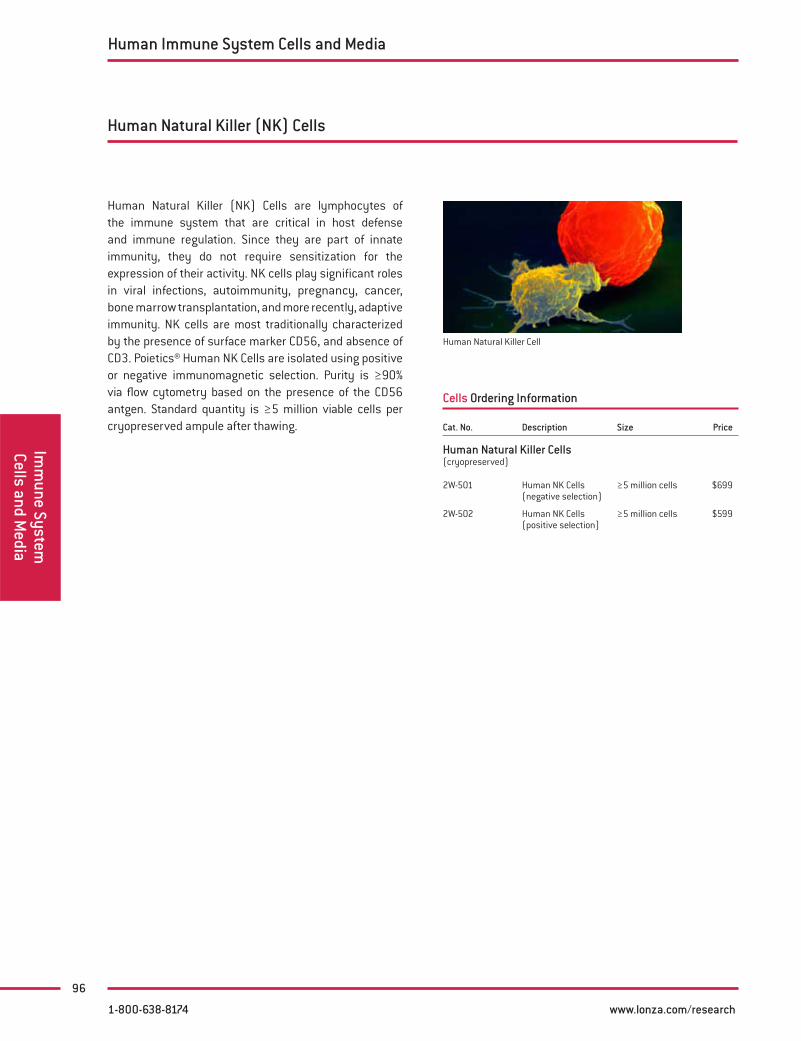
96
Imm
une System
Cells and Media
Human Natural Killer (NK) Cells
Human Natural Killer (NK) Cells are lymphocytes of the immune system that are critical in host defense and immune regulation. Since they are part of innate immunity, they do not require sensitization for the expression of their activity. NK cells play significant roles in viral infections, autoimmunity, pregnancy, cancer, bone marrow transplantation, and more recently, adaptive immunity. NK cells are most traditionally characterized by the presence of surface marker CD56, and absence of CD3. Poietics® Human NK Cells are isolated using positive or negative immunomagnetic selection. Purity is ≥90% via flow cytometry based on the presence of the CD56 antgen. Standard quantity is ≥5 million viable cells per cryopreserved ampule after thawing.
Cells Ordering Information
Human Natural Killer Cell
Human Immune System Cells and Media
1-800-638-8174 www.lonza.com/research
Cat. No. Description Size Price
Human Natural Killer Cells(cryopreserved)
2W-501 Human NK Cells (negative selection)
≥5 million cells $699
2W-502 Human NK Cells (positive selection)
≥5 million cells $599

97
Poietics® Neural Progenitor Cells and Media contains NHNP-Normal Human Neural Progenitor Cells and optimized medium for their sustenance. This cell system can be useful for research applications in drug development, neurotoxicity, neurogenesis, electrophysiology, CNS function, and neurotransmitter disorders.
Poietics® NHNP Cells are cryopreserved in primary passage as “spheroids”. We guarantee that Poietics® NHNP will express markers described when plated onto laminin coated plates.
Performance and Quality Tests
Poietics® Cells, Medium, and Reagents are quality –tested together and guaranteed for optimal performance as a complete system
All cells test negative for HIV-1, Hepatitis B and C, –mycoplasma, bacteria, yeast, and fungi
Poietics® Normal Human Neural Progenitor Cells –test positive for -tubulin III (neurons) and GFAP (astrocytes)
We now offer two media kits specially formulated to support the growth, expansion, and differentiation of the NHNP cells.
NPMM Neural Progenitor Maintenance Medium BulletKit® contains the necessary supplements and media for optimal NHNP cell growth and differentiation. This kit includes:
CC-3210 – Neural Progenitor Basal Medium –
CC-4241 – Neural Progenitor Maintenance Medium –SingleQuots® Kit (contains hEGF and hFGF)
CC-4242 – Neural Progenitor Supplement SingleQuots® –Kit (contains NSF-1 and GA)
NPDM™ Neural Progenitor Differentiation Medium BulletKit® contains the necessary supplements and media for optimal NHNP differentiation. The medium can be customized by supplementation with differentiation-promoting agents such as Brain Derived Neurotrophic Factor. This kit includes:
CC-3210 – Neural Progenitor Basal Medium –
CC-4242 – Neural Progenitor Supplement SingleQuots® –Kit
Cells Ordering Information
NHNP stained for -tubulin III and GFAP
Sold under license from StemCells, Inc. US patents 5,968,829 and 1. 5,851, 832.
References
For a comprehensive list of references and additional —technical information, please visit www.lonza.com/research
Human Neural Progenitor Cells and Media
Neur
al P
roge
nito
r Ce
lls a
nd M
edia
Media Ordering Information
Neural Progenitor Media
1-800-638-8174 www.lonza.com/research
Cat. No. Size Price
NHNP - Normal Human Neural Progenitor CellsRecommended Medium: NPMM™ BulletKit®
PT-2599 ≥1.2 106 cells/ampoule $599
NHNP - Cell Pellet in RNALater
PX-2599RL �10 million cells/pellet $1,299
Cat. No. Size Price
NPMM™ Neural Progenitor Maintenance Medium BulletKit®Includes Basal Medium and SingleQuots® Kit
CC-3209 Kit $110
NPDM™ Neural Progenitor Differentiation Medium BulletKit®Includes basal medium and SingleQuots® Kit
CC-3229 Kit $95
NPBM™ Neural Progenitor Basal Medium
CC-3210 200 ml $61
NPMM™ Neural Progenitor Maintenance Medium SingleQuots® Kit
CC-4241 — $65
Neural Progenitor Supplement SingleQuots® Kit
CC-4242 — $83
For related neural cell and media systems, see page 49, 73 – 76

98
Consult the cell culture experts and harness the power of Lonza’s extensive knowledge and experience delivering custom cell solutions. Choose from an endless variety of human and animal primary cell types, cryopreserved, plated or in culture flasks, for your convenience. Save time and money by avoiding the aggravation of tissue acquisition, failed isolations and low yields.
Quality and experience
As the cell culture experts, Lonza maintains an ISO 9001:2001 certified custom cell isolation laboratory staffed by some of the world’s finest cell culture technicians, providing a variety of cell types for your individual research needs. Partner with Lonza and benefit from over 60 years of combined cell culture isolation experience.
We offer
Expert custom cell isolation solutions made to your –specifications
Extensive quality testing and cellular charac- –terization available
Years of isolation experience with a wide variety of –cell types
Custom formats: cryopreserved, plates or flasks –
State-of-the-art cell isolation capabilities –
Delays in obtaining vital primary cell cultures can be very costly. Discover our superior custom cell isolation capabilities including:
Primary cell isolations from human and animal –tissues
Cell expansion and testing –
Donor-matched cell sets –
Cryopreserved or proliferating formats –
Wide array of QC and cell characterization services, –including PCR
Individual customer consultation to ensure correct –order fulfillment
Custom primary cell isolations from human, rat, mouse, porcine, bovine, monkey and many other species. Examples of successful isolations include:
Bovine adrenal gland capillary endothelial cells –
Bovine embryonic kidney cells –
Bovine and porcine preadipocytes –
Canine umbilical vein endothelial cells –
Cat iris smooth muscle cells –
Guinea pig kidney cells –
Human bladder epithelial cells –
Human bronchial/tracheal epithelial cells – Cystic –Fibrosis
Human diabetic CD34 – + cells
Human microvascular retinal endothelial cells –
Human small intestine disassociated cells –
Murine dermal fibroblasts –
Porcine mesangial cells –
Rat mesenchymal stem cells –
Custom Cell Isolation Services
Custom Cell Isolation
Services
1-800-638-8174 www.lonza.com/research

99
Clonetics® FAQs
Q. What is a BulletKit®?
A. A BulletKit® contains a bottle of basal medium and a SingleQuots® Kit. It is possible to purchase each of these components separately. Once the basal medium is supplemented with the SingleQuots® Kit, it becomes a complete growth medium.
Q. What are SingleQuots® Supplements and Growth Factors?
A. SingleQuots® Supplements and Growth Factors contain several vials that may include growth factors, FBS, and antibiotics in pre-measured aliquots designed to be added to a bottle of basal medium. The concentrations of these growth factors are proprietary.
Q. Do I need to add antibiotics to your media?
A. For most kits, gentamicin-amphotericin-B mixture is provided at a final concentration of 30 μg/ml gentamicin and 15 ng /ml amphotericin-B. Antibiotics are not provided for the KGM®-CD Kit and will need to be added if you so desire.
Q. Can I use a medium other than the one you offer to culture the Clonetics® Cells?
A. We have an optimized medium for each Clonetics® Cell type. The cells are grown and tested in their specific medium system. We cannot guarantee cell performance if our recommended medium is not used.
Q. What is the difference between the EGM® BulletKit® (CC-3124) and the EGM®-2 BulletKit® (CC-3162)?
A. The primary difference between the two media types is in the growth factors. The EGM® BulletKit® contains: BBE, hEGF, hydrocortisone, FBS, and GA-1000. In the EGM®-2 BulletKit®, we have removed the BBE and replaced it with known growth factors: VEGF, hFGF-B, ascorbic acid, insulin-like growth factor, and heparin. The cells proliferate much better in the EGM®-2 Medium. It has been reported that the EGM® BulletKit® works better for VEGF, FGF, and angiogenesis research.
Q. What is a complete cell and media system?
A. A complete cell and media system allows the growth, subculture and continued growth of normal human cells in vitro (in cell culture plastic). A Clonetics® Cell and Media System includes three components: one cryopreserved ampoule or proliferating cell culture, Clonetics® Optimized Growth Media, and a ReagentPack™ Subculture Reagents containing all reagents necessary for subculturing the cells. All of our products are quality tested using the complete Clonetics® Cells and Media System and must conform to standardized cell culture performance before release. Cells, media, and reagents must all be purchased separately.
Q. Do I need to use a gelatin, �bronectin, collagen, or Matrigel® coating?
A. Most Clonetics® Cells do not require an extracellular matrix when grown on tissue culture treated plastic – flasks, dishes, well plates (with the exception of hepatocytes, bovine brain microvascular endothelial cells, and the rat/mouse neuronal and dorsal root ganglion neurons, rat cardiac myocytes which do require a coating). However, we have found that glass slides, chamber slides, and other formats may require a coating prior to seeding the cells. Please contact Scientific Support for further information.
Q. Can Clonetics® Cells be transfected?
A. Yes, see pages 191, 203-205 for transfection reagents that are optimized to work with our primary cells.
Q. Your instructions do not mention centrifuging the amp of cells upon thawing. Won’t the DMSO kill the cells?
A. Clonetics® Normal Human Cells are very fragile out of cryopreservation and should not be centrifuged (exception – bovine brain microvascular endothelial cells and hepatocytes). Residual DMSO will be removed by a media change after 24 hours.
Q. I thawed and plated my cells yesterday and most of the cells are �oating, what happened?
A. Clonetics® Cells will not be confluent on day one. After 24 hours, you will see few attached cells per field under the microscope. Aspirate off the media and the floating cells and replace with fresh, pre-warmed media. Those attached cells will begin to proliferate and a confluent monolayer should be established in 5 –10 days.
Clon
etic
s® F
AQs
Media & Reagents
1-800-638-8174 www.lonza.com/research

100
Clonetics® FAQsContinued
Clonetics® FAQs
Q. I’ve had my cells in culture for a few months, they’re no longer growing and don’t look like healthy cells, what’s wrong?
A. All Clonetics® Cells are primary cells with a finite life span. Most Clonetics® Cells are guaranteed for 10 – 15 population doublings, which equates to about 2 – 3 passages. As cell death begins to occur, morphology and cell performance will begin to change and growth will slow drastically.
Q. I don’t see the speci�c cell type I want in your catalog or on your website. Do you do custom isolations?
A. Yes, Lonza offers custom isolations and formats for various types of non-catalog cell products. Please see pages 79 & 98 for additional information.
Q. Can I seed my ampoule of cells into one T-25 �ask?
A. No. Each cell type has a recommended seeding density optimized for cell attachment and proliferation. Using information from the Certificate of Analysis shipped with each cryopreserved vial of cells, use the following equations to calculate the number of flasks or dishes that can be set up:
Number of cells per amp percent viability
= Maximum number of cm2 that can be inoculatedRecommended seeding
density
Maximum number of cm2 that can be plated =
Maximum number of culture vessels that can be set upEffective growth area of flask
*Note: Cells seeded into well plates should be seeded at 10,000 viable cells/cm2.
Media & Reagents
1-800-638-8174 www.lonza.com/research

101
Clonetics® Technical Information: Cells and MediaOverview of cell culture process for Clonetics® Human and Animal Cells*
Storage Requirements:
CellsUpon arrival, immediately remove cryo-preserved cells from dry ice and place immediately into liquid nitrogen. If no dry ice is left in the package, thaw cells, immediately place them into culture vessels and call your Technical Specialist.
MediumStore Clonetics® Cell Culture Medium in a 4°C refrigerator. When using medium, under sterile conditions, take the amount you need and return bottle to the refrigerator. Always bring medium to room temperature before use.
Supplements and ReagentsIf you to plan to subculture within three days, store all growth supplements, HEPES Buffered Saline Solution and Trypsin Neutralizing Solution at 4°C. Trypsin/EDTA Solution has a limited shelf life at 4°C. If, upon arrival, Trypsin/EDTA is thawed, immediately aliquot and refreeze at -20°C. If Trypsin/EDTA is frozen, store at -20°C. If you do not plan to set up the cell culture within 3 days, store all growth supplements and subculture reagents in a -20°C freezer.
Unpack and store cells
Prepare Medium
Prepare Cells
Add BPE or BBEAdd SingleQuots® Kit
Maintenance: Every 40 hours change medium, increase as
necessary
When at 60 – 90% con�uence, subculture
Assess cell yield and viability, optimize as necessary
Maintenance: After 24 hours, change medium
Incubate and re-examine culture
Calculate number of �asks/chambers/dishes required
Thaw cells in water bath
Resuspend cells in cryovial
Dispense equal amounts of cells into �ask
Add medium to culture vessel(s) and incubate for at least 30
minutes
Incubate 3 – 4 hours
Replace medium and incubate
Proliferating Cultures Cryopreserved Cultures
BulletKit® Supplemented Media
1 – 2 DAYS
5 – 9 DAYS
*Exceptions: NHEM, Rat and Mouse neural cells, Rat Cardiac Myocytes, h NHEPS®, sm NHEPS®, rt NHEPS®, and bMVEC-B
Clon
etic
s®
Tech
nica
l Inf
orm
atio
n
1-800-638-8174 www.lonza.com/research

102
As a precaution against contamination, follow all procedures for handling products of human origin outlined in —“Guidelines to Avoid Personnel Contamination By Infective Agents in Research Laboratories That Use Human Tissues,” from the J. of Tissue Culture Methods.
Always wear gloves and safety glasses when working with all materials. Exercise caution when working with cryopreserved —cells; rapid temperature changes may cause splattering of liquid nitrogen.
Wash hands thoroughly after performing all procedures. —
Do not smoke, eat or drink in areas where reagents or cells are handled. —
Never pipette by mouth. —
Products of human and animal origin are potential biohazards. Although provided human cells negative for HIV-1, —Hepatitis B and C, proper precautions must be taken to avoid inadvertent exposure.
WARNING: Clonetics® and Poietics® Products contain human sourced material. Treat as potentially infectious.
Safety Precautions with Clonetics® Cells
Perform the following steps in a sterile �eld
For a bottle of fully supplemented medium, do the following:
Add BBE or BPE, if required, to a 500 ml bottle of 1. basal medium.
Detach the BBE or BPE supplement from the medium 2. bottle.
Decontaminate the vial and medium bottle with 3. ethanol or isopropanol.
Add the entire contents of the vial (approximately 4. 2 ml) to the medium with a pipette; rinse the vial with medium and pipette the contents back into the 500 ml bottle.
Replace the cap and swirl the medium gently a few 5. times to mix.
Record the date the BBE or BPE was added on the 6. medium label.
Media Preparation
For BulletKit®, perform the following steps
Decontaminate the external surfaces of the 1. SingleQuots® Cryovials and the basal media bottle with ethanol or isopropanol.
Aseptically open each cryovial and add the entire 2. amount to the basal medium with a pipette.
Rinse each cryovial with the medium. It may not 3. be possible to recover the entire volume listed for each cryovial; small losses, even up to 10%, should not affect the cell growth characteristics of the supplemented medium.
Transfer the label provided with each kit to the basal 4. medium bottle being supplemented. Use it to record the date and amount of each supplement added; we recommend that you place the completed label over the basal medium label to avoid confusion or possible double supplementation.
Record the new expiration date on the label based on 5. the shelf life. A fully reconstituted BulletKit® should be used within 30 days; this supplemented medium will now be referred to as a growth medium.
NOTE: If there is concern that sterility was compromised during the supplementation process, the entire newly prepared growth medium may be re-filtered to assure sterility. If you re-filter, use a sterile 0.2-micron filter. Routine re-filtration is not recommended as some protein loss may occur with each filtration.
Continued on next page
Clonetics® Technical Inform
ation
1-800-638-8174 www.lonza.com/research

103
Growth Area of Common Plasticware
Media PreparationContinued
Before You BeginPerform the following steps before you begin media or cell preparation:
MultiwellPlates
EffectiveGrowth Area
Per Well
Cell CultureMedium Required
Per Well/Total
Initial Number of Cells to Seed
at 10,000 cells/cm2
6 well 9.60 cm2 2 ml / 12 ml 96,00012 well 3.80 cm2 1 ml / 12 ml 38,00024 well 2.00 cm2 0.5 ml / 12 ml 20,00048 well 0.75 cm2 150 μl / 7 ml 7,50096 well 0.32 cm2 100 μl / 10 ml 3,200
DishesEffective
Growth AreaCell Culture
Medium Required
Initial Number of Cells to Seed
at 2,500 cells/cm2
Initial Number of Cells to Seed at3,500 cells/cm2
Initial Number of Cells to Seed at5,000 cells/cm2
35 mm 9.6 cm2 2 ml 20,000 28,000 40,00060 mm 21 cm2 5 ml 52,500 73,500 105,000
100 mm 55 cm2 11 ml 137,500 192,500 275,000150 mm 148 cm2 30 ml 370,000 518,000 740,000
Clon
etic
s®
Tech
nica
l Inf
orm
atio
n
Flasks
Effective
Growth Area
Cell Culture
Medium Required
Initial Number of Cells to Seed
at 2,500 cells/cm2
Initial Number of Cells to Seed at3,500 cells/cm2
Initial Number of Cells to Seed at5,000 cells/cm2
T-25 25 cm2 5 ml 62,500 87,500 125,000T-75 75 cm2 15 ml 187,500 262,500 375,000
T-150 150 cm2 30 ml 375,000 525,000 750,000
Prepare a sterile field:1. A sterile field consists of a Class II biological safety –cabinet with a front access opening and filtered laminar airflow, or an equivalent device
Determine the amount of medium required:2. Review the – Growth Area of Common Plasticware table to determine the amount of medium to be used
Sterile instruments and vessels required:3. Sterile disposable serological pipettes –
Micropipettes and sterile pipette tips –
Adjustable multichannel pipette or repeating pipette –
Sterile reservoirs for use with multichannel pipette –
Sterile 15 ml centrifuge tubes –
Cell culture flasks, or multiwell, flat-bottom tissue –culture plates
Hemacytometer or cell counter –
Other required supplies:4. 70% alcohol (ethanol or isopropanol) –
Growth medium (cell type specific) –
Protective gloves and garments –
Trypan Blue –
Plan and prepare for initial set up:5. Base the set-up on the number of cells indicated –on the Certificate of Analysis accompanying the product
Check the calibration on humidified incubator. 6. Incubator should be humidified and set to 5% CO2, 95% air, and 37°C.
1-800-638-8174 www.lonza.com/research

104
These instructions do not apply to bMVEC-B, hepatocytes, NHEM, rat & mouse cells, and Poietics® Cells.
Calculate the number of vessels to be set up. 1. Refer to your Certificate of Analysis for the exact number of cells in your cryovial. Refer to table on page 103 “Growth Area of Common Plasticware”, for help in adjusting this calculation.
Use the following calculations to determine the number of vessels to be set-up for the recommended seeding densities of 2,500 cells/cm2, 3,500 cells/cm2, or 5,000 cells/cm2
If you use a T-25 with an effective growth area of 25 cm2
The advantage of setting up this number of T-25 flasks from the initial cryovial, as opposed to larger flasks, is that it reduces the risk of losing large numbers of cells. That is, if you experience di� culty trypsinizing the first T-25 flask, there are other remaining T-25 flasks to use.
Label each flask with the passage number, cell 2. type, lot number, and date.
In a sterile field, carefully open the supplemented 3. bottle of growth medium and aseptically transfer the medium to new culture vessels by adding 1 ml growth medium for every 5 cm2 surface area of the flask.
Example: 5 ml growth medium for a 25 cm2 flask.
Tighten vented caps on vessels. If vented caps 4. are not being used, twist caps until tight, then loosen about 1/2 turn. Allow the culture vessels to warm and equilibrate in a 37°C, 5% CO2, humidified incubator for at least 30 minutes.
Culture Set Up – Adherent Cell Types
Thawing Cells – Adherent Cell Types
Aseptically add the recommended amount of medium to the �ask and equilibrate for 30 minutes in a 5% CO2, 37°C incubator.
Have a micropipette ready prior to thawing.1. Remove the cryovial of cells from storage. Wipe 2. cryovial with ethanol or isopropanol before opening. In a sterile field, briefly twist the cap a quarter turn to relieve the internal pressure, then retighten; do not open the cryovial completely.Holding the cryovial, dip the bottom 3/4 of the 3. cryovial in a 37°C water bath and swirl gently for 1 – 2 minutes until contents are thawed. Watch the cryovial closely; when the last sliver of ice melts remove it; DO NOT submerge it completely. Thawing the cells for longer than 2 minutes may result in less than optimal results.Remove the cryovial immediately, wipe it 4. dry, and transfer to a sterile field where the equilibrated flasks should be waiting, ready to seed. Rinse the cryovial with 70% alcohol, then wipe to remove excess.Note the color of the thawed cryovial. Ideally, 5.
the color of the thawed cryovial should be pink. If the color is not pink, seed the cells, note the color and mention this fact to Scientific Support if seeding is not successful.
NOTES:If more than one cryovial is to be thawed, thaw one cryovial –at a time and keep other cryovials in liquid nitrogen until ready for use
Cryopreserved cells are very delicate; thaw and return them –to culture as quickly as possible with minimal handling
Wear eye protection when handling frozen cells; rapid –temperature changes may cause splattering of liquid nitrogen
Centrifugation should not be performed to remove cells –from the cryoprotectant cocktail; this action is more damaging than the effects of DMSO residue in the culture
It is not recommended to thaw frozen cells directly onto –glass slides, chamber slides, gridded plates or multiwell plate configurations (6, 12, 24, 96…); optimal performance is achieved when initial seeding out of cryopreservation is performed into T-25 flasks; for further instructions follow the directions in the set-up section in the cell culture instructions provided
No. of cells available × Percent viability= Max. no. of cm2 that can
be platedRecommended seeding density
Max. no. of cm2 that can be plated = Max. no. of flasks that
can be set up Effective growth area of flask
Example: A cryovial of HMVEC-L with 520,000 cells and 80% viability
520,000 × 0.80 = 83 cm2, to be set up
5,000
83 cm2
= 3 flasks (rounded down to the nearest whole number of flasks)25 cm2
Clonetics® Technical Inform
ation
1-800-638-8174 www.lonza.com/research

105
Remove the cap, being careful not to touch the interior threads with your fingers.
Using a 1,000 μl micropipette set to 800 μl, put the 1. tip into the cryovial and resuspend the cells with a gentle, slow and steady up and down pipetting motion no more than five times. DO NOT resuspend quickly and keep the tip near the bottom to avoid making bubbles.Dispense an equal amount of cells into the flasks 2. set up earlier (as determined by the recommended seeding density and number of cells/vial). If four T-25 flasks were prepared, set micropipette to 250 μl and dispense. If eight T-25 flasks were prepared, set micropipette to 125 μl and dispense.Replace the cap or cover and gently rock the 3. vessels to evenly distribute the cells. Loosen caps if necessary to permit gas exchange.Return the culture vessels to a 37°C incubator 4. with 5% CO2. Lay them flat on the shelf, providing the largest surface for cells to attach. The cells will anchor to the bottom surface of the flask.
NOTE: Do not dispense the entire contents of the cryovial into one T-25 flask!
After SeedingCells are not tolerant to rapid temperature fluctuations or nutrient-deficient medium. Feeding them with fresh growth medium that has been warmed will avert potential problems. (Remember to warm only the amount needed.) Check and feed the cells on the schedule below, even on weekends and holidays.
Change the growth medium the day after seeding 1. (to remove residual DMSO and unattached cells), then every other day thereafter while examining them daily.
NOTE: A change of medium requires removal of the medium by aspirating with a sterile pipette on the opposite side of the flask from where the cells are attached. Then warm, fresh medium is added down the same side.
Successfully recovered cultures will exhibit the 2. following:
a. Cells with clear non-granular cytoplasm. b. Numerous mitotic figures after day 2.
Feed the cells a larger volume of medium as 3. they become more confluent. Use this table as a guideline:
If cells are: Then feed them: Under 25% confluent… 1 ml per 5 cm2
From 25 – 45% confluent… 1.5 ml per 5 cm2
Exceeding 45% confluence… 2 ml per 5 cm2
Continue feeding the cells until 60 – 90% 4. confluence. If specific cell types are allowed to become over-confluent (i.e., epithelial cells) and stay at confluence for more than 2 days, they can suffer irreversible contact inhibition and may detach from the flask and/or be di� cult to trypsinize.
Seeding – Adherent Cell Types
Examine the culture microscopically for any signs 1. of distress during shipment (i.e., detachment, rounding-up, or atypical morphology). Check the relative cell density and estimate % confluency. The culture should be 30 – 100% confluent upon receipt. Some cellular detachment is normal. Please call Scientific Support immediately if cells look severely distressed.Decontaminate the external surface of the cell culture 2. flask or multiwell dish by wiping with 70% ethanol or isopropanol.Incubate the sealed flask or multiwell dish at 37°C, 5% 3. CO2, for 3 – 4 hours to equilibrate temperature.Warm an appropriate amount of growth medium 4.
to 37°C in a sterile container. Warming the entire bottle can shorten the life of the medium. Never warm medium under hot running water or any other uncontrolled temperature source. NEVER MICROWAVE. In a sterile field, carefully open the cell culture flask 5. or multiwell dish, remove the medium and replace it with the warmed, fresh medium. Aseptically remove any medium inside the neck or cap area because it can facilitate microbial contamination.If you are using a flask with a non-vented cap, loosen 6. the cap and return the flask to the 37°C humidified incubator with 5% CO2 for at least 24 hours.
Clon
etic
s®
Tech
nica
l Inf
orm
atio
n
Proliferating Cells – Adherent Cell Types
1-800-638-8174 www.lonza.com/research

106
Subculturing – Adherent Cell Types
Clonetics® Technical Inform
ation
Storage Information for Subculture Reagents
Subculture reagents are sterile-filtered and then 1. stored at -20°C until shipped from Lonza’s Distribution Centers.
Subculture reagents may thaw during transport. 2. They may be refrozen once.
Subculture reagents can be stored at -20°C until 3. expiration date, after thawing once and refreezing.
To keep Trypsin/EDTA fresh and active after thawing, 4. you may aliquot it into sterile centrifuge tubes and refreeze at -20°C. Trypsin/EDTA may be stored frozen until the expiration date.
We recommend that HEPES-BSS and the Trypsin 5. Neutralization Solution once stored at 4°C be used within one month.
The following instructions are for a 25 cm2 flask. Adjust all volumes accordingly for other size flasks.
PreparationPreparation for subculturing the first flask:
Subculture the cells when they are 60 – 90% confluent 1. and contain many mitotic figures throughout the flask.
For each 25 cm2. 2 of cells to be subcultured: a. Thaw 2 ml of Trypsin/EDTA and allow to come to room temperature. b. Allow 7 - 10 ml of HEPES Buffered Saline Solution (HEPES-BSS) to
come to room temperature. c. Allow 4 ml of Trypsin Neutralizing Solution (TNS) to come to room
temperature.
Remove growth medium from 4°C storage and allow 3. to start warming to room temperature.
Prepare new culture vessels:4. a. Prepare 1 – 3 T-75 flasks. The number of flasks needed depends
upon confluence and total yield. Larger flasks may be used to save plasticware and time spent on subsequent subcultures. Smaller flasks reduce the risk of losing a substantial part of your culture.
b. As before, label each flask with the passage number, lot number, cell type, and date.
c. In a sterile field, carefully open the bottle and transfer growth medium to new culture vessels by adding 1 ml growth medium for every 5 cm2 surface area of the flask.
Example: 15 ml growth medium for a 75 cm2 flask d. If not using vented caps, loosen caps of flasks. Place the new
culture vessels into a 37°C humidified incubator with 5% CO2 and equilibrate the flasks for at least 30 minutes. Subculture one flask at a time. All flasks following the first flask will be subcultured following an optimization of this protocol (explained later in this procedure), based on calculated cell count, cell viability, and seeding density.
NOTE: Use only Clonetics® Trypsin/EDTA. The concentration of Trypsin/EDTA from other suppliers may be 10X our concentration, which will detrimentally effect Clonetics® Cells.
In a sterile �eld:(T-25 flask used as example. Increase volume proportionately for others flasks.)
Aspirate the medium from one culture vessel.1.
Rinse the cells with 5 ml of room temperature 2. HEPES Buffered Saline Solution (HEPES-BSS). DO NOT forget this step. The medium contains complex proteins that neutralize the trypsin.
Aspirate the HEPES-BSS from the flask.3.
Cover the cells with 2 ml of Trypsin/EDTA solution.4.
Tighten the cap and begin monitoring the flask 5. under the microscope.
Continue to examine the cell layer microscopically.6. a. Allow the trypsinization to continue until approximately 90% of
the cells are rounded up.
NOTE: Rounded up cells are spherical, have smooth edges and are refractile or shiny. If the cells still have protruding nubs which are still attached to the flask, they need more time to trypsinize. This entire process takes about 2 – 6 minutes, depending on cell type.
b. At this point, rap the flask against the palm of your hand to release the majority of cells from the culture surface. If only a few cells detach, you may not have let them trypsinize long enough. Wait 30 seconds and rap again. If cells still do not detach, wait and rap every 30 seconds thereafter.
NOTE: Do not try to get all cells to detach by rapping them severely. This action may damage the cells.
After cells are released, neutralize the trypsin in 7. the flask with 4 ml of room temperature Trypsin Neutralizing Solution. If the majority of cells do not detach within seven minutes, the trypsin is either not warm enough or not active enough to release the cells. Harvest the culture vessel as described above, and either re-trypsinize with fresh, warm Trypsin/EDTA Solution or rinse with Trypsin Neutralizing Solution and then add fresh, warm medium to the culture vessel and return to an incubator until fresh trypsinization reagents are available.
Quickly transfer the detached cells to a sterile 8. 15 ml centrifuge tube.
Rinse the flask with a final 2 ml of HEPES-BSS to 9. collect residual cells, and add this rinse to the centrifuge tube.
Examine the harvested flask under the microscope 10. to make sure the harvest was successful by looking at the number of cells left behind. This should be less than 5%.
Centrifuge the harvested cells at 220 × g for 5 minutes 11. to pellet the cells.
a. Aspirate most of the supernatent, except for 100 μl to 200 μl.
b. Flick the centrifuge tube with your finger to loosen the pellet.
1-800-638-8174 www.lonza.com/research

107
Subculturing – Adherent Cell TypesContinued
Clon
etic
s®
Tech
nica
l Inf
orm
atio
n
Dilute the cells in 4 ml to 5 ml of growth medium 12. and note the total volume of the diluted cell suspension.
NOTE: To obtain the best results from your cells, assess cell yield and viability with Trypan Blue.
Count the cells with a hemacytometer or cell 13. counter and calculate the total number of cells. (See instructions above.) Make a note of your cell yield for later use.
NOTE: The cell suspension should contain between 250,000 to 1,000,000 cell/ml for greatest accuracy.
If necessary, dilute the suspension with HEPES 14. Buffered Saline Solution (HEPES-BSS) to achieve the desired “cells/ml” and re-count the cells.
Assess cell viability using Trypan Blue.15.
Use the following equation to determine the total 16. number of viable cells.
Total cell count × percent viability = Total # of viable cells
Example: 1,000,000 cells × 60% = 600,000 viable cells
Determine the total number of flasks to inoculate 17. by using the equation below. The number of flasks needed depends upon cell yield and seeding density. Larger flasks may be used to save plasticware and time spent on subsequent subcultures. Smaller flasks reduce the risk of losing a substantial part of your culture if contamination occurs.
The recommended seeding density could be 2,500 cells/cm2, 3,500 cells/cm2, or 5,000 cells/cm2 for flasks and 10,000 cells /cm2 for well plates
Total # of viable cells= Total # of flasks to
inoculateRecommended seeding density × flask size
600,000 viable cells = 3 T-75 flasks (rounded down to
nearest whole number)75 cm2 × 2,500 cells/cm2
Use the following equation to calculate the volume 18. of cell suspension to seed into your flasks.
Total volume of diluted cell suspension= Seeding volume
# of flasks as determined in step 17
4.3 ml of diluted cell suspension = 1.43 ml per T-75 flask
3 T-75 flasks
Prepare flasks by labeling each flask with the 19. passage number, lot number, cell type, and date.Carefully open the medium bottle and transfer 20. growth medium to new culture vessels by adding 1 ml growth medium for every 5 cm2 surface area of the flask (1 ml/5 cm2).Example: 15 ml growth medium for a 75 cm2 flask.
After mixing the diluted cells with a 5 ml pipette to 21. ensure a uniform suspension, dispense the volume of suspension calculated above into the prepared subculture flasks.
After dispensing the cells, gently rock flask to 22. promote even distribution.
If not using vented caps, loosen caps of flasks. 23. Place the new culture vessels into a 37°C humidified incubator with 5% CO2.
1-800-638-8174 www.lonza.com/research

108
Clonetics® Technical Inform
ation
OverviewA culture flask of normal human cells is harvested by trypsinization and subsequent trypsin inhibitor treatment. The cells are centrifuged, resuspended in growth medium and counted. The desired number of cells is then added to wells of sterile 96-well tissue culture plates. The plates are incubated in a 37°C, 5% CO2 humidified incubator for 1 – 3 days to allow for cell adherence and growth. Seeding densities will vary somewhat with your experimental requirements. A density of 10,000 cells/cm2 for multiwell plates is ideal. See the Cell Culture Instruction Sheet* for the cell type you are using for specific information.*Available at www.lonza.com/research or contact Scientific Support.
Required Materials:T-25 flask of proliferating normal human cells between 1. 60% and 90% confluence96-well flat bottom, tissue culture plates2. 37°C humidified incubator with 5% CO3. 2/95% airLaminar flow hood or other sterile environment4. Adjustable multichannel pipette (8 or 12 channel) or 5. repeating pipetteSterile reservoir(s) for use with multichannel 6. pipette
ProcedureFollow the steps for subculture preparation and 1. subculturing. Then follow steps 2 – 4 below.Since the cells/ml calculation computed is per ml, 2. the cell concentration must be increased by 4 times before seeding 96-well plates (to accommodate the 1:4 dilution when adding 250 μl of suspended cells per well). When making the cell suspension, adjust the cell concentration with growth medium.Transfer the diluted cell suspension to a sterile 3. reservoir. Using a multichannel (8 or 12 channel) pipette equipped with sterile pipette tips, add 250 μl of the diluted cell suspension to each well of the labeled 96-well flat bottom, tissue culture plate(s).
RESUSPEND THE CELL SUSPENSION OFTEN DURING THE SEEDING PROCEDURE TO ENSURE A UNIFORM NUMBER AND DISTRIBUTION OF CELLS INTO EACH WELL BY PIPETTING UP AND DOWN A FEW TIMES BETWEEN EVERY OTHER DISPENSING.
Cover and incubate the plates for 1 – 3 days at 4. 37°C/5% CO2. (Incubation periods exceeding 3 days are generally not recommended because of evaporation of medium from the edge wells of the plate).
Note: Before using the 96-well plate culture in a bioassay, examine the cells microscopically for the presence of mitotic figures as a confirmation that the cells have resumed active growth.
Subculturing into 96-well plates
1-800-638-8174 www.lonza.com/research

109
Cryopreservation may compromise cell quality and performance. Performance of the cells cannot be guaranteed after cryopreservation. These instructions do not apply to hepatocytes, bMVEC-B, melanocytes, mouse and rat neuronal cells, rat cardiac cells, and Poietics® Cells.
Instructions
Sterile filter freezing medium using a cell culture 1. rated 0.2-micron filter.
Harvest cells and spin them down.2.
Resuspend cells in cold freezing solution at 3. �500,000 to 2,000,000 cells/ml. Work quickly. Once exposed to DMSO, cells become very fragile.
Pipet aliquots (1 ml each) into freezing vials or 4. ampoules and seal.
Insulate aliquots with a Styrofoam® or propanol 5. freezing canister.
Store cells at -70°C overnight.6.
Within 12 – 24 hours, place in LN7. 2 (-200°C) for long-term storage. Cells will be compromised by prolonged storage at -70°C.
Cryopreservation Solutions
For mesenchymal stem cells70% MSCBM10% DMSO20% Human Serum Albumin (25% solution)
For skeletal muscle cells70% SKGM®20% FBS10% DMSO
For all other cell types80% Clonetics® Growth Medium of choice10% FBS10% DMSO
Instructions for Cryopreservation
Clon
etic
s®
Tech
nica
l Inf
orm
atio
n
1-800-638-8174 www.lonza.com/research

110
Several factors, or a combination of factors, can contribute to low cell count and low cell viability. If cell yield or viability is unsatisfactory, use the following information to increase the success rate of future cultures.
Improving Cell YieldIf your cell yield is low (less than 50%), determine the cause(s) and possible solution(s) using the table below. Then subculture one more flask applying the appropriate solution(s).
Improving Cell Yield and Viability During Subculture
Low Yield (Cell Count)
Low Viability (<50% viable)
If your cell viability is low (less than 50%), determine the possible cause(s) and solution(s) using the table below. Then subculture one more flask applying the appropriate solution(s).
Condition Possible Causes Solutions
Majority of cells did not detach 1. Inactive or cold Trypsin/EDTA 1. Use Trypsin/EDTA at room temperature
2. Improper storage of Trypsin/EDTA 2. Store at -20°C until ready for use; thaw and allow it to come to room temperature briefly before subculturing
3. Exposure time to Trypsin/EDTA was too short 3. Exposure time to Trypsin/EDTA is usually 5 –6 minutes
4. Trypsin/EDTA has been neutralized 4 . Be sure to rinse the culture completely with HEPES-BSS before trypsinization
5. Vessel was not rapped firmly 5. Use a moderate amount of force when rapping during trypsinization
Low yield, 95% of the cells detached but the yield was low
Culture was under confluent at trypsinization Be sure to trypsinize at 60 – 90% confluence with numerous mitotic figures throughout the flask
Condition Possible Causes Solutions
Trypsin/EDTA damaged the cells 1. Used another vendor’s Trypsin/EDTA 1. Use only Clonetics® Trypsin
2. Exposure time of the cells to Trypsin/EDTA was too long 2. Do not trypsinize longer than 7 minutes
3. Trypsin/EDTA was used above room temperature. Trypsin becomes more active at temperatures above room temperature
3. Do not use even mildly heated Trypsin/EDTA
4. Failed to neutralize the trypsin. Prolonged exposure to trypsin will damage cells.
4. Neutralize the Trypsin/EDTA with Trypsin Neutralizing Solution to eliminate cell damage due to trypsin
5. Vessel was rapped too firmly during trypsinization. Rapping too hard to release cells causes cell membrane damage
5. Use moderate force when rapping flask to dislodge cells during trypsinization
Culture vessel was too confluent; was completely covered with cells
Culture was too confluent at trypsinization Be sure to trypsinize at 60 – 90% confluence with about five mitotic figures per field of view
Cell growth slowed before 80% confluence and cells look dull and non refractile
The most probable cause is failure to increase the volume of medium used as the cell confluency increasedThe cells become mildly starved and are not able to recover after trypsinization
Change medium and increase volume as recommended. Please observe all guidelines
Clonetics® Technical Inform
ation
1-800-638-8174 www.lonza.com/research

111
Medium Speci�cations
Medium is formulated for optimal growth of specific types of normal human cells. It can be purchased as basal medium without supplements, as fully supplemented growth medium, or in a conveniently packaged BulletKit® that allows user control over supplement type and concentration. Each type of medium is tested for its ability to support growth of the intended normal human cell. Biochemical and sterility tests are also performed on every lot of medium. Certificates of Analysis are available upon request for all medium products.
Basal Medium
Basal medium has been optimized for specific types of normal human cells. Basal medium does not contain growth factors necessary for propagation of cells. Growth factors must be added to enhance plating e� ciency and cellular proliferation. Optimized formulations make it possible to perform research on a wide variety of normal human cell types. Results of years of media development are available to you for use in your own research.
Complete Medium
Complete medium is fully supplemented growth medium (BPE and BBE are packaged separately) and contains all of the growth factors and supplements necessary for the propagation of specific types of normal human cells in culture. Undefined supplements are avoided when possible and used only at minimal levels when necessary. Standard formulations of all growth media include antimicrobials. Antimicrobial-free media are available as a special order.
BulletKit®
A BulletKit® provides flexibility in final medium formulation and increased shelf life. Each BulletKit® contains basal medium and premeasured, single-use aliquots (SingleQuots®) of growth factors and antimicrobial agents to formulate the fully supplemented growth medium of your choice.
Clonetics® Media
Clon
etic
s® M
edia
1-800-638-8174 www.lonza.com/research

112
Airway cell media are serum-free media that have been optimized for the proliferation of airway cells. Each component of the basal medium and each growth supplement are carefully titered for optimal growth by Lonza’s R&D team. Lonza currently offers two media choices for the growth of airway cells, allowing for desired performance and formulation flexibility. When selecting a medium to use, refer to specific media recommendations or call Scientific Support for assistance.
BEGM® BulletKit®
Best growth of NHBE - Normal Human Bronchial/ —Tracheal Epithelial Cells in medium containing antimicrobials
SAGM™ BulletKit®
Superior growth for SAEC - Human Small Airway —Epithelial Cells
Airway Cell Media Options
Product Information
Cat. No. Product Description
CC-3170 BEGM® BulletKit® Bronchial Epithelial Cell Growth Medium BulletKit®, Serum-freeCC-3171 BEBM® Basal Medium Bronchial Epithelial Cell Basal Medium, Serum-free CC-4175 BEGM® SingleQuots® Kit Formulates BEBM® to BEGM® BPE, 2 ml; Hydrocortisone, 0.5 ml; hEGF, 0.5 ml; Epinephrine, 0.5 ml; Transferrin, 0.5 ml; Insulin, 0.5 ml; Triiodothyronine, 0.5 ml; GA-1000, 0.5 ml; Retinoic Acid, 0.5 ml CC-3118 SAGM™ BulletKit® Small Airway Epithelial Cell Growth Medium BulletKit®, Serum-freeCC-3119 SABM™ Basal Medium Small Airway Epithelial Cell Basal Medium, Serum-free CC-4124 SAGM™ SingleQuots® Kit Formulates SABM™ to SAGM™ BPE, 2 ml; Hydrocortisone, 0.5 ml; hEGF, 0.5 ml; Epinephrine, 0.5 ml; Transferrin, 0.5 ml; Insulin, 0.5 ml; Triiodothyronine, 0.5 ml; GA-1000, 0.5 ml; Retinoic Acid, 0.5 ml; BSA-FAF, 5.0 ml
Clonetics® Media
1-800-638-8174 www.lonza.com/research

113
Endothelial cell media are low serum media optimized for the proliferation of endothelial cells. Each component of the basal medium and each growth supplement is carefully titered for optimal growth by Lonza’s R&D team. Lonza currently offers four Clonetics® Media for the growth of endothelial cells allowing for desired performance and formulation flexibility. When selecting a medium to use, refer to specific medium recommendations or call Scientific Support for assistance.
EGM® and EGM® BulletKit®
Basal medium developed for normal human endothelial —cells in a low-serum environment
EGM® is supplemented growth medium and includes —an attached aliquot of Bovine Brain Extract (BBE)
EGM® BulletKit® includes basal medium with supple- —ments and growth factors in separate, frozen aliquots
Final serum concentration is 2% —
EGM® can be used to grow all of Clonetics® Endothelial —Cells except microvascular, coronary artery, umbilical artery, and iliac artery
EGM® MV BulletKit®
Developed for microvascular and coronary artery —endothelial cells
Same basal medium as in EGM® —
Final serum concentration is 5% —
EGM®-MV can be used to grow most Clonetics® —Endothelial Cells
EGM®-2 BulletKit®
Refinements to basal medium and the growth factors —
Does not contain BBE —
Final serum concentration is 2% —
Improved cell proliferation over EGM® —
EGM®-2 can be used to grow all of Clonetics® Endothelial —Cells except microvascular, coronary artery, umbilical artery, and iliac artery
EGM®-2MV BulletKit®
Developed for the enhanced growth of lung micro- —vascular endothelial cells
Does not contain BBE —
Final serum concentration increased to 5% —
EGM®-2MV can be used to grow all Clonetics® Endothelial —Cells except HUVEC
Endothelial Cell Media Options
Clon
etic
s® M
edia
1-800-638-8174 www.lonza.com/research

114
Product Information
Cat. No. Product DescriptionCC-3125 EGM® MV BulletKit® Microvascular Endothelial Cell Growth Medium BulletKit® w/ 5% FBSCC-3121 EBM® Basal Medium Endothelial Cell Basal Medium, Serum-free CC-4143 EGM® MV SingleQuots® kit Formulates EBM® to EGM® MV BBE, 2 ml; hEGF, 0.5 ml; Hydrocortisone, 0.5 ml; FBS, 25 ml; GA-1000, 0.5 mlCC-3202 EGM®-2 MV BulletKit® Microvascular Endothelial Cell Growth Medium-2 BulletKit® w/ 5% FBSCC-3156 EBM®-2 Basal Medium-2 Endothelial Cell Basal Medium-2, Serum-freeCC-4147 EGM®-2 MV SingleQuots® Kit Formulates EBM®-2 to EGM®-2 MV hEGF, 0.5 ml; Hydrocortisone, 0.2 ml; FBS, 25 ml; VEGF, 0.5 ml; hFGF-B, 2 ml; R3-IGF-1, 0.5 ml; Ascorbic Acid, 0.5 ml; GA-1000, 0.5 ml (No BBE)CC-3024 EGM® Complete Medium Endothelial Cell Growth Medium w/ 2% FBSCC-3124 EGM® BulletKit® Endothelial Cell Growth Medium BulletKit® w/ 2% FBSCC-3121 EBM® Basal Medium Endothelial Cell Basal Medium, Serum-free CC-3129 EBM®-PRF EBM® w/o Phenol Red CC-4133 EGM® SingleQuots® Kit Formulates EBM® to EGM® BBE, 2 ml; hEGF, 0.5 ml; Hydrocortisone, 0.5 ml; FBS, 10 ml; GA-1000, 0.5 mlCC-3162 EGM®-2 BulletKit® Endothelial Cell Growth Medium-2 BulletKit® w/ 2% FBS CC-3156 EBM®-2 Basal Medium-2 Endothelial Cell Basal Medium-2, Serum-freeCC-4176 EGM®-2 SingleQuots® Kit Formulates EBM®-2 to EGM®-2 hEGF, 0.5 ml; Hydrocortisone, 0.2 ml; FBS, 10 ml; VEGF, 0.5 ml; hFGF-B, 2 ml; R3-IGF-1, 0.5 ml; Ascorbic Acid, 0.5 ml; GA-1000, 0.5 ml, Heparin, 0.5 ml (No BBE)
Endothelial Cell Media OptionsContinued
Clonetics® Media
1-800-638-8174 www.lonza.com/research

115
Fibroblast media has been optimized for the proliferation of fibroblasts. Each component of the basal medium and each growth supplement is carefully titered for optimal growth by Lonza’s R&D team. Lonza currently offers two media choices for the growth of fibroblasts, allowing for desired performance and formulation flexibility. When selecting a medium to use, refer to specific media recommendations or call Scientific Support for assistance.
FGM® BulletKit®
FGM® is a defined medium system and does not contain —serum
FGM®-2 BulletKit®
Contains a vial of FBS for a final serum concentration —of 2%
Product Information
Cat. No. Product Description
CC-3132 FGM®-2 BulletKit® Fibroblast Growth Medium BulletKit®-2, w/ 2% FBSCC-3131 FBM® Basal Medium Fibroblast Basal Medium CC-4126 FGM® SingleQuots® Kit Formulates FBM® to FGM-2 hFGF-B, 0.5 ml; Insulin, 0.5 ml FBS, 10 ml; GA-1000, 0.5 mlCC-3130 FGM® BulletKit® Fibroblast Growth Medium BulletKit®, Defined CC-3131 FBM® Basal Medium Fibroblast Basal Medium CC-4134 FGM® SingleQuots® Kit Formulates FBM® to FGM hFGF-B, 0.5 ml; Insulin, 0.5 ml; GA-1000, 0.5 ml
Fibroblast Cell Media Options
Product Information
Cat. No. Product Description
CC-3198 HCM™ BulletKit® Hepatocyte Culture Medium, Phenol red-freeCC-3199 HBM™ Basal Medium Hepatocyte Basal Medium, Phenol red-free, Serum-freeCC-4182 HCM™ SingleQuots® Kit Formulates HBM™ to HCM™ hEGF, 0.5 ml; Transferrin 0.5 ml; Hydrocortisone, 0.5 ml; BSA, 10.0 ml; Ascorbic Acid 0.5 ml; GA-1000, 0.5 ml, Insulin, 0.5 mlCC-3197 HMM™ Medium HMM™, Hepatocyte Maintenance Medium, Phenol red-free, Serum-freeCC-4192 HMM™ SingleQuots® Kit HMM™ SingleQuots®, required supplements for use with HMM™ to provide optimal maintenance of cells, Insulin; 0.5 ml Dexamethasone, 0.5 ml; GA-1000, 0.5 ml
Hepatocyte Media
Clon
etic
s® M
edia
1-800-638-8174 www.lonza.com/research

116
Keratinocyte media has been optimized for the proliferation of keratinocytes. Each component of the basal medium and each growth supplement is carefully titered for optimal growth by Lonza’s R&D team. Lonza currently offers three media choices for the growth of keratinocytes, allowing for desired performance and formulation flexibility. When selecting a medium to use, refer to specific media recommendations in the cell systems sections or call your Scientific Support for assistance.
KGM® and KGM® BulletKit®
Optimized for Clonetics® NHEK – Normal Human —Epidermal Keratinocytes in a serum-free environment
KGM® includes supplemented medium and an attached —aliquot of Bovine Pituitary Extract (BPE)
KGM® BulletKit® includes basal medium with all —supplements and growth factors in separate, frozen aliquots
KGM® can be used to grow all Clonetics® — NHEK Normal Human Epidermal Keratinocytes
KGM®-2 BulletKit®
Refinements to the basal medium and the growth —factors
Grows normal human keratinocytes up to 20% faster —and allows for additional population doublings when compared to KGM® and other commercially available keratinocyte media
KGM®-2 can be used to grow all Clonetics® — NHEK Normal Human Epidermal Keratinocytes
KGM®-CD
Supports isolation and proliferation of primary human —keratinocytes in culture
Chemically defined – No serum and no animal or plant —extracts
Minimized variable experimental results due to —unknown effects of animal or plant-derived components
Obtain cleaner and more accurate results quickly —
Product Information
Cat. No. Product Description
CC-3107 KGM®-2 BulletKit® Keratinocyte Growth Medium-2 BulletKit®, Serum-freeCC-3103 KBM®-2 Basal Medium-2 Keratinocyte Basal Medium-2CC-3108 KGM®-2 w/o Ca++ BulletKit® Keratinocyte Growth Medium-2 BulletKit®, Calcium-Free, Serum-freeCC-3158 KBM®-2 w/o Ca++ Keratinocyte Basal Medium-2, Calcium-Free, Serum-freeCC-4152 KGM®-2 SingleQuots® Kit Formulates KBM®-2 to KGM®-2 BPE, 2 ml; hEGF, 0.5 ml; Insulin, 0.5 ml Hydrocortisone, 0.5 ml; Epinephrine, 0.5 ml; Transferrin, 0.5 ml; GA-1000, 0.5 mlCC-3001 KGM® Complete Medium Keratinocyte Growth Medium, Serum-freeCC-3111 KGM® BulletKit® Keratinocyte Growth Medium BulletKit®, Serum-freeCC-3101 KBM® Basal Medium Keratinocyte Basal Medium, Serum-freeCC-3112 KGM® BulletKit® w/o Ca++ Keratinocyte Growth Medium BulletKit®, Calcium-free, Serum-freeCC-3104 KBM® w/o Ca++ Keratinocyte Basal Medium, Calcium-free, Serum-freeCC-4131 KGM® SingleQuots® Kit Formulates KBM® to KGM® BPE, 2 ml; hEGF, 0.5 ml; Insulin, 0.5 ml; Hydrocortisone, 0.5 ml; GA-1000, 0.5 mlCC-4455 KGM®-CD BulletKit® Keratinocyte Growth Media – Serum-free, Non-animal origin componentsCC-3255 KBM®-CD Basal Medium Keratinocyte Basal Media – Chemically DefinedCC-4456 KGM®-CD SingleQuots® Kit Formulates KBM®-CD to KGM®-CD Recombinant human insulin 1 ml; growth supplement, 5 ml
Keratinocyte Cell Media Options
Clonetics® Media
1-800-638-8174 www.lonza.com/research

117
Product Information
Cat. No. Product Description
CC-3051 MEGM® Complete Medium Mammary Epithelial Cell Growth Medium, Serum-free, complete medium CC-3150 MEGM® BulletKit® Mammary Epithelial Cell Growth Medium BulletKit®, Serum-freeCC-3151 MEBM® Basal Medium Mammary Epithelial Cell Basal Medium, Serum-free CC-3153 MEBM®-PF free Mammary Epithelial Cell Basal Medium Phenol Red Free, Serum-free CC-3152 MEBM®-Bicarb free Mammary Epithelial Cell Basal Medium Sodium Bicarbonate Free, Serum-free CC-4136 MEGM® SingleQuots® Kit Formulates MEBM® to MEGM® BPE, 2 ml; Hydrocortisone, 0.5 ml; hEGF, 0.5 ml; Insulin, 0.5 ml; GA-1000, 0.5 ml
Product Information
Cat. No. Product Description
CC-3186 AGM™ BulletKit® Astrocyte Growth Medium BulletKit®, w/3.0% FBSCC-3187 ABM™ Basal Medium Astrocyte Basal Medium, Serum-free, without L-glutamineCC-4123 AGM™ SingleQuots® Kit Formulates ABM™, to AGM™ Insulin, 1.25 ml; rhEGF, 0.5 ml; FBS, 15.0 ml; Ascorbic Acid, 0.5 ml; L-glutamine, 5.0 ml; GA-1000, 0.5 ml
Product Information
Cat. No. Product Description
CC-3249 MGM® BulletKit® Melanocyte Growth MediumCC-3250 MBM®-4 Basal Medium-4 Melanocyte Basal Medium, Serum-freeCC-4435 MGM®-4 SingleQuots® Kit Formulates MBM®-4 to MGM®-4 CaCl2, 1.0 ml; hFGF-B, 1.0 ml; PMA, 0.5 ml; rhInsulin, 1.0 ml; Hydrocortisone, 0.5 ml; BPE, 2.0 ml; FBS, 2.5 ml; Gentamicin/Amphotericin B, 0.5 ml
Clonetics® Melanocyte Cell Medium has been optimized for the growth and proliferation of normal human primary Melanocytes in culture. This medium system has been shown to deliver superior results as compared to other existing commercial media systems. Melanocytes in culture maintain >90% functionality based on the conversion of L-dopa to dopa-melanin. The Melanocyte Media system also allows for normal morphology and proliferative capacity after recovery from cryopreservation and throughout serial passaging.
MGM-4
Melanocyte Growth Media has been qualified and —tested together to provide optimum performance
The media system is offered as BulletKit® (basal —medium and separately packaged growth factors) to allow for flexibility with your research project
Adult melanocytes also require the addition of Et-3, —sold separately
Melanocyte Cell Medium, Serum-free
Neural Cell Medium, Low Serum
Mammary Epithelial Cell Media, Serum-free
Clon
etic
s® M
edia
1-800-638-8174 www.lonza.com/research

118
Product Information
Cat. No. Product Description
CC-3166 PrEGM™ BulletKit® Prostate Epithelial Cell Growth Medium BulletKit®, Serum-freeCC-3165 PrEBM™ Basal Medium Prostate Epithelial Cell Basal Medium, Serum-free CC-4177 PrEGM™ SingleQuots® Kit Formulates PrEBM® to PrEGM® BPE, 2.0 ml; Hydrocortisone, 0.5 ml; hEGF, 0.5 ml; Epinephrine, 0.5 ml; Transferrin, 0.5 ml; Insulin, 0.5 ml; Retinoic Acid, 0.5 ml; Triiodothyronine, 0.5 ml; GA-1000, 0.5 ml
Prostate Epithelial Cell Medium, Serum-free
Product Information
Cat. No. Product Description
CC-3190 REGM™ BulletKit® Renal Epithelial Cell Growth Medium BulletKit®, w/ 0.5% FBSCC-3191 REBM™ Basal Medium Renal Epithelial Cell Basal Medium, Serum-freeCC-4127 REGM™ SingleQuots® Kit Formulates REBM® to REGM® Hydrocortisone, 0.5 ml; hEGF, 0.5 ml; FBS, 2.5 ml; Epinephrine, 0.5 ml; Triiodothyronine, 0.5 ml; Transferrin, 0.5 ml; Insulin, 0.5 ml; GA-1000, 0.5 ml CC-3146 MsGM™ BulletKit® Mesangial Cell Growth Medium BulletKit®, w/ 5% FBSCC-3147 MsBM™ Basal Medium Mesangial Cell Basal Medium, Serum-freeCC-4146 MsGM™ SingleQuots® Kit Formulates MsBM™ to MsGM™ FBS, 25 ml; GA-1000, 0.5 ml
Product Information
Cat. No. Product Description
CC-4461 PNGM™ BulletKit® Primary Neuron Growth Medium BulletKit® CC-3256 PNBM™ Basal Medium Primary Neuron Basal Medium CC-4462 PNGM™ SingleQuots® Kit Formulates PNBM to PNGM™ NSF-1, 4 ml; L-glutamine, 2 ml; GA-1000, 0.2 mlCC-4512 PNGM™-A BulletKit® Primary Neuron Growth Media-AdultCC-4511 PNGM™-A SingleQuots® Kit Formulates PNBM to PNGM™-Adult NSF-1, 4 ml; L-glutamine, 2 ml; Pen/Strep, 2 ml
Renal Cell Media, Low Serum
Primary Neuron Growth Medium, Serum-freeClonetics® M
edia
1-800-638-8174 www.lonza.com/research

119
Product Information
Cat. No. Product Description
CC-3207 OGM™ BulletKit® Osteoblast Growth Medium BulletKit®, w/ 10% FBS CC-3208 OGM™ Basal Medium Osteoblast Cell Basal Medium, Serum-freeCC-4193 OGM™ SingleQuots® Kit Formulates OBM™ to OGM™ FBS, 50 ml; Ascorbic Acid, 0.5 ml; GA-1000, 0.5 mlCC-4194 OGM™ Differentiation SingleQuots® Kit Induces osteoblast differentiation and bone mineralization. Hydrocortisone-21-Hemisuccinate, 0.5 ml; -Glycerophosphate, 5.0 mlCC-3216 CGM™ BulletKit® Chondrocyte Growth Medium BulletKit®CC-3217 CBM Basal Medium Chondrocyte Basal MediumCC-4409 CGM™ SingleQuots® Kit Formulates CBM to CGM™ R3-IGF-1, 1.0 ml; bFGF, 2.5 ml; Insulin, 1.0 ml; Transferrin, 0.5 ml; FBS, 25 ml; Gentamicin/Amphotericin-B, 0.5 mlCC-3225 CDM™ BulletKit® Chondrocyte Differentiation Medium BulletKit®, Serum-freeCC-3226 CDM™ Basal Medium Chondrocyte Differentiation Basal MediumCC-4408 CDM™ SingleQuots® Kit Formulates CDM™ Basal Medium to CDM™ Differentiation Medium TGF- , 1.25 ml; R3-IGF-1, 0.5 ml; Insulin, 0.5 ml; Transferrin, 0.5 ml; FBS, 12.5 ml; GA-1000, 0.25 ml
Skeletal Cell Media
Product Information
Cat. No. Product Description
CC-3160 SkGM® BulletKit® Skeletal Muscle Growth Medium BulletKit®, Serum-freeCC-3161 SkBM® Basal Medium Skeletal Muscle Basal Medium, Serum-freeCC-4139 SkGM® SingleQuots® Kit Formulates SKBM® to SKGM® hEGF, 0.5 ml; Insulin, 5 ml; BSA, 5 ml; Fetuin, 5 ml; Dexamethasone, 0.5 ml; GA-1000, 0.5 mlCC-3245 SkGM®-2 BulletKit® Skeletal Muscle Myoblast Growth Medium-2 BulletKit®, with 10% FBSCC-3246 SKBM®-2 Basal Medium-2 Skeletal Muscle Myoblast Basal Medium-2, Serum-free no L-glutamineCC-3244 SkGM®-2 SingleQuots® Kit Formulates SKBM®-2 to SKGM®-2 FBS, 50.0 ml; GA-1000, 0.5 ml; rhEGF, 0.5 ml; Dexamethasone, 0.5 ml; L-glutamine,10.0 ml
Skeletal Muscle Cell Media Clon
etic
s® M
edia
1-800-638-8174 www.lonza.com/research

120
Smooth muscle medium has been optimized for the proliferation of smooth muscle cells. Each component of the basal medium and each growth supplement is carefully titered for optimal growth by Lonza’s R&D team.
SmGM®-2 BulletKit®
SmGM®-2 BulletKit® was developed for better cell —morphology and proliferation
Final serum concentration is 5% —
SmGM®-2 can be used to grow all smooth muscle cells —
Product Information
Cat. No. Product Description
CC-3182 SmGM®-2 BulletKit® Smooth Muscle Growth Medium-2 BulletKit® w/ 5% FBSCC-3181 SmBM® Basal Medium Smooth Muscle Basal Medium, Serum-freeCC-4149 SmGM®-2 SingleQuots® Kit Formulates SmBM® to SmGM®-2 hEGF, 0.5 ml; Insulin, 0.5 ml; hFGF- B, 1 ml; FBS, 25 ml; GA-1000, 0. 5 ml
Smooth Muscle Cell Medium
Clonetics® Media
Product Information
Cat. No. Product Description
CC-3205 SCGM™ BulletKit® Stromal Cell Growth Medium BulletKit®, w/ 5% FBSCC-3204 SCBM™ Basal Medium Stromal Basal Medium, w/o phenol red, Serum-freeCC-4181 SCGM™ SingleQuots® Kit Formulates SCBM™ to SCGM™ hFGF-B, 0.5 ml; Insulin 0.5 ml; FBS, 25 ml; GA-1000, 0.5 ml
Stromal Cell Medium, Low Serum
1-800-638-8174 www.lonza.com/research

121
Q. What is a BulletKit®?
A. A BulletKit® contains basal medium and SingleQuots® Kit of supplements and growth factors. In most cases, it is possible to purchase each of these com ponents separately. Once the basal medium is supplemented with the SingleQuots® Kit, it becomes a complete growth or differentiation medium.
Q. What are SingleQuots® Supplements and Growth Factors?
A. SingleQuots® Supplement and Growth Factors contains several vials of growth factors, supplements and antibiotics in premeasured aliquots designed to be added to a bottle of basal medium. Serum will be provided in the SingleQuots® Kit if it is to be added to the basal medium. The concentrations of these growth factors are proprietary.
Q. What media should I use for Poietics® Cells?
A. Information regarding specific media selections can be found in the Technical Data Sheet or the Instructions. Please contact Scientific Support for more information.
Q. What is the difference between the different cell sources: bone marrow, cord blood, or fetal liver?
A. Bone Marrow: provides a hematopoietic micro-environment and the most number of different donors.
Cord Blood: more naïve cells useful in transplant and stem cell research.
Fetal Liver: best for developmental studies and they are highly proliferative.
Q. Are the fresh cells you provide equivalent to their cryopreserved counterparts?
A. Yes, the cells perform equivalently by all measures we have tested. Some cells do die in the cryopreservation and thawing processes, but it does not seem to be selective to a particular cell type. We put additional cells in each vial to account for this loss.
Poietics® FAQs
Q. I don’t see the speci�c cell type I want in your catalog or on your website, do you do custom isolations?
A. Yes, Lonza offers custom isolations and formats for various types of non-catalog cell products. See page 79 & 98 for additional information.
Q. Can I expand the mononuclear cells, CD34+ progenitors, or the CD133+ cells without differentiating them?
No, the cells will proliferate and differentiate simultaneously upon being placed in culture. Varying the cytokine cocktail can influence the differentiation and proliferation of the cells. Please contact Scientific Support for more information.
Q. Can I expand the human mesenchymal stem cells prior to differentiating them?
A. These cells are frozen in 2nd passage and we recommend they are used before passage 5.
Q. Can I expand the preadipocytes prior to differ-entiating them?
A. The subcutaneous cells can be passaged twice prior to differentiation. The visceral cells can be passaged once prior to being differentiated.
Q. Can I get subcutaneous and visceral preadipocytes from the same donor?
A. Yes, please contact Scientific Support for more information.
Poie
tics®
FAQ
s
Media
1-800-638-8174 www.lonza.com/research

122
Poietics® Technical Inform
ation
Warm medium containing 10% FBS or 1% BSA. For 1. mononuclear cells and hematopoietic progenitors, DNase I (20 U/ml) should also be added.*
Quickly thaw the vial of frozen cells in a 37°C water 2. bath. Wipe the outside of the vial with 70% ethanol.
Aseptically transfer a maximum of 2 ml of cell 3. suspension to a 50 ml conical tube. For one million cells or less, use a 15 ml conical tube.
Rinse the vial with 1 ml of medium. Add the rinse 4. dropwise to the cells while gently swirling the tube (�1 minute).
Slowly add enough medium dropwise to the cells 5. until the total volume is 5 ml, while gently swirling after each addition of several drops of medium (�3 minutes).
Slowly bring the volume up to fill the tube by adding 6. 1 ml to 2 ml volumes of medium dropwise, while gently swirling after each addition of medium (�5 – 10 minutes).
Centrifuge the cell suspension at 200 × g at room 7. temperature for 15 minutes.
Carefully remove by pipette (and save in a second 8. tube) most of the wash, leaving a few mls behind so the cell pellet is not disturbed. Gently resuspend the cell pellet in the remaining medium. If you are using a 50 ml tube, transfer the cells to a 15 ml conical tube and rinse the 50 ml tube with 5 ml of medium. Slowly add the 5 ml wash medium to the cell suspension with gentle swirling.
Slowly bring the volume up to fill the tube by adding 9. 1 ml to 2 ml volumes of medium while gently swirling after each addition of medium.
Centrifuge the cell suspension at 200 × g at room 10. temperature for 15 minutes.
Carefully remove by pipette all but 2 ml of the wash. 11. Gently resuspend the cell pellet in the remaining 2 ml of medium and count. If cell count is lower than expected, centrifuge the wash saved in step 8 at a higher speed, count and combine if necessary.
Rest the cells for 1 hour at 37°C and 5% CO12. 2. Count the cells a second time. The cells are ready to be put in culture.
* For the addition of DNase, prepare 20 ml of medium containing 10% FBS and 20 U/ml of DNase I (Sigma D 4513). Proceed as above, using the DNase-containing medium to dilute the cells. Centrifuge the cells and continue with step #8.
Procedure for Thawing CD34+ and Precursor Cells
1-800-638-8174 www.lonza.com/research

123
Poie
tics®
Med
ia
Product Information
Cat. No. Product Description
PT-3001 MSCGM™ BulletKit® Mesenchymal Stem Cell Growth Medium BulletKit®
PT-3238 MSCGM™ Basal Medium Mesenchymal Stem Cell Basal Medium
PT-4105 MSCGM™ SingleQuots® Kit Formulates MSCBM to MSCGM™ Growth MediumMCGS, 50 ml; L-glutamine, 10 ml; GA-1000, 0.5 ml
PT-3002 hMSC Differentiation Kit– Osteogenic Mesenchymal Stem Cell Differentiation Kit – Osteogenic
PT-3924 hMSCBM – Osteogenic Basal Medium Mesenchymal Stem Cell Basal Medium – Osteogenic
PT-4120 hMSC SingleQuots® Kit – Osteogenic Formulates Osteogenic Basal Medium to Osteogenic Differentiation Medium Dexamethasone, 1 ml; -Glycerophosphate, 2 ml; Ascorbate, 1 ml; Penicillin /Streptomycin, 2 ml; MCGS, 20 ml; L-glutamine, 4 ml
PT-3003 hMSC Differentiation Kit – Chondrogenic Mesenchymal Stem Cell Differentiation Kit – Chondrogenic
PT-3925 hMSCBM – Chondrogenic Basal Medium Mesenchymal Stem Cell Basal Medium – Chondrogenic
PT-4121 hMSC SingleQuots® Kit – Chondrogenic Formulates Chondrogenic Basal Medium to Chondrogenic Differentiation Medium ITS+, 2 ml; Sodium Pyruvate, 2 ml; Proline, 2 ml; Dexamethasone, 1 ml; Ascorbate, 2 ml; GA-1000, 0.2 ml; L-glutamine, 4 ml
PT-4124 TGF- 3 Required component sold separately
PT-3004 hMSC Differentiation Kit – Adipogenic Mesenchymal Stem Cell Differentiation Kit – Adipogenic
PT-3102A hMSC Adipogenic Maintenance Medium Mesenchymal Stem Cell Maintenance Medium – Adipogenic
PT-3102B hMSC Adipogenic Induction Medium Mesenchymal Stem Cell Induction Medium – Adipogenic
PT-4122 hMSC Maintenance SingleQuots® Kit – Adipogenic Formulates Adipogenic MM to Adipogenic Differentiation Medium rhInsulin, 2 ml; GA-1000, 0.2 ml; MCGS, 20 ml; L-glutamine, 4 ml
PT-4135 hMSC Induction SingleQuots® Kit – Adipogenic Formulates Adipogenic IM to Adipogenic Differentiation Medium Indomethacin, 0.4 ml; IBMX, 0.2 ml; rhInsulin, 2 ml; Dexamethasone, 1 ml; GA-1000, 0.2 ml; MCGS, 20 ml; L-glutamine, 4 ml
Product Information
Cat. No. Product Description
PT-8002 PGM™-2 BulletKit® Preadipocyte Growth Medium-2 BulletKit®
PT-8202 PBM-2 Basal Medium-2 Preadipocyte Basal Medium-2
PT-9502 PGM™-2 SingleQuots® Kit Formulates PBM-2 to PGM™-2 FBS (10%), 50 ml; L-Glutamine, 5 ml; GA-1000, 0.2 ml, 5 ml; rhInsulin, 2 ml; Dexamethasone, 0.2 ml; Indomethacin, 0.4 ml; IBMX, 0.2 ml
Human Mesenchymal Stem Cell Media
Preadipocyte Growth Medium
1-800-638-8174 www.lonza.com/research

124
Poietics® Media
Product Information
Cat. No. Product Description
PT-8001 Osteoclast Growth Medium BulletKit® Osteoclast Growth Medium BulletKit®
PT-8201 OBM™ Osteoclast Basal Medium
PT-9501 Osteoclast Growth Medium SingleQuots® Kit Formulates OBM™ to Osteoclast Growth Medium FBS (10%), 10 ml; L-glutamine, 1 ml; Penicillin/Streptomycin, 1 ml; M-CSF, 0.1 ml; Soluble RANK Ligand, 2 �g
Product Information
Cat. No. Product Description
CC-3209 NPMM™ BulletKit® Neural Progenitor Maintenance Medium BulletKit®
CC-3210 NPBM™ Basal Medium Neural Progenitor Basal Medium
CC-4242 NPDM SingleQuots® Kit Formulates NPBM™ to NPMM™ (for differentiation) NSF-1, 4 ml; GA-1000, 0.4 ml
CC-4241 NPMM™ SingleQuots® Kit Formulates NPBM™ to NPMM™ (for maintenance) rhEGF, 0.4 ml; rhFGF, 0.4 ml
CC-3229 NPDM BulletKit® Neural Progenitor Differentiation Medium BulletKit®
CC-3210 NPBM™ Basal Medium Neural Progenitor Basal Medium
CC-4242 NPDM SingleQuots® Kit Formulates NPBM™ to NPDM (for differentiation) NSF-1, 4 ml; GA-1000, 0.4 ml
Osteoclast Growth Medium
Neural Progenitor Growth Medium
1-800-638-8174 www.lonza.com/research

Amaxa® Nucleofection® and TransfectionChapter 2

126
Amaxa® Nucleofection®
Introduction 127Amaxa® Nucleofector® Technology 128 – 131Nucleofector® II Device 13296-well Shuttle® Device 133Nucleofector® Extended Guarantee and Service Contracts 13496-well Shuttle® Automation Package 135 – 137List of Nucleofector® Kits for Primary Cells 138 – 139Nucleofector® Kits for Blood Cells 140 – 150Nucleofector® Kits for Dermal Cells 151 – 155Nucleofector® Kits for Endothelial Cells 156 – 158Nucleofector® Kits for Epithelial Cells 159 – 163Nucleofector® Kits for Hepatocytes 164 – 165Nucleofector® Kits for Neural Cells 166 – 171Nucleofector® Kits for Smooth Muscle Cells 172 – 173Nucleofector® Kits for Stem Cells 174 – 179Nucleofector® Kits for Rat Cardiomyocytes 180Nucleofector® Kits for Human Chondrocytes 181Nucleofector® Kits for Mouse Embryonic Fibroblasts (MEF) 182List of Optimized Protocols for Cell Lines 183 – 184Cell Line Nucleofector® Kits 185Cell Line Optimization Nucleofector® Kits 186 – 187cGMP Cell Line Nucleofector® Kits 188Basic Parasite Nucleofector® Kits 188FAQs on Nucleofection® 189 – 190
Transfection Reagents
HiFect® Transfection Reagent 191Transport™ Protein Delivery Reagent 202PrimeFect™ Transfection Reagent 203 & 204FAQs Transport Reagent 205
Vectors
pmaxFP® Fluorescent Protein Expression Vectors 192pmaxFP® Vector Backbone Information 197pmaxFP®-Green Vectors 198pmaxFP®-Yellow Vectors 199pmaxFP®-Red Vectors 200pmaxCloning™ Vector 201
Antibiotics and Antimycotics
MycoZap™ Mycoplasma Elimination Reagent 198Antibiotics and Antimycotics 199
Other Transfection Products
siRNA Test Kit 200Amaxa® Peptide Transfection Control 201
Nucleofection® and Transfection
Amaxa® Nucleofection® and Transfection
888-632-9110 www.lonza.com/research

127
Nucl
eofe
ctio
n®
and
Tran
sfec
tion
Amaxa® Nucleofection® and Transfection
Selecting the Ideal Technology and Product for your Needs
Lonza provides comprehensive information and protocols regarding the introduction of many bio-molecules and particles. This information and more, is available online at www.lonza.com/celldatabase or through Scientific Support. Nucleofector® Devices and Kits are available in both single sample and 96-well formats to suit your individual throughput needs and to ensure that you have continuity of transfection method as your processing capacity increases over time. The selection chart below, and others hosted on the Lonza website, are designed to help you quickly focus your transfection methodologies.
Substrate
Cell type
DNAPlasmid DNA, cDNAs, Gene constructs
RNAsiRNA, shRNA, miRNA
ProteinPeptide, Protein
Other Morpholinos, pNAs, etc.
Primary cells Nucleofector® Device96-well Shuttle® System
Nucleofector® Device96-well Shuttle® System
Nucleofector® Device96-well Shuttle® System
Nucleofector® Device96-well Shuttle® System
Difficult-to-transfectcell lines
Nucleofector® Device96-well Shuttle® System
Nucleofector® Device96-well Shuttle® System
Nucleofector® Device96-well Shuttle® System
Nucleofector® Device96-well Shuttle® System
Standard cell lines HiFect® ReagentNucleofector® Device96-well Shuttle® System
HiFect® ReagentNucleofector® Device96-well Shuttle® System
Nucleofector® Device96-well Shuttle® System
Nucleofector® Device96-well Shuttle® System
Identify the right methodology based on your cell type and substrate
Contact Lonza Scientific Support to review your selection and discover more about the solution that fits your needs!
888-632-9110 www.lonza.com/research
Scientific Support
USA Europe/World– Phone: 888-632-9110 – Phone: +49(0)221-99199-400 – Fax: (888) 632-9112 – Fax: +49(0)221-99199-499 – Email: [email protected] – Email: [email protected]

128
Nucleofector® Technology
Amaxa® Nucleofector® Technology
The application of systems biology and multidisciplinary approaches require that cells and model systems display ’in-vivo-like’ cellular functionality. This means that the future of cell transfection is in using primary cell types, and that transfecting these physiologically relevant cell types presents researchers with a series of technical challenges. The most physiologically relevant cell types are typically the most difficult to transfect using traditional methods. In contrast, when using relevant cell lines as model systems, the critical issues are to achieve reproducibly efficient transfection with high levels of viability while matching throughput capability with the number of transfections required at each project phase - from proof of concept, through scale-up and screening-like applications.
Amaxa® Nucleofection® – your unique advantageAmaxa® Nucleofection® is a proprietary technology based on the momentary creation of small pores in cell membranes by applying an electrical pulse. The comprehensive way in which Nucleofector® Pulse Conditions and cell-type specific solutions are developed ensures that nucleic acid substrates are delivered not only to the cytoplasm, but also through the nuclear membrane
and into the nucleus. Transfected cells retain excellent viability and the function of intracellular systems is highly conserved. Whatever your application, Amaxa® Transfection Specialists are available to assist you in rapidly optimizing your new application.
One technology – the broadest choice of cell typesUsing the Nucleofector® Technology, cell lines, as well as primary cells, can be reliably transfected at high efficiency. Delivery of nucleic acid substrates directly into both the nucleus and cytoplasm ensures transfection efficiencies of up to 99% in both cell lines and primary cells. More than 200 ready-to-use Amaxa® Optimized Protocols contain cell-type specific guidance and Lonza's Amaxa® Cell Database contains user-developed methods and data for more than 1300 cell types.
Substrate flexibility – explore the limits of your imaginationThe �exibility that Nucleofection® allows in planning ex peri mental projects stems from Lonza's proven capability to transfect the widest range of substrate molecules into the widest range of cell types. Imagine the �exibility to first complement a knock-out mutant by transfecting a DNA vector, then to explore the regulation of the gene(s) of interest using multiple or single shRNA, siRNA, or miRNA substrates using the same Nucleofector® Kit and conditions.
Re�ecting the focus of molecular biologists, the �exibility of Nucleofection® now extends beyond nucleic acid
Normal human dermal fibroblasts - neonatal were transfected with 2.5 μg TMR-labeled plasmid DNA encoding eGFP. After 2 hours, cells were fixed with 3.5% PFA and analyzed by confocal microscopy. TMR label is shown in (A), GFP �uorescence in (B), DAPI nuclear staining in (C) and a merge of all three �uorescent labels in (D).
A
C
B
D
Structural Genomics
Functional Genomics
DrugCandidates
Peptide
Antibodies
Expression
shRNA
Plasmid Pre-miRNA
DNA
RNA
siRNA
miRNA
mRNA
AntisenseOligos
Protein
ProteinSmallMolecules
Fluoresentconjugates
System Biology
Drug Discovery
DNA delivery straight into the nucleus
Amaxa® Nucleofector® TechnologyContinued
888-632-9110 www.lonza.com/research

129
substrates, to include peptides, proteins, and antibodies. This allows yet further characterization and validation of the cellular functions that interest your research group, e.g., allowing researchers to identify and characterize related signaling or protein modification pathways using inhibitory peptides, before tracking active protein trafficking, or localization of active target molecules using antibody-conjugated nano-�uorophores or quantum-dots.
Two platforms – fulfilling your choice of sample throughputs
The use of Nucleofector® Technology allows your trans-fection capabilities to grow in pace with your group, to adapt to changing applications that require different levels of sample throughput, to accelerate the pace of your research, or to transfect only the number of cells that fits your unique application.
The Nucleofector® II Device and 96-well Shuttle® SystemBoth the Nucleofector® Device and 96-well Shuttle® System deliver unique electrical parameters that are different from other commercially available electroporation instruments. Electrical settings are pre-programmed for each optimized cell type and can be selected using the Nucleofector® Device or the PC-based software controlling the 96-well Shuttle® Device.
The Nucleofector® and 96-well Shuttle® KitsKits for Nucleofector® Technology contain Amaxa® speci-fied plasticware, pipettes, pmaxGFP® Vector, Nucleofector® Solution and Supplement. Each solution and supplement is individually developed for every primary cell type. For cell lines, a set of different Nucleofector® Solutions is available. All solutions provide a protective environment that ensures the highest transfection efficiency and cell viability, while helping maintain physiologically relevant cellular functions.
Cell Line Optimization Kits provide the ideal tools to conveniently determine the optimal Nucleofection® parameters of your cell line of interest within a single experiment.
The Components of the Nucleofector® Technology
Proven – in leading labsThe Nucleofector® Technology is an established and trusted transfection method in opinion-leading labs worldwide. Several thousand peer-reviewed publications illustrate the importance of Nucleofection® in hundreds of leading-edge research applications using primary cells, such as neurons and stem cells, as well as difficult-to-transfect and standard cell lines.
Low throughput Low to high throughput
Device Nucleofector® Device 96-well Shuttle® System
Samples per run 1 1 - 96
Reaction volume 100 μl 20 μl
Cell number 2 x 10⁵ to 2 x 10⁷ 5 x 10⁴ to 1x 10⁶
Substrate amount Oligonucleotides:
0.2 - 200 pmol (2 nM - 2 μM)
Oligonucleotides:
0.04 - 40 pmol (2 nM - 2 μM)
Vector DNA:
1 - 5 μM
Vector DNA:
1 - 5 μM
Two platforms
Amaxa® Nucleofector® TechnologyContinued
Nuc
leof
ecto
r®
Tech
nolo
gy
888-632-9110 www.lonza.com/research

130
Widest Range of Substrates
Jurkat cells were transfected in the presence of a FAM-labeled octapeptide (30 uM). Images were captured 2 hours post-transfection.
Rat hippocampal neurons (E17) were transfected with the shRNA vector pSUPERIOR targeting CDC10 using the 96-well Shuttle® System. A and B: Efficient Nucleofection® of pSUPERIOR is shown by eGFP expression after 1 day in vitro. C: Immunostaining of CDC10 (red �uorescence) shows reduced endogenous CDC10 protein levels in transfected neurons (green) after 4 days in vitro compared to untransfected cells (red). Western blot analysis (D) and quantification (E) of CDC10 down regulation.(Data courtesy of Prof. Kiebler, Medical University of Vienna, Vienna, Austria.)
Knockdown on mRNA level measured by qRT-PCR. 15 samples compared to control (C) set to 100%.(Data kindly provided by C. Merz, Bayer Schering Pharma AG, Berlin.)
siRNA-mediated depletion of vimentin in human T cells
Gene silencing in primary neurons by transfection of an shRNA vector – Quantitive down-regulation of CDC10 protein
Transfection of Jurkat cells
120
100
↑
50
40
30
20
10
0
Rela
tive
Expr
essi
on (%
pul
se o
nly,
con
trol
sam
ple
C)
11 3 5 7 9 11 13 15 1
100
80
60
40
20
0
CD C
10 S
igna
l (%
of C
alne
xin
Sign
al)
siCDC10 siPum2 misPum2
A B C ED
CDC10
siCDC10misPum2
siPum2
Calnexin
Amaxa® Nucleofector® TechnologyContinued Nucleofector®
Technology
888-632-9110 www.lonza.com/research

131
The Ideal Tool for Transfection of Cell Lines and Primary Cells
Contact Lonza today regarding your unique application or to arrange to test the Nucleofector® Technology with your cells and in your lab.
Cell Lines
Broad cell line coverage –––
From standard to difficult-to-transfect cells, including suspension cellsMore than 100 ready-to-use protocolsFast and easy optimization
High efficiencies ––
Up to 90% efficiency with plasmid DNAUp to 99% efficiency with siRNA duplexes
Reliable results – High viability and reproducibility
Safe and easy procedure – Easy and convenient handling
Fast experiments – Expression within hours – from transfection to analysis in one day
Primary Cells
High efficiencies ––
Up to 90% efficiency with plasmid DNAUp to 99% efficiency with siRNA duplexes
Reliable results – Reproducible high viability and functionality
No optimization – Ready-to-use kits for a multitude of cell types, e.g. neurons and stem cells
Reproducible results – Optimized Protocols for each cell type with details on cell isolation and culture
Unique advantages – Direct transport of nucleic acids into the nucleus thus enabling transfection even of non-dividing cells
Amaxa® Nucleofector® TechnologyContinued
Nuc
leof
ecto
r®
Tech
nolo
gy
NOTE: The Nucleofector® Technology, comprising Nucleofection® Process, Nucleofector® Device, Nucleofector® Solutions, Nucleofector® 96-well Shuttle® System and Nucleocuvette® plates and modules is covered by patent and/or patent pending rights owned by Lonza Cologne AG.
888-632-9110 www.lonza.com/research

132
Clonetics® Technical Inform
ation Nucleofector®
Technology
The Nucleofector® II Device delivers electrical parameters that differ from other commercially available electro-poration instrument. Electrical settings are pre-programmed for each optimized cell type, while an integrated chip-card reader allows quick program updates for future applications. In addition to programs for transfection of eukaryotic cells, the Nucleofector® II Device program list (from version N5-5 S4-2 on) includes programs for bacteria transformation. It also offers a language selection (English or Japanese). Weighing only 2.8 kg (6.2 lb), the device is conveniently sized so that it easily fits under a laminar-�ow hood enabling sterile work with cells.
Features of the Nucleofector® II Device
Easy handling
Motor-driven carousel for automatic cuvette handling –
High convenience
Large graphic display for easy day-to-day use –
Great flexibility
Software enables the customized naming of –individual programs
Future-proof
Program delivery unit for 96-well Shuttle® System –
Nucleofector® II Device
Technical Specifications
Dimensions (w x d x h) 30 x 23 x 11 cm (11.81 x 9.06 x 4.33 in)
Weight 2.8 kg (6.2 lb)
Power supply 100-110 VAC or 230 VAC50-60 Hz, self-regulating
Power consumption 50 VA/fuse T630mA L250V
Protection Class IP 20, EN 61010-1, UL 61010A-1
Ordering Information
888-632-9110 www.lonza.com/research
Cat. No. Price
Nucleofector® II Device
AAD-1001 on request

133
The 96-well Shuttle® Device is a high throughput extension to the Nucleofector® II Device. The complete Nucleofector® 96-well Shuttle® System consists of three components, the 96-well Shuttle® Device, the Nucleofector® II Device and a laptop computer. The Nucleofector® II Device serves as the program delivery unit. The 96-well Shuttle® Device is the contacting unit which mediates the transfer of the respective 96-well program to a specific well of the 96-well Nucleocuvette® Plate. The interaction between Nucleofector® II Device and Nucleofector® 96-well Shuttle® Device is controlled via the 96-well Shuttle® Software installed on the laptop.
Features of the 96-well Shuttle® Device
Transfection flexibility
Modular 6 x 16 Nucleocuvette® Plate for –scalable throughput
Variable cell numbers – from 10 – 4 – 106 cells per reaction
Rapid optimization
Up to 96 independent programs can be run per plate –
Optimization of any difficult-to-transfect cell line in –just one plate
Safety
Disposable plates minimize the risk of cross –contamination
Nucleocuvette® Plate with innovative conductive –polymer electrodes – no metal ion release
HTS compatibility
Automatic processing of a 96-well plate –in 3 – 4 minutes
Fulfills prerequisites for liquid handling integration –
96-well Shuttle® Device
Nucleofector® 96-well Shuttle® System
Technical Specifications
Dimensions (w x d x h) 34 x 27 x 10 cm (13.39 x 10.63 x 3.94 in)
Weight 3.0 kg (6.6 lb)
Power supply 110 VAC +10%/-20% or 230 VAC +10%/-20% 50-60 Hz, self-regulating
Power consumption 20 VA
Protection Class IP 22
96-w
ell S
hutt
le®
De
viceOrdering Information
Ordering Information
888-632-9110 www.lonza.com/research
Cat. No. Price
96-well Shuttle® Device
AAM-1001 on request
including:Laptop and 96-well Shuttle® SoftwareThe Nucleofector® II Device must be purchased independently
Cat. No. Price
96-well Shuttle® System Rack
AXA-1001 on request

134
Nucleofector® Device Extended Guarantee and Service Contracts
96-well Shuttle® Device Extended Guarantee and Service Contracts
Lonza provides the highest possible level of support to insure continuous performance of the 96-well Shuttle® Device. To further improve our service, Lonza offers the 96-well Shuttle® Extended Guarantee and Service Contracts that helps you pre-plan your costs and keep down-time as short as possible. The 96-well Shuttle® Extended Guarantee has to be purchased together with the 96-well Shuttle® Device. Service Contracts can be purchased at any time during a guarantee period.
Contract Benefits
1 or 2 additional years of guarantee beyond the basic –device guarantee of 1 year
Diagnosis and repair of your 96-well Shuttle® Device –including all replacement parts
Costs for return shipments of repaired device –
Replacement of 96-well Shuttle® Device within –5 business days*
*only for Gold Service Contracts
Lonza provides an exceptional level of support for and around the Nucleofector® Technology to guarantee high performance of our products and to ensure user satisfaction. To better serve your needs, Lonza offers the Nucleofector® Extended Guarantee and Service Contracts that provides you with a number of benefits (see below). The Nucleofector® Extended Guarantee has to be purchased together with a Nucleofector® Device. Service Contracts can be purchased at any time during a guarantee period.
Contract Benefits
1 or 2 additional years of guarantee beyond the basic –device guarantee of 1 year
Diagnosis and repair of your Nucleofector® Device –including all replacement parts
Costs for return shipments of repaired device –
Replacement of Nucleofector® Device within –5 business days*
*only for Gold Service Contracts
Nucleofector® Device
Ordering Information
Ordering Information
888-632-9110 www.lonza.com/research
Cat. No. Price
96-well Shuttle® Device Guarantee – 1 year Extension
AWM-1001 on request
96-well Shuttle® Device Guarantee – 2 year Extension
AWM-1002 on request
Service Contract 96-well Shuttle® Device Silver
AWB-1001 on request
Service Contract 96-well Shuttle® Device Gold
AWB-2002 on request
Cat. No. Price
Nucleofector® Device Guarantee – 1 year Extension
AWD-1001 on request
Nucleofector® Device Guarantee – 2 year Extension
AWD-1002 on request
Service Contract Nucleofector® Device Silver
AWA-1001 on request
Service Contract Nucleofector® Device Gold
AWA-2002 on request

135
96-well Shuttle® Automation Package
Lonza’s 96-well Shuttle® Automation Package is the first high throughput transfection device suitable for the transfection of difficult-to-transfect cell lines and primary cells. For a complete automation of the Nucleofection® Process, the Nucleofector® 96-well Shuttle® System has to be integrated into an automated environment providing liquid handling and plate handling capabilities such as Liquid Handling platforms from Tecan, Beckman Coulter, Hamilton, or other suppliers. For the integration, additional software is necessary to control the Nucleofection® Process via the Liquid Handling Software (LHS).
Please note: Automation Nucleofector® Kits are provided as customized kits. Please contact our Scientific Support or your local account manager for detailed information.
96-w
ell S
hutt
le®
Auto
mat
ion
Pack
age
Ordering Information
888-632-9110 www.lonza.com/research
Cat. No. Size Price
96-well Shuttle® Automation Package
SBA-1001 on request
Software and Documentation:– CD containing the 96-well Shuttle® Automation Software– Manual with detailed interface documentation
Support:– Support to LHS supplier during integration process– On-site test support– On-site installation support
HeLaS3 96-well Automation Nucleofector® Kit
VHCA-4001 96 reactions (smallest unit) on request
Neuro2a 96-well Automation Nucleofector® Kit
VHCA-4003 96 reactions (smallest unit) on request
HeLa 96-well Automation Nucleofector® Kit
VHCA-4004 96 reactions (smallest unit) on request

136
96-well Shuttle®
Automation Package
96-well Shuttle® Automation Package
Example for Fully Automated Cell Transfection with Tecan and Lonza’s New Combined Platform
Combining the Nucleofector® 96-well Shuttle® System and a Tecan® Freedom EVO® liquid handling workstation allows fully automated, precise and efficient transfection of difficult-to-transfect cell lines and primary cells. The integrated system is ideal for large-scale studies that involve high throughput transfection stages such as RNAi-based screening for target identification and validation, or screening of cDNA libraries. The present platform will be used for siRNA experiments based on human T lymphocytes and Jurkat cells using focused siRNA libraries.
Integration and Interaction
The 96-well Shuttle® System is positioned on the Freedom EVO® Worktable using a special adapter plate. Nucleocuvette® Plates, which are first prepared using the eight channel liquid handling arm or the multichannel option with stainless steel or disposable tips, can be loaded on the 96-well Shuttle® System and retrieved after the Nucleofection® Process using the robotic manipulator. The 96-well Shuttle® Software is controlled by Tecan’s Freedom EVOware® Robotics Control Software using the interface provided by Lonza.
Rapid, Precise and Reliable Liquid Handling
The freely configurable Freedom EVO® Workstation can accommodate a wide range of modules, including the robotic manipulator arm to transfer plates to and from the 96-well Shuttle® System. Integrating the 96-well Shuttle® System and a robotic incubator with the Freedom EVO® Workstation completely substitutes all manual steps for cell preparation and transfection, generating consistent reproducible results. The platform can process a 96-well Nucleocuvette® Plate in just minutes, depending on the protocol used.
The Freedom EVO® Workstation is available in different sizes and with a variety of options, including:
Liquid handling arm with fixed or disposable (filter) tips
Multichannel pipettors for plating cells or direct transfer of compounds from plate to plate
Robotic CO 2 incubator for automated cell storage, delivery and incubation
Option for harvesting of adherent cells from robotic-friendly cell culture �asks
Cooled or heated carriers for media, nucleic acids and Nucleofector® Solution
Integrated thermocyclers for PCR applications
888-632-9110 www.lonza.com/research

137
Process Control
Using the Lonza interface included in the 96-well Shuttle® Automation Package the entire Nucleofection® Process is controlled by the Freedom EVOware® Software, which allows for the implementation of individual and specific plate preparation protocols.
Nucleofection® conditions for the 96-well Shuttle® System are defined in the Lonza software and the resulting parameter files are uploaded from Freedom EVOware® Software and executed on the 96-well Shuttle® System.
All necessary steps for transfection can be automated, including:
Cell harvesting
Cell counting, diluting to the desired density and plating
Overnight incubation and cell washing
DNA/RNA normalization
Preparation and incubation of reagent mixes
Resuspension of cells and substrates in Nucleofector® Solution prior to Nucleofection® Process
Nucleofection® Process
Incubation of transfected cells prior to analysis
Analysis of transfection results
96-well Shuttle® Automation Package
96-w
ell S
hutt
le®
Auto
mat
ion
Pack
age
888-632-9110 www.lonza.com/research

138
Nucleofector® Kits
List of Nucleofector® Kits for Primary Cells
Cell Type Kit TransfectionEfficiency
Viability
Blood Cells
B cell, human NF 36% 84 – 92%
B cell, human 96 28% 70%
B cell, mouse, stimulated NF 49 – 59% 27 – 47%
B cell, mouse, stimulated 96 55 – 56% 41 – 87%
CD34+ cell, human NF 71%
Dendritic cell, human NF 49% 71%
Dendritic cell, mouse, immature, BALB/c NF 58% 62%
Dendritic cell, mouse, immature, C57BL/6 NF 54% 52%
Dendritic cell, mouse, mature, BALB/c NF 49% 78%
Dendritic cell, mouse, mature, C57BL/6 NF 37% 63%
Dendritic cell, mouse, immature, BALB/c 96 27 – 43% 37 – 49%
Dendritic cell, mouse, immature, C57BL/6 96 23 – 34% 41 – 58%
Dendritic cell, mouse, mature, BALB/c 96 32% 85%
Dendritic cell, mouse, mature, C57BL/6 96 29% 88%
Macrophage, human NF 55 – 59% 87 – 88%
Macrophage, human 96 42% 60%
Macrophage, mouse, BALB/c NF 34 – 45% 84 – 92%
Macrophage, mouse, C57BL/6 NF 24 – 47% 80 – 88%
Monocyte, human NF 55 – 56% 62 – 81%
Monocyte, human 96 64% 77%
Natural killer (NK) cell, human NF 16% 50 – 60%
T cell, human, stimulated NF 36 – 47%
T cell, human, unstimulated NF 66%
T cell, human, stimulated 96 70% 59%
T cell, human, unstimulated 96 68 – 80% 53 – 79%
T cell, mouse, BALB/c NF 32 – 44% 18 – 55%
T cell, mouse, C57BL/6 NF 20 – 28% 17 – 45%
T cell, mouse, BALB/c 96 45% 32%
T cell, mouse, C57BL/6 96 43% 23%
Dermal Cells
Fibroblast, dermal (NHDF), human, adult NF 42 – 69% 74 – 77%
Fibroblast, dermal (NHDF), human, neonatal NF 82 – 90%
Fibroblast, dermal (NHDF), human, neonatal 96 86 – 98% 86 – 91%
Keratinocyte, human, adult NF 36 – 51% 40 – 60%
Keratinocyte, human, neonatal NF 39 – 53% 50 – 60%
Melanocyte, (NHEM-neo), human, neonatal NF 70% 55 – 60%
Endothelial Cells
Endothelial cell, human, coronary artery (HCAEC) NF 57% 42%
Endothelial cell, human, lung (HMVEC-L) NF 52% 52%
Endothelial cell, human, umbilical vein (HUVEC) NF 90% 74%
Endothelial cell, human, umbilical vein (HUVEC) 96 74% 66%
NF: Nucleofector® Kit available for this particular cell type 96: 96-well Shuttle® Kit available for this particular cell type
888-632-9110 www.lonza.com/research

139
Nuc
leof
ecto
r® K
its
List of Nucleofector® Kits for Primary CellsContinued
Cell Type Kit TransfectionEfficiency
Viability
Epithelial Cells
Epithelial cell, human, bronchial (NHBE) NF 52% 51%
Epithelial cell, human, bronchial (NHBE) 96 54% 53%
Epithelial cell, human, mammary (HMEC) NF 56 – 73% 66 – 98%
Epithelial cell, human, mammary (HMEC) 96 51% 66%
Epithelial cell, human, prostate (PrEC) NF 43% 64%
Epithelial cell, human, prostate (PrEC) 96 67% 48%
Hepatocytes
Hepatocyte, human 96 54% 59 – 69%
Hepatocyte, mouse NF 54% 80%
Hepatocyte, rat NF 52% 78%
Neural Cells
Astrocyte, mouse NF 60% 60 – 70%
Astrocyte, rat NF 59-67% 70 – 80%
Neuron, chicken, hippocampal NF 43%
Neuron, chicken, dorsal root ganglion NF 30%
Neuron, mouse, hippocampal NF 58%
Neuron, mouse, dorsal root ganglion NF 30 – 60% 25 – 65%
Neuron, rat, hippocampal/cortical NF 54 – 64% 47 – 52%
Neuron, rat, hippocampal/cortical 96 60%
Neuron, rat, dorsal root ganglion NF 41%
Oligodendrocyte, rat NF 44% 60%
Smooth Muscle Cells
Smooth muscle cell, human, aortic NF 68% 90 – 96%
Stem Cells
CD34+ cell, human NF 71%
Embryonic stem cell (H9), human 96 64% 98%
Embryonic stem cell, human NF 20 – 78% 50 – 96%
Embryonic stem cell, mouse NF 80%
Embryonic stem cell, mouse 96 77 – 90% 68 – 81%
Mesenchymal stem cell (MSC), human NF 30 – 47% 78 – 86%
Mesenchymal stem cell (MSC), human 96 67%
Neural stem cell (NSC), mouse NF 40 – 60%
Neural stem cell (NSC), rat NF 42 – 46%
Other Cells
Cardiomyocyte, rat, neonatal NF 35% 50 – 60%
Chondrocyte, human NF 65% 60 – 70%
Chondrocyte, human 96 74% 84%
Embryonic fibroblast (MEF), mouse NF 43% 60 – 80%
NOTE: Nucleofector® Kits contain a proprietary nucleic acid coding for a proprietary copepod �uorescent protein intended to be used as positive control with this Lonza product only. Any use of proprietary nucleic acid or �uorescent protein other than as positive control with this Lonza product is strictly prohibited. USE IN ANY OTHER APPLICATION REQUIRES LICENSE FROM EVROGEN. To obtain such a license, please contact Evrogen at [email protected].
888-632-9110 www.lonza.com/research

140
Clonetics® Technical Inform
ation
Ordering Information
Ordering Information
Nucleofector® KitsBlood Cells
Human B Cell 96-well Nucleofector® Kit
Human B Cell Nucleofector® Kit
Promoter studies in primary B cells prove that NF-κB regulates the activity of the human CR2 promoter. Primary human B cells were transiently co-transfected with a CAT reporter plasmid driven by wild-type (WT) CR2 promoter, and plasmids encoding NF-κB subunit p50 or p65 or a control plasmid. Cells were assayed for CAT activity 15 hours post Nucleofection®. Values represent percentage of CAT activity, considering the activity of the empty vector control 0% and the activity of the wild-type CR2 promoter 100%. The results demonstrate that both NF-κB subunits clearly induce CAT activity. (Data reproduced from Tolnay et al. (2002) J Immunology, with permission from the Journal of Immunology.)
Well-to-well uniformity of reporter gene expression of human B cells transfected by 96-well Nucleofection®. Freshly isolated human B cells were transfected by Nucleofection® with the pmaxGFP® Vector using the Human B Cell 96-well Nucleofector® Kit. 24 hours post Nucleofection®, cells were analyzed on a BD FACSCalibur™ Flow Cytometer with HTS option. Wells without GFP expression are negative controls of cells in 96-well Nucleofector® Solution and plasmid DNA, but without Nucleofection®.
High throughput Nucleofection® of human B cells becomes feasible with the Human B Cell 96-well Nucleofector® Kit. The unique Nucleocuvette® Plate allows working with cell numbers of 1 x 10⁶ cells per well and lower. Moreover, its modular design offers highest �exibility concerning the number of transfections performed per experiment.
Low cell numbers –Low plate variation –Excellent performance with up to 28% efficiency –and 70% viability Suitable for siRNA, shRNA, DNA plasmids, etc. –
Efficient transfection of human B cells is a pre-requisite to enable research in fields such as chemokine/antigen response or B cell activation. Now cited in a growing number of publications, the Human B Cell Nucleofector® Kit has been used for DNA, RNA and siRNA transfection. The versatility of the Nucleofector® Technology is ideal for co-transfection of siRNA with a DNA plasmid as reporter gene, a setup required in rescue experiments or as transfection control.
For unstimulated and stimulated B cells –Up to 36% transfection efficiency –One protocol for DNA, siRNA, RNA –Suitable for transfection of CLL cells derived from –patient material
400
350
300
250
200
150
100
50
0 CR2 promoter CR2 promoter+ p50
WT Control
CR2 promoter+ p65
% CA
T ac
tivity
70
60
50
40
30
20
10
0
% Tr
ansf
ectio
n effi
cien
cy
Well C = Control=
//C C C
Buffy 2Buffy 1
2 4 6 8 10 12 14 16 18 20 22 24 26 28 30 32 66 68 70 72 74 76 78 80 82 84 86 88 90 92 94 96
888-632-9110 www.lonza.com/research
Cat. No. Size Price
Human B Cell 96-well Nucleofector® Kits
VHPA-1001 96 reactions $480
VHPA-2001 960 reactions $3,840
Customized kits for more reactions are available upon request.
Cat. No. Size Price
Human B Cell Nucleofector® Kit
VPA-1001 25 reactions $347

141
Ordering Information
Ordering Information
Example showing typical transfection results of stimulated mouse B cells. Prior to Nucleofection®, mouse B cells were stimulated with LPS for 24 hours. Stimulated cells were transfected with program 96-DI-100 using 0.4 μg pmaxGFP® Vector. 24 hours post-transfection, GFP expression was analyzed by light (A) and �uorescence microscopy (B).
Mouse B cells play a central role in the humoral immune response, thus understanding their functionality for example by loss and gain of function studies is of vital interest for research groups in academia and industry. The Mouse B Cell 96-well Nucleofector® Kit for stimulated B cells allows high throughput transfection with a wide variety of substrates enabling scientists to perform these studies with much less time and labor input.
Allowing best performance on various donor strains –Up to 55% transfection efficiency –Cells maintain their functionality –
Mouse B Cell 96-well Nucleofector® Kit
Nucleofection® of mouse B cells. Primary mouse B cells were transfected by Nucleofection® using the Mouse B Cell Nucleofector® Kit and a plasmid encoding maxGFP® Reporter Protein. Cells were then stimulated with LPS. 48 hours post Nucleofection® cells were analyzed by light (A) and �uorescence microscopy (B).
Mouse B cells are a frequently used model system in various research fields, such as tumor immunology, signal transduction and B cell development. However, transfection efficiencies reached by non-viral methods like lipofection or electroporation were far below scientists' expectations. The new Mouse B Cell Nucleofector® Kit will bridge this methodological gap, therefore enabling scientific approaches that are otherwise impossible with today’s technology.
Up to 59% efficiency and 47% viability –Expression of cell typical marker proteins not effected –One protocol for DNA, siRNA and RNA transfection –
Mouse B Cell Nucleofector® Kit
A
A
B
B
Nucl
eofe
ctor
® Ki
tsBl
ood
Cells
888-632-9110 www.lonza.com/research
Cat. No. Size Price
Mouse B Cell Nucleofector® Kit
VPA-1010 25 reactions $347
Cat. No. Size Price
Mouse B Cell 96-well Nucleofector® Kit
VHPA-1010 96 reactions $480
VHPA-2010 960 reactions $3,840

142
Ordering Information
Nucleofection® of human DCs with siRNA. Co-Nucleofection® of cells with two plasmids and siRNA: a CMV-promoter driven fire�y luciferase construct (pCMV-Luc), a TK-promoter driven Renilla luciferase construct (pRL-TK) as internal control reporter for normalization, and siRNA against fire�y luciferase (siRNA luc) or scrambled control siRNA (siRNA scr). 24 hours post Nucleofection®, an assay was performed measuring fire�y and Renilla luciferase expression in a consecutive fashion. Viability was determined with an ATP-content assay.(Data reproduced from Stallwood et al. (2006) J Immunol 177(2), 885-895 with permission of the authors.)
A rapidly growing interest in dendritic cells (DCs) as both research and therapeutic tools re�ects an increased understanding and appreciation of their role at the heart of the immune system and the broad range of functions they perform. Human dendritic cells can be transfected using the same Nucleofection® conditions with different substrates such as RNA, DNA or siRNA.
Transfection efficiencies up to 50% –For immature and mature monocyte-derived –dendritic cellsFor short term expression of up to 48 hours –
Human Dendritic Cell Nucleofector® Kit
120
110
100
90
80
70
60
50
40
30
20
10
0
% Fi
refly
luci
fera
se e
xpre
ssio
n
no
(Normalized to Renilla luciferase as internal control reporter)
100 scr
100 luc
500scr
500 luc
120
110
100
90
80
70
60
50
40
30
20
10
0
% V
iab
ility
no100 scr
100 luc
500scr
500 luc
Long-term transgene expression after Nucleofection® in blood and bone marrow derived CD34+ cells. Kinetics of deltaLNGFR (Low Affinity Nerve Growth Factor Receptor) expression were determined by �ow cytometric analysis. 39 ± 5.9% of peripheral blood stem cells showed deltaLNGFR staining 4 hours after transfection with a continuous decrease (n = 3, 3 patients). Bone marrow stem cells showed maximal deltaLNGFR expression with 26 ± 9.7% 84 hours after transfection, which then decreased in the proliferating culture (n = 3, single patient).(Data courtesy of Greiner et al., University Hospital of Ulm, Ulm, Germany.)
In their role as hematopoietic progenitors, CD34+ cells are of great interest to immunologists. They have been shown to differentiate into other non-hematopoietic cells, such as endothelial precursors. So far, transfection of CD34+ progenitor cells has only been possible with viral methods or by electroporating stimulated cells. Using Nucleofection®, unstimulated CD34+ cells can now be transfected non-virally. For Nucleofection®, CD34+ cells can be derived from cord blood or leukapheresis material. Both fresh or cryopreserved material can be used.
Up to 70% transfection efficiency –No in�uence on hematopoietic cell differentiation –Cited for DNA and siRNA transfection –For unstimulated primary CD34 – + cells
Human CD34+ Cell Nucleofector® Kit
50
40
30
20
10
0
% Po
sitiv
e ce
lls 50
40
30
20
10
0
% Po
sitiv
e ce
lls
4 h 36 h 84 hTime
120 h 200 h 4 h 36 h 84 hTime
120 h 200 h
Peripheral Blood Stem Cells Bone Marrow Stem Cells
Nucleofector® KitsBlood Cells
Ordering Information
888-632-9110 www.lonza.com/research
Cat. No. Size Price
Human Dendritic Cell Nucleofector® Kit
VPA-1004 25 reactions $347
Cat. No. Size Price
Human CD34+ Cell Nucleofector® Kit
VPA-1003 25 reactions $347

143
Clon
etic
s®
Tech
nica
l Inf
orm
atio
n
Well-to-well uniformity of reporter gene expression of mouse dendritic cells transfected by 96-well Nucleofection®. Mouse dendritic cells were transfected by Nucleofection® with the pmaxGFP® Vector using the Mouse Dendritic Cell 96-well Nucleofector® Kit. 24 hours post Nucleofection®, cells were analyzed on a BD FACSCalibur™ Flow Cytometer with HTS option. Wells without GFP expression are negative controls of cells in Nucleofector® Solution and plasmid DNA, but without Nucleofection®.
Mouse dendritic cells play a central role in the mammalian immune system. Understanding dendritic cell functionali - ty is of vital interest for both research groups and industry. The two Mouse Dendritic Cell 96-well Nucleofector® Kits allow high throughput transfection with a wide variety of substrates, enabling scientists to perform such studies more efficiently than ever before. Reproducible high performance is guaranteed for both mature and immature mouse dendritic cells using kits dedicated for either mature or immature cells.
Up to 43% transfection efficiency –High cell viability (up to 80%) –Low well-to-well variation –
Mouse Dendritic Cell 96-well Nucleofector® Kit
Functionality post Nucleofection®. The graph displays functionality of immature mouse DCs (isolated from mice strain Balb/C) post Nucleofection® (Sample) on the standard Nucleofector® Device. 2 hours post Nucleofection®, cells were stimulated by LPS. 22 hours later, functionality was analyzed by IL-6 specific ELISA and is given in percent compared to non-transfected control. (A)
Transfection Performance. Mouse DC (Balb/C) were transfected according to the appropriate optimized protocol for Nucleofection® using pmaxGFP® Vector. Cells were analyzed 24 hours post Nucleofection® by �ow cytometry for maxGFP® Reporter Protein expression and viability. Cell viability is given in percent compared to non-transfected control. (B)
Lonza’s R&D team has developed a Nucleofector® Kit for the transfection of immature and mature mouse dendritic cells. For the first time, it is possible to investigate the role and function of dendritic cells in the murine model system using a non-viral transfection approach. Gene over-expression studies or RNAi mediated gene silencing can then easily be performed in these cells that play such a pivotal role within immune defense.
Mouse Dendritic Cell Nucleofector® Kit
110
100
90
80
70
60
50
40
30
20
10
0
% Fu
nctio
nalit
y
SampleControl
ViabilityEfficiency110
100
90
80
70
60
50
40
30
20
10
0
% Tr
ansf
ectio
n effi
cien
cy
100
90
80
70
60
50
40
30
20
10
0
% Tr
ansf
ectio
n effi
cien
cy
Well C = Control=
//C C C C C C C C
1 2 3 4 5 6 7 8 9 10 11 12 13 14 15 16 81 82 83 84 85 86 87 88 89 90 91 92 93 94 95 96
A B
Nucl
eofe
ctor
® Ki
tsNe
uron
s
Ordering Information
Ordering Information
888-632-9110 www.lonza.com/research
Cat. No. Size Price
Mouse Dendritic Cell 96-well Nucleofector® Kits
VHPA-1011 (Immature) 96 reactions $480
VHPA-1012 (Mature) 96 reactions $480
VHPA-2011 (Immature) 960 reactions $3,840
VHPA-2012 (Mature) 960 reactions $3,840
Cat. No. Size Price
Mouse Dendritic Cell Nucleofector® Kit
VPA-1011 (Immature) 25 reactions $347
VPA-1012 (Mature) 25 reactions $347

144
Well-to-well uniformity of SEAP reporter gene expression after 96-well Nucleofection®. 1 x 10⁵ cells were transfected by Nucleofection® with 0.4 μg of an expression vector encoding the secreted alkaline phosphatase (SEAP) using the Human Macrophage 96-well Nucleofector® Kit. 24 hours post Nucleofection®, SEAP enzyme activity was measured (n=36, SD = ± 14% from mean). Wells without SEAP enzyme activity are negative controls of cells in 96-well Nucleofector® Solution and plasmid DNA but without Nucleofection®.
Human macrophages play a key role in cell-mediated immunity. Hence, studying their functionality and role in health and disease is of great interest for pharmacologists, immunologists and cancer research scientists. Lonza’s R&D team developed a Human Macrophage 96-well Nucleofector® Kit enabling scientists to conduct high throughput transfection in these cells. RNAi library screenings and other high throughput screening approaches should help to elucidate the role of these key mediators within the immunological system and may thus become an essential tool to accelerate drug discovery processes.
Human Macrophage 96-well Nucleofector® Kit
Nucleofection® of human macrophages with pmaxGFP® Vector. Primary human macrophages were transfected by Nucleofection® using the Human Macrophage Nucleofector® Kit with a plasmid encoding maxGFP® Reporter Protein. 24 hours post Nucleofection®, cells were analyzed by light (A, C) and �uorescence microscopy (B, D). A and B show cells at 10 x magnification. At 40 x magnification (C, D), transfected macrophages reveal cytoplasmic extrusions important for phagocytic function of macrophages.
With their multifunctional roles in antigen processing and presentation, phagocytosis and cytokine secretion, macrophages are a prominent effector in cellular immunity. The efficient transfection of primary macrophages (derived from peripheral blood monocytes) with Nucleofector® Technology now opens new options for immunologists and cancer researchers.
Up to 60% transfection efficiency –For transfection of resting macrophages –Cited for DNA and siRNA transfection –Maintenance of functionality (e.g., activation) –
Human Macrophage Nucleofector® Kit
A
C
B
D
1.7
1.6
1.5
1.4
1.3
1.2
1.1
1.0
0.9
0.8
0.7
0.6
0.5
0.4
0.3
0.2
0.1
0
A405
Well C = Control=
//C C C
1 2 3 4 5 6 7 8 9 10 11 12 13 14 15 16 81 82 83 84 85 86 87 88 89 90 91 92 93 94 95 96
Nucleofector® KitsBlood Cells
Ordering Information
Ordering Information
888-632-9110 www.lonza.com/research
Cat. No. Size Price
Human Macrophage 96-well Nucleofector® Kit
VHPA-1008 96 reactions $480
VHPA-2008 960 reactions $3,840
Cat. No. Size Price
Human Macrophage Nucleofector® Kit
VPA-1008 25 reactions $347

145
Nucleofection® of mouse macrophages with pmaxGFP® Vector. Primary mouse macrophages were transfected by Nucleofection® using the Mouse Macrophage Nucleofector® Kit with a plasmid encoding maxGFP® Reporter Protein. 24 hours post Nucleofection®, cells were analyzed by light (A, C) and �uorescence microscopy (B, D). A and B show cells at 10 x magnification. At 40 x magnification (C, D), transfected macrophages reveal cytoplasmic extrusions important for phagocytic function.
Macrophages are frequently used in studies on basic immunology and cancer research. With the Mouse Macrophage Nucleofector® Kit, you can now address areas such as gene regulation, signaling pathways or identification of genes responsible for differentiation in the murine in vitro model system.
Evaluated for macrophages from C57BL/6 and –BALB/c strainsFor transfection of resting bone-marrow derived –macrophagesMaintenance of functionality (e.g., activation) –
Mouse Macrophage Nucleofector® Kit
A
C
B
D
Nucl
eofe
ctor
® Ki
tsBl
ood
Cells
Ordering Information
888-632-9110 www.lonza.com/research
Cat. No. Size Price
Mouse Macrophage Nucleofector® Kit
VPA-1009 25 reactions $347

146
The efficient transfection of primary human monocytes allows research in areas such as cellular immunology, signaling, differentiation, and activation to be addressed. The Human Monocyte Nucleofector® Kit contains 100 ml of a specialized recovery medium that helps improve post-transfection survival of monocytes transfected by Nucleofection®.
Transfection efficiencies up to 60% –Viability of 70% after 24 hours –Complete solution – optimized recovery –medium includedCited for DNA and siRNA transfections –
Human Monocyte Nucleofector® Kits
Nucleofection® of human monocytes with Stealth™ RNAi (sRNAi). (A) Efficiency of transfection was determined with 100 nM �uo rescein-labeled dsRNA oligomer (same length, charge and configuration as the sRNAi) monitored 24 hours later by �ow cytometry. Blue curve shows auto�uorescence. (B and C) Knockdown with Stealth™ RNAi [Invitrogen]. Oxidized linoleic acid metabolites (like 9-HODE, 9-hydroxy-10E, 12Z-octadecadienoic acid ester), components of oxidized LDL found in large amounts in atherosclerotic plaque, are able to specifically induce differentiation of human monocytes to macrophages accompanied by a switch of chemokine receptor expression (CCR2-off and CX3CR1-on). CX3CR1 then mediates macrophage adhesion to coronary artery smooth muscle cells (CASMCs). The effects of the lipids on receptor expression are mediated by the nuclear receptor peroxisome proliferator-activated receptor (PPAR) gamma. Down regulation of PPARgamma with sRNAi (200 nM, [Invitrogen]) dramatically reduced receptor switch (B) and consequently macrophage adhesion to CASMCs in an adhesion assay (C). (Data extracted from Barlic et al. (2006) Circulation 114(8), 807-19 with permission from the authors.)
None Control PPARg #1None Control PPARg #150
45
40
35
30
25
20
15
10
5
0
25
20
15
10
5
0
% CX
3CR1
+ ce
lls
% CX
3CR1
+ ce
lls
– 9-HODE – 9-HODE
Lipid Lipid
No stimulus 9-HODE240
200
160
120
80
40
0
Coun
ts 14
12
10
8
6
4
2
0
Adhe
sion
(end
poin
t flu
ores
cenc
e U/
ml x
10-
6
FL1-H
–
sRNAi
Control10 101 102 103 104 PPARg #1
96.37%
A B
C D
Nucleofector® KitsBlood Cells
Ordering Information
888-632-9110 www.lonza.com/research
Cat. No. Size Price
Human Monocyte Nucleofector® Kit
VPA-1007 25 reactions $398
Human Monocyte Nucleofector® Medium
VZB-1003 1 x 500 ml $35
VZB-4003 4 x 500 ml $115

147
Human monocytes are involved in the pathogenesis of atherosclerosis. They play an important role in immune defense, in�ammation and tissue remodeling. Understanding these processesis a key issue for drug development. The Human Monocyte 96-well Nucleofector® Kit is tailored to high throughput application needs in industry and academia.
First high throughput transfection technology for –human monocytesExcellent well-to-well reproducibility –75% cell viability –Up to 76% transfection efficiency –80% fewer cells required compared to standard –Nucleofector® Device
Human Monocyte 96-well Nucleofector® Kit
Well-to-well uniformity of reporter gene expression of enriched human monocytes transfected by 96-well Nucleofection®. Freshly enriched human monocytes were transfected by Nucleofection® with pmaxGFP® Vector using the Human Monocyte 96-well Nucleofector® Kit. 24 hours post Nucleofection®, cells were analyzed on a BD FACSCalibur™ Flow Cytometer with HTS option. Wells without GFP expression are negative controls of cells in 96-well Nucleofector® Solution and plasmid DNA, but without Nucleofection®.
100
90
80
70
60
50
40
30
20
10
0
% Tr
ansf
ectio
n effi
cien
cy
Well C =Control=
//C C CCC C
1 2 3 4 5 6 7 8 9 10 11 12 13 14 15 16 81 82 83 84 85 86 87 88 89 90 91 92 93 94 95 96
Nucl
eofe
ctor
® Ki
tsBl
ood
Cells
Ordering Information
888-632-9110 www.lonza.com/research
Cat. No. Size Price
Human Monocyte 96-well Nucleofector® Kits
VHPA-1007 96 reactions $530
VHPA-2007 960 reactions $4,040
Human Monocyte Nucleofector® Medium
VZB-1003 1 x 500 ml $35
VZB-4003 4 x 500ml $115

148
Nucleofection® of primary human NK cells. Polyclonal human NK cells generated from PBMC co-cultured with the feeder cell line RPMI 8866 for 9 days were transfected by Nucleofection® with a plasmid encoding eGFP protein. Cells were analyzed by �ow cytometry 24 hours post Nucleofection®. eGFP expression in natural killer cells is shown after Nucleofection® without (A) and with plasmid DNA (B).(Courtesy of J. Sundbäck and K. Kärre, Karolinska Institute, Microbiology and Tumor Biology Center, Stockholm, Sweden.)
Cancer and immunology research was frequently hampered due to the inability to transfect primary natural killer cells non-virally. With the Human Natural Killer (NK) Cell Nucleofector® Kit, a major breakthrough was accomplished, enabling primary cells to be transfected instead of cell lines. This kit has overcome the major obstacles of NK cell transfection and, with efficiencies of up to 20%, can provide a major advancement to your research.
First non-viral transfection technology for –primary NK cellsUp to 20% transfection efficiency –Viability post Nucleofection® 50 - 60% –
Human Natural Killer Cell Nucleofector® Kit
A
eGFP
SSC
SSC
NK cells NK cells-DNA +DNA
0.5% 16.9%
eGFP
B
Nucleofector® KitsBlood Cells
Ordering Information
888-632-9110 www.lonza.com/research
Cat. No. Size Price
Human Natural Killer Cell Nucleofector® Kit
VPA-1005 25 reactions $347

149
Ordering Information
Well-to-well uniformity of reporter gene expression of human T cells transfected with the Nucleofector® 96-well Shuttle® System. Human T cells were transfected by Nucleofection® with pmaxGFP® Vector using the Human T Cell 96-well Nucleofector® Kit. 24 hours post Nucleofection®, cells were analyzed on a BD FACSCalibur™ Flow Cytometer with HTS option. Wells without GFP expression are negative controls of cells in Nucleofector® Solution and plasmid DNA, but without Nucleofection®.
Ordering Information
Human T Cell 96-well Nucleofector® Kit
As human T cells play such a central role in cell-mediated immunity, they are of major interest for immunological studies and, consequently, for drug discovery studies. The Human T Cell 96-well Nucleofector® Kit enables efficient non-viral high throughput transfection of unstimulated, as well as stimulated, human T cells for the first time. RNAi screenings performed directly in primary T cells instead of T cell lines may thus become a new useful approach to address questions within basic as well as pharmaceutical research.
High transfection performance – up to 68% efficiency –at 87% viabilityTransfected cells preserve their biochemical –functionality
Nucleofection® of human T cells with pmaxGFP® Vector. PBMC were freshly isolated from buffy coat and transfected by Nucleofection® with pmaxGFP® Vector. 24 hours post Nucleofection®, cells were analyzed by �ow cytometry. Lymphocytes were gated according to forward/side scatter (A). T cells were stained with antibody directed against CD3. Dead cells were excluded by propidium iodide staining and gating (B/C). maxGFP® Reporter Protein expression of T cells is shown after Nucleofection® without (D) and with plasmid DNA (E).
The Nucleofection® of unstimulated and stimulated human T cells has become a significant tool in immunology research. For optimal transfection results, Lonza offers specially developed optimized protocols for stimulated and unstimulated T cells. A huge number of publications in which T cells have been transfected with DNA plasmids, shRNA vectors and siRNA prove that T cells transfected by Nucleofection® maintain functionality. RNAi-mediated gene silencing could thus be shown to address a variety of scientific questions in primary T cells.
For unstimulated and stimulated T cells –Up to 70% transfection efficiency –
Human T Cell Nucleofector® Kit
100
90
80
70
60
50
40
30
20
10
0
% Tr
ansf
ectio
n effi
cien
cy
=
//CC C C
Buffy 2Buffy 1
Well C = Control
1 2 3 4 5 6 7 8 9 10 11 12 13 14 15 16 81 82 83 84 85 86 87 88 89 90 91 92 93 94 95 96
A
D E
B C
FL1
+ DNA
0.14% 60.64%
forward scatter FL1 FL2
FL1
+ DNA
CD3-
APC
side
sca
tter
CD3-
APC
PJ
CD3-
APC
Nucl
eofe
ctor
® Ki
tsBl
ood
Cells
888-632-9110 www.lonza.com/research
Cat. No. Size Price
Human T Cell 96-well Nucleofector® Kits
VHPA-1002 96 reactions $480
VHPA-2002 960 reactions $3,840
Cat. No. Size Price
Human T Cell Nucleofector® Kit
VPA-1002 25 reactions $347

150
Ordering Information
Ordering Information
Well-to-well uniformity of reporter gene expression of mouse T cells trans-fected by 96-well Nucleofection®. Freshly isolated mouse T cells (BALB/c) were transfected by Nucleofection® with 0.5 μg pmaxGFP® Vector using the Mouse T Cell 96-well Nucleofector® Kit. 24 hours post Nucleofection®, cells were analyzed on a BD FACSCalibur™ Flow Cytometer with HTS option. Wells without GFP expression are negative controls of cells in 96-well Nucleofector® Solution and plasmid DNA, but without Nucleofection®.
Mouse T cells are an excellent in vitro model system to elucidate the role of T cells in the immune system. Using the Mouse T Cell 96-well Nucleofector® Kit over-expression studies or RNAi mediated gene silencing can now be studied in a high throughput framework in these primary cells.
Transfection efficiencies up to 50% –Post-transfection cell viability up to 35% –Optimized protocols for C57BL/6 and BALB/c strains –80% fewer cells required compared to standard –Nucleofector® Device
Mouse T Cell 96-well Nucleofector® Kit
Transfected and non-transfected mouse T cells can be stimulated equally well. Primary C57BL/6 mouse T cells were transfected using Nucleofection® with pmaxGFP® Vector. 3 hours post Nucleofection®, cells were stimulated with plate bound anti-CD3 and anti-CD28. 48 and 72 hours post Nucleofection®, CD4+ cells were analyzed for CD25 surface expression. Figure shows proportion of CD25-expressing cells among living CD4+ T cells. (%CD25 expression in unstimulated samples ranged from 10-20%.)
Transfection of primary mouse T cells represents a major breakthrough in addressing research areas such as T cell function, activation, regulation, and signaling in this model in vitro system. The combination of specific Nucleofector® Solution, electrical parameters and optimized recovery medium is essential for transfection efficiency and post-transfection survival of mouse T cells transfected by Nucleofection®.
Transfection efficiencies up to 40% –Viability after transfection up to 55% –Evaluated for C57BL/6 and BALB/c strains –Maintenance of functionality (e.g., stimulation) –Complete solution – optimized recovery –medium included
Mouse T Cell Nucleofector® Kit
90
80
70
60
50
40
30
20
10
0
% Tr
ansf
ectio
n effi
cien
cy
Well C = Control=
//C CC CCCC C
1 2 3 4 5 6 7 8 9 10 11 12 13 14 15 16 81 82 83 84 85 86 87 88 89 90 91 92 93 94 95 96
120
100
80
60
40
20
0 –Nucleofection® /–DNA +Nucleofection® /–DNA +Nucleofection® /+DNA
% CD
25 e
xpre
ssin
g CD
4+ cel
ls
72h48h
Nucleofector® KitsBlood Cells
888-632-9110 www.lonza.com/research
Cat. No. Size Price
Mouse T Cell 96-well Nucleofector® Kits
VHPA-1006 96 reactions $530
VHPA-2006 960 reactions $4,040
Cat. No. Size Price
Mouse T Cell Nucleofector® Kit
VPA-1006 25 reactions $398

151
Ordering Information
Well-to-well uniformity of reporter gene expression of NHDF-Neo transfected with the Nucleofector® 96-well Shuttle® System. NHDF-Neo cells were transfected by Nucleofection® with the pmaxGFP® Vector using the NHDF 96-well Nucleofector® Kit. 24 hours post Nucleofection®, cells were analyzed on a BD FACSCalibur™ Flow Cytometer with HTS option. Wells without GFP expression are negative controls of cells in 96-well Nucleofector® Solution and plasmid DNA but without Nucleofection®.
Human dermal fibroblasts are commonly used to elucidate the molecular basis of various skin diseases. Pharmacological studies to reveal the function of genes related to fibroblast based diseases, like fibrosarcoma or fibrosis, involve high throughput knockdown using siRNA. With the new Human Dermal Fibroblast 96-well Nucleofector® Kit, it is possible to perform Nucleofection® of various substrates in 96-well format.
Human Dermal Fibroblast (NHDF) 96-well Nucleofector® Kit
Nucleofection® of adult human dermal fibroblasts with eGFP cDNA. Adult NHDF were transfected by Nucleofection® using the Human Dermal Fibroblast Nucleofector® Kit, a plasmid encoding eGFP and program U-23/U-023. 24 hours post Nucleofection®, cells were analyzed by light (A) and �uorescence microscopy (B).
Related Products
NHDF-Ad - Human Adult Dermal Fibroblasts 38
NHDF-Neo - Neonatal Human Dermal Fibroblasts 38
FGM®-2 BulletKit® 38
Related Products
NHDF-Ad - Human Adult Dermal Fibroblasts 38
NHDF-Neo - Neonatal Human Dermal Fibroblasts 38
FGM®-2 BulletKit® 38
Ordering InformationHuman dermal fibroblasts are used to study the biochemical basis of various normal and disease processes. The efficient and reproducible transfection of NHDFs with the Nucleofector® Technology has provided a major advancement in studying diseases related to fibroblast cells such as fibrosarcoma, fibrosis, scleroderma, and xeroderma pigmentosum.
Up to 90% transfection efficiency –
Human Dermal Fibroblast (NHDF) Nucleofector® Kit
BA
1009080706050403020100
% Tr
ansf
ectio
n effi
cien
cy
0.1 μg pmaxGFP® Vector 0.4 μg pmaxGFP® Vector
//C C C
Well C = Control 2 4 6 8 10 12 14 16 18 20 22 24 26 28 30 32 66 68 70 72 74 76 78 80 82 84 86 88 90 92 94 96
Nucl
eofe
ctor
® Ki
tsDe
rmal
Cel
ls
888-632-9110 www.lonza.com/research
Cat. No. Size Price
NHDF 96-well Nucleofector® Kits
VHPD-1001 96 reactions $480
VHPD-2001 960 reactions $3,840
Cat. No. Size Price
Human Dermal Fibroblast (NHDF) Nucleofector® Kit
VPD-1001 25 reactions $347

152
Ordering Information
Keratinocytes transfected by Nucleofection® survive and show eGFP expression for more than one week. Neonatal keratinocytes were transfected by Nucleofection® with eGFP cDNA and analyzed by light and �uorescence microscopy after 1, 2 and 5 days.
Transfection of primary keratinocytes is an essential tool for studying various cellular functions such as gene expression, intracellular signaling and promoter studies. Commonly used non-viral transfection methods usually display certain drawbacks such as low transfection efficiency and the induction of terminal differentiation. Nucleofection® offers a new highly effective non-viral alternative that does not induce terminal differentiation in transfected keratinocytes (Distler et al. 2005).
Up to 55% transfection efficiency –For neonatal and adult keratinocytes –Maintenance of functionality –(e.g., no terminal differentiation)Cited for DNA and siRNA transfection –
Human Keratinocyte Nucleofector® Kit
Day
1Da
y 2
Day
5
Nucleofector® KitsDerm
al Cells
888-632-9110 www.lonza.com/research
Cat. No. Size Price
Human Keratinocyte Nucleofector® Kit
VPD-1002 25 reactions $347

153
Nucleofection® of NHEM-Neo with eGFP cDNA. NHEM-Neo were transfected by Nucleofection® using the NHEM-Neo Nucleofector® Kit and a plasmid encoding enhanced green �uorescent protein, eGFP. 24 hours post Nucleofection®, cells were analyzed by light (A) and �uorescence microscopy (B).
Melanocytes, the pigment-producing cells of the epi der- mis, undergo malignant transformation in malignant melanoma. Melanoma cells can metastasize to almost every major organ and tissue. Nucleofection® of neonatal melanocytes is a major pre-requisite to elucidate this transformation process.
Up to 80% transfection efficiency –Reproducible non-viral transfection –For DNA and siRNA transfection –
NHEM-Neo (Normal Human Epidermal Melanocyte-Neo) Nucleofector® Kit
A B
Nucl
eofe
ctor
® Ki
tsDe
rmal
Cel
ls
Ordering Information
888-632-9110 www.lonza.com/research
Cat. No. Size Price
NHEM-Neo Nucleofector® Kit
VPD-1003 25 reactions $347

154
Ordering Information
Related Products
Fibroblast Cells 36 – 39
FGM®-2 BulletKit® 38
Basic Nucleofector® Kit for Primary Mammalian Fibroblasts
The Basic Nucleofector® Kit for Primary Mammalian Fibroblasts is the ideal tool to transfect a multitude of fibroblasts from different mammalian species (e.g., mouse, rat, human) and from various organs (e.g., lung, dermis).
Suitable for virtually any primary mammalian –fibroblast cell typeTesting of just five selected programs with one –Nucleofector® SolutionAlready tested for macaque dermal fibroblasts, bovine –fibroblasts, human colon myofibroblasts, mouse lung fibroblasts, etc.
Knockdown of N-cadherin by Nucleofection® of primary human colon myofibroblasts with siRNA. (A) Western blot showing the knockdown of N-cadherin upon Nucleofection® of myofibroblasts with combinations of specific siRNAs derived from different regions of N-cadherin cDNA (siN-cad 2+3 or 3+4) or control siRNA (con). Cadherin-11 expression is unchanged. Tubulin was used as loading control. (B) Inhibiting effect of N-cadherin knockdown on TGF-β-stimulated invasion of myofibroblasts in a spheroid-cell- collagen-invasion-assay after 2 and 4 days.(Data reproduced from De Wever et al. (2004) J Cell Sci 117(Pt 20), 4691-703 with permission of the Company of Biologists and of the authors.)
rTGF-β1 2 dayssiN-cad
N-cadherin
relative intensity 1 1.03 0.05
con 2+3 3+4
1 1.07 1.19
1 0.89 0.99
relative intensity
relative intensity
cadherin-11
tubulin
con
2+3
3+4
SIN-
CAD
rTGF-β1 4 daysBA
Nucleofector® KitsDerm
al Cells
888-632-9110 www.lonza.com/research
Cat. No. Size Price
Basic Nucleofector® Kit for Primary Mammalian Fibroblasts
VPI-1002 25 reactions $347

155
Nucleofection® of primary fibroblasts. NHDF-adult cells were transfected with 0.4 μg pmaxGFP® Vector using the Basic 96-well Nucleofector® Kit for Primary Mammalian Fibroblasts. 48 hours post Nucleofection®, cells were analyzed by light (A) and �uorescence (B) microscopy.
Related Products
Fibroblast Cells 36 – 39
FGM®-2 BulletKit® 38
Fibroblasts are commonly used to elucidate the molecular basis of various fibroblast-based diseases like fibrosarco- ma or fibrosis. Furthermore, fibroblasts are utilized as "feeder cells" in human embryonic stem cell research and for reprogramming into iPS (induced pluripotent stem) cells. The Basic 96-well Nucleofector® Starter Kit and the Basic 96-well Nucleofector® Kits 1 and 2 for Primary Mammalian Fibroblasts allow high throughput Nucleofection® with any type of mammalian fibroblasts.
Economical starter kit for fast determination –of ideal Nucleofection® conditionsTwo specialized basic kits suited to your –specific cell type
Basic 96-well Nucleofector® Kits for Primary Mammalian Fibroblasts
A B
Nucl
eofe
ctor
® Ki
tsDe
rmal
Cel
ls
Ordering Information
888-632-9110 www.lonza.com/research
Cat. No. Size Price
Basic 96-well Nucleofector® Starter Kit for Primary Mammalian Fibroblasts
VHPI-1002 64 reactions $240
Basic 96-well Nucleofector® Kit 1 for Primary Mammalian Fibroblasts
VHPI-1012 96 reactions $480
VHPI-2012 960 reactions $3,840
Basic 96-well Nucleofector® Kit 2 for Primary Mammalian Fibroblasts
VHPI-1022 96 reactions $480
VHPI-2022 960 reactions $3,840

156
Related Products
HMVEC-L - Lung Microvascular Endothelial Cells 26
EGM®-2MV BulletKit® 33
Example for the Nucleofection® of HMVEC-L. HMVEC-L were transfected by Nucleofection® using the HMVEC-L Nucleofector® Kit and a plasmid encoding the enhanced green �uorescent protein eGFP. 25 hours post Nucleofection®, cells were analyzed by light (A) and �uorescence microscopy (B).
The vascular endothelium serves as a barrier between blood and tissues. It participates in physiological processes and disorders, such as angiogenesis or atherosclerosis. Human microvascular endothelial cells of the lung (HMVEC-L) are frequently used as a model system for microvascular pathobiology. Nucleofector® Technology enables efficient transfection of HMVEC-L without the use of a viral system.
Up to 50% transfection efficiency –
Human Microvascular Endothelial Cell-Lung (HMVEC-L) Nucleofector® Kit
Related Products
HCAEC - Coronary Artery Endothelial Cells 25
EGM®-2MV BulletKit® 33
Example for the Nucleofection® of HCAEC. HCAEC were transfected by Nucleofection® with a plasmid encoding the �uorescent protein eGFP using the HCAEC Nucleofector® Kit. 25 hours post Nucleofection®, cells were analyzed by light (A) and �uorescence microscopy (B).
The coronary endothelium is important for cardiac function and coronary blood �ow. The efficient gene transfer into coronary endothelial cells was previously limited to viral systems. With Nucleofection® of coronary endothelial cells, you can now address various fields in cardiovascular research such as thrombosis, atherosclerosis and hypertension.
Up to 60% transfection efficiency –
Human Coronary Artery Endothelial Cell (HCAEC) Nucleofector® Kit
A
A
B
B
Nucleofector® KitsEndothelial Cells
Ordering Information
Ordering Information
888-632-9110 www.lonza.com/research
Cat. No. Size Price
HMVEC-L Nucleofector® Kit
VPB-1003 25 reactions $347
Cat. No. Size Price
HCAEC Nucleofector® Kit
VPB-1001 25 reactions $347

157
Related Products
HUVEC - Human Umbilical Vein Endothelial Cells 33
EGM®-2 BulletKit® 35
Well-to-well uniformity of reporter gene expression of 96-well transfected primary HUVECs. HUVECs were transfected by Nucleofection® with pmaxGFP® Vector using the HUVEC 96-well Nucleofector® Kit. 24 hours post Nucleofection®, cells were analyzed on a BD FACSCalibur™ Flow Cytometer with HTS option. Wells without GFP expression are negative controls of cells in 96-well Nucleofector® Solution and plasmid DNA but without Nucleofection®.
The 96-well Nucleofector® Kit for HUVEC cells allows non viral, high throughput transfection of HUVEC with up to 96 different substrates in parallel. It opens the possibility to perform projects like siRNA screening within your drug discovery process.
Excellent performance with up to 85% efficiency –and 65% viability
Human Umbilical Vein Endothelial Cell (HUVEC) 96-well Nucleofector® Kit
Related Products
HUVEC - Human Umbilical Vein Endothelial Cells 33
EGM®-2 BulletKit® 35
Nucleofection® of HUVECs with siRNA. Knockdown of the NAD(P)H oxidase Nox4 with siRNA shows that Nox4 is the major source for superoxide production in the nucleus of HUVECs. 48 hours after Nucleofection® of HUVECs with Nox4 siRNA, the nuclear fraction was prepared and superoxide production was determined as superoxide dismutase (SOD)-inhibitable chemiluminescence detected with a luminol-based test. The reaction was started by the addition of NADH and stopped by addition of SOD.(Data from Kuroda et al. (2005) Genes Cells 10(12), 1139-1151.)
HUVEC is an easy-to-isolate endothelial cell type, commonly used in cancer and cardiovascular research to address areas such as angiogenesis and cardiovascular diseases. Using the HUVEC Nucleofector® Kit, high transfection efficiencies and protein expression levels can be achieved. It is the ideal tool for protein over-expression studies or RNAi experiments.
Human Umbilical Vein Endothelial Cell (HUVEC) Nucleofector® Kit
Time (min)10 20 30
NADH
SOD
+ siRNA
- siRNA
3.5
3.0
2.0
1.0
0Ch
emilu
min
esce
nce
(x10
4 cp
m)
80
70
60
50
40
30
20
10
0
% Tr
ansf
ectio
n effi
cien
cy
Well C = Control =
//C CC CCCC C
1 2 3 4 5 6 7 8 9 10 11 12 13 14 15 16 81 82 83 84 85 86 87 88 89 90 91 92 93 94 95 96
Nucl
eofe
ctor
® Ki
tsEn
doth
elia
l Cel
ls
Ordering Information
Ordering Information
888-632-9110 www.lonza.com/research
Cat. No. Size Price
HUVEC 96-well Nucleofector® Kits
VHPB-1002 96 reactions $480
VHPB-2002 960 reactions $3,840
Cat. No. Size Price
HUVEC Nucleofector® Kit
VPB-1002 25 reactions $347

158
Related Products
Endothelial Cells & Media 23 – 35
EGM®-2 BulletKit® 35
Example for Nucleofection® of primary porcine endothelial cells. Primary porcine trabecular meshwork cells (derived from eye) were transfected by Nucleofection® with the Basic Nucleofector® Kit for Primary Mammalian Endothelial Cells, program T-23/T-023 and a plasmid encoding the green �uorescent maxGFP® Reporter Protein. 24 hours post Nucleofection®, the cells were analyzed by light (A) and �uorescence microscopy (B).(Data courtesy of Dr. Ted Acott, Oregon Health & Science University, USA.)
The Basic Nucleofector® Kit for Primary Endothelial Cells is the ideal tool to transfect a multitude of endothelial cells from different mammalian species (e.g., mouse, rat, human) and from various organs (e.g., lung, brain, liver).
Suitable for virtually any primary mammalian –endothelial cell typeTesting of just five selected programs with one –Nucleofector® SolutionAlready tested for porcine capillary endothelial cells, –sheep uterine artery endothelial cells, etc.
Basic Nucleofector® Kit for Primary Mammalian Endothelial Cells
A B
Nucleofector® KitsEndothelial Cells
Ordering Information
888-632-9110 www.lonza.com/research
Cat. No. Size Price
Basic Nucleofector® Kit for Primary Mammalian Endothelial Cells
VPI-1001 25 reactions $347

159
Ordering Information
Ordering Information
Related Products
NHBE - Bronchial /Tracheal Epithelial Cells 19
BEGM® BulletKit® 19
Well-to-well uniformity of SEAP reporter gene expression after 96-well Nucleofection®. 7.5 x 10⁴ cells were transfected by Nucleofection® with 0.4 μg of an expression vector encoding the secreted alkaline phosphatase (SEAP) using the NHBE 96-well Nucleofector® Kit. 24 hours post Nucleofection®, SEAP enzyme activity was measured (n=60, SD = ± 11% from mean). Wells without SEAP enzyme activity are negative controls of cells in 96-well Nucleofector® Solution and plasmid DNA but without Nucleofection®.
In the past, high throughput transfection of primary hu- man bronchial epithelial cells has constituted a challenge. However, the use of cell lines instead of primary NHBE limit the expressiveness of experimental data. The new NHBE 96-well Nucleofector® Kit allows researchers to study cancer related genes in NHBE cells directly, thus overcoming all limitations coupled to the use of cell lines.
Normal Human Bronchial Epithelial Cell (NHBE) 96-well Nucleofector® Kit
Related Products
NHBE - Bronchial /Tracheal Epithelial Cells 19
BEGM® BulletKit® 19
Example of Nucleofection® of NHBE. Normal human bronchial epithelial cells were transfected with 2 μg pmaxGFP® Vector using the Normal Human Bronchial Epithelial Cell Nucleofector® Kit. 24 hours post Nucleofection®, cells were analyzed by light (A) or �uorescence (B) microscopy.
Revealing a gene’s role in human bronchial cells may help in understanding the formation and progression of human lung carcinoma. Transfection of primary human bronchial epithelial cells isolated from a carcinoma is an important method to study gene function. The NHBE Nucleofector® Kit enables you to easily verify previous cell line results in the analogous primary cell type.
Normal Human Bronchial Epithelial Cell (NHBE) Nucleofector® Kit
A B
1.1
1.0
0.9
0.8
0.7
0.6
0.5
0.4
0.3
0.2
0.1
0
A405
Well C = Control=
//
C C C CCC CC
1 2 3 4 5 6 7 8 9 10 11 12 13 14 15 16 81 82 83 84 85 86 87 88 89 90 91 92 93 94 95 96
Nucl
eofe
ctor
® Ki
tsEp
ithel
ial C
ells
888-632-9110 www.lonza.com/research
Cat. No. Size Price
Normal Human Bronchial Epithelial Cell (NHBE) 96-well Nucleofector® Kits
VHPK-1001 96 reactions $480
VHPK-2001 960 reactions $3,840
Cat. No. Size Price
Normal Human Bronchial Epithelial Cell (NHBE) Nucleofector® Kit
VPK-1001 25 reactions $347

160
Nucleofector® KitsEpithelial Cells
Related Products
HMEC - Mammary Epithelial Cells 48
MEGM® BulletKit® 48
Ordering InformationBreast cancer is one of the most widespread carcinomas in humans. Transfection of cells derived from the original tumor, i.e., primary human mammary epithelial cells, is one important tool to explore a gene’s role in the development and progression of breast cancer. These cells can now be efficiently transfected without the need of a viral system using the HMEC Nucleofector® Kit. For the transfection of mammary epithelial cells from other mammalian species, we recommend using our Basic Nucleofector® Kit for Primary Epithelial Cells.
First efficient non-viral transfection method – for HMECsUp to 75% transfection efficiency after 24 hours –More than 90% viability –High reproducibility –
Human Mammary Epithelial Cell (HMEC) Nucleofector® Kit
siRNA Nucleofection® in HMECs show that Rit42 inhibition leads to decreased accumulation of acetylated α-tubulin and disrupts spindle fiber formation. Cells were transfected by Nucleofection® with Rit42- or control (luciferase) siRNA. 48 hours after transfection, cells were co-stained with DAPI (blue), anti-α-tubulin monoclonal antibody (green), and anti-Rit42 polyclonal antibodies (red). Merged images are shown in the last column.(Courtesy of Kyung-tae Kim, Hematology and Oncology Division, Beth Israel Deaconess Medical Center and Harvard Medical School, Boston, Massachusetts, USA.)
DAPI
Lucif.-SiRNA
Rit42-siRNA
Rit42 Mergeα-tubulin
888-632-9110 www.lonza.com/research
Cat. No. Size Price
Human Mammary Epithelial Cell (HMEC) Nucleofector® Kit
VPK-1002 25 reactions $347

161
Nucl
eofe
ctor
® Ki
tsEp
ithel
ial C
ells
Related Products
HMEC - Mammary Epithelial Cells 48
MEGM® BulletKit® 48
Well-to-well uniformity of SEAP reporter gene expression after 96-well Nucleofection®. 1 x 10⁵ cells were transfected by Nucleofection® with 1 μg of an expression vector encoding the secreted alkaline phosphatase (SEAP) using the HMEC 96-well Nucleofector® Kit. 24 hours post Nucleofection®, SEAP enzyme activity was measured (n = 72, SD = ± 11.5% from mean). Wells without SEAP enzyme activity are negative controls of cells in 96-well Nucleofector® Solution and plasmid DNA but without Nucleofection®.
Ordering InformationHMECs are the ideal model system to elucidate the role of breast cancer related genes in a primary tissue context. However, time and labor efficient high throughput studies were hard to perform in these cells due to the lack of appropriate and easy-to-use transfection systems. The Human Mammary Epithelial Cell (HMEC) 96-well Nucleofector® Kit now renders RNAi mediated gene silencing, as well as over-expression approaches possible.
Up to 51% efficiency –Around 66% viability –Low well-to-well variation –No lot-to-lot variations –
Human Mammary Epithelial Cell (HMEC) 96-well Nucleofector® Kit
1.4
1.3
1.2
1.1
1.0
0.9
0.8
0.7
0.6
0.5
0.4
0.3
0.2
0.1
0
A 405
Well C = Control=
//
C C C CCC CC
1 2 3 4 5 6 7 8 9 10 11 12 13 14 15 16 81 82 83 84 85 86 87 88 89 90 91 92 93 94 95 96
888-632-9110 www.lonza.com/research
Cat. No. Size Price
Human Mammary Epithelial Cell (HMEC) 96-well Nucleofector® Kits
VHPK-1002 96 reactions $480
VHPK-2002 960 reactions $3,840

162
Related Products
PrEC - Prostate Epithelial Cells 52
PrEGM™ BulletKit® 52
Well-to-well uniformity of SEAP reporter gene expression after 96-well Nucleofection®. 1 x 10⁵ cells were transfected by Nucleofection® with 0.4 μg of an expression vector encoding the secreted alkaline phosphatase (SEAP) using the hPrEC 96-well Nucleofector® Kit. 24 hours post Nucleofection®, SEAP enzyme activity was measured (n = 73, SD = ± 10% from mean). Wells without SEAP enzyme activity are negative controls of cells in 96-well Nucleofector® Solution and plasmid DNA but without Nucleofection®.
Normal human prostate epithelial cells are often used to study potential causes of cancer by knock down or induction of cancer related genes. The new hPrEC 96-well Nucleofector® Kit is the perfect tool for such projects.
Human Prostate Epithelial Cell (hPrEC) 96-well Nucleofector® Kit
Related Products
PrEC - Prostate Epithelial Cells 52
PrEGM™ BulletKit® 52
Example for Nucleofection® of human PrECs. Normal human prostate epithelial cells were transfected with 2 μg pmaxGFP® Vector using the Human Prostate Epithelial Cell (hPrEC) Nucleofector® Kit. 24 hours post Nucleofection®, cells were analyzed by light (A) or �uorescence microscopy (B).
Over-expression or down-regulation of genes using transfection is a helpful tool for the identification and validation of target genes. However, the lack of an efficient non-viral transfection method for primary human prostate epithelial cells often made this a laborious task. With the hPrEC Nucleofector® Kit, primary human prostate epithelial cells can now be efficiently transfected - fast and easy.
Human Prostate Epithelial Cell (hPrEC) Nucleofector® Kit
A B
1.0
0.9
0.8
0.7
0.6
0.5
0.4
0.3
0.2
0.1
0
A 405
Well C = Control=
//C CC C
1 2 3 4 5 6 7 8 9 10 11 12 13 14 15 16 81 82 83 84 85 86 87 88 89 90 91 92 93 94 95 96
Nucleofector® KitsEpithelial Cells
Ordering Information
Ordering Information
888-632-9110 www.lonza.com/research
Cat. No. Size Price
Human Prostate Epithelial Cell (hPrEC) 96-well Nucleofector® Kits
VHPK-1003 96 reactions $480
VHPK-2003 960 reactions $3,840
Cat. No. Size Price
Human Prostate Epithelial Cell (hPrEC) Nucleofector® Kit
VPK-1003 25 reactions $347

163
Related Products
Airway Epithelial Cells & Media 18 – 20
Renal Cells & Media 54 – 57
Ordering InformationWith the new Basic 96-well Nucleofector® Kit for Primary Mammalian Epithelial Cells, Nucleofection® of epithelial cells, is now a high throughput application. It can be used for screening approaches with DNA as well as siRNA and shRNA vectors.
Basic 96-well Nucleofector® Kit for Primary Mammalian Epithelial Cells
Example for Nucleofection® of primary human renal proximal tubular epithelial cells. Human renal proximal tubular epithelial cells were transfected by Nucleofection® with the Basic Nucleofector® Kit for Primary Mammalian Epithelial Cells, program U-17/U-017 and a plasmid encoding the green �uorescent protein, eGFP. 48 hours post Nucleofection®, cells were analyzed by �uorescence microscopy.(Data courtesy of Chang Xu, Robert L. Bacallao*, and Seth L. Alper. Department of Medicine, Beth Israel Deaconess Medical Center and Harvard Medical School, Boston, Massachusetts, USA, *University of Indiana School of Medicine, Indianapolis, USA.)
The Basic Nucleofector® Kit for Primary Epithelial Cells is the ideal tool to transfect a multitude of epithelial cells from different mammalian species (e.g., mouse, rat, human) and from various organs (e.g., lung, liver, kidney).
Basic Nucleofector® Kit for Primary Mammalian Epithelial Cells
96-CM-102 30 3096-DC-100 50 4096-EA-104 70 6096-EL-110 70 7096-CM-113 60 6096-DS-109 50 50
Program Transfection
efficiency (%) Viability (%)
> > >
1. Choose your primary epithelial cell type of interest.
2. Test one 96-well Nucleofector® Solution in combination with seven different 96-well Nucleofector® Programs. In addition, you can also titrate different cell numbers.
3. Identify the optimal 96-well Nucleofector® Program (and additionally, optimal cell number) for best transfection performance.
The Approach
1. 2. 3.Nu
cleo
fect
or®
Kits
Epith
elia
l Cel
ls
Ordering Information
888-632-9110 www.lonza.com/research
Cat. No. Size Price
Basic 96-well Nucleofector® Kits for Primary Mammalian Epithelial Cells
VHPI-1005 64 reactions $240
Basic 96-well Nucleofector® Kit 1 for Primary Mammalian Epithelial Cells
VHPI-1015 96 reactions $480
VHPI-2015 960 reactions $3,840
Basic 96-well Nucleofector® Kit 2 for Primary Mammalian Epithelial Cells
VHPI-1025 96 reactions $480
VHPI-2025 960 reactions $3,840
Cat. No. Size Price
Basic Nucleofector® Kit for Primary Mammalian Epithelial Cells
VPI-1005 25 reactions $347

164
Hepatocytes transfected by Nucleofection® maintain functionality. 24 hours post-plating, primary mouse hepatocytes transfected by Nucleofection® and non Nucleofection® Transfected Control Cultures were analyzed for albumin secretion (normalized to cell number) by ELISA.
Nucleofection® of mouse hepatocytes. Primary mouse hepatocytes were transfected with 4 μg pmaxGFP® Vector using the Mouse Hepatocyte Nucleofector® Kit. 24 hours post Nucleofection®, cells were analyzed by light (A) and �uorescence microscopy (B).
Transfection of primary mouse hepatocytes represents a major breakthrough in gastroenterology research. Efficiencies of up to 60% can be achieved using Nucleofection®. The Nucleofector® Kit for freshly isolated mouse hepatocytes is suitable for both DNA and siRNA transfer, while functionality of cells is not affected.
Up to 60% transfection efficiency –
Up to 80% cell viability –
Mouse Hepatocyte Nucleofector® Kit
Example showing typical Nucleofection® results of human hepatocytes. Cryopreserved human hepatocytes were transfected with 1 μg pmaxGFP® Vector using the Human Hepatocyte 96-well Nucleofector® Kit. 120 hours post Nucleofection®, cells were analyzed by light (A) and �uorescence microscopy (B).
Human hepatocytes are frequently used to study metabolic pathways and toxic effects of new therapeutic agents. However, due to their low proliferation ability in-vitro, primary hepatocytes are difficult to transfect by classical, non-viral methods. Our new Nucleofector® Kit solves this problem by offering excellent transfection rates for DNA and siRNA.
Up to 54% transfection efficiency –
Up to 69% cell viability –
Cells remain functionality for up to 120 hours –
Human Hepatocyte 96-well Nucleofector® Kit
A
A
B
B0.25
0.2
0.15
0.1
0.05
0 Untreated cells Cells transfected by Nucleofection®Al
bum
in s
ecre
tion
[pg/
cell/
hour
]
Nucleofector® KitsHepatocytes
Ordering Information
Ordering Information
888-632-9110 www.lonza.com/research
Cat. No. Size Price
Mouse Hepatocyte Nucleofector® Kit
VPL-1002 25 reactions $347
Cat. No. Size Price
Human Hepatocyte 96-well Nucleofector® Kits
VHPL-1001 96 reactions $480
VHPL-1002 960 reactions $3,840

165
Hepatocytes transfected by Nucleofection® maintain their morphology and polarization. Primary rat hepatocytes were transfected by Nucleofection® with the pmaxGFP® Vector (A) or a plasmid containing the cDNA sequence for a plasma membrane receptor-YFP fusion protein (B). Cells were stained with antibodies against desmoplakin (A; blue) to visualize cell boundaries and against multidrug resistance protein 2 (MRP2; A+B; red) to show the apical, canalicular membrane. maxGFP® Reporter Protein was located in the cytosol of transfected cells (A). YFP-fusion protein was correctly targeted to both the basolateral and the apical membrane domain as shown by co-localization with MRP2 (B). These data prove normal formation of bile canaliculi in hepatocytes transfected by Nucleofection®.(Data courtesy of V. Keitel, F. Schliess and D. Häussinger, Department for Gastroenterology, Hepatology and Infectiology, Heinrich-Heine-University Düsseldorf, Germany.)
Rat hepatocytes are involved in numerous metabolic pathways and have important functions that include detoxification and synthesis of proteins, lipids and bile acids. With the first efficient non-viral transfection method for primary rat hepatocytes in hand, you can now get a better understanding of new therapeutic agents and toxicity mechanisms
Up to 55% transfection efficiency –Up to 80% viability –Suitable for both DNA and siRNA –Maintenance of functional properties –
Rat Hepatocyte Nucleofector® Kit
A B
N
N
NN
N
* ***
N = nuclei; * = bile canaliculi
Nucl
eofe
ctor
® Ki
tsHe
pato
cyte
s
Ordering Information
888-632-9110 www.lonza.com/research
Cat. No. Size Price
Rat Hepatocyte Nucleofector® Kit
VPL-1003 25 reactions $347

166
Expression of GFAP and eGFP in rat astrocytes transfected by Nucleofection®.Primary rat astrocytes were isolated from rat embryos (E17) and cultured for 10 days until cells reached con�uency. These cells were transfected using the Rat Astrocyte Nucleofector® Kit, program T-20/T-020 and 5 μg of a plasmid encoding the enhanced green �uorescent protein, eGFP. 24 hours post Nucleofection®, cells were analyzed by �uorescence microscopy for expression of eGFP (A) and GFAP (B), an astrocyte-specific marker protein. Nuclei were stained with DAPI (C). Fig. D shows an overlay of the images. (Photographs courtesy of Dr. Hyun-Ju Kim and Dr. Tim Vartanian, Beth Israel Deaconess Medical Center, Dept. of Neurology, Boston, Massachusetts, USA.)
Astrocytes are essential for the communication between neural cells and provide nutrients to the brain areas they serve. Apart from this knowledge, however, the function of astrocytes remains a great mystery. The Nucleofector® Technology provides the ability to learn more about these cells.
Transfection efficiencies up to 70% –Maintenance of functionality –
Rat Astrocyte Nucleofector® Kit
Nucleofection® of mouse astrocytes. Primary mouse astrocytes isolated from mouse embryos (E14) were transfected by Nucleofection® using the Mouse Astrocyte Nucleofector® Kit, program T-20/T-020 and a plasmid encoding enhanced green �uorescent protein, eGFP. 2 days post Nucleofection®, the cells were analyzed by �uorescence microscopy.(Photograph courtesy of Dr. S. Franken, Dr. M. Eckhardt, Prof. V. Gieselmann, Institute for Physiological Chemistry, University of Bonn, Germany.)
Astrocytes play an important role in providing support to neurons and controlling the structural and functional elasticity of synapses. They may be involved in the pathogenesis of brain disorders. The Mouse Astrocyte Nucleofector® Kit presents a powerful tool to gain further insights into the biological role and function of these cells.
Transfection efficiencies up to 60% –Suitable for mouse astrocytes from cortex, midbrain –or striatumMaintenance of functionality –
Mouse Astrocyte Nucleofector® Kit
A
C
B
D
Nucleofector® KitsNeural Cells
Ordering Information
Ordering Information
888-632-9110 www.lonza.com/research
Cat. No. Size Price
Rat Astrocyte Nucleofector® Kit
VPG-1007 25 reactions $347
Cat. No. Size Price
Mouse Astrocyte Nucleofector® Kit
VPG-1006 25 reactions $347

167
Nucleofection® of primary mouse hippocampal neurons. Primary dissociated neurons of mixed glial cultures were transfected using the Mouse Neuron Nucleofector® Kit and a plasmid encoding the enhanced green �uorescent protein eGFP. 48 hours post Nucleofection®, the cells were analyzed by light (A) and �uorescence microscopy (B).(Photograph courtesy of A. Dityatev, G. Dityateva and M. Hammond, Center for Molecular Neurobiology, Hamburg, Germany.)
Mouse neurons are highly interesting scientifically as they are used to develop new therapies for severe disorders such as Alzheimer’s or Parkinson’s Diseases. The Nucleofector® Technology offers the first non-viral method for efficient gene transfer into primary mouse neurons.
Up to 60% transfection efficiencies –Transgene expression for more than one week –Maintenance of morphological and functional –properties
Mouse Neuron Nucleofector® Kit
Formation of normal growth cones indicates maintenance of functionality of dorsal root ganglia after Nucleofection®. DRG neurons from chicken were transfected by Nucleofection® with a plasmid encoding the GFP protein. After cultivation on pre-coated glass coverslips overnight, single cells were analyzed for formation of normal growth cones (A-C), F-actin localization after staining with Alexa 568 conjugated phalloidin (A and C) and GFP expression (B and C).(Photograph courtesy of B. Eickholt, King’s College, London, Great Britain.)
Chicken neurons are used as a model system to elucidate neurological disorders, such as Parkinson’s Disease. The Nucleofector® Technology is providing a new insight that will enable you to learn more about neurodegenerative diseases using this in vitro model system.
Transfection efficiencies of more than 40% –Transgene expression for more than one week –Suitable for hippocampal neurons and –dorsal root gangliaMaintenance of morphological and –functional properties
Chicken Neuron Nucleofector® Kit
A
A
C
B
B
Nucl
eofe
ctor
® Ki
tsNe
ural
Cel
ls
Ordering Information
Ordering Information
888-632-9110 www.lonza.com/research
Cat. No. Size Price
Mouse Neuron Nucleofector® Kit
VPG-1001 25 reactions $347
Cat. No. Size Price
Chicken Neuron Nucleofector® Kit
VPG-1002 25 reactions $347

168
Normal neuronal development after 96-well Nucleofection®. Freshly isolated neonatal rat hippocampal neurons (P0) transfected by Nucleofection® with pSyn-GFP and pDsRed were plated onto glass coverslips and examined after 7 DIV for GFP (green) and RFP (red) expression. Transfected neurons show an extensive dendritic network and develop dendritic protrusions that already resemble mature mushroom shaped dendritic spines (arrows). Surrounding, transfected glia cells are shown by DAPI staining (blue).(Data courtesy of M. Kiebler, Department of Neuronal Cell Biology, Medical University of Vienna, Vienna, Austria.)
96-well Nucleofection® of neonatal rat hippocampal neurons. Freshly isolated rat cells (P0) were transfected by Nucleofection® with program 96-CU-133 and pSyn-GFP (eGFP under control of the synapsin promoter). After 7 DIV, neurons were fixed and analyzed by light (A) and �uorescence (B) microscopy. Neuron morphology was unaltered compared to non-transfected neurons.
Rat neurons are a versatile in vitro model system to study neural cell function and interaction of neurons in the nervous system. Efficient high throughput transfection of these cells is a key issue to elucidate neurolo- gical pathways. With the Rat Neuron 96-well Nucleofector® Kit, this gap is closed. High throughput Nucleofection® of rat neurons offers up to 50% transfection efficiency.
Rat Neuron 96-well Nucleofector® Kit
Ordering Information
Expression of miR132 induces neurite sprouting by targeting a protein that represses neurite outgrowth (p250GAP). Rat neonatal cortical neurons were transfected with a GFP reporter (green) and co-transfected with vector control, or expression constructs for premiR1-1 or premiR132 using the Rat Neuron Nucleofector® Kit. Cells were immunostained for the neuronal marker MAP2 (red). Only cells transfected with premiR132 show neurite sprouting.(Vo et al., reproduced from the Proc Natl Acad Sci USA, 102(45): 16429 by copyright of the National Academy of Science and by permission of the authors.)
Rat neurons are frequently used as an experimental model to study neural cell function and to develop new therapies for neurodegenerative diseases. The Nucleofector® Technology offers the first non-viral method for efficient gene transfer into primary rat neurons.
Transfection efficiencies of up to 60% –Transgene expression for more than one week –Protocols for hippocampal neurons, cortical neurons –and dorsal root ganglia neuronsMaintenance of morphological and functional –propertiesProven performance for siRNA, shRNA, miRNA, and –antisense oligos
Rat Neuron Nucleofector® Kit
Vector miR1-1 miR132
A B
DIV = Days in vitro
Nucleofector® KitsNeural Cells
Ordering Information
888-632-9110 www.lonza.com/research
Cat. No. Size Price
Rat Neuron 96-well Nucleofector® Kits
VHPG-1003 96 reactions $480
VHPG-2003 960 reactions $3,840
Cat. No. Size Price
Rat Neuron Nucleofector® Kit
VPG-1003 25 reactions $347

169
Comparison of conventional electroporation, lipofection and Nucleofection® for transfection of rat neuronal progenitor cells. Ventral mesencephalic progenitor (VMP) cells from rat brain, which are the important source of dopaminergic neurons for cell replacement strategies in Parkinson’s Disease, were transfected with two different plasmids expressing DsRed or EGFP. For transfection, conventional electroporation (EasyjecT from EquiBio, 100 μg plasmid per 500,000 cells), lipofection (Lipofectamine™ 2000 Reagent, 0.5 μg DNA per 60,000 cells), or Nucleofection® (5 μg DNA per 2,000,000 cells) were used.(Data from Cesnulevicius et al. (2006) Stem Cells 24(12), 2776-91.)
Basic Nucleofector® Kit for Primary Mammalian Neurons
Nucleofection® of rat oligodendrocytes. Primary rat oligodendrocyte precursor cells were transfected by Nucleofection® using the Rat Oligodendrocyte Nucleofector® Kit, program O-17 and 1 μg of a plasmid encoding the enhanced green �uorescent protein eGFP (A) or 0.25 μg of a plasmid encoding a histone 2B-tagged enhanced green �uorescent protein eGFP (B) which is predominantly located in the nucleus. 3 days post Nucleofection®, cells were analyzed by �uorescence microscopy. In figure (C) same cells were stained for the oligodendrocyte-specific marker MBP. (Photograph courtesy of W. Deng, Children‘s Hospital, Boston, Massachusetts, USA (A) and H. Colognato, Dept. of Pathology, Univ. of Cambrige, UK (B, C).)
During multiple sclerosis, oligodendrocytes as well as the myelin sheath which insulates axons, are destroyed. Nucleofector® Technology is the first technology which permits an efficient non-viral gene transfer into these cells. This provides further insights into oligodendrocyte biology and dysfunction.
Up to 50% transfection efficiency –Maintenance of functionality –
Rat Oligodendrocyte Nucleofector® Kit
The Basic Nucleofector® Kit for Primary Mammalian Neurons is the ideal tool to transfect a multitude of neuronal cells from different mammalian species (e.g., mouse, rat, human) and from various origins (e.g., cerebrellum, cortex, hippocampus).
Suitable for primary mammalian neural cells –Testing of just five selected programs with one –Nucleofector® Solution necessarySuccessful transfection of rat neural progenitor cells, –cow cromaffin cells, etc.
A
C
B
100
90
80
70
60
50
40
30
20
10
0 Electroporation Lipofection Nucleofection®
DsRedEGFP
% Tr
ansf
ectio
n effi
cien
cy
Nucl
eofe
ctor
® Ki
tsNe
ural
Cel
ls
Ordering Information
Ordering Information
888-632-9110 www.lonza.com/research
Cat. No. Size Price
Basic Nucleofector® Kit for Primary Mammalian Neurons
VPI-1003 25 reactions $347
Cat. No. Size Price
Rat Oligodendrocyte Nucleofector® Kit
VPG-1009 25 reactions $347

170
96-CA-13896-CL-13396-CU-11096-DC-10496-DR-114
40505035
10
4096-EM-110
Program Transfection
efficiency (%)
> > >
Ordering InformationPrimary mammalian neurons are a versatile tool to study the physiology, the interaction and the regenerative capacities of neuronal cells. The Basic 96-well Nucleofector® Kit for primary mammalian neurons allows the transfection of a multitude of neuronal cells from different mammalian species like mouse, rat, human, and different tissue origins at high throughput. Moreover, the unique Nucleocuvette® Plate guarantees the most efficient transfection of these precious cells by reducing the cell number per transfection down to 5 x 10⁴ cells.
Testing of just 7 selected programs with one –Nucleofector® Solution necessary (within one Nucleocuvette® Module)Optimize Nucleofector® Program and cell number in –one experiment80% fewer cells required compared to standard –Nucleofector® DeviceMaintenance of morphological and functional –properties
Basic 96-well Nucleofector® Kit for Primary Mammalian Neurons
The Approach
1. 2. 3. 1. Choose your primary mammalian neuron of interest.
2. Test one 96-well Nucleofector® Solution in combination with seven different 96-well Nucleofector® Programs. You can also titrate different cell numbers.
3. Identify the optimal Nucleofector® 96-well Program (and additionally, the optimal cell number) for best transfection performance.
Nucleofector® KitsNeural Cells
888-632-9110 www.lonza.com/research
Cat. No. Size Price
Basic 96-well Nucleofector® Kits for Primary Mammalian Neurons
VHPI-1003 96 reactions $480
VHPI-2003 960 reactions $3,840

171
SCN Nucleofection® of freshly isolated mouse dorsal root ganglion (DRG) cells. 15,000 DRGs (E15.5) were transfected with SCN Basic Neuro Program 6 and 0.4 μg pmaxGFP® Vector (green, B) and seeded on a coated coverslip. 24 hours post Nucleofection®, cells were fixed and immunostained for beta-tubulin III (red, neuronal marker, A). In this experiment, transfection efficiency as determined by �uorescence microscopy was approximated at 60%. (Data courtesy of B. Eickholt, MRC Centre for Developmental Neurobiology, King‘s College, London, United Kingdom.)
The Basic Neuron Small Cell Number (SCN) Nucleofector® Kit makes high efficiency transfection of mammalian primary neurons possible for extremely low cell numbers. Now you can transfect just the number of cells needed for later culture and analysis (e.g. microscopic analysis of neuron growth and morphology). Benefit from a drastic reduction in both number of donor animals and amount of preparation time. The new, optimized SCN Nucleofector® Solution, SCN Cuvettes and Programs make transfection efficiencies of up to 60% possible with down to 15,000 cells per Nucleofection®.
Transfection of down to 15,000 cells per Nucleofection® –(i.e., more experiments with fewer donor animals)Excellent non-viral transfection efficiencies –and viabilities (i.e., 50% and higher even for very difficult-to-transfect neurons like DRGs)
Basic Neuron SCN Nucleofector® Kit
A B
Nucl
eofe
ctor
® Ki
tsNe
ural
Cel
ls
Ordering Information
888-632-9110 www.lonza.com/research
Cat. No. Size Price
Basic Neuron SCN Nucleofector® Kit
VSPI-1003 25 reactions $347

172
Related Products
AoSMC - Aortic Smooth Muscle Cells 65
SmGM®-2 BulletKit® 65
Example for the transfection of human AoSMC with eGFP. Human AoSMC were transfected by Nucleofection® using the Human AoSMC Nucleofector® Kit, program U-25/U-025 and a plasmid encoding the enhanced green �uorescent protein eGFP. 14 hours post Nucleofection®, the cells were analyzed by light (A) or, �uorescence microscopy (B).
Time course of transient expression of transfected human AoSMC. Human AoSMC were transfected by Nucleofection® using the Human AoSMC Nucleofector® Kit and a plasmid encoding the mouse MHC class I heavy chain molecule H-2Kk. 1, 2, 7, 14, 21, and 28 days post Nucleofection®, the cells were analyzed for their H-2Kk expression by �ow cytometry. Dead cells were excluded from the analysis by propidium iodide staining and gating.
Ordering InformationAortic smooth muscle cells (AoSMC) are a well established cell system that enables studies on human vascular disorders, such as atherosclerosis and stroke. As a result of the slow proliferation rate of aortic smooth muscle cells, the expression of a transiently transfected gene can be monitored for up to three weeks (see below).
Up to 70% transfection efficiency –
10-fold higher transfection efficiency compared –to lipofection
For aortic and vascular smooth muscle cells –
Suitable for transient long-term expression –
Human Aortic Smooth Muscle Cell Nucleofector® Kit
A B
100
90
80
70
60
50
40
30
20
10
0 1 d 2 d 7 d 14 d 21 d 28 d
% Tr
ansf
ecte
d ce
lls
Expression kinetics for human AoSMCs
Nucleofector® KitsSm
ooth Muscle Cells
888-632-9110 www.lonza.com/research
Cat. No. Size Price
Human Aortic Smooth Muscle Cell Nucleofector® Kit
VPC-1001 25 reactions $347

173
Related Products
Smooth Muscle Cells & Media 64 – 68
SmGM®-2 BulletKit® 65
Ordering Information
Nucleofection® of primary smooth muscle cells. Primary pulmonary artery smooth muscle cells were transfected with 0.2 μg the pmaxGFP® Vector using the Basic 96-well Nucleofector® Kit for Primary Smooth Muscle Cells. 24 hours post Nucleofection®, cells were analyzed by light (A) and �uorescence (B) microscopy.
Smooth muscle cells (SMCs) can be found within the tunica media layer of arteries and veins, the bladder, uterus, male and female reproductive tracts, gastrointestinal tract, respiratory tract, the ciliary muscle, and iris of the eye. Vascular smooth muscle cells represent a well established system to study vascular disorders such as atherosclerosis and stroke. The Basic 96-well Nucleofector® Kit for Primary Smooth Muscle Cells allows high throughput Nucleofection® with almost any type of mammalian SMCs.
Basic 96-well Nucleofector® Kit for Primary Smooth Muscle Cells
Related Products
Smooth Muscle Cell Systems 64 – 68
SmGM®-2 BulletKit® 65
Ordering Information
Example for Nucleofection® of Primary Porcine Vascular Smooth Muscle Cells. Primary porcine vascular smooth muscle cells were transfected by Nucleofection® with the Basic Nucleofector® Kit for Primary Smooth Muscle Cells, program A-33/A-033 and a plasmid encoding the green �uorescent maxGFP® Reporter Protein. 48 hours post Nucleofection®, the cells were analyzed by �uorescence microscopy. A transfection efficiency of around 80 - 90% was achieved.(Data courtesy of Dr. TaoWang and Dr. CathyM. Holt, University of Manchester, United Kingdom.)
The Basic Nucleofector® Kit for Primary Mammalian Smooth Muscle Cells (SMCs) is the ideal tool to transfect a multitude of SMCs from different mammalian species (e.g., mouse, rat, human) and from various organs (e.g., blood vessels, intestine).
Basic Nucleofector® Kit for Primary Smooth Muscle Cells
A
A B
Nucl
eofe
ctor
® Ki
tsSm
ooth
Mus
cle
Cells
888-632-9110 www.lonza.com/research
Cat. No. Size Price
Basic 96-well Nucleofector® Kits for Primary Smooth Muscle Cells
VHPI-1004 96 reactions $480
VHPI-2004 960 reactions $3,840
Cat. No. Size Price
Basic Nucleofector® Kit for Primary Smooth Muscle Cells
VPI-1004 25 reactions $347

174
Related Products
NHNP - Normal Human Neural Progenitors Cells 97
Ordering Information
Average transfection efficiency of human stem cell lines. Different human stem cell lines were transfected by Nucleofection® using the pmaxGFP® Vector according to the guidelines given in the human Stem Cell Nucleofector® Starter Kit. (Data for Nucleofection® of human stem cells are compiled from experiments performed by leading stem cell research customers.)
Embryonic stem cells (ES cells) are derived e.g. from the inner cell mass of preimplantation embryos and retain the developmental potency of embryonic founder cells, being able to differentiate into cells and tissues of all three germ layers in vitro and in vivo. Since ESCs can be differentiated into many cell types in vitro, they provide a resource not only for studying basic human developmental biology, but also for therapeutic clinical approaches to replace diseased cells in humans.
Tedious creation of viruses unnecessary –Higher transfection efficiencies compared –to other methodsLess DNA and lower cell number needed –
Human Stem Cell Nucleofector® Kit
100
90
80
70
60
50
40
30
20
10
0 H 1 H 9 H 9.2 Hues-9 BG01V Customer
Tran
sfec
tio
n e
ffic
ien
cy /
via
bili
ty (%
)
ViabilityEfficiency
Nucleofector® KitsStem
Cells
888-632-9110 www.lonza.com/research
Cat. No. Size Price
Human Stem Cell Nucleofector® Starter Kit
VPH-5002 18 reactions $347
Human Stem Cell Nucleofector® Kit 1
VPH-5012 25 reactions $347
Human Stem Cell Nucleofector® Kit 2
VPH-5022 25 reactions $347

175
Related Products
hMSC - Human Mesenchymal Stem Cells 90
NHNP - Nomal Human Neural Progenitors Cells 97
Example for Nucleofection® of human embryonic stem cells. Human embryonic HS306 cells were transfected with the pmaxGFP® Vector using the Basic 96-well Nucleofector® Kit for Human Stem Cells. 24 hours post Nucleofection®, cells were analyzed by light (A) and �uorescence microscopy (B). (Data courtesy of Jennifer Moore, Rutgers University, Piscataway, USA.)
Human embryonic stem cells are the subject of increasing scientific interest because of their potential utility in numerous biomedical applications. Since stem cells are a self-renewing population of cells, they can be continuously cultured in an undifferentiated state and give rise to more specialized cells of the body, such as heart, liver, fat, blood and nerve cells. The new Basic 96-well Nucleofector® Kit for Human Stem Cells now allows high throughput Nucleofection® of other human embryonal stem cell lines combined with the ability to use low cell numbers.
Economical starter kit for fast determination of ideal –Nucleofection® conditions
Successfully tested for H1, H9, H14 and HS306 cells –
Basic 96-well Nucleofector® Kits for Human Stem Cells
A B
Nucl
eofe
ctor
® Ki
tsSt
em C
ells
Ordering Information
888-632-9110 www.lonza.com/research
Cat. No. Size Price
Basic 96-well Nucleofector® Starter Kit for Human Stem Cells
VHPH-5002 64 reactions $240
Basic 96-well Nucleofector® Kit 1 for Human Stem Cells
VHPH-5012 96 reactions $480
VHPH-5212 960 reactions $3,840
Basic 96-well Nucleofector® Kit 2 for Human Stem Cells
VHPH-5022 96 reactions $480
VHPH-5222 960 reactions $3,840

176
Long-term transgene expression after Nucleofection® in blood and bone marrow derived CD34+ cells. Kinetics of deltaLNGFR (Low Affinity Nerve Growth Factor Receptor) expression were determined by �ow cytometric analysis. 39 ± 5.9% of peripheral blood stem cells showed deltaLNGFR staining 4 hours after transfection with a continuous decrease (n = 3, 3 patients). Bone marrow stem cells showed maximal deltaLNGFR expression with 26 ± 9.7% 84 hours after transfection, which then decreased in the proliferating culture (n = 3, single patient).(Data courtesy of Greiner et al., University Hospital of Ulm, Ulm, Germany.)
In their role as hematopoietic progenitors, CD34+ cells are of great interest to immunologists. They have been shown to differentiate into other non-hematopoietic cells, such as endothelial precursors. So far, transfection of CD34+ progenitor cells has only been possible with viral methods or by electroporating stimulated cells. Using Nucleofection®, unstimulated CD34+ cells can now be transfected non-virally. For Nucleofection®, CD34+ cells can be derived from cord blood or leukapheresis material. Both fresh or cryopreserved material can be used.
Up to 70% transfection efficiency –No in�uence on hematopoietic cell differentiation –Cited for DNA and siRNA transfection –For unstimulated primary CD34 – + cells
Human CD34+ Cell Nucleofector® Kit
H9 cells preserve pluripotency post Nucleofection®. H9 cells transfected with the pmaxGFP® Vector maintain their undifferentiated state. Analysis after 24 hours shows expression of GFP (green) as well as of the pluripotency markers SSEA4 (red) and Oct4 (purple). The blue signals refer to nuclear staining by DAPI.(Data kindly provided by Jennifer Moore, Rutgers University, Piscataway, USA.)
The genetic manipulation of human stem cells has been difficult due to their resistance to high efficiency transfection. With the release of Nucleofector® Kits dedicated to transfect these cells, this bottleneck has been overcome. Now, the new Human Stem Cell (H9) 96-well Nucleofector® Kit allows high performance Nucleofection® of the stem cell line H9 in a 96-well format, thus enabling high throughput approaches to elucidate various aspects of stem cell differentiation.
64% transfection efficiency –Up to 98% cell viability –Excellent preservation of pluripotency –
Human Stem Cell (H9) 96-well Nucleofector® Kit
50
40
30
20
10
0
% Po
sitiv
e ce
lls 50
40
30
20
10
0
% Po
sitiv
e ce
lls
4 h 36 h 84 hTime
120 h 200 h 4 h 36 h 84 hTime
120 h 200 h
Peripheral Blood Stem Cells Bone Marrow Stem CellsNucleofector® Kits
Stem Cells
Ordering Information
Ordering Information
888-632-9110 www.lonza.com/research
Cat. No. Size Price
Human CD34+ Cell Nucleofector® Kit
VPA-1003 25 reactions $347
Cat. No. Size Price
Human Stem Cell (H9) 96-well Nucleofector® Kit
VHPH-5003 96 reactions $480
VHPH-5203 960 reactions $3,840

177
Efficiency and viability of the Mouse ES Cell 96-well Nucleofector® Kit. 5 x 10⁴ cells were transfected by Nucleofection® using 0.4 μg pmaxGFP®. Cells were analyzed 24 hours post Nucleofection® for GFP expression and viability using �ow cytometry. Cell viability is given in percent compared to non-transfected control.
Mouse ES cells are often used to study the biological basics of cell differentiation, tissue formation and embryo development. In addition, transfected Mouse ES cells are used to generate knock-out mice.
Transfection efficiencies up to 90% –Viability of up to 80% after 24 hours –Preservation of cell functionality –(ability to differentiate)
Mouse ES Cell 96-well Nucleofector® Kit
Comparison of Nucleofection® and electroporation for transfection of mouse ES cells. Mouse ES cells were transfected by Nucleofection® using program A-13/A-013 and compared to mock-transfected (no DNA) and electroporated (EP) ES cells using Bio-Rad® Gene Pulser®. Cells were stained 48 hours after transfection for transient lacZ expression.(Data courtesy of S. Boljahn, A. Rode, M. Joao da Silva, T. Hennek and B. Zevnik, Artemis Pharmaceutical GmbH, Cologne, Germany.)
Mouse ES cells offer the ability to differentiate into various cell types and tissues, enabling them to be employed for cell differentiation studies or the generation of knock-out mice.
Tested with several mouse ES cell lines –(e.g., R1, D3, E14)Successfully used to generate germline chimeras –Homogenous transient gene expression pattern –No in�uence on – in vitro or in vivo differentiation potential
Mouse ES Cell Nucleofector® Kit
100
90
80
70
60
50
40
30
20
10
0 E14 D3
Tran
sfec
tio
n e
ffic
ien
cy /
via
bili
ty (%
)
ViabilityTransfection efficiency
A-13A-13 no DNA EP
Nucl
eofe
ctor
® Ki
tsSt
em C
ells
Ordering Information
Ordering Information
888-632-9110 www.lonza.com/research
Cat. No. Size Price
Mouse ES Cell 96-well Nucleofector® Kit
VHPH-1001 96 reactions $480
VHPH-2001 960 reactions $3,840
Cat. No. Size Price
Mouse ES Cell Nucleofector® Kit
VPH-1001 25 reactions $347

178
Related Products
hMSC - Human Mesenchymal Stem Cells 90
MSCGM™ Mesenchymal Stem Cell Growth Medium BulletKit® 90
MSCs were transfected by Nucleofection® with 2 μg pcDNA3/NT-GFP using either Nucleofection® (Human MSC Nucleofector® Kit and program U-023) or the lipid-based Fugene® 6 or DOTAP Reagents (both Roche Applied Science).MSCs transfected by Nucleofection® were analzyed for transfection efficiency roughly 60 hours post Nucleofection®, cells transfected with Fugene® 6 or DOTAP Reagents were analyzed after 72 hours. Transfection efficiency was scored by �ow cytometric analysis and reported as percentage of GFP+ cells. The percentage of viable cells was estimated by trypan blue exclusion.(Data courtesy of Aluigi M, Fogli M, Curti A, Isidori A, Gruppioni E, Chiodoni C, Colombo MP, Versura P, D’Errico-Grigioni A, Ferri E, Baccarani M and Lemoli RM, Institute of Hematology and Medical Oncology, Bologne, Italy.)
The ability of bone marrow derived MSCs to differentiate into various cell types has generated enormous interest in many different research fields. For the first time, an efficient non-viral transfection of these cells is possible through the use of the Human MSC Nucleofector® Kit.
Up to 70% transfection efficiency –Maintenance of functional properties –
Human MSC (Mesenchymal Stem Cell) Nucleofector® Kit
100
90
80
70
60
50
40
30
20
10
0 FUGENE® 6
% T
ran
sfec
tio
n e
ffic
ien
cy
ViabilityEfficiency
Nucleofection® DOTAP
Nucleofector® KitsStem
Cells
Ordering Information
888-632-9110 www.lonza.com/research
Cat. No. Size Price
Human MSC (Mesenchymal Stem Cell) Nucleofector® Kit
VPE-1001 25 reactions $347

179
Ordering Information
Nucleofection® of Rat NSCs. Primary NSCs isolated from rat embryos (E14) were transfected by Nucleofection® using the Rat NSC Nucleofector® Kit, program A-31/A-031 and a plasmid encoding enhanced green �uorescent protein eGFP under control of an EF1alpha promoter (pcDNAEF1-eGFP). Post Nucleofection®, cells were cultured with bFGF for 2 days, then for 5 additional days without bFGF to differentiate into neurons. Cells were analyzed 2 days (A) and 7 days (B) post Nucleofection® by �uorescence microscopy.(Photograph courtesy of S.H. Lee, College of Medicine, Dept. of Biochemistry, Hanyang University, Seoul, South Korea.)
The use of Rat NSCs in research will lead to a better understanding of normal brain development. Additionally, neural cells derived from NSCs have the ability to replace damaged neurons or degenerate tissue. The Nucleofector® Technology offers the first non-viral method for efficient gene transfer into primary neural stem cells.
More than 40% transfection efficiency –Transgene expression for several days –Suitable for both neurospheres and adherent cells –Differentiation into neurons and astrocytes possible –
Rat NSC (Neural Stem Cell) Nucleofector® Kit
Nucleofection® of Mouse NSCs. Primary Mouse NSCs isolated from the lateral ventrical wall of an adult mouse were transfected by Nucleofection® using the Mouse NSC Nucleofector® Kit, program A-33/A-033 and a plasmid encoding the enhanced green �uorescent protein eGFP. 48 hours post Nucleofection®, the cells were analyzed by light (A) and �uorescence microscopy (B).(Photograph courtesy of Dr. L. Wikstrom et al., NeuroNova, Stockholm, Sweden.)
NSC from mouse brain can be differentiated into various neural and glial cell types. This may potentially contribute to the development of therapies for CNS disorders via replacement of damaged neural tissue. The Nucleofector® Technology presents a powerful tool to gain further insights into how these cells function.
Up to 70% transfection efficiency –Up to 90% viability –Transgene expression for several days –Differentiation into neurons and astrocytes possible –
Mouse NSC (Neural Stem Cell) Nucleofector® Kit
A
A
B
B
Nucl
eofe
ctor
® Ki
tsSt
em C
ells
Ordering Information
888-632-9110 www.lonza.com/research
Cat. No. Size Price
Rat NSC (Neural Stem Cell) Nucleofector® Kit
VPG-1005 25 reactions $347
Cat. No. Size Price
Mouse NSC (Neural Stem Cell) Nucleofector® Kit
VPG-1004 25 reactions $347

180
Nucleofector® KitsRat Cardiom
yocytes
Example for Nucleofection® of neonatal rat cardiomyocytes with DsRed2 cDNA. Primary neonatal rat cardiomyocytes were transfected by Nucleofection® using the Rat Cardiomyocyte - Neonatal Nucleofector® Kit, program G-09/G-009 and 4 μg of a plasmid encoding DsRed [Clontech]. 2 days post Nucleofection®, the cells were analyzed by �uorescence microscopy. Fig. (A) shows DsRed expressing cells. Cardiomyocytes stained with FITC-labeled tropomyosin antibody are shown in fig. (B). Fig. (C) is an overlay of images (A) and (B).(Photograph courtesy of F. Engel and M. Keating, Cardiology Department, Children’s Hospital, Havard Medical School, Boston, Massachusetts, USA.)
The efficient introduction of DNA and RNAi substrates into cardiomyocytes is of great importance in the molecular characterization of cardiac muscle function and development. The lack of satisfactory non-viral transfection methods for cardiac cells, such as neonatal rat cardiomyocytes, was a major obstacle in cardio- vascular research. With Nucleofector® Technology, you can now achieve efficient transfection and address areas such as cardiac gene regulation and differentiation and the characterization of functional cardiac proteins.
Up to 35% transfection efficiency –Ideal for neonatal rat cardiomyocytes –First efficient non-viral transfection technology –Includes detailed cell isolation protocol –
Rat Cardiomyocyte – Neonatal Nucleofector® Kit
A
C
B
Ordering Information
888-632-9110 www.lonza.com/research
Cat. No. Size Price
Rat Cardiomyocyte - Neonatal Nucleofector® Kit
VPE-1002 25 reactions $347

181
Nucl
eofe
ctor
® Ki
tsHu
man
Cho
ndro
cyte
s
It is of vital interest for regenerative medicine and tissue engineering to decipher the functionality of human chondrocytes. High throughput Nucleofection® with the Human Chondrocyte 96-well Nucleofector® Kit marks a milestone for this scientific challenge. It allows functional studies of genes involved in degenerative processes and cartilage formation.
Human Chondrocyte 96-well Nucleofector® Kit
Example of the transfection of human chondrocytes with eGFP. Human chondrocytes were transfected by Nucleofection® using the Human Chondrocyte Nucleofector® Kit, program U-24/U-024 and 5 μg of a plasmid encoding the enhanced green �uorescent protein eGFP. Cell membranes were �uorescently stained in red with the substance R18 (Octadecylrhodamine-B-chloride, Molecular Probes). 24 hours post Nucleofection®, the cells were analyzed by �uorescence microscopy. The image shows an overlay of eGFP and R18 �uorescence.(Data courtesy of Dr. Schmid and Dr. Aigner, University of Erlangen, Germany.)
Example showing typical Nucleofection® results of human chondrocytes. Primary human chondrocytes were transfected with 1 μg pmaxGFP® Vector usíng the Human Chondrocyte 96-well Nucleofector® Kit. 24 hours post Nucleofection®, cells were analyzed by light, A, and �uorescence microscopy, B. (Pictures by courtesy of Dr. Jochen Haag, University of Leipzig.)
The study of chondrocytes, the matrix-forming cells of cartilage, is of major importance in tissue engineering and regenerative medicine. Nucleofection® of chondrocytes allows for a better understanding of the basic principles of degenerative processes such as osteoarthritis.
Up to 65% transfection efficiency –First efficient non-viral transfection technology –
Human Chondrocyte Nucleofector® Kit
A B
Ordering Information
Ordering Information
888-632-9110 www.lonza.com/research
Cat. No. Size Price
Human Chondrocyte 96-well Nucleofector® Kits
VHPF-1001 96 reactions $480
VHPF-2001 960 reactions $3,840
Cat. No. Size Price
Human Chondrocyte Nucleofector® Kit
VPF-1001 25 reactions $347

182
Nucleofector® KitsM
EF
Example of Nucleofection® of primary MEFs. Spontaneously immortalized mouse embryonic fibroblasts (strain: C57BL/6 x 129Sv) were transfected by Nucleofection® using the MEF Nucleofector® Kit 1 and a plasmid encoding the enhanced green �uorescent protein eGFP. 24 hours post Nucleofection®, the cells were analyzed by �uorescence microscopy.(Photograph courtesy of Dr. H. Hermanns and Prof. P.H. Heinrich, University of Aachen, Germany.)
MEFs display significant genotypic and phenotypic variations depending on the strain and the genetic background of the mice they were isolated from, as well as the immortalization strategy used. Some MEF cell types were impossible to transfect as each could behave differently with respect to the optimal conditions for efficient transfection. Lonza has resolved this drawback by developing two MEF Nucleofector® Kits (1 and 2) and an easy-to-use MEF Starter Nucleofector® Kit.
Fast and individual Nucleofection® of your MEF cells –
Two different MEF kits (1 and 2) to address genotypic –and phenotypic variations
MEF Starter Kit for fast determination of ideal –Nucleofection® Conditions
Up to 50% transfection efficiency after 24 hours –
MEF (Mouse Embryonic Fibroblast) Nucleofector® Kit
Ordering Information
888-632-9110 www.lonza.com/research
Cat. No. Size Price
MEF Starter Nucleofector® Kit
VPD-1006 10 reactions $177
MEF 1 Nucleofector® Kit
VPD-1004 25 reactions $347
MEF 2 Nucleofector® Kit
VPD-1005 25 reactions $347

183
Optim
ized
Pro
toco
ls fo
r Cel
l Lin
es
Optimized Protocols for Cell Lines
NF: An Optimized Protocol is available using the Amaxa® Nucleofector® Device
96: An Optimized Protocol is available using the Amaxa® 96-well Shuttle® System
Cell Type Optimized Protocol
TransfectionEfficiency
Viability
293 NF 84%
293 96 83% 93%
32D NF 79% 61%
3T3-L1 ad NF 25% 90%
3T3-L1 pre-ad NF 73% 59%
A-10 NF 64% 74%
A-375 NF 72% 97%
A-431 NF 45% 83%
A20 NF 74% 81%
A2058 NF 81% 94%
A549 NF 72% 81%
A7r5 NF 49% 81%
AGS NF 73% 62%
ARPE-19 NF 83% 92%
ARPE-19 96 87%
B16-F0 NF 84% 91%
B16-F10 NF 91% 96%
BA/F3 NF 88% 79%
BHK-21 NF 85% 78%
BJ NF 52% 76%
BxPC-3 NF 28% 62%
C2C12 NF 82% 93%
C6 NF 70 – 94% 80%
Caco-2* NF 59% 70%
Capan-1 NF 29% 78%
CCRF-CEM NF 68% 79%
CHO [suspension] NF 84 – 92% 82%
CHO-K1 NF 94% 53 – 87%
CHO-K1 96 86% 97%
CHO-S [suspension] NF 90 – 98% 67 – 72%
CHO-S [suspension] 96 84 – 86% 55 – 57%
COS-1 NF 49% 64%
COS-7 NF 99% 94%
COS-7 96 91 – 99% 80 – 96%
D1 ORL UVA NF 61% 97%
DU 145 NF 47% 89%
EL4 NF 65% 76%
EL4 96 70 – 80%
FDC-P1 NF 82% 84%
GH3 NF 77% 84%
GH3 96 60 – 80% 60 – 70%
H9c2(2-1) NF 86% 90%
HaCaT NF 43%
HCT 116 NF 78% 76%
HCT 116 96 70 – 80% 65 – 75%
HeLa NF 70%
HeLa 96 75% 89%
*Caco-2 was developed by the Memorial Sloan Kettering Cancer Center and is accordingly subject to certain third party rights and commercial use restrictions. Please contact Memorial Sloan Kettering Cancer Center or Navicyte Scientific for licensing information.
888-632-9110 www.lonza.com/research

184
Optimized Protocols
for Cell Lines
Optimized Protocols for Cell LinesContinued
Cell Type Optimized Protocol
TransfectionEfficiency
Viability
Neuro-2a [N2a] NF 76% 79%
Neuro-2a [N2a] 96 67% 82%
NG108-15 NF 64% 82%
NIH/3T3 NF 84% 87%
NIH/3T3 96 95% 93%
NK-92 NF 26% 40%
NRK NF 44% 91%
NS0 NF 83% 54%
NTERA-2 cl.D1 NF 90% 94%
P19 NF 85% 80%
P815 NF 62%
PANC-1 NF 68% 75%
PC-12 NF 92% 81%
PC-3 NF 81 – 88% 59 – 66%
Raji NF 84% 81%
Ramos NF 27% 72%
Ramos 96 20 – 30% 70 – 86%
RAW 264.7 NF 65% 74%
RAW 264.7 96 60% 86%
RBL-1 NF 83% 67%
RBL-2H3 NF 42% 93%
S49 NF 81% 68 – 95%
Saos-2 NF 82% 79%
Schneider's Drosophila Line 2 NF 76% 70%
Sf9 NF 59 – 82% 76 – 79%
SH-SY5Y NF 63 – 82% 40%
SH-SY5Y 96 81% 80%
SK-BR-3 NF 50% 94%
SK-N-SH NF 85% 73%
SK-OV-3 NF 89% 53%
SW480 NF 60% 86%
T-47D NF 51% 94%
T-47D 96 80%
T/G HA-VSMC NF 58% 79%
T2 NF 60% 68%
T84 NF 52% 83%
TF-1 NF 38% 82%
THP-1 NF 47 – 68% 40 – 58%
THP-1 96 65% 81%
U-2 OS NF 98% 88%
U-87 MG NF 43% 91%
U-937 NF 67% 85%
U-937 96 36% 85%
U266B1 NF 86% 91%
Vero NF 79% 97%
WEHI-231 NF 77% 62%
WI-38 NF 75% 91%
Cell Type Optimized Protocol
TransfectionEfficiency
Viability
HeLa S3 NF 67% 95%
HeLa S3 96 61 – 85% 62 – 95%
Hep G2 NF 41 – 64% 86 – 94%
Hep G2 96 96% 93%
HL-60 NF 50 – 90% 50 – 65%
HL-60 96 58% 61%
HT-1080 NF 74% 76%
HT-29 NF 16 – 51% 57 – 94%
HuT 78 NF 53% 64%
HUV-EC-C NF 75% 77%
IMR-32 NF 80% 62%
IMR-90 NF 51% 70%
Jurkat NF 84 – 88% 75 – 90%
Jurkat 96 71 – 92% 71 – 80%
K-562 NF 89% 88%
K-562 96 92% 95%
KG-1 NF 70% 84%
KG-1a NF 86% 79%
L-428 NF 78% 73%
L-428 96 70 – 80% 85%
L6 NF 59% 92%
LNCaP NF 82% 80%
MCF7 NF 77% 60%
MCF7 96 72% 89%
MDA-MB-231 NF 79% 77%
MDA-MB-231 96 73 – 89%
MDA-MB-453 NF 54% 90%
MDA-MB-468 NF 60% 81%
MDCK NF 73% 83%
MDCK 96 80 – 99% 80 – 99%
MDCK II NF 80% 88%
MEG-01 NF 80% 66%
MG-63 NF 62% 90%
MOLT-4 NF 55% 61%
MV-4-11 NF 29% 79%
NALM-6 NF 64% 87%
NB-4 NF 71% 66%
NCI-H1299 [H1-299] NF 99% 75%
NCTC clone 929 NF 67% 91%
NF: An Optimized Protocol is available using the Amaxa® Nucleofector® Device
96: An Optimized Protocol is available using the Amaxa® 96-well Shuttle® System
888-632-9110 www.lonza.com/research

185
Nucl
eofe
ctio
n® o
f Ce
ll Li
nes
Ordering Information
Ordering Information
For the high throughput transfection of cell lines using the 96-well Shuttle® System, Lonza offers three different Amaxa® Cell Line 96-well Nucleofector® Kits: SE, SF and SG which are available in two different sizes (96 and 960 reactions). Optimized Protocols outlining the optimal Nucleofector® Kit for a selection of cell lines are already available and can be downloaded from www.lonza.com/celldatabase. Lonza’s R&D Team is continuously optimizing the Nucleofection® Protocols for new cell lines. Make sure you are up-to-date with our latest development by visiting our cell database.
High throughput transfection with excellent –performance and high reliability
siRNA and shRNA screening in cell lines –with high medical relevance
High throughput protein expression in –suspension cell lines
High Throughput Nucleofection® of Cell Lines
For the transfection of cell lines, Lonza offers five different Amaxa® Cell Line Nucleofector® Solutions: C, L, R, T, and V. Optimized Protocols outlining the optimal Nucleofector® Kit for a large selection of cell lines are already available and can be downloaded from www.lonza.com/celldatabase. Lonza’s R&D Team is continuously optimizing the Nucleofection® Protocols for new cell. Make sure you are up-to-date with our latest developments by visiting our cell line database.
Achieve transfection efficiencies of up to 90% with –high cell viability
Up to 99% transfection efficiency with siRNA duplexes –even in suspension cells
Expression within hours - from transfection to –analysis in a day
Nucleofection® of Cell Lines
888-632-9110 www.lonza.com/research
Cat. No. Size Price
Cell Line 96-well Nucleofector® Kit SE
VHCA-1001 96 reactions $480
VHCA-2001 960 reactions $3,840
Cell Line 96-well Nucleofector® Kit SF
VHCA-1002 96 reactions $480
VHCA-2002 960 reactions $3,840
Cell Line 96-well Nucleofector® Kit SG
VHCA-1003 96 reactions $480
VHCA-2003 960 reactions $3,840
Cat. No. Size Price
Cell Line Nucleofector® Kit C
VCA-1004 25 reactions $347
Cell Line Nucleofector® Kit L
VCA-1005 25 reactions $347
Cell Line Nucleofector® Kit R
VCA-1001 25 reactions $347
Cell Line Nucleofector® Kit T
VCA-1002 25 reactions $347
Cell Line Nucleofector® Kit V
VCA-1003 25 reactions $347

186
2
3
> > > +
>>
>
1 Solution Solution VL
Program 1 A - 0 2 0
T - 0 2 0
Program 3 T - 0 3 0 T - 0 3 0
Program 4 X - 0 0 1 X - 0 0 1
Program 5 X - 0 0 5 X - 0 0 5
Program 6 L - 0 2 9 L - 0 2 9
Program 7 D - 0 2 3 D - 0 2 3
Ordering InformationThe advanced Amaxa® Cell Line Optimization Nucleofector® Kit is the ideal tool for the transfection of virtually any difficult-to-transfect cell line. It enables youto conveniently determine the optimal Nucleofection®condition of your cell line of interest within oneexperiment. The kit has been significantly simplified and now requires only 18 reactions instead of the former 30. Moreover, the optimization strategy has been updatedusing a series of new programs and Nucleofector® Solutioncombinations. The kit contains two different Amaxa® Cell Line Nucleofector® Solutions, V and L, which shouldbe tested with seven different Nucleofector® Programs.Fine-tuning to achieve optimal results can then be performed together with Lonza’s Scientific Support Team.
Efficient transfection of virtually any –difficult-to-transfect cell line
Simple and rapid optimization completed within just –one experiment
Up-to-date optimization strategy – new program and–Amaxa® Cell Line Nucleofector® Solution combinations
Cell Line Optimization Nucleofector® Kit
Step 1The cell line of interest is transfected with the Nucleofector® Solutions L and V in combination with seven different Nucleofector® Programs.
Step 2The Nucleofector® Solution and program which result in highest transfection efficiencies with lowest mortality are selected.
Step 3 A further fine tuning of the Nucleofection® conditions can be performed with the help of our Scientific Support Team.
Related Products
Classical Media 251 – 260
Nucleofection® of Cell Lines
888-632-9110 www.lonza.com/research
Cat. No. Size Price
Cell Line Optimization Nucleofector® Kit
VCO-1001 18 reactions $267

187
2
3
+
1 SE SF SG
1 2 3 4 5 6 7 8 9 10 11 12
A
B
C
D
E
F
G
H
96-well Nucleofector®
Solution SF
> > >
>>
>
Ordering Information
Cell Line Optimization 96-well Nucleofector® Kit
With the unique capability of the 96-well Shuttle® System to address up to 96 different programs in one Nucleocuvette® Plate, cell line optimizations are easily performed. The Amaxa® Cell Line Optimization 96-well Nucleofector® Kit is the ideal tool to conveniently and rapidly determine the optimal 96-well Nucleofection® Condition of virtually any difficult-to-transfect cell line within one experiment. 31 different programs plus one control are tested with the Cell Line 96-well Nucleofector® Solutions SE, SF and SG in one 96-well Nucleocuvette® Plate. Once the best Cell Line Nucleofector® Solution and program have been determined, Lonza‘s Scientific Support Team is available to assist you in improving the results even further.
Transfection of virtually any difficult-to-transfect –cell lineSimple and rapid optimization within just one –experiment and plate
Step 1 The cell line of interest is transfected with the 96-well Nucleofector® Solutions SE, SF and SG in combination with 31 different Nucleofector® Programs plus a negative control. Control wells: encircled dark pink dots.
Step 2 The 96-well Nucleofector® Solution and program which result in highest transfection efficiency with lowest mortality are selected. Optimal Nucleofection® Condition: dark pink dot; suboptimal conditions: medium pink dots; inappropriate conditions: light pink dots.
Step 3 (optional) A further fine tuning of the Nucleofection® Conditions can be performed with the help of our Scientific Support Team.
Related Products
Classical Media 251 – 260
Nucl
eofe
ctio
n® o
f Ce
ll Li
nes
888-632-9110 www.lonza.com/research
Cat. No. Size Price
Cell Line Optimization 96-well Nucleofector® Kit
VHCO-1001 96 reactions $600

188
Basic Parasite Nucleofector® Kit
Nucleofection® of the rodent malaria parasite Plasmodium berghei. Plasmodium berghei parasites were transfected with a reporter vector containing two genes encoding for Green Fluorescent Protein (GFP) and Red Fluorescent Protein (RFP) under control of sex-specific promoters. After selection of transgenic parasites, sexual cells (gametocytes) of these parasites were analyzed by �uorescence microscopy. Male cells showed green and female cells a red �uorescence.(Data kindly provided by Chris Janse, Blandine Franke-Fayard and Andrew Waters, Leiden Malaria Research Group, Department of Parasitology, Leiden University Medical Centre, Netherlands.)
Parasitic protozoa infect vertebrates and invertebrates and some are even parasitic in plants. In humans, they can cause severe diseases such as Malaria (Plasmodium), Sleeping Sickness (Trypanosoma) or Leishmaniasis (Leishmania). Nucleofection® has proven to provide strong increased transfection efficiencies (e.g., in Plasmodium berghei and Trypanosoma brucei) compared to standard methods such as electroporation or particle bombardment. Due to significant genotypic and phenotypic diversity between species and life cycles, Lonza has developed two Amaxa® Basic Parasite Nucleofector® Kits (1 and 2) and an easy-to-use Amaxa® Basic Parasite Nucleofector® Starter Kit.
Basic Parasite Nucleofector® Kits
The Amaxa® cGMP Cell Line Nucleofector® Solutions enable you to perform your transfection experiments under animal component-free conditions. They are, therefore, the ideal tool for the Nucleofection® of cell lines used to generate stable protein-producing clones for large-scale protein production.
Efficient transfection of cell lines relevant to protein –production, e.g., CHO, suspension CHO, suspension 293, NS0Non-animal-origin free formulation –Production under cGMP conditions reduces –regulatory concernsCertified for absence of DNA/RNA, DNase/RNase, –particles and endotoxins
cGMP Cell Line Nucleofector® Kits
Ordering Information
Ordering Information
888-632-9110 www.lonza.com/research
Cat. No. Size Price
Basic Parasite Starter Nucleofector® Kit
VMI-1001 10 reactions $177
Basic Parasite Nucleofector® Kit 1
VMI-1011 25 reactions $347
Basic Parasite Nucleofector® Kit 2
VMI-1021 25 reactions $347
Cat. No. Size Price
cGMP Cell Line Nucleofector® Kit C
VGA-1004 25 reactions $2,990
cGMP Cell Line Nucleofector® Kit L
VGA-1005 25 reactions $2,990
cGMP Cell Line Nucleofector® Kit R
VGA-1001 25 reactions $2,990
cGMP Cell Line Nucleofector® Kit T
VGA-1002 25 reactions $2,990
cGMP Cell Line Nucleofector® Kit V
VGA-1003 25 reactions $2,990
cGMP Cell Line 96-well Nucleofector® Kits can be provided on request.

189
Q. Why is the Amaxa® Nucleofector® Technology ideal for primary cells and difficult-to-transfect cell lines?
A. There are several reasons to choose the Amaxa® Nucleofector® Technology for your gene transfer experiments. One is the direct transfer of DNA into the cell nucleus. This makes gene expression independent from cell division. Therefore, the technique is the ideal tool for primary cells, even for non-dividing cells, such as neurons. In cell lines as well as primary cells, this yields gene expression shortly after transfection and in many cases transfection efficiencies of over 50% can be detected in as little as 2-4 hours.
Q. What optimization is necessary to get the technology to work?
A. In most cases none. For each cell type in our pro- duct list, we offer a cell-type-specific solution and thoroughly optimized electrical parameters. These are already pre-programmed in the Nucleofector® Device. Our Amaxa® Optimized Protocols give you recommendations regarding cell culture and handling for optimum transfection results.
Q. What can I do when my cell of interest is not in your range of products?
A. Lonza is constantly developing new Amaxa® Nucleofector® Kits and Optimized Protocols for an increasing number of primary cells and cell lines. In order to obtain the latest information, check our website or contact our Scientific Support Team.
If your target cell is a cell line, we offer the Cell Line Optimization Nucleofector® Kit. This kit enables a rapid and simple determination of the optimal Nucleofection® Conditions for your cell line of interest, including those which are difficult-to-transfect.
Q. What are your recommendations for minimum and maximum cell numbers for Nucleofection®?
A. The recommended cell number will vary depending on which Amaxa® Optimized Protocol is being used. In general for the Nucleofector® Device (single cuvette format), using less than 2x10⁵ cells per reaction causes a major increase in cell mortality. For some cell lines, we have tried cell numbers up to 3x10⁷. The problem here is that you end up with a very large cell pellet leading to a dilution of the Nucleofector® Solution as the maximum cuvette volume is 100 μl to 110 μl. We suggest increasing the cell number successively from experiment to experiment but not exceeding a cell pellet volume of 50 μl. Our Small Cell Number Nucleofector® Kit for primary neurons represents an exception to
this. It comes with different cuvettes designed for 20 μl sample volume. With the Small Cell Number Nucleofector® Kit 20,000-100,000 cells per sample are used.
For the 96-well Shuttle® System, the sample volume is 20 μl (typically cutting the cell number recommended for the single cuvette by 1/5). The lower limits are often dictated by the minimum amount of cells required for optimal plating density on 96-well cell culture plates.
Q. When transfecting cancer cell lines, do I need to be concerned about passage number?
A. For the most efficient gene transfer, we recommend using cells that are in logarithmic growth phase and at a passage number of 10-15 (from the time of thaw). This is because some cell lines differentiate and change their features after many passages.
Q. What is the optimal size of DNA that you recommend for Nucleofection®?
A. We routinely use plasmids of 4-7 kb in our laboratories and plasmids up to approximately 20 kb should not be a problem. The optimized conditions are cell-type-specific. That means you can use the identical set up for DNA, siRNA, mRNA, peptide and large molecules like bacterial artificial chromosomes (BACs) as well.
It is very likely that you will see a decrease in the transient efficiency when BACs are used as a substrate, but in general it works very well. You also have to consider that a) by using 5-10 μg DNA you have less copies in your experiment than with standard constructs which leads to lower brightness of the cells, and b) the quality of the DNA is very important. So try to use endotoxin free DNA preparations, and check the DNA quality on an agarose gel to be sure that the DNA isn’t nicked.
FAQs on Nucleofection®
FAQs
on
Nucl
eofe
ctio
n®
888-632-9110 www.lonza.com/research

190
FAQs on Nucleofection®
Q. How do you determine cell death?A. We determine cell death in two ways: 1) ToxiLight® Non-Destructive Cytotoxicity
BioAssay Kit. ToxiLight® is a patented luminescent assay that measures the release of adenylate kinase. Unequivocal results are available in less than 10 minutes. It can detect as few as 10 dead cells per well without requiring a lysis step. Please see more details on page 226.
2) Flow cytometry determination of viable/dead cells by propidium iodide staining. We normally analyze transfection efficiency in living cells by �ow cytometry:
We first exclude cellular debris by gating for the "normal" population, (regarding size and granularity) in the forward-sidescatter. From this gated population we determine dying cells by propidium iodide staining and exclude them from analysis by setting another gate. So, only those cells which are in the FSC-SSC gate and are not propidium iodide positive are analyzed for efficiency.
To be even more precise in the determination of the mortality for adherent cells, we also collect the detached cells in the supernatant and combine these after trypsinization with the former adherent cells and include them in the �ow cytometry analysis. This helps ensure that our data is complete and accurate.
3) Flow cytometry determination of total cell loss induced by Nucleofection® (optional method):
In order to get an idea about the total cell loss, we compare the initial cell number per sample with the final cell number per sample.
For that we use APC-labeled beads from BD Biosciences (to detect APC your �ow cytometry needs a 4 channel laser, because APC is detected on channel 4).
We prepare a stock of beads (1000 beads/μl in PBS). After 5 minutes of vortexing, we add 50 μl of the beads to the sample we want to analyze by �ow cytometry.
To count the beads by �ow cytometry you have to lower the threshold in the FSC/SSC down to 20 because they are much smaller than cells.
After analysis, we do the following calculation to define the number of used living cells in the analyzed sample (input X, unknown value):
input X = number of used beads*/counted beads x counted cells in R2**
* input: 50 μl = 50000 beads ** R2 gate means: normal cell population in FSC/SSC without cellular
debris (gate R1) AND without PI-postive cells in FL-2/FL-3
By comparing the X-values for the untreated sample and the transfected samples you can calculate how many dead cells you have in your treated samples.
Q. Does your system work for co-transfections of plasmids?
A. In principle, co-transfection of different plasmids should not be a problem. However, the total amount of DNA used is a crucial point because high amounts of DNA could cause decreased cell viability. For the Nucleofector® Device (single cuvette format), we recommend starting co-transfection experiments with 1 μg - 5 μg total DNA first and then successively increasing the DNA amount if necessary.
Also verify that both plasmids will be expressed in this cell type. For the 96-well Shuttle® System you should calculate 1/5 of the amount; 0.2 μg - 1 μg DNA per vector.
Q Can I use the Amaxa® Nucleofector® Technology for RNAi applications? How do I start?
A. Yes. The Amaxa® Nucleofector® Technology can be easily applied for any RNAi substrate (siRNA, shRNA, miRNA). You can use the same conditions described in the cell-type-specific optimized protocol for DNA vectors (cDNA, shRNA or miRNA expressing plasmids) or oligonucleotides (siRNA, miRNA inhibtors).
Prior to beginning comprehensive experiments, multiple parameters associated with experimental design need to be optimized:
Selection of appropriate controls (non-targeting siRNA, siRNA targeting a house-keeping gene, untreated cells).
Determination of minimum effective siRNA amount for your target gene in your cell type of interest.
Determination of the optimal analysis time point (kinetics of down-regulation depend on target gene, cell type and analysis level).
Identification of a suitable and robust analysis method (i.e. mRNA, protein or phenotypic analysis).
Q. Do you have any helpful calculations or conversion information for siRNA?
A. On our website, we provide a siRNA calculator which allows you to calculate from either amount to concentration or from concentration to amount and from either molar amount to weight or from weight to molar amount.
FAQs on Nucleofection®
888-632-9110 www.lonza.com/research

191
Cells maintain their normal morphology. 293 cells were transfected with the pmaxGFP® Vector (A) or a plasmid containing the cDNA sequence for a plasma membrane receptor-YFP fusion protein (B) using HiFect® Reagent. Cells were stained with propidium iodide to visualize cell nuclei (A; red). maxGFP® Reporter Protein was located in the cytosol of transfected cells (A). YFP fusion protein was correctly targeted to the cell membrane (B). The overall transfection efficiency was 80%. HepG2 cells were transfected with a YFP expression plasmid using HiFect® Reagent (C). The overall transfection efficiency was 40%.(Data courtesy of V. Keitel, F. Schliess and D. Häussinger, Clinic for Gastroenterology, Hepatology and Infectiology, Heinrich-Heine-University Düsseldorf, Germany.)
HiFect® Transfection Reagent provides high efficiencies. Different cells have been transfected with the pmaxGFP® Vector using either HiFect® Reagent or other often used transfection reagents (competitor reagent L, competitor reagent F). Efficiencies were measured by �ow cytometry 24 hours post-transfection.
The HiFect® Transfection Reagent is Lonza’s stand alone biochemical transfection reagent for standard cell lines. While the well established Nucleofector® Technology remains the best system for gene transfer into notoriously difficult-to-transfect cell lines and primary cells, the HiFect® Transfection Reagent is the cost-effective alternative for standard cell lines. As a company with great expertise in transfection technology, Lonza prides itself on having the ideal solution for any transfection challenge.
Superior transfection efficiencies and cell viabilities –Cost effective –Suitable for DNA and siRNA transfections –Suitable for high throughput applications –Scientific support by our trusted and experienced team –
High Performance
Most transfection reagents are optimized for either high efficiency or high cell viability. With Lonza's HiFect® Transfection Reagent you can address both features equally. The data clearly show that using HiFect® Reagent enables you to achieve superior efficiencies and high viabilities in parallel, while cells maintain their morphological and functional properties.
HiFect® Transfection Reagent
HiFe
ct®
Tr
ansf
ectio
n Re
agen
t
A
C
B
Ordering Information
High Efficiency Combined with Low Cytotoxicity
You are working on a wide range of standard cell lines and would like to have optimized transfection conditions for all of them. With HiFect® Reagent – the transfection reagent optimized by Lonza – you no longer have to keep several reagents in stock. Compared to other leading transfection reagents, HiFect® Reagent ensures excellent efficiencies and the lowest mortality rates.
Competitor reagent LHiFect™
Competitor reagent F
100
90
80
70
60
50
40
30
20
10
0 COS-7
% Tr
ansf
ectio
n effi
cien
cy
CHO-K1 [ATCC® CCL-61™] MCF7 [ATCC® HTB-22™]
888-632-9110 www.lonza.com/research
Cat. No. Size Price
HiFect® Reagent
VBA-1001 1 x 825 μl $169
VBA-4001 4 x 825 μl $525

192
Vectors
Lonza offers pmaxFP® Vectors expressing green, yellow and red �uorescent proteins.
Monitor gene expression and protein localization in living mammalian cells –
Generate green, yellow or red �uorescent fusion proteins –
Study gene-regulation by using promoter-less (PRL) versions –
Your benefits
Easy analysis
Very bright green, yellow or red �uorescence –
Non-toxic
Suitable for transient and stable expression –
Affordable
Affordable pricing and license-free use for research purposes for for-profit entities –during the first six months after purchase
Flexible
Various fusion constructs available; compatible with Nucleofector® Technology –
pmaxFP® Fluorescent Protein Expression Vectors
HUVECs were transfected by Nucleofection® with 2 μg pmaxFP®-Green-C (A), pmaxFP®-Yellow-C (B) or pmaxFP®-Red-C Vectors (C) and analyzed by �uorescence microscopy after 24 hours.
A CB
Propagation of pmaxFP® Vectors in E. coli
Suitable host strains DH5α HB101, and other general purpose strains; single-stranded DNA production requires a host containing an F plasmid such as JM109 or XL1-Blue; for production of non-methylated DNA JM110 or SCS110 is recommended.
Selectable marker Plasmid confers resistance to kanamycin (30 μg/ml) to E. coli hosts
E. coli replication origin pUC
Copy number ~500
Plasmid incompatibility group
pMB1/ColE1
NOTE: pmaxFP® Vector products contain a proprietary nucleic acid coding for a proprietary �uorescent protein(s) intended to be used for research purposes only. Any use of proprietary nucleic acid or �uorescent proteins coding by proprietary nucleic acid other than for research use is strictly prohibited. USE IN ANY OTHER APPLICATION REQUIRES LICENSE FROM EVROGEN. To obtain such a license, please contact Evrogen at [email protected].
888-632-9110 www.lonza.com/research

193
Vect
ors
pmaxFP® Vector Backbone Information
pmaxFP® Vectors for C-terminal Fusions
Immediate early promoter of cytomegalovirus (PCMV IE) for protein expression
SV40 origin for replication in mammalian cells
pUC origin of replication for propagation in E. coli
f1 origin for single-stranded DNA production
SV40 early promoter provides neomycin resistance gene expression to select stably transfected eukaryotic cells using G418
Bacterial promoter (P) provides kanamycin resistance gene expression in E. coli
To increase maxFP®-Green, -Yellow or -Red mRNA translation efficiency, Kozak consensus translation initiation site is generated upstream of maxFP®-Green, -Yellow or -Red Gene coding sequence
Multiple cloning site (MCS) is located between maxFP®-Green, -Yellow or -Red Gene coding sequence and SV40 polyadenylation signal (SV40 poly A)
pmaxFP® Vectors for N-terminal Fusions
Immediate early promoter of cytomegalovirus (PCMV IE) for protein expression
SV40 origin for replication in mammalian cells
pUC origin of replication for propagation in E. coli
f1 origin for single-stranded DNA production
SV40 early promoter provides neomycin resistance gene expression to select stably transfected eukaryotic cells using G418
Bacterial promoter (P) provides kanamycin resistance gene expression in E. coli
To increase maxFP®-Green, -Yellow or -Red Gene mRNA translation efficiency, Kozak consensus translation initiation site is generated upstream of maxFP®-Green, -Yellow or -Red Gene coding sequence
Multiple cloning site (MCS) is located between PCMV IE and maxFP®-Green, -Yellow or -Red Gene coding sequence
Promoterless (PRL) pmaxFP® Vectors
SV40 origin for replication in mammalian cells
pUC origin of replication for propagation in E. coli
f1 origin for single-stranded DNA production
SV40 early promoter provides neomycin resistance gene expression to select stably transfected eukaryotic cells using G418
Bacterial promoter (P) provides kanamycin resistance gene expression in E. coli
To increase maxFP®-Green, -Yellow or -Red Gene mRNA translation efficiency, Kozak consensus translation initiation site is generated upstream of maxFP®-Green, -Yellow or -Red Gene coding sequence
Multiple cloning site (MCS) is located upstream of maxFP®-Green, -Yellow or -Red Gene coding sequence
888-632-9110 www.lonza.com/research

194
Vectors
pmaxFP®-Green Vectors are eukaryotic (mammalian) expression vectors encoding green �uorescent protein maxFP®-Green, an improved maxGFP® Reporter Protein variant from copepod Pontellina plumata. Different from maxGFP® Reporter Protein, which is expressed by our positive control pmaxGFP® Vector (provided in all Nucleofector® Kits), maxFP®-Green Reporter Protein is suitable for stable expression and generation of fusion proteins. pmaxFP®-Green Vectors carry a synthetic
maxFP®-Green Gene with humanized codon usage, i.e., it is optimized for high expression in mammalian cells.
Extra fast protein maturation allowing early signal –detection
Bright green �uorescence, easy-to-detect –
No aggregation, good performance in fusions –
Suitable for stable expression –
pmaxFP®-Green Vectors
pmaxFP®-Green-N Vector Ordering Information
pmaxFP®-Green-PRL Vector Ordering Information
Ordering InformationpmaxFP®-Green-C Vector
*not unique site.sites are blocked by methylation. If you wish to digest the vector using these sites, you will need to transform the vector into a dam- host and make fresh DNA.
*not unique site.
*not unique site.
maxFP®-Green Reporter Protein Properties
Molecular weight 26 kDa Brightness1 37.1
Quantum yield 0.53 Excitation max 482 nm
Polypeptide length 232 AA pKa 5.2
Extinction 70,000 M-1 cm-1 Emission max 502 nm
Fluorescence green Structure monomer1 Brightness is a product of extinction coefficient and quantum yield, divided by 1000.
888-632-9110 www.lonza.com/research
Cat. No. Size Price
pmaxFP®-Green-N Vector
VDF-1012 20 μg Concentration: 0.5 μg/μl
$545
Use:– Generation of fusions to the N-terminus of maxFP®-Green Reporter
Protein for expression and localization studies– Expression of maxFP®-Green Reporter Protein in mammalian cells
Cat. No. Size Price
pmaxFP®-Green-PRL Vector
VDF-1013 20 μg Concentration: 0.5 μg/μl
$545
Use:– Promoter studies in mammalian cells
Cat. No. Size Price
pmaxFP®-Green-C Vector
VDF-1011 20 μg Concentration: 0.5 μg/μl
$545
Use:– Generation of fusions to the C-terminus of the maxFP®-Green Reporter
Protein for expression and localization studies– Expression of maxFP®-Green Reporter Protein in mammalian cells

195
Vect
ors
pmaxFP®-Yellow Vectors are eukaryotic (mammalian) expression vectors encoding �uorescent maxFP®-Yellow or maxFP®-Yellow-m Reporter Proteins, respectively. maxFP®-Yellow Reporter Protein is a mutant of a natural yellow �uorescent protein from jellyfish Phialidium sp. However, maxFP®-Yellow Reporter Protein was found to be inappropriate for generating fusions to its C-terminus due to folding problems. To overcome this problem, maxFP®-Yellow-m Reporter Protein mutant suitable for
generation of fusion to its C-terminus was produced by random mutagenesis of maxFP®-Yellow Reporter Protein. pmaxFP®-Yellow Vectors carry maxFP®-Yellow and maxFP®-Yellow-m Genes with humanized codon usage.
No aggregation, good performance in fusions –
maxFP®-Yellow-m: for C-terminal fusions –
maxFP®-Yellow: for N-terminal fusions –
pmaxFP®-Yellow Vectors
maxFP®-yellow Reporter Protein Properties
Molecular weight 26 kDa Brightness1 52.0
Quantum yield 0.40 Excitation max 525 nm
Polypeptide length 234 AA pKa 6.0
Extinction 130,000 M-1 cm-1 Emission max 537 nm
Fluorescence yellow Structure monomer
maxFP®-yellow-m Reporter Protein Properties
Molecular weight 26 kDa Brightness1 48.4
Quantum yield 0.39 Excitation max 525 nm
Polypeptide length 234 AA pKa 6.0
Extinction 124,000 M-1 cm-1 Emission max 537 nm
Fluorescence yellow Structure weak dimmer
pmaxFP®-Yellow-N Vector Ordering Information
pmaxFP®-Yellow-PRL Vector Ordering Information
Ordering InformationpmaxFP®-Yellow-C Vector
*not unique site.# sites are blocked by methylation. If you wish to digest the vector using these sites, you will need to transform the vector into a dam- host and make fresh DNA.
1 Brightness is a product of extinction coefficient and quantum yield, divided by 1000. 1 Brightness is a product of extinction coefficient and quantum yield, divided by 1000.
*not unique site.
*not unique site.
888-632-9110 www.lonza.com/research
Cat. No. Size Price
pmaxFP®-Yellow-N Vector
VDF-1022 20 μg Concentration: 0.5 μg/μl
$545
Use:– Expression of maxFP®-Yellow Reporter Protein in mammalian cells – Generation of fusions to the N-terminus of maxFP®-Yellow Reporter
Protein for expression, localization and cellular dynamic studies
Cat. No. Size Price
pmaxFP®-Yellow-PRL Vector
VDF-1023 20 μg Concentration: 0.5 μg/μl
$545
Use:– Promoter studies in mammalian cells
Cat. No. Size Price
pmaxFP®-Yellow-C Vector
VDF-1021 20 μg Concentration: 0.5 μg/μl
$545
Use:– Expression of maxFP®-Yellow-m Reporter Protein in mammalian cells– Generation of fusions to the C-terminus of maxFP®-Yellow-m Reporter
Protein for expression, localization and cellular dynamic studies

196
Vectors
pmaxFP®-Red Vectors are eukaryotic (mammalian) expression vectors carrying maxFP®-Red Gene with humanized codon usage. maxFP®-Red Reporter Protein is a novel red �uorescent protein obtained by mutagenesis of an Anthomedusae jellyfish chromoprotein.
True red �uorescence (no interference with green or –yellow �uorescence)
No aggregation, good performance in fusions –
Suitable for stable expression –
pmaxFP®-Red Vectors
pmaxFP®-Red-N Vector Ordering Information
pmaxFP®-Red-PRL Vector Ordering Information
Ordering InformationpmaxFP®-Red-C Vector
# sites are blocked by methylation. If you wish to digest the vector using these sites, you will need to transform the vector into a dam- host and make fresh DNA.
maxFP®-Red Reporter Protein Properties
Molecular weight 27 kDa Brightness1 8.8
Quantum yield 0.20 Excitation max 584 nm
Polypeptide length 242 AA pKa 5.0
Extinction 44,000 M-1 cm-1 Emission max 610 nm
Fluorescence red Structure monomer1 Brightness is a product of extinction coefficient and quantum yield, divided by 1000.
888-632-9110 www.lonza.com/research
Cat. No. Size Price
pmaxFP®-Red-N Vector
VDF-1032 20 μg Concentration: 0.5 μg/μl
$545
Use:– Expression of maxFP®-Red Reporter Protein in mammalian cells– Generation of fusions to the N-terminus of maxFP®-Red Reporter Protein
for expression and localization studies, protein-protein interaction and co-localization studies
Cat. No. Size Price
pmaxFP®-Red-PRL Vector
VDF-1033 20 μg Concentration: 0.5 μg/μl
$545
Use:– Promoter studies in mammalian cells
Cat. No. Size Price
pmaxFP®-Red-C Vector
VDF-1031 20 μg Concentration: 0.5 μg/μl
$545
Use:– Expression of maxFP®-Red Reporter Protein in mammalian cells – Generation of fusions to the C-terminus of maxFP®-Red Reporter Protein
for expression and localization studies, protein-protein interaction and co-localization studies

197
Ordering Information
Vect
ors
High expression rates in mammalian cells –
License-free use for research purposes –
Multiple cloning site for convenient insertion of the –gene-of-interest
pmaxCloning™ Vector
pmaxCloning™ Vector
NOTE: The CMV promoter is covered under the U.S. patents 5,168,062 and 5,385,839 and its use is permitted for research purposes only. Any other use of the CMV promoter requires a license from the University of Iowa Research Foundation, 214 Technology Innovation Center, Iowa City, IA.
888-632-9110 www.lonza.com/research
Cat. No. Size Price
pmaxCloning™ Vector
VDC-1040 20 μg Concentration: 0.5 μg/μl
$360

198
MycoZap™
MycoZap™ Mycoplasma Elimination Reagent
Reports are that 5 - 35% of all cells in continuous culture are contaminated with mycoplasma. A cell line infection can affect every known process in the cell and can seriously impact the reliability, reproducibility and consistency of results. Additionally, mycoplasma infections can spread easily within the culture environment. To monitor for such infections, regular testing of cell lines should be performed using Lonza’s MycoAlert® Mycoplasma Detection Kit. Where contamination has occurred, and the sample absolutely cannot be discarded, the MycoZap™ Mycoplasma Elimination Reagent has been optimized to eliminate mycoplasma with minimal toxic effects on the cells.
The MycoZap™ Reagent eliminates mycoplasma by using a combination of antibiotic and antimetabolic agents. This approach allows for a highly reliable and definite elimination of mycoplasma that cannot be achieved by the use of antibiotics alone.
Cultures should be tested with the MycoAlert® Mycoplasma Detection Kit at regular intervals for 4-6 weeks after mycoplasma elimination to ensure fresh infections do not arise. MycoZap™ Mycoplasma Elimination Reagent is:
Easy — – simply add the reagent to your culture media
Universal — – MycoZap™ Reagent can be used to eradicate Mollicutes, including mycoplasma, acholeplasma, spiroplasma and entomoplasma species in cell cultures
Thorough — – the combination of antibiotic and antimetabolic agents ensure total removal of the contamination
Complete — – the kit contains all the required reagents
Applications
All cell culture –
Mycoplasma elimination –
Storage Conditions
2°C – 8°C
Ordering Information
– Both competitors' products take at least 2 weeks to eliminate the mycoplasma infection
– Competitor 2’s treatment adversely affects cell viability– The MycoZap™ Reagent treatment eliminates mycoplasma in as few as
4 days– The MycoZap™ Reagent treatment kills mycoplasma with minimal impact
on cell viability
0.1
1
10
100
Day 0 Day 2 Day 4 Day 7 Day 9 Day 11 Day 16
Myc
oAle
rt®
ratio
ViaL
ight
® Pl
us R
LUs
1
10
100
1000
10000
100000
1000000
10000000
No mycoplasma detected
Mycoplasma detected
untreated treated untreated viability treated viability
Competitor 1 – Effect on cell viability and mycoplasma removal
0.1
1
10
100
Day 0 Day 3 Day 8 Day 10 Day 14 Day 17 Day 20
Myc
oAle
rt®
ratio
1
10
100
1000
10000
100000
1000000
10000000
ViaL
ight
® Pl
us R
LUs
untreated treated untreated viability treated viability
No mycoplasma detected
Mycoplasma detected
Competitor 2 – Effect on cell viability and mycoplasma removal
0.1
1
10
100
Day 0 Day 4 Day 8 Day 11 Day17
Myc
oAle
rt®
ratio
1
10
100
1000
10000
100000
1000000
10000000Vi
aLig
ht®
Plus
RLU
s
untreated treated untreated viability treated viability
No mycoplasma detected
Mycoplasma detected
MycoZap™ Reagent – Effect on cell viability and mycoplasma removal
Related Products
MycoAlert® Mycoplasma Detection Kit 220
888-632-9110 www.lonza.com/research
Cat. No. Size Price
MycoZap™ Mycoplasma Elimination ReagentLT07-818 1 treatment $135
LT07-918 5 treatments $623

199
Ordering Information Ordering Information
Antib
iotic
s an
d An
timyc
otic
s
Antibiotics and Antimycotics
888-632-9110 www.lonza.com/research
Cat. No. Size Price
Penicillin-Streptomycin Mixturecontains 5,000 units potassium penicillin and 5,000 μg streptomycin sulfate per ml in 0.85% salineStorage Conditions: -10°C – -20°C
17-603E 100 ml $13.50
Penicillin-Streptomycin Mixturecontains 10,000 units potassium penicillin and 10,000 μg streptomycin sulfate per ml in 0.85% salineStorage Conditions: -10°C – -20°C
17-602E 100 ml $14.50
17-602F 500 ml $68.80
Penicillin-Streptomycin Mixturecontains 20,000 units potassium penicillin and 20,000 μg streptomycin sulfate per ml in 0.85% salineStorage Conditions: -10°C – -20°C
09-757F 500 ml $43.00
Penicillin-Streptomycin Mixturecontains 25,000 units potassium penicillin and 25,000 μg streptomycin sulfate per ml in 0.85% salineStorage Conditions: -10°C – -20°C
17-719R 25 4.5 ml $194.00
Penicillin-Streptomycin-Amphotericin B Mixturecontains 10,000 units potassium penicillin, 10,000 μg streptomycin sulfate and 25 μg Amphotericin B per ml in 0.85% salineStorage Conditions: -10°C – -20°C
17-745H 20 ml $12.90
17-745E 100 ml $23.60
Penicillin-Streptomycin-L-glutamine Mixturecontains 25,000 units potassium penicillin, 25,000 μg streptomycin sulfate and 29.2 mg L-glutamine per mlStorage Conditions: -10°C – -20°C
17-718R 25 4.5 ml $165.00
Cat. No. Size Price
Amphotericin Bcontains 250 μg/ml amphotericin BStorage Conditions: -10°C – -20°C
17-836R 20 ml $22.00
17-836E 100 ml $64.50
Gentamicin Sulfate 50 mg/mlscrew cap vialStorage Conditions: 15°C – 30°C
17-518Z 1 10 ml $35.00
17-518L 10 10 ml $279.00
Gentamicin Sulfate 50 mg/mlcrimp top vialStorage Conditions: 15°C – 30°C
17-528Z 1 10 ml $36.60
Gentamicin Sulfate 10 mg/mlscrew cap vialStorage Conditions: 15°C – 30°C
17-519Z 1 10 ml $10.00
17-519L 10 10 ml $102.00
Gentamicin Sulfate10 mg/mlStorage Conditions: 15°C – 30°C
BE02-012E 100 ml Europe only

200
siRNA Test Kit
Ordering Information
Example for Nucleofection® of NIH/3T3 cells with pmaxGFP® Vector and siRNA. NIH/3T3 cells (ATCC® CRL-1658™) were transfected by Nucleofection® with 0.5, 1 or 2 μg of the pmaxGFP® Vector only (A, B, C top row) or co-trans- fected with 0.5, 1 or 2 μg the pmaxGFP® Vector and 1.5 μg siRNA directed against maxGFP® Reporter Protein (D, E, F bottom row). Gene silencing of maxGFP® Reporter Protein expression was monitored by �uorescence microscopy, 24 hours post Nucleofection®.
Establish your siRNA experiments faster than ever before. The siRNA Test Kit is based on co-transfection of the pmaxGFP® Vector and an siRNA duplex directed against maxGFP® Reporter Protein. Knock-down of maxGFP® Reporter Protein expression can be easily detected by �uorescence microscopy. The kit has already been evaluated in multiple cell types such as Jurkat, THP-1, U-937, NIH/3T3, or primary NHDF cells. It is not suitable for use in primary human blood cells.
Easy set-up of siRNA transfections –
Suitable for cell lines and primary adherent cells –
siRNA Test Kit
0.5 �g 1 �g 2 �g
A
D
B
E
C
F
siRNA technology licensed to QIAGEN is covered by various patent applications, owned by the Massachusetts Institute of Technology, Cambridge, MA, USA and others.
888-632-9110 www.lonza.com/research
Cat. No. Size Price
siRNA Test Kit
VSC-1001 25 - 50 reactions $113

201
Pept
ide
Tr
ansf
ectio
n Co
ntro
l
Ordering Information
Bright field and fluorescence images of SH-SY5Y cells 2 hours after Nucleofection® with the Amaxa® Peptide Transfection Control at the given concentrations. Cells show ‘normal’ morphology and intracellular distribution of the �uorescently labeled (FAM) peptideis homogenous.(Image courtesy of Prof. R.Brock, Dept. of Biochemistry, Nijmegen Centre for Molecular Life Sciences, Netherlands.)
Nucleofection® of Amaxa® Peptide Transfection Control
Start Nucleofection® of peptides quickly and easily. The Amaxa® Peptide Transfection Control offers a simple method to control the peptide transfection process when used together with your regular cell-type specific Nucleofector® or 96-well Shuttle® Kits. The �uorescently labeled (FAM) control peptide is visible in the cytoplasm up to several hours following transfection and shows that the Nucleofector® Program enabled delivery of extracellular cargos, such as your experimental peptides.
Easily control peptide transfection optimizations –
Explore peptide function with direct delivery into the –cytoplasm
Analyze active protein pathways with the same –program as for DNA and RNA
Peptide Transfection ControlNucleofection® for Peptide Transfection
888-632-9110 www.lonza.com/research
Cat. No. Size Price
Peptide Transfection Control
VFP-1001 50 μg (lyophilized) $250

202
Transfection Reagents
Ordering InformationTransport™ Protein Delivery Reagent is a revolutionary new tool for non-cytotoxic delivery of biologically active cargo molecules (proteins, peptides, or small molecules) directly into live cells. Transport™ Reagent allows you to quickly and easily study cell and protein function without the problems of DNA transfections. This newly discovered peptide (patent pending) translocates itself and attaches cargo molecules into cultured cells when added to the culture medium. Transport™ Reagent-cargo complex delivery is very efficient (up to 100% of cultured primary cells or cell lines).
The conjugation of Transport™ Reagent and a cargo molecule occurs through the 2-pyridyldithio group on the reagent that forms a disulfide bond with any free thiol (cysteine) situated on the cargo molecule. Coupling of Transport™ Reagent with cargo molecules is a simple process – simply incubate appropriate amounts of each component at room temperature for 15 – 30 minutes, dilute with culture medium and apply to cells in culture. 400 μg of Transport™ Protein Delivery Reagent is sufficient for 40 – 80 reactions in 6-well plates or 1,000 96-well transfections.
Fast — – Deliver macromolecules into cells in less than 1 – 2 hours. Assay for activity immediately
Efficient — – Deliver biologically active proteins, peptides, and other macromolecules in up to 100% of cells
— Flexible – Transfect a wide variety of cell types including difficult-to-transfect primary cells
Non-cytotoxic — – Assay live cells
Storage Conditions
-20°C
Simple Protocol Combine Transport™ Reagent with your molecule, incubate at room temperature for 15 – 30 minutes, then add to your cells.
Transport™ Reagent and another commercial protein delivery reagent (Company A) were compared for delivering -Galactosidase into a variety of cell types. Activity post transfection was measured.
TransportTM ReagentCompany A
NIH3T3 HUVEC NHBE AoSMC
Cell Type
Activ
ity (O
D)
4
3.5
3
2.5
2
1.5
1
0.5
0
Protein Delivery
Transport™ Protein Delivery ReagentSuperior delivery of protein into live cells
Transport™ Reagent and protein delivery reagents from Company A and Company B were used to transfect cells with -Galactosidase. Note higher activity of cells transfected with Transport™ Reagent and absence of precipitation.
Transport™ Reagent Company B
NIH3T3 CellsTransport™ Reagent Company A
AoSM Cells
Cells Successfully Transfected with Transport™ Reagent.
Cell Type
Human Bronchial Epithelial Cells
Human Epidermal Keratinocytes
Human Aortic Smooth Muscle Cells
Human Umbilical Vein Endothelial Cells
Human Dermal Fibroblasts
Human Neural Progenitors
NIH3T3 Cells
293 Cells
C2C12 Cells
CHO Cells
Rat Hepatocytes
Rat Hippocampal/Cortical Neurons
1-800-638-8174 www.lonza.com/research
Cat. No. Size Price
Transport™ Protein Delivery Reagent50568 400 μg $301

203
Tran
sfec
tion
Reag
ents
PrimeFect™ I and II DNA Transfection Reagents, for plasmid DNA delivery, and PrimeFect™ siRNA Transfection Reagent for siRNA delivery, offer the advantages of proven high transfection efficiency and low cytotoxicity when used with Clonetics® Primary Human Cells and Media. (Table below)
Simple and fast protocol — – Already optimized for use with Clonetics® Primary Human Cells
High transfection efficiency — – Achieve expression in a large population of cells for experimental success
Low cytotoxicity — – Maintain cell density and obtain a higher yield of transfections
Transfection in the presence or absence of serum — – Does not require media changes and can be carried out in serum containing media
Storage Conditions
2°C – 8°C, do not freeze
Applications
– Plasmid DNA delivery
– siRNA delivery
PrimeFect™ DNA and siRNA Transfection ReagentsOptimized for delivery of DNA and siRNA into primary cells
Ordering Information
Related Products
Clonetics® Primary Cells and Media 2 –79
Recommended PrimeFect™ Transfection Reagents for Clonetics® and Poietics® Primary Human Cells
Plasmid DNA was complexed with reagent and applied to 50 – 80% con�uent monolayers in 6-well tissue culture dishes in accordance with the manufacturer’s instructions. Cells were harvested 48 hours post-transfection and quantitated by �ow cytometry.
AoSMC – Human Aortic Smooth Muscle Cells, SkMC – Human Skeletal Muscle Cells, HMVEC-dBl – Human Blood Microvascular Endothelial Cells, HMEC – Human Mammary Epithelial Cells, NHEK – Normal Human Epidermal Keratinocytes.
Company ICompany RPrimeFectTM I
80
70
60
50
40
30
20
10
0 AoSMC SkMC HMVEC-dBI HMEC NHEK
Primary Cell Type
% of
Tran
sfec
ted
Cells
A comparison of PrimeFect™ I Reagent with other leading transfection reagents
Clonetics® or Poietics®Primary Cell Type
Plasmid DNA siRNA
PrimeFect™ I PrimeFect™ II PrimeFect™ siRNA
Human Umbilical Vein Endothelial Cells – HUVEC
Human Blood Microvascular Endothelial Cells – HMVEC-dBl
Human Mammary Epithelial Cells – HMEC
Human Mesenchymal Stem Cells – HMSC
Human Coronary Artery Endothelial Cells – HCAEC
Normal Human Epidermal Keratinocytes – NHEK
Human Aortic Endothelial Cells – HAEC
Human Aortic Smooth Muscle Cells – AoSMC
Human Skeletal Muscle Cells – SkMC
Normal Human Bronchial Epithelial Cells – NHBE
Human Prostate Epithelial Cells – PrEC
1-800-638-8174 www.lonza.com/research
Cat. No. Size Price
PrimeFect™ I DNA Transfection ReagentPA-3267 1 ml $291
PrimeFect™ II DNA Transfection ReagentPA-3268 0.5 ml $209
PrimeFect™ siRNA Transfection ReagentPA-3269 0.5 ml $230
PrimeFect™ Dilution BufferPA-3271 50 ml $27

204
Transfection Reagents
A reporter plasmid expressing green �uorescent protein DNA (1�g) was transfected into HMVEC-d Bl - Human Blood Microvascular Endothelial Cells using PrimeFect™ I and HAEC - Human Aortic Endothelial Cells using PrimeFect™ II. Cells were photographed and analyzed by �ow cytometry at 48 hours post-transfection. Cy3-labelled siRNA (25 nM final conc.) was transfected into HMEC - Human Mammary Epithelial Cells using PrimeFect™ siRNA. Cells were photographed and analyzed by �ow cytometry at 24 hours post-transfection. (All images are 10X magnification).
Measurements of transfection efficiency using PrimeFect™ Transfection Reagents
PrimeFect™ I PrimeFect™ II PrimeFect™ siRNA
PrimeFect™ DNA and siRNA Transfection ReagentsContinued
Ordering InformationRecombinant human fibronectin fragment CH-296 produced in E. coli. When coated on the surface of �asks and plates, Retronectin® significantly enhances retrovirus-mediated gene transfer into mammalian cells.
Storage Conditions
-20°C
Retronectin® Recombinant Human Fibronectin Fragment
NOTE: PrimeFect™ I: Use of this product is covered under patents and patents pending. This product is sold under license from Synvolux Therapeutics BV and its use is limited solely to research purposes.NOTE: PrimeFect™ II or PrimeFect™ siRNA: Use of this product is covered under patents and patents pending. This product is sold under license from Mirus Bio Corporation and its use is limited solely to research purposes.
1-800-638-8174 www.lonza.com/research
Cat. No. Size Price
Retronectin® Recombinant Human Fibronectin Fragment(Lyophilized)
T100A 0.5 mg $188
T100B 2.5 mg $687
Retronectin® Dish (Retronectin® pre-coated 35 mm dishes)
T110A 10 dishes (35 mm) $377

205
Tran
spor
t™
Reag
ents
FAQ
s
Transport™ Reagents FAQ's
Q. What type of studies would one do with Transport™ Protein Delivery Reagent?
A. Studies include understanding interactions of proteins within the cell, using inhibitors to study signal transduction, effects of transient over-expression of proteins, and screening peptide libraries.
Q. What cells have been transfected using Transport™ Reagent?
A. Several cell lines and primary cells have been successfully transfected with Transport™ Reagent, including:
– Human bronchial epithelial cells
– Human epidermal keratinocytes
– Human aortic smooth muscle cells
– HUVEC
– Human dermal fibroblasts
– Human neural progenitors
– NIH3T3
– 293
– C2C12
– CHO
– Rat hepatocytes
– Rat hippocampal/cortical neurons
Q. What happens to the Transport™ Reagent-Cargo complex after it enters the cell?
A. After entering the cell, the disulfide bond connecting Transport™ Reagent to the cargo molecule is reduced due to the intracellular environment and the complex dissociates.
1-800-638-8174 www.lonza.com/research

206
Notes
1-800-638-8174 www.lonza.com/research

ArraysChapter 3

208
Array Products and Services
Introduction 209GeneSieve™ Query 210StellARray™ qPCR Arrays 211Global Pattern Recognition™ Analysis Tool 210Human Gene StellARray™ qPCR Arrays 211
Alzheimer's Desease 211AMPK Signaling 211Antigen Processing & Presentation 211Apoptosis 211Autophagy 211B cell Receptor and Signaling 211Blood Coagulation 211Cell Cycle Tox and Cancer 211Cytokines and Receptors 211Diabesity 211DNA Damage and Repair 212Electron Transport Toxicology 212FoxP3 Target Genes 212General Toxicology 212Growth and Development Tox 212Hematopoetic Cell Lineage 212Huntingtons Disease 212Immune Complement 212Immune System Phenotyping 212 Immunology 212Immunological Targets of NFKB 212Innate Immune Signaling 212Interferon Signaling & Response 212Lymphoma and Leukemia 212Mood Disorder 212Neurogeneration 213NFKB Signaling 213Notch Signaling 213Obesity 213Osteoporosis 213p53 Signaling 213Parkinson's 213Stem Cell 213Stress Response 213T Cell Receptor Signaling 213T helper 17 (Th 17) 213T Regulatory Phenotyping 213Targets of Wnt/β-catenin Signaling 213TGF-β Signaling 213Toll-like Receptor Signaling 213Wnt Signaling 213
StellARray™ Gene Expression Systems
StellARray™ Gene Expression System
s
Mouse Gene StellARray™ qPCR Arrays 214
Alzheimer's Disease 214AMPK Signaling 214Antigen Processing & Presentation 214Apoptosis 214Autophagy 214B cell Receptor 214B cell Receptor and Signaling 214Blood Coagulation 214Cell Cycle Tox and Cancer 214Cytokines and Receptors 214Diabesity 214DNA Damage and Repair 214Electron Transport Toxicology 214FoxP3 Target Genes 214General Toxicology 214Growth and Development Tox 215Hematopoetic Cell Lineage 215Huntingtons Disease 215Immune Complement 215Immune System Phenotyping 215 Immunology 215Immunological Targets of NFKB 215Innate Immune Signaling 215Interferon Signaling & Response 215Lymphoma and Leukemia 215Mood Disorder 215Natural Killer Cell 215Neurogeneration 215NFKB Signaling 215Notch Signaling 215Obesity 215Osteoporosis 216p53 Signaling 216Parkinson's 216Pathogen Recognition 216Stem Cell 216Stress Response 216T cell Receptor Signaling 216T helper 17 (Th 17) 216T Regulatory Phenotyping 216Targets of Wnt/β-catenin Signaling 216TGF-β Signaling 216Toll-like Receptor Signaling 216Wnt Signaling 216
1-800-638-8174 www.lonza.com/research

209
A fundamental hinderance to the advancement of basic research and drug discovery is the availability of tools that allow simple comparisons of “like” things in one experiment. Our challenge is to deliver arrays of hard-to-source components that, when seen together in one experiment, deliver valuable context to the researcher.
StellARray™ Gene Expression Systems
Arrays can come in the form of tissues from several donors for a specific disease and the respective normal margin tissue on one plate. It can take the form of oligonucleotide sequences relevant to a specific genome bound to a solid support. In the case of StellARray™ qPCR Arrays, PCR primer sets of genes relevant to a specific disease or cell state are offered on one 96-well or 384-well plate for real-time PCR gene expression analysis.
We look forward to developing this new product offering for you, as the need for basic research and drug discovery mature and meaningful comparative experiments become more complex.
Stel
lARr
ay™
Gene
Ex
pres
sion
Sys
tem
s
1-800-638-8174 www.lonza.com/research

210
StellARray™ Gene Expression Systems
GeneSieve™ Query
Challenging Dogma – Your cells are telling you more than you realize.
When using pre-defined normalizers, one often ignores small changes in gene expression. The dogma is that those small changes represent noise and are not relevant. In actuality, small changes in expression of key genes can significantly impact metabolic pathways. Consequently, cell behavior is altered. Even housekeeping genes change expression profiles, depending on the media or environmental factors to which they are exposed.
Fundamentally, researchers have operated under two axioms when measuring gene or protein expression.
Axiom 1. You can predict a priori which gene is invariant and thus useful as a normalizer. Yet, the expression levels of all genes change at some time and there is normalizer drift. So how can you select the best normalizer?
A xiom 2. Only high fold change equals biological significance, yet small message level changes may yield important protein level information.
By using the logic of pre-selecting normalizers, your lab could miss significant expression changes that potentially represent significant variations in cellular activity.
The simplest way to recognize relevant gene expression changes is the StellARray™ Gene Expression System.
Follow these three steps from database search to publishable results:
GeneSieve™ Query Search a gene or biological pathway to – screen 16 million publications and find the most appropriate StellARray™ Product
Return a list of biological pathways and related –StellARray™ qPCR Arrays
Type in a disease name or gene and see relevant –arrays in seconds
400
300
200
100
0
-100
-200
CD83
IGH
A1 BLK
CD79
ALR
MP
IL21
RIG
HD
CD80
CD79
BFO
SB FOS
BCL1
1A REL
LMO2
CD40
POU2
AF1
PTPR
CTN
RSF1
3CEZ
H2
DTX1
CD19
LEF1
RAD5
1IK
ZF1
IL8
CD86
IL2R
ATR
AF4
PAX5 LT
B
18s NormalizerGAPDH NormalizerB2M Normalizer
Fold
Cha
nge
Axiom 1. The top 30 (of 75) GPR-ranked genes are shown. Fold change values were calculated using the ddCt method. The data was derived from the Lonza Human Lymphoma and Leukemia 384 StellARray™ qPCR Array (Cat. No. 00188333) deter-mining the expression level changes in cDNA derived from Human Lymphoma compared to Normal Adjacent Tissue RNA (Applied Biosystems, Inc., Part No. AM7268).
Note: The varying profiles represent single gene normalizer bias. GPR ranks the statistical differences between sample groups AND THEN calculates a fold change value. The ranking is NOT based on the magnitude of fold change.
Axiom 2. The top 30 (of 75) GPR-ranked genes are shown. Fold change values were calculated using the GPR multi-normalizer method. The data was derived from the Lonza Human Lymphoma and Leukemia 384 StellARray™ qPCR Array (Cat. No. 00188333) determining the expression level changes in cDNA derived from Human Lymphoma compared to Normal Adjacent Tissue RNA (Applied Biosystems, Inc., Part No. AM7268). The ranking via GPR reveals the true profile without normaliza-tion-based or fold change-based bias.
Note: The GPR statistically ranked list reveals a different profile from the Normalizer/Fold Change-biased profile. GPR reveals a profile with fold change values asymptoti-cally approaching the cells’ real state.
80
60
40
20
0
-20
-40
-60
-80
-100
CD83
IGH
A1 BLK
CD79
ALR
MP
IL21
RIG
HD
CD80
CD79
BFO
SB FOS
BCL1
1A REL
LMO2
CD40
POU2
AF1
PTPR
CTN
FRSF
13C
EZH
2DT
X1CD
19LE
F1RA
D51
IKZF
1IL
8CD
86IL
ZRA
TRAF
4PA
X5 LTB
GPR Analysis
Stat
istic
ally
Ran
ked
Fold
Cha
nge
StellARray™ Gene Expression System
s
Global Pattern Recognition™ (GPR) fold change values
Single gene normalized fold change values for three commonly used ‘normalizers’, sorted with respect to GPR ranking
1-800-638-8174 www.lonza.com/research

211
StellARray™ Gene Expression SystemsContinued
Ordering Information Ordering Information
Human Gene StellARray™ qPCR Arrays
StellARray™ qPCR ArraysPrimers arranged in 96-well and – 384-well formats for real-time PCR
Determine gene expression levels or genomic DNA –copy number
Screen an entire biological pathway in one run –
Each 96-well StellARray™ qPCR Array contains –94 gene-specific PCR primer pairs, plus genomic and RNA controls
Global Pattern Recognition™ (GPR) Analysis ToolAnalyze – data in minutes without housekeeping genes
Allows for quick and reliable analysis of real-tme PCR –data
Returns a ranked gene list based on p-values –
Normalizers determined by the experiment, not –a priori by a scientist
Stel
lARr
ay™
Gene
Ex
pres
sion
Sys
tem
s
1-800-638-8174 www.lonza.com/research
Cat. No. Plates Price
Alzheimer’s Disease 96 StellARray™ qPCR Array00188211 2 x 96-well plates $500
00188257 2 x Fast 96-well plates $500
Alzheimer’s Disease 384 StellARray™ qPCR Array00188329 2 x 384-well plates $1,000
Alzheimer’s Disease 4 x 96 StellARray™ qPCR Array00188293 2 x 384-well plates $1,000
AMPK Signaling 96 StellARray™ qPCR Array00188658 2 x 96-well plates $500
00188659 2 x Fast 96-well plates $500
AMPK Signaling 4 x 96 StellARray™ qPCR Array00188657 2 x 384-well plates $1,000
Antigen Processing & Presentation 96 StellARray™ qPCR Array00188212 2 x 96-well plates $500
00188258 2 x Fast 96-well plates $500
Antigen Processing & Presentation 4 x 96 StellARray™ qPCR Array00188294 2 x 384-well plates $1,000
Apoptosis 96 StellARray™ qPCR Array00188213 2 x 96-well plates $500
00188259 2 x Fast 96-well plates $500
Apoptosis 4 x 96 StellARray™ qPCR Array00188295 2 x 384-well plates $1,000
Autophagy 96 StellARray™ qPCR Array00188214 2 x 96-well plates $500
00188260 2 x Fast 96-well plates $500
Autophagy 4 x 96 StellARray™ qPCR Array00188296 2 x 384-well plates $1,000
Cat. No. Plates Price
B cell Receptor and Signaling 96 StellARray™ qPCR Array00188215 2 x 96-well plates $500
00188261 2 x Fast 96-well plates $500
B cell Receptor and Signaling 4 x 96 StellARray™ qPCR Array00188297 2 x 384-well plates $1,000
Blood Coagulation 96 StellARray™ qPCR Array00188216 2 x 96-well plates $500
00188262 2 x Fast 96-well plates $500
Blood Coagulation 4 x 96 StellARray™ qPCR Array00188298 2 x 384-well plates $1,000
Cell Cycle Tox and Cancer 96 StellARray™ qPCR Array00188217 2 x 96-well plates $500
00188263 2 x Fast 96-well plates $500
Cell Cycle Tox and Cancer 4 x 96 StellARray™ qPCR Array00188299 2 x 384-well plates $1,000
Cytokines and Receptors 96 StellARray™ qPCR Array00188218 2 x 96-well plates $500
00188264 2 x Fast 96-well plates $500
Cytokines and Receptors 4 x 96 StellARray™ qPCR Array00188300 2 x 384-well plates $1,000
Diabesity 384 StellARray™ qPCR Array00188330 2 x 384-well plates $1,000

212
StellARray™ Gene Expression SystemsContinued
Ordering Information Ordering Information
Human Gene StellARray™ qPCR Arrays
StellARray™ Gene Expression System
s
1-800-638-8174 www.lonza.com/research
Cat. No. Plates Price
DNA Damage and Repair 96 StellARray™ qPCR Array00188219 2 x 96-well plates $500
00188265 2 x Fast 96-well plates $500
DNA Damage and Repair 4 x 96 StellARray™ qPCR Array00188301 2 x 384-well plates $1,000
Electron Transport Toxicology 96 StellARray™qPCR Array00188220 2 x 96-well plates $500
00188266 2 x Fast 96-well plates $500
Electron Transport Toxicology 4 x 96 StellARray™ qPCR Array00188302 2 x 384-well plates $1,000
FoxP3 Target Genes 96 StellARray™ qPCR Array00188231 2 x 96-well plates $500
00188267 2 x Fast 96-well plates $500
FoxP3 Target Genes 4 x 96 StellARray™ qPCR Array00188303 2 x 384-well plates $1,000
General Toxicology 96 StellARray™ qPCR Array00188232 2 x 96-well plates $500
00188268 2 x Fast 96-well plates $500
General Toxicology 384 StellARray™ qPCR Array00188331 2 x 384-well plates $1,000
General Toxicology 4 x 96 StellARray™ qPCR Array00188304 2 x 384-well plates $1,000
Growth and Development Tox 96 StellARray™ qPCR Array00188233 2 x 96-well plates $500
00188269 2 x Fast 96-well plates $500
Growth and Development Tox 4 x 96 StellARray™ qPCR Array00188305 2 x 384-well plates $1,000
Hematopoetic Cell Lineage 96 StellARray™ qPCR Array00188234 2 x 96-well plates $500
00188270 2 x Fast 96-well plates $500
Hematopoetic Cell Lineage 4 x 96 StellARray™ qPCR Array00188306 2 x 384-well plates $1,000
Huntingtons Disease 96 StellARray™ qPCR Array00188235 2 x 96-well plates $500
00188271 2 x Fast 96-well plates $500
Huntingtons Disease 4 x 96 StellARray™ qPCR Array00188307 2 x 384-well plates $1,000
Cat. No. Plates Price
Immune Complement 96 StellARray™ qPCR Array00188236 2 x 96-well plates $500
00188272 2 x Fast 96-well plates $500
Immune Complement 4 x 96 StellARray™ qPCR Array00188308 2 x 384-well plates $1,000
Immune System Phenotyping 96 StellARray™ qPCR Array00188237 2 x 96-well plates $500
00188273 2 x Fast 96-well plates $500
Immune System Phenotyping 4 x 96 StellARray™ qPCR Array00188309 2 x 384-well plates $1,000
Immunology 384 StellARray™ qPCR Array00188332 2 x 384-well plates $1,000
Immunological Targets of NFKB 96 StellARray™ qPCR Array00188238 2 x 96-well plates $500
00188274 2 x Fast 96-well plates $500
Immunological Targets of NFKB 4 x 96 StellARray™ qPCR Array00188310 2 x 384-well plates $1,000
Innate Immune Signaling 96 StellARray™ qPCR Array00188239 2 x 96-well plates $500
00188275 2 x Fast 96-well plates $500
Innate Immune Signaling 4 x 96 StellARray™ qPCR Array00188311 2 x 384-well plates $1,000
Interferon Signaling & Response 96 StellARray™ qPCR Array00188240 2 x 96-well plates $500
00188276 2 x Fast 96-well plates $500
Interferon Signaling & Response 4 x 96 StellARray™ qPCR Array00188312 2 x 384-well plates $1,000
Lymphoma and Leukemia 384 StellARray™ qPCR Array00188333 2 x 384-well plates $1,000
Mood Disorder 96 StellARray™ qPCR Array00188241 2 x 96-well plates $500
00188277 2 x Fast 96-well plates $500
Mood Disorder 4 x 96 StellARray™ qPCR Array00188313 2 x 384-well plates $1,000
Mood Disorder 384 StellARray™ qPCR Array00188334 2 x 384-well plates $1,000

213
StellARray™ Gene Expression SystemsContinued
Ordering Information Ordering Information
Human Gene StellARray™ qPCR Arrays
Stel
lARr
ay™
Gene
Ex
pres
sion
Sys
tem
s
1-800-638-8174 www.lonza.com/research
Cat. No. Plates Price
Neurodegeneration 96 StellARray™ qPCR Array00188242 2 x 96-well plates $500
00188278 2 x Fast 96-well plates $500
Neurodegeneration 4 x 96 StellARray™ qPCR Array00188314 2 x 384-well plates $1,000
NFKB Signaling 96 StellARray™ qPCR Array00188243 2 x 96-well plates $500
00188279 2 x Fast 96-well plates $500
NFKB Signaling 4 x 96 StellARray™ qPCR Array00188315 2 x 384-well plates $1,000
Notch Signaling 96 StellARray™ qPCR Array00188244 2 x 96-well plates $500
00188280 2 x Fast 96-well plates $500
Notch Signaling 4 x 96 StellARray™ qPCR Array00188316 2 x 384-well plates $1,000
Obesity 96 StellARray™ qPCR Array00188245 2 x 96-well plates $500
00188281 2 x Fast 96-well plates $500
Obesity 4 x 96 StellARray™ qPCR Array00188317 2 x 384-well plates $1,000
Osteoporosis 96 StellARray™ qPCR Array00188246 2 x 96-well plates $500
00188282 2 x Fast 96-well plates $500
Osteoporosis 4 x 96 StellARray™ qPCR Array00188318 2 x 384-well plates $1,000
p53 Signaling 96 StellARray™ qPCR Array00188247 2 x 96-well plates $500
00188283 2 x Fast 96-well plates $500
p53 Signaling 4 x 96 StellARray™ qPCR Array00188319 2 x 384-well plates $1,000
Parkinson’s 96 StellARray™ qPCR Array00188248 2 x 96-well plates $500
00188284 2 x Fast 96-well plates $500
Parkinson’s 4 x 96 StellARray™ qPCR Array00188320 2 x 384-well plates $1,000
Stem Cell 384 StellARray™ qPCR Array00188335 2 x 384-well plates $1,000
Stress Response 96 StellARray™ qPCR Array00188249 2 x 96-well plates $500
00188285 2 x Fast 96-well plates $500
Cat. No. Plates Price
Stress Response 4 x 96 StellARray™ qPCR Array00188321 2 x 384-well plates $1,000
T cell Receptor Signaling 96 StellARray™ qPCR Array00188250 2 x 96-well plates $500
00188286 2 x Fast 96-well plates $500
T cell Receptor Signaling 4 x 96 StellARray™ qPCR Array00188322 2 x 384-well plates $1,000
T helper 17 (Th17) 96 StellARray™ qPCR Array00188251 2 x 96-well plates $500
00188287 2 x Fast 96-well plates $500
T helper 17 (Th17) 4 x 96 StellARray™ qPCR Array00188323 2 x 384-well plates $1,000
T Regulatory Phenotyping 96 StellARray™ qPCR Array00188252 2 x 96-well plates $500
00188288 2 x Fast 96-well plates $500
T Regulatory Phenotyping 4 x 96 StellARray™ qPCR Array00188324 2 x 384-well plates $1,000
Targets of Wnt/ -catenin Signaling 96 StellARray™ qPCR Array00188253 2 x 96-well plates $500
00188289 2 x Fast 96-well plates $500
Targets of Wnt/ -catenin Signaling 4 x 96 StellARray™ qPCR Array00188325 2 x 384-well plates $1,000
TGF- Signaling 96 StellARray™ qPCR Array00188254 2 x 96-well plates $500
00188290 2 x Fast 96-well plates $500
TGF- Signaling 4 x 96 StellARray™ qPCR Array00188326 2 x 384-well plates $1,000
Toll-like Receptor Signaling 96 StellARray™ qPCR Array00188255 2 x 96-well plates $500
00188291 2 x Fast 96-well plates $500
Toll-like Receptor Signaling 4 x 96 StellARray™ qPCR Array
00188327 2 x 384-well plates $1,000
Wnt Signaling 96 StellARray™ qPCR Array00188256 2 x 96-well plates $500
00188292 2 x Fast 96-well plates $500
Wnt Signaling 4 x 96 StellARray™ qPCR Array00188328 2 x 384-well plates $1,000

214
StellARray™ Gene Expression SystemsContinued
Ordering Information Ordering Information
Mouse Gene StellARray™ qPCR Arrays
StellARray™ Gene Expression System
s
1-800-638-8174 www.lonza.com/research
Cat. No. Plates Price
Alzheimer’s Disease 96 StellARray™ qPCR Array00188072 2 x 96-well plates $400
00188365 2 x Fast 96-well plates $400
Alzheimer’s Disease 384 StellARray™ qPCR Array00188199 2 x 384-well plates $800
Alzheimer’s Disease 4 x 96 StellARray™ qPCR Array00188424 2 x 384-well plates $800
AMPK Signaling 96 StellARray™ qPCR Array00188661 2 x 96-well plates $400
00188662 2 x Fast 96-well plates $400
AMPK Signaling 4 x 96 StellARray™ qPCR Array00188660 2 x 384-well plates $800
Antigen Processing & Presentation 96 StellARray™ qPCR Array00188073 2 x 96-well plates $400
00188366 2 x Fast 96-well plates $400
Antigen Processing & Presentation 4 x 96 StellARray™ qPCR Array00188425 2 x 384-well plates $800
Apoptosis 96 StellARray™ qPCR Array00188074 2 x 96-well plates $400
00188367 2 x Fast 96-well plates $400
Apoptosis 4 x 96 StellARray™ qPCR Array00188426 2 x 384-well plates $800
Autophagy 96 StellARray™ qPCR Array00188075 2 x 96-well plates $400
00188368 2 x Fast 96-well plates $400
Autophagy 4 x 96 StellARray™ qPCR Array00188427 2 x 384-well plates $800
B cell Developmental and Function 96 StellARray™ qPCR Array00188076 2 x 96-well plates $400
00188369 2 x Fast 96-well plates $400
B cell Developmental and Function 4 x 96 StellARray™ qPCR Array00188428 2 x 384-well plates $800
B cell Receptor and Signaling 96 StellARray™ qPCR Array00188077 2 x 96-well plates $400
00188370 2 x Fast 96-well plates $400
B cell Receptor and Signaling 4 x 96 StellARray™ qPCR Array00188429 2 x 384-well plates $800
Cat. No. Plates Price
Blood Coagulation 96 StellARray™ qPCR Array00188078 2 x 96-well plates $400
00188371 2 x Fast 96-well plates $400
Blood Coagulation 4 x 96 StellARray™ qPCR Array00188430 2 x 384-well plates $800
Cell Cycle Tox and Cancer 96 StellARray™ qPCR Array00188079 2 x 96-well plates $400
00188372 2 x Fast 96-well plates $400
Cell Cycle Tox and Cancer 96 StellARray™ qPCR Array00188431 2 x 384-well plates $800
Cytokines and Receptors 96 StellARray™ qPCR Array00188139 2 x 96-well plates $400
00188373 2 x Fast 96-well plates $400
Cytokines and Receptors 4 x 96 StellARray™ qPCR Array00188432 2 x 384-well plates $800
Diabesity 384 StellARray™ qPCR Array00188200 2 x 384-well plates $800
DNA Damage and Repair 96 StellARray™ qPCR Array00188140 2 x 96-well plates $400
00188374 2 x Fast 96-well plates $400
DNA Damage and Repair 4 x 96 StellARray™ qPCR Array00188433 2 x 384-well plates $800
Electron Transport Toxicology 96 StellARray™ qPCR Array00188161 2 x 96-well plates $400
00188375 2 x Fast 96-well plates $400
Electron Transport Toxicology 4 x 96 StellARray™ qPCR Array00188434 2 x 384-well plates $800
FoxP3 Target Genes 96 StellARray™ qPCR Array00188162 2 x 96-well plates $400
00188376 2 x Fast 96-well plates $400
FoxP3 Target Genes 4 x 96 StellARray™ qPCR Array00188435 2 x 384-well plates $800
General Toxicology 96 StellARray™ qPCR Array00188163 2 x 96-well plates $400
00188377 2 x Fast 96-well plates $400
General Toxicology 384 StellARray™ qPCR Array00188201 2 x 384-well plates $800
General Toxicology 4 x 96 StellARray™ qPCR Array00188436 2 x 384-well plates $800

215
StellARray™ Gene Expression SystemsContinued
Ordering Information Ordering Information
Mouse Gene StellARray™ qPCR Arrays
Stel
lARr
ay™
Gene
Ex
pres
sion
Sys
tem
s
1-800-638-8174 www.lonza.com/research
Cat. No. Plates Price
Growth and Development Tox 96 StellARray™ qPCR Array00188164 2 x 96-well plates $400
00188378 2 x Fast 96-well plates $400
Growth and Development Tox 4 x 96 StellARray™ qPCR Array00188437 2 x 384-well plates $800
Hematopoetic Cell Lineage 96 StellARray™ qPCR Array00188165 2 x 96-well plates $400
00188379 2 x Fast 96-well plates $400
Hematopoetic Cell Lineage 4 x 96 StellARray™ qPCR Array00188438 2 x 384-well plates $800
Huntingtons Disease 96 StellARray™ qPCR Array00188166 2 x 96-well plates $400
00188380 2 x Fast 96-well plates $400
Huntingtons Disease 4 x 96 StellARray™ qPCR Array00188439 2 x 384-well plates $800
Immune Complement 96 StellARray™ qPCR Array00188381 2 x Fast 96-well plates $400
00188167 2 x 96-well plates $400
Immune Complement 4 x 96 StellARray™ qPCR Array00188440 2 x 384-well plates $800
Immune System Phenotyping 96 StellARray™ qPCR Array00188168 2 x 96-well plates $400
00188382 2 x Fast 96-well plates $400
Immune System Phenotyping 4 x 96 StellARray™ qPCR Array00188441 2 x 384-well plates $800
Immunology 384 StellARray™ qPCR Array00188202 2 x 384-well plates $800
Immunological Targets of NFKB 96 StellARray™ qPCR Array00188169 2 x 96-well plates $400
00188383 2 x Fast 96-well plates $400
Immunological Targets of NFKB 4 x 96 StellARray™ qPCR Array00188442 2 x 384-well plates $800
Innate Immune Signaling 96 StellARray™ qPCR Array00188170 2 x 96-well plates $400
00188384 2 x Fast 96-well plates $400
Innate Immune Signaling 4 x 96 StellARray™ qPCR Array00188443 2 x 384-well plates $800
Cat. No. Plates Price
Interferon Signaling & Response 96 StellARray™ qPCR Array00188171 2 x 96-well plates $400
00188385 2 x Fast 96-well plates $400
Interferon Signaling & Response 4 x 96 StellARray™ qPCR Array00188444 2 x Fast 96-well plates $800
Lymphoma and Leukemia 384 StellARray™ qPCR Array00188203 2 x 384-well plates $800
Mood Disorder 96 StellARray™ qPCR Array00188172 2 x 96-well plates $400
00188386 2 x Fast 96-well plates $400
Mood Disorder 4 x 96 StellARray™ qPCR Array00188445 2 x 384-well plates $800
Mood Disorder 384 StellARray™ qPCR Array00188204 2 x 384-well plates $800
Natural Killer Cell 96 StellARray™ qPCR Array00188173 2 x 96-well plates $400
00188387 2 x Fast 96-well plates $400
Natural Killer Cell 4 x 96 StellARray™ qPCR Array00188446 2 x 384-well plates $800
Neurodegeneration 96 StellARray™ qPCR Array00188174 2 x 96-well plates $400
00188388 2 x Fast 96-well plates $400
Neurodegeneration 4 x 96 StellARray™ qPCR Array00188447 2 x 384-well plates $800
NFKB Signaling 96 StellARray™ qPCR Array00188183 2 x 96-well plates $400
00188389 2 x Fast 96-well plates $400
NFKB Signaling 4 x 96 StellARray™ qPCR Array00188448 2 x 384-well plates $800
Notch Signaling 96 StellARray™ qPCR Array00188184 2 x 96-well plates $400
00188390 2 x Fast 96-well plates $400
Notch Signaling 4 x 96 StellARray™ qPCR Array00188449 2 x 384-well plates $800
Obesity 96 StellARray™ qPCR Array00188185 2 x 96-well plates $400
00188391 2 x Fast 96-well plates $400

216
StellARray™ Gene Expression SystemsContinued
Ordering Information Ordering Information
Mouse Gene StellARray™ qPCR Arrays
StellARray™ Gene Expression System
s
1-800-638-8174 www.lonza.com/research
Cat. No. Plates Price
Obesity 4 x 96 StellARray™ qPCR Array00188450 2 x 384-well plates $800
Osteoporosis 96 StellARray™ qPCR Array00188186 2 x 96-well plates $400
00188392 2 x Fast 96-well plates $400
Osteoporosis 4 x 96 StellARray™ qPCR Array00188451 2 x 384-well plates $800
p53 Signaling 96 StellARray™ qPCR Array00188187 2 x 96-well plates $400
00188393 2 x Fast 96-well plates $400
p53 Signaling 4 x 96 StellARray™ qPCR Array00188452 2 x 384-well plates $800
Parkinson’s 96 StellARray™ qPCR Array00188188 2 x 96-well plates $400
00188394 2 x Fast 96-well plates $400
Parkinson’s 4 x 96 StellARray™ qPCR Array00188453 2 x 384-well plates $800
Pathogen Recognition 96 StellARray™ qPCR Array00188189 2 x 96-well plates $400
00188395 2 x Fast 96-well plates $400
Pathogen Recognition 4 x 96 StellARray™ qPCR Array00188454 2 x 384-well plates $800
Stem Cell 384 StellARray™ qPCR Array00188205 2 x 384-well plates $800
Stress Response 96 StellARray™ qPCR Array00188190 2 x 96-well plates $400
00188396 2 x Fast 96-well plates $400
Stress Response 4 x 96 StellARray™ qPCR Array00188455 2 x 384-well plates $800
T cell Receptor Signaling 96 StellARray™ qPCR Array00188191 2 x 96-well plates $400
00188397 2 x Fast 96-well plates $400
T cell Receptor Signaling 4 x 96 StellARray™ qPCR Array00188456 2 x 384-well plates $800
T helper 17 (Th17) 96 StellARray™ qPCR Array00188192 2 x 96-well plates $400
00188398 2 x Fast 96-well plates $400
T helper 17 (Th17) 4 x 96 StellARray™ qPCR Array00188457 2 x 384-well plates $800
Cat. No. Plates Price
T Regulatory Phenotyping 96 StellARray™ qPCR Array00188193 2 x 96-well plates $400
00188399 2 x Fast 96-well plates $400
T Regulatory Phenotyping 4 x 96 StellARray™ qPCR Array00188458 2 x 384-well plates $800
Targets of Wnt/ -catenin Signaling 96 StellARray™ qPCR Array00188194 2 x 96-well plates $400
00188400 2 x Fast 96-well plates $400
Targets of Wnt/ -catenin Signaling 4 x 96 StellARray™ qPCR Array00188459 2 x 384-well plates $800
TGF- Signaling 96 StellARray™ qPCR Array00188195 2 x 96-well plates $400
00188401 2 x Fast 96-well plates $400
TGF- Signaling 4 x 96 StellARray™ qPCR Array00188460 2 x 384-well plates $800
Toll-like Receptor Signaling 96 StellARray™ qPCR Array00188196 2 x 96-well plates $400
00188402 2 x Fast 96-well plates $400
Toll-like Receptor Signaling 4 x 96 StellARray™ qPCR Array00188461 2 x 384-well plates $800
Wnt Signaling 96 StellARray™ qPCR Array00188197 2 x 96-well plates $400
00188403 2 x Fast 96-well plates $400
Wnt Signaling 4 x 96 StellARray™ qPCR Array00188462 2 x 384-well plates $800

BioAssaysChapter 4

218
BioAssays
BioAssay Products and Services
Introduction 219MycoAlert® Mycoplasma Detection Kit 220MycoZap™ Mycoplasma Elimination Reagent 222ViaLight® Cell Proliferation and Cytotoxicity BioAssay Kits 223ToxiLight® Non-destructive Cytotoxicity BioAssay Kit 226ApoGlow® Rapid Apoptosis Screening Kit 228PKLight® HTS Protein Kinase Assay Kit 230PDELight® HTS cAMP Phosphodiesterase Assay Kit 232PPiLight® Inorganic Pyrophosphate Assay 234AdipoRed™ Assay Reagent 236OsteoAssay™ Human Bone Plate 237OsteoLyse™ Assay Kit (Human Collagen) 238CalciFluor™ Quantitative Bone Resorption Assay 239BioAssay Accessory Products 241Cell Analysis Reagents 242G Protein-Coupled Receptors 245BioAssay Services 246BioAssays Frequently Asked Questions 247 New Products
OsteoImage™ Mineralization Assay 240
BioAssays
1-800-638-8174 www.lonza.com/research

219
BioA
ssay
s
When you need to perform high-throughput in vitro toxicology, drug discovery research, or assess the health of your cells, we have you covered. We offer a powerful line of products and services designed to deliver increased speed, sensitivity, and convenience over conventional methods. Whether you’re studying cell proliferation, apoptosis, cytotoxicity, cell differentiation, or enzyme activities, we have a line of bioassay kits that will deliver
BioAssay Selection Guide
BioAssay ApplicationSensitivity
Detection LimitsDetection
Method Plate
Compatibility
MycoAlert® Mycoplasma Detection Kit
Mycoplasma detection <50 cfu/ml Luminescence cuvette or 96
ViaLight® Plus Kit Cell proliferation, cytotoxicity 10 cells Luminescence 96, 384ViaLight® MDA Plus Kit Cell proliferation, cytotoxicity 1,000 bacterial
cells, 100 yeast cells
Luminescence 96, 384
ToxiLight® Kit Cytotoxicity 10 cells Luminescence 96, 384ApoGlow® Screening Kit Apoptosis, necrosis 100 cells Luminescence 96PKLight® HTS Protein Kinase Assay Kit
Kinase activity <10 ng Luminescence 96, 384, 1536
PDELight® HTS cAMP Phosphodiesterase Assay Kit
cAMP dependent phosphodiesterase activity
<0.001 mU Luminescence 96, 384, 1536
PPiLight® Inorganic Pyrophosphate Assay
Detection of inorganic phosphate
0.02 μM Luminescence 96, 384
OsteoAssay™ Human Bone Plate
Bone resorption, osteoclast precursor differentiation, osteoclast enzyme activity
— — 96
OsteoLyse™ Assay Kit(Human Collagen)
Bone resorption, osteoclast precursor differentiation
— Fluorescence 96
CalciFluor™ Quantitative Bone Resorption Assay
Bone resorption, osteoclast precursor differentiation
— Fluorescence 96
OsteoImage™ Mineralization Assay
Bone formation — Fluorescence 96
BioAssays
unprecedented performance. The bioassay product family provides an integrated assay platform for cell biology research that is complemented by our Clonetics® and Poietics® Cells and Media. Assays are offered in kit format or can be supplied in bulk, thus providing appropriate reagent quantities for each phase of the research or drug discovery process.
1-800-638-8174 www.lonza.com/research

220
MycoAlert® M
ycoplasma
Detection Kit
Cell culture supernatant (100 μl)
Add MycoAlert® Reagent(Wait 5 min)
Measure luminescence(Reading A)
Add MycoAlert® Substrate(Incubate 10 min)
Measure luminescence(Reading B)
Ratio B/A >1 equalsmycoplasma contamination
Simple protocol
The MycoAlert® Mycoplasma Detection Kit is designed to detect mycoplasma contamination in cell cultures. The MycoAlert® Kit is simple to use, is very fast to perform, and gives reliable results, even with very low levels of mycoplasma contamination. The speed, convenience, and reliability of the assay now make mycoplasma detection so easy that it can be performed at every cell passaging to ensure that your cultures are clean and your results are untainted.
The MycoAlert® Kit is a selective biochemical test that exploits the activity of mycoplasmal enzymes. The presence of these enzymes provides a rapid screening procedure allowing sensitive detection of contaminating mycoplasma in a test sample. The viable mycoplasma are lysed and the enzymes react with the MycoAlert® Substrate, catalyzing the conversion of ADP to ATP. By measuring the level of ATP in a sample both before and after the addition of the MycoAlert® Substrate, a ratio can be obtained which is indicative of the presence or absence of mycoplasma.
The MycoAlert® Assay Control Set (sold separately) is available with positive and negative controls for ensuring proper assay performance. The control set does not contain mycoplasma.
Fast — – Get results in less than 20 minutes to quickly assess the health of your cultures; other methods take several hours to weeks to get results
Convenient — – Simply mix two reagents with 100 μl of your culture supernatant and take two readings on your luminometer
Universal — – Detect all mycoplasma and acholeplasma infections commonly contaminating cell cultures
Sensitive — – Detect <50 cfu/ml mycoplasma and avoid false negative results often encountered with PCR and fluorescent staining procedures
Complete — – Each kit contains all of the reagents needed to perform the assay, unlike commercial PCR kits, which require you to supply Taq Polymerase, nucleotides, restriction enzymes, and agarose gel electrophoresis reagents (assay controls sold separately)
MycoAlert® Kit is a mycoplasma detection assay. The assay and kits are covered by UK patent number GB2,357,336 and pending patent applications.
Standard assay protocols were performed by an independent testing lab.
Comparison of mycoplasma detection results of HepG2 cells infected with A. laidlawii A. laidlawii (cfu/ml) Culture Fluorescence PCR MycoAlert®
0 Neg Neg Neg Neg20 Pos Pos Neg Pos 200 Pos Pos Pos Pos 20,000 Pos Pos Pos Pos 200,000 Pos Pos Neg Pos
MycoAlert® Mycoplasma Detection KitDetect mycoplasma contamination in less than 20 minutes
Ordering Information
Applications
Mycoplasma contamination detection –
Cell culture screening –
Storage Conditions
2°C – 8°C, do not freeze
1-800-638-8174 www.lonza.com/research
Cat. No. Quantity Price
MycoAlert® Mycoplasma Detection KitLT07-118 10 tests $131
LT07-218 25 tests $255
LT07-418 50 tests $431
LT07-318 100 tests $647
MycoAlert® Assay Control SetLT07-518 10 tests $126

221
Myc
oAle
rt®
Myc
opla
sma
Dete
ctio
n Ki
t
MycoAlert® Mycoplasma Detection KitContinued
Specifications
Number of tests per kit: 10 tests (5 tests/bottle)25 tests (5 tests/bottle)50 tests (25 tests/bottle)100 tests (100 tests/bottle)
Detection limit: <50 cfu/ml
Assay time: <20 minutes
Detects: Mollicutes
Operating temperature: Ambient
Recommended equipment: Cuvette or microplate luminometerLiquid scintillation counter with “luminescence” (i.e. out of coincidence) mode.
Note: Absorbance readers, fluorometers, and ELISA plate readers are not suitable instruments for the detection of bioluminescence. Check the luminometer list at www.lonza.com for recommended luminometers.
For rapid, reliable mycoplasma detection, MycoAlert® Kits outperform other procedures.
Assay kinetics of light output for infected and uninfected cells
Reading A Reading B
M.hyorhinis infected: B/A ration 114Uninfected control: B/A ratio <1
Time(minutes)
Lum
ines
cenc
e (R
LU)
0 2 4 6 8 10 12 14 16
10,000
1,000
100
10
1
Comparison of mycoplasma detection methods
14-21 days
Test
Met
hod
Time (hours)0 1 2 3 4 5 6
MycoAlert® Kit
PCR Amplification
FluorescenceStaining
Culture Method
Related Products
MycoZap™ Mycoplasma Elimination Agent 222
1-800-638-8174 www.lonza.com/research

222
MycoZap™ M
ycoplasma
Elimination Reagent
MycoZap™ Mycoplasma Elimination Reagent
Reports are that 5 - 35% of all cells in continuous culture are contaminated with mycoplasma. A cell line infection can affect every known process in the cell and can seriously impact the reliability, reproducibility and consistency of results. Additionally, mycoplasma infections can spread easily within the culture environment. To monitor for such infections, regular testing of cell lines should be performed using Lonza’s MycoAlert® Mycoplasma Detection Kit. Where contamination has occurred, and the sample absolutely cannot be discarded, the MycoZap™ Mycoplasma Elimination Reagent has been optimized to eliminate mycoplasma with minimal toxic effects on the cells.
The MycoZap™ Reagent eliminates mycoplasma by using a combination of antibiotic and antimetabolic agents. This approach allows for a highly reliable and definite elimination of mycoplasma that cannot be achieved by the use of antibiotics alone.
Cultures should be tested with the MycoAlert® Mycoplasma Detection Kit at regular intervals for 4-6 weeks after mycoplasma elimination to ensure fresh infections do not arise. MycoZap™ Mycoplasma Elimination Reagent is:
Easy — – simply add the reagent to your culture media
Universal — – MycoZap™ Reagent can be used to eradicate mollicutes, including mycoplasma, acholeplasma, spiroplasma and entomoplasma species in cell cultures
Thorough — – the combination of antibiotic and antimetabolic agents ensure total removal of the contamination
Complete — – the kit contains all the required reagents
Applications
All cell culture –
Mycoplasma elimination –
Storage Conditions
2°C – 8°C
Ordering Information
– Both competitors' products take at least 2 weeks to eliminate the mycoplasma infection
– Competitor 2’s treatment adversely affects cell viability– The MycoZap™ Reagent treatment eliminates mycoplasma in as few as
4 days– The MycoZap™ Reagent treatment kills mycoplasma with minimal impact
on cell viability
0.1
1
10
100
Day 0 Day 2 Day 4 Day 7 Day 9 Day 11 Day 16
Myc
oAle
rt®
ratio
ViaL
ight
® Pl
us R
LUs
1
10
100
1000
10000
100000
1000000
10000000
No mycoplasma detected
Mycoplasma detected
untreated treated untreated viability treated viability
Competitor 1 – Effect on cell viability and mycoplasma removal
0.1
1
10
100
Day 0 Day 3 Day 8 Day 10 Day 14 Day 17 Day 20
Myc
oAle
rt®
ratio
1
10
100
1000
10000
100000
1000000
10000000
ViaL
ight
® Pl
us R
LUs
untreated treated untreated viability treated viability
No mycoplasma detected
Mycoplasma detected
Competitor 2 – Effect on cell viability and mycoplasma removal
0.1
1
10
100
Day 0 Day 4 Day 8 Day 11 Day17
Myc
oAle
rt®
ratio
1
10
100
1000
10000
100000
1000000
10000000Vi
aLig
ht®
Plus
RLU
s
untreated treated untreated viability treated viability
No mycoplasma detected
Mycoplasma detected
MycoZap™ Reagent – Effect on cell viability and mycoplasma removal
Related Products
MycoAlert® Mycoplasma Detection Kit 220
1-800-638-8174 www.lonza.com/research
Cat. No. Size Price
MycoZap™ Mycoplasma Elimination ReagentLT07-818 1 treatment $135
LT07-918 5 treatments $623

223
ViaL
ight
® B
ioAs
say
Kits
ViaLight® Cell Proliferation and Cytotoxicity BioAssay KitsFast and accurate cell proliferation or cytotoxicity analysis
ViaLight® Cell Proliferation and Cytotoxicity BioAssay Kits are designed to provide unprecedented speed and sensitivity for cytotoxicity and cell proliferation studies, and are safer than traditional radioactive methods. The ViaLight® Kit incorporates bioluminescent detection of cellular ATP as a measure of viability. These convenient, homogeneous assays are scalable for high-throughput applications in both 96- and 384-well formats, and can be run on a variety of luminmeters or scintillation counters.
ViaLight® Plus Cell Proliferation and Cytotoxicity BioAssay Kit
The ViaLight® Plus Cell Proliferation and Cytotoxicity BioAssay Kit is designed to deliver high, stable luminescent signals for an extended period of time, for greater experimental design flexibility. The easy, two-step assay is scalable for high throughput applications and can be run on a variety of luminometers or scintillation counters.
Fast — – Results from a 96-well plate can be processed and analyzed in <15 minutes
Sensitive — – Detect as few as 10 cells allowing lower seeding densities and more assays
Convenient — – Simply add two reagents directly to your culture well and read; no cell washing or medium removal required
Flexible — – Dynamic range of 5 decades and excellent performance with both adherent or suspension cultures allows you to design the experiment for your needs
Robust — – Stable bioluminescent signal (half-life >6 hours) facilitates manual or batch processing; unlike MTT or Alamar Blue assays, ViaLight® Kits are not prone to interference due to culture condition changes
Ordering Information
ViaLight® HS Cell Proliferation and Cytotoxicity BioAssay Kit
The ViaLight® HS (High Sensitivity) Cell Proliferation and Cytotoxicity BioAssay Kit, the original ATP viability assay, can detect less than 10 mammalian cells per microwell and measures over a dynamic range of 5 decades. The assay takes <15 minutes overall to measure all 96 wells and is suitable for both adherent and non-adherent cells.
ViaLight® MDA Plus Microbial Proliferation and Cytotoxicity Kit
The ViaLight® MDA Plus Microbial Proliferation and Cytotoxicity Kit has been optimized for use with bacteria or yeast. The basic reaction remains the same as the ViaLight® Plus Kit, however, the lysis reagent has been optimized for bacteria and yeast. Sensitivity is 1,000 bacterial cells or 100 yeast cells per well.
Continued on next page
1-800-638-8174 www.lonza.com/research
Cat. No. Quantity Price
ViaLight® Plus Cell Proliferation and Cytotoxicity BioAssay KitLT07-221 500 tests $179
LT07-121 1,000 tests $321
LT07-321 10,000 tests $2,349
LT17-221 500 tests, with 5 white TC plates $212
ViaLight® HS Cell Proliferation and Cytotoxicity BioAssay KitLT07-211 500 tests $186
LT07-111 1,000 tests $332
LT07-311 10,000 tests $2,453
ViaLight® MDA Plus Microbial Proliferation and Cytotoxicity KitLT07-122 1,000 tests $348
LT07-322 10,000 tests $2,412

224
ViaLight® Cell Proliferation and Cytotoxicity BioAssay Kits are:Highly sensitive – The high sensitivity of the ViaLight® Plus and HS BioAssay Kits enable lower seeding densities to be used, reducing the high costs of culture media and plastics that other assays require.
Fast — – Results from a complete 96-well plate are available in <15 minutes (including the cell lysis step) for the ViaLight® Plus and HS BioAssay Kits; the same number of tests with the tritiated thymidine method would take 8 hours to complete
Simple — – ViaLight® Kits are simple to perform and can be fully automated for laboratory robots using a microplate luminometer
Robust — – Stable bioluminescent signal (half-life >6 hrs) with the ViaLight® Plus BioAssay Kit at many different cell concentrations facilitates manual or batch processing
Flexible — – Dynamic range of 5 decades and specially designed kits for mammalian, yeast or bacterial cells, or manual and automated modes offer exceptional experimental design flexibility
Applications
Cell proliferation studies –
Cytotoxicity studies –
Cell viability studies –
High-throughput screening –
Storage Conditions
2°C – 8°C, do not freeze
ViaLight® Cell Proliferation and Cytotoxicity BioAssay KitsContinued
Related Products
ATP Standard 241
Clear Bottom, White Walled Tissue Culture Plates 241
ExPro™ Luminometer Injector Cleaning Solution 241
ToxiLight® Non-destructive Cytotoxicity BioAssay Kit 226
References
Crouch, S., Kozlowski, R., Slater, K., and Fletcher, J. (1993) The use of 1. ATP bioluminescence as a measure of cell proliferation and cytotoxicity. J Immunol Methods 160: 81 – 88. Slater, K. (2001) Cytotoxicity tests for high throughput drug discovery. 2. Current Op. in Biotech. 12: 70 – 74.
Crouch, S. and Slater, K. (2001) High-throughput cytotoxicity screening: 3. hit and miss. DDT 6 (Suppl.).
ViaLight® BioAssay Kits
Time (hours)
Lum
ines
cenc
e (R
LU)
0 1 2 3 4 5 60
10
100
1,000
10,000
100,000
1,000,000
100,000 cells/well 10,000 cells/well 1,000 cells/well
100 cells/well 10 cells/well
Extended luminescent signal stability (half life >6 hours) regardless of the number of cells used facilitates batch processing and ensures consistent results.
ViaLight® Plus BioAssay Kit signal stability
ViaLight® Plus CompetitorTime (minutes)
Rela
tive
Lum
ines
cenc
e
0 20 40 60 80 100 120 140 160 180 200 220
160%
140%
120%
100%
80%
60%
40%
20%
0%
Light output with ViaLight® Plus BioAssay Kit and a competitive luminescent assay kit with A549 cells was compared over time. The consistent light output, 10-fold lower background and exceptional lysis capabilities of the ViaLight® BioAssay Kit ensure superior results.
Comparison of signal stability over time
Lum
ines
cenc
e (R
LU)
Cells/Well
Background
0.1 10 100 1,000 10,000 100,0000.1
10
100
1,000
10,000
100,000
T=1 minT=1 hourT=2 hoursT=3 hoursT= 4 hoursT=5 hoursT=6 hours
Light output at various times after reagent addition and with increasing numbers of K562 cells demonstrate the exceptional sensitivity and dynamic range delivered by the ViaLight® Plus BioAssay Kit.
ViaLight® Plus BioAssay Kit sensitivity and extended linearity
1-800-638-8174 www.lonza.com/research

225
ViaL
ight
® Bi
oAss
ay K
its
ViaLight® Cell Proliferation and Cytotoxicity BioAssay KitsContinued
Cell Type Treatment
HL60, human acute promyelocytic leukemia Cold shock, Ara-C, camptothecin, etoposide, daunorubucin
Jurkat, human acute T-lymphoblastic leukemia Ara-C, etoposide
CEM7, human acute T-lymphoblastic leukemia Dexamethasone
U937, human histiocytic lymphoma Camptothecin
K562, human acute promyelocytic leukemia Daunorubicin
TF-1, human erythroleukemia +/- GM-CSF
B9, mouse IL-6 dependent +/- IL-6 (murine)
PC12, rat adrenal, pheochromocytoma +/- NGF (rat)
A549, human lung carcinoma Etoposide, staurosporine, ATP analogue
L929, mouse connective tissue TNF (murine)
NFS-60, myeloid G-CSF (murine)
HUVECs, human umbilical cord endothelial cells Camptothecin
Dermal fibroblasts Irradiation
Dendritic cells Etoposide, adriamicin
AML primary cell cultures Ara-C
Lymph node primary cell cultures Etoposide, Ara-C
MCF7, breast cancer Several cytotoxic agents
Uveal melanoma Several cytotoxic agents
H36, chicken lens Hydrogen peroxide
Human bladder carcinoma Mitomycin
Examples of cell types and treatments tested using ViaLight® BioAssay Kits
Specifications
Number of tests per kit: 500 (5 96-well plates)1,000 (10 96-well plates)10,000 (100 96-well plates)
Detection limit: ViaLight® Plus and HS BioAssay Kits – 10 mammalian cells per wellViaLight® MDA Plus BioAssay Kit – 1,000 bacterial cells per well, 100 yeast cells per well
Assay time: ViaLight® Plus and HS BioAssay Kits – <15 minutes per plateViaLight® MDA Plus BioAssay Kit – <15 minutes per plate
Suitable cell types: ViaLight® Plus and HS BioAssay Kits – All mammalian cells, adherent and non-adherentViaLight® MDA Plus BioAssay Kit – All bacterial (excluding spores) and yeast cells
Operating temperature: Ambient
Linear range: Greater than 5 orders of magnitude
Reproducibility: Typical Coefficient of Variation (CV) is ≈6%
Correlation: Shows excellent correlation with tritiated thymidine (typically, R2 = 0.995, p<0.0001)
Recommended equipment: Microplate luminometer with or without reagent injectors Microplate liquid scintillation counter with “luminescence” (i.e., out of coincidence) mode
1-800-638-8174 www.lonza.com/research

226
The ToxiLight® Non-destructive Cytotoxicity BioAssay Kit is a bioluminescent, non-destructive cytolysis assay kit designed to measure the release of the enzyme, adenylate kinase (AK), from damaged cells. AK is a robust protein present in all eukaryotic cells, which is released into the culture medium when cells die. The enzyme actively phosphorylates ADP to form ATP and the resultant ATP is then measured using the bioluminescent firefly luciferase reaction. As the level of cytolysis increases, the amount of AK in the supernatant also increases, which results in emission of higher light intensity by the ToxiLight® Reagent.
Because the ToxiLight® BioAssay Kit exploits the fact that AK is released from cells when they die, there is no need for cell lysis (unlike many other cytotoxicity assays). Repeated samples of supernatant can therefore be taken over time without disrupting the cells themselves. This allows for kinetic analysis of cell death. The ToxiLight® BioAssay Kit also facilitates high content screening by allowing other tests to be performed on the original cells. For example, the ToxiLight® BioAssay Kit is fully compatible with reporter gene constructs, thus facilitating measurement of cytotoxicity and gene expression in the same sample.
All the components required for AK detection are contained within a single reagent making the assay very simple to perform. The kit provides outstanding sensitivity with a detection limit of 10 cells per microwell with a dynamic range of over 3 orders of magnitude.
A 100% Lysis Control Set (sold separately) is available for determining the total AK activity in the reaction.
Highly sensitive — – Exploits the cyclic nature of the AK reaction whereby a small amount of the AK enzyme can generate high concentrations of ATP; this enables the detection of very subtle changes in cytotoxicity and reduces false negatives
Non-destructive — – Detects cellular AK present in cell culture supernatants, eliminating the need to lyse cells to perform the assay, and allowing multiple tests to be performed on the same sample
Simple — – Assay requires the addition of a single reagent, which can be added directly to wells in which cells are growing, or to a small aliquot of culture supernatant
Fast — – Results from a 96-well plate can be processed and analyzed in <10 minutes
Flexible — – Cell supernatants can be frozen with no loss of AK activity, allowing for long-term kinetic studies
Safe — – No hazardous chemicals are required
ToxiLight® Non-destructive Cytotoxicity BioAssay KitSingle-reagent, high content cytotoxicity assay
Related Products
Clear Bottom, White Walled Tissue Culture Plates 241
ExPro™ Luminometer Injector Cleaning Solution 241
ViaLight® Plus Cell Proliferation and Cytotoxicity BioAssay Kit 223
Ordering Information
Applications
Cytotoxicity studies –
High content screening –
Combination assays –
Storage Conditions
2°C – 8°C, do not freeze
ToxiLight® Non-destructive Cytotoxicity BioAssay Kit
1-800-638-8174 www.lonza.com/research
Cat. No. Quantity Price
ToxiLight® Non-destructive Cytotoxicity BioAssay KitLT07-217 500 tests $197
LT07-117 1,000 tests $349
LT17-217 500 tests, with 5 white TC plates $223
ToxiLight® 100% Lysis Control Set (sold separately)LT07-517 10 ml (200 tests) $98

227
Toxi
Ligh
t® N
on-d
estr
uctiv
e Cy
toto
xici
ty B
ioAs
say
Kit
ToxiLight® Non-destructive Cytotoxicity BioAssay KitContinued
Specifications
Number of tests per kit: 500 (5 96-well plates)1,000 (10 96-well plates)10,000 (100 96-well plates)
Detection limit: 10 dead cells per well in homogeneous mode and 50 dead cells per well when the supernatant is sampled from the wells
Assay time: 1 second integrated reading per sample<15 minutes per 96-well plate
Suitable cell types: All mammalian cells, adherent and non-adherent
Operating temperature: Ambient
Linear range: Greater than 3 orders of magnitude
Reproducibility: Typical Coefficient of Variation (CV) ≈5%
Correlation: Shows excellent correlation with other membrane permeability assays such as propidium iodide
Recommended equipment: Microplate luminometer with or without reagent injectorsMicroplate liquid scintillation counter with “luminescence” (i.e. out of coincidence) mode
0 2 4 6 24
60,000
50,000
40,000
30,000
20,000
10,000
0
Time (hours)
Lum
ines
cenc
e (R
LU)
Samples (20 μl) of culture supernatant from cells treated with camptothecin were collected at various times and assayed for cytotoxicity.
Kinetics of cytotoxicity
Cells/Well
Lum
ines
cenc
e (R
LU)
R2 = 0.9979
0 10,000 20,000 30,000 40,000 50,000
120,000
100,000
80,000
60,000
40,000
20,000
0
Exceptional sensitivity and wide dynamic range results in exceptional experimental flexibility.
ToxiLight® BioAssay Kit is sensitive to 10 cells
0 50 100 250 500 1000 2000 5000
70,000
60,000
50,000
40,000
30,000
20,000
10,000
0
ViaLight® PlusToxiLight®
30,000
25,000
20,000
15,000
10,000
5,000
0Camptothecin (nM)
ViaL
ight
® RL
Us
ToxiLight® RLUs
Comparison of ViaLight® Plus and ToxiLight® Kits using HUVECs dosed with camptothecin. The ATP levels indicated by the ViaLight® Plus RLUs reduce steadily in a dose-dependent manner. At the lower drug doses, the AK released from the cells is relatively low compared with that of the control, only increasing dramatically at the highest drug doses.
Identify dose-dependent activities in cells
Jurkat cells were seeded at 105 cells/ml and immediately lysed using the ToxiLight® 100% Lysis Reagent. The stability of the released AK was measured at 24h, 48h, and 72h after release with no significant loss in activity.
1000
2000
3000
4000
5000
6000
7000
0
RLUs
Day 1 Day 2 Day 3
Days after lysate
Adenylate kinase is stable over 3 days
1-800-638-8174 www.lonza.com/research

228
ApoGlow® Rapid Apoptosis
Screening Kit
The ApoGlow® Rapid Apoptosis Screening Kit offers sensitive, safe and rapid detection of apoptosis, necrosis and cell proliferation in a convenient 96-well format that can be fully automated. The ApoGlow® Screening Kit uses bioluminescence catalyzed by the firefly enzyme luciferase to measure intracellular changes in ADP/ATP ratios. ApoGlow® Assays are run on luminometers or scintillation counters (luminometers with reagent injection facilities are strongly recommended) and are designed for high-throughput applications.
Efficient — – One of three possible outcomes – apoptosis, necrosis, or proliferation – can be determined from a single assay when compared to a drug-free control, saving time and reagent costs
Reliable — – Data from ApoGlow® Kits correlates with established apoptosis assays including TUNEL, propidium iodide, Annexin V, and caspase assays
Versatile — – Enables adherent and non-adherent cells to be tested with the same degree of convenience and level of performance (as opposed to Annexin V and TUNEL, which are unsuitable for adherent cells)
Rapid — – All samples in a 96-well plate can be processed and analyzed in as little as 20 minutes
Suitable for automation — – The ApoGlow® Screening Kit is compatible with robotic sample processors, reducing research time and increasing cost-effectiveness in cell viability assays
ApoGlow® Screening Kit is an apoptosis assay. The assay and kits are covered by US patent number 6,004,767 and UK patent number GB2,323,167.
Applications
Apoptosis screening –
Necrosis screening –
Cell proliferation studies –
Storage Conditions
2°C – 8°C, do not freeze
ApoGlow® Rapid Apoptosis Screening KitDifferentiate between apoptosis, necrosis and cell proliferation with a single assay
Related Products
ATP Standard 241
Clear Bottom, White Walled Tissue Culture Plates 241
ExPro™ Luminometer Injector Cleaning Solution 241
ToxiLight® Non-destructive Cytotoxicity BioAssay Kit 226
ViaLight® Cell Proliferation and Cytotoxicity BioAssay Kits 223
References
Bradbury, D., Simmons, T., Slater, K., and Crouch, S. (2000) Measurement 1. of the ADP:ATP ratio in human leukaemic cell lines can be used as an indicator of cell viability, necrosis and apoptosis. J. Immunol. Methods 243:167-190.Slater, K. (2001) Cytotoxicity tests for high throughput drug discovery.2. Current Opinion in Biotechnology 12:70-74.Crouch, S., and Slater, K. (2001) High-throughput cytotoxicity screening: 3. hit and miss. DDT 6: (Suppl) S48-S53.
0.35
0.3
0.25
0.2
0.15
0.1
0.05
60
50
40
30
20
10
0
ADP/
ATP
Ratio
% Apoptosis
0 10 25 50 75 100 150 200
Dexamethasone (nM)
Ratio% Apoptosis (PI)
Graph shows a comparison between ApoGlow® Screening Kit ratios and % apoptosis determined by flow cytometric analysis of the sub G0/G1 peak obtained with PI staining. CEM-7 cells were incubated with increasing concen trations of dexamethasone for 72 hours. The data is shown as means +/- SEM for 83 separate experiments.
Apoptosis detection using the ApoGlow® Screening Kit vs. flow cytometry
Ordering Information
1-800-638-8174 www.lonza.com/research
Cat. No. Quantity Price
ApoGlow® Rapid Apoptosis Screening KitLT07-115 200 tests $415

229
ApoG
low
® Ra
pid
Apop
tosi
s Sc
reen
ing
Kit
ApoGlow® Rapid Apoptosis Screening KitContinued
Cell Type Treatment
Suspension cells
B-9, mouse IL-6 dependent Removal of IL-6
BCP-1, human B lymphoma Serum withdrawal
CEM7, human acute T-lymphoblastic leukemia Dexamethasone, etoposide
Dermal fibroblasts UV radiation
HL60, human acute promyelocytic leukemia Camptothecin, Ara-C, etoposide, ceramide
Jurkat, human acute T-lymphoblastic leukemia Ara-C, dexamethasone, etoposide, staurosporine, vincristine
TF-1, human erythroleukemia Removal of GM-CSF
K562, human acute promyelocytic leukemia Etoposide
U937, human histiocytic lymphoma Ara-C, camptothecin, vincristine
Adherent cells
A549, human lung carcinoma Vincristine, staurosporine, etoposide
CHO, Chinese hamster ovary Lonomycin, staurosporine
HepG2, human hepatocyte carcinoma Vinblastine
H-36, chicken lens cells Hydrogen peroxide
L929, mouse fibroblasts TNF
PC12, rat adrenal pheochromocytoma cells Removal of NGF, Ara-C
SWISS 3T3, mouse embryo fibroblasts Vincristine
T47D, human breast carcinoma Staurosporine
MCF7, human breast adenocarcinoma Unknown compounds
Primary cultures
Peripheral blood mononuclear cells Serum withdrawal
Neutrophils, peripheral blood polymorphonuclear cells Serum withdrawal
AML blast cells, acute myeloid leukemia cell line Ara-C, etoposide
Islets of Langerhans, porcine and human Biocompatibility agents; alginates
B cell lymphoma Ara-C, etoposide
Breast carcinoma cells Isolation from biopsies
Specifications
Number of tests per kit: 200 (2 96-well plates)
Detection limit: 100 mammalian cells per well, 10% of necrotic cells in an actively proliferating population
Assay time: 20 minutes per plate
Suitable cell types: All mammalian cells, adherent and non-adherent.
Operating temperature: Ambient
Reproducibility: Typical Coefficient of Variation (CV) is <10%
Correlation: Shows excellent correlation with PI (r2 = 0.831, p<0.00001); TUNEL (r2 = 0.799, p<0.0001)
Recommended equipment: Microplate luminometer with reagent injectors. (Luminometers without reagent injectors may also be used, but sample throughput is compromised); microplate liquid scintillation counter with “luminescence” (i.e. out of coincidence) model
Apoptotic Models Tested Using the ApoGlow® Rapid Apoptosis Screening Kit
1-800-638-8174 www.lonza.com/research

230
The PKLight® HTS Protein Kinase Assay Kit is designed for rapid screening of protein kinase activity. The assay utilizes the bioluminescent measurement of ATP to provide a generic endpoint detection system for determination of a wide range of protein kinases. The PKLight® Kit is non-radioactive, homogeneous, robust and simple, and is suitable for the screening of potentially all protein kinases in 96-, 384- and 1536-well formats.
During a kinase reaction, the level of free ATP in the reaction mixture drops as the -phosphate is cleaved and transferred to the kinase substrate. This drop in free ATP can then be measured using a bioluminescent reaction, which is quantified by a reduction in light emission.
The PKLight® Kit is catalyzed by the firefly luciferase enzyme and provides speed, sensitivity and convenience. Lonza supplies a patented reagent system containing luciferase and luciferin, which emits a stable light signal, the intensity of which is proportional to the concentration of ATP present in the well.
Homogeneous assay — – Add only one reagent following completion of the kinase reaction
Generic platform — – Single endpoint for all kinase/substrate pairs, including serine/threonine, tyrosine and lipid kinases
Safe — – Bioluminescent assay eliminates hazardous radioactivity and minimizes disposal costs
Rapid — – Read an entire plate in less than 3 minutes
Stable light emission — – Extended light output is perfect for batch processing applications
Highly sensitive — – Detect low levels of protein kinase
PKLight® Kit is a protein kinase assay. The assay and kits are covered by UK patent number 2,375,171, US patent numbers 6,599,711 and 6,911,319, Swiss patent number CH694,096, and pending patent applications.
PKLight® HTS Protein Kinase Assay KitRapid, generic assay for protein kinases
Applications
Protein kinase activity screening –
Storage Conditions
2°C – 8°C, do not freeze
References
Singh, P., Harden, B.J., Lillywhite, B.J., and Broad, P.M. (2004) Identification 1. of kinase inhibitions by an ATP depletion method. Assay and Drug Development Technologies 2: 161-169.
Related Products
Clear Bottom, White-walled Tissue Culture Plates 241
ExPro™ Luminometer Injector Cleaning Solution 241
PDELight® HTS cAMP Phosphodiesterase Assay Kit 232
PKLight® HTS Protein Kinase Assay Kit
Ordering Information
ATP levels drop as a result of kinase activity
1-800-638-8174 www.lonza.com/research
Cat. No. Quantity Price
PKLight® HTS Protein Kinase Assay KitLT07-500 500 tests $175
LT07-501 5,000 tests $855
10 + Inquire
Bulk reagents for high-throughput screening applications are also available. Please Inquire.

231
PKLi
ght®
HTS
Pro
tein
Ki
nase
Ass
ay K
it
PKLight® HTS Protein Kinase Assay KitContinued
Specifications
Number of tests per kit: 500 (500 assay points per 96- or 384-well plate)5,000 (5,000 assay points per 96- or 384-well plate)
ATP range: <1 μM – 20 μM
Assay time: < 3 minutes per plate
Protein kinases: Serine/threonine, tyrosine, lipid kinases
Operating temperature: Ambient
Reproducibility: Typical Coefficient of Variation (CV) is <5%. Z´ Factor is >0.7
Recommended equipment: Microplate luminometerMicroplate liquid scintillation counter with “luminescence” (i.e. out of coincidence) mode
300,000
250,000
200,000
150,000
100,000
50,000
0
Lum
ines
cenc
e (R
LU)
(SAPK2 ) μg/ml
0 1.25 2.50 5.00 10.00 20.00 40.00
Concentration dependent consumption of ATP (starting concentration 10 μM) by SAPK2 mediated phosphorylation of myelin basic protein (10 μg in reaction mixture) as measured using the PKLight® HTS Protein Kinase Assay Kit.
PKLight® Assay results with protein kinase SAPK2a
100 ng of kinase was used to phosphorylate 10 μg of dephosphorylated myelin basic protein in the presence of 10 μM ATP over a 1 hour incubation period. A concentration dependent effect can clearly be observed using the assay. Western blot data clearly verifies the phosphorylation event.
280,000
260,000
240,000
220,000
200,000
180,000
160,000
140,000
120,000
100,000
Lum
ines
cenc
e (R
LU)
(SB203580) nM Neg 0 0.1 1 10 100 1,000 10,000 Control
Kinase inhibitor (SB203580) inhibition of Kinase p38B
1-800-638-8174 www.lonza.com/research

232
The PDELight® HTS cAMP Phosphodiesterase Assay Kit is a generic, homogeneous assay designed for use in high throughput screening to identify inhibitors of phospho-diesterse activity and IC50 determinations. The assay utilizes a robust and highly sensitive luciferase-based bioluminescent system to quantify the AMP produced from the hydrolysis of cyclic AMP by phosphodiesterases. The AMP is directly converted to ATP and quantitated as light, with nearly a photon of light emitted for every molecule of ATP produced. The assay is sensitive, robust and reproducible. Unlike other phosphodiesterase assays, the PDELight® Kit does not require costly radioactivity, the use of beads, modified substrates, or antibodies.
The PDELight® Kit is ideally suited for primary and secondary screening of cAMP dependent phospho-diesterases.
Simple, homogeneous assay — – A single reagent is added to the completed reaction and read
Generic platform — – The same assay can be used for all cAMP dependent phosphodiesterases
Non-radioactive — – The assay contains no hazardous reagents, eliminating costly disposal expenses
Rapid assay — – Complete a 384-well plate in less than 3 minutes
Bioluminescent sensitivity — – Use small amounts of enzyme in 96-, 384-, or 1536-well formats
Reproducible, robust assay — – Low false positive rates, typical Z´ values > 0.8, and few artifacts result in good clean hits
PDELight® HTS cAMP Phosphodiesterase Assay KitRapid, generic assay for cAMP dependent phosphodiesterases
PDELight® Kit protocol
Applications
cAMP dependent phosphodiesterase activity –screening
IC – 50 determinations
Storage Conditions
2°C – 8°C, do not freeze
The PDELight® Kit measures the AMP produced as a result of phosphodiesterase activity. The PDELight® Detection Reagent measures AMP up to 20 μM. The PDELight® Detection Reagent is specific for AMP and not cAMP.
107
106
105
104
103
102
Lum
ines
cenc
e (R
LU)
AMP or cAMP (μM)
0.01 0.1 1 10 100
r2 = 0.9992
AMPcAMP
Linear detection of AMP
Signal (S) and background (B) determinations were assessed using a single phosphodiesterase demonstrating typical high quality data. Z´ results are typically greater than 0.8 with excellent signal to background ratios.
1,600,000
1,750,000
800,000
400,000
0
Lum
ines
cenc
e (R
LU)
Sample No.
0 10 20 30 40 5
Z´ = 0.86 S/B = 28.3
Reproducible, high signal to background ratios
PDELight® HTS cAMP
Assay Kit
Ordering Information
10 μl of inhibitor (40 μM in 10% DMSO)
10 μl phosphodiesterase
20 μl of cAMP substrate (40 μM) (Incubate for 30 – 60 min
at room temp)
20 μl of PDELight® Detection Reagent
(Incubate for 10 min at room temp)
Measure luminescence (0.1 – 1 second/well)
1-800-638-8174 www.lonza.com/research
Cat. No. Quantity Price
PDELight® HTS cAMP Phosphodiesterase Assay KitLT07-600 500 tests $226
Bulk reagents for high-throughput screening applications are also available. Please Inquire.

233
PDEL
ight
® HT
S cA
MP
As
say
Kit
PDELight® HTS cAMP Phosphodiesterase Assay KitContinued
Specifications
Number of tests per kit: 500 (500 assay points per 96- or 384-well plate)
AMP range: <10 nM – 20 μM
Assay time: < 3 minutes per plate
Phosphodiesterases: cAMP phosphodiesterases
Operating temperature: Ambient
Reproducibility: Typical Coefficient of Variation (CV) is <5%. Typical Z´ value > 0.8
Recommended equipment: Microplate luminometerMicroplate liquid scintillation counter with “luminescence” (i.e. out of coincidence) mode
1,000,000
750,000
500,000
250,000
0
Lum
ines
cenc
e (R
LU)
IBMX (μM)
0.01 0.1 1 10 100 1,0
IC50 = 6.2 μM
Left Image. A library containing 150 pharmacologically active compounds was screened using the PDELight® Kit. The compounds MMPX and 3-IBMX were identified as inhibiting phosphodiesterase activity greater than 50% at 10 μM. Right Image. An IC50 of 6.2 μM was determined for 3-IBMX.
Rapid screening and IC50 determinations with the PDELight® Assay Kit100
80
60
40
20
0
-20
-40
% In
hibi
tion
Compounds
MMPX
3-IBMX
1-800-638-8174 www.lonza.com/research

234
PPiLight® Inorganic Pyrophosphate Assay
Inorganic pyrophosphate (also referred to as diphosphate, pyrophosphoric acid or PPi) is a small diphosphate molecule that is required as a substrate or is the product formed from a number of different enzymatic reactions. Enzymes that utilize PPi as a substrate may include phosphotransferases (EC 2.7) and pyrophosphatases (EC 3.6.1.1). Enzymes that generate PPi are more numerous and may include cyclases (EC 4.6.1), hydrolases (EC 3) and ligases (EC 6).
The PPiLight® Inorganic Pyrophosphate Assay is a non-radioactive bioluminescent assay for the detection of inorganic pyrophosphate. In the presence of PPi, the detection reagent catalyses the conversion of AMP to ATP. The assay uses luciferase, which produces light from the newly formed ATP and luciferin following the reaction schematic shown below.
Fast — – Measure enzyme activity via pyrophosphate consumption in 1 hour
Simple — – The 2-step, luminescent assay doesn’t require radioactive substrates, beads, detection antibodies or modified substrates
Wide detection range — – Sensitive to 0.02 μM with a linear range to 10 μM
Versatile — – Scalable to 96-, 384-, 1586-well formats
Applications
Measure activity of: –
– Phosphotransferases
– Pyrophosphatases
– Cyclases
– Ligases
– Hydrolases
– DNA polymerases
Storage Conditions
2°C – 8°C
PPiLight® Inorganic Pyrophosphate Assay protocol
Prepare reaction mixture in an appropriate volume.
Continuous Kinetics Convert and Detect
Add PPiLight® Add PPiLight® Converting Converting & Detection Reagent to sample. Reagent Mix to sample. Incubate for 30 min. Incubate for desired period of measurement. Add PPiLight® Detection Reagent to sample. Incubate for minimum of 30 min. Measure luminescence. Measure luminescence.
Bioluminescent reaction
PPi + AMP
ATP Luciferin + Mg2+ Luciferase Oxyluciferin + AMP + PPi + CO2 + Light
PPiLight® Inorganic Pyrophosphate Assay
Ordering Information
continued on next page
1-800-638-8174 www.lonza.com/research
Cat. No. Size Price
PPiLight® Inorganic Pyrophosphate AssayLT07-610 500 tests $195

235
PPiL
ight
® In
orga
nic
Pyro
phos
phat
e As
say
1,600,000
1,200,000
800,000
400,000
0 0.00 0.25 0.50 0.75 1.00 1.25 1.50
RLUs
0 0.9994 10 0.9993 20 0.9997 30 0.9996 40 0.9998 50 0.9999 60 0.9998
PPi μM
The linearity of the signal generated with varying concentrations of PPi was assessed over 1 hour. Linearity is not affected by PPi concentration increase.
Linear signal
PPiLight® Inorganic Pyrophosphate AssayContinued
Light increases proportionally to PPi concentration
1,600,000
1,200,000
800,000
400,000
0
Mins 0 10 20 30 40 50 60 70
RLUs
0.1560.3130.6251.25
PPi (μM)
PPi signal increases over time at a steady, proportional rate as PPi concentration increases. Signal linearity is constant throughout a 1 hour incubation.
8,000,000
6,000,000
4,000,000
2,000,000
0 0.0 2.5 5.0 7.5 10.0 12.5
RLUs
PPi μM
300,000
200,000
100,000
0 0.0 0.1 0.2 0.3 0.4
Typical linear PPi standard curve using the PPiLight® Inorganic Pyrophosphate Assay. Sensitivity is typically 0.02 μM to 10 μM with r 2 values >0.95.
Light produced directly proportional to PPi present
Specifications
Number of tests: 500 Tests in 96- or 384-well plates
PPi range: 0.02 μM – 10 μM in a 100ul sample
Assay time: 1 hour
Operating temperature: Ambient
Reproducibility: r2 value > 0.95
1-800-638-8174 www.lonza.com/research

236
The AdipoRed™ Assay Reagent is designed for assessing the effect of compounds on the differentiation of pre adipocytes or on lipid utilization in mature adipocytes. The lipophilic AdipoRed™ Assay Reagent specifically partitions into the fat droplets of differentiated adipocytes and fluoresces at 572 nm.
This objective, high-throughput, homogeneous, fluores-cence-based assay quantifies the accumulation of intracellular triglycerides and provides significant advantages to drug discovery efforts in the field of obesity and diabetes. It is more sensitive than other methods such as the Oil Red O assay, and is much faster and easier than Northern and Western blots.
Convenient — – Simply replace cell culture medium with PBS, add AdipoRed™ Reagent and read in a standard fluorimeter
Fast — – Process an entire 96-well plate in as little as 20 minutes
Effective — – Provides objective, high throughput measurement of the accumulation of intracellular triglycerides, with high signal-to-noise ratios
AdipoRed™ Assay ReagentQuantify intracellular lipid accumulation
Applications
Differentiation of preadipocytes –
Lipid utilization –
Storage Conditions
Room temperature
Related Products
Human Subcutaneous or Visceral Preadipocyte Cells 91
PGM™-2 Preadipocyte Growth Medium-2 BulletKit® 91
0
2,000
4,000
6,000
8,000
10,000
Fluo
resc
ence
Day 0 Day 4 Day 4TNF
Day 4Mifeprestone
Poietics® Primary Human Preadipocytes were induced to differentiate in the presence of TNF , Mifepristone or no inhibitor. Lipid accumulation was assayed after 4 days in culture.
Inhibition of adipocyte differentiation assayed with AdipoRed™ Assay Reagent
Poietics® Primary Human Preadipocytes were induced to differentiate and assayed using the AdipoRed™ Assay Reagent.
Preadipocytes
Days of Induction
Fluo
resc
ence
0 4 7 100
20,000
40,000
60,000
80,000
Quantitation of intracellular triglyceride accumulation
AdipoRed™ Assay Reagent
Ordering Information
1-800-638-8174 www.lonza.com/research
Cat. No. Size Price
AdipoRed™ Assay ReagentPT-7009 5 4.0 ml $96

237
Oste
oAss
ay™
Hu
man
Bon
e Pl
ate
The OsteoAssay™ Human Bone Plate provides a thin layer of adherent human bone for the culture of primary human or non-human osteoclasts, osteoclast precursors, and immortalized cell lines. Cells can be stained with standard cytochemical (e.g. TRAP) or immunofluorescent techniques. Assays for measuring bone resorption, and/or enzyme activity can be performed easily by sampling the cell culture supernatant.
Convenient — – Ready-to-use plates with human bone chips attached to wells eliminates the need for dentine or animal bone slices
Simple — – Cells can be seeded onto the surface of the OsteoAssay™ Plate as used in traditional cell culture protocols
Flexible — – Can be used with a variety of cell types and cell-based assays
Novel — – Contains real human bone for more biologically relevant results
Applications
Bone resorption –
Osteoclast precursor differentiation –
Osteoclast enzymatic activity –
Storage Conditions
-20°C
OsteoAssay™ Human Bone PlateMeasure osteoclastic bone resorption
ControlDifferentiated
24 hr 48 hr 96 hr0
100
200
300
400
Colla
gen
Telo
pept
ide
(nM
)
The release of collagen peptides from the OsteoAssay™ Plate by differentiating primary human osteoclasts is linear with time. Poietics® Osteoclast Precursors were seeded onto an OsteoAssay™ Plate at 10,000 cells/well and cultured in medium containing M-CSF +/- soluble RANK ligand. After 5 days of culture, the medium was renewed. Samples of supernatant were harvested after an additional 24, 48 and 96 hours and used in an EIA assay for a telopeptide.
Time course of in vitro osteoclast resorptive activity assayed with the OsteoAssay™ Plate and a Telopeptide EIA Kit
Colla
gen
Helic
al P
eptid
e (n
g/m
l)
Differentiated Control
OsteoAssay™ PlateDentine disc
0
20
40
60
80
100
120
140
160
OsteoAssay™ Plate is superior to dentine slices
Comparison of primary human osteoclast function (in vitro bone degradation) when cultured on an OsteoAssay™ Plate vs. dentine slices. Poietics® Human Osteoclast Precursors were seeded at 10,000 cells/well in the presence of either M-CSF alone (undifferentiated control) or both M-CSF and soluble RANK ligand (differentiated) for 5 days. Media were renewed after 5 days and supernatants were harvested after an additional 1 day of culture and assayed for collagen peptides. Related Products
OCP Osteoclast Precursor BulletKit® 92
Human Osteoclast Precursors 92
CalciFluor™ Quantiative Bone Resorption Assay 239
OsteoLyse™ Assay Kit (Human Collagen) 238
OsteoImage™ Mineralization Assay 240
Ordering Information
1-800-638-8174 www.lonza.com/research
Cat. No. Size Price
OsteoAssay™ Human Bone PlatePA-1000 96 wells $346

238
The OsteoLyse™ Assay Kit provides easy-to-use reagents for quantitatively measuring in vitro osteoclast-mediated bone matrix resorption in a high-throughput format. The kit includes a 96-well cell culture plate coated with europium-labeled human Type I collagen, and a bottle of Fluorophore Releasing Agent. Osteoclasts can be seeded onto the OsteoLyse™ Plate using traditional cell culture protocols. The assay directly measures the release of europium-labeled collagen fragments (resorptive activity) into the osteoclast cell culture supernatant via time resolved fluorescence.
Convenient — – Human collagen is bound to wells in the plate eliminating the need to purchase bone matrices separately
Easy-to-use — – Cells can be seeded onto the surface of the OsteoLyse™ Plate as used in traditional cell culture protocols
Homogeneous — – Resorptive activity is easily measured by simply sampling the cell culture supernatant and counting via time-resolved fluorescence
OsteoLyse™ Assay Kit (Human Collagen)Measure bone resorption in minutes
Day 7 Day 8 Day 9 Day 10
DifferentiatedUndifferentiated controls
0
200,000
400,000
600,000
800,000
1,000,000
1,200,000
Activ
ity (R
FU)
Time in culture
Poietics® Primary Human Osteoclast Precursors were seeded onto an OsteoLyse™ Plate at 10,000 cells/well and differentiated with M-CSF and soluble RANK ligand. At days 7, 8, 9 and 10 of culture, 10 μl of supernatant was removed and counted. The blue bars represent counts obtained when the precursors were cultured with M-CSF only.
Human osteoclast activity measured by collagen release using the OsteoLyse™ Assay Kit
TRAP stainOsteoLyse™
0
50
100
150
200
250
0 0.12 0.23 0.47 0.94 1.88 3.75 8 15 0
0.2
0.4
0.6
0.8
1.0
1.2
1.4
1.6
Alendronate (mM)
Mul
tinuc
leat
ed TR
AP-P
ositi
ve C
ells
OsteoLyse™ (RFU X 106)
Poietics® Primary Human Osteoclast Precursors were seeded onto an OsteoLyse™ Plate at 10,000 cells/well and differentiated with M-CSF and soluble RANK ligand in the presence of interferon . At day 10 of culture, 10 μl of supernatant was removed and counted. The red line denotes TRAP data (day 10 multinucleated TRAP-positive cells/well) while the green line represents OsteoLyse™ Assay data.
A comparison of TRAP and OsteoLyse™ Assay Kits in alendronate-mediated osteoclast function
OsteoLyse™ Assay Kit (Hum
an Collagen)
Ordering Information
Applications
Osteoporosis –
Bone resorption –
Osteoclast precursor differentiation –
Mature osteoclast enzyme activity –
Cancer research: metastasis/collagen degradation –
Storage Conditions
4°C – 8°C
Related Products
OCP Osteoclast Precursor BulletKit® 92
Human Osteoclast Precursors 92
CalciFluor™ Quantiative Bone Resorption Assay 239
OsteoLyse™ Assay Kit (Human Collagen) 238
OsteoImage™ Mineralization Assay 240
1-800-638-8174 www.lonza.com/research
Cat. No. Quantity Price
OsteoLyse™ Assay Kit (Human Collagen)PA-1500 96 tests $520

239
Related Products
OCP Osteoclast Precursor BulletKit® 92
Human Osteoclast Precursors 92
OsteoAssay™ Human Bone Plate 237
OsteoLyse™ Assay Kit (Human Collagen) 238
OsteoImage™ Mineralization Assay 240
Calc
iFlu
or™
Quan
titat
ive
Bone
Res
orpt
ion
Assa
y
CalciFluor™ Quantitative Bone Resorption AssayQuantitative measurement of bone resorption
Ordering InformationIf you are evaluating osteoclast mediated bone degrada-tion, the CalciFluor™ Quantitative Bone Resorption Assay offers a faster, simpler, more economical, fluorescent assay with results in one hour with four easy steps.
Replace your TRAP, bone pitting, CrossLaps® CTx Assay or Helical Peptide EIA assay with the rapid, easy CaliciFluor™ Assay.
CalciFluor™ Assay works well with human osteoclast precursors and murine bone marrow mononuclear cells. (Fig. 1a, 1b)
CaliciFluor™ Assay vs. other assays
CalciFluor™ Assay Helical Peptide EIA CrossLaps® CTx
Format: 96-well plate 96-well plate 96-well plate
Kit Size: 3 x 96-well plates 1 x 96-well plate 1 x 96-well plate
Measures: Calcium Type I collagen helical peptide 1(I) 620-633
C-telopeptide fragments of collagen type I
Sample Amount: 10 μl 20 μl 30 μl
Assay Time: 1 hour 24 hours 4-5 hours
Number of Steps: 4 steps 8 steps 9 steps
MeasurementCalculation of Data:
Fluorescence at 570 nmExcel®
Absorbance at 405 nmRequires special software
Absorbance at 450 nmExcel®
Figure 1a. Human OCPs were seeded onto a human bone substrate at 10,000 cells/well and cultured with M-CSF +/- sRANKL. Medium was renewed on day 7 and on days 8, 9, and 10 of culture, supernatants were removed and assayed for calcium.
Time of sampling (days)
Calc
ium
(mM
)
8 9 10
control
differentiated
-0.2
0.0
0.2
0.4
0.6
0.8
Time of sampling (days)
Calc
ium
(mM
)
11 12 13
controldifferentiated
-0.2
0.0
0.2
0.4
0.6
0.8
1.0
1.2
1.4
Figure 1b. Murine BM-MNCs were seeded into a T-25 flask and cultured overnight. Nonadherent cells were harvested and seeded onto a human bone substrate at 50,000 cells/well and cultured with M-CSF +/- sRANKL. Medium was renewed on days 5 and 10 of culture and on days 11, 12, and 13 supernatants were removed and assayed for calcium.
Poietics® Human Osteoclast Precursor Cells Murine Bone Marrow Mononuclear Cells
Measurement of Osteoclast-Mediated Bone Degradation using CalciFluor™ Assay
1-800-638-8174 www.lonza.com/research
Cat. No. Size Price
CalciFluor™ Quantitative Bone Resorption AssayPA-4490 3 96 wells $540

240
OsteoImage™ Mineralization AssayRapid, flourescent assay for bone mineralization
OsteoImage™
Mineralization Assay
The OsteoImage™ Mineralization Assay is a rapid, fluorescent, in vitro assay for assessing bone cell mineralization. The OsteoImage™ Assay can quantitate in vitro mineralization by osteogenic stem cells, primary osteoblasts, and osteoblast-like cell lines. It is based on specific binding of the fluorescent OsteoImage™ Staining Reagent to the hydroxyapatite portion of bone-like nodules deposited by cells.
Unlike typical histochemical methods such as von Kossa and Alizarin red, neither of which is hydroxyapatite specific, the OsteoImage™ Assay eliminates multiple steps or tedious extraction steps.
The OsteoImage™ Assay is the newest addition to Lonza’s line of products for bone research. Increase the speed, sensitivity and ease of measuring mineralization in your cell cultures with the OsteoImage™ Mineralization Assay.
The OsteoImage™ Mineralization Assay:
– Delivers qualitative, visual fluorescent microscopy or quantitative plate reader results
– Can be used with primary osteoblasts, osteoblast stem cells, and osteoblast cell lines
– Measures hydroxyapatite, similar to real bone
– Completed in less than 90 minutes, without tedious extractions
– Sensitive enough to detect time-dependent increases in mineralization in differentiating cells
– Scalable for use in 6-well up to 96-well plates
Ordering Information
1,000
800
600
400
200
0 UMR-106, Day 7 Saos-2, Day 11 NHOst, Day 21 hMSC, Day 28
UndifferentiatedDifferentiated
RFU
(492
/520
)Figure 1. Osteoblast cell lines, Clonetics® NHOst-Normal Human Osteoblasts, and osteoblast-differentiated Poietics® hMSC Human Mesenchymal Stem Cells were evaluated for mineralization with the OsteoImage™ Mineralization Assay on a 96-well plate reader.
600
500
400
300
200
100
0 Day 7 Day 14 Day 21
Day of Assessment
UndifferentiatedDifferentiated
RFU
(492
/520
)
Figure 2. NHOst-Normal Human Osteoblasts were seeded at 3,200 cells/well in a 96-well plate. Cells were cultured as undifferentiated control cells or with differentia-tion factors. Mineralization was quantitated on a plate reader after staining with the OsteoImage™ Assay on days 7, 14 and 21.
Simple Protocol
Culture cells
Remove media and wash with PBS
Fix cells (20 min and wash)
Add OsteoImage™ Staining Reagent (incubate 30 min)
Wash 3 times
View under fluorescence microscope or read on plate reader
The OsteoImage™ Mineralization Assay can quantitate in vitro mineralization by osteogenic stem cells, primary osteoblasts, and osteoblast-like cell lines (Figure 1). The assay is sufficiently sensitive to detect the time-dependent increases in mineralization in differentiating osteoblast cultures (Figure 2).
Works with stem cells, primary cells, and cell lines
Detects mineralization with time
Related Products
OCP Osteoclast Precursor BulletKit® 92
Human Osteoclast Precursors 92
OsteoAssay™ Human Bone Plate 237
OsteoLyse™ Assay Kit (Human Collagen) 238
OsteoImage™ Mineralization Assay 239
1-800-638-8174 www.lonza.com/research
Cat. No. Size Price
OsteoImage™ Mineralization AssayPA-1503 5 x 96 wells $342

241
BioA
ssay
Acc
esso
ry
Prod
ucts
ExPro™ Luminometer Injector Cleaning Solution
ExPro™ Luminometer Injector Cleaning Solution is a specially formulated detergent specifically designed for bioluminescent applications. ExPro™ Solution is intended to clean injector lines (e.g., on luminometers and sample processors) between running different luminescent applications where there is a risk of transferring reagent components between different assays. For example, reagents used in ATP measurement (i.e., ViaLight® BioAssay Kits) contain high levels of luciferase, which can contaminate luciferase assays when used in reporter gene analysis. ExPro™ Solution will neutralize the luciferase enzyme and prevent the risk of contaminating one assay with reagents used in a different assay.
NOTE: ExPro™ Solution has only been tested with luciferase-containing assays. Performance is not guaranteed for other applications.
Storage Conditions
2°C – 8°C
Clear Bottom, White Walled Tissue Culture Plates
Clear Bottom White Walled Tissue Culture Plates are white-walled 96-well plastic plates designed specifically for use with any bioluminescent bioassay kit.
ATP Standard
The ATP Standard is a specialized aqueous preparation of adenosine triphosphate (ATP) and is primarily intended for use in research to calibrate ATP assays based on the luciferase bioluminescence technique. Each vial contains 5 ml of 10 μM ATP.
Storage Conditions
-20°C
Ordering InformationOrdering Information
Ordering Information
BioAssay Accessory Products
1-800-638-8174 www.lonza.com/research
Cat. No. Size Price
ExPro™ Luminometer Injector Cleaning SolutionLT27-040 50 ml $44
Cat. No. Size Price
Clear Bottom, White Walled Tissue Culture PlatesLT27-102 25 plates $163
Cat. No. Size Price
ATP StandardLT27-008 5 ml $64

242
Cell Analysis Reagents
Cell Analysis Reagents
Fluorescent reagents selected for optimal performance with Clonetics® and Poietics® Primary Cells
Reagents for Cellular Staining
Fluorescent probes for cells enable certain structures to be visualized in cell-based assays e.g. viability/cytotoxity assays, fluorescent microscopy, and flow cytometry. The development and improvement of these probes have advanced the studies of different cell types including primary cells from different organs. Cell permeant and impermeant fluorescent probes are available for 2 and 3 color stains e.g. nucleic acid, organelles and cytoskeletal structures.
Phalloidin, Alexa Fluor® 488 Conjugate
Phalloidin, Alexa Fluor® 488 Conjugate stains for F-actin at nanomolar concentrations. Labeled phalloidin has the same affinity for large and small filaments, binding in a stoichiometric ratio with one molecule per actin subunit in muscle and nonmuscle cells from plants and animals. It is suitable for labeling and identifying F-actin in tissue sections or cell cultures.
Storage Conditions
-20°C, protect from light
CellTracker™ Green Fluorescent Probe
(5-chloromethylfluorescein diacetate)
CellTracker™ Green readily crosses the plasma membrane of intact cells and is retained intracellularly after reacting with cytoplasmic thiols. Cell labeling is stable for 24 hours or more in most cell types, making CellTracker™ Green well suited to applications requiring a long-lasting fluorescent marker.
Storage Conditions
-20°C, protect from light
CellTracker™ Orange Fluorescent Probe
(5-(and-6)-(((4-chloromethyl)benzoyl)amino) tetramethylrhodamine )
CellTracker™ Orange is similar to CellTracker™ Green but with longer wavelength absorption and emission maxima. Many cell types loaded with the CellTracker™ Probes are both brightly fluorescent and viable for at least 24 hours after loading, and labeling can often persist through several cell divisions.
Storage Conditions
-20°C, protect from light
MitoTracker® Red CMXRos Mitochondrial Probe
MitoTracker® Red is a cell-permeant fluorescent probe that accumulates in active mitochondria. It labels living cells at submicromolar concentrations and is retained in mitochondria following aldehyde-based fixation and cell permeabilization, thus allowing subsequent staining with antibodies or other non-permeant fluorescent probes.
Storage Conditions
-20°C, protect from light
Human skeletal muscle myoblasts fixed and stained with Phalloidin, Alexa Fluor® 488 and DAPI
Normal human astrocytes stained live with MitoTracker® Red
Ordering Information
1-800-638-8174 www.lonza.com/research
Cat. No. Size Price
Phalloidin, Alexa Fluor® 488 ConjugatePA-3010 150 U $272
CellTracker™ Green Fluorescent ProbePA-3011 250 μg $83
CellTracker™ Orange Fluorescent ProbePA-3012 250 μg $83
MitoTracker® Red CMxRos Mitochondrial ProbePA-3017 5 × 50 μg $77

243
Cell Analysis ReagentsContinued
DAPI Nucleic Acid Stain
The blue fluorescent DAPI Nucleic Acid Stain preferentially stains dsDNA. In addition, DAPI binds to RNA and exhibits a longer-wavelength fluorescence emission maximum than DNA binding. DAPI has limited cell permeability and therefore is typically used as a nuclear counterstain with multicolor fluorescent labeling of fixed cells.
Storage Conditions:
Room temperature, protect from light
Hoechst 33342 Nuclear Counterstain
Hoechst is a cell-permeant nuclear counterstain that emits blue fluorescence when bound to dsDNA. It is often used to distinguish condensed pycnotic nuclei in apoptotic cells, and used for cell cycle studies in combination with BrdU.
Storage Conditions
4°C – 8°C, protect from light
LysoTracker® Red Lysosomal Probe
LysoTracker® Red is an acidotropic probe useful for selective fluorescent labeling and tracking of acidic organelles in live cells. It is highly cell-permeant and labels living cells at nanomolar concentrations.
Storage Conditions
-20°C, protect from light
Live / Dead® Viability / Cytotoxicity Assay Kit
This two-color, fluorescence cell viability assay kit allows for the simultaneous determination of live and dead eukaryotic cells. Calcein AM is cleaved by intracellular esterases to brightly label live cells with green fluorescence. Ethidium homodimer (EthD-1) enters cells that have lost plasma membrane integrity, producing bright red fluorescent labeling of dead cells. The kit is suitable for use with fluorescence microscopes or multi-well plate scanners and easily adaptable for use with flow cytometers or other fluorescence detection systems.
Storage Conditions
-20°C, protect from light
Hoechst 33342 Nuclear Counterstain in live microvascular endothelial cells
Human skeletal muscle myoblasts stained with LysoTracker® Red and SYTO® Green.
Cell
Anal
ysis
Rea
gent
s
Ordering Information
1-800-638-8174 www.lonza.com/research
Cat. No. Size Price
DAPI Nucleic Acid StainPA-3013 10 mg $77
Hoechst 33342 Nuclear Counterstain PA-3014 10 ml $73
LysoTracker® Red Lysosomal ProbePA-3015 5 50 μl $88
Live / Dead® Viability / Cytotoxicity Assay KitPA-3016 Kit $340

244
Cell Analysis Reagents
Cell Analysis ReagentsContinued
SYTO® Green 11 Nucleic Acid Stain
SYTO® Green is a cell-permeant nucleic acid stain useful for visualization of DNA and RNA in both live and fixed cells. It shows a large fluorescent enhancement upon binding nucleic acids, and may be used with eukaryotic cells and Gram-positive and Gram-negative bacteria. Eukaryotic cells labeled with SYTO® Green will typically exhibit bright nuclear staining as well as diffuse cytoplasmic staining.
Storage Conditions
-20°C, protect from light
SYTOX® Green Nucleic Acid Stain
SYTOX® Green is chemically similar to SYTO® Green but is not cell permeant. SYTOX® Green may be used as a green nuclear counterstain for multicolor fluorescence of fixed cells or as a dead cell indicator in viability assays.
Storage Conditions
-20°C, protect from light
Vybrant® DiI Cell-Labeling Solution
DiI is a lipophilic tracer useful for labeling the membranes of living cells. Vybrant® DiI is an easy-to-use cell-labeling solution that can be added directly to aqueous culture media to uniformly label the membranes of attached or suspended cells.
Storage Conditions
-20°C, protect from light
Annexin V, Alexa Fluor ® 488 Conjugate
Annexin V conjugates bind to phosphatidylserine (PS) on apoptotic cell surfaces in the presence of Ca2+. It can also pass through the compromised membranes of necrotic cells and bind to PS in the interior of the cell. Annexin V, Alexa Fluor® 488 is useful for identifying apoptotic cells by flow cytometry or fluorescence microscopy.
Storage Conditions
4°C – 8°C, protect from light
Differentiated human skeletal muscle myoblasts stained with SYTOX® Green
Differentiated NHNP stained live with Vybrant® Dil (red) and SYTO® Green
Cat. No. Cell Type Excitation/Emission (nm)
PA-3010 Phallodin, Alexa Fluor® 488 Conjugate 495/519
PA-3011 CellTracker™ Green Fluorescent Probe 492/517
PA-3012 CellTracker™ Orange Fluorescent Probe 541/565
PA-3013 DAPI Nucleic Acid Stain 358/461
PA-3014 Hoechst 33342 Nuclear Counterstain 350/461
PA-3015 LysoTracker® Red Lysosomal Probe 590/617
PA-3016 LIVE/DEAD® Viability/Cytotoxicity Assay Kit 494/517 528/617
PA-3017 MitoTracker® Red CMXRos Mitochondrial Probe 579/599
PA-3018 SYTO® 11 Green Nucleic Acid Stain 499/522
PA-3019 SYTOX® Green Nucleic Acid Stain 504/511
PA-3020 Vybrant® Dil Cell-Labeling Solution 549/565
PA-3021 Annexin V, Alexa Fluor® 488 Conjugate 495/519
Cell analysis reagent excitation/emmission specifications
Ordering Information
1-800-638-8174 www.lonza.com/research
Cat. No. Size Price
SYTO® Green 11 Nucleic Acid StainPA-3018 250 μl $199
SYTOX® Green Nucleic Acid StainPA-3019 250 μl $199
Vybrant® DiI Cell-Labeling SolutionPA-3020 1 ml $110
Annexin V, Alexa Fluor® 488 ConjugatePA-3021 125 μl (25 assays) $154

245
G protein-coupled receptors (GPCRs) are a large super-family of cell-surface proteins whose function is to transduce information from extracellular space to the cell interior by stimulating (or inhibiting) second messenger systems. Included in this family are receptors for neurotransmitters (such as receptors for dopamine, acetylcholine, histamine, etc.) as well as receptors for brain and gut peptides (e.g. bradykinin, gallanin, and various neuropeptides).
GPCRs are an extremely important superfamily comprised of some 800 different members. Drugs currently on the market that interact with GPCRs interact with only about 100 out of the 800 identified GPCRs. Approximately 30% of all pharmaceuticals on the market today exert their therapeutic effect by interacting with a GPCR. GPCRs still represent untapped potential for drug discovery.
Of these 800 genes, approximately 400 represent olfactory receptors, and approximately 50 represent pseudogenes. Of the remaining 340 or so genes, approximately 200 represent known receptors and 140 represent unknown or orphan receptors. Orphan receptors are receptors that are known to be part of the GPCR family by virtue of their molecular sequence but whose biology is unknown (in the
sense that it is not known what endogenous molecules activate these receptors). They represent a tremendous opportunity to uncover novel ligands and novel therapeutic approaches to major diseases.
In the past few years, a new paradigm for drug discovery has been elucidated, the “reverse pharmacological” approach to drug discovery. In a “normal” drug discovery project, a target would be identified and validated as a proper target for a particular therapeutic intervention, then a screening program would be constructed around that target. In a reverse pharmacological approach, the screening of an orphan receptor precedes the target’s validation for a particular therapeutic intervention.
Many orphan GPCRs have been characterized in the past few years by the discovery of the endogenous substances which activate these receptors. The remaining orphan GPCRs are likely to contribute additional targets to these and other disease areas.
We are pleased to present this GPCR offering and look forward to announcing new GPCRs to you routinely.
G Protein-Coupled ReceptorsQuality, quantity, quickly
Additional available information through the web site, www.lonza.com/research, or Scientific Support at 1-800-521-0390
A full listing of cell lines and membranes offered
Your Sales Representative contact information and quote request
General recommended assay conditions
Host cell
Protein concentration
GenBank accession number
EC50
Binding assay data lot specific
Storage Conditions
Storage at -80°C is recommended, repeated freeze-thawing of this product is not recommended
G Pr
otei
n-Co
uple
d
Rece
ptor
s
7,500
5,000
2,500
0
Carbachol (M) -10 -8 -6 -4
GTP
35S
Boun
d (c
pm)
Recombinant human M1 muscarinic receptor.
Representative GPCR characterization data
For more information concerning licensing opportunities, please call 301-898-7025, ext. 7350.
1-800-638-8174 www.lonza.com/research

246
Cytotoxicity and Apoptosis BioAssays
Lonza Bioservices offers a number of bioassays to address cell viability, cytotoxicity, apoptosis, and proliferation in culture to determine test compound toxicity in the drug discovery process. These bioassays take advantage of the power of ATP bioluminescent detection for exceptional sensitivity, reproducibility, and rapid turnaround.
Exceptionally sensitive with a detection limit of as few —as 10 cells per well
Highly reproducible CV’s of <5% —
Effective tests for adherent and suspension —mammalian cells as well as bacteria and yeast
Apoptosis bioassays correlate well with established —apoptosis assays, including TUNEL, propidium iodide, Annexin V, and caspase assays
Toxic ControlCompound 1Compound 2Compound 3Compound 4
.0005 .005 .05 .5 1 5 100
2,000
4,000
6,000
8,000
10,000
12,000
14,000
Lum
ines
cenc
e (R
LU)
Concentration (μM)
Primary Human Hepatocytes
Toxic ControlCompound 1Compound 2Compound 3Compound 4
Lum
ines
cenc
e (R
LU)
Concentration (μM).005 .05 .1 .5 1 10 100100
1,000
10,000
100,000
Primary Human CD34+ Cells
Typical cytotoxicity results for several drug compounds using hepatocytes and hematopoietic progenitors
Bioservices
Bioassays are a critical part of the drug development pipeline, from discovery through clinical trials to lot release. The diverse skills of our Bioservices group make us an excellent partner, whatever your bioassay needs, whether for research and development purposes, or per cGMP or cGLP guidelines. We combine client-specific project management with rapid and flexible turnaround time to meet your requirements. Bioservices specializes in the development, qualification, and validation of cell-based bioassays. We apply the same world class expertise used in development of more than a thousand cell culture products. No one knows more about cells than we do.
BioAssay Method Development
We have developed a wide range of bioassays to characterize biological and biopharmaceutical products.Our capabilities include, but are not limited to:
Cell-Based BioAssays Biological Systems AnalyzedApoptosis — Adipogenesis —
Differentiation — Angiogenesis —
Proliferation — Hematopoiesis —
Purity — Neurology —
Cytotoxicity — Osteogenesis —
Cytokine induction — Immunology —
Necrosis —
Neutralization —
BioAssay Qualification and Validation
We will qualify your bioassay and then validate the bioassay’s parameters, including accuracy, precision, specificity, linearity and robustness, in accordance with regulatory guidelines.
BioAssay Performance
We perform validated, stability-indicating bioassays and potency bioassays to meet your regulatory requirements.
Contact our Bioservices Team for detailed discussions of your needs.
Phone: 800-654-4452, ext.2375 301-898-7025, ext. 2375Fax: 301-845-0165E-mail: [email protected]
BioservicesCombining cell culture expertise with innovative bioassays and development capabilities
1-800-638-8174 www.lonza.com/research

247
BioAssays FAQs
MycoAlert® Mycoplasma Detection Kit
Q. Does the kit recognize the whole spectrum of mycoplasma?
A. The mycoplasma specific metabolism detected by the MycoAlert® Kit is present in all members of the mollicutes family. 96% of all mycoplasma infections of cells grown in culture are caused by just six species. The MycoAlert® Kit can detect these species and many more; the list is ongoing. Please contact Scientific Support for further information.
Q. What type of sample do I need? Can it be stored?
A. The MycoAlert® Kit requires the use of cell free supernatant only! Ideally you should sample your cell cultures and test immediately. If needed you can store the supernatant at room temperature for 8 hours, 2°C – 8°C for 1 week or 2 months at -20°C. (Thaw sample without the aid of artificial heat and equilibrate to room temperature before use in assay).
Q. What about a positive control?
A. The MycoAlert® Assay Control Set (LT07-518) is available separately and includes a lyophilized positive control (1 ml) and assay buffer (2 ml) for reconstitution. The assay buffer also serves as a negative control. The positive and negative controls are designed for use with the MycoAlert® Mycoplasma Detection Kit for the detection of mycoplasma contaminations in cell cultures. The Assay Control Set may be used to ensure that the operator and all of the assay reagents are performing properly.
The positive control is proprietary, but does NOT contain mycoplasma. The positive or negative control is simply substituted for a sample in the standard assay protocol. The positive control will give a ratio of >1 while the negative control will give a ratio <1.
ViaLight® Cell Proliferation and Cytotoxicity Assays
Q. How does the performance of the ViaLight® Assays compare with other assays routinely used in cell proliferation/cytotoxicity assays?
A. The combination of extreme sensitivity and wide dynamic range of bioluminescent measurement allows the ViaLight® Assays to determine very subtle changes in the viability status of cultures which reveal cytotoxicity missed by other methods such as tetrazolium dyes, resazurin reagents and LDH assays.
Q. What is the difference between the ViaLight® HS and ViaLight® Plus Cell Proliferation and Cytotoxicity BioAssay Kits?
A. ViaLight® HS Assay can detect less than 10 mammalian cells per microwell and measures over a dynamic range of 5 decades. The ViaLight® Plus Assay is designed to give enhanced signal stability and light output – signal half-life is in excess of 5 hours with greater reagent stability with the same sensitivity of the ViaLight® HS Assay.
BioA
ssay
FAQ
s
Continued on next page
1-800-638-8174 www.lonza.com/research

248
ToxiLight® Non-destructive Cytotoxicity BioAssay
Q. Can the ToxiLight® Assay be used for all cell types?
A. Yes, the ToxiLight® Kit is intended for use with all mammalian cell types, with both adherent and non-adherent cells. As adenylate kinase (AK) is ubiquitous, the principle can also be applied to bacterial, fungal and plant cells.
Q. How does the ToxiLight® Kit compare with conventional assays for the determination of cytolysis?
A. The ToxiLight® Kit compares extremely favorably with all other routinely used assays including lactate dehydrogenase (LDH), chromium staining and the nucleic acid stain propidium iodide. LDH is measured using a colorimetric substrate; disadvantages include poor dynamic range, a lack of sensitivity, interference by cellular debris and long pre-incubation periods prior to measurement. 51Chromium release uses radioisotopes and as such is in rapid decline.
Q. Can I measure the kinetics of my cytolysis studies?
A. Yes, as the ToxiLight®Kit is a non-destructive assay, repeated samples of supernatant can be collected at different points throughout the course of an experiment without disrupting the cells themselves. This enables the kinetic study of cell death.
Q. What are the differences between ToxiLight® and ViaLight® Kits?
A. The ToxiLight® Kit is a method of quantifying the lysis of the cell with the enzyme adenylate kinase in the terminal stages of cell death, whereas the ViaLight® Kits measures ATP levels and therefore the number of metabolically active cells.
ApoGlow® Rapid Apoptosis Screening Assay
Q. How does the ApoGlow® Assay differentiate between apoptosis and necrosis?
A. Interpretation of the results is based on the ratio of ATP to ADP. The interpretation of different ratios obtained, within each experimental situation, may vary according to the cell types and conditions used. However, in general terms, if a test gives considerably lower ATP levels than the control, but greatly increased ADP level, it would indicate necrosis. Whereas, if the test gives slightly lower levels of ATP to control, but shows an increase in ADP, it would point to apoptosis. (These ratios vary according to the degree of apoptosis in the cell population). It is the relationship between the ratios and the control that is the key to the indication of the mode of cell death. See ApoGlow® Kit insert or contact Scientific Support for further guidance.
Q. How do ApoGlow® Assay test results correlate with other apoptosis assay methods?
A. The ApoGlow® Assay correlates very well with other methods of cell death identification, including flow cytometric analysis using propidium iodide and TUNEL, giving significant improvements in reproducibility.
Q. What is the principle behind the ApoGlow® Assay?
A. Changes in the ratio of adenylate nucleotides, ADP and ATP, can be used to detect and measure apoptosis, necrosis and cell proliferation. Apoptosis is an energy driven process, and as such, ATP is required for many of the early events of apoptosis. As the process continues, ATP levels will eventually drop to a level where the cell can no longer perform basic functions and the cell will die. The ApoGlow® Assay uses bioluminescence catalyzed by the firefly enzyme luciferase to measure these intracellular nucleotides in a stepwise reaction, to give a sensitive and rapid detection of cell death/proliferation. Through extensive research, we have demonstrated that a change in the ratio of ADP to ATP occurs when mitochondrial function becomes compromised in apoptosis, and that specifically cells in the early stages of apoptosis show an increase in ADP without an appreciable drop in ATP. Bioluminescent detection of adenylate nucleotides is very simple and extremely reproducible.
BioAssays FAQsContinued
BioAssay FAQs
1-800-638-8174 www.lonza.com/research

BioWhittaker® Cell CultureChapter 5

250
BioWhittaker®
Cell Culture
Classical Media
Introduction 251BioWhittaker® Classical Media 253
Specialty Media
Introduction 261General Purpose Serum-free Media 262 X-VIVO™ Chemically Defined, Serum-free Hematopoietic Cell Media 265CHO Media 266 NAO Freezing Medium 269Renal Media 269Specialty Media - IVF 272Specialty Media - Cytogenetics 273Hybridoma Media 274Insect-XPRESS™ Protein-free Insect Cell Medium 278ProNS0™ Chemically Defined, Protein-free Media 279 Cell Culture Reagents
Introduction 281Balanced Salt Solutions 282Reagents 283Antibiotics and Antimycotics 285Buffers and Buffered Saline 286 Animal and Human Sera
Introduction 289Fetal Bovine Sera 294Equine Sera 294Bovine Sera 295Human Sera 295Fetal Bovine Sera – Available Outside the USA and Canada 296
For further manufacturing or laboratory use. Not for use in diagnostic procedures.
BioWhittaker® Cell Culture
Cell Culture Technical Information
Adaptation of Cell Cultures to Serum-free Medium 298Protocols for Weaning Cell Cultures 299Cryopreservation and Reconstitution 300Determination of Cell Numbers 301Powdered Medium Preparation 303Subculturing Procedures for Mammalian Cells 304Media FAQs 306Sera FAQs 307Cell Culture Reagents FAQs 308Formulations for BioWhittaker® Classic Media 309
New Products
PowerCHO® -GS Chemically Defined, Serum-free CHO Medium 268
1-800-638-8174 www.lonza.com/research

251
BioW
hitt
aker
® Cl
assi
cal M
edia
Liquid Cell Culture Media
Chemically defined liquid media are used to provide nutrients for cell culture growth in research, diagnostic, and manufacturing applications. In order to meet the diverse needs of the markets we serve, the production and quality control procedures for liquid media emphasize control. Our goal is to provide customers with high quality products that are consistent from batch to batch. With all of the time that you take to optimize your systems, we want to provide you with products you can trust.
All BioWhittaker® Classical Media products produced are manufactured in accordance with cGMP regulations. A Device Master Record (DMR) is prepared for every liquid medium product. It defines the procedures for production from receipt of raw materials to final product release, the environmental and processing controls required, and product specifications, including packaging and labeling. Product manufacturing is consistent with the requirements defined in the DMR, which ensures that each lot of a product is consistent with all other lots of the same product.
Chemicals used to prepare liquid media products are purchased according to the raw material qualifications from approved suppliers. Each lot must meet established component specifications before it is released by Quality Assurance for use. We manufacture all liquid cell culture media using Water for Injection (WFI) quality water, which has been prepared by ultrafiltration, reverse osmosis, deionization, and distillation. Liquid media is sterile filtered through pharmaceutical-grade sterilizing filters. The for -mulations used for standard liquid media products are those recommended by the Tissue Culture Association.
continued on next page
BioWhittaker® Classical Media
1-800-638-8174 www.lonza.com/research

252
BioWhittaker®
Classical Media
BioWhittaker® Classical MediaContinued
Quality Control
In order to maintain consistent quality in sterile cell culture media products, strict quality control of each production lot is essential. Written procedures in accordance with current Good Manufacturing Practices (cGMPs) provide quality control from start to finish for each product produced. Final product testing includes the following:
USP Sterility – Tests are performed on representative samples using the membrane filtration procedure in accordance with the US Pharmacopoeia or EP Pharmacopoeia. Culture media used are fluid thioglycollate medium (FTM) and trypticase soy broth (TSB). All test media must pass growth promotion testing before use. The test samples are filtered and the filters are immersed in FTM and TSB. TSB cultures are incubated at 22.5°C ± 2.5°C. FTM cultures are incubated at 32.5°C ± 2.5°C. The cultures are incubated for 14 days, during which they are periodically examined and sterility results are recorded.
Chemistries (pH and osmolality) – Tests are performed on representative samples from each lot using routinely calibrated equipment. Osmolality is determined by means of the highly-repeatable freezing point method.
Cell Growth Promotion – Media and supplements are tested for their growth promoting properties using primary, diploid, and heteroploid cell lines unless other cells are appropriate based on the product use. Results are obtained using the Lowry protein method or manual cell count to assess cell replication. Visual screening of cultures is also done to ensure characteristic cell morphology and lack of toxicity.
Additional testing for toxicity, function and/or activity may also be conducted if one or more is required in the product release specifications.
Endotoxin – Products are tested for endotoxin content using the Kinetic-QCL® Assay. Test dilutions used are screened for inhibition and enhancement in the Kinetic-QCL® Assay. Endotoxin levels for these products are available on Certificates of Analysis.
The combined result of all in-process monitoring and final product testing is the further assurance that each lot has been prepared not only according to approved written procedures, but has passed test criteria, and will meet design specifications. Samples from each lot of standard product are retained and stored at label temperature in the event follow-up testing is required. Subsequent testing may be in response to customer inquiries or for shelf life studies.
Micronized Media (Powder)
Some tissue culturists prefer to prepare their own liquid medium from powder. Therefore, we are committed to offering you the same high quality powdered media as our customers have come to expect with our liquid media. We utilize unique manufacturing processes, which produce the powder in a controlled environment, guaranteeing maximum stability and minimum degradation due to chemical oxidation. Standard package sizes are 10 L and 50 L.
Quality Control TestingAll powdered media are subject to rigorous quality control testing. These tests include: pH, osmolality, cell growth, endotoxin, moisture content, and a sieve test.
Shelf LifePowdered media are generally very stable at refrigerated temperatures. Shelf life is two years on most powdered products.
For information about how to prepare powdered media, please refer to the Technical Information section at the end of this chapter, page 303
1-800-638-8174 www.lonza.com/research

253
BioW
hitt
aker
®
Clas
sica
l Med
ia
BioWhittaker® Classical MediaContinued
Ordering Information
Basal Medium Eagle (BME)
A minimal medium suitable for a variety of cell types. It is a historical precursor to Minimum Essential Medium (MEM).
Cat. No. Size With
out L
-glu
tam
ine
With
L-gl
utam
ine
With
out s
odiu
m p
yruv
ate
With
out p
heno
l red
With
HEP
ES
With
1.0
g/L
glu
cose
With
4.5
g/L
glu
cose
Pow
der
Hybr
idom
a s
cree
ned
With
Ultr
aGlu
tam
ine
I
12-614F 12-614Q
500 ml 1 L
15-614D 1 10 L
12-604F12-604Q
500 ml 1 L
BE12-604F/U1 500 ml
15-604DBE15-604F
1 10 L1 50 L
12-914F 500 ml
12-917F 500 ml
12-707F 500 ml
12-708F 500 ml
12-709F 500 ml
12-733F 12-733Q
500 ml1 L
12-741F 500 ml
Please see the Product Formulations section for exact compositions.
DMEM is used in a wide range of mammalian cell culture applications. The high glucose version is well suited to high density suspension culture. The low glucose formula is used for adherent dependent cells.
Dulbecco’s Modified Eagle’s Medium (DMEM)
Ordering Information
1-800-638-8174 www.lonza.com/research
Cat. No. Size Price
Dulbecco’s Modified Eagle’s Mediumwith 4.5 g/L glucose, without L-glutamineStorage Conditions: 2°C – 8°C
12-614F 500 ml $15.6012-614F/12 12 x 500 ml $161.0012-614Q 1 L $24.3012-614Q/12 12 x 1 L $242.00
Dulbecco’s Modified Eagle’s Medium, Powderwith 4.5 g/L glucose, without L-glutamine or sodium pyruvateStorage Conditions: 2°C – 8°C
15-614D 1 10 L $47.40
Dulbecco’s Modified Eagle’s Mediumwith 4.5 g/L glucose, with L-glutamineStorage Conditions: 2°C – 8°C
12-604F 500 ml $15.6012-604F/12 12 x 500 ml $154.0012-604Q 1 L $23.0012-604Q/12 12 x 1 L $245.00
Dulbecco’s Modified Eagle’s Mediumwith 4.5 g /L glucose, with UltraGlutamine IStorage Conditions: 2°C – 8°C
BE12-604F/U1 500 ml $16.40
Dulbecco’s Modified Eagle’s Medium, Powderwith 4.5 g/L glucose, L-glutamine, without sodium pyruvateStorage Conditions: 2°C – 8°C
15-604D 1 10 L $55.00
BE15-604F 1 50 L Europe only
Dulbecco’s Modified Eagle’s Mediumwith 4.5 g/L glucose, without L-glutamine (hybridoma screened)Storage Conditions: 2°C – 8°C
12-914F 500 ml $23.00
Dulbecco’s Modified Eagle’s Mediumwith 4.5 g/L glucose, without L-glutamine or phenol redStorage Conditions: 2°C – 8°C
12-917F 500 ml $25.30
Cat. No. Size Price
Cryoprotective Mediumwithout L-glutamine, with 15% DMSO (Use 1:1 with growth medium)Storage Conditions: 15°C – 30°C
12-132A 100 ml $24.30
See also ProFreeze™-CDM, Non-Animal Origin, Chemically Defined Freeze Medium, Cat. No. 12-769E, page 269.See page 300 for a basic procedure for cryopreservation of cells.
Basal Medium Eagle with Earle’s BSSwithout L-glutamineStorage Conditions: 15°C – 30°C
12-105F 500 ml $26.80

254
Ordering Information
BioWhittaker®
Classical Media
BioWhittaker® Classical MediaContinued
Ordering Information DMEM:F12 Medium
DMEM:F12 combines the richness of F12 with the higher component concentrations of DMEM. This media is well suited for clonal density cultures.
Cat. No. Size With
L-gl
utam
ine
With
HEP
ES
With
out H
EPES
With
Ultr
aGlu
tam
ine
I
12-719F 12-719Q
500 ml 1 L
BE04-687Q 1 L
BE04-687F/U1 500 ml
Please see the Product Formulations section for exact compositions.
1-800-638-8174 www.lonza.com/research
Cat. No. Size Price
Dulbecco’s Modified Eagle’s Mediumwith 1.0 g/L glucose, without L-glutamineStorage Conditions: 2°C – 8°C
12-707F 500 ml $18.80
Dulbecco’s Modified Eagle’s Mediumwith 1.0 g/L glucose and 25 mM HEPES, without L-glutamineStorage Conditions: 2°C – 8°C
12-708F 500 ml $24.30
Dulbecco’s Modified Eagle’s Medium with 4.5 g/L glucose and 25 mM HEPES, without L-glutamineStorage Conditions: 2°C – 8°C
12-709F 500 ml $24.30
Dulbecco’s Modified Eagle’s Mediumwith 4.5 g/L glucose, without L-glutamine or sodium pyruvateStorage Conditions: 2°C – 8°C
12-733F 500 ml $15.60
12-733Q 1 L $24.30
Dulbecco’s Modified Eagle’s Mediumwith 4.5 g/L glucose, with L-glutamine, without sodium pyruvateStorage Conditions: 2°C – 8°C
12-741F 500 ml $14.50
Cat. No. Size Price
DMEM:F-12, 1:1 Mixturewith 3.151 g/L glucose, L-glutamine, and 15 mM HEPESStorage Conditions: 2°C – 8°C
12-719F 500 ml $19.90
12-719Q 1 L $36.40
DMEM:F-12, 1:1 Mixturewith 3.151 g/L glucose, L-glutamine, without HEPESStorage Conditions: 2°C – 8°C
BE04-687Q 1 L $21.60
DMEM:F12, 1:1 Mixturewith 3.151 g /L glucose, with UltraGlutamine I, without HEPESStorage Conditions: 2°C – 8°C
BE04-687F/U1 500 ml $23.60

255
BioW
hitt
aker
® Cl
assi
cal M
edia
BioWhittaker® Classical MediaContinued
Glasgow Minimum Essential Medium (GMEM)
Designed to support BHK-21 cells.
Ordering Information
Ordering Information
Cat. No. Size With
out L
-glu
tam
ine
With
L-gl
utam
ine
With
HEP
ES
Hybr
idom
a sc
reen
ed
12-722F 12-722Q
500 ml1 L
12-726F 12-726Q
500 ml1 L
12-915F 500 ml
IMDM is suitable for fast growing cells. All formulas contain HEPES for added buffering.
Iscove’s Modified Dulbecco’s Medium (IMDM)
Please see the Product Formulations section for exact compositions.
Ham’s Medium
Developed for low density (clonal) growth of CHO cells.
Grace’s Insect Medium
1-800-638-8174 www.lonza.com/research
Cat. No. Size Price
Ham’s F10 Mediumwith L-glutamineStorage Conditions: 2°C – 8°C
12-618F 500 ml $15.60
Ham’s F10 Mediumwith UltraGlutamine I, without thymidineStorage Conditions: 2°C – 8°C
BE02-014F 500 ml Europe only
Ham’s F12 Mediumwith L-glutamineStorage Conditions: 2°C – 8°C
12-615F 500 ml $15.60
Cat. No. Size Price
Iscove’s Modified Dulbecco’s Mediumwith 25 mM HEPES and L-glutamineStorage Conditions: 2°C – 8°C
12-722F 500 ml $19.90
12-722Q 1 L $30.60
Iscove’s Modified Dulbecco’s Medium with 25 mM HEPES, without L-glutamineStorage Conditions: 2°C – 8°C
12-726F 500 ml $19.90
12-726F/12 12 x 500 ml $214.00
12-726Q 1 L $32.30
12-726Q/12 12 x 1 L $341.00
Iscove’s Modified Dulbecco’s Medium with 25 mM HEPES and L-glutamine (hybridoma screened)Storage Conditions: 2°C – 8°C
12-915F 500 ml $23.00
Cat. No. Size Price
Grace’s Insect Mediumwithout lactalbumin hydrolysate, yeastolate, gentamicin and FBSStorage Conditions: 2°C – 8°C
04-457F 500 ml $24.30
Grace’s Complete Insect Medium(TNM-FH) with 10% heat-inactivated FBS, yeastolate, lactalbumin hydrolysate, and gentamicinStorage Conditions: 2°C – 8°C
04-501Q 1 L $96.70
Grace’s Insect Mediumwith lactalbumin hydrolysate, yeastolate, and gentamicin, without FBSStorage Conditions: 2°C – 8°C
04-649F 500 ml $26.80
Cat. No. Size Price
Glasgow Minimum Essential Medium(G-MEM, BHK-21) Storage Conditions: 2°C – 8°C
12-739F 500 ml $21.00

256
BioWhittaker®
Classical Media
BioWhittaker® Classical MediaContinued
L-15 (Leibovitz) Medium
Developed for fast growing tumor cells, this formula does not require a CO2 enriched atmosphere. The bicarbonate-free medium is buffered with elevated levels of amino acids.
Ordering Information
Cat. No. Size With
out L
-glu
tam
ine
With
L-gl
utam
ine
With
sod
ium
pyr
uvat
e
With
out p
heno
l red
With
HEP
ES
With
HBS
S
With
EBS
S
Pow
der
With
NEA
A
With
pen
icill
in, s
trep
tom
ycin
, am
phot
eric
in B
, gen
tam
icin
, FBS
With
out N
a-Bi
carb
onat
e
With
out c
alci
um
With
nuc
leos
ides
With
Ultr
aGlu
tam
ine
I
With
D-v
alin
e, n
ot L-
valin
e
12-169F 500 ml
BE02-002F 500 ml
12-611F12-611Q
500 ml1 L
15-611D 1 10 L
12-125F12-125Q
500 ml1 L
12-662F12-662Q
500 ml1 L
12-136F12-136Q
500 ml1 L
12-736E12-736F
100 ml500 ml
12-684F (10X) 500 ml
12-668E (2X) 100 ml
06-174G 450 ml
BE02-020F 500 ml
12-127F 500 ml
12-137F 500 ml
04-719Q (Joklik’s) 1 L
Minimum Essential Medium (E-MEM) is suitable for a diverse spectrum of mammalian cell types. It is available with either Hanks' or Earle’s salts. MEM-Hanks’ (12-127F or 12-137F) does not require a CO2 enriched atmosphere. Joklik’s modification is intended for suspension culture.
Minimum Essential Medium - Eagle (MEM Eagle) (E-MEM)
McCoy’s 5A Medium
Designed for human lymphocyte culture.
Ordering Information
Please see the Product Formulations section for exact compositions.
1-800-638-8174 www.lonza.com/research
Cat. No. Size Price
L-15 (Leibovitz) Mediumwithout L-glutamine Storage Conditions: 2°C – 8°C
12-700F 500 ml $21.00
12-700Q 1 L $38.30
L-15 (Leibovitz) Modified Medium (2X)(2X) except L-tyrosine (1X), without L-glutamine or phenol red (virus plaquing medium)Storage Conditions: 2°C – 8°C
12-669E 100 ml $22.00
Cat. No. Size Price
McCoy’s 5A Mediumwith L-glutamineStorage Conditions: 2°C – 8°C
12-688F 500 ml $15.60
McCoy’s 5A Mediumwith L-glutamine and 25 mM HEPES Storage Conditions: 2°C – 8°C
12-168F 500 ml $23.00

257
BioWhittaker® Classical MediaContinued
BioW
hitt
aker
® Cl
assi
cal M
edia
Ordering Information Ordering Information
1-800-638-8174 www.lonza.com/research
Cat. No. Size Price
MEM Alpha Eagle with Earle's BSSwithout L-glutamine, deoxyribonucleosides or ribonucleosidesStorage Conditions: 2°C – 8°C
12-169F 500 ml $18.80
MEM Alpha Eaglewith ribonucleosides and deoxyribonucleosides, with UltraGlutamine IStorage Conditions: 2°C – 8°C
BE02-002F 500 ml $24.70
MEM Eagle with Earle’s BSS with L-glutamineStorage Conditions: 2°C – 8°C
12-611F 500 ml $14.50
12-611F/12 12 x 500 ml $154.00
12-611Q 1 L $23.00
12-611Q/12 12 x 1 L $262.00
MEM Eagle with Earle’s BSS, Powderwith L-glutamine Storage Conditions: 2°C – 8°C
15-611D 1 10 L $31.70
MEM Eagle with Earle’s BSSwithout L-glutamineStorage Conditions: 15°C – 30°C
12-125F 500 ml $14.50
12-125F/12 12 x 500 ml $154.00
12-125Q 1 L $24.30
12-125Q/12 12 x 1 L $262.00
MEM Eagle with Earle’s BSSwith non-essential amino acids and sodium pyruvate, without L-glutamineStorage Conditions: 2°C – 8°C
12-662F 500 ml $18.80
12-662Q 1 L $24.60
MEM Eagle with Earle’s BSSwith 25 mM HEPES, without L-glutamine Storage Conditions: 15°C – 30°C
12-136F 500 ml $23.00
12-136Q 1 L $35.00
Cat. No. Size Price
MEM Eagle with Earle’s BSSCell culture maintenance medium 12-136 with NEAA, L-glutamine, 25 mM HEPES, 10 μg/ml gentamicin, 50 units/ml penicillin, 50 μg/ml streptomycin, 2.5 μg/ml amphotericin B, and 2.0% heat-inactivated FBSStorage Conditions: 2°C – 8°C
12-736E 100 ml $13.50
12-736F 500 ml $60.10
MEM Eagle with Earle’s BSS (10X) without NaHCO3 or L-glutamine Storage Conditions: 15°C – 30°C
12-684F 500 ml $29.60
MEM Eagle with Earle’s BSS (2X)without L-glutamine or phenol red (virus plaquing medium)Storage Conditions: 15°C – 30°C
12-668E 100 ml $11.10
BE12-668F 500 ml $33.90
MEM Eagle with Earle’s BSS with non-essential amino acids and L-glutamine, without calciumStorage Conditions: 2°C – 8°C
06-174G 450 ml $31.10
MEM Eagle with Earle’s BSS D-Val Mediumwithout L-glutamine, with D-valine substituted for L-valine, use dialyzed FBS (14-810F) to supplementStorage Conditions: 15°C – 30°C
BE02-020F 500 ml $32.90
MEM Eagle with Hanks’ BSSwithout L-glutamineStorage Conditions: 15°C – 30°C
12-127F 500 ml $17.70
MEM Eagle with Hanks’ BSSwith 25 mM HEPES, without L-glutamineStorage Conditions: 15°C – 30°C
12-137F 500 ml $24.30
MEM Eagle Joklik’s Formulationfor suspension cultures, with L-glutamineStorage Conditions: 2°C – 8°C
04-719Q 1 L $28.50

258
BioWhittaker® Classical MediaContinuedBioW
hittaker® Classical M
edia
Medium 199
Medium 199 was originally formulated for chick embryo fibroblast culture. These four formulations require a CO2 enriched atmosphere.
Ordering Information
Cat. No. Size With
out L
-glu
tam
ine
With
L-gl
utam
ine
With
out p
heno
l red
With
HEP
ES
Pow
der
With
pen
icill
in –
str
epto
myc
in
With
out N
a-bi
carb
onat
e
With
MOP
S bu
ffer
With
Ultr
aGlu
tam
ine
I
With
out D
-glu
cose
12-702F 12-702Q
500 ml1 L
BE12-702F/U1 500 ml
15-702D 1 10 L
12-167F12-167Q
500 ml1 L
08-012B 10 L
BE12-115E12-115F12-115Q
100 ml500 ml
1 L
BE12-115F/U1 500 ml
04-525F 500 ml
09-774E09-774F
100 ml500 ml
12-918F 500 ml
BE12-752F 500 ml
RPMI is a general purpose media with a broad range of applications for mammalian cells, especially hematopoietic cells. RPMI with MOPS (04-525F) is for certain mycological assays.
RPMI 1640 Medium
Cat. No. Size With
L-gl
utam
ine
With
HEP
ES
With
HBS
S
With
EBS
S
With
1.4
g/L
Na-
bica
rbon
ate
12-109F 500 ml
12-117F12-117Q
500 ml1 L
12-118F 500 ml
12-119F 500 ml
Please see the Product Formulations section for exact compositions.
Please see the Product Formulations section for exact compositions.
NCTC-109 Medium
A complex formula used to supplement hybridoma medium.
Richter’s CM Medium
Also known as I-MEM – Improved MEM with zinc.
1-800-638-8174 www.lonza.com/research
Cat. No. Size Price
Medium 199 with Hanks’ BSSwith L-glutamine and 1.4 g/L NaHCO3Storage Conditions: 2°C – 8°C
12-109F 500 ml $17.70
Medium 199 with Earle’s BSSwith L-glutamine, 25 mM HEPES, and 2.2 g/L NaHCO3Storage Conditions: 2°C – 8°C
12-117F 500 ml $21.90
12-117Q 1 L $26.30
Medium 199 with Hanks’ BSSwith L-glutamine, 25 mM HEPES, and 1.4 g/L NaHCO3Storage Conditions: 2°C – 8°C
12-118F 500 ml $25.30
Medium 199 with Earle’s BSSwith L-glutamine and 2.2 g/L NaHCO3Storage Conditions: 2°C – 8°C
12-119F 500 ml $16.70
Cat. No. Size Price
NCTC-109 Medium with Earle’s BSSwith L-glutamine (hybridoma screened)Storage Conditions: 2°C – 8°C
12-923E 100 ml $33.30
Cat. No. Size Price
Richter’s CM Mediumwith L-glutamineStorage Conditions: 2°C – 8°C
04-648F 500 ml $19.90

259
BioW
hitt
aker
® Cl
assi
cal M
edia
BioWhittaker® Classical MediaContinued
Ordering Information Ordering Information
Schneiders' Drosophila Medium
TC 100 Insect Medium
Waymouth's MB 752/1 Medium
Williams' Medium E
1-800-638-8174 www.lonza.com/research
Cat. No. Size Price
RPMI 1640 Mediumwith L-glutamineStorage Conditions: 2°C – 8°C
12-702F 500 ml $14.3012-702F/12 12 x 500 ml $148.0012-702Q 1 L $23.0012-702Q/12 12 x 1 L $242.00
RPMI 1640 Mediumwith UltraGlutamine IStorage Conditions: 15°C – 30°C
BE12-702F/U1 500 ml Europe only
RPMI 1640 Medium, Powderwith L-glutamine Storage Conditions: 2°C – 8°C
BE15-702D 1 10 L $79.20
RPMI 1640 Mediumwithout L-glutamine Storage Conditions: 15°C – 30°C
12-167F 500 ml $14.2012-167F/12 12 x 500 ml $148.0012-167F/24 24 x 500 ml $272.0012-167Q 1L $23.0012-167Q/12 12 x 1 L $242.00
08-012B 10 L $163.0010 L size is provided in a flexible bag with one female and one male luer connector.
RPMI 1640 Mediumwith L-glutamine and 25 mM HEPESStorage Conditions: 2°C – 8°C
BE12-115E 100 ml Europe only
12-115F 500 ml $19.9012-115F/12 12 x 500 ml $214.0012-115Q 1 L $32.8012-115Q/12 12 x 1 L $360.00
RPMI 1640 Mediumwith UltraGlutamine I and 25 mM HEPESStorage Conditions: 15°C – 30°C
BE12-115F/U1 500 ml Europe only
RPMI 1640, MediumFor certain mycological assays. with L-glutamine and 165 mM MOPS, without sodium bicarbonate Storage Conditions: 2°C – 8°C
04-525F 500 ml $37.10
Cat. No. Size Price
RPMI 1640 Mediumwith L-glutamine, 25 mM HEPES, 100 units/ml penicillin, and 50 μg/ml streptomycinStorage Conditions: 2°C – 8°C
09-774E 100 ml $11.30
09-774F 500 ml $22.00
RPMI 1640 Mediumwithout L-glutamine or phenol redStorage Conditions: 15°C – 30°C
12-918F 500 ml $18.80
RPMI 1640 Mediumwith L-glutamine, without D-glucoseStorage Conditions: 2°C – 8°C
BE12-752F 500 ml Europe only
Cat. No. Size Price
Schneiders' Drosophila Medium, Modifiedwith L-glutamineStorage Conditions: 2°C – 8°C
04-351Q 1 L $64.00
Cat. No. Size Price
TC 100 Insect Mediumwith L-glutamineStorage Conditions: 2°C – 8°C
BE02-011F 500 ml Europe only
Cat. No. Size Price
Waymouth's MB 752 /1 Mediumwith L-glutamineStorage Conditions: 2°C – 8°C
BE12-738F 500 ml $29.80
Cat. No. Size Price
Williams' Medium Ewithout L-glutamineStorage Conditions: 2°C – 8°C
BE12-761F 500 ml Europe only
Williams' Medium Ewithout L-glutamine or phenol redStorage Conditions: 2°C – 8°C
BE02-019F 500 ml $25.70

260
Notes
1-800-638-8174 www.lonza.com/research

261
Spec
ialty
Med
ia
Serum-free media and reagents have a wide range of applications, including production of monoclonal antibodies, viral antigens, and recombinant proteins using a variety of mammalian and invertebrate cell lines. There are numerous advantages associated with the use of serum-free media formulations:
Increased definition
Increased lot-to-lot consistency
Simplified purification and downstream processing
Better control over the physiological condition of cultures
Ability to optimize formulations for specific cell types
Serum-free media formulations must satisfy a number of nutritional and physical requirements of cells that are normally addressed by the presence of serum. Serum proteins such as albumin, fibronectin, and fetuin serve a variety of functions that include adsorbing toxic compounds, providing protection against shear forces in bioreactors, creating a matrix for cellular attachment to surfaces, and acting as a carrier for lipids and other growth factors.
For information about adaptation of cell cultures to serum-free media, please refer to the Technical Information section, page 298.
Specialty Media
Specialty Media
Introduction 261UltraCULTURE™ Serum-free Medium 262PC-1™ Chemically Defined, Serum-free Medium 263UltraMEM™ Reduced Serum Media 264X-VIVO™ Chemically Defined, Serum-free Hematopoietic Cell Media 265UltraCHO™ Serum-free CHO Cell Media 266ProCHO™ Protein-free CHO Media 267PowerCHO® Chemically Defined, Serum-free CHO Media 268ProFreeze™ -CDM, NAO, Chemically Defined Freeze Medium (2X) 269UltraMDCK™ Chemically Defined, Serum-free Renal Cell Medium 269Pro293™ Chemically Defined, Serum-free Media 270
ProVero™ 1 Serum-free Medium 270ProPer™ 1 Chemically Defined, Serum-free Medium 271Human Tubal Fluid 272Lymphochrome™ Medium 272Amniochrome® II Modified Medium 273Amniochrome® Plus Medium 273Amniochrome® Pro Medium 273HL-1™ Chemically Defined, Serum-free Media 274UltraDOMA™ Serum-free Hybridoma Medium 276ProDoma™ Serum-free Hydridoma Media 277UltraDOMA-PF™ Protein-free Hydridoma Media 277Insect-XPRESS™ Protein-free Insect Cell Medium 278ProNS0™ Chemically Defined, Protein-free Media 279
1-800-638-8174 www.lonza.com/research

262
UltraCULTURE™ Serum-free Medium is a complete, all- purpose, serum-free medium designed for the cultivation of a wide variety of adherent and non-adherent mammalian cell types. UltraCULTURE™ Medium can be used to support fusion of cells during hybridoma formation, growth of monocyte, macrophage, epithelial, and fibroblastic cells lines, and generation of virus particles for vaccine production. The medium is supplemented with recombinant human insulin, bovine transferrin, and a purified mixture of bovine serum proteins, including albumin. The total protein concentration of UltraCULTURE™ Medium is approximately 3 mg/ml. UltraCULTURE™ Medium can be supplemented with Cryoprotective Medium (Cat. No. 12-132A) to cryopreserve cells in a serum-free environment. UltraCULTURE™ Medium does not contain L-glutamine; add 5 ml of 200 mM L-glutamine solution (Cat. Nos. 17-605 or 17-905) prior to use.
The formulation for UltraCULTURE™ Medium has been submitted to the FDA as a Master File. Permission to cross-reference the Master File may be obtained by contacting the Regulatory Affairs Department.
Applications
Cultivation of adherent and non-adherent mammalian –cells
Generation of viral particles for vaccine production –
Performance and Quality Tests
Turbidity may develop in UltraCULTURE™ Medium; –experiments have determined that the turbidity will not alter the performance of the product
Storage Conditions
2°C – 8°C
UatraCULTURE™ Serum
-free Medium
UltraCULTURE™ Serum-free MediumGeneral purpose serum-free medium
Related Products
L-glutamine 283
ProFreeze™-CDM, NAO, Chemically Defined, Freeze Medium (2X) 269
Cell Line Source Cell Type
HEL, N-10 Human Fetal lung diploid fibroblast
HeLa Human Uterine cancer
HuL-1,2 Human Liver (normal)
HuK-1 Human Kidney (normal)
HuS-1AT Human Skin
HEC Human Embryonic cancer
HL-60 Human Acute promyelocytic leukemia
Raji Human Burkitt’s lymphoma
EB-3 Human Burkitt’s lymphoma
K-562 Human Chronic myelocytic leukemia
HNK Human Neonatal kidney (primary)
HTC29 Human Colon cancer
TT Human Medullary thyroid tumor
MB231 Human Breast carcinoma
U138 Human Glioma
FM3A Mouse Breast cancer
NS-1 Mouse Myeloma
L Mouse Subcutaneous
P388D1 Mouse Macrophage-like
P815 Mouse Mast cell tumor
T3 Mouse Pituitary
B82 Mouse L cells - connective tissue
RPL-1 Rat Peritoneum
RSP-2 Rat Spleen
RLG-1 Rat Lung
Lym-1 Rat Lymph node
RCR-1 Rat Brain
235-1, MMQ Rat Pituitary
GC, GH3 Rat Pituitary
CA77 Rat Medulary thyroid tumor
Rat-1 Rat Fibroblast
JTC-12 Monkey Kidney
COS1, COS7 African green monkey
SV40 transformed kidney
Partial list of cell cultures cultivated with UltraCULTURE™ Medium
Ordering Information
1-800-638-8174 www.lonza.com/research
Cat. No. Size Price
UltraCULTURE™ Serum-free Mediumwithout L-glutamine
12-725F 500 ml $43.00

263
PC-1
™ Se
rum
-free
Med
ium
PC-1™ Chemically Defined, Serum-free Medium is a low-protein, serum-free medium intended for the culture of primary cells and anchorage-dependent cell lines. PC-1™ Medium contains a complete HEPES buffering system with known amounts of insulin, transferrin, fatty acids, and proprietary proteins assembled under strict quality control procedures. PC-1™ Medium is intended for a variety of research and industrial applications and is formulated using defined components for optimal cell growth, while maintaining the lowest possible protein content.
Applications
Cultivation of primary and anchorage-dependent cells –
Storage Conditions
2°C – 8°C
PC-1™ Chemically Defined, Serum-free MediumGeneral purpose serum-free medium
Cell Line Source Cell Type
HeLa Human Epithelial carcinoma, cervix
HTB-72 Human Malignant melanoma, epithelial-like
HTB-4 Human Bladder tumor
WISH Human Amnion, epithelial-like
WI-38 Human Lung, diploid
HEP2 Human Transformed larynx, epidermoid carcinoma
MRC-5 Human, male Embryonal lung, fibroblast-like
BHK-21 Syrian hamster Kidney, fibroblast-like
CHO-K1 Chinese hamster Ovary, epithelial-like
NRK Rat Normal kidney, epithelial/fibroblast-like
C6 Rat Glioma, primary
T9 Rat Glioma
ARL6T Rat Normal liver
3T3 Mouse Embryonic, fibroblast-like
STO Mouse Transformed fibroblast
VERO African green monkey Fibroblast
MDCK Dog Madin Darby canine kidney, epithelial-like
SIRC Rabbit Cornea
Partial list of cell cultures cultivated with PC-1™ Chemically Defined, Serum-free Medium
Source Cell Type
Human: Neuroblastoma, foreskin fibroblast, bladder carcinoma, renal papillary collecting tubule (primary), colon epithelium (primary), colon carcinoma (primary)
Rat: Dermal fibroblast (primary), mammary carcinoma (primary), neonatal normal cardiac muscle (primary), thyroid epithelium (primary), astrocytes (primary)
Baboon: (Paprocynocephalus) spinal ganglia
Swine: Testes cell
Bovine: Kidney
Other cell types/sources include:
Related Products
L-glutamine 283
ProFreeze™-CDM, NAO, Chemically Defined, Freeze Medium (2X) 269
Ordering Information
1-800-638-8174 www.lonza.com/research
Cat. No. Size Price
PC-1™ Chemically Defined, Serum-free MediumComplete medium system, including frozen supplement, without L-glutamine
77232 2 500 ml $138.00

264
UltraMEM™ Reduced Serum Media are chemically-defined media designed to support growth and maintenance of several anchorage-dependent cell types under reduced serum concentrations (see table). When supplemented with 2 – 4% serum, UltraMEM™ Media growth performance is comparable and, in some instances, exceeds that of standard media supplemented with 10% fetal bovine serum. Weaning is not necessary for most applications. Further reduction in serum concentration (<2%) can be achieved over several passages. In addition, confluent cultures can be maintained with minimal amounts of serum (≤1%) or no serum at all. Growth performance in UltraMEM™ Media can be further increased by the addition of insulin, transferrin, selenium, and ethanolamine (ITS or ITES) to the basal medium. UltraMEM™ Reduced Serum Media are offered as a protein-free “basal” medium without L-glutamine, and as a “complete” low protein medium supplemented with ITES (Cat. No. 17-839Z). Recombinant human insulin and transferrin are the only protein components of the “complete” formulation and are present at a total concentration of 20 μg/ml.
Applications
Growth and maintenance of anchorage-dependent –cell types
Storage Conditions
2°C – 8°C
UltraMEM
™ Serum
-free Media
UltraMEM™ Reduced Serum MediaGeneral purpose serum-free media
Related Products
ITES 283
ITS 283
Cell Type Recommended UltraMEM™1
Recommended % Serum 2
Diploid
CEF, chicken fibroblast with ITS/ITES 2% FBS
WI-38, human fibroblast with or without ITES 3% FBS
RHMK, monkey kidney with or without ITS/ITES
2 – 4% FBS
MRC-5, human fibroblast with ITS/ITES 3% FBS
RK, rabbit kidney with ITS/ITES 3% FBS
BGMK, monkey kidney with ITS/ITES 2 – 3% FBS
HEL, human fibroblast with ITS/ITES 2 – 3% FBS
CMK, monkey kidney with ITS/ITES 3 – 4% FBS
Heteroploid
RD, human sarcoma without ITS/ITES 3 – 4% FBS
VERO, monkey kidney with or without ITS/ITES
3% FBS
CRFK, cat kidney with ITS/ITES 2 – 3% FBS
L929, mouse fibroblast with or without ITS/ITES
4% FBS
MDCK, dog kidney with ITS/ITES 2 – 3% FBS
McCOY, mouse fibroblast with ITS/ITES 4% FBS
3T3, mouse fibroblast with ITS/ITES 3 – 4% FBS
CHO-K1, hamster ovary with ITS/ITES 3% FBS
PK-15, pig kidney with ITS/ITES 3 – 4% FBS
MK2, monkey kidney with or without ITS/ITES
3 – 4% FBS
Partial list of cell cultures cultivated with UltraMEM™ Reduced Serum Medium
ITS/ITES should not be refrozen. It can be kept refrigerated (2°C – 8°C) for up to a month.1. ITS/ITES means either one supplement or the other.2. In the following cell lines Fetal Bovine Serum can be replaced with Serum
Supreme (Cat. No. 14-492F): CRFK, MDCK, 3T3, CHO-K1, PK-15, BGMK and MK2.
Ordering Information
1-800-638-8174 www.lonza.com/research
Cat. No. Size Price
UltraMEM™ Reduced Serum Mediumprotein-free basal medium without L-glutamine
12-745F 500 ml $26.00
UltraMEM™ Reduced Serum Mediumlow protein medium containing ITES and L-glutamine
12-743F 500 ml $26.00

265
X-VI
VO™
Med
ia
X-VIVO™ Chemically Defined, Serum-free Hematopoietic Cell Media provide nutritionally complete and balanced environments for a variety of cells including lymphokine activated killer (LAK) cells, peripheral blood lymphocytes (PBL), and tumor infiltrating lymphocytes (TIL). These media do not contain any exogenous growth factors, artificial stimulators of cellular proliferation, or undefined supplements. They are devoid of any protein kinase C stimulators and are suitable for the investigation of second messenger systems in the activation of human and murine lymphocytes. The formulations are complete and contain pharmaceutical grade human albumin, recombinant human insulin, and pasteurized human transferrin.
All X-VIVO™ Media products are manufactured under current GMPs and are listed with the FDA in a product Master File. Permission to cross-reference the Master File may be obtained by contacting the Regulatory Affairs Department.
X-VIVO™ 10 Chemically Defined, Serum-free Hematopoietic Cell MediaThe X-VIVO™ 10 Media formulations are designed to support the generation of LAK cells in a serum-free environment. The original protocols involved the incubation of patient or normal donor peripheral blood lymphocytes (PBL) at 1.0 – 3.0 106 cells/ml for a period of 3 days in the presence of 1,000 Cetus units of rIL-2/ml. Optimal LAK cell generation is achieved when peri pheral blood lymphocytes are incubated for 3 – 10 days at a density of 1.0 – 6.0 106 cells/ml in the presence of 100 – 1,000 Cetus units of rIL-2. X-VIVO™ 10 Media is available as a 1X liquid in two convenient formulations.
X-VIVO™ 15 Chemically Defined, Serum-free Hematopoietic Cell MediaX-VIVO™ 15 Media are similar in composition to X-VIVO™ 10 Media and have been optimized for the proliferation of tumor infiltrating lymphocytes (TIL) under serum-free conditions. X-VIVO™ 15 Media support the proliferation of purified CD3+ cells isolated from peripheral blood and human tumors. X-VIVO™ 15 Media can also be used to support the growth of human monocytes, macrophage cells and cell lines, PBL, granulocytes, and natural killer (NK) cells. In addition, X-VIVO™ 15 Media provide a serum-free environment for the expansion of HUT-78 and related human lymphocytic cell lines.
X-VIVO™ Chemically Defined, Serum-free Hematopoietic Cell MediaSerum-free media for hematopoietic cells
X-VIVO™ 20 Chemically Defined, Serum-free Hematopoietic Cell MediumX-VIVO™ 20 Medium is optimized to support the generation of lymphokine activated killer (LAK) cells from monocyte-depleted peripheral blood lymphocytes (PBL) at high density. Initial cell densities between 2.0 – 3.0 107 cells/ml can be used to successfully generate LAK cells. X-VIVO™ 20 Medium may also be used as a growth medium for PBL and tumor infiltrating lymphocytes (TIL).
Applications
Proliferation of peripheral blood lymphocytes –
Proliferation of tumor infiltrating lymphocytes –
Cryopreservation and transplantation of organs –
Cultivation of human monocytes and macrophages –
Cultivation of stem cells –
Cultivation of dendritic cells –
Storage Conditions
2°C – 8°C
Ordering Information
1-800-638-8174 www.lonza.com/research
Cat. No. Size Price
X-VIVO™ 10 Chemically Defined, Serum-free Hematopoietic Cell Mediumwith L-glutamine, gentamicin, and phenol red
04-380Q 1 L $74.00
X-VIVO™ 10 Chemically Defined, Serum-free Hematopoietic Cell Mediumwith L-glutamine, without gentamicin or phenol red
04-743Q 1 L $81.00
X-VIVO™ 15 Chemically Defined, Serum-free Hematopoietic Cell Mediumwith L-glutamine, gentamicin, and phenol red
BE04-418F 500 ml Europe only
04-418Q 1 L $74.00
X-VIVO™ 15 Chemically Defined, Serum-free Hematopoietic Cell Mediumwith L-glutamine, without gentamicin or phenol red
04-744Q 1 L $81.00
X-VIVO™ 20 Chemically Defined, Serum-free Hematopoietic Cell Mediumwith L-glutamine, gentamicin, and phenol red
04-448Q 1 L $74.00

266
UltraCHO™ Serum-free CHO Cell Media are optimized to support the growth of transfected and non-transfected Chinese Hamster Ovary (CHO) cells and expression of recombinant proteins. UltraCHO™ Media are composed of a modified DMEM:F-12 base. The formulation is supplemented with insulin, transferrin, and proprietary purified proteins. UltraCHO™ Media are available as a complete 1X liquid or as a powder with a frozen supplement. UltraCHO™ Media contains less than 300 μg/ml protein.
UltraCHO™ Media are manufactured under FDA current GMPs and the formulation has been submitted to the FDA as a Master File. Permission to cross-reference the file may be obtained from the Regulatory Affairs Department.
Applications
Growth of CHO cells –
Recombinant protein production –
Storage Conditions
Liquid medium: 2°C – 8°C
Powdered medium: 2°C – 8°C
Supplement: -10°C – -20°C
UltraCHO™ Serum-free CHO Cell MediaCHO expression media
UltraCHO™ Media
Ordering Information
Partial list of cell types cultivated with UltraCHO™ Medium
Transfected and non-transfected CHO cell lines –
HeLa cells (suspension or attached) –
Human leukemia cell lines –
1-800-638-8174 www.lonza.com/research
Cat. No. Size Price
UltraCHO™ Serum-free CHO Cell Medium1X liquid with L-glutamine
12-724Q 1 L $54.00
UltraCHO™ Serum-free CHO Cell Medium, Powder15-724D 1 10 L $135.00
15-724F 1 50 L $364.00
UltraCHO™ Supplementnon-sterile, for use with UltraCHO™ Powdered Medium
17-811A 100 ml $193.00

267
ProCHO™ Protein-free CHO Media were developed specifically to facilitate the production and downstream processing of recombinant proteins expressed in CHO cells. These protein-free formulations support high-density cultures without the need for animal derived components. Very low levels of recombinant insulin facilitate both downstream purification and regulatory compliance. The following media systems are available:
ProCHO™ 4 Medium – For concurrent transition of —adherent CHO cells to serum-free and suspension culture; supports faster doubling times
ProCHO™ 5 Medium – For CHO cells already growing in —suspension; supports increased protein production
ProCHO™ AT Medium – For adherent culture of CHO —cells
ProMEDIA SELECT® CHO Media Optimization KitKit contains 16 distinct formulas that contain no materials of animal origin. It can be used to develop the most optimized formulas for protein expression in CHO cells. Media formulations are scalable to production volumes.
Applications
Recombinant protein expression in CHO cells –
Storage Conditions
2°C – 8°C
ProC
HO™
Pr
otei
n-fr
ee M
edia
ProCHO™ Protein-free CHO MediaNon-animal origin CHO expression media
Related Products
ProFreeze™-CDM, NAO, Chemically Defined Freeze Medium (2X) 269
ProHT™ Supplement (100X) 283
Ordering Information
1-800-638-8174 www.lonza.com/research
Cat. No. Size Price
ProCHO™ 4 Protein-free CHO Mediumwith 0.1% Pluronic® F-68 and phenol red, without hypoxanthine, thymidine, or L-glutamine
12-029Q 1 L $64.00
ProCHO™ 4 Protein-free CHO Mediumwith 0.1% Pluronic® F-68, without phenol red, hypoxanthine, thymidine, or L-glutamine
04-919Q 1 L $64.00
ProCHO™ 4 Protein-free CHO Medium, Powderincludes base powder and required supplementswith 0.1% Pluronic® F-68, without phenol red, hypoxanthine, thymidine, or L-glutamine
BE15-029D 1 10 L $321.00
BE15-029F 1 50 L $1,595.00
ProCHO™ 5 Protein-free CHO Mediumwith 0.1% Pluronic® F-68, without phenol red, hypoxanthine, thymidine, or L-glutamine
12-766Q 1 L $58.00
ProCHO™ 5 Protein-free CHO Medium, Powderwith 0.1% Pluronic® F-68, without phenol red, hypoxanthine, thymidine, or L-glutamine
BE15-766D 1 10 L $279.00
BE15-766F 1 50 L $1,393.00
ProCHO™ AT Serum-free CHO Mediumwith L-glutamine, without hypoxanthine or thymidine; for adherent CHO cells
BE02-016Q 1 L $58.00
ProMEDIA Select® CHO Media Optimization Kitwithout L-glutamine, hypoxanthine, or thymidine
BE18-002Q Kit, 16 x 1 L $1,800.00

268
PowerCHO® 1, 2, and 3 Chemically Defined, Serum-free CHO Media are the next generation in CHO media, optimized for both cell growth and protein production. PowerCHO® Media are hydrolysate-free, serum-free, and non-animal origin media for supporting high-density CHO cells in suspension. For therapeutic bioprocessing applications, these protein-free formulations also facilitate both downstream purification and regulatory compliance.
Maximum culture performance through balanced —formulation
Maintain high viability (>90%) at high cell densities —
Confidence in performance lot-to-lot with chemically —defined, serum-free media
Easily scaleable to support high-density, large scale —production volumes
PowerCHO® -GS is designed for use with Lonza's Proprietary GS Gene Expression System™. Please visit www.lonza.com/geneexpressions for more information on the GS System™.
Applications
Recombinant protein expression in CHO cells –
Storage Conditions
2°C – 8°C
PowerCHO® Chemically Defined, Serum-free CHO MediaNon-animal origin CHO expression media
PowerCHO®
Serum-free M
edia
Related Products
L-glutamine 283
ProFreeze™-CDM, NAO, Chemically Defined Freeze Medium (2X) 269
2500
2000
1500
1000
500
0 ProCHOTM 4 PowerCHO® 1 PowerCHO® 2 PowerCHO® 3 Commercial Supplier A
Commercial Supplier B
Commercial Supplier C
Commercial Supplier D
lgG
(ng/
ml)
PowerCHO® Media and ProCHO™ Media IgG production
Ordering Information
1-800-638-8174 www.lonza.com/research
Cat. No. Size Price
PowerCHO® 1 Chemically Defined, Serum-free CHO Mediumwith HEPES and Pluronic® F-68, without phenol red, hypoxanthine, thymidine, or L-glutamine
12-770Q 1 L $64.00
PowerCHO® 2 Chemically Defined, Serum-free CHO Mediumwith HEPES and Pluronic® F-68, without phenol red, hypoxanthine, thymidine, or L-glutamine
12-771Q 1 L $64.00
PowerCHO® 2 Chemically Defined, Serum-free CHO Medium, Powderwith HEPES and Pluronic® F-68, without phenol red, hypoxanthine, thymidine, or L-glutamine
BE15-771D 10 L $185.00
BE15-771J 100 L $1,500.00
PowerCHO® 3 Chemically Defined, Serum-free CHO Mediumwith HEPES and Pluronic® F-68, without phenol red, hypoxanthine, thymidine, or L-glutamine
12-772Q 1 L $64.00
PowerCHO®-GS Chemically Defined, Serum-free CHO Mediumwith HEPES and Pluronic® F-68, without insulin, phenol red or L-glutamine
00189269 1 L $64.00

269
NAO
Free
zing
Med
ium
Rena
l Med
ia
UltraMDCK™ Chemically Defined, Serum-free Renal Cell Medium is a defined, serum-free medium designed to support the growth of MADIN-DARBY Canine Kidney (MDCK) cells at low and high plating densities. UltraMDCK™ Medium contains low levels of recombinant human insulin and bovine transferrin, yielding a very low protein formulation. MDCK cells grown in UltraMDCK™ Medium are smaller and more densely packed than cells grown in the presence of serum, and cultures can stay confluent for at least two weeks without medium change.
Applications
Growth of MDCK cells –
Storage Conditions
2°C – 8°C
ProFreeze™- CDM, NAO, Chemically Defined Freeze Medium (2X)Non-animal origin freezing medium
ProFreeze™- CDM, NAO, Chemically Defined Freeze Medium (2X) is universally suitable for cryopreserving many cell types in the absence of fetal bovine serum (FBS). However, it is used to greatest advantage with cells cultured in a serum-free and animal component-free environment. This protein-free freezing medium contains no animal derived components, insulin, or hydrolysate, and maintains high cell viability upon recovery from frozen storage.
ProFreeze™ Medium requires the addition of 15% reagent or spectrophotometric grade dimethylsulfoxide (DMSO) at time of use. One bottle will make 117.6 ml of complete 2X concentrated freezing medium after the addition of 17.6 ml DMSO. For best results, keep on ice during use.
Storage Conditions
2°C – 8°C
UltraMDCK™ Chemically Defined, Serum-free Renal Cell MediumRenal cell expression medium
Ordering Information
Ordering Information
1-800-638-8174 www.lonza.com/research
Cat. No. Size Price
UltraMDCK™ Chemically Defined, Serum-free Renal Cell Mediumwith L-glutamine
12-749Q 1 L $47.00
Cat. No. Size Price
ProFreeze™- CDM, NAO, Chemically Defined Freeze Medium (2X)non-animal origin (NAO), chemically definedPlease see complete instructions at:www.lonza.com/profreeze
12-769E 100 ml $122.00

270
ProVero™ 1 Serum-free MediumNon-animal origin renal cell expression medium
ProVero™ 1 Serum-free Medium is a protein-free medium designed to support the growth of MDCK and vero cells. ProVero™ 1 Medium includes HEPES and sodium bicarbonate buffer. The absence of proteins and only very low levels of human recombinant insulin facilitate both downstream processing and regulatory compliance.
Some vero cell strains require additional supplementation with 5.0 μg/L rhEGF for optimal vero cell growth.
Applications
Recombinant protein production –
Virus production –
Storage Conditions
2°C – 8°C
Related Products
ProFreeze™- CDM, NAO, Chemically-Defined Freeze Medium (2X) 269
Pro293™ and ProVero™ Renal M
edia
Pro293™ Chemically Defined, Serum-free MediaNon-animal origin renal cell expression media
Pro293™ Chemically Defined, Serum-free Media were optimized to support high-density growth and recombinant protein production in 293 neonatal kidney cells. They are chemically defined to ease regulatory compliance and downstream protein purification. They contain very low levels of recombinant human insulin, and are free of animal-origin components.
Pro293™s Medium – for 293 cells growing in —suspension culture
Pro293™ a Medium– for 293 cells growing in adherent —culture
Applications
Recombinant protein production in 293 cells –
Storage Conditions
2°C – 8°C
Related Products
L-glutamine 283
ProFreeze™-CDM, NAO, Chemically Defined Freeze Medium (2X) 269
Ordering Information
Ordering Information
1-800-638-8174 www.lonza.com/research
Cat. No. Size Price
Pro293™s Chemically Defined, Serum-free Mediumwithout L-glutamine or phenol red, with 0.1% Pluronic® F-68
12-765Q 1 L $66.00
Pro293™a Chemically Defined, Serum-free Mediumwithout L-glutamine or phenol red, with 0.1% Pluronic® F-68
12-764Q 1 L $66.00
Cat. No. Size Price
ProVero™ 1 Serum-free Mediumwith L-glutamine
BE02-030Q 1 L $63.00

271
ProP
er™
1Se
rum
-free
Med
ium
ProPer™ 1 Chemically Defined, Serum-free MediumNon-animal origin medium for human embryonic retinoblast cells
ProPer™ 1 Chemically Defined, Serum-free Medium is an animal origin component-free, chemically defined, serum-free medium for growth of human embryonic retinoblast cells (PER.C6® and related cell lines) in suspension. The medium contains a low amount of human recombinant insulin. HEPES as well as sodium bicarbonate is present in the formulation.
Applications
Recombinant protein and virus production –
Growth of human embryonic retinoblast cells (PER.C6® –and related cell lines) in suspension
Storage Conditions
2°C – 8°C
PER.C6® cells growing in ProPer™ 1 Medium.
Related Products
L-glutamine 283
ProFreeze™-CDM, NAO, Chemically Defined Freeze Medium (2X) 269
Ordering Information
1-800-638-8174 www.lonza.com/research
Cat. No. Size Price
ProPer™ 1 Chemically Defined, Serum-free Mediumwith 0.1% Pluronic® F-68, without phenol red, or L-glutamine
BE02-028Q 1 L $77.00

272
IVF CultureSpecialty M
edia
Lymphochrome™ MediumKaryotyping medium
Complete, ready-to-use, serum-free medium for the cultivation of lymphocytes from peripheral blood. Serum-free medium provides a high lot-to-lot consistency with high mitotic index and good chromosome pattern. For in vitro diagnostic use.
Storage Conditions
-10°C – -20°C
IVF Culture MediaMouse embryo tested
HTF with GentamicinRecommended for IVF, GIFT and ZIFT procedures.
Storage Conditions
2°C – 8°C
Ordering Information
Ordering Information
Human Tubal Fluid
1-800-638-8174 www.lonza.com/research
Cat. No. Size Price
Lymphochrome™ Mediumwith L-glutamine and phytohaemagglutinin
BE02-015E 100 ml Europe only
BE02-015BOX 10 5 ml Europe only
BE02-015F 500 ml Europe only
CE marked according to IVD Directive 98/79/EC
Cat. No. Size Price
HTF with Gentamicin
BE02-036F 500 ml Europe only
HTF HEPES with Gentamicin
BE02-037F 500 ml Europe only

273
Cyto
gene
tics
Spec
ialty
Med
ia
Amniochrome® II Modified MediumCytogenetics research
Amniochrome® II Modified Medium has been developed for the culture of human amniotic fluid cells obtained from amniocentesis, a procedure extensively used for clinical prenatal diagnosis. The Amniochrome® II Modified Medium is performance tested by a cytogenetic reference laboratory for the cultivation of amniotic fluid cells for cytogenetic analysis.
Complete medium for the primary culture of amniotic fluid and chorionic villi cells used in cytogenetic applications. The system includes an enriched basal medium and growth supplement. For diagnostic use.
Storage Conditions
Basal medium: 2°C – 8°C
Supplement: -10°C – -20°C
Amniochrome® Plus MediumCytogenetics research
Complete, ready-to-use medium for the primary culture of amniotic fluid and chorionic villi cells used in cytogenetic applications. Quick attachment of the cells and high growth speed of cells are the main advantages of this new media formulation. For in vitro diagnostic use.
Storage Conditions
-10°C – -20°C
Amniochrome® Pro MediumCytogenetics medium
Complete, ready-to-use medium for the primary culture of amniotic fluid and chorionic villi cells used in cytogenetic applications. Contains the necessary growth factors, L-glutamine, phenol red, sodium bicarbonate, and FBS. The complete formulation reduces handling steps and the possibility of contamination. For diagnostic use.
Storage Conditions
-10°C – -20°C
Ordering Information
Ordering Information
Ordering Information
Cytogenetics Media
1-800-638-8174 www.lonza.com/research
Cat. No. Size Price
Amniochrome® II Modified Medium
BE12-756EZM 100 ml Europe only
BE12-756FCM 500 ml Europe only
CE marked according to IVD Directive 98/79/EC
Cat. No. Size Price
Amniochrome® Plus Medium
BE02-026E 100 ml Europe only
BE02-026F 500 ml Europe only
CE marked according to IVD Directive 98/79/EC
Cat. No. Size Price
Amniochrome® Pro Medium
BE02-035E 100 ml Europe only
BE02-035F 500 ml Europe only
CE marked according to IVD Directive 98/79/EC

274
HL-1™ Chemically Defined, Serum-free MediaChemically defined hybridoma medium
HL-1™ Chemically Defined, Serum-free Media is a culture medium containing less than 30 μg protein per ml. Components of HL-1™ Medium include HEPES buffer, insulin, transferrin, sodium selenite, ethanolamine, a variety of saturated and unsaturated fatty acids and proprietary stabilizing proteins. HL-1™ Medium contains no bovine serum albumin or other undefined protein mixtures. HL-1™ Medium supports the serum-free growth of various hybridomas, including those derived from P3X63Ag8.653 and Sp2/0-Ag14 myelomas, as well as other differentiated cells of lymphoid origin.
Applications
Serum-free growth of hybridomas and differentiated –cells of lymphoid origin
Storage Conditions
2°C – 8°C
HL-1™ Chemically Defined, Serum-free Medium Supplement (100X)HL-1™ Chemically Defined, Serum-free Medium Supplement (100X) is a chemically defined medium additive that can be used to replace serum or significantly reduce its concentration in a variety of basal media. It contains less than 30 μg protein/ml when diluted 1:100 in medium and it does not contain bovine serum albumin or other undefined protein ingredients.
Storage Conditions
15°C – 30°C
Related Products
L-glutamine 283
ProFreeze™-CDM, NAO, Chemically Defined Freeze Medium (2X) 269
HL-1™ Serum-free
Hybridoma M
edia
Ordering Information
1-800-638-8174 www.lonza.com/research
Cat. No. Size Price
HL-1™ Chemically Defined, Serum-free Medium1X liquid, without L-glutamine, HL-1™ Supplement (77227) is not required.
77201 2 500 ml $85.00
HL-1™ Chemically Defined, Serum-free Medium, Powderwithout L-glutamine, HL-1™ Supplement (77227) is not required.
77204 50 L $792.00
HL-1™ Chemically Defined, Serum-free Medium Supplement (100X)
77227 10 ml $69.00

275
HL-1
™ Se
rum
-free
Hybr
idom
a M
edia
Partial list of cell cultures cultivated with HL-1™ Chemically Defined, Serum-free Medium
Cell Line Source Cell Type
U937 Human Macrophage
RaJi Human B lymphoblastic
MCF-7 (NIH) Human Breast carcinoma
MCF-7 (MCF) Human Breast carcinoma
NIH ZR-75 Human Breast carcinoma
COLO 302 HSR Human Colon carcinoma
J82 Human Bladder carcinoma
SW 1738 Human Bladder carcinoma
SW780 Human Bladder carcinoma
CCL 119 Human Lymphoid
CCL 213 Human Burkitt lymphoma
C91/PL Human T lymphoma
Undesignated Human Astrocytoma
Undesignated Human Hepatoma
MOLT-3 Human Acute lymphoblastic leukemia
MOLT-4 Human Acute lymphoblastic leukemia
NAMALWA Human Burkitt lymphoma
THP-1 Human Monocytic leukemia
BB88 Mouse Erythroid (leukemia)
P815 Mouse Macrophage
P388D1 Mouse Macrophage
WeHi3 Mouse Monocyte
JLS-V5 Mouse Spleen cell
70Z-3 Mouse Pre-B lymphoma
70Z/3.12 Mouse B lymphoma
S49 and variants Mouse T lymphoma
RAW309F1.1 Mouse T lymphoma
WeHi7 Mouse T lymphoma
I-10 Mouse Leydig-tumor
EL4 Mouse T lymphoma
RL1 Mouse T lymphoma
BW5147.3 Mouse T lymphoma
LBRM-33 Mouse T lymphoma
Friend leukemia Mouse Leukemia
C57BL6 Mouse Embryo (C57 X DBA)
L5178Y Mouse Lymphoma (DBA/2)
VERO African green monkey Fibroblast
MDCK Dog Madin Darby canine kidney
CHO K1 Hamster Chinese hamster ovary (epithelial-like)
GCL2 Hamster/mouse B lymphoma X Normal B
Hybridoma Source Fusion Partner
8A1 Human CLLC
undesignated Human WI-L2-729-HF2
undesignated Human LICR-LON-HMY2
HB44 Mouse Sp2/0-Ag14
HB45 Mouse Sp2/0-Ag14
HB56 Mouse NS-1
HB59 Mouse NS-1
HB60 Mouse P3X63Ag 8.653
53-7.313 Mouse NS-1
MI/9.3.4HL-2 Mouse NS-1
MI/70.15.1 Mouse NS-1
ARB Mouse Hybridoma
P3U Mouse P3X63Ag 8.653
BCS12 Mouse P3X63Ag 8.653
BCS 2002 Mouse P3X63Ag 8.653
undesignated Mouse NS-1
undesignated Mouse P3X63Ag 8.653
TIB 175 Rat/mouse S194
TIB 104 Rat/mouse NS-1
TIB 105 Rat/mouse NS-1
TIB 109 Rat/mouse NS-1
TIB 128 Rat/mouse NS-1
TIB 166 Rat/mouse NS-1
TIB 168 Rat/mouse NS-1
RS Rat/mouse P3X63Ag 8.653
HL-1™ Chemically Defined, Serum-free MediaContinued
Transformed and established cell lines Hybridomas
Cell Type
Human peripheral blood mononuclear cells
Mink lymphocytes
Human fetal adrenal
Human blood monocytes
Human peripheral blood T lymphocytes
Primary cells
1-800-638-8174 www.lonza.com/research

276
UltraDOMA™ Serum-free Hybridoma Medium is a formulation designed for the cultivation of murine, human, and chimeric hybridomas in batch culture and in hollow fiber bioreactors. UltraDOMA™ Medium is supplemented with recombinant human insulin, bovine transferrin and bovine albumin. The total protein concentration is 30 μg/ml. UltraDOMA ™ Medium does not contain L-glutamine.
Cells that are adapted for growth in UltraDOMA™ Medium can be maintained in the medium indefinitely and can be cryo preserved in UltraDOMA™ Medium supplemented with Cryoprotective Freezing Medium (Cat. No. 12-132A).
Applications
Hybridoma cell growth –
Monoclonal antibody production –
Performance and Quality Tests
UltraDOMA™ Medium contains no human-derived –proteins
Storage Conditions
2°C – 8°C
UltraDOMA™ Serum-free Hybridoma Medium Hybridoma medium
Related Products
L-glutamine 283
ProFreeze™-CDM, NAO, Chemically Defined Freeze Medium (2X) 269
UltraGlutamine 284
Cell Type
Murine hybridomas
NS-1 derived myelomas
SP-2 derived myelomas
Human hybridomas (with 0.5% FBS)
Partial list of cell types cultivated with UltraDOMA™ Medium
UltraDOMA™ Serum
-freeHybridom
a Media
Ordering Information
1-800-638-8174 www.lonza.com/research
Cat. No. Size Price
UltraDOMA™ Serum-free Hydridoma Mediumwithout L-glutamine
12-723B 500 ml (glass bottle) $29.00
BE12-723F 500 ml (plastic bottle) Europe only

277
UltraDOMA-PF™ Protein-free Hydridoma Media are formulations designed for use with hybridoma cell lines of murine, human, and chimeric origin. UltraDOMA-PF™ Media are completely defined media and do not contain peptides or tissue extracts. The use of UltraDOMA-PF™ Media significantly simplifies downstream processing since all proteins present in a given cell culture supernatant are produced by the cells. UltraDOMA-PF™ Media are designed for lab scale or industrial scale use. It is available as a 1X liquid or as a powder. L-glutamine and HEPES buffer are included in the formulation.
Applications
Hybridoma and myeloma growth –
Monoclonal antibody production –
Storage Conditions
2°C – 8°C
UltraDOMA-PF™ Protein-free Hydridoma MediaNon-animal origin hybridoma media
Cell Type
Murine hybridomas
NS-1 derived myelomas
SP-2 derived myelomas
Human hybridomas (with 0.5% FBS)
Rat hybridomas
Some transfected Chinese Hamster Ovary (CHO) cell lines
Human lymphoid origin cells
Murine lymphoid origin cells
Partial list of cell types cultivated with UltraDOMA-PF™ Medium
Hybr
idom
a M
edia
ProDoma™ Serum-free Hybridoma Media have been developed for cultivation of murine, human, and chimeric hybridomas. ProDoma™ Media are protein-free with a low amount of human recombinant insulin. All ProDoma™ Media include HEPES as well as sodium bicarbonate in the formulation.
Applications
Hybridoma cell growth –
Monoclonal antibody production –
Storage Conditions
2°C – 8°C
ProDoma™ Serum-free Hybridoma MediaNon-animal origin hybridoma media
Ordering Information
Ordering Information
1-800-638-8174 www.lonza.com/research
Cat. No. Size Price
UltraDOMA-PF™ Protein-free Hydridoma Medium1X liquid; with L-glutamine
12-727F 500 ml $29.00
UltraDOMA-PF™ Protein-free Hydridoma Medium, Powder
15-727D 1 10 L $355.00
Cat. No. Size Price
ProDoma™ 1 Chemically Defined, Serum-free Hydridoma Mediumwith 0.1% Pluronic® F-68, without phenol red or L-glutamine
BE02-029Q 1 L $62.40
ProDoma™ 3 Serum-free Hydridoma Mediumwith 0.1% Pluronic® F-68, without phenol red or L-glutamine
BE02-032Q 1 L $62.40

278
Insect-XPRESS™Protein-free M
edium
Insect-XPRESS™ Protein-free Insect Cell Medium is a formulation designed to support the growth of insect cell lines derived from Spodoptera frugiperda (Sf9 and Sf21). Cell densities in excess of 8.3 × 10⁶ cells/ml can be achieved with suspension cultures of Sf9 cells using Insect-XPRESS™ Medium and an excess of oxygen. This formulation can also be used for stationary mono layer cultures and shake-flask cultures. Insect-XPRESS™ Medium contains L-glutamine and supports superior production of recom-binant proteins when using the Baculovirus Expression Vector System (BEVS). For cryopreser vation of insect cells, Insect-XPRESS™ Medium can be mixed 50:50 with Cryoprotective Freeze Medium (Cat. No. 12-132A).
Applications
Baculovirus propagation –
Recombinant protein expression –
Storage Conditions
2°C – 8°C
Insect-XPRESS™ Protein-free Insect Cell MediumInsect cell expression medium
Related Products
ProFreeze™-CDM, NAO, Chemically Defined Freeze Medium (2X) 269
Ordering Information
1-800-638-8174 www.lonza.com/research
Cat. No. Size Price
Insect-XPRESS™ Protein-free Insect Cell Mediumwith L-glutamine
12-730F 500 ml $29.00
12-730Q 1 L $49.90

279
ProNS0™ Chemically Defined, Protein-free MediaNon-animal origin NS0 expression media
The NS0 cell line is widely used for recombinant mammalian protein expression. Some of the reasons to select this mouse myeloma platform are:
Forms stably producing hybrid cells with high levels of —protein production
Lacks ability to secrete endogenous antibody or —antibody fragments
Resists aggregation clumping in suspension culture —
Lacks adverse protease activity on recombinant —protein product (in many cases)
ProNS0™ 1 and 2 CD Media, together with ProNS0™ Lipid CD Supplement, are designed to meet a growing demand for optimized NS0 formulations.
Two optimized formulas are available to cover the —broadest range of NS0 nutritional needs
Further maximize protein production by titration with —ProNS0™ Lipid CD Supplement
Protein-free and chemically defined formulations —
Product purification is simplified –
Lot-to-lot consistency ensures dependable –performance
Non-animal origin components reduce regulatory —burdens
The data in Figure 1 show that both ProNS0™ 1 and 2 CD Media yield superior protein production compared to competitive media. Other experiments displayed the same trends (data not shown).
Applications
High density culture of NS0 cells –
Storage Conditions
Basal media: 2°C – 8°C
Lipid supplement: -10° – -20°C
Ordering Information
Protein Production – ProNS0™ Media
ProNS0™1 CD ProNS0™2 CD Competitor 1 Competitor 2 Competitor 30
0.05
0.1
0.15
0.2Ig
G (m
g/m
l) Day 6 mg/ml
Figure 1. ProNS0™ Media outperform competitive media for recombinant IgG production in NS0 cells. Duplicate 125 ml shaker flasks were seeded at a density of 200,000 NS0 cells per ml in a 30 ml volume. Shake rate was 100 rpm. Cells were cultured in their respective test media for one passage prior to test initiation.
ProN
S0 ™
Prot
ein-
free
Med
ia
Related Products
L-glutamine 283
ProFreeze™-CDM, NAO, Chemically Defined Freeze Medium (2X) 269
1-800-638-8174 www.lonza.com/research
Cat. No. Size Price
ProNS0™ 1 Chemically Defined, Protein-free Mediumwith HEPES and Pluronic®, without L-glutamine, phenol red or cholesterol
12-773Q 1 L $54.00
ProNS0™ 2 Chemically Defined, Protein-free Mediumwith HEPES and Pluronic®, without L-glutamine, phenol red or cholesterol
12-774Q 1 L $54.00
ProNS0™ Lipid Chemically Defined Supplementwith cholesterol, suggested use is 5 ml/L.
12-775J 5 ml $16.60

280
Notes
1-800-638-8174 www.lonza.com/research
281
Cell
Cultu
re R
eage
nts
BioWhittaker® Cell Culture Reagents include a number of different products, such as amino acids, antibiotics, and buffers, all of which are used routinely in research, and manufacturing applications involving cell culture.
Products are manufactured under the same cGMP conditions as our other cell culture products.
Chemicals used to prepare cell culture reagents are purchased according to the raw material qualifications from approved suppliers. Each lot must meet established component specifications before it is released by Quality Assurance for use. We manufacture all liquid cell culture reagents using Water for Injection (WFI) quality water, which has been prepared by ultrafiltration, reverse osmosis, deionization, and distillation. Liquid products are sterile filtered through pharmaceutical-grade sterilizing filters.
Cell Culture Reagents
Cell Culture Reagents
Balanced Salt Solutions 282Reagents 283Antibiotics and Antimycotics 285Buffers and Buffered Saline 286
1-800-638-8174 www.lonza.com/research

282
Ordering Information
Ordering Information
Balanced Salt Solutions
Balanced Salt Solutions
NOTE: All balanced salt solutions contain phenol red and NaHCO3, unless otherwise indicated.
Cat. No. Size With
phe
nol r
ed
With
out p
heno
l red
With
cal
cium
and
mag
nesi
um
With
out c
alci
um o
r mag
nesi
um
10-508F 10-508Q
500 ml1 L
10-543F 10-543Q
500 ml1 L
10-527F 500 ml
10-547F 500 ml
04-315Q 1 L
08-003A 4 L (bag)
Please see the Reagents Formulations section for exact compositions.
Hanks’ BSS (1X)
1-800-638-8174 www.lonza.com/research
Cat. No. Size Price
Hanks’ BSS (1X)with phenol red, calcium and magnesiumStorage Conditions: 15°C – 30°C
10-508F 500 ml $13.50
10-508Q 1 L $21.00
Hanks’ BSS (1X)with phenol red, without calcium or magnesiumStorage Conditions: 15°C – 30°C
10-543F 500 ml $13.50
10-543Q 1 L $22.00
Hanks’ BSS (1X)without phenol redStorage Conditions: 15°C – 30°C
10-527F 500 ml $13.50
10-527F/12 12 500 ml $154.00
Hanks’ BSS (1X)without phenol red, without calcium or magnesiumStorage Conditions: 15°C – 30°C
10-547F 500 ml $13.50
04-315Q 1 L $25.30
08-003A 4 L $152.80
The 4 L size is provided in a flexible package with one female luer connector, one blood spike receptacle, and one male MPC connector with cap.
Cat. No. Size Price
Earle’s BSSwith phenol redStorage Conditions: 15°C – 30°C
BE10-502F 500 ml Europe only
Earle’s BSSwith 20 mM HEPES, 1.8 g/L sodium bicarbonate and phenol redStorage Conditions: 15°C – 30°C
BE02-027F 500 ml Europe only

283
Reag
ents
Ordering Information Ordering Information
Reagents
1-800-638-8174 www.lonza.com/research
Cat. No. Size Price
MEM Non-Essential Amino Acid Solution (100X)100X stock solution contains a 10 mM concentration of each non-essential amino acid Storage Conditions: 2°C – 8°C
13-114E 100 ml $11.30
MEM Eagle Vitamin Mixture (100X)Storage Conditions: -20°C
13-607C 50 ml $11.30
ProHT™ Supplement (100X)Hypoxanthine, Thymidine supplement (100X) from non-animal origin; optimized for use with ProCHO™ Media (12-766Q, 12-029Q, 04-919Q, BE02-016Q)Storage Conditions: -10°C – -20°C
BE17-855E 100 ml $26.70
Sodium Bicarbonate Solution, 7.5%7.5% aqueous solutionStorage Conditions: 15°C – 30°C
17-613E 100 ml $6.80
Sodium Bicarbonate, PowderStorage Conditions: 15°C – 30°C
15-613I 500 g $12.60
Sodium Pyruvate Solution (100 mM)11.1 g/LStorage Conditions: 2°C – 8°C
13-115E 100 ml $$7.80
Trypan Blue 0.4% SolutionPrepared in 0.85% NaClStorage Conditions: 15°C – 30°C
17-942E 100 ml $12.90
Cat. No. Size Price
ITS (500X)Supplement of insulin, transferrin, and seleniumStorage Conditions: -10°C – -20°C
17-838Z 5 ml $93.40
ITES (500X)Supplement of insulin, transferrin, ethanolamine, and seleniumStorage Conditions: -10°C – -20°C
17-839Z 5 ml $93.40
Lymphocyte Separation Medium 1.077Density 1.077, for the isolation of human lymphocytesStorage Conditions: 15°C – 30°C
17-829E 100 ml $25.30
17-829E/24 24 x 100 ml $535.00
L-glutamine (200 mM)Supplied at 29.3 mg/ml in 0.85% NaClStorage Conditions: -10°C – -20°C
17-605C 50 ml $11.30
17-605E 100 ml $16.70
17-605F 500 ml $54.80
L-glutamine (200 mM)Supplied at 29.3 mg/ml in 0.85% NaCl and hybridoma screenedStorage Conditions: -10°C – -20°C
17-905C 50 ml $16.70
L-glutamine, PowderStorage Conditions: 2°C – 8°C
15-605G 25 g $52.70

284
Reagents
Ordering Information Ordering Information
ReagentsContinued
1-800-638-8174 www.lonza.com/research
Cat. No. Size Price
Versene® (EDTA) 0.02%0.2 g/L Ethylenediaminetetraacetic acid (0.53 mM) in DPBS, without calcium or magnesiumStorage Conditions: 15°C – 30°C
17-711E 100 ml $9.10
Water for Cell CultureWater for Injection (WFI) quality water is prepared by ultrafiltration, reverse osmosis, deionization, distillation, and sterile filtrationStorage Conditions: 15°C – 30°C
17-724F 500 ml $9.70
17-724Q 1 L $12.90
04-585P 20 L (carboy, screw-cap) $161.00
08-199F 20 L (flexible packaging) $210.00
08-199D 100 L (flexible packaging) $685.00
08-199E 200 L (flexible packaging) $790.00
BE02-038P20 20 L Europe only
BE02-038P200 200 L Europe only
Please call for custom WFI products information.
Cat. No. Size Price
Trypsin 1:250 (10X) 2.5%2.5% in Modified Hanks’ BSS without calcium or magnesium, manufactured with irradiated porcine trypsin; tested for porcine parvovirus and mycoplasmaStorage Conditions: -10°C – -20°C
17-160E 100 ml $17.70
17-160F 500 ml $44.00
Trypsin /EDTA (10X)Contains 5 g/L trypsin 1:250 and 2 g/L Versene® (EDTA); manufactured with irradiated porcine trypsin; tested for porcine parvovirus and mycoplasmaStorage Conditions: -10°C – -20°C
BE02-007E 100 ml Europe only
Trypsin-Versene® (EDTA) Mixture (1X)Contains 0.5 g/L trypsin 1:250 and 0.2 g/L Versene® (EDTA); manufactured with irradiated porcine trypsin; tested for porcine parvovirus and mycoplasmaStorage Conditions: -10°C – -20°C
17-161E 100 ml $7.50
17-161F 500 ml $25.30
UltraGlutamine I Supplement 200 mM (100X)200 mM Alanyl-L-glutamine in normal salineStorage Conditions: 15°C – 30°C
BE17-605E/U1 100 ml $20.60
UltraGlutamine II Supplement 200 mM (100X)200 mM L-Gycyl-L-glutamine in normal salineStorage Conditions: 15°C – 30°C
BE04-684E 100 ml Europe only

285
Ordering Information Ordering Information
Antib
iotic
s an
d An
timyc
otic
s
Antibiotics and Antimycotics
Cat. No. Size 5,00
0 un
its p
enic
illin
/
5,00
0 μg
str
epto
myc
in
10,0
00 u
nits
pen
icill
in /
10
,000
μg
stre
ptom
ycin
20,0
00 u
nits
pen
icill
in /
20,0
00 μ
g st
rept
omyc
in
25,0
00 u
nits
pen
icill
in /
25,0
00 μ
g st
rept
omyc
in
25 μ
g/m
l Am
phot
eric
in B
With
L-gl
utam
ine
17-603E 100 ml
17-602E 17-602F
100 ml500 ml
09-757F 500 ml
17-719R 25 4.5 ml
17-745H 17-745E
20 ml100 ml
17-718R 25 4.5 ml
Please see the Reagents Formulations section for exact compositions.
Penicillin-Streptomycin Mixtures
1-800-638-8174 www.lonza.com/research
Cat. No. Size Price
Penicillin-Streptomycin Mixturecontains 5,000 units potassium penicillin and 5,000 μg streptomycin sulfate per ml in 0.85% salineStorage Conditions: -10°C – -20°C
17-603E 100 ml $13.50
Penicillin-Streptomycin Mixturecontains 10,000 units potassium penicillin and 10,000 μg streptomycin sulfate per ml in 0.85% salineStorage Conditions: -10°C – -20°C
17-602E 100 ml $14.50
17-602F 500 ml $68.80
Penicillin-Streptomycin Mixturecontains 20,000 units potassium penicillin and 20,000 μg streptomycin sulfate per ml in 0.85% salineStorage Conditions: -10°C – -20°C
09-757F 500 ml $43.00
Penicillin-Streptomycin Mixturecontains 25,000 units potassium penicillin and 25,000 μg streptomycin sulfate per ml in 0.85% salineStorage Conditions: -10°C – -20°C
17-719R 25 4.5 ml $194.00
Penicillin-Streptomycin-Amphotericin B Mixturecontains 10,000 units potassium penicillin, 10,000 μg streptomycin sulfate and 25 μg Amphotericin B per ml in 0.85% salineStorage Conditions: -10°C – -20°C
17-745H 20 ml $12.90
17-745E 100 ml $23.60
Penicillin-Streptomycin-L-glutamine Mixturecontains 25,000 units potassium penicillin, 25,000 μg streptomycin sulfate and 29.2 mg L-glutamine per mlStorage Conditions: -10°C – -20°C
17-718R 25 4.5 ml $165.00
Cat. No. Size Price
Amphotericin Bcontains 250 μg/ml amphotericin BStorage Conditions: -10°C – -20°C
17-836R 20 ml $22.00
17-836E 100 ml $64.50
Gentamicin Sulfate 50 mg/mlscrew cap vialStorage Conditions: 15°C – 30°C
17-518Z 1 10 ml $35.00
17-518L 10 10 ml $279.00
Gentamicin Sulfate 50 mg/mlcrimp top vialStorage Conditions: 15°C – 30°C
17-528Z 1 10 ml $36.60
Gentamicin Sulfate 10 mg/mlscrew cap vialStorage Conditions: 15°C – 30°C
17-519Z 1 10 ml $10.00
17-519L 10 10 ml $102.00
Gentamicin Sulfate10 mg/mlStorage Conditions: 15°C – 30°C
BE02-012E 100 ml Europe only

286
Ordering Information
Buffers and Buffered Salines
Buffers and Buffered Salines
Ordering Information
Dulbecco’s Phosphate Buffered Saline
Cat. No. Size With
out p
heno
l red
With
cal
cium
and
mag
nesi
um
With
out c
alci
um o
r mag
nesi
um
With
glu
cose
With
sod
ium
pyr
uvat
e
Pow
der
17-513F 17-513Q
500 ml1 L
17-512F17-512Q
500 ml1 L
15-456D15-456FBE15-512DBE15-512F
1 10 L1 50 L1 10 L1 50 L
04-479Q 1 L
17-515F (10X)17-515Q (10X)
500 ml 1 L
Please see the Reagents Formulations section for exact compositions.
1-800-638-8174 www.lonza.com/research
Cat. No. Size Price
Dulbecco’s Phosphate Buffered Saline (1X)0.0095 M (PO4) with calcium and magnesiumStorage Conditions: 15°C – 30°C
17-513F 500 ml $12.30
17-513Q 1 L $22.00
Dulbecco’s Phosphate Buffered Saline (1X)0.0095 M (PO4) without calcium or magnesiumStorage Conditions: 15°C – 30°C
17-512F 500 ml $12.30
17-512F/12 12 x 500 ml $129.00
17-512F/24 24 x 500 ml $232.00
17-512Q 1 L $22.00
17-512Q/12 12 x 1 L $226.00
Dulbecco’s Phosphate Buffered Saline, Powder0.0095 M (PO4) without calcium or magnesium; this is a powdered version of 17-512Storage Conditions: 15°C – 30°C
15-456D 1 10 L $52.70
15-456F 1 50 L $316.00
BE15-512D 1 10 L Europe only
BE15-512F 1 50 L Europe only
Dulbecco’s Phosphate Buffered Saline (1X)0.0095 M (PO4) with calcium, magnesium, 1 g/L glucose, and 36 mg/L sodium pyruvate. Used in animal embryo transfer procedures.Storage Conditions: 15°C – 30°C
04-479Q 1 L $24.30
Dulbecco’s Phosphate Buffered Saline (10X)0.095 M (PO4) without calcium or magnesiumStorage Conditions: 15°C – 30°C
17-515F 500 ml $22.00
17-515Q 1 L $32.30
HEPES Buffer (1 M)contains 238.3 g/L in normal salineStorage Conditions: 15°C – 30°C
17-737E 100 ml $43.00
17-737F 500 ml $215.00
Cat. No. Size Price
ACK Lysing Buffer (1X)used to lyse red blood cells in preparations containing white blood cellsStorage Conditions: 15°C – 30°C
10-548E 100 ml $12.70
Dextrose-Gelatin-Veronal (DGV)Storage Conditions: 15°C – 30°C
10-539B 500 ml $25.30

287
Buffe
rs a
nd
Buffe
red
Salin
es
Ordering Information
Buffers and Buffered SalinesContinued
Cat. No. Size With
out p
heno
l red
With
out c
alci
um o
r mag
nesi
um
With
HEP
ES
Phosphate Buffered Saline
17-516F17-516Q
500 ml1 L
04-409R (Carboy) 15 L
17-517Q (10X) 1 L
UltraSaline A
12-747F 500 ml
Please see the Reagents Formulations section for exact compositions.
Buffered Saline
1-800-638-8174 www.lonza.com/research
Cat. No. Size Price
PBS-ETPBS used in animal embryo transfer procedures; bottle with septum capStorage Conditions: 15°C – 30°C
BE02-018T 1 L Europe only
PBS EDTAused as buffer in procedures of generating human dendritic cells from monocytes, pH 7.5 solutionStorage Conditions: 15°C – 30°C
BE02-017F 500 ml $$13.40
Phosphate Buffered Saline (1X)0.0067 M (PO4) without calcium or magnesiumStorage Conditions: 15°C – 30°C
17-516F 500 ml $13.10
17-516F/12 12 x 500 ml $141.00
17-516F/24 24 x 500 ml $257.00
17-516Q 1 L $17.70
17-516Q/12 12 x 1 L $198.00
Phosphate Buffered Saline (1X)0.0067 M (PO4) without calcium or magnesium Storage Conditions: 15°C – 30°C
04-409R 15 L $148.30
Carboy with dispensing rigging, one male luer fitting and 5 feet of tubing.
Phosphate Buffered Saline (10X)0.067 M (PO4) without calcium or magnesiumStorage Conditions: 15°C – 30°C
17-517Q 1 L $19.90
TL HEPES SolutionStorage Conditions: 2°C – 8°C
04-616F 500 ml $25.80
UltraSaline AHEPES buffered saline without phenol red; enhances trypsin action when used to rinse monolayer before subcultureStorage Conditions: 2°C – 8°C
12-747F 500 ml $14.50
Veronal Buffer (5X)Storage Conditions: 15°C – 30°C
12-624E 100 ml $7.80

288
Notes
1-800-638-8174 www.lonza.com/research

289
Anim
al a
nd H
uman
Ser
a
BioWhittaker® Brand Sera and Media products from Lonza have been the leaders in quality for over 50 years. Our strict adherence to current Good Manufacturing Practices (cGMP) results in the highest quality serum products available. From raw material collection to final product release, in-process quality assurance tests are designed to ensure that the products we supply meet our own high quality standards as well as our clients’ stringent requirements.
All US-origin animal sera are collected from abattoirs inpected by the USDA and located within the contiguous 48 states. USDA approved source fetal bovine serum (Cat. No. 14-502) is USDA certified and approved for importation into the United States.
All sera, including bovine, equine and human, are aseptically collected and must meet established component specifi-cations before they are accepted and released by Quality Assurance for use in further manufacturing. Daily collection records indicate the day, month and year the raw blood was collected and provide traceability to the individual pool, which provides traceability back to the USDA establishment number and abattoir or donor herd from which the raw material originated.
Prior to filtration, the serum is dispensed into a batch tank and mixed to assure bottle-to-bottle homogeneity. All fetal bovine serum is sterile filtered through three 0.1-micron filters. continued on next page
Animal and Human Sera
Animal and Human Sera
Introduction 289Fetal Bovine Sera 294Equine Sera 294Bovine Sera 295Human Sera 295Fetal Bovine Sera – Available Outside the USA and Canada 296
1-800-638-8174 www.lonza.com/research

290
Quality Assurance
Lonza produces a variety of serum products for use in cell culture media, production of biological and diagnostic products and other laboratory procedures that demand the finest quality serum available. In order to maintain consistent quality in serum products, strict quality assurance of each production lot is maintained.
Raw Sera Quality ControlAll individual pools of raw serum are evaluated and must meet established specifications before they are released for further manufacturing. Each pool must be free of detectable mycoplasma and meet stringent specifications for endotoxin, hemoglobin, pH, osmolality and protein. Fetal bovine serum lots are also tested for adventitious bovine viruses prior to acceptance. Only those pools meeting our standards of quality are accepted for use in further manufacturing.
Finished Product Quality ControlSamples of each lot of finished product are selected following 9CFR guidelines and tested in accordance with written final product specifications. Final product quality control tests include the following:
Sterility – Tests are performed on representative samples using a modification of the membrane filtration method described in US Pharmacopeia (USP). A representative sample of serum is filtered. Filters are subsequently immersed in fluid thioglycolate medium (FTM) and trypticase soy broth (TSB). TSB cultures are incubated at 22.5°C ± 2.5°C. FTM cultures are incubated at 32.5°C ± 2.5°C. The cultures are incubated for 14 days, during which they are periodically examined to ensure absence of microbial contamination.
Mycoplasma – Final products are tested for mycoplasma by two separate methods: the large volume biological test (Barile and Kern) and a DNA staining technique (Hoechst method).
Adventitious Viruses – All bovine serum lots receive full compliance 9CFR testing for adventitious viral agents including bovine viral diarrhea (BVD), infectious bovine rhino tracheites (IBR), parainfluenza virus type 3 (PI3), bovine parvovirus, bovine adenovirus 3 and 5, rabies virus, blue tongue, BRSV and reovirus. All donor horse serum lots receive full compliance 9CFR testing for adventitious viral agents including BVD, ERV/EHV-1, EVA, EIA, rabies virus, RED, IBR (CPE) and PI3 (hemadsorption).
Animal and Hum
an Sera
Animal and Human SeraContinued
Assay FBS Calf Newborn
Serum Supreme
Donor Horse Human
Mycoplasma Negative Negative Negative Negative Negative Negative
Virus Full 9CFR Tested Full 9CFR Tested Full 9CFR Tested Full 9CFR Tested Full 9CFR Tested Tested and Reported*
USP Sterility Negative Negative Negative Negative Negative Negative
Endotoxin EU/ml
≤10 ≤25 Tested and Reported
≤25 ≤25 ≤10
Hemoglobin mg/dl
≤20 ≤50 Tested and Reported
≤50 ≤20 **
pH 6.8 – 8.3 6.8 – 8.3 6.8 – 8.3 6.8 – 8.3 6.8 – 8.3 6.8 – 8.3
Osmolality 291 – 327 277 – 341 281 – 321 Tested and Reported
270 – 305 271 – 307
Biochemical Profile
Tested and Reported
Tested and Reported
Tested and Reported
Tested and Reported
Tested and Reported
N/A
Growth Promotion
≥75 % of control ≥75 % of control ≥75 % of control ≥75 % of control ≥75 % of control N/A
Filtration 3 × 0.1 micron filtered
3 × 0.2 micron filtered
3 × 0.2 micron filtered
3 × 0.2 micron filtered
3 × 0.2 micron filtered
2 × 0.2 micron filtered
*Virus testing completed on raw serum, not finished product.**14-402E, ≤10 mg/dl; 14-490E, 14-491E, 14-498E, ≤25 mg/dl
1-800-638-8174 www.lonza.com/research

291
Anim
al a
nd H
uman
Ser
a
Cell Growth Promotion – All serum lots of animal origin are tested for their growth promoting properties using primary, diploid, and heteroploid cell lines unless other cells are appropriate based on the product’s use. Results are obtained using the Lowry protein method to assess cell replication. Visual screening of cultures is also done to ensure characteristic cell morphology and lack of toxicity.
Chemistry – pH, osmolality, total protein and hemoglobin determinations are performed for each lot of serum. Osmolality is determined by means of the highly repeatable freezing point method. Total protein is determined by the Biuret method. An extensive panel of chemical tests, including a biochemical profile, is run on each lot of animal serum. The biochemical profile includes:
Albumin Gamma Globulin SGOTAlkaline Phosphatase Glucose SGPTBlood Urea Nitrogen Glutamyl Transpeptidase SodiumCalcium % Gamma Globulin Total BilirubinChloride Iron Total Protein Cholesterol Lactic Dehydrogenase TriglyceridesCreatinine Phosphorus Uric AcidFree Fatty Acids Potassium
Serum Identity – Samples are tested for characteristic electro phoretic separation of proteins that appear as visible bands or precipitate when the appropriate antiserum is allowed to react with them. This technique allows verification of serum speciation.
Endotoxin – Serum products are routinely tested for the presence of endotoxin. Testing is done by the Quantitative Chromogenic LAL method (Kinetic-QCL®) to yield a specific value reported in endo toxin units (EU/ml). Results of each lot are available on the Certificate of Analysis issued for each lot.
Immunoglobulin G – Fetal bovine serum is quantitatively tested by radial immunodiffusion.
Bacteriophage – Fetal bovine serum lots are tested for the presence of bacteriophage.
Serum Tests for Special Applications
Hybridoma Support—Each new lot of fetal bovine serum (Cat. No. 14-901) is labeled for use in hybridoma applications and is evaluated for its ability to support the growth of hybridoma cells.
Animal and Human SeraContinued
Chlamydia Functional Testing—Fetal bovine serum (Cat. No. 14-902E) labeled for chlamydia isolation is tested in a controlled functional test. Dilutions of LGV1 or LGV2 are isolated in McCoy or L929 cells. Cultures are stained with Jones Iodine for inclusion counts which must be >75% of the control.
Origin and Types of Sera
Fetal Bovine Serum, Premium, US OriginAll whole fetal bovine serum is aseptically collected using a cardiac puncture technique from USDA inspected abattoirs located within the 48 contiguous United States. Each lot is completely traceable back to the USDA establishment number from which the raw material was obtained. Donor animals receive ante- and post-mortem inspection by an on-site USDA veterinary inspector and appear to be free of infectious diseases.
Fetal Bovine Serum, USDA Approved SourceAll whole fetal bovine serum is aseptically collected using a cardiac puncture technique from USDA approved sources. Each lot produced is safety tested for exotic viruses and approved for importation into the United States by the USDA. Donor animals receive ante- and post-mortem inspection by an on-site USDA veterinary inspector and appear to be free of infectious diseases.
Fetal Bovine Serum, Premium, US Origin, IrradiatedIrradiated fetal bovine serum is produced in the same manner and from the same high quality US origin fetal bovine serum as our premium FBS and irradiated to reduce or destroy any bovine viruses. In addition, the premium FBS is irradiated in its final product container, instead of in an unfinished state to assure that the effectiveness of the irradiation process is not compromised due to further product packaging after irradiation. Each lot must pass complete growth promotion testing following irradiation to assure our customers that the irradiation treatment has not compromised the growth promoting abilities of serum.
Calf Serum, (Bovine)Whole bovine calf blood is of US origin and is collected from animals that are 10 days to six months of age. Careful handling and quick processing ensure that low levels of endotoxin are maintained.
Calf Serum, Newborn (Bovine)Whole newborn calf blood is of US origin and is collected from animals that are 0 – 10 days of age. Newborn calf serum is a popular alternative to fetal bovine serum.
continued on next page
1-800-638-8174 www.lonza.com/research

292
Origin and Types of Sera Continued
Horse Serum, Donor – US OriginWhole donor horse blood is collected from a donor herd located in the United States. Donors are fasted prior to bleeding to reduce lipid concentration. Each animal receives routine Coggins tests for equine infectious anemia (EIA).
Human Serum – US CollectionWhole human blood is obtained from normal human donors who test negative for Hepatitis B surface antigen (HBsAg), antibodies to Hepatitis C (HCV), and Human Immunodeficiency Virus 1 and 2 (HIV-1 and HIV-2). Serum is obtained off the clot, centrifuged to remove red blood cells, pooled and sterile filtered.
Human Serum, AB – US CollectionWhole human AB blood is obtained from normal human donors who test negative for Hepatitis B surface antigen (HBsAg), antibodies to Hepatitis C (HCV), and Human Immuno deficiency Virus 1 and 2 (HIV-1 and HIV-2). Serum is obtained off the clot, centrifuged to remove red blood cells, pooled and sterile filtered.
Human Serum, AB (Male only) – US CollectionWhole human AB blood is obtained from normal human male donors who test negative for Hepatitis B surface antigen (HBsAg), antibodies to Hepatitis C (HCV), and Human Immuno deficiency Virus 1 and 2 (HIV-1 and HIV-2). Serum is obtained off the clot, centrifuged to remove red blood cells, pooled and sterile filtered.
Human Serum, AB (Male only), Plasma Derived – US CollectionHuman AB serum, derived from plasma, is obtained from normal human male donors who test negative for Hepatitis B surface antigen (HBsAg), antibodies to Hepatitis C (HCV), and Human Immunodeficiency Virus 1 and 2 (HIV-1 and HIV-2). Serum is pooled and sterile filtered.
Serum Supreme, Iron Supplemented Calf SerumOur affordable FBS alternative is of US Origin and supports a wide range of cell types.
Animal and Hum
an Sera
Animal and Human SeraContinued
Handling Serum Products
Animal serum varies from lot-to-lot. To minimize the variability, it must be handled consistently.
Receipt – All serum is stored frozen and shipped with dry ice. A fee will be added to your invoice for dry ice packing. It is critical that all boxes containing serum be emptied upon receipt.
Storage – We recommend that serum be stored at its labeled temperature.
Using Serum – Although the product has been sterile filtered, aseptic procedures must be followed at all times. Whenever possible use a biological safety cabinet that has been certified according to the National Sanitation Foundation Standard 49.
Granules, flocculent material or turbidity may develop after thawing. This particulate matter does not alter the performance of the serum as a supplement for cell culture medium. Repeated freezing/thawing of serum may increase the amount of precipitate and is therefore not recommended. If you do not intend to use an entire bottle of serum within 2 freeze/thaw cycles, aliquot it into usable quantities in sterile containers before freezing a second time.
Wipe the outside of each bottle with a disinfectant solution, proven effective against the normal bioburden in your laboratory, before setting it on the work surface. Remove the heat seal and wipe the outside of the cap with the disinfectant solution.
Inactivation of Viruses and Mycoplasma by Gamma Irradiation
As commercialization of biological products continues to escalate, regulatory agencies are looking more closely at ingredients of animal origin being used in the manufacture of biological products.
To satisfy the regulatory concerns our clients face, we have performed a study on the efficiency of using gamma irradiation to remove bovine viruses from serum. Our process of gamma irradiation delivers a precise dose of gamma irradiation to each bottle by multi-dimensional dose mapping and standardized handling and packaging configurations.
Our validation study includes demonstrating up to a six-log reduction in bovine viruses, as well as confirming the serum’s ability to support cell growth after irradiation. The complete validation study is available to review during an on-site audit of Lonza Walkersville, Inc.
1-800-638-8174 www.lonza.com/research

293
Anim
al a
nd H
uman
Ser
a
FBS, US Origin, Irradiated (14-471) is routinely tested for the presence of bovine viruses by full compliance 9CFR test procedures. These results include test results for the following bovine viruses: Bovine Viral Diarrhea (BVD), IBR (CPE), PI3 (hemadsorption), Bovine parvovirus, Bovine adenovirus 3 and 5, Rabies virus, Blue tongue, Bovine syncitial respiratory virus (BRSV) and Reovirus.
In addition, each lot receives our full cell growth test following irradiation and a Certificate of Irradiation is provided confirming the product received the proper dose required to inactivate bovine viruses.
Special Serum Treatments
We will heat inactivate or gamma irradiate serum on an individual basis. Customers may designate approved lots to be heat inactivated or gamma irradiated by informing customer service when placing an order. There will be an additional charge for these services. Please allow 4 weeks or more, for these procedures.
Animal and Human SeraContinued
Heat Inactivation Irradiation$3.00/2,50 € per 50 ml $11.00/9,00 € per 50 ml
$3.00/2,50 € per 100 ml $11.00/9,00 € per 100 ml
$6.00/5,00 € per 500 ml $11.00/9,00 € per 500 ml
$6.00/5,00 € per 1,000 ml $11.00/9,00 € per 1,000 ml
Minimum Charge: $10.00/8,50 €
Minimum Charge: $300.00/250,00 €
Special Handling Fees
NOTE: Please contact your local Lonza office or your local distributor for pricing outside the US or Europe.
Heat Inactivation of SerumWe recommend the following protocol for heat inactivation:
Place the serum at room temperature for 3 – 4 hours 1. before thawing the serum completely in a 37°C water bath.
Preheat a water bath to 56°C.2.
Agitate the bottles to mix the contents. Set them into 3. the water bath so that the water level is above serum level.
Place a thermometer in a bottle with an equivalent 4. volume of water in the water bath as a control.
Mix the serum occasionally as the temperature rises. 5. Monitor the temperature of the water bath to ensure that it stays at 56°C. When the temperature of the water in the control bottle reaches 56°C, set a timer for 30 minutes.
At regular intervals, ensure that the temperature is 6. 56°C and swirl the bottles.
At the end of 30 minutes, remove the bottles from the 7. water bath and label them as “heat inactivated”.
NOTE: Unless there is a compelling reason to heat inactivate your FBS, we recommend that it not be done. Heat inactivation was historically done for many reasons that are no longer valid. Generally, FBS performs better in cell culture without heat treatment.
1-800-638-8174 www.lonza.com/research

294
Ordering Information
Ordering Information Ordering Information
Fetal Bovine andEquine Sera
Fetal Bovine Sera
Equine Sera
1-800-638-8174 www.lonza.com/research
Cat. No. Size Price
Horse Serum, Donor, US OriginUS origin; screened for viruses by full compliance 9CFR (113.53) and the absence of mycoplasmas; 3 0.2-micron filteredStorage Conditions: -10°C – -20°C
14-403E 100 ml $23.10
14-403F 500 ml $45.10
Cat. No. Size Price
Fetal Bovine Serum Premium, US OriginScreened for viruses by full compliance 9CFR (113.53) and the absence of mycoplasmas; 3 0.1-micron filteredStorage Conditions: -10°C – -20°C
14-501E 100 ml Inquire
14-501F 500 ml Inquire
Fetal Bovine Serum, USDA Approved SourceScreened for viruses by full compliance 9CFR (113.53)and the absence of mycoplasmas; 3 0.1-micron filteredStorage Conditions: -10°C – -20°C
14-502E 100 ml Inquire
14-502F 500 ml Inquire
Fetal Bovine Serum, USDA Approved Source, Heat InactivatedHeat inactivated at 56°C for 30 minutes; screened for viruses by full compliance 9CFR (113.53) and the absence of mycoplasmas; 3 0.1-micron filteredStorage Conditions: -10°C – -20°C
14-507E 100 ml Inquire
14-507F 500 ml Inquire
Fetal Bovine Serum Premium, US Origin, IrradiatedIrradiation dose is 25 – 40 kGy; screened for viruses by full compliance 9CFR (113.53) and the absence of mycoplasmas; 3 0.1-micron filteredStorage Conditions: -10°C – -20°C
14-471E 100 ml Inquire
14-471F 500 ml Inquire
Cat. No. Size Price
Fetal Bovine Serum Premium, US Origin, Heat InactivatedHeat inactivated at 56°C for 30 minutes; screened for viruses by full compliance 9CFR (113.53) and the absence of mycoplasmas; 3 0.1-micron filteredStorage Conditions: -10°C – -20°C
14-503E 100 ml Inquire
14-503F 500 ml Inquire
Fetal Bovine Serum Premium, US Origin, Hybridoma ScreenedScreened to support hybridoma growth; screened for viruses by full compliance 9CFR (113.53) and the absence of mycoplasmas; 3 0.1-micron filteredStorage Conditions: -10°C – -20°C
14-901E 100 ml Inquire
14-901F 500 ml Inquire
Fetal Bovine Serum Premium, US Origin, for Testing ChlamydiaScreened to support chlamydia growth; contains 10X L-glutamine and 10X nystatin, a chlamydia isolation component; screened for viruses by full compliance 9CFR (113.53) and the absence of mycoplasmas; 3 0.1-micron filteredStorage Conditions: -10°C – -20°C
14-902E 100 ml Inquire

295
Bovi
ne a
nd H
uman
Ser
a
Ordering Information
Human Sera
Bovine Sera
Ordering Information Ordering Information
1-800-638-8174 www.lonza.com/research
Cat. No. Size Price
Calf Serum, US OriginBovine, screened for viruses by full compliance 9CFR and the absence of mycoplasmas; 3 0.2-micron filteredStorage Conditions: -10°C – -20°C
14-401E 100 ml $44.00
14-401F 500 ml $102.00
Cat. No. Size Price
Calf Serum, Newborn, US OriginBovine, screened for viruses by full compliance 9CFR and the absence of mycoplasmas; 3 0.2-micron filteredStorage Conditions: -10°C – -20°C
14-416E 100 ml (US origin) $33.50
14-416F 500 ml (US origin) $45.10
DE14-417E 100 ml (Australian origin) Europe only
DE14-417F 500 ml (Australian origin) Europe only
Serum Supreme, Iron Supplemented Calf SerumUS origin; screened for viruses by full compliance 9CFR and the absence of mycoplasmas; 3 0.2-micron filteredStorage Conditions: -10°C – -20°C
14-492E 100 ml $21.80
14-492F 500 ml $44.70
Cat. No. Size Price
Human SerumStorage Conditions: -10°C – -20°C
14-402E 100 ml $105.00
Human Serum, ABStorage Conditions: -10°C – -20°C
14-490E 100 ml $212.00
Human Serum, AB (Male only)Storage Conditions: -10°C – -20°C
14-498E 100 ml $226.00
Human Serum, AB (Male only), Plasma DerivedStorage Conditions: -10°C – -20°C
14-491E 100 ml $169.00
Special Handling Instructions: Treat as potentially infectious. Each serum/plasma donor unit used in the preparation of this product has been tested by an FDA approved method and found non-reactive for the presence of HBsAg, Hepatitis C, and antibody to HIV-1, and HIV-2. No known test method can offer complete assurance that Hepatitis B virus, Hepatitis C virus, HIV-1, HIV-2 or other infectious agents are absent. The serum/plasma is also tested for the absence of mycoplasma. All human blood-based products should be handled in accordance with currently acceptable biosafety practices and guidelines for the prevention of blood-borne viral infections.

296
All sera listed follow EC regulations on import for serum into the European community. Products manufactured using those sera might be subject to import restrictions in some countries (e.g. USA).
The EC approved fetal bovine sera are manufactured in a European ISO 9001 certified facility.
Finished Product Quality Control
Samples of each lot of finished product are selected and tested in accordance with written product specifications. Final product quality control includes the following:
Sterility – – Tests are performed on representative samples using a modification of the membrane filtration method described in the EP Pharmacopoeia
Mycoplasma – – Final products are tested for myco-plasma using the culture test described European Pharmacopoeia (EP)
Adventitious Viruses – – All fetal bovine serum lots receive testing for adventitious viral agents including bovine viral diarrhea (BVD), infectious bovine rhinotracheites (IBR) and parainfluenza virus type 3 (PI3)
Cell Growth Promotion – – All fetal bovine serum lots are tested for their growth promoting properties using MRC5 and L929 cell lines
Chemistry, serum identity, endotoxin and immuno- –globulin testing is equal to that described in the section for US manufactured sera
Applications:
Support growth of undifferentiated embryonic stem –cells
Low unspecific stimulation –
Ordering Information
Fetal Bovine Serum (Available Outside USA and Canada)The following sera are ONLY available for customers outside the USA and Canada
Fetal Bovine SeraOutside USA and Canada
1-800-638-8174 www.lonza.com/research
Cat. No. Size Price
Fetal Bovine Serum, EU Standard (Low IgG, IgG content lower than 100 microgram/ml)South American origin (Brazil), screened for viruses (IBR, PI3 and BVD) and the absence of mycoplasma, 0.2 μm filteredStorage Conditions: -10°C – -20°C
DE14-870E 100 ml Europe only
DE14-870F 500 ml Europe only
Fetal Bovine Serum, EU Standard (Delipidized)South American origin (Brazil), screened for viruses (IBR, PI3 and BVD) and the absence of mycoplasma, 0.2 μm filteredStorage Conditions: -10°C – -20°C
DE14-840E 100 ml Europe only
DE14-840F 500 ml Europe only
Fetal Bovine Serum, EU Standard (ES qualified serum)South American origin (Brazil), screened for viruses (IBR, PI3 and BVD) and the absence of mycoplasma, 0.2 μm filteredStorage Conditions: -10°C – -20°C
DE14-850E 100 ml Europe only
DE14-850F 500 ml Europe only

297
Fetal Bovine Serum (Available Outside USA and Canada)Continued
Ordering Information
Feta
l Bov
ine
Sera
Outs
ide
USA
and
Cana
da
Ordering Information
1-800-638-8174 www.lonza.com/research
Cat. No. Size Price
Fetal Bovine Serum EU Standard (UltraLow IgG, <8 mg/ml)South American origin (Brazil), screened for viruses (IBR, PI3 and BVD) and the absence of mycoplasma, 0.2 μm filteredStorage Conditions: -10°C – -20°C
DE14-860F 500 ml Europe only
Fetal Bovine Serum EU StandardSouth American origin (Brazil), screened for viruses (IBR, PI3 and BVD) and the absence of mycoplasma, 0.2 μm filteredStorage Conditions: -10°C – -20°C
DE14-801E 100 ml Europe only
DE14-801F 500 ml Europe only
Fetal Bovine Serum EU Standard, Research GradeSouth American origin, screened for viruses (IBR, PI3 and BVD) and the absence of mycoplasma, 0.2 μm filteredStorage Conditions: -10°C – -20°C
DE14-802E 100 ml Europe only
DE14-802F 500 ml Europe only
Fetal Bovine Serum, USDA Country of OriginAustralian origin, screened for viruses (IBR, PI3 and BVD) and the absence of mycoplasma, 0.2 μm filteredStorage Conditions: -10°C – -20°C
DE14-701E 100 ml Europe only
DE14-701F 500 ml Europe only
Cat. No. Size Price
Fetal Bovine Serum EU Standard, DialyzedSouth American origin (Brazil), screened for viruses (IBR, PI3 and BVD) and the absence of mycoplasma. Dialyzed against physiological NaCl solution or PBS using a 10 kDa molecular weight cut-off membrane until the glucose level is < 10 mg/dl. To prevent precipitation and inactivation of serum peptides we do not perform exhaustive dialysis. Thus we do not certify that all low molecular weight dialyzable components, such as amino acids, are totally removed by our processing. The dialyzed serum is sterile filtered using 0.2 μm filter.Storage Conditions: -10°C – -20°C
DE14-810E 100 ml Europe only
DE14-810F 500 ml Europe only
Fetal Bovine Serum EU Standard, Charcoal Dextran TreatedSouth American origin (Brazil), screened for viruses (IBR, PI3 and BVD) and the absence of mycoplasma, Charcoal Dextran treated, 0.2 μm filteredStorage Conditions: -10°C – -20°C
DE14-820E 100 ml Europe only
DE14-820F 500 ml Europe only
Fetal Bovine Serum EU Standard, Tetracycline FreeSouth American origin (Brazil), screened for viruses (IBR, PI3 and BVD) and the absence of mycoplasma, 0.2 μm filteredStorage Conditions: -10°C – -20°C
DE14-830E 100 ml Europe only
DE14-830F 500 ml Europe only

298
We offer an extensive line of cell culture media and reagents backed by years of experience and innovation. Through our own internal efforts as well as work with exceptional collaborators, we are able to provide ongoing technical support in the form of protocols, detailed product information, and troubleshooting tips for our broad range of media and reagents. In this section we address many
of the commonly asked questions relating to cell culture techniques by providing instructions and tips for adapting cultures to serum-free medium, cryopreservation and reconstitution, preparing powdered media, and more. Our Scientific Support Department is prepared to assist you with many other technical questions and concerns related to cell culture and media.
Cell Culture Technical Information
The conversion of a particular cell or cell line from growth in serum-containing medium to serum-free medium is achieved through the weaning process. However, weaning is not required for all cell types. Rapid conversion of a cell population to serum-free conditions can be achieved by pelleting the cells and resuspending them in the serum-free medium. While this may be successful for some types of cells, a gradual conversion is more likely to yield the desired result.
Weaning is actually a process by which a subpopulation of cells that can proliferate in the absence of serum is selected. The degree of difficulty in selecting these cells is a function of the physical and nutritional requirements of the cells and the complexity of the serum-free formulation. Conversion of cells to growth in UltraCULTURE™ Medium can be relatively simple because it is a complex formula. Other formulations may contain reduced amounts of protein (i.e., UltraCHO™ Medium and UltraDOMA™ Medium) or be entirely devoid of proteins and peptides (i.e., UltraDOMA-PF™ Medium). In practice, these formulations require slightly more attention during the weaning process. However, the benefits of a low protein serum-free growth environment and subsequent reduction in downstream processing procedures more than offset the extra time spent in the weaning process.
Maintenance of cellular function is an aspect of the weaning process that must be monitored. One needs to ensure that the subpopulation selected exhibits the same characteristics with respect to cellular function as the population that was culti vated in the presence of serum. These functions are diverse and may include receptor expression, viral susceptibility, monoclonal antibody production, and recombinant gene expression. In many cases, an increase in product yield has been noted when
cells are converted to a serum-free environment. However, each investigator should monitor the cellular function of interest to their application during the weaning process.
We recommend two protocols for the conversion of cell populations to a serum-free environment. These protocols may be used for mammalian and invertebrate cell types. The first protocol may be used with attachment independent cells or cells that are loosely adherent and do not require trypsini zation. It involves the gradual dilution of the serum-containing medium with serum-free medium. The second protocol may be used with both attachment dependent and independent cell types and begins with the serum-free medium supplemented with serum. A gradual reduction in the serum concentration is performed at each subculture until serum-free growth is achieved. This latter protocol has the added advantage of establishing the limit of serum concentration for the cell type. Some cells (especially transfected lines) require small amounts of serum (i.e., 0.1 – 0.5% v/v). This method allows the investigator to titrate the serum to the lower limit.
The two weaning protocols are presented on the following page. They represent our recommended procedures, however, each investigator may choose to make modifcations that better suit their particular application. In our experience, the minimum cell density maintained during the conversion process has a major effect on the outcome. We recommend that the cells be maintained above 3.0 × 10⁵/ml for attachment independent and above 30% confluency for attachment dependent cells.
Adaptation of Cell Cultures to Serum-free Medium
Adaption of Cell Cultures to Serum
-free Medium
1-800-638-8174 www.lonza.com/research

299
Prot
ocol
s fo
r Wea
ning
Ce
ll Cu
lture
s
Protocol #1: Medium Replacement – for adherent independent cells (suspensions)
Approximate time required: 2 weeks – 6 weeks
Culture conditions: Mammalian cells: 95% air, 5% CO – 2, 35°C – 37°C
Invertebrate cells: air, 25°C – 27°C –
Begin with cultures at maximum cell density.1. NOTE: Attachment dependent cells that are exposed to trypsin during subculturing should be converted to serum-free growth using Protocol #2.
Split cells 1:2 using serum-free medium as the 2. diluent.
Incubate cells until the maximum cell density is 3. achieved.
Split cells 1:5 or to 3.0 4. 105 cells/ml for attachment independent cells or 30% confluency for attachment dependent cells using serum-free medium as the diluent.
Incubate cells until the maximum cell density is 5. achieved.
If the cell viability is >85% at this point, and the 6. generation time is similar to that observed with serum-containing medium, the culture may be maintained in serum-free medium using a similar split schedule as originally optimized for serum-containing medium.
If the cells exhibit slow growth or low viability, maintain 7. the split ratio at 1:2 or 1:5 for 3 successive splits. The minimum cell density should be above 3.0 × 10⁵ cells/ml or 30% confluency during this period.
Gradually increase the split ratio to obtain a maximum 8. value for the cell type being used.
NOTE: Some cells may require a small amount of serum for growth. If the cells have not adapted to serum-free cultivation using the above protocol, add 0.1% – 0.5% serum to the culture or contact Scientific Support at [email protected].
Protocol #2: Serum Dilution – for adherent dependent cells
Approximate time required: 2 weeks–6 weeks
Culture conditions: Mammalian cells: 95% air, 5% CO – 2, 35°C – 37°C
Invertebrate cells: air, 25°C – 27°C –
Begin with cultures at maximum cell density.1.
Trypsinize attachment dependent cultures and 2. transfer to an appropriately sized centrifuge tube. Attachment independent cells may be transferred directly to the centrifuge tube.
Sediment the cells by centrifugation at 350 × g for 3. 5 minutes.
Resuspend the cells in serum-free medium containing 4. 5% serum (v/v).
Adjust the cell concentration using the serum 5. supplemented serum-free medium to a maximum of 3.0 × 10⁵ cells/ml for attachment independent cells or a density to achieve not less than 30% confluency for attachment dependent cells.
Plant the cells and incubate until a maximum cell 6. density is achieved.
Repeat steps 2 – 6 using a lower concentration of 7. serum at each split. We recommend beginning at 5% serum and lowering to 2%, 1%, 0.5%, and finally 0.1% prior to eliminating serum from the culture.
NOTE: If the culture viability drops below 80% or if the generation time increases markedly following a decrease in the serum concentration, increase the serum level to the previous value and maintain the cells for 2 split cycles before lowering the level of serum again. It may be necessary to institute a more gradual decline in serum concentration with these cells. Some cell types may require a small amount of serum for growth. If the cells have not adapted to serum-free cultivation using the protocol described above, add 0.1 – 0.5% serum to the culture or contact Scientific Support at [email protected].
Protocols for Weaning Cell Cultures
1-800-638-8174 www.lonza.com/research

300
Basic Procedure for Cryopreservation and Reconstitution of Cultured Cells
NOTE: Not applicable to Clonetics® and Poietics® Primary Human or Animal Cells.
Cryopreservation:Select a flask of cells at or near confluency.1.
Cells should be first removed by trypsinization.2.
Adjust the cell concentration to between 2 × 10³. 6 and 8 × 10⁶ cells/ml with EMEM (Cat. No. 12-136) containing 20% Fetal Bovine Serum (Cat. No. 14-501). Centrifugation may be necessary.
Add the above cell suspension to an equal volume of 4. cold (+4°C) Cryoprotective Freezing Medium (Cat. No. 12-132A).
Mix continuously to ensure homogeneity.5.
6. Dispense either 4 ml of the cells suspended in Cryoprotective Freezing Medium into 5 ml glass ampoules, or 1 ml into plastic screw cap vials suitable for freezing in the liquid or vapor phase of liquid nitrogen.
Cells are now ready for the freezing cycle. Cells should 7. not be allowed to remain in the Cryoprotective Freezing Medium for more than 1 hour before freezing.
The temperature of the contents in the ampoule 8. must then be lowered at a rate of 0.5°C – 2°C/minute throughout the range of +4°C to -30°C.
After the temperature has reached -30°C, the rate of 9. the temperature drop to -70°C (which is the warmest temperature at which cells can be stored) can be done very quickly. An automatic programmable freezer system is the most reliable means of obtaining controlled rate freezing.
Storage of the ampoules or vials must be at -70°C 10. or colder. A storage temperature of -196°C (liquid nitrogen) is best. It is essential that the temperature of the contents of the ampoule be -70°C or colder at all times until reconstitution. For prolonged or indefinite storage, the use of liquid nitrogen is strongly recommended. Storage in dry ice or a mechanical freezer (-70°C) should be limited to less than 3 months.
Ampoule Handling Recommendations
Receipt of AmpouleUpon receipt, transfer the ampoule(s) from the shipping container to a -70°C freezer or a vapor phase nitrogen tank. For long-term storage (over 3 months), a vapor phase nitrogen tank is preferable to prevent significant loss of viability. Immersion of screw cap ampoules in liquid nitrogen is not recommended.
To use:CAUTION: Wear protective facemask and clothing as ampoule explosions can occur.
Remove an ampoule from the freezer and place it into 1. a 37°C waterbath. Do not submerge the ampoule or allow water to get under the cap.
After thawing, disinfect the ampoule with 70% 2. isopropanol, then open aseptically in a laminar flow safety cabinet. Dilute the contents 1:10 in the appropriate growth medium.
Determine the viability and cell concentration of the 3. thawed cells by using the Trypan Blue exclusion cell counting method.
Adjust the cell concentration as desired for seeding 4. culture vessels.
Twenty-four hours after cells have been seeded, remove 5. the medium and refeed with the appropriate growth medium. This will remove the cryopreservative, if the alternative method described below is not used.
As an alternative, the cryopreservative may be removed6. prior to cell viability determination by centrifugation. This is done by centrifuging the resuspended cells at low speed for 10 minutes (200 g). The supernatant is removed and the cells are resuspended in the appropriate growth medium.
Reference
Freshney, R.I. (2000) Culture of Animal Cells: A Manual of Basic Technique, 4th edition, Wiley-Liss, Inc., New York, pp. 297 – 308.
Cryopreservation and Reconstitution
Cryoprerservation and Reconstitution
1-800-638-8174 www.lonza.com/research

301
Dete
rmin
atio
n of
Ce
ll Nu
mbe
rs
Counting cells by use of a hemacytometer is a convenient and practical method of determining cell numbers in suspension culture or from dispersed monolayer cultures. The hemacyto meter consists of two chambers, each of which is divided into nine 1.0 mm squares. A cover glass is supported 0.1 mm over these squares so that the total volume over each square is 1.0 mm 0.1 mm or 0.1 mm3, or 10–4 cm3. Since 1 cm3 is approximately equivalent to 1 ml, the cell concentration per ml will be the average count per square 104.
Hemacytometer counts are subject to the following sources of error:
Unequal cell distribution in the sample.1.
Improper filling of chambers.2.
Failure to adopt a convention for counting cells in 3. contact with boundary lines or with each other.
Statistical error.4.
With careful attention to detail, the overall error can be reduced to about 15%. It is assumed that the total volume in the chamber represents a random sample. This will not be a valid assumption unless the suspension consists of individual separated cells. Cell distribution in the hemacytometer chamber depends on the particle number, not particle mass. Thus, cell clumps will distribute the same as single cells and can distort the final result. Unless 90% or more of the cells are free from contact with other cells, the count should be repeated with a new sample. Cells that are difficult to obtain in uniform suspensions, or in which extensive clumping cannot be avoided, may be counted by separating nuclei. This method is more time-consuming than direct counting and is subject to additional error if the population contains multinucleate cells. A sample will not be representative if the cells are permitted to settle before a sample is taken. Always mix the cell suspension thoroughly before sampling.
With a 10X objective and a 10X ocular, one square (1 mm2) will approximately fill the microscope field (the circle on the representation of a hemacytometer grid). The cell suspension should be diluted so that each such square has between 20 and 50 cells (2 – 5 × 10⁵ cells/ml). A total of 300 to 400 cells should be counted since the counting
error is approximated by the square root of the total count. A common convention is to count cells that touch the middle line (of the triple lines) to the left and top of the square, but not to count cells similarly located to the right and bottom (see diagram).
In order to fill the hemacytometer chamber properly by capillary action, the cover slip, chamber, and the pipette used to fill the chamber must be scrupulously clean. The chamber and cover slip are cleaned first with distilled water, then with absolute ethanol, and wiped dry.
Hemacytometer counts do not distinguish between living and dead cells. A number of stains are useful to make this distinction. Trypan blue, among others (erythrosin B, nigrosin), is excluded by the membrane of the viable cells, whereas the nuclei of damaged or dead cells take up the stain. Although this distinction has been questioned, it has the virtue of being simple and giving a good approximation. If more than 20% of the cells are stained, the result is probably significant.
Determination of Cell Numbers
= count; = do not count
Diagram of a hemacytometer, improved Neubauer ruling, 0.1 mm deep Brackets indicate 1 mm2 squares. Circle is the approximate area covered at 100X magnification.
1-800-638-8174 www.lonza.com/research

302
MaterialsClean hemacytometer and cover glass1.
Pasteur pipettes2.
Hanks’ Balanced Salt Solution (HBSS) 3. (Cat. No. 10-543)
Trypan Blue, 0.4% in BSS (Cat. No. 17-942E)4.
Microscope5.
Tubes (12 × 75 mm)6.
Hand counter7.
Cell suspension8.
ProcedureDilute 0.2 ml of Trypan Blue with 0.8 ml of HBSS.1.
Place cover glass over hemacytometer chamber.2.
Transfer 0.5 ml of agitated cell suspension to a 12 3. 75 mm tube and add 0.5 ml of diluted Trypan Blue.
With Pasteur pipette, fill both chambers of the 4. hemacytometer (without overflow) by capillary action. Cells will settle in the tube and in the pipette by gravity within a few seconds. Work quickly.
Using a microscope with a 10X ocular and a 10X 5. objective, count the cells in each of 10 squares (1 mm2 each). If over 10% of the cells represent clumps, repeat entire sequence. If fewer than 200 or more than 500 cells are present in the 10 squares, repeat with a more suitable dilution factor.
Calculate the number of cells per ml, and total number6. of cells in the original culture as follows:
Cells/ml = average count per square × 104 × dilution factor (i.e., 2, if 0.5 ml of cells plus 0.5 ml of Trypan Blue is used)Total cells = cells/ml × total volume of cell preparation from which sample was taken
Repeat count to check reproducibility.7.
Determination of Cell NumbersContinued
Determination of
Cell Numbers
1-800-638-8174 www.lonza.com/research

303
Pow
ered
Med
ia
Prep
arat
ion
Powdered Media Preparation
NaHCO3 Solution ml/L
NaHCO3 Powder g/LProduct
Description Cat. No. 17-613 15-613
DMEM w/L-glutamine 15-604 49.3 3.700
DMEM w/o L-glutamine 15-614 49.3 3.700
MEM Eagle w/L-glutamine 15-611 29.3 2.200
RPMI w/L-glutamine 15-702 26.7 2.000
Sodium bicarbonate addition table
Product Description
L-glutamine Solution ml/L
L-glutamine Powder g/L
Cat. No. 17-605 15-605
DMEM w/o L-glutamine 15-614 20.0 [4.0 mM]
0.585 [4.0 mM]
L-glutamine addition table
Preparation of Media for FiltrationPowdered media or salt mixtures are extremely hygroscopic and must be protected from atmospheric moisture. The entire contents of each package should be used immediately after opening. The preparation of medium in concentrated form is not recommended as some free-base amino acids have low solubility coefficients and insoluble salt complexes may precipitate in concentrated solution. All powdered products do not contain sodium bicarbonate, with the exception UltraCHO™ Media (Cat. No. 15-724).
Supplements can be added prior to filtration or introduced aseptically to sterile medium. Note: The nature of the supplement may affect the storage conditions and shelf life of the medium.
Procedure
Select a suitable container as close in size to the final 1. volume as possible. Measure out 90% of the final volume of deionized or distilled water (cell culture grade water is available from Lonza in volumes from 500 ml to 200 L).
While gently stirring, add the powdered medium or 2. salt mixture. Continue stirring until dissolved. Do not heat the water.
Rinse the original package with a small amount of 3. deionized or distilled water. Add to solution.
For each liter of final volume of medium being prepared, 4. add to the solution the required amount of sodium bicarbonate and/or L-glutamine (consult tables below). Continue stirring until completely dissolved.
While stirring, adjust the pH to 0.2 – 0.3 pH units 5. below the desired pH. Use 1 N HCl or 1 N NaOH. The pH will normally rise 0.1 – 0.3 units during filtration.
Add additional deionized or distilled water to bring the 6. medium to final volume.
Sterilize immediately by filtration using a 0.22-micron 7. or smaller filter. To reduce the loss of CO2, positive pressure (3 – 15 psi), with an inert gas (i.e., nitrogen) should be used for filtering. The use of CO2 is not recommended, as it will alter the pH of the medium.
After filtration, aseptically transfer into sterile8. containers. Store medium at 2°C – 4°C in the dark until ready to use.
1-800-638-8174 www.lonza.com/research

304
Subculturing Procedures for Mammalian Cells
NOTE: Not applicable to Clonetics® and Poietics® Primary Human or Animal Cells.
In cell culture there is frequently the need to subculture cells. In doing so, cells can be propagated for the purposes of increasing cell numbers or providing cells in a culture vessel suitable to one’s needs. There are a number of ways to remove cells from one culture vessel and pass them to another vessel. Cells may be removed from surfaces on which they are attached by:
Mechanical means (scraping)1.
Chelating agents, ethylenediaminetetraacetic acid 2. [EDTA]
Enzymes (trypsin, pronase, collagenase)3.
Enzymes and chelating agents are often used in combi-nation. Trypsin is an aqueous crude extract prepared from porcine pancreas. It is the most common means used for removal of cells from surfaces and from intact tissue. Trypsin is, to some extent, a misnomer because in addition to trypsin, the preparation contains other proteases, lipases, and carbohydrases. The multitude of digestive enzymes produced by the pancreas would be expected to be found in trypsin preparations. Pure crystalline trypsin can be used, but it is more expensive than crude trypsin and often does not work as well, especially when preparing cells from intact tissue.
The optimum conditions for trypsin activity are a pH range of 7.6 – 7.8 and a temperature of 37°C. The effect of trypsin is to break down the intracellular matrix that binds cells to each other or to a substrate surface.
There are no chemical standards for trypsin activity. We conduct quality assurance tests on trypsin to determine its capacity to detach cells from a substrate surface in a standard time period without damage. This is in addition to the usual tests for sterility.
Trypsin is typically used at concentrations between 0.05% and 0.25%, although some applications may require concentrations outside this range. Versene® (EDTA) enhances trypsin action, and therefore lowers the required trypsin concentration for effective performance. Concentrated trypsin (2.5%, Cat. No. 17-160) should be diluted in calcium- and magnesium-free balanced salt solution (BSS) (Hanks’ BSS, Cat. No. 10-543; or Dulbecco’s Phosphate Buffered Saline, Cat. No. 17-512). Dilution in water is not recommended since the solution will be hypotonic and produce cell damage. Dilution in saline alone is also damaging to cells.
Subculturing Procedures for M
amm
alian Cells
Trypsinization Procedure
Cell cultures are normally subcultured (“split”) when the cultures are at or near confluency. As a general rule, the longer the time frame between when confluency is first achieved and subculturing, the longer it will take for the trypsin to act.
Decant medium from the culture vessel. Serum 1. inhibits trypsin activity, so complete removal of serum-containing medium is necessary.
Rinse the cell sheet with BSS without calcium 2. and magnesium before addition of Trypsin/Versene® (Cat. No. 17-161). The monolayer should be thoroughly covered with BSS. This rinse is instantaneous but the BSS can remain on the cell sheet for up to 4 hours, if desired.
Pour off rinse medium. Trypsin/Versene® is to be added 3. to each vessel as follows:
75 cm2 flask 2.5 ml to 5.0 ml 150 cm2 flask 5.0 ml to 10.0 ml 850 cm2 roller bottle 10.0 ml to 20.0 ml
Cover the monolayer thoroughly with Trypsin/Versene®. 4. Since different lots of Trypsin/Versene® may vary in strength, it is acceptable to monitor the trypsinization process at room temperature for the first 30 seconds. This will ensure that the trypsinization process is not occurring too rapidly.
The culture vessel should then be moderately hit 5. against the palm of the hand to see if the cells are being dislodged. Hold the vessel up to a light in a vertical position and look for signs of the cell sheet sloughing off of the surface. If the entire monolayer is dislodged, proceed to step #6. If not, incubate at 37°C and observe the vessel every minute for dissociation. The culture vessel should again be hit against the palm of the hand to ensure all cells have been dislodged. Remove culture vessel from the incubator.
Immediately transfer dissociated cells to a vessel 6. containing medium supplemented with 10% serum. All of the cells should be removed. Aspirate the medium plus cells with a pipette onto the surface to remove all remaining cells. It is essential that this aspiration be done as completely as possible with a small bore pipette so as to obtain individual, dispersed cells. If the cells are not separated, the new culture will contain numerous microcolonies. Cells added to the vessel should be stirred with a magnetic stir bar at a speed that avoids vortexing (approximately 100 – 200 rpm),
1-800-638-8174 www.lonza.com/research

305
Subc
ultu
ring
Proc
edur
es
for M
amm
alia
n Ce
lls
Subculturing Procedures for Mammalian CellsContinued
or agitated frequently. It is important at this point to add medium containing serum at least 10 times the volume of Trypsin/Versene® used. This will ensure that the digestive agent is inhibited.
Add sufficient fresh medium to the aspirated 7. suspension so that the total volume will cover the surface of two culture vessels, each having the same surface area as the original culture vessel (or use a single culture vessel having twice the floor area of the original vessel). This is a 1:2 split. Other split ratios can be used for vigorously growing cell populations.
Incubate the culture vessel (or vessels) at 37°C.8.
When making 1:2 splits, subculturing of human 9. diploid cell cultures should be done on a rigid 3 or 4 day schedule, at which time confluent sheets should occur. Surplus cells can be frozen and stored in liquid nitrogen.
Populations that can be cultivated indefinitely can be 10. subcultured serially each time confluency is reached. If the culture is a diploid population with a finite doubling capacity, increase the population doubling level (PDL) number by one at each 1:2 subculturing (split).
By making repeated 1:2 splits (twice a week) it can be 11. seen that the number of culture vessels can be built up geo metrically (1, 2, 4, 8, 16, 32, 64, etc.) in a short period of time for the production of large quantities of cells for various purposes.
Although the line will be eventually lost as a 12. continuously passaged line, it will not be lost for use since frozen ampoules can be obtained at almost every passage and thus the line can be restored to continuous passage again, up to a cumulative total of about 50 population doublings. By repeating this procedure, the number of cells that can be obtained is almost unlimited for all practical purposes.
A human embryonic diploid line has an in vitro life 13. span of about fifty 1:2 subcultivations, or population doublings, at which time the cells will cease to divide and eventually die.
Using split ratios higher than 1:2 results in the 14. advantage of minimizing the number of manipulations necessary to obtain a specific cell density or number of culture vessels. Since human embryonic diploid cell lines pass through a finite number of population doublings in vitro, it is necessary to keep a record of
the number of population doublings that have elapsed. With a 1:2 split ratio this is achieved by simply adding “1” to each split since this ratio yields one population doubling. Larger split ratios can be used. For example, a split ratio of 1:4 would yield 2 doublings per 1:4 split; a 1:8 split ratio would yield 3 doublings per 1:8 split. In order to have knowledge of the approach of cessation, it is essential to keep records of the number of elapsed population doublings.
Since human diploid cells multiply by fission, the 15. increase in population may be expressed per cell as follows:1 2 4 8 16 …Number of cells0 1 2 3 4 …Population Doubling Level (PDL)
References
Hayflick, L. and Moorhead, P.S. (1961) The serial cultivation of human 1. diploid cell strains. Exp. Cell Research 25:585.
Hayflick, L. (1970) Aging under glass. 2. Exp. Geront. 5:291.
Hayflick, L. (1965) The limited 3. in vitro lifetime of human diploid cell strains. Exp. Cell Res. 37:614.
Hayflick, L. (1968) Human cells and aging. 4. Scientific American 218:32.
Hayflick, L. (1973) Subculturing human diploid fibroblast cultures. 5. Methods and Applications of Tissue Culture Eds. Patterson, M.K. and Kruse, P.F., Academic Press, N.Y.
Freshney, R.I. (1983) 6. Culture of Animal Cells: A Manual of Basic Technique. Alan R. Liss, Inc., New York.
Trypsinization Procedure, continued
1-800-638-8174 www.lonza.com/research

306
Cell Culture FAQs
Cell Culture FAQs
Q. Are all SFM (serum-free media) protein-free?
A. No, this is a common misconception.
Q. What is chemically defined, serum-free medium?
A. Chemically defined media contain only components of known molecular structure. Constituents such as extracts or hydrolysates are examples of undefined materials.
Q. What are the advantages of using serum-free media?
A. – Reduces media variables
Eliminates the expense and time required –to perform the extensive testing required by most labs to qualify new lots of serum
Improves cell performance consistency –
Eases downstream protein purification –
Q. Does Lonza manufacture a serum-free medium that supports the growth of a wide variety of cell lines?
A. UltraCULTURE™ Medium (12-725F) is a complete serum-free medium designed for the cultivation of a wide variety of mammalian cell types including adherent and non-adherent primary cells and established cell lines.
Q. Can media products be used past their expiration date? Why do certain media products not have an expiration date?
A. We do not recommend the use of a product past its expiration date. Lonza has an ongoing shelf-life monitoring program that supports the labeled expiration dates for its products.
Products that are labeled for Research Use are non-In Vitro Diagnostic (IVD) products and are not required to be labeled with an expiration date. Establishing appropriate shelf-life may be problematic because of the variety of applications for which these products are used. Lonza labels non-IVD products with the date of production so that the end user can calculate an appropriate expiration date based on the functionality of the medium for their particular application.
Q. If medium is accidentally frozen, will it still function properly?
A. In most cases medium that is frozen should not be adversely affected. Upon thawing the medium should be examined for precipitate. If any evidence of precipitation is present, the medium should be incubated in a 37°C water bath for a few minutes. The bottles should be gently swirled while in the water bath and the precipitate should go back into solution. If the precipitate remains, the medium should be discarded.
Q. Can you manufacture custom media formu-lations?
A. Yes, Lonza provides custom media formulations in bottles and bags of various sizes. If you are interested in a custom production, please contact your sales representative.
Q. Why might customers want phenol red-free media?
A. Phenol red is a somewhat variable material shown to contain trace impurities that can mimic the effects of estrogen. Some customers want to avoid the risk of potential adverse impact of these estrogen-like effects.
Q. Can I expect comparable results using ProFreeze™-CDM Medium as compared to serum-containing cryopreservation cocktails?
A. ProFreeze™-CDM Medium achieves these same protective results, but with a chemically defined formulation that contains no animal derived components. ProFreeze™-CDM Freezing Medium is universally suitable for cryopreserving many cell types in the absence of fetal bovine serum (FBS). However, it is used to greatest advantage with cells cultured in a serum-free and animal component-free environment. This protein-free freezing medium contains no animal derived components, insulin, or hydrolysate, and maintains high cell viability upon recovery from frozen storage.
Media FAQs
1-800-638-8174 www.lonza.com/research

307
Cell Culture FAQsContinued
Q. What is the difference between Fetal Bovine Serum (FBS)-USA and FBS-USDA?
A. FBS-USA are sera collected from USDA approved sites within the United States. FBS-USDA are sera collected from USDA approved sites outside the United States.
Q. Is FBS tested for viruses?
A. Yes, all lots are full 9CFR 113.53 (c) [113.46, 113.47] tested for Bovine Viral Diarrhea (BVD), Parainfluenza virus type 3 (P13), Bovine rhino tracheitis (IBR), Bovine parvovirus, Bovine adeno 3, Bovine adeno 5, Rabies virus, Blue tongue, Bovine respiratory syncytial virus (BRSV), and Reovirus.
Q. What is the difference between calf serum and newborn calf serum?
A. Newborn calf serum is obtained from animals up to 10 days old. Calf serum is obtained from calves 10 days to six months of age and weighing up to 200 kg.
Q. What are your endotoxin and hemoglobin specifications for FBS?
A. We sell only Type I FBS. Our specifications for FBS are endotoxin ≤10 EU/ml and hemoglobin of ≤20 mg/deciliter. Typical endotoxin and hemoglobin results of our FBS are undetected and 6 – 9 mg/deciliter, respectively. Type II sera have endotoxin and hemoglobin above these threshold levels. Endotoxin and hemoglobin levels exceeding these threshold levels are indicative of poor handling and processing practices. Endotoxin impacts over 30 biological activities and can lead to cell death.
Q. Do I need to heat inactivate the fetal bovine serum?
A. Unless you have a compelling scientific reason to heat inactivate your FBS, we would not recommend doing it. Heat inactivation was historically done for many reasons that are no longer valid. Generally, FBS performs better in cell culture without heat treatment.
Cell
Cultu
re F
AQs
Q. The medium I purchased from Lonza already contains L-glutamine. Should I further supplement the medium with additional L-glutamine prior to use?
A. Further supplementation of L-glutamine is not necessary if the medium is used within expiration period and stored according to product label. See the L-glutamine technical data sheet for more information on the web at https://bcprd.lonza.com/shop/deeplink/area.do?area=1133510
Q. What is the difference between L-glutamine and UltraGlutamine?
A. UltraGlutamine is an extremely stable dipeptide form of L-glutamine that avoids accumulation of toxic ammonia produced by spontaneous degradation in the medium. Applications that may benefit from UltraGlutamine are ammonia sensitive systems (bioreactors), long-term toxicity studies, and long incubations without feeding (cloning assays).
Sera FAQs
1-800-638-8174 www.lonza.com/research

308
Cell Culture FAQsContinued
Reagent FAQs
Q. Some suppliers report endotoxin in nanograms. How does that compare to EU (endotoxin units)?
A. One nanogram is equivalent to ten endotoxin units.
Q. What working concentration do you suggest for your penicillin-streptomycin solutions?
A. Lonza offers various solutions with varying concentrations of penicillin-streptomycin. We suggest these solutions be diluted to 100 U penicillin/100 μg streptomycin for most cell culture applications.
Q. What is the concentration of your Trypsin-Versene® (EDTA) mixture (17-161E, 17-161F)?
A. This product contains 0.5 g/L trypsin 1:250 and 0.2 g/L Versene® (EDTA) (0.05% Trypsin/0.02% EDTA).
Q. What working concentration do you suggest for your gentamicin sulfate?
A. Lonza offers various solutions with varying concentrations. For most applications, we suggest these solutions be diluted to a working concentration of 10 – 50 μg/ml.
Q. What are the concentrations of the ITS (17-838Z) and ITES (17-839Z) supplements?
A. Insulin – 5 mg/ml
Transferrin – 5 mg/ml
Selenium – 5 μg/ml
Ethanolamine – 5 mM
Cell Culture FAQs
Sera FAQs
Q. Is your human sera derived from plasma or whole blood?
A. We offer both. Most of our sera is "off-the-clot" from whole blood. Whole blood is collected and pooled. The blood is then allowed to clot and the clot removed. The cells are removed and the sera is then sterile filtered.
We offer Human Serum, AB (Male only), 14-491E, derived from plasma through the process of plasmapheresis. Plasmapheresis collection allows larger and more frequent donations from a limited number of donors. Type AB male donors comprise less than one percent of the population. Long-term or large volume needs for human AB serum are best addressed with 14-491E because plasma derived product is more abundant.
Q. Which human serum product will best meet my needs?
A. Immunological naiveté may be a factor in selecting the serum best suited to your needs. Pooled sera from gender non-specific donors contain naturally occurring antibodies to ABO blood group antigens. Sera from blood type AB donors do not contain naturally occurring antibodies to other blood group antigens. Pools of gender non-specific sera also contain antibodies raised in response to fetal antigens as a result of pregnancy. Sera from male AB donors lack antibodies attributed to pregnancy.
Q. How can I be assured that your human sera products are of high quality?
A. Human blood or plasma is collected at FDA-licensed facilities within the United States from healthy normal individuals, pooled and sterile filtered.
They are manufactured in strict compliance with cGMP requirements.
Donors test non-reactive/negative using FDA-approved methods for the following:
– Hepatitis B surface antigen (HBsAg)
– Antibodies to Hepatitis C (HCV)
– Human Immunodeficiency Virus I (HIV-1 Ag)
– Antibodies to HIV-1 and HIV-2
– Syphilis
In addition, human sera products are QC tested for sterility, pH, osmolality, mycoplasma, total protein, and endotoxin.
1-800-638-8174 www.lonza.com/research

309
Form
ulat
ions
for
Clas
sica
l Med
ia
Formulations for BioWhittaker® Classical Media
Basal Medium Eagle (BME)
Cat. No.
COMPONENT
12-132 (Freezing Medium)Liquidmg/L
12-105
Liquidmg/L
INORGANIC SALTS
CaCl2 20 158.00 265.00
KCl 340.00 400.00
KH2PO4 51.00
MgSO4 2O 170.00 200.00
NaCl 6,800.00 6,800.00
NaHC03 298.00 2,200.00
Na2HPO4 2O 77.00
NaH2PO4 (anhyd.) 140.00
OTHER COMPONENTS
Dimethylsulfoxide 150 ml
Glucose 850.00 1,000.00
17.00 10.00
AMINO ACIDS
17.94 21.10
L -Cystine 10.20 12.00
20 8.93 10.50
L -Isoleucine 22.27 26.20
L -Leucine 22.27 26.20
31.03 36.50
L -Methionine 6.38 7.50
L -Phenylalanine 14.03 16.50
L -Threonine 20.23 23.80
L -Tryptophan 3.40 4.00
L -Tyrosine 15.39 18.10
L -Valine 19.89 23.40
VITAMINS
d-Biotin 0.21 0.24
D-Ca Pantothenate 0.20 0.24
Choline Chloride 0.12 0.14
Folic Acid 0.38 0.44
Nicotinamide 0.10 0.12
0.17 0.20
Riboflavin 0.03 0.04
0.29 0.34
1-800-638-8174 www.lonza.com/research

310
Formulations for
Classical Media
Formulations for BioWhittaker® Classical MediaContinued
Cat. No.
Component
12-604Liquidmg/L
15-604Powdermg/L
12-614Liquidmg/L
15-614Powdermg/L
12-707Liquidmg/L
12-708Liquidmg/L
12-709Liquidmg/L
12-733Liquidmg/L
12-741Liquidmg/L
12-914Liquidmg/L
12-917Liquidmg/L
BE12-604/U1Liquidmg/L
INORGANIC SALTS
CaCl2 (anhyd.) 200.00 200.00 200.00 200.00 200.00 200.00 200.00 200.00 200.00 200.00 200.00 200.00
Fe(NO3)3 20 0.10 0.10 0.10 0.10 0.10 0.10 0.10 0.10 0.10 0.10 0.10 0.10
KCl 400.00 400.00 400.00 400.00 400.00 400.00 400.00 400.00 400.00 400.00 400.00 400.00
MgSO4 (anhyd.) 97.66 97.66
MgSO4 20 200.00 200.00 200.00 200.00 200.00 200.00 200.00 200.00 200.00 200.00
NaCl 6,400.00 6,400.00 6,400.00 6,400.00 6,400.00 5,400.00 5,400.00 6,400.00 6,400.00 6,400.00 6,400.00 6,400.00
NaHC03 3,700.00 3,700.00 3,700.00 3,700.00 3,700.00 3,700.00 3,700.00 3,700.00 3,700.00 3,700.00
NaH2PO4 108.69 108.69
NaH2P04 20 125.00 125.00 125.00 125.00 125.00 125.00 125.00 125.00 125.00 125.00
OTHER COMPONENTS
Glucose 4,500.00 4,500.00 4,500.00 4,500.00 1,000.00 1,000.00 4,500.00 4,500.00 4,500.00 4,500.00 4,500.00 4,500.00
HEPES 5,957.50 5,957.50
Phenol Red 15.00 15.00 15.34 15.00 15.34 15.00 15.00 15.00 15.00 15.00 15.00
Sodium Pyruvate 110.00 110.00 110.00 110.00 110.00 110.00 110.00 110.00
AMINO ACIDS
84.00 84.00 84.00 84.00 84.00 84.00 84.00 84.00 84.00 84.00 84.00 84.00
L-Cystine 48.00 48.00 48.00 48.00 48.00 48.00 48.00 48.00 48.00 48.00
62.58 62.58
L-Glutamine 584.00 584.00 584.00
L-Alanyl-L-Glutamine (UltraGlutamine I)
868.00
Glycine 30.00 30.00 30.00 30.00 30.00 30.00 30.00 30.00 30.00 30.00 30.00 30.00
20 42.00 42.00 42.00 42.00 42.00 42.00 42.00 42.00 42.00 42.00 42.00 42.00
L-Isoleucine 104.80 104.80 104.80 104.80 104.80 104.80 104.80 104.80 104.80 104.80 104.80 104.80
L-Leucine 104.80 104.80 104.80 104.80 104.80 104.80 104.80 104.80 104.80 104.80 104.80 104.80
146.20 146.20 146.20 146.20 146.20 146.20 146.20 146.20 146.20 146.20 146.20 146.20
L-Methionine 30.00 30.00 30.00 30.00 30.00 30.00 30.00 30.00 30.00 30.00 30.00 30.00
L-Phenylalanine 66.00 66.00 66.00 66.00 66.00 66.00 66.00 66.00 66.00 66.00 66.00 66.00
L-Serine 42.00 42.00 42.00 42.00 42.00 42.00 42.00 42.00 42.00 42.00 42.00 42.00
L-Threonine 95.20 95.20 95.20 95.20 95.20 95.20 95.20 95.20 95.20 95.20 95.20 95.20
L-Tryptophan 16.00 16.00 16.00 16.00 16.00 16.00 16.00 16.00 16.00 16.00 16.00 16.00
L-Tyrosine 72.00 72.00 72.00 72.00 72.00 72.00 72.00 72.00 72.00 72.00
103.79 103.79
L-Valine 93.60 93.60 93.60 93.60 93.60 93.60 93.60 93.60 93.60 93.60 93.60 93.60
VITAMINS
D-Ca Pantothenate 4.00 4.00 4.00 4.00 4.00 4.00 4.00 4.00 4.00 4.00 4.00 4.00
Choline Chloride 4.00 4.00 4.00 4.00 4.00 4.00 4.00 4.00 4.00 4.00 4.00 4.00
Folic Acid 4.00 4.00 4.00 4.00 4.00 4.00 4.00 4.00 4.00 4.00 4.00 4.00
i-Inositol 7.00 7.00 7.00 7.00 7.00 7.00 7.00 7.00 7.00 7.00 7.00 7.00
Nicotinamide 4.00 4.00 4.00 4.00 4.00 4.00 4.00 4.00 4.00 4.00 4.00 4.00
4.00 4.00 4.00 4.00 4.00 4.00 4.00 4.00 4.00 4.00 4.00 4.00
Riboflavin 0.40 0.40 0.40 0.40 0.40 0.40 0.40 0.40 0.40 0.40 0.40 0.40
4.00 4.00 4.00 4.00 4.00 4.00 4.00 4.00 4.00 4.00 4.00 4.00
Dulbecco’s Modified Eagle's Medium (DMEM)
Dulbecco, R. and G. Freeman (1959) Virology 8:396 – 397.
1-800-638-8174 www.lonza.com/research

311
Form
ulat
ions
for
Clas
sica
l Med
ia
Formulations for BioWhittaker® Classical MediaContinued
DMEM: F-12
Cat. No.
Component
12-719 Liquidmg/L
BE04-687Liquidmg/L
BE04-687/U1Liquidmg/L
INORGANIC SALTSCaCl2 (anhyd.) 116.00 116.00 116.00CuSO4 20 0.0012 0.0012 0.0012Fe(NO3)3 20 0.05 0.05 0.05FeSO4 20 0.42 0.42 0.42KCl 311.83 311.83 311.83MgCl2 20 61.00 61.00 61.00MgSO4 20 100.00 100.00 100.00NaCl 6,999.50 6,999.50 6,999.50NaHCO3 1,200.00 1,200.00 1,200.00Na2H2P04 20 134.00 134.00 134.00NaH2P04 20 62.50 62.50 62.50ZnSO4 20 0.43 0.43 0.43OTHER COMPONENTSGlucose 3,151.00 3,151.00 3,151.00HEPES 3,574.50Hypoxanthine 2.04 2.04 2.04Linoleic Acid 0.04 0.04 0.04Lipoic Acid 0.10 0.103 0.103
8.00 8.00 8.000.08 0.08 0.08
Sodium Pyruvate 110.00 110.00 110.00Thymidine 0.36 0.36 0.36AMINO ACIDSL-Alanine 4.46 4.46 4.46
147.35 147.35 147.35
20 7.50 7.50 7.50L-Aspartic Acid 6.66 6.66 6.66
20 17.56 17.56 17.56L-Cystine 24.00 24.00 24.00L-Glutamic Acid 7.36 7.36 7.36L-Glutamine 365.10 365.10L-Alanyl-L-Glutamine (UltraGlutamine 1)
868.00
Glycine 18.76 18.76 18.76
20 31.48 31.48 31.48L-Isoleucine 54.37 54.37 54.37L-Leucine 58.96 58.96 58.96
91.37 91.37 91.37L-Methionine 17.24 17.24 17.24L-Phenylalanine 35.48 35.48 35.48L-Proline 17.27 17.27 17.27L-Serine 26.26 26.26 26.26L-Threonine 53.56 53.56 53.56L-Tryptophan 9.02 9.02 9.02L-Tyrosine 38.72 38.72 38.72L-Valine 52.66 52.66 52.66VITAMINSd-Biotin 0.004 0.004 0.004D-Ca Pantothenate 2.12 2.12 2.12Choline Chloride 8.98 8.98 8.98Folic Acid 2.66 2.66 2.66i-Inositol 12.51 12.50 12.50Nicotinamide 2.02 2.02 2.02
2.03 2.03 2.03Riboflavin 0.22 0.22 0.22
2.17 2.17 2.17Vitamin B12 0.68 0.68 0.68
Ham’s Medium (F-12 and F-10)
1. Ham, R.G. (1965) Proc. Natl. Acad. Sci. USA 53: 288 – 293.2. Ham, R.G. (1963) Exp. Cell Res. 29:515 – 526.
Europe Only
Cat. No.
Ham’sF-1212-615Liquid
Ham’sF-1012-618Liquid
Ham's F-10BE02-014Liquid
Component mg/L mg/L mg/L
INORGANIC SALTSCaCl2 20 44.11 44.10 44.10CuSO4 20 0.0025 0.0025 0.0025FeSO4 20 0.83 0.83 0.83KCl 223.65 285.00 285.00KH2PO4 83.00 83.00MgCl2 20 122.00MgSO4 20 152.80 152.80NaCl 7,599.00 7,400.00 7,400.00NaHCO3 1,176.00 1,200.00 1,200.00Na2HPO4 20 268.10 290.00 290.00ZnSO4 20 0.86 0.29 0.29OTHER COMPONENTSGlucose 1,801.60 1,100.00 1,100.00Hypoxanthine 4.08 4.08 4.08Linoleic Acid 0.08Lipoic Acid 0.21 0.20 0.20
1.24 1.20 1.200.16
Sodium Pyruvate 110.00 110.00 110.00Thymidine 0.73 0.73AMINO ACIDSL-Alanine 8.91 8.91 8.91
210.70 211.00 211.00
20 15.01 15.00 15.00L-Aspartic Acid 13.31 13.30 13.30
20 35.12 35.13 35.13L-Glutamic Acid 14.71 14.70 14.70L-Glutamine 146.20 146.20
446.00Glycine 7.51 7.51 7.51
20 20.96 21.00 23.00L-Isoleucine 3.94 2.60 2.60L-Leucine 13.12 13.10 13.10
36.54 29.30 29.30L-Methionine 4.48 4.48 4.48L-Phenylalanine 4.96 4.96 4.96L-Proline 34.53 11.50 11.50L-Serine 10.51 10.50 10.50L-Threonine 11.91 3.57 3.57L-Tryptophan 2.04 0.60 0.60L-Tyrosine 5.44 1.81 1.81L-Valine 11.71 3.50 3.50VITAMINSd-Biotin 0.007 0.024 0.024D-Ca Pantothenate 0.24 0.72 0.72Choline Chloride 13.96 0.70 0.70Folic Acid 1.32 1.32 1.32i-Inositol 18.00 0.54 0.54Nicotinamide 0.037 0.620 0.62
0.062 0.210 0.21Riboflavin 0.038 0.380 0.38
0.34 1.01 1.01Vitamin B12 1.36 1.36 1.36
1-800-638-8174 www.lonza.com/research

312
Formulations for
Classical Media
Formulations for BioWhittaker® Classical MediaContinued
Grace's Insect Medium
Cat. No. 04-457 04-649 04-501Liquid Liquid Liquid
COMPONENT mg/L mg/L mg/LINORGANIC SALTSCaCl2 2O 993.00 993.00 993.00KCl 4,100.00 4,100.00 4,100.00MgCl2 2O 2,280.00 2,280.00 2,280.00MgSO4 (anhyd.) 1,358.00MgSO4 2O 2,780.00 2,780.00NaHCO3 350.00 350.00 350.00NaH2PO4 2O 1,008.00 1,008.00 1,008.00OTHER COMPONENTSFetal Bovine Serum 10%(v/v)(Heat
Inactivated)Fructose 400.00 400.00 400.00Fumaric Acid 55.00 55.00 55.00Gentamicin Sulfate 50.00 55.00Glucose 700.00 700.00 700.00
-Ketoglutaric Acid 370.00 370.00 370.00
Lactalbumin Hydrolysate 3,330.00 3,330.00Malic Acid 670.00 670.00 670.00Succinic Acid 60.00 60.00 60.00Sucrose 26,680.00 26,680.00 26,680.00Yeastolate 3,330.00 3,330.00AMINO ACIDSL-Alanine 225.00 225.00 225.00ß-Alanine 200.00 200.00 200.00
700.00 700.00 700.00
2O 398.00 398.00 398.00L-Aspartic Acid 350.00 350.00 350.00
28.29 28.29 28.29L-Glutamic Acid 600.00 600.00 600.00L-Glutamine 600.00 600.00 600.00Glycine 650.00 650.00 650.00L-Histidine 2,500.00 2,500.00 2,500.00L-Isoleucine 50.00 50.00 50.00L-Leucine 75.00 75.00 75.00
625.00 625.00 625.00L-Methionine 50.00 50.00 50.00L-Phenylalanine 150.00 150.00 150.00L-Proline 350.00 350.00 350.00DL-Serine 1,100.00 1,100.00 1,100.00L-Threonine 175.00 175.00 175.00L-Tryptophan 100.00 100.00 100.00
2O 72.08 72.08 72.08L-Valine 100.00 100.00 100.00VITAMINSp-Aminobenzoic Acid 0.02 0.02 0.02d-Biotin 0.01 0.01 0.01D-Ca Pantothenate 0.02 0.02 0.02Choline Chloride 0.20 0.20 0.20Folic Acid 0.02 0.02 0.02i-Inositol 0.02 0.02 0.02Nicotinic Acid 0.02 0.02 0.02
0.02 0.02 0.02Riboflavin 0.02 0.02 0.02
0.02 0.02 0.02
Insect Medium
Europe OnlySchneider's TC-100
Cat. No. 04-351 BE02-011Liquid Liquid
COMPONENT mg/L mg/LINORGANIC SALTSCaCl2 (anhyd.) 600.00 996.66KCl 1,600.00 2,870.00KH2PO4 450.00MgCl2 (anhyd.) 1,068.19MgCl2 2OMgSO4 (anhyd.) 1,357.86MgSO4 2O 3,700.00MnCl2 2O 1,321.00NaCl 2,100.00NaHCO3 400.00 350.00NaH2PO4 2O 876.92OTHER COMPONENTSFumaric Acid 100.00Glucose 2,000.00 1,000.00
-Ketoglutaric Acid 200.00
L-Malic Acid 100.00Succinic Acid 100.00
2O 2,000.00Tryptose Broth 2,600.00Yeastolate 2,000.00AMINO ACIDSL-Alanine 225.00ß-Alanine 500.00 700.00
350.00400.00
L-Asparagine 1,300.00 350.00L-Aspartic Acid 400.00 350.00L-Cysteine 60.00 L-Cystine 100.00 19.20L-Glutamic Acid 800.00 600.00L-Glutamine 1,800.00 600.00 Glycine 250.00 650.00L-Histidine 400.00 2,500.00L-Hydroxyproline 550.00L-Isoleucine 150.00 50.00L-Leucine 150.00 75.00
1,650.00 625.00L-Methionine 800.00 50.00L-Phenylalanine 150.00 150.00L-Proline 1,700.00 350.00L-Serine 250.00 550.00L-Threonine 350.00 175.00L-Tryptophan 100.00L-Tyrosine 500.00
2O 72.20L-Valine 300.00 100.00VITAMINSp-Aminobenzoic Acid 0.02d-Biotin 0.01D-Ca Pantothenate 0.02Choline Chloride 0.20Folic Acid 0.02i-Inositol 0.02Niacin 0.02
0.02Riboflavin 0.02
0.02
1-800-638-8174 www.lonza.com/research

313
Form
ulat
ions
for
Clas
sica
l Med
ia
L-15 (Leibovitz) Medium McCoy’s 5A Medium Iscove’s Modified Dulbecco’s Medium (IMDM)
Iscove, N.N. and F. Melchers (1978) J. of Exptl.Med.147: 923 – 933.
Cat. No. 12-722 12-726 12-915Liquid Liquid Liquid
COMPONENT mg/L mg/L mg/LINORGANIC SALTSCaCl2 (anhyd.) 165.00 165.00 165.00KCl 330.00 330.00 330.00KNO3 0.076 0.076 0.076MgSO4 (anhyd.) 97.66 97.67 97.67NaCl 4,505.00 4,505.00 4,505.00NaHCO3 3,024.00 3,024.00 3,024.00NaH2PO4 2O 125.00 125.00 125.00Na2SeO3 20 0.01 0.01 0.01OTHER COMPONENTSGlucose 4,500.00 4,500.00 4,500.00HEPES 5,958.00 5,958.00 5,958.00
15.00 15.00 15.00Sodium Pyruvate 110.00 110.00 110.00AMINO ACIDSL-Alanine 25.00 25.00 25.00
84.00 84.00 84.00
2O 28.40 28.40 28.40L-Aspartic Acid 30.00 30.00 30.00
91.24 91.24 91.24L-Glutamic Acid 75.00 75.00 75.00L-Glutamine 584.00 584.00Glycine 30.00 30.00 30.00
2O 42.00 42.00 42.00L-Isoleucine 105.00 105.00 105.00L-Leucine 105.00 105.00 105.00
146.00 146.00 146.00L-Methionine 30.00 30.00 30.00L-Phenylalanine 66.00 66.00 66.00L-Proline 40.00 40.00 40.00L-Serine 42.00 42.00 42.00L-Threonine 95.00 95.00 95.00L-Tryptophan 16.00 16.00 16.00
2O 104.20 104.20 104.20L-Valine 94.00 94.00 94.00VITAMINSd-Biotin 0.013 0.013 0.013D-Ca Pantothenate 4.00 4.00 4.00Choline Chloride 4.00 4.00 4.00Folic Acid 4.00 4.00 4.00i-Inositol 7.20 7.20 7.20Nicotinamide 4.00 4.00 4.00
4.00 4.00 4.00Riboflavin 0.40 0.40 0.40
4.00 4.00 4.00Vitamin B12 0.013 0.013 0.013
Leibovitz:, A. (1963) Am. J. Hyg. 78: 173 – 180.
Cat. No. 12-669 12-7002X Liquid Liquid
COMPONENT mg/L mg/LINORGANIC SALTSCaCl2(anhyd.) 280.00 140.00KCl 800.00 400.00KH2PO4 120.00 60.00MgCl2 anhyd.) 187.34 93.67MgSO4 (anhyd.) 195.46 97.70NaCl 16,000.00 8,000.00Na2HPO4 (anhyd.) 380.00 190.00OTHER COMPONENTSD(+) Galactose 1,800.00 900.00
10.00Sodium Pyruvate 1,100.00 550.00AMINO ACIDSDL-Alanine 900.00 450.00L-Arginine 1,000.00 500.00L-Asparagine 500.00 250.00L-Cysteine 240.00 120.00Glycine 400.00 200.00L-Histidine 500.00 250.00DL-Isoleucine 500.00 250.00L-Leucine 250.00 125.00L-Lysine 150.00 75.00DL-Methionine 300.00 150.00DL-Phenylalanine 500.00 250.00L-Serine 400.00 200.00DL-Threonine 1,200.00 600.00L-Tryptophan 40.00 20.00
2O 370.00 370.00DL-Valine 400.00 200.00VITAMINSDL-Ca Pantothenate 2.00 1.00Choline Chloride 2.00 1.00Folic Acid 2.00 1.00i-Inositol 4.00 2.00Nicotinamide 2.00 1.00
2.00 1.00Riboflavin-5-PO4 0.20 0.10Thiamine 2.00 1.00
McCoy, T.A., M. Maxwell, and P.F. Kruse, Jr. (1959) Proc. Soc. Exptl. Biol. Med. 100: 115 – 118.
Cat. No. 12-168 12-688Liquid Liquid
COMPONENT mg/L mg/LINORGANIC SALTSCaCl2 (anhyd.) 100.00 100.00KCl 400.00 400.00MgSO4 7H2O 200.00 200.00NaCl 5,460.00 6,460.00NaHCO3 2,200.00 2,200.00NaH2PO4 H2O 580.00 580.00OTHER COMPONENTSBactopeptone 600.00 600.00Glucose 3,000.00 3,000.00Glutathione (reduced) 0.50 0.50HEPES 5,957.40Phenol Red Na 10.00 10.00AMINO ACIDSL-Alanine 13.36 13.36L-Arginine HCl 42.14 42.14L-Asparagine H2O 45.03 45.03L-Aspartic Acid 19.97 19.97L-Cysteine HCl H2O 35.12 35.12L-Glutamic Acid 22.07 22.07L-Glutamine 219.15 219.15Glycine 7.51 7.51L-Histidine HCl H2O 20.96 20.96Hydroxy L Proline 19.67 19.67L-Isoleucine 39.36 39.36L-Leucine 39.36 39.36L-Lysine HCl 36.54 36.54L-Methionine 14.92 14.92L-Phenylalanine 16.52 16.52L-Proline 17.27 17.27L-Serine 26.28 26.28L-Threonine 17.87 17.87L-Tryptophan 3.06 3.06L-Tyrosine 18.12 18.12L-Valine 17.57 17.57VITAMINSp-Aminobenzoic Acid 1.00 1.00Ascorbic Acid 0.50 0.50d-Biotin 0.20 0.20D-Ca Pantothenate 0.20 0.20Choline Chloride 5.00 5.00Folic Acid 10.00 10.00i-Inositol 36.00 36.00Nicotinamide 0.50 0.50Nicotinic Acid 0.50 0.50Pyridoxal HCl 0.50 0.50Pyridoxine HCl 0.50 0.50Riboflavin 0.20 0.20Thiamine HCl 0.20 0.20Vitamin B12 2.00 2.00
Formulations for BioWhittaker® Classical MediaContinued
1-800-638-8174 www.lonza.com/research

314
Formulations for
Classical Media
Formulations for BioWhittaker® Classical MediaContinued
Medium 199
Cat. No. 12-109 12-117 12-118 12-119Liquid Liquid Liquid Liquid
COMPONENT mg/L mg/L mg/L mg/LINORGANIC SALTSCaCl2 2H2O 186.00 265.00 186.00 265.00Fe(NO3)3 9H2O 0.72 0.72 0.72 0.72KCl 400.00 400.00 400.00 400.00KH2PO4 60.00 60.00MgSO4 7H2O 200.00 200.00 200.00 200.00NaCl 8,000.00 5,800.00 7,000.00 6,800.00NaHCO3 1,400.00 2,200.00 1,400.00 2,200.00Na2HPO4 7H2O 90.00 90.00NaH2PO4 H2O 140.00 140.00OTHER COMPONENTSAdenine Sulfate 10.00 10.00 10.00 10.00Adenosine Monophosphate
0.20 0.20 0.20 0.20
Adenosine Triphosphate 2 Na
1.00 1.00 1.00 1.00
Deoxyribose 0.50 0.50 0.50 0.50Glucose 1,000.00 1,000.00 1,000.00 1,000.00Glutathione (reduced)
0.05 0.05 0.05 0.05
Guanine HCl 0.30 0.30 0.30 0.30HEPES 5,957.40 5,957.40Hypoxanthine 0.30 0.30 0.30 0.30Phenol Red Na 15.00 15.00 15.00 15.00Ribose 0.50 0.50 0.50 0.50Sodium Acetate 3H2O
83.00 83.00 83.00 83.00
Thymine 0.30 0.30 0.30 0.30Tween 80 4.90 4.90 4.90 4.90Uracil 0.30 0.30 0.30 0.30Xanthine 0.30 0.30 0.30 0.30AMINO ACIDSL-Alanine 25.00 25.00 25.00 25.00L-Arginine HCl 70.00 70.00 70.00 70.00L-Aspartic Acid 30.00 30.00 30.00 30.00L-Cysteine HCl H2O 0.10 0.10 0.10 0.10L-Cystine 20.00 20.00 20.00 20.00L-Glutamic Acid 67.00 67.00 67.00 67.00L-Glutamine 100.00 100.00 100.00 100.00Glycine 50.00 50.00 50.00 50.00L-Histidine HCl H2O 22.00 22.00 22.00 22.00Hydroxy L Proline 10.00 10.00 10.00 10.00L-Isoleucine 20.00 20.00 20.00 20.00L-Leucine 60.00 60.00 60.00 60.00L-Lysine HCl 70.00 70.00 70.00 70.00L-Methionine 15.00 15.00 15.00 15.00L-Phenylalanine 25.00 25.00 25.00 25.00L-Proline 40.00 40.00 40.00 40.00L-Serine 25.00 25.00 25.00 25.00L-Threonine 30.00 30.00 30.00 30.00L-Tryptophan 10.00 10.00 10.00 10.00L-Tyrosine 40.00 40.00 40.00 40.00L-Valine 25.00 25.00 25.00 25.00
Morgan, J.F., H.J. Morton, and R.C. Parker (1950) Proc. Soc. Exptl. Biol. Med. 73:1 – 8.
Continued
Cat. No. 12-109 12-117 12-118 12-119Liquid Liquid Liquid Liquid
COMPONENT mg/L mg/L mg/L mg/LVITAMINSp-Aminobenzoic Acid
0.05 0.05 0.05 0.05
Ascorbic Acid 0.05 0.05 0.05 0.05d-Biotin 0.01 0.01 0.01 0.01Calciferol 0.10 0.10 0.10 0.10D-Ca Pantothenate 0.01 0.01 0.01 0.01Cholesterol 0.20 0.20 0.20 0.20Choline Chloride 0.50 0.50 0.50 0.50Folic Acid 0.01 0.01 0.01 0.01i-Inositol 0.05 0.05 0.05 0.05Menadione 0.01 0.01 0.01 0.01Nicotinamide 0.025 0.025 0.025 0.025Nicotinic Acid 0.025 0.025 0.025 0.025Pyridoxal HCl 0.025 0.025 0.025 0.025Pyridoxine HCl 0.025 0.025 0.025 0.025DL-Tocopheryl Phosphate Na2
0.01 0.01 0.01 0.01
Riboflavin 0.01 0.01 0.01 0.01Thiamine HCl 0.01 0.01 0.01 0.01Vitamin A Acetate 0.14 0.14 0.14 0.14
1-800-638-8174 www.lonza.com/research

315
Form
ulat
ions
for
Clas
sica
l Med
ia
Minimum Essential Medium (MEM)
Formulations for BioWhittaker® Classical MediaContinued
Cat. No. 04-719 06-174 12-125 12-127 12-136 12-137 12-169 12-611 15-611 12-662 12-668Liquid Liquid Liquid Liquid Liquid Liquid Liquid Liquid Powder Liquid Liquid 2X
Component mg/L mg/L mg/L mg/L mg/L mg/L mg/L mg/L mg/L mg/L mg/LINORGANIC SALTSCaCl2 2O 265.00 186.00 265.00 186.00 265.00 265.00 264.87 265.00 530.00KCl 400.00 400.00 400.00 400.00 400.00 400.00 400.00 400.00 400.00 400.00 800.00KH2PO4 60.00 60.00MgCl2 2O 200.00MgSO4 (anhyd.) 97.67MgSO4 2O 200.00 200.00 200.00 200.00 200.00 200.00 200.00 200.00 400.00NaCl 6500.00 6800.00 6800.00 8000.00 5800.00 7000.00 6800.00 6800.00 6800.00 6800.00 13600.00NaHCO3 2000.00 2200.00 2200.00 350.00 2200.00 350.00 2200.00 2200.00 2200.00 4400.00NaH2PO4 (anhyd.) 121.74NaH2PO4 2O 1327.00 140.00 140.00 140.00 140.00 140.00 140.00 280.00NaH2PO4 2O 90.00 90.00OTHER COMPONENTSGlucose 2000.00 1000.00 1000.00 1000.00 1000.00 1000.00 1000.00 1000.00 1000.00 1000.00 2000.00HEPES 5957.40 5957.50Lipoic Acid 0.20
10.00 10.00 10.00 20.00 10.00 20.00 10.00 10.00 10.20 10.00Sodium Pyruvate 110.00 110.00AMINO ACIDSL-Alanine 8.90 25.00 8.90
126.00 126.40 126.40 126.40 126.40 126.40 126.40 126.40 126.98 126.40 252.80L-Asparagine 13.21
2O 13.21 50.00L-Aspartic Acid 13.30 30.00 13.30Cysteine HCl H2O 100.00
32.40 31.29L-Cystine 24.00 24.00 24.00 24.00 24.00 24.00 24.00 24.00 48.00L-Glutamic Acid 14.70 75.00 14.70L-Glutamine 294.00 292.00 292.00 292.00Glycine 7.50 50.00 7.50L-Histidine, free base 31.00
2O 42.00 42.00 42.00 42.00 42.00 42.00 42.00 42.00 42.00 84.00L-Isoleucine 52.00 52.40 52.40 52.40 52.40 52.40 52.40 52.40 52.00 52.40 104.80L-Leucine 52.00 52.40 52.40 52.40 52.40 52.40 52.40 52.40 52.00 52.40 104.80L-Lysine, free base 58.00
73.00 73.00 73.00 73.00 73.00 73.00 73.00 72.46 73.00 146.00L-Methionine 15.00 15.00 15.00 15.00 15.00 15.00 15.00 15.00 15.00 15.00 30.00L-Phenylalanine 32.00 33.00 33.00 33.00 33.00 33.00 33.00 33.00 32.00 33.00 66.00L-Proline 11.50 40.00 11.50L-Serine 10.50 25.00 10.50L-Threonine 48.00 47.60 47.60 47.60 47.60 47.60 47.60 47.60 48.00 47.60 95.20L-Tryptophan 10.00 10.20 10.20 10.20 10.20 10.20 10.20 10.20 10.00 10.20 20.40L-Tyrosine 36.20 36.20 36.20 36.20 36.20 36.20 36.20 36.20 72.40
51.90
2O 47.00L-Valine 46.00 46.80 46.80 46.80 46.80 46.80 46.80 46.80 46.00 46.80 93.60VITAMINSAscorbic Acid 50.00d-Biotin 0.10D-Ca Pantothenate 1.00 1.00 1.00 1.00 1.00 1.00 1.00 1.00 1.00 1.00 2.00Choline Chloride 1.00 1.00 1.00 1.00 1.00 1.00 1.00 1.00 1.00 1.00 2.00Folic Acid 1.00 1.00 1.00 1.00 1.00 1.00 1.00 1.00 1.00 1.00 2.00i-Inositol 2.00 2.00 2.00 2.00 2.00 2.00 2.00 2.00 2.00 2.00 4.00Nicotinamide 1.00 1.00 1.00 1.00 1.00 1.00 1.00 1.00 1.00 1.00 2.00
1.00 1.00 1.00 1.00 2.00 1.00 1.00 1.00 1.00 1.00 1.00
Riboflavin 0.10 0.10 0.10 0.10 0.10 0.10 0.10 0.10 0.10 0.10 0.201.00 1.00 1.00 1.00 1.00 1.00 1.00 1.00 1.00 1.00 2.00
Vitamin B12 1.33
1-800-638-8174 www.lonza.com/research

316
Minimum Essential Medium (MEM)
Cat. No. 12-684 12-739 BE02-002 BE02-020Liquid 10X Liquid Liquid Liquid
COMPONENT mg/L mg/L mg/L mg/LINORGANIC SALTSCaCl2 2H2O 2,650.00 264.92 265.00 265.00Fe(NO3)3 9H2O 0.10KCl 4,000.00 400.00 400.00 400.00MgSO4 (anhyd.) 97.67MgSO4 7H2O 2000.00 200.00 200.00NaCl 68,000.00 6,400.00 6,800.00 6,800.00NaHCO3 2,750.00 2,200.00 2,200.00NaH2PO4 (anhyd.) 107.83NaH2PO4 H2O 1400.00 124.00 140.00 140.00OTHER COMPONENTSGlucose 10,000.00 4,500.00 1,000.00 1,000.00HEPESLipoic Acid 0.20Phenol Red Na 100.00 15.30 10.00 10.00Sodium Pyruvate 110.00AMINO ACIDSL-Alanine 25.00L-Arginine HCl 1,264.00 42.00 126.40 126.40L-Asparagine H2O 50.00L-Aspartic Acid 30.00Cysteine HCl H2O 100.00L-Cystine 2HCl 31.00L-Cystine 240.00 24.00 24.00L-Glutamic Acid 75.00L-Glutamine 292.00L-Alanyl-L-Glutamine (UltraGlutamine 1)
434.00
Glycine 50.00L-Histidine HCl H2O 420.00 21.00 42.00 42.00L-Isoleucine 524.00 52.40 52.40 52.40L-Leucine 524.00 52.40 52.40 52.40L-Lysine HCl 730.00 73.10 73.00 73.00L-Methionine 150.00 15.00 15.00 15.00L-Phenylalanine 330.00 33.00 32.00 32.00L-Proline 40.00L-Serine 25.00L-Threonine 476.00 47.60 47.60 47.60L-Tryptophan 102.00 8.00 10.20 10.20L-Tyrosine 362.00 36.20 36.20L-Tyrosine 2Na 2H2O 52.90L-Valine 468.00 46.80 46.80D-Valine 46.80
Cat. No. 12-684 12-739 BE02-002 BE02-020Liquid 10X Liquid Liquid Liquid
COMPONENT mg/L mg/L mg/L mg/LVITAMINSAscorbic Acid 50.00d-Biotin 0.10D-Ca Pantothenate 10.00 2.00 1.00 1.00Choline Chloride 10.00 2.00 1.00 1.00Folic Acid 10.00 2.00 1.00 1.00i-Inositol 20.00 3.60 2.00 2.00Nicotinamide 10.00 2.00 1.00 1.00Pyridoxine HCl 10.00 2.00 1.00 1.00Riboflavin 1.00 0.20 0.10 0.10Thiamine HCl 10.00 2.00 1.00 1.00Vitamin B12 1.33RIBONUCLEOSIDESAdenosine 10.00Cytidine 10.00Guanosine 10.00Uridine 10.00Thymidine 10.00DEOXYRIBONUCLEOSIDES2´ Deoxyadenosine 10.002´ Deoxycytidone HCl 11.002´ Deoxyguanosine 10.002´ Deoxythymidine 10.00
Formulations for
Classical Media
Formulations for BioWhittaker® Classical MediaContinued
Continued
1-800-638-8174 www.lonza.com/research

317
Form
ulat
ions
for
Clas
sica
l Med
ia
Formulations for BioWhittaker® Classical MediaContinued
Cat. No. 12-923Liquid
COMPONENT mg/LINORGANIC SALTSCaCl2 (anhyd.) 200.00KCl 400.00MgSO4 (anhyd.) 100.00NaCl 6,800.00NaHCO3 2,200.00NaH2PO4 H20 140.00OTHER COMPONENTSCocarboxylase (TPP) 1.00Coenzyme A (CoA) 2.50Deoxyadenosine 10.00Deoxycytidine HCl 10.00Deoxyguanosine 10.00Diphosphopyridine Nucleotide 4H2O 7.00Flavin Adenine Dinucleotide 2Na 1.00D-Glucosamine HCl 3.85Glucose 1,000.00D-Glucuronolactone 1.80Glutathione (reduced) 10.005-Methylcytosine HCl 0.16Phenol Red Na 20.00Sodium Acetate 3H2O 50.00Sodium Glucuronate H2O 1.80Thymidine 10.00Triphosphopyridine Nucleotide Na 1.00Tween 80 5.40Uridine 5-Triphosphate 3Na 2H2O 1.00AMINO ACIDSL-Alanine 31.48L-Amino Butyric Acid 5.51L-Arginine HCl 31.16L-Asparagine H2O 9.19L-Aspartic Acid 9.91L-Cysteine HCl H2O 288.57L-Cystine 10.49L-Glutamic Acid 8.26L-Glutamine 135.70Glycine 13.51L-Histidine HCl H2O 26.65Hydroxy L Proline 4.09L-Isoleucine 18.04L-Leucine 20.44L-Lysine HCl 38.43L-Methionine 4.44L-Ornithine HCl 9.41L-Phenylalanine 16.53L-Proline 6.13L-Serine 10.75L-Taurine 4.18L-Threonine 18.93L-Tryptophan 17.50L-Tyrosine 16.40L-Valine 25.00
NCTC 109 Medium
Cat. No. 04-648Liquid
COMPONENT mg/LINORGANIC SALTSCaCl2 2H20 264.90FeCl3 6H2O 0.54KCl 400.00MgCl2 6H2O 183.00MgSO4 7H2O 25.00NaCl 6,800.00NaHCO3 2,200.00NaH2PO4 H20 150.00ZnSO4 7H2O 0.14OTHER COMPONENTSGlucose 2,000.00Linoleic Acid 80.00Lipoic Acid 0.20Phenol Red Na 10.00Putrescine 2HCl 0.16Sodium Pyruvate 110.00AMINO ACIDSL-Arginine HCl 127.00L-Asparagine H2O 60.00L-Cystine 24.00L-Glutamine 584.00L-Histidine HCl H2O 42.00L-Isoleucine 52.00L-Leucine 131.20L-Lysine HCl 72.60L-Methionine 15.00L-Phenylalanine 32.00L-Serine 42.00L-Threonine 48.00L-Tryptophan 10.00L-Tyrosine 36.00L-Valine 46.00VITAMINSd-Biotin 0.10Choline Chloride 56.00D-Ca Pantothenate 1.00Folic Acid 2.20i-Inositol 36.00Nicotinamide 1.00Pyridoxine HCl 1.00Riboflavin 0.10Thiamine HCl 1.00Vitamin B12 1.36
Richter’s CM Medium
Cat. No. 12-923Liquid
COMPONENT mg/LVITAMINSp-Aminobenzoic Acid 0.125Ascorbic Acid 50.00d-Biotin 0.025Calciferol 0.250D-Ca Pantothenate 0.030Choline Chloride 1.250Folic Acid 0.025i-Inositol 0.125Menadione 0.025Niacinamide 0.063Niacin 0.063Pyridoxine HCl 0.063Pyridoxal HCI 0.063DL-Tocopherol phosphate Na2 0.025Riboflavin 0.025Thiamine HCl 0.025Vitamin A Acetate 0.275Vitamin B12 10.00
McQuilkin, W.T., V.J. Evans, and W.R. Earle (1957) J. Nat. Canc. Inst. 19:885 – 907.
Continued
1-800-638-8174 www.lonza.com/research

318
Formulations for
Classical Media
Formulations for BioWhittaker® Classical MediaContinued
RPMI Medium
Moore, G.E., R.E. Gerner, and H.A. Franklin (1967) J. Am. Med. Assoc. 199:87 – 92.
— Europe Only —Cat. No. 09-774 12-115 12-167 12-918 12-702 04-525 15-702 BE12-752 BE12-115/U1 BE12-702/U1
Liquid Liquid Liquid Liquid Liquid Liquid Powder Liquid Liquid LiquidCOMPONENT mg/L mg/L mg/L mg/L mg/L mg/L mg/L mg/L mg/L mg/LINORGANIC SALTSCa(NO3)2 4H2O 100.00 100.00 100.00 100.00 100.00 100.00 100.00 100.00 100.00 100.00KCl 400.00 400.00 400.00 400.00 400.00 400.00 400.00 400.00 400.00 400.00MgSO4 (anhyd.) 48.83MgSO4 7H2O 100.00 100.00 100.00 100.00 100.00 100.00 100.00 100.00 100.00NaCl 5,000.00 5,000.00 6,000.00 6,000.00 6,000.00 6,000.00 6,000.00 6,000.00 5,000.00 6,000.00NaHCO3 2,000.00 2,000.00 2,000.00 2,000.00 2,000.00 2,000.00 2,000.00 2,000.00Na2HPO4 800.49Na2HPO4 7H2O 1,512.00 1,512.00 1,512.00 1,512.00 1,512.00 1,512.00 1,512.00 1,512.00 1,512.00OTHER COMPONENTSGlucose 2,000.00 2,000.00 2,000.00 2,000.00 2,000.00 2,000.00 2,000.00 2,000.00 2,000.00Glutathione (reduced) 1.00 1.00 1.00 1.00 1.00 1.00 1.00 1.00 1.00 1.00HEPES 5,957.50 5,957.40 5,957.50MOPS 34,535.00Penicillin G Potassium (units/ml) 100Phenol Red Na 5.00 5.00 5.00 5.00 5.00 5.10 5.00 5.00 5.00Streptomycin (mcg/ml) 50.00AMINO ACIDSL-Arginine 200.00 200.00 200.00 200.00 200.00 200.00 200.00 200.00 200.00L-Arginine HCl 241.86L-Asparagine H2O 50.00 50.00 50.00 50.00 50.00 50.00 50.00 50.00 50.00 50.00L-Aspartic Acid 20.00 20.00 20.00 20.00 20.00 20.00 20.00 20.00 20.00 20.00L-Cystine 2HCl 65.19L-Cystine 50.00 50.00 50.00 50.00 50.00 50.00 50.00 50.00 50.00L-Glutamic Acid 20.00 20.00 20.00 20.00 20.00 20.00 20.00 20.00 20.00 20.00L-Glutamine 300.00 300.00 300.00 300.00 300.00 300.00L-Alanyl-L-Glutamine (UltraGlutamine 1) 446.00 446.00Glycine 10.00 10.00 10.00 10.00 10.00 10.00 10.00 10.00 10.00 10.00L-Histidine 15.00 15.00 15.00 15.00 15.00 15.00 15.00 15.00 15.00L-Histidine HCl H2O 20.27 Hydroxy L Proline 20.00 20.00 20.00 20.00 20.00 20.00 20.00 20.00 20.00 20.00L-Isoleucine 50.00 50.00 50.00 50.00 50.00 50.00 50.00 50.00 50.00 50.00L-Leucine 50.00 50.00 50.00 50.00 50.00 50.00 50.00 50.00 50.00 50.00L-Lysine HCl 40.00 40.00 40.00 40.00 40.00 40.00 40.00 40.00 40.00 40.00L-Methionine 15.00 15.00 15.00 15.00 15.00 15.00 15.00 15.00 15.00 15.00L-Phenylalanine 15.00 15.00 15.00 15.00 15.00 15.00 15.00 15.00 15.00 15.00L-Proline 20.00 20.00 20.00 20.00 20.00 20.00 20.00 20.00 20.00 20.00L-Serine 30.00 30.00 30.00 30.00 30.00 30.00 30.00 30.00 30.00 30.00L-Threonine 20.00 20.00 20.00 20.00 20.00 20.00 20.00 20.00 20.00 20.00L-Tryptophan 5.00 5.00 5.00 5.00 5.00 5.00 5.00 5.00 5.00 5.00L-Tyrosine 20.00 20.00 20.00 20.00 20.00 20.00 20.00 20.00 20.00L-Tyrosine 2Na 28.83L-Valine 20.00 20.00 20.00 20.00 20.00 20.00 20.00 20.00 20.00 20.00VITAMINSp-Aminobenzoic Acid 1.00 1.00 1.00 1.00 1.00 1.00 1.00 1.00 1.00 1.00d-Biotin 0.20 0.20 0.20 0.20 0.20 0.20 0.20 0.20 0.20 0.20D-Ca Pantothenate 0.25 0.25 0.25 0.25 0.25 0.25 0.25 0.25 0.25 0.25Choline Chloride 3.00 3.00 3.00 3.00 3.00 3.00 3.00 3.00 3.00 3.00Folic Acid 1.00 1.00 1.00 1.00 1.00 1.00 1.00 1.00 1.00 1.00i-Inositol 35.00 35.00 35.00 35.00 35.00 35.00 35.00 35.00 35.00 35.00Nicotinamide 1.00 1.00 1.00 1.00 1.00 1.00 1.00 1.00 1.00 1.00Pyridoxine HCl 1.00 1.00 1.00 1.00 1.00 1.00 1.00 1.00 1.00 1.00Riboflavin 0.20 0.20 0.20 0.20 0.20 0.20 0.20 0.20 0.20 0.20Thiamine HCl 1.00 1.00 1.00 1.00 1.00 1.00 1.00 1.00 1.00 1.00Vitamin B12 0.005 0.005 0.005 0.005 0.005 0.005 0.005 0.005 0.005 0.005
1-800-638-8174 www.lonza.com/research

319
Form
ulat
ions
for
Clas
sica
l Med
ia
Formulations for BioWhittaker® Classical MediaContinued
Cat. No. BE12-738Liquid
COMPONENT mg/LINORGANIC SALTSCaCl2 (anhyd.) 90.61KCl 150.00KH2PO4 80.00MgCl2 (anhyd.) 112.56MgSO4 (anhyd.) 97.67NaCl 6,000.00NaHCO3 2,240.00Na2HPO4 (anhyd.) 300.00OTHER COMPONENTSGlucose 5,000.00Glutathione (reduced) 15.00Hypoxanthine Na 29.00Phenol Red Na 10.20AMINO ACIDSL-Arginine HCl 75.00L-Aspartic Acid 60.00Cysteine HCl H2O 100.29L-Cystine 2 HCl 20.00L-Glutamic Acid 150.00L-Glutamine 350.00Glycine 50.00L-Histidine HCl H2O 164.10L-Isoleucine 25.00L-Leucine 50.00L-Lysine HCl 240.00L-Methionine 50.00L-Phenylalanine 50.00L-Proline 50.00L-Threonine 75.00L-Thryptophan 40.00L-Tyrosine 2Na 2H2O 57.66L-Valine 65.00VITAMINSAscorbic Acid 17.50d-Biotin 0.02D-Ca Pantothenate 1.00Choline Chloride 250.00Folic Acid 0.40i-Inositol 1.00Nicotinamide 1.00Pyridoxine HCl 1.00Riboflavin 1.00Thiamine HCl 10.00Vitamin B12 0.20
Waymouth's MB 752/1 Medium
Williams' Medium E
Europe Only
Cat. No. BE12-761 BE02-019Liquid Liquid
COMPONENT mg/L mg/LINORGANIC SALTSCaCl2 (anhyd.) 200.00 200.00CuSO4 5H2O 0.0001 0.0001Fe(NO3)3 9H2O 0.0001 0.0001KCl 400.00 400.00MgSO4 (anhyd.) 400.00 400.00MgSO4 7H2O 200.00 200.00MnCl2 4H2O 0.0001 0.0001NaCl 6,800.00 6,800.00NaHCO3 2,200.00 2,200.00NaH2PO4 H2O 140.00 140.00ZnSO4 7H2O 0.0002 0.0002OTHER COMPONENTSGlucose 2,000.00 2,000.00Glutathione (reduced) 0.05 0.05Linoeic Acid Methyl Ester 0.03 0.03Phenol Red Na 10.00Sodium Pyruvate 25.00 25.00Tween 80 1.836 1.836AMINO ACIDSL-Alanine 90.00 90.00L-Arginine 50.00 50.00L-Asparagine H2O 20.00 20.00L-Aspartic Acid 30.00 30.00L-Cysteine 40.00 40.00L-Cystine 20.00 20.00L-Glutamic Acid 50.00 50.00Glycine 50.00 50.00L-Histidine 15.00 15.00L-Isoleucine 50.00 50.00L-Leucine 75.00 75.00L-Lysine HCl 87.50 87.50L-Methionine 15.00 15.00L-Phenylalanine 25.00 25.00L-Proline 30.00 30.00L-Serine 10.00 10.00L-Threonine 40.00 40.00L-Thryptophan 10.00 10.00L-Tyrosine 35.00 35.00L-Valine 50.00 50.00VITAMINSAscorbic Acid 2.00 2.00d-Biotin 0.50 0.50D-Ca Pantothenate 1.00 1.00Choline Chloride 1.50 1.50Ergocalciferol 0.10 0.10Folic Acid 1.00 1.00i-Inositol 2.00 2.00Menadione Na bisulfite 0.01 0.01Nicotinamide 1.00 1.00Pyridoxal HCl 1.00 1.00DL-Tocopherol phos-phate Na2
0.01 0.01
Riboflavin 0.10 0.10Thiamine HCl 1.00 1.00Vitamin A Acetate 0.10 0.10Vitamin B12 0.20 0.20
1-800-638-8174 www.lonza.com/research

320
Formulations for
Classical Media
Formulations for BioWhittaker® Classical MediaContinued
Cat. No. 13-607Liquid
COMPONENT mg/LD-Ca Pantothenate 100.00Choline Chloride 100.00Folic Acid 100.00i-Inositol 200.00Nicotinamide 100.00Pyridoxine HCl 100.00Riboflavin 10.00Thiamine HCl 100.00
MEM Vitamin Solution
Cat. No. 09-757 17-518 17-528
17-519 17-602 17-603 17-718 17-719 17-745 17-836 02-012
COMPONENT / UNITS Liquid Liquid Liquid Liquid Liquid Liquid Liquid Liquid Liquid LiquidAmphotericin B (mg/L) 25.00 250.00Gentamicin sulfate (mg/L) 50,000.00 10,000.00 10,000.00L-Glutamine (mg/L) 29,200.00NaCl (mg/L) 8,500.00 8,500.00 8,500.00 8,500.00Potassium Penicillin (units/ml)
20,000.00 10,000.00 5,000.00 25,000.00 25,000.00 10,000.00
Streptomycin sulfate (mcg/ml)
20,000.00 10,000.00 5,000.00 25,000.00 25,000.00 10,000.00
Antibiotic Solutions
— EBSS Europe Only — — HBSS —Cat. No. 10-502 02-027 04-315 10-508 10-527 10-543 10-547
Liquid Liquid Liquid Liquid Liquid Liquid LiquidCOMPONENT mg/L mg/L mg/L mg/L mg/L mg/L mg/LINORGANIC SALTSCaCl2 2H2O 265.00 265.00 186.0 186.0KCl 400.00 400.00 400.00 400.00 400.00 400.00 400.00KH2PO4 60.00 60.00 60.00 60.00 60.00MgSO4 7H2O 200.00 200.00 200.00 200.00NaCl 6,800.00 6,800.00 8,000.00 8,000.00 8,000.00 8,000.00 8,000.00NaHCO3 2,200.00 1,800.00 350.00 350.00 350.00 350.00 350.00Na2HPO4 2H2O 158.00 158.00Na2HPO4 7H2O 90.00 90.00 90.00 90.00 90.00OTHER COMPONENTSGlucose (anhyd.) 1,000.00 1,000.00 1,000.00 1,000.00 1,000.00Glucose H2O 1,100.00 1,100.00HEPES 4,766.00Phenol Red Na 10.60 10.60 20.00 20.00
Balanced Salt Solutions (EBSS and HBSS)Lymphocyte Separation Medium
Cat. No. 17-829 LiquidCOMPONENT mg/LFicoll 400 62,000.00*Sodium Diatrizoate 104,000.00Specific Gravity 1.077 g/ml
* Slight adjustments may be required to obtain the specified specific gravity
MEM Non-essential Amino Acids
Cat. No. 13-114Liquid
COMPONENT mg/LL-Alanine 890.00L-Asparagine 1,321.00L-Aspartic Acid 1,330.00L-Glutamic Acid 1,470.00Glycine 750.00L-Proline 1,150.00L-Serine 1,050.00
Cat. No. 04-479 17-512 17-513 17-515Liquid Liquid Liquid Liquid 10X
COMPONENT mg/L mg/L mg/L mg/LINORGANIC SALTSCaCl2 2H2O 130.00 130.00KCl 200.00 200.00 200.00 2,000.00KH2PO4 200.00 200.00 200.00 2,000.00MgCl2 6H2O 100.00 100.00MgCl2 (anhyd.)NaCl 8,000.00 8,000.00 8,000.00 80,000.00Na2HPO4
Na2HPO4 7H2O 2,160.00 2,160.00 2,160.00 21,600.00OTHER COMPONENTSGlucose 1,000.00Sodium Pyruvate 36.00
Dulbecco’s Phosphate Buffered Saline (DPBS)
1-800-638-8174 www.lonza.com/research

321
Form
ulat
ions
for
Clas
sica
l Med
ia
Cell Culture Reagent Formulations
Cat. No. 04-409 17-516 17-517 BE02-017Liquid Liquid Liquid 10x Liquid
Component mg/L mg/L mg/L mg/LINORGANIC SALTS CaCl2 2H2O 280.00KH2PO4 144.00 144.00 1,440.00 260.00NaCl 9,000.00 9,000.00 90,000.00 8,120.00Na2HPO4 795.00 795.00 7950.00Na2HPO4 2H2O 1,170.00Titriplex III (EDTA Na2) 460.00LiCl 426.70
Phosphate Buffered Saline
Europe OnlyCat. No. 17-160 17-161 17-711 BE02-007E
Liquid Liquid Liquid LiquidComponent mg/L mg/L mg/L mg/LGlucose 1,000.00 1,000.00KCl 400.00 400.00 200.00KH2PO4 60.00 200.00NaCl 8,000.00 8,000.00 8,000.00 9,000.00NaHCO3 580.00Na2HPO4 7H2O 90.00 2,170.00Phenol Red Na 2.00Trypsin 25,000.00 500.00 5,000.00Versene® (EDTA) 2Na 2H2O
200.00 200.00 2,000.00
Trypsin and Versene® Solutions
DGV Buffer ACK Lysing Buffer
HEPES Buffer
TL HEPES Solution
UltraSaline A
Cat. No. 10-539 10-548 17-737 04-616 12-747Liquid Liquid Liquid Liquid Liquid
COMPONENT mg/L mg/L mg/L mg/L mg/LINORGANIC SALTSCaCl2 (anhyd.) 20.00CaCl2 2H2O 300.00EDTA, Na2 3.72KCl 240.00 223.68KHCO3 1001.00MgCl2 6H2O 100.00MgSO4 7H2O 120.00NaCl 8,500.00 8,500.00 6,660.00 7,130.00NaHCO3 168.00NaH2PO4 H2O 47.60Na2HPO4 7H2O 268.07NH4Cl 8,023.50OTHER COMPONENTSBSA Fraction V 3,000.00DL-Lactic Acid (Na Salt) (60% syrup)
1.86 ml
Gelatin 600.00Glucose 10,000.00 720.64HEPES 238,300.00 2,400.00 7,150.00Phenol Red (0.5% in DPBS)
1.0 ml
Sodium Veronal 380.00Veronal 580.00
Miscellaneous Buffer Solutions
Cat. No. 12-624 LiquidCOMPONENT mg/LCaCl2 2H20 185.00MgCl2 6H20 840.00NaCl 42,500.00Sodium Barbital 1,870.00Barbital 2,870.00
Veronal Buffer (5X)
–––– Europe Only —–––ProHT™ Ultra Glutamine I Ultra Glutamine II HAT
Cat. No. BE17-855 BE17-605/U1 BE04-684 BE02-024Liquid Liquid Liquid Liquid
COMPONENT mg/L mg/L mg/L mg/LINORGANIC SALTSNaCl 8,500.00 8,500.00 OTHER COMPONENTSAminopterin 8.80Hypoxanthine 1,927.00 680.00Thymidine 100.00 194.00AMINO ACIDSL-Arginine HClL-CystineL-Alanyl-L-Glutamine (UltraGlutamine I)
43,444.00
L-Glycyl-L-Glutamine H2O (UltraGlutamine II)
44,240.00
Reagents
1-800-638-8174 www.lonza.com/research

322
Notes
1-800-638-8174 www.lonza.com/research

Endotoxin DetectionChapter 6

324
Endotoxin Detection
Endotoxin Detection Reagents
Introduction 325Gel Clot LAL Assays 326Endpoint Chromogenic LAL Assay 328Fluorescent Assay 329LAL Accessory Products 330
Endotoxin Detection
1-800-638-8174 www.lonza.com

325
Endo
toxi
n De
tect
ion
Endotoxin is a pyrogen from Gram-negative bacteria. Endotoxin impacts over 30 biological activities (including macrophage activation, mitogenic activity, induction of interferon and colony stimulating factor). Endotoxin can lead to cell death by intitiating complement activation.
Blood cells (amebocytes) from horseshoe crabs were found to clot in the presence of endotoxin and this technology was used in the development of endotoxin detection assays. The Limulus Amebocyte Lysate (LAL) test was commercially introduced in the 1970’s. More recently, Lonza scientists have produced a recombinant form of Factor C, the first component in the Limulus polyphemus clotting cascade activated by endotoxin.
As an industry leader in endotoxin testing sales and technology, Lonza offers a wide variety of assays for endotoxin detection. For the research customer, we have included in this catalog Gel Clot LAL (PYROGENT®), Endpoint Chromogenic LAL (QCL-1000®), and PyroGene® Recombinant Factor C Assays, as well as instrumentation, software and accessories to run these assays.
In addition, Lonza offers kinetic quantitative assays and a Testing Service. More information can be found on the web at www.lonza.com or call Scientific Support.
Endotoxin DetectionFast and easy endotoxin detection for your research
1-800-638-8174 www.lonza.com

326
PYROGENT® Gel Clot LAL AssayThe PYROGENT® Gel Clot LAL Assay is a qualitative LAL test for Gram-negative bacterial endotoxin. PYROGENT® Assays are available as a lysate kit with sensitivities of 0.03, 0.06, 0.125 and 0.25 EU/ml. Standard kit sizes include 250 tests, 80 tests, and a 25 test-kit in a single test vial format. These kits do not include a matched control standard endotoxin. However, the standard can be purchased separately (see Control Standard Endotoxin, page 327). For your convenience, Certificates of Analysis are available at www.lonza.com/coa.
Storage Conditions
2°C – 8°C
PYROGENT® Plus Gel Clot LAL AssayThe PYROGENT® Plus Gel Clot LAL Assay combines PYROGENT® LAL with a matched control standard endotoxin. For your convenience, the Certificate of Analysis documenting the FDA and USP required RSE/CSE correlation is available at www.lonza.com/coa. PYROGENT® Plus Kits are available with sensitivities of 0.03, 0.06, 0.125 and 0.25 EU/ml. Standard kit sizes are 200 tests, 64 tests, and a 24-test kit in a single test vial format is available.
Storage Conditions
2°C – 8°C
Gel Clot LAL Assays
Gel Clot LAL Assays
Ordering Information
Ordering Information
1-800-638-8174 www.lonza.com
Cat. No. Size Sensitivity EU/ml Price
PYROGENT® Gel Clot LALContains:80 tests = 5 16 tests/vial of lysate250 tests = 5 50 tests/vial of lysate
N194-03 250 tests 0.03 $445
N183-06 80 tests 0.06 $210
N194-06 250 tests 0.06 $445
N183-125 80 tests 0.125 $210
N194-125 250 tests 0.125 $445
N184-25 250 tests 0.25 $445
PYROGENT® Gel Clot LAL Single Test VialsContains: 25 single test vials of lysate
N189-06 25 tests 0.06 $110
N189-125 25 tests 0.125 $110
N189-25 25 tests 0.25 $110
Cat. No. Size Sensitivity EU/ml Price
PYROGENT® Plus Gel Clot LAL AssayContains:1 10 ng/vial endotoxin64 Tests = 4 16 tests/vial of lysate200 Tests = 4 50 tests/vial of lysate
N294-03 200 tests 0.03 $371
N283-06 64 tests 0.06 $173
N294-06 200 tests 0.06 $371
N283-125 64 tests 0.125 $173
N294-125 200 tests 0.125 $371
N284-25 200 tests 0.25 $371
PYROGENT® Plus Gel Clot LAL Single Test VialsContains:1 1 ml vial endotoxin24 single test vials of lysate
N289-06 24 tests 0.06 $109
N289-125 24 tests 0.125 $109
N289-25 24 tests 0.25 $109

327
Gel C
lot L
AL A
ssay
s
Lonza’s Control Standard Endotoxin is referenced against the USP Reference Standard Endotoxin following the procedures set forth in the “Guidelines on Validation of the Limulus Amebocyte Lysate Test as an End-Product Endotoxin Test for Human and Animal Parenteral Drugs, Biological Products, and Medical Devices”. Certificates of Analysis showing potency are available upon request.
Storage Conditions
2°C – 8°C
Control Standard Endotoxin for Gel Clot LAL
Ordering Information
* high potency for depyrogenation studies
Gel Clot LAL Assays
PYROGENT® Ultra Gel Clot LAL AssayThe PYROGENT® Ultra Gel Clot LAL Assay contain 4 vials of lysate and a series of pre-mixed liquid endotoxin standards that are at the concentrations necessary for a standard curve and positive product control. This results in substantially reduced preparation time. Standard dilutions are 2 lambda, 1 lambda, 0.5 lambda, 0.25 lambda, and 20 lambda. Each kit is sufficient for 200 tests.
Storage Conditions
2°C – 8°C
Ordering Information
1-800-638-8174 www.lonza.com
Cat. No. Size Sensitivity EU/ml Price
Endotoxin (E. coli) for Gel Clot AssaysStrain O55:B5
N185 5 vials 2.5 mg/vial* $178
N186 5 vials 10 ng/vial $135
Cat. No. Size Sensitivity EU/ml Price
PYROGENT® Ultra Gel Clot LAL AssayContains:200 Tests = 4 50 tests/vial of lysate 5 5 ml liquid endotoxin standards
N594-03 200 tests 0.03 $422
N594-06 200 tests 0.06 $422
N594-125 200 tests 0.125 $422

328
Endpoint Chromogenic LAL Assay
QCL-1000® Endpoint Chromogenic LAL AssayThe QCL-1000® EndPoint Chromogenic LAL Assay, the most rapid of the LAL tests, is a 16-minute, quantitative endpoint assay that has a sensitivity range from 1.0 EU/ml to 0.1 EU/ml. This chromogenic LAL method forms a yellow color and is measured photometrically at 405 – 410 nm. With the QCL-1000® Assay, a multichannel pipette and a 96-well plate, you can run 96 reaction wells at one time. This assay can also be run in tubes. For your convenience, Certificates of Analysis are available at www.lonza.com/coa.
Storage Conditions
2°C – 8°C
Endpoint Chromogenic
LAL Assay
Ordering Information
1-800-638-8174 www.lonza.com
Cat. No. Size Sensitivity EU/ml Price
QCL-1000® Endpoint Chromogenic LAL Assay
50-647U 120 tests 0.1 – 1.0 $463
Contains:5 1.4 ml vials lysate1 1 ml vial endotoxin2 30 ml vials LAL Reagent Water2 6.5 ml vials chromogenic substrate
50-648U 300 tests 0.1 – 1.0 $716
Contains:5 3 ml vials lysate2 1 ml vial endotoxin5 6.5 ml vials chromogenic substrate
Endotoxin for QCL-1000® Assay Strain O111:B4
E50-640 1 1 ml vial endotoxinApproximately 15 – 40 EU/ml
$21

329
Fluorescent Assay
PyroGene®Recombinant Factor C AssayThe PyroGene® Recombinant Factor C Assay offers improvements for endotoxin detection testing. Lonza scientists have produced a recombinant form of Factor C, the first component in the Limulus polyphemus clotting cascade activated by endotoxin. Recombinant Factor C (rFC) is activated by endotoxin binding, and the active moiety created then acts to cleave a synthetic substrate which results in the generation of a fluorogenic compound. The reaction is run in a 96-well microplate and measured at time zero and after a one-hour incubation in a fluorescent microplate reader using excitation/emission wavelengths of 380/440 nm.
Advantages of PyroGene® Assay:
Greater endotoxin specificity —
Elimination of false positive glucan reactions —
Less lot-to-lot variability —
No animal utilization —
The PyroGene® Assay represents a reliable, environmentally friendly alternative to LAL. We invite you to experience the PyroGene®Assay alternative.
Storage Conditions
2°C – 8°C
Fluo
resc
ent
Assa
y
EC-6 O55:B5 Pseudomonas0.0
0.5
1.0
1.5
2.0
Rela
tive
Endo
toxi
n Po
tenc
yKinetic-QCL® AssayPyroGene® AssayPYROGENT®-5000 Assay
Similar endotoxin potency is detected in purified lipopolysaccharides using PyroGene®, Kinetic-QCL® (kinetic chromogenic) and PYROGENT®-5000 (kinetic turbimetric) Assays.
The rFC Assay and LAL Assays measure similar endotoxin potency
EC-6 Endotoxin (EU/ml)
Fluo
resc
ence
(FU)
0.001 0.01 0.1 1 10 100
Lot 061101Lot 051401Lot 111201Lot 041601
1e+0
1e+1
1e+2
1e+3
1e+4
1e+5
The log net fluorescence is proportional to the log endotoxin concentration and is linear in the 0.01 – 10 EU/ml range. Lot-to-lot standard curves exhibit excellent reproducibility.
rFC endotoxin standard curves using 4 different lots of rFC
Ordering Information
1-800-638-8174 www.lonza.com
Cat. No. Size Sensitivity EU/ml Price
PyroGene® Recombinant Factor C Assay
50-658U 192 Tests 0.01 – 10 $418
Contains:2 1.2 ml vials rFC2 6 ml vials fluorogenic substrate2 5 ml vials assay buffer2 10 ng /vial endotoxin2 30 ml vials LAL Reagent Water
For instrumentation and software see page 330

330
LAL Accessory Products
LAL Accessory Products
Ordering Information
Ordering Information
Ordering Information
1-800-638-8174 www.lonza.com
Name Cat. No. Size Storage Conditions Comments Price
Reagents
-G-Blocker N190 5 5 ml vials 2°C – 8°C Used to block glucan interference $100
PYROSPERSE® Dispensing Agent N188 5 5 ml vials 2°C – 30°C Dispersing Agent $154
MgCl2 S50-641 30 ml vial 2°C – 30°C 10 mM Solution $13
Tris Buffer S50-642 30 ml vial 2°C – 30°C 50 mM Solution $16
LAL Reagent Water W50-640 30 ml 2°C – 8°C <0.005 EU/ml $8
W50-100 100 ml 15°C – 30°C <0.005 EU/ml $10
W50-500 500 ml 15°C – 30°C <0.005 EU/ml $14
Name Cat. No. Size Quantity Comments Price
Labware
Pyrogen-free Test Tubes N201 10 75 mm 50/pkg w/polypropylene screw caps $50
Pyrogen-free Test Tubes N205 10 75 mm 50/pkg Foil wrapped without caps $30
Pyrogen-free Dilution Tubes N207 13 100 mm 30/pkg Foil wrapped without caps $31
Reagent Reservoir 25-364 — 10/pkg For use with 8-channel pipettes $14
Sample Container, 80-507L 10 ml 25/box Glass bottles with screw caps $100Depyrogenated 80-507U 100/box $379
Eppendorf® Biopur Pipette Tips
25-415 200 μl 5 trays/pkg; 96 tips/tray
$74
25-416 300 μl Only for multichannel pipettors $7425-417 1,000 μl $78
Stepped Neutral Density Plate
25-342 — For use in ELx808LBS plate readers $1,413
Ground Aluminum Block with Lid
25-038 — Each Holds a 96-well plate in a dry bath multiblock heater
$523
LAL Reagent Grade Plates 25-340 — 50/case 96-well, <0.0005 EU/well $233
Name Cat. No. Comments Price
Instrumentation and Software
ELx808LBS Incubating Microplate Reader 25-315 For use with chromogenic assays $10,500
FLx800LBS Fluorescent Microplate Reader with Ex/Em 380/440 25-344 For use with fluorogenic assay $15,000
WinKQCL® 4 Software Package 00189910 For use with ELx808LBS and FLx800LBS plate readers $5,500

Flexible Bioprocess ContainersChapter 7

332
Flexible Bioprocess Containers
Flexible Bioprocess Containers
Introduction 333 Platinum UltraPAK™ Bioprocess Containers 336 Bioprocess Tank Liners 337ProEVA PAK™ Bioprocess Containers 338
Flexible Bioprocess Containers
1-800-638-8174 www.lonza.com

333
Flex
ible
Bio
proc
ess
Cont
aine
rs
Lonza is committed to providing innovative products for the life science community. Our Platinum UltraPAK™ Bioprocess Containers are no exception, providing the best single-use container for your bioprocessing needs.
Flexible bioprocess containers are the efficient alternative packaging to traditional, rigid wall containers used for the transfer of liquid media, buffers, or product intermediates. Platinum UltraPAK™ Single-use, Pillow-style Containers are available in multiple configurations using a 4-web proprietary film. For the past 10 years, Lonza has continually produced high quality, flexible packaging for the pharmaceutical and biotech community. Lonza bioprocess containers offer extensive customization capabilities with a large variety of connectors and tubing types, while maintaining a short turn around time for custom made bags.
Lonza Platinum UltraPAK™ Bioprocess Containers are available in multiple sizes from 50 ml up to 3000 L, in pillow-style or 3D, made from the same film material, eliminating the time consuming task of validating another film when scaling up production. As with all of our products, quality is incorporated into every aspect of our manufacturing process, from raw materials to the finished product.
Platinum UltraPAK™ FilmAn important component of our packaging is our proprietary polyethylene film formulation. The Platinum UltraPAK™ Film was created by our engineers to provide
durability and flexibility. Aside from having superior gas barrier properties, undetectable endotoxin, and certification as a USP Class VI material, our film is non-animal in origin (NAO). As a result, our film provides complete protection and reliability to many products that come in contact with it such as:
Cell culture media
Buffers
Water for Injection (WFI)
Final product yielded from downstream manufacturing processes
We chose a four-web, polyethylene film design, providing more flexibility and less rigidity compared to single-web films, without sacrificing durability. More rigid films have a tendency to generate pinhole leaks.
Quality SystemsPlatinum UltraPAK™ Bioprocess Containers are manufactured under strict quality standards to ensure the integrity of the product. All of our packaging is manufactured in an FDA registered, class 10,000 clean room, adhering to cGMP and ISO 9001 standards. Film, fitment and perimeter seals undergo thorough visual inspection. Furthermore, we routinely perform leak testing according to ASTM F2095-01. From 50 ml to 3000 L, our flexible bioprocess containers can be customized to match your specific application. continued on next page
Flexible Bioprocess Containers
1-800-638-8174 www.lonza.com

334
Flexible Bioprocess Containers
Validation Testing Methods & Results
The list below describes the validation testing methods that were performed on the inner and outer layers of the Platinum UltraPAK™ Polyethylene Film, followed by actual testing results.
Validation Test MethodEndotoxin – Testing was completed by the Quantitative Chromogenic LAL method on irradiated (25-45 kGy) film. Endotoxin was not detected in the manufacturing process.
Physiochemical Assay – The physical and chemical properties of the film and its extracts, using the Non-volatile Residue, Residue on Ignition, Heavy Metals and Buffering Capacity Assays, were determined according to the protocol referenced from USP <661> Containers Physiochemical Tests-Plastics.
In vitro and In vivo Biological Reactivity Tests – Samples of irradiated and artificially aged irradiated film were assayed for biological reactivity with mammalian cell culture and biological response of animals following the requirements of Class VI plastics, as specified in USP <87> Biological Reactivity Test, in vitro and in vivo.
Moisture Vapor Transmission Rate – We demonstrated the barrier properties of irradiated and artificially aged irradiated film by determining the Rate of Water Vapor Transmission (in grams/100 square inches/day). The quantitative data was determined by performing the protocol referenced from ASTM F1249-01.
Oxygen and Carbon Dioxide Transmission Rate – We demonstrated the barrier properties of irradiated and artificially aged irradiated film by determining the Rate of Oxygen and Carbon Dioxide Transmission (in grams/100 square inches/day). The quantitative data was determined by performing the protocol referenced from ASTM D3985.
Heat Seal and Fitment Seal Strength – To ensure the strength of a heat sealed seam (either the film to itself or the film to a port), we performed an assay referenced from ASTM D882. To test the heat seal strength, all four sides of a pillow-style flexible container were assayed. The data demonstrated superior strength.
Integrity Testing – To ensure the durability of our flexible packaging as a complete system, bioprocess containers produced with irradiated and artificially aged irradiated
Platinum UltraPAK™ Film were filled with Trypticase Soy Broth. The filled systems were incubated at two separate temperatures: (1) at 20˚C – 25˚C for two weeks followed by; (2) 30˚C – 35˚C for an additional two weeks. After incubation, the filled bioprocess containers were shipped cross-country, returning to the point of origin and incubated a second time at the two separate temperature ranges and durations as described previously. Integrity was maintained during this stringent test, with no evidence of contamination.
Brittleness Temperature Test – Samples of irradiated and artificially aged irradiated film were subjected to –80˚C in accordance with ASTM D1790-02. The Platinum UltraPAK™ Film showed no visible evidence of damage when subjected to this rigorous test.
Freezing Integrity Study – To ensure the durability of our flexible packaging as a complete system for frozen applications and conditions, bioprocess containers produced with irradiated Platinum UltraPAK™ Film were filled with Trypticase Soy Broth. The filled systems were incubated at two separate temperatures: (1) at 20° C - 25°C for two weeks followed by; (2) 30°C - 35°C for an additional two weeks, to demonstrate sterility prior to freezing. After incubation, the filled bioprocess containers were frozen to – 20°C and incubated a second time at the two separate temperature ranges and durations as described previously. Integrity and sterility were maintained during this rigorous test with no evidence of contamination.
Polyethylene with Additives for Containers for Parenteral Preparation EP 5.4, 2006, 3.1.5 – Irradiated bags were tested to meet the requirements of the European Pharmacopoeia regarding “polyethylene with additives for containers for parenteral preparation and for ophthalmic preparations”. The study was based upon the European Pharmacopoeia current version: Chapter 3.1.5. Based on the evaluation criteria, Platinum UltraPAK™ Bioprocess Containers meet the requirements of the EP current version, Chapter 3.1.5.
Plastic Containers for Parenteral Infusion EP 5.4, 2006, 3.2.2.1 – Irradiated bags were tested to meet the requirements of the European Pharmacopoeia regarding plastic containers for aqueous solutions for parenteral infusion. Based on the evaluation criteria, Platinum UltraPAK™ Bioprocess Containers meet the requirements of the EP 5.4, 2006, Chapter 3.2.2.1.
Flexible Bioprocess Containers
1-800-638-8174 www.lonza.com

335
Flex
ible
Bio
proc
ess
Cont
aine
rs
Assay Irradiated Film Artificially Aged Irradiated Film
Endotoxin Less than 0.005 EU/ml N/A
Class VI Plastic Meets USP Class VI Meets USP Class VI
Physiochemical TestingNon-Volatile Residue –Residue on Ignition –Heavy Metals –Buffering Capacity –
Meets <661> per USP N/A
In-Vitro Biological Reactivity Meets <87> per USP Meets <87> per USP
ISO Elution Method No evidence of cell lysis or toxicity No evidence of cell lysis or toxicity
In-Vivo Biological ReactivitySystemic Toxicity –Intracutaneous Toxicity –Muscle Implantation –
Meets <88> per USPNo mortality or evidence of systemic toxicity; no evidence of irritation and insignificant macroscopic reaction
Meets <88> per USP 25No mortality or evidence of systemic toxicity; no evidence of irritation and insignificant macroscopic reaction
Moisture Vapor Transmission Rate 0.09 gm/100in2/day 0.09 gm/100in2/day
Oxygen Transmission Rate 0.103 cc/100in2/day 0.092 cc/100in2/day
Carbon Dioxide Transmission Rate 0.27 cc/100in2/day 0.24 cc/100in2/day
Heat Seal StrengthBottom Seal –Top Seal –Right Seal –Left Seal –
No failure during testing31.09 lb/in 30.44 lb/in27.27 lb/in27.57 lb/in
No failure during testing32.24 lb/in33.20 lb/in29.09 lb/in29.76 lb/in
Fitment Seal StrengthWidth –Length –Perpendicular –
41.7 lb/in28.3 lb/in102.4 lb/in
26.4 lb/in39.0 lb/in105.0 lb/in
Shipping Integrity Test Sterile Sterile
Brittleness Temperature Test -80° C, no evidence of visible damage -80° C, no evidence of visible damage
Freezing Integrity Test Integrity of the bag is kept; no visual signs of contamination of the medium after incubation
N/A
Polyethylene with additives for containers for parenteral preparation and for ophthalmic preparations
Meets EP 3.1.5 N/A
Plastic containers for aqueous solutions for parenteral infusion
Meets EP 3.2.2.1 N/A
Validation Test Results
Flexible Bioprocess Containers
The Platinum UltraPAK™ Outer Film material is a laminate of nylon and polyethylene, providing structural support and excellent gas barrier properties.
The Platinum UltraPAK™ Film fluid contact layer is a high quality polyethylene.
Inner film layer construction
Polyethylene
Outer film layer construction
PolyetheyleneEVOH
PolyethyleneTie layer
PVDCNylon
{
1-800-638-8174 www.lonza.com

336
Flexible Bioprocess Containers
Platinum UltraPAK™ Advantages
Aside from the superior properties of our film, Lonza engineers continue to research new designs and features to enhance the benefits of our product. Platinum UltraPAK™ Bioprocess Containers offer features such as:
Continuous sealing process — – 50 ml – 200 L bags have a continuous seal generated by our automated sealing process, eliminating possible seam leaks
Unmatched versatility in connectivity — – Standard systems offer MPC and luer connections, along with needle-less injection sites for sampling; custom systems offer additional connectivity devices such as steam-in-place (SIP) and aseptic connections, just to name a few
Convenient carrier handles — – Available on 5 L, 10 L and 20 L sizes; the carrier handle is sealed separately and does not contact the inner product, eliminating areas of gas transmission
Robust ports — – Lonza's goal was to maximize the surface area of the Platinum UltraPAK™ Film contacting the inner product; as a result, our ports offer a small surface area, minimizing a point of gas exchange, and at the same time, maintaining a robust connection between the barb and tubing
Tubing Sets and Connectors
Lonza can design and produce a myriad of different tubing sets to meet your specific applications. With multiple tubing types and connectors at our disposal, designing a specific configuration unique to your applications is easy for us to produce. When used in conjunction with our Platinum UltraPAK™ Bioprocess Containers, you can be confident in solving connectivity issues efficiently, every time.
Flexible Bioprocess Containers
Standard Platinum UltraPAK™ Bag Configurations
The list below describes the components on our premium pre-made containers. All bags are individually wrapped and irradiated at 25-45 kGy.
Ordering Information
1-800-638-8174 www.lonza.com
Cat. No. Size Price
Platinum UltraPAK™ w/C-Flex® Tubing 1 LPort 1: 18" of 1/4" C-Flex® tubing with female luer needle-less injection sitePort 2: 18" of 1/4" PVC tubing with male MPC with female cap
08-E1322 1 L, 25 bags per case $557
Platinum UltraPAK™ w/C-Flex® Tubing 5 L, 10 L, 20 LPort 1: 18" of 1/4" C-Flex® tubing with female luer needle-less injection sitePort 2: 18" of 1/4" silicone tubing with female MPC with male capPort 3: 18" of 1/4" silicone tubing with male MPC with female capHanging Bar
08-E1323 5 L, 20 bags per case $900
08-E1324 10 L, 20 bags per case $921
08-E1325 20 L, 20 bags per case $921
Platinum UltraPAK™ w/C-Flex® Tubing 50 L, 100 L, 200 LPort 1: 18" of 1/4" C-Flex® tubing with female luer needle-less injection sitePort 2: 72" of 1/4" silicone tubing with male MPC with female capPort 3: 72" of 1/4" silicone tubing with female MPC with male cap
08-E313 50 L, 5 bags per case $385
08-E314 100 L, 5 bags per case $385
08-E315 200 L, 5 bags per case $418

337
Flex
ible
Bio
proc
ess
Cont
aine
rs
Flexible Bioprocess Containers
Bioprocess Tank Liners
Bioprocess tank liners are the efficient, alternative solution to traditional, rigid wall containers used for the transfer of liquid media, buffers, or product intermediates. Single-use, pillow-style tank liners are available in sizes ranging from 50 L to 1360 L, and gusseted tank liners are available in 50 L to 200 L using a LLDPE film.
Validated — – Passes USP Class VI Medical Grade Testing
Polyethylene — – Product contact layer and a poly-ethylene/nylon barrier layer that offers durability and superior gas barrier properties
Non-Animal Origin — – The film is completely non-animal origin (NAO), including slip agents used in the manufacturing process
Integrity Tested — – Complete systems were rigorously tested to ensure shipping stability and durability
Brittleness Temperature Tested — – Subjected to -80°C in accordance with ASTM D1790-02
Broad Range of Sizes — – Pillow-style tank liners range in size from 50 L to 1360 L, while our gusseted tank liners range in size from 50 L to 200 L
Convenient Single-Use — – Eliminates cross-contami-nation; cleaning of dedicated equipment and cleaning validation
Gamma Irradiated — – Off-the-shelf ready; no in-plant sterilization needed
Flexibility — – Tank liners provide quick scale-up and scale-down; reduces loss time between manufacturing campaigns
Minimal Training — – Nominal operator training and preparation time
All bioprocess tank liners are manufacturing in a cGMP, FDA Registered, class 10,000 clean room to ensure product consistency and quality assurance. Quality testing includes complete cGMP traceability and exceeds ISO 9001 standards.
The LLDPE film is tested for thickness, seal properties, and bond strength prior to manufacturing. A thorough visual inspection is conducted on tank liner film after manufacturing and prior to irradiation.
Ordering Information – Tank Liners
1-800-638-8174 www.lonza.com
Cat. No. Size Price
Pillow-style Tank Liners
08-E269 50 L, 20 bags per case 13” x 27” $428
08-E270 130 L, 20 bags per case
18" 33" $600
08-E271 200 L, 20 bags per case
22.5" 34". $622
08-E272 340 L, 20 bags per case
24" 48" $643
08-E273 400 L, 20 bags per case
28" 42" $814
08-E275 560 L, 10 bags per case
30" 48" $449
08-E276 790 L, 10 bags per case
38" 48" $557
08-E277 1,090 L, 10 bags per case
42" 48" $600
08-E278 1,360 L, 10 bags per case
48" 48" $664
Gusseted-style Tank Liners
08-E282 50 L, 20 bags per case 13.5” x 29” $493
08-E283 100 L, 20 bags per case
18.5" 33" $557
08-E284 200 L, 20 bags per case
22.5" 38" $707

338
Flexible Bioprocess Containers
ProEVA PAK™ Bioprocess Containers
Flexible bioprocess containers are the efficient, alternative packaging to traditional, rigid wall containers used for the transfer of liquid media, buffers, or product intermediates. Our ProEVA PAK™ Single-use, Pillow-style Bioprocess Containers are available in 1, 5, 10 and 20 L standard configurations using a 4-web LDPE based on EVA film.
Lonza ProEVA PAK™ Bioprocess Containers have the following features:
Ethylene-Vinyl Acetate — – Product contact layer and a polyethylene/EVOH barrier layer that offers durability and superior gas barrier properties
Unmatched Versatility in Connectivity — – Our standard systems offer MPC and luer connections, along with needleless injection sites for sampling
Continuous Sealing Process — – Generated by our auto-mated sealing process, eliminating possible seam leaks
Convenient Carrier Handles — – Available on our 5, 10 and 20 L sizes; the carrier handle is sealed separately and does not contact the inner product eliminating areas of gas transmission
Robust Ports — – Offering small surface area, minimizing a point of gas exchange, while maintaining a robust connection between the barb and tubing
Validated — – Passes USP Class VI Medical Grade Testing
Non-Animal Origin — – Both layers are completely non-animal origin (NAO) including slip agents used in the manufacturing process
Flexible Bioprocess Containers
Standard ProEVA PAK™ Bag Configurations
The list below describes the components on our premium premade containers. All bags are individually wrapped and irradiated at 25-45 kGy.
Ordering Information
1-800-638-8174 www.lonza.com
Cat. No. Size Price
ProEVA PAK™ 1 LPort 1: male luer with 3/16" barbPort 2: female luer with 3/16" barbPort 3: break-off injection site
08-E287 1 L, 25 units per case $321
ProEVA PAK™ 5 L, 10 L, and 20 LPort 1: male MPC with 1/4" barbPort 2: male MPC with 1/4" barbPort 3: female luer with 3/16" barb with male luer cap and built in injection site
08-E279 5 Liters, 20 units per case $449
08-E280 10 Liters, 20 units per case $536
08-E281 20 Liters, 20 units per case $579

Custom ServicesChapter 8

340
Custom Services
Lonza Custom Manufacturing
Lonza established the concept of custom manufacturing over 25 years ago, recognizing the trend toward outsourcing in the pharmaceutical industry.
Since then, Lonza Custom Manufacturing has been helping both emerging and established pharmaceutical and Biopharmaceutical companies improve and advance their products.
Whether for pre-clinical or commercial supply, Lonza’s broad technology platforms, complete range of development services, and cutting-edge manufacturing processes enable your products to reach their full potential within a desirable timeframe.
A Snapshot of LonzaDedicated custom manufacturing organization (CMO) —
Proven track record (over 25 years) —
Focused on the life-sciences market —
Worldwide, multi-product cGMP facilities —
Pre-clinical to commercial supply —
Full R&D service provider —
Broad Product ExperienceAPIs & HAPIs —
Advanced intermediates —
Monoclonal antibodies —
Antibody fragments —
Recombinant proteins —
Cell-based therapeutics —
Drug conjugates —
Vaccines —
Plasmid DNA —
Peptides —
Oligonucleotides —
Fine chemicals —
1-800-638-8174 www.lonza.com

341
Cust
om S
ervi
ces
Lonza Custom Manufacturing
Five Key TechnologiesAdvanced Chemical Synthesis—
Peptide & Oligonucleotide Synthesis—
Microbial Fermentation—
Mammalian Cell Culture—
Cell Therapy—
Complete R&D ServicesAggreSolve™ Protein Aggregation Predictor—
The GS Gene Expression System™—
XS Microbial Expression Technologies™—
Cell line/Strain design—
Bio/Catalyst design—
Process development—
Process scale-up—
Process validation—
Process transfer—
Technology development—
Regulatory support—
Contact LCM Sales & Marketing
PortsmouthConshohocken
HopkintonWalkersvillePorriño
VispBraine
Slough Verviers
Nansha
Singapore
Kourim
North AmericaSteve JohnsonLonza, Inc.90 Boroline RoadAllendale. NJ 07401United StatesTel: +1 201 316 9200E-mail: [email protected]
Rest of WorldOlivier LoeillotLonza, LtdMuenchensteinerstrasse 38CH-4002 BaselSwitzerlandTel: +41 61 316 81 11E-mail: [email protected]
GS System™E-mail: [email protected]: +44 1753 777 000www.lonza.com/geneexpressions.html
1-800-638-8174 www.lonza.com

342
Custom Services
Protocol optimization for either your Nucleofector® II Device or your 96-well Shuttle® System
Generation of fully characterized stable transfectants
All of Lonza services are designed to meet your needs and to assist you in successfully achieving your transfection goals. The Lonza Services Team has a broad range of expertise to offer you the most efficient solution to your research needs while guiding you through the entire process quickly.
Meet key milestones and objectives in your research faster
Lonza Scientists will optimize transfection protocols using our proprietary non-viral technology for virtually any cell type and substrate, allowing you the freedom to focus your expertise on analysis and experimentation, not the creation of the clone or protocol.
A service you can trust
Lonza’s team of scientists has years of transfection experience. They will provide you with fast, reliable results along with expert scientific support.
Lonza's in-house scientists will:
Optimize transfection parameters for either the –Nucleofector® II Device or 96-well Shuttle® System for your cell lines and primary cells
Create stably transfected cells in either pools of stable –transfectants or single cell-derived clones
After optimizing the transfection conditions you will receive:
Detailed description of the Nucleofection® Procedure –
Nucleofection® Parameters for optimal results –
Data on transfection efficiency and viability –
A detailed description of cell handling before and –after Nucleofection®
After generation of stable transfectants you will receive:
Your stably transfected cells (batch or single clones) –
Detailed information on how clones were generated –
Clone data sheet including expression –characterization
For more information about protocol optimization or development of stably transfected cells, please contact your local Sales Representative.
Transfection Services
1-800-638-8174 www.lonza.com

IndicesChapter 9

344
Indices
Numerical Index
Cat. No. Description Page Number Cat. No. Description Page Number50568 Transport™ Reagent 400 μg . . . . . . . . . . . . . . . . . . . . . . 20277201 HL-1™ Liquid w/o L-Glut 2 × 500 ml . . . . . . . . . . . . . . . . 27477204 HL-1™ Powder Kit w/o L-Glut, 50 L . . . . . . . . . . . . . . . . . 27477227 HL-1™ Supplement 100X 10 ml. . . . . . . . . . . . . . . . . . . . 27477232 PC-1™ Liquid 2 × 500 ml . . . . . . . . . . . . . . . . . . . . . . . . . 26300188072 Alzheimer’s Disease 2 x 96-well . . . . . . . . . . . . . . . . . .21400188073 Antigen Processing & Presentation 2 x 96-well . . . . .21400188074 Apoptosis 2 x 96-well . . . . . . . . . . . . . . . . . . . . . . . . . . . .21400188075 Autophagy 2 x 96-well. . . . . . . . . . . . . . . . . . . . . . . . . . . .21400188076 B cell Developmental and Function 2 x 96-well . . . . .21400188077 B cell Receptor and Signaling 2 x 96-well. . . . . . . . . . .21400188078 Blood Coagulation 2 x 96-well . . . . . . . . . . . . . . . . . . . . .21400188079 Cell Cycle Tox and Cancer 2 x 96-well . . . . . . . . . . . . . .21400188139 Cytokines and Receptors 2 x 96-well . . . . . . . . . . . . . .21400188140 DNA Damage and Repair 2 x 96-well . . . . . . . . . . . . . . .21400188161 Electron Transport Toxicology 2 x 96-well . . . . . . . . . .21400188162 FoxP3 Target Genes 2 x 96-well . . . . . . . . . . . . . . . . . . .21400188163 General Toxicology 2 x 96-well . . . . . . . . . . . . . . . . . . . .21400188164 Growth and Development Tox 2 x 96-well. . . . . . . . . . .21500188165 Hematopoetic Cell Lineage 2 x 96-well. . . . . . . . . . . . .21500188166 Huntington’s Disease 2 x 96-well . . . . . . . . . . . . . . . . . .21500188167 Immune Complement 2 x 96-well . . . . . . . . . . . . . . . . .21500188168 Immune System Phenotyping 2 x 96-well. . . . . . . . . .21500188169 Immunological Targets of NFKB 2 x 96-well . . . . . . . . .21500188170 Innate Immune Signaling 2 x 96-well . . . . . . . . . . . . . .21500188171 Interferon Signaling & Response 2 x 96-well . . . . . . .21500188172 Mood Disorder 2 x 96-well . . . . . . . . . . . . . . . . . . . . . . . .21500188173 Natural Killer Cell 2 x 96-well. . . . . . . . . . . . . . . . . . . . . .21500188174 Neurodegeneration 2 x 96-well. . . . . . . . . . . . . . . . . . . .21500188183 NFKB Signaling 2 x 96-well. . . . . . . . . . . . . . . . . . . . . . . .21500188184 Notch Signaling 2 x 96-well . . . . . . . . . . . . . . . . . . . . . . .21500188185 Obesity 2 x 96-well. . . . . . . . . . . . . . . . . . . . . . . . . . . . . . .21500188186 Osteoporosis 2 x 96-well . . . . . . . . . . . . . . . . . . . . . . . . 21600188187 p53 Signaling 2 x 96-well . . . . . . . . . . . . . . . . . . . . . . . . 21600188188 Parkinson’s 2 x 96-well . . . . . . . . . . . . . . . . . . . . . . . . . . 21600188189 Pathogen Recognition 2 x 96-well . . . . . . . . . . . . . . . . 21600188190 Stress Response 2 x 96-well . . . . . . . . . . . . . . . . . . . . . 21600188191 T cell Receptor Signaling 2 x 96-well . . . . . . . . . . . . . . 21600188192 T helper 17 (Th17) 2 x 96-well. . . . . . . . . . . . . . . . . . . . 21600188193 T Regulatory Phenotyping 2 x 96-well . . . . . . . . . . . . 21600188194 Targets of Wnt/ß-catenin Signaling 2 x 96-well . . . . 21600188195 TGF-ß Signaling 2 x 96-well . . . . . . . . . . . . . . . . . . . . . . 21600188196 Toll-like Receptor Signaling 2 x 96-well. . . . . . . . . . . . 21600188197 Wnt Signaling 2 x 96-well . . . . . . . . . . . . . . . . . . . . . . . . 21600188199 Alzheimer’s Disease 2 x 384-well. . . . . . . . . . . . . . . . . .21400188200 Diabesity 2 x 384-well. . . . . . . . . . . . . . . . . . . . . . . . . . . .21400188201 General Toxicology 2 x 384-well . . . . . . . . . . . . . . . . . . .21400188202 Immunology 2 x 384-well. . . . . . . . . . . . . . . . . . . . . . . . .21500188203 Lymphoma and Leukemia 2 x 384-well . . . . . . . . . . . .21500188204 Mood Disorder 2 x 384-well . . . . . . . . . . . . . . . . . . . . . . .21500188205 Stem Cell 2 x 384-well . . . . . . . . . . . . . . . . . . . . . . . . . . . 21600188211 Alzheimer’s Disease 2 x 96-well . . . . . . . . . . . . . . . . . . 21100188212 Antigen 2 x 96-well . . . . . . . . . . . . . . . . . . . . . . . . . . . . . 21100188213 Apoptosis 2 x 96-well . . . . . . . . . . . . . . . . . . . . . . . . . . . 21100188214 Autophagy 2 x 96-well. . . . . . . . . . . . . . . . . . . . . . . . . . . 21100188215 B cell Receptor and Signaling 2 x 96-well. . . . . . . . . . 21100188216 Blood Coagulation 2 x 96-well . . . . . . . . . . . . . . . . . . . . 21100188217 Cell Cycle Tox and Cancer 2 x 96-well . . . . . . . . . . . . . 21100188218 Cytokines and Receptors 2 x 96-well . . . . . . . . . . . . . 21100188219 DNA Damage and Repair 2 x 96-well . . . . . . . . . . . . . . .21200188220 Electron Transport Toxicology 2 x 96-well . . . . . . . . . .21200188231 FoxP3 Target Genes 2 x 96-well . . . . . . . . . . . . . . . . . . .21200188232 General Toxicology 2 x 96-well . . . . . . . . . . . . . . . . . . . .21200188233 Growth and Development Tox 2 x 96-well. . . . . . . . . . .21200188234 Hematopoetic Cell Lineage 2 x 96-well. . . . . . . . . . . . .212
00188235 Huntington’s Disease 2 x 96-well . . . . . . . . . . . . . . . . . .21200188236 Immune Complement 2 x 96-well . . . . . . . . . . . . . . . . .21200188237 Immune System Phenotyping 2 x 96-well. . . . . . . . . .21200188238 Immunological Targets of NFKB 2 x 96-well . . . . . . . . .21200188239 Innate Immune Signaling 2 x 96-well . . . . . . . . . . . . . .21200188240 Interferon Signaling & Response 2 x 96-well . . . . . . .21200188241 Mood Disorder 2 x 96-well . . . . . . . . . . . . . . . . . . . . . . . .21200188242 Neurodegeneration 2 x 96-well. . . . . . . . . . . . . . . . . . . .21300188243 NFKB Signaling 2 x 96-well. . . . . . . . . . . . . . . . . . . . . . . .21300188244 Notch Signaling 2 x 96-well . . . . . . . . . . . . . . . . . . . . . . .21300188245 Obesity 2 x 96-well. . . . . . . . . . . . . . . . . . . . . . . . . . . . . . .21300188246 Osteoporosis 2 x 96-well . . . . . . . . . . . . . . . . . . . . . . . . .21300188247 p53 Signaling 2 x 96-well . . . . . . . . . . . . . . . . . . . . . . . . .21300188248 Parkinson’s 2 x 96-well . . . . . . . . . . . . . . . . . . . . . . . . . . .21300188249 Stress Response 2 x 96-well . . . . . . . . . . . . . . . . . . . . . .21300188250 T cell Receptor Signaling 2 x 96-well . . . . . . . . . . . . . . .21300188251 T helper 17 (Th17) 2 x 96-well. . . . . . . . . . . . . . . . . . . . .21300188252 T Regulatory Phenotyping 2 x 96-well . . . . . . . . . . . . .21300188253 Targets of Wnt/ß-catenin Signaling 2 x 96-well . . . . .21300188254 TGF-ß Signaling 2 x 96-well . . . . . . . . . . . . . . . . . . . . . . .21300188255 Toll-like Receptor Signaling 2 x 96-well. . . . . . . . . . . . .21300188256 Wnt Signaling 2 x 96-well . . . . . . . . . . . . . . . . . . . . . . . . .21300188257 Alzheimer’s Disease 2 x Fast 96-well . . . . . . . . . . . . . 21100188258 Antigen 2 x Fast 96-well . . . . . . . . . . . . . . . . . . . . . . . . . 21100188259 Apoptosis 2 x Fast 96-well . . . . . . . . . . . . . . . . . . . . . . . 21100188260 Autophagy 2 x Fast 96-well . . . . . . . . . . . . . . . . . . . . . . 21100188261 B cell Receptor and Signaling 2 x Fast 96-well . . . . . 21100188262 Blood Coagulation 2 x Fast 96-well . . . . . . . . . . . . . . . 21100188263 Cell Cycle Tox and Cancer 2 x Fast 96-well . . . . . . . . . 21100188264 Cytokines and Receptors 2 x Fast 96-well. . . . . . . . . 21100188265 DNA Damage and Repair 2 x Fast 96-well . . . . . . . . . .21200188266 Electron Transport Toxicology 2 x Fast 96-well. . . . . .21200188267 FoxP3 Target Genes 2 x Fast 96-well . . . . . . . . . . . . . . .21200188268 General Toxicology 2 x Fast 96-well . . . . . . . . . . . . . . . .21200188269 Growth and Development Tox 2 x Fast 96-well . . . . . .21200188270 Hematopoetic Cell Lineage 2 x Fast 96-well . . . . . . . .21200188271 Huntington’s Disease 2 x Fast 96-well . . . . . . . . . . . . .21200188272 Immune Complement 2 x Fast 96-well . . . . . . . . . . . . .21200188273 Immune System Phenotyping 2 x Fast 96-well . . . . .21200188274 Immunological Targets of NFKB 2 x Fast 96-well . . . .21200188275 Innate Immune Signaling 2 x Fast 96-well. . . . . . . . . .21200188276 Interferon Signaling & Response 2 x Fast 96-well . . .21200188277 Mood Disorder 2 x Fast 96-well. . . . . . . . . . . . . . . . . . . .21200188278 Neurodegeneration 2 x Fast 96-well . . . . . . . . . . . . . . .21300188279 NFKB Signaling 2 x Fast 96-well . . . . . . . . . . . . . . . . . . .21300188280 Notch Signaling 2 x Fast 96-well . . . . . . . . . . . . . . . . . .21300188281 Obesity 2 x Fast 96-well . . . . . . . . . . . . . . . . . . . . . . . . . .21300188282 Osteoporosis 2 x Fast 96-well . . . . . . . . . . . . . . . . . . . . .21300188283 p53 Signaling 2 x Fast 96-well . . . . . . . . . . . . . . . . . . . .21300188284 Parkinson’s 2 x Fast 96-well . . . . . . . . . . . . . . . . . . . . . .21300188285 Stress Response 2 x Fast 96-well . . . . . . . . . . . . . . . . .21300188286 T cell Receptor Signaling 2 x Fast 96-well . . . . . . . . . .21300188287 T helper 17 (Th17) 2 x Fast 96-well . . . . . . . . . . . . . . . .21300188288 T Regulatory Phenotyping 2 x Fast 96-well . . . . . . . . .21300188289 Targets of Wnt/ß-catenin Signaling 2 x Fast 96-well 21300188290 TGF-ß Signaling 2 x Fast 96-well . . . . . . . . . . . . . . . . . . .21300188291 Toll-like Receptor Signaling 2 x Fast 96-well . . . . . . . .21300188292 Wnt Signaling 2 x Fast 96-well . . . . . . . . . . . . . . . . . . . .21300188293 Alzheimer’s Disease 2 x 384-well. . . . . . . . . . . . . . . . . 21100188294 Antigen 2 x 384-well . . . . . . . . . . . . . . . . . . . . . . . . . . . . 21100188295 Apoptosis 2 x 384-well . . . . . . . . . . . . . . . . . . . . . . . . . . 21100188296 Autophagy 2 x 384-well . . . . . . . . . . . . . . . . . . . . . . . . . 21100188297 B cell Receptor and Signaling 2 x 384-well . . . . . . . . 21100188298 Blood Coagulation 2 x 384-well. . . . . . . . . . . . . . . . . . . 21100188299 Human Cell Cycle Tox and Cancer 2 x 384-well. . . . . 211
1-800-638-8174 www.lonza.com/research

345
Indi
ces
Cat. No. Description Page Number
Numerical IndexContinued
Cat. No. Description Page Number00188300 Cytokines and Receptors 2 x 384-well . . . . . . . . . . . . 21100188301 DNA Damage and Repair 2 x 384-well . . . . . . . . . . . . . .21200188302 Electron Transport Toxicology 2 x 384-well . . . . . . . . .21200188303 FoxP3 Target Genes 2 x 384-well . . . . . . . . . . . . . . . . . .21200188304 General Toxicology 2 x 384-well . . . . . . . . . . . . . . . . . . .21200188305 Growth and Development Tox 2 x 384-well . . . . . . . . .21200188306 Hematopoetic Cell Lineage 2 x 384-well. . . . . . . . . . . .21200188307 Huntington’s Disease 2 x 384-well. . . . . . . . . . . . . . . . .21200188308 Immune Complement 2 x 384-well . . . . . . . . . . . . . . . .21200188309 Immune System Phenotyping 2 x 384-well . . . . . . . .21200188310 Immunological Targets of NFKB 2 x 384-well. . . . . . . .21200188311 Innate Immune Signaling 2 x 384-well . . . . . . . . . . . . .21200188312 Interferon Signaling & Response 2 x 384-well . . . . . .21200188313 Mood Disorder 2 x 384-well . . . . . . . . . . . . . . . . . . . . . . .21200188314 Neurodegeneration 2 x 384-well . . . . . . . . . . . . . . . . . .21300188315 NFKB Signaling 2 x 384-well . . . . . . . . . . . . . . . . . . . . . .21300188316 Notch Signaling 2 x 384-well . . . . . . . . . . . . . . . . . . . . . .21300188317 Obesity 2 x 384-well . . . . . . . . . . . . . . . . . . . . . . . . . . . . .21300188318 Osteoporosis 2 x 384-well . . . . . . . . . . . . . . . . . . . . . . . .21300188319 p53 Signaling 2 x 384-well. . . . . . . . . . . . . . . . . . . . . . . .21300188320 Parkinson’s 2 x 384-well. . . . . . . . . . . . . . . . . . . . . . . . . .21300188321 Stress Response 2 x 384-well . . . . . . . . . . . . . . . . . . . . .21300188322 T cell Receptor Signaling 2 x 384-well . . . . . . . . . . . . . .21300188323 T helper 17 (Th17) 2 x 384-well . . . . . . . . . . . . . . . . . . .21300188324 Human T Regulatory Phenotyping 2 x 384-well . . . . .21300188325 Targets of Wnt/ß-catenin Signaling 2 x 384-well . . . .21300188326 TGF-ß Signaling 2 x 384-well . . . . . . . . . . . . . . . . . . . . . .21300188327 Toll-like Receptor Signaling 2 x 384-well . . . . . . . . . . .21300188328 Wnt Signaling 2 x 384-well. . . . . . . . . . . . . . . . . . . . . . . .21300188329 Alzheimer’s Disease 2 x 384-well. . . . . . . . . . . . . . . . . 21100188330 Diabesity 2 x 384-well. . . . . . . . . . . . . . . . . . . . . . . . . . . 21100188331 General Toxicology 2 x 384-well . . . . . . . . . . . . . . . . . . .21200188332 Immunology 2 x 384-well. . . . . . . . . . . . . . . . . . . . . . . . .21200188333 Lymphoma and Leukemia 2 x 384-well . . . . . . . . . . . .21200188334 Mood Disorder 2 x 384-well. . . . . . . . . . . . . . . . . . . . . . .21200188335 Stem Cell 2 x 384-well . . . . . . . . . . . . . . . . . . . . . . . . . . . .21300188365 Alzheimer’s Disease 2 x Fast 96-well . . . . . . . . . . . . . .21400188366 Antigen Processing & Presentation 2 x Fast 96-well 21400188367 Apoptosis 2 x Fast 96-well . . . . . . . . . . . . . . . . . . . . . . . .21400188368 Autophagy 2 x Fast 96-well . . . . . . . . . . . . . . . . . . . . . . .21400188369 B cell Developmental and Function 2 x Fast 96-well 21400188370 B cell Receptor and Signaling 2 x Fast 96-well . . . . . .21400188371 Blood Coagulation 2 x Fast 96-well . . . . . . . . . . . . . . . .21400188372 Cell Cycle Tox and Cancer 2 x Fast 96-well . . . . . . . . . .21400188373 Cytokines and Receptors 2 x Fast 96-well. . . . . . . . . .21400188374 DNA Damage and Repair 2 x Fast 96-well . . . . . . . . . .21400188375 Electron Transport Toxicology 2 x Fast 96-well. . . . . .21400188376 FoxP3 Target Genes 2 x Fast 96-well . . . . . . . . . . . . . . .21400188377 General Toxicology 2 x Fast 96-well . . . . . . . . . . . . . . . .21400188378 Growth and Development Tox 2 x Fast 96-well . . . . . .21500188379 Hematopoetic Cell Lineage 2 x Fast 96-well . . . . . . . .21500188380 Huntington’s Disease 2 x Fast 96-well . . . . . . . . . . . . .21500188381 Immune Complement 2 x Fast 96-well . . . . . . . . . . . . .21500188382 Immune System Phenotyping 2 x Fast 96-well . . . . .21500188383 Immunological Targets of NFKB 2 x Fast 96-well . . . .21500188384 Innate Immune Signaling 2 x Fast 96-well. . . . . . . . . .21500188385 Interferon Signaling & Response 2 x Fast 96-well . . .21500188386 Mood Disorder 2 x Fast 96-well. . . . . . . . . . . . . . . . . . . .21500188387 Natural Killer Cell 2 x Fast 96-well . . . . . . . . . . . . . . . . .21500188388 Neurodegeneration 2 x Fast 96-well . . . . . . . . . . . . . . .21500188389 NFKB Signaling 2 x Fast 96-well . . . . . . . . . . . . . . . . . . .21500188390 Notch Signaling 2 x Fast 96-well . . . . . . . . . . . . . . . . . .21500188391 Obesity 2 x Fast 96-well . . . . . . . . . . . . . . . . . . . . . . . . . .21500188392 Osteoporosis 2 x Fast 96-well . . . . . . . . . . . . . . . . . . . . 21600188393 p53 Signaling 2 x Fast 96-well . . . . . . . . . . . . . . . . . . . 216
00188394 Parkinson’s 2 x Fast 96-well . . . . . . . . . . . . . . . . . . . . . 21600188395 Pathogen Recognition 2 x Fast 96-well. . . . . . . . . . . . 21600188396 Stress Response 2 x Fast 96-well . . . . . . . . . . . . . . . . 21600188397 T cell Receptor Signaling 2 x Fast 96-well . . . . . . . . . 21600188398 T helper 17 (Th17) 2 x Fast 96-well . . . . . . . . . . . . . . . 21600188399 T Regulatory Phenotyping 2 x Fast 96-well . . . . . . . . 21600188400 Targets of Wnt/ß-catenin Signaling 2 x Fast 96-well 21600188401 TGF-ß Signaling 2 x Fast 96-well . . . . . . . . . . . . . . . . . . 21600188402 Toll-like Receptor Signaling 2 x Fast 96-well . . . . . . . 21600188403 Wnt Signaling 2 x Fast 96-well . . . . . . . . . . . . . . . . . . . 21600188424 Alzheimer’s Disease 2 x 384-well. . . . . . . . . . . . . . . . . .21400188425 Antigen Processing & Presentation 2 x 384-well . . . .21400188426 Apoptosis 2 x 384-well . . . . . . . . . . . . . . . . . . . . . . . . . . .21400188427 Autophagy 2 x 384-well . . . . . . . . . . . . . . . . . . . . . . . . . .21400188428 B cell Developmental and Function 2 x 384-well . . . .21400188429 B cell Receptor and Signaling 2 x 384-well . . . . . . . . .21400188430 Blood Coagulation 2 x 384-well. . . . . . . . . . . . . . . . . . . .21400188431 Cell Cycle Tox and Cancer 2 x 384-well . . . . . . . . . . . . .21400188432 Cytokines and Receptors 2 x 384-well . . . . . . . . . . . . .21400188433 DNA Damage and Repair 2 x 384-well . . . . . . . . . . . . . .21400188434 Electron Transport Toxicology 2 x 384-well . . . . . . . . .21400188435 FoxP3 Target Genes 2 x 384-well . . . . . . . . . . . . . . . . . .21400188436 General Toxicology 2 x 384-well . . . . . . . . . . . . . . . . . . .21400188437 Growth and Development Tox 2 x 384-well . . . . . . . . .21500188438 Hematopoetic Cell Lineage 2 x 384-well. . . . . . . . . . . .21500188439 Huntington’s Disease 2 x 384-well. . . . . . . . . . . . . . . . .21500188440 Immune Complement 2 x 384-well . . . . . . . . . . . . . . . .21500188441 Immune System Phenotyping 2 x 384-well . . . . . . . .21500188442 Immunological Targets of NFKB 2 x 384-well. . . . . . . .21500188443 Innate Immune Signaling 2 x 384-well . . . . . . . . . . . . .21500188444 Interferon Signaling & Response 2 x 384-well . . . . . .21500188445 Mood Disorder 2 x 384-well . . . . . . . . . . . . . . . . . . . . . . .21500188446 Natural Killer Cell 2 x 384-well . . . . . . . . . . . . . . . . . . . .21500188447 Neurodegeneration 2 x 384-well . . . . . . . . . . . . . . . . . .21500188448 NFKB Signaling 2 x 384-well . . . . . . . . . . . . . . . . . . . . . .21500188449 Notch Signaling 2 x 384-well . . . . . . . . . . . . . . . . . . . . . .21500188450 Obesity 2 x 384-well . . . . . . . . . . . . . . . . . . . . . . . . . . . . 21600188451 Osteoporosis 2 x 384-well . . . . . . . . . . . . . . . . . . . . . . . 21600188452 p53 Signaling 2 x 384-well. . . . . . . . . . . . . . . . . . . . . . . 21600188453 Parkinson’s 2 x 384-well. . . . . . . . . . . . . . . . . . . . . . . . . 21600188454 Pathogen Recognition 2 x 384-well . . . . . . . . . . . . . . . 21600188455 Stress Response 2 x 384-well . . . . . . . . . . . . . . . . . . . . 21600188456 T cell Receptor Signaling 2 x 384-well . . . . . . . . . . . . . 21600188457 T helper 17 (Th17) 2 x 384-well . . . . . . . . . . . . . . . . . . 21600188458 T Regulatory Phenotyping 2 x 384-well . . . . . . . . . . . 21600188459 Targets of Wnt/ß-catenin Signaling 2 x 384-well . . . 21600188460 TGF-ß Signaling 2 x 384-well . . . . . . . . . . . . . . . . . . . . . 21600188461 Toll-like Receptor Signaling 2 x 384-well . . . . . . . . . . 21600188462 Wnt Signaling 2 x 384-well. . . . . . . . . . . . . . . . . . . . . . . 21600188657 AMPK Signaling 2 x 384-well . . . . . . . . . . . . . . . . . . . . . 21100188658 AMPK Signaling 2 x 96-well . . . . . . . . . . . . . . . . . . . . . . 21100188659 AMPK Signaling 2 x Fast 96-well. . . . . . . . . . . . . . . . . . 21100188660 AMPK Signaling 2 x 384-well . . . . . . . . . . . . . . . . . . . . . .21400188661 AMPK Signaling 2 x 96-well . . . . . . . . . . . . . . . . . . . . . . .21400188662 AMPK Signaling 2 x Fast 96-well. . . . . . . . . . . . . . . . . . .21400189269 PowerCHO® -GS . . . . . . . . . . . . . . . . . . . . . . . . . . . . . . . . . 26800189910 WinKQCL® 4 Software Package . . . . . . . . . . . . . . . . . . . 33004-351Q Schneider’s Drosophila 1 L. . . . . . . . . . . . . . . . . . . . . . . 25904-380Q X-VIVO™ 10 1 L . . . . . . . . . . . . . . . . . . . . . . . . . . . . . . . . . . 26504-409R PBS-1X 15 L . . . . . . . . . . . . . . . . . . . . . . . . . . . . . . . . . . . . 28704-418Q X-VIVO™ 15 1 L . . . . . . . . . . . . . . . . . . . . . . . . . . . . . . . . . . 26504-448Q X-VIVO™ 20 1 L . . . . . . . . . . . . . . . . . . . . . . . . . . . . . . . . . . 26504-457F Grace’s Insect Medium 500 ml . . . . . . . . . . . . . . . . . . . 25504-479Q DPBS (1X) w/Ca++ and Mg++ 1 L. . . . . . . . . . . . . . . . . . . 28604-501Q Grace’s Complete Insect 1 L. . . . . . . . . . . . . . . . . . . . . . 255
1-800-638-8174 www.lonza.com/research

346
Indices
Numerical IndexContinued
Cat. No. Description Page Number Cat. No. Description Page Number04-525F RPMI 1640 500 ml . . . . . . . . . . . . . . . . . . . . . . . . . . . . . . 25904-585P Water for Cell Culture 20 L . . . . . . . . . . . . . . . . . . . . . . . 28404-616F TL HEPES Buffer 500 ml . . . . . . . . . . . . . . . . . . . . . . . . . 28704-648F Richter’s CM Medium 500 ml. . . . . . . . . . . . . . . . . . . . . 25804-649F Grace’s Insect 500 ml . . . . . . . . . . . . . . . . . . . . . . . . . . . 25504-719Q MEM Eagle Joklik 1 L . . . . . . . . . . . . . . . . . . . . . . . . . . . . 25704-743Q X-VIVO™ 10 1 L w/o gentamicin or phenal red . . . . . . 26504-744Q X-VIVO™ 15 1 L . . . . . . . . . . . . . . . . . . . . . . . . . . . . . . . . . . 26504-919Q ProCHO™ 4 1 L . . . . . . . . . . . . . . . . . . . . . . . . . . . . . . . . . . 26706-174G MEM Eagle-EBSS 450 ml. . . . . . . . . . . . . . . . . . . . . . . . . 25708-003A Hanks’ BSS (1X) 4 L. . . . . . . . . . . . . . . . . . . . . . . . . . . . . 28208-012B RPMI 1640 10 L . . . . . . . . . . . . . . . . . . . . . . . . . . . . . . . . . 25908-199D Water for Cell Culture 100 L . . . . . . . . . . . . . . . . . . . . . . 28408-199E Water for Cell Culture 200 L . . . . . . . . . . . . . . . . . . . . . . 28408-199F Water for Cell Culture 20 L . . . . . . . . . . . . . . . . . . . . . . . 28408-E1322 Platinum UltraPAK™ 1 L . . . . . . . . . . . . . . . . . . . . . . . . . . 33608-E1323 Platinum UltraPAK™ 5 L . . . . . . . . . . . . . . . . . . . . . . . . . . 33608-E1324 Platinum UltraPAK™ 10 L . . . . . . . . . . . . . . . . . . . . . . . . . 33608-E1325 Platinum UltraPAK™ 20 L . . . . . . . . . . . . . . . . . . . . . . . . . 33608-E269 Pillow Tank Liner 50 L . . . . . . . . . . . . . . . . . . . . . . . . . . . 33708-E270 Pillow Tank Liner 130 L . . . . . . . . . . . . . . . . . . . . . . . . . . 33708-E271 Pillow Tank Liner 200 L . . . . . . . . . . . . . . . . . . . . . . . . . . 33708-E272 Pillow Tank Liner 340 L . . . . . . . . . . . . . . . . . . . . . . . . . . 33708-E273 Pillow Tank Liner 400 L . . . . . . . . . . . . . . . . . . . . . . . . . . 33708-E275 Pillow Tank Liner 560 L . . . . . . . . . . . . . . . . . . . . . . . . . . 33708-E276 Pillow Tank Liner 790 L . . . . . . . . . . . . . . . . . . . . . . . . . . 33708-E277 Pillow Tank Liner 1090 L . . . . . . . . . . . . . . . . . . . . . . . . . 33708-E278 Pillow Tank Liner 1360 L . . . . . . . . . . . . . . . . . . . . . . . . . 33708-E279 ProEVA PAK™ 5 L. . . . . . . . . . . . . . . . . . . . . . . . . . . . . . . . . 33808-E280 ProEVA PAK™ 10 L . . . . . . . . . . . . . . . . . . . . . . . . . . . . . . . 33808-E281 ProEVA PAK™ 20 L . . . . . . . . . . . . . . . . . . . . . . . . . . . . . . . 33808-E282 Gusseted Tank Liner 50 L . . . . . . . . . . . . . . . . . . . . . . . . 33708-E283 Gusseted Tank Liner 100 L. . . . . . . . . . . . . . . . . . . . . . . 33708-E284 Gusseted Tank Liner 200 L. . . . . . . . . . . . . . . . . . . . . . . 33708-E287 ProEVA PAK™ 1 L. . . . . . . . . . . . . . . . . . . . . . . . . . . . . . . . . 33808-E313 Platinum UltraPAK™ 50 L . . . . . . . . . . . . . . . . . . . . . . . . . 33608-E314 Platinum UltraPAK™ 100 L. . . . . . . . . . . . . . . . . . . . . . . . 33608-E315 Platinum UltraPAK™ 200 L. . . . . . . . . . . . . . . . . . . . . . . . 33609-757F Pen/Strep Stock 500 ml . . . . . . . . . . . . . . . . . . . . 199, 28509-774E RPMI 1640 100 ml . . . . . . . . . . . . . . . . . . . . . . . . . . . . . . 25909-774F RPMI 1640 500 ml . . . . . . . . . . . . . . . . . . . . . . . . . . . . . . 25910-508F Hanks’ BSS 500 ml. . . . . . . . . . . . . . . . . . . . . . . . . . . . . . 28210-508Q Hanks’ BSS 1 L. . . . . . . . . . . . . . . . . . . . . . . . . . . . . . . . . . 28210-539B DGV Buffer 500 ml . . . . . . . . . . . . . . . . . . . . . . . . . . . . . . 28610-548E ACK Lysing Buffer 100 ml . . . . . . . . . . . . . . . . . . . . . . . . 28612-029Q ProCHO™ 4 1 L . . . . . . . . . . . . . . . . . . . . . . . . . . . . . . . . . . 26712-105F BME with Earle’s BSS 500 ml . . . . . . . . . . . . . . . . . . . . . 25312-109F Medium199 500 ml . . . . . . . . . . . . . . . . . . . . . . . . . . . . . 25812-115F RPMI 1640 500 ml . . . . . . . . . . . . . . . . . . . . . . . . . . . . . . 25912-115F/12 RPMI 1640 12 x 500 ml . . . . . . . . . . . . . . . . . . . . . . . . . . 25912-115Q RPMI 1640 1 L . . . . . . . . . . . . . . . . . . . . . . . . . . . . . . . . . . 25912-115Q/12 RPMI 1640 12 x 1 L. . . . . . . . . . . . . . . . . . . . . . . . . . . . . . 25912-117F Medium199 500 ml . . . . . . . . . . . . . . . . . . . . . . . . . . . . . 25812-117Q Medium199 1 L . . . . . . . . . . . . . . . . . . . . . . . . . . . . . . . . . 25812-118F Medium199 500 ml . . . . . . . . . . . . . . . . . . . . . . . . . . . . . 25812-119F Medium 199 500 ml . . . . . . . . . . . . . . . . . . . . . . . . . . . . 25812-125F MEM Eagle EBSS 500 ml . . . . . . . . . . . . . . . . . . . . . . . . . 25712-125F/12 MEM Eagle EBSS 12 x 500 ml . . . . . . . . . . . . . . . . . . . . 25712-125Q MEM Eagle EBSS 1 L. . . . . . . . . . . . . . . . . . . . . . . . . . . . . 25712-125Q/12 MEM Eagle EBSS 12 x 1 L . . . . . . . . . . . . . . . . . . . . . . . . 25712-127F MEM Eagle HBSS 500 ml. . . . . . . . . . . . . . . . . . . . . . . . . 25712-132A Cryoprotective Medium 100 ml . . . . . . . . . . . . . . . . . . 25312-136F MEM Eagle EBSS 500 ml . . . . . . . . . . . . . . . . . . . . . . . . . 25712-136Q MEM Eagle EBSS 1 L. . . . . . . . . . . . . . . . . . . . . . . . . . . . . 25712-137F MEM Eagle HBSS 500 ml. . . . . . . . . . . . . . . . . . . . . . . . . 257
12-167F RPMI 1640 500 ml . . . . . . . . . . . . . . . . . . . . . . . . . . . . . . 25912-167F/12 RPMI 1640 12 x 500 ml . . . . . . . . . . . . . . . . . . . . . . . . . . 25912-167F/24 RPMI 1640 24 x 500 ml . . . . . . . . . . . . . . . . . . . . . . . . . . 25912-167Q RPMI 1640 1 L . . . . . . . . . . . . . . . . . . . . . . . . . . . . . . . . . . 25912-167Q/12 RPMI 1640 12 x 1 L. . . . . . . . . . . . . . . . . . . . . . . . . . . . . . 25912-168F McCoy’s 5A 500 ml. . . . . . . . . . . . . . . . . . . . . . . . . . . . . . 25612-169F MEM, Alpha 500 ml. . . . . . . . . . . . . . . . . . . . . . . . . . . . . . 25712-604F DMEM 4.5 g/L Glucose 500 ml . . . . . . . . . . . . . . . . . . . 25312-604F/12 DMEM 4.5 g/L Glucose 12 x 500ml . . . . . . . . . . . . . . . 25312-604Q DMEM 4.5 g/L Glucose 1 L . . . . . . . . . . . . . . . . . . . . . . . 25312-604Q/12 DMEM 4.5 g/L Glucose 12 x 1 L . . . . . . . . . . . . . . . . . . . 25312-611F MEM Eagle EBSS 500 ml . . . . . . . . . . . . . . . . . . . . . . . . . 25712-611F/12 MEM Eagle EBSS 12 x 500 ml . . . . . . . . . . . . . . . . . . . . 25712-611Q MEM Eagle EBSS 1 L. . . . . . . . . . . . . . . . . . . . . . . . . . . . . 25712-611Q/12 MEM Eagle EBSS 12 x 1 L . . . . . . . . . . . . . . . . . . . . . . . . 25712-614F DMEM 4.5 g/L Glucose 500ml . . . . . . . . . . . . . . . . . . . . 25312-612F/12 DMEM 4.5 g/L Glucose 12x 500ml . . . . . . . . . . . . . . . . 25312-614Q DMEM 4.5 g/L Glucose 1 L . . . . . . . . . . . . . . . . . . . . . . . 25312-614Q/12 DMEM 4.5 g/L Glucose 12x 1 L . . . . . . . . . . . . . . . . . . . 25312-615F Ham’s F-12 Med w/L-Gln 500 ml . . . . . . . . . . . . . . . . . . 25512-618F Ham’s F10 Medium 500 ml . . . . . . . . . . . . . . . . . . . . . . 25512-624E Veronal Buffer (5X) 100 ml . . . . . . . . . . . . . . . . . . . . . . 28712-662F MEM Eagle EBSS 500 ml . . . . . . . . . . . . . . . . . . . . . . . . . 25712-662Q MEM Eagle EBSS 1 L. . . . . . . . . . . . . . . . . . . . . . . . . . . . . 25712-668E MEM Eagle EBSS (2X) 100 ml . . . . . . . . . . . . . . . . . . . . 25712-669E L-15 (Leibovitz) (2X) 100 ml . . . . . . . . . . . . . . . . . . . . . 25612-684F MEM Eagle EBSS (10X) 500 ml. . . . . . . . . . . . . . . . . . . 25712-688F McCoy’s 5A 500 ml. . . . . . . . . . . . . . . . . . . . . . . . . . . . . . 25612-700F L-15 (Leibovitz) 500 ml. . . . . . . . . . . . . . . . . . . . . . . . . . 25612-700Q L-15 (Leibovitz) 1 L . . . . . . . . . . . . . . . . . . . . . . . . . . . . . 25612-702F RPMI 1640 500 ml . . . . . . . . . . . . . . . . . . . . . . . . . . . . . . 25912-702F/12 RPMI 1640 12 x 500 ml . . . . . . . . . . . . . . . . . . . . . . . . . . 25912-702Q RPMI 1640 1 L . . . . . . . . . . . . . . . . . . . . . . . . . . . . . . . . . . 25912-702Q/12 RPMI 1640 12 x 1 L. . . . . . . . . . . . . . . . . . . . . . . . . . . . . . 25912-707F DMEM 1.0 g/L Glucose 500 ml . . . . . . . . . . . . . . . . . . . 25412-708F DMEM 1.0 g/L Glucose 500 ml . . . . . . . . . . . . . . . . . . . 25412-709F DMEM 4.5 g/L Glucose 500 ml . . . . . . . . . . . . . . . . . . . 25412-719F DMEM:F12 500 ml . . . . . . . . . . . . . . . . . . . . . . . . . . . . . . 25412-719Q DMEM:F12 1 L . . . . . . . . . . . . . . . . . . . . . . . . . . . . . . . . . . 25412-722F IMDM 500 ml . . . . . . . . . . . . . . . . . . . . . . . . . . . . . . . . . . . 25512-722Q IMDM 1 L . . . . . . . . . . . . . . . . . . . . . . . . . . . . . . . . . . . . . . . 25512-723B UltraDOMA™ 500 ml. . . . . . . . . . . . . . . . . . . . . . . . . . . . . . 27612-724Q UltraCHO™ 1 L . . . . . . . . . . . . . . . . . . . . . . . . . . . . . . . . . . . 26612-725F UltraCULTURE™ 500 ml. . . . . . . . . . . . . . . . . . . . . . . . . . . 26212-726F IMDM 500 ml . . . . . . . . . . . . . . . . . . . . . . . . . . . . . . . . . . . 25512-726F/12 IMDM 12 x 500 ml . . . . . . . . . . . . . . . . . . . . . . . . . . . . . . . 25512-726Q IMDM 1 L . . . . . . . . . . . . . . . . . . . . . . . . . . . . . . . . . . . . . . . 25512-726Q/12 IMDM 12 x 1 L. . . . . . . . . . . . . . . . . . . . . . . . . . . . . . . . . . . 25512-727F UltraDOMA™-PF™ 500 ml . . . . . . . . . . . . . . . . . . . . . . . . . 27712-730F Insect-XPRESS™ 500 ml. . . . . . . . . . . . . . . . . . . . . . . . . . 27812-730Q Insect-XPRESS™ 1 L . . . . . . . . . . . . . . . . . . . . . . . . . . . . . 27812-733F DMEM 4.5 g/L Glucose 500 ml . . . . . . . . . . . . . . . . . . . 25412-733Q DMEM 4.5 g/L Glucose 1 L . . . . . . . . . . . . . . . . . . . . . . . 25412-736E MEM Eagle EBSS 100 ml . . . . . . . . . . . . . . . . . . . . . . . . . 25712-736F MEM Eagle EBSS 500 ml . . . . . . . . . . . . . . . . . . . . . . . . . 25712-739F MEM, Glasgow’s 500 ml . . . . . . . . . . . . . . . . . . . . . . . . . 25512-741F DMEM 4.5 g/L Glucose 500 ml . . . . . . . . . . . . . . . . . . . 25412-743F UltraMEM™ 500 ml . . . . . . . . . . . . . . . . . . . . . . . . . . . . . . 26412-745F UltraMEM™ 500 ml . . . . . . . . . . . . . . . . . . . . . . . . . . . . . . 26412-747F UltraSaline A 500 ml . . . . . . . . . . . . . . . . . . . . . . . . . . . . 28712-749Q UltraMDCK™ 1 L . . . . . . . . . . . . . . . . . . . . . . . . . . . . . . . . . 26912-764Q Pro293™A-CDM™ 1 L . . . . . . . . . . . . . . . . . . . . . . . . . . . . . 27012-765Q Pro293™S-CDM™ 1 L . . . . . . . . . . . . . . . . . . . . . . . . . . . . . 27012-766Q ProCHO™ 5 1 L . . . . . . . . . . . . . . . . . . . . . . . . . . . . . . . . . . 26712-769E ProFreeze™-CDM™ 100 ml . . . . . . . . . . . . . . . . . . . . . . . . 269
1-800-638-8174 www.lonza.com/research

347
Indi
ces
Numerical IndexContinued
Cat. No. Description Page Number Cat. No. Description Page Number12-770Q PowerCHO®-1 1 L . . . . . . . . . . . . . . . . . . . . . . . . . . . . . . . . 26812-771Q PowerCHO®-2 1 L . . . . . . . . . . . . . . . . . . . . . . . . . . . . . . . 26812-772Q PowerCHO®-3 1 L. . . . . . . . . . . . . . . . . . . . . . . . . . . . . . . . 26812-773Q ProNS0™ 1 1 L . . . . . . . . . . . . . . . . . . . . . . . . . . . . . . . . . . 27912-774Q ProNS0™ 2 1 L . . . . . . . . . . . . . . . . . . . . . . . . . . . . . . . . . . 27912-775J ProNS0™ Lipid 5 ml . . . . . . . . . . . . . . . . . . . . . . . . . . . . . . 27912-914F DMEM 4.5 g/L Glucose 500 ml . . . . . . . . . . . . . . . . . . . 25312-915F IMDM 500 ml . . . . . . . . . . . . . . . . . . . . . . . . . . . . . . . . . . . 25512-917F DMEM 4.5 g/L Glucose 500 ml . . . . . . . . . . . . . . . . . . . 25312-918F RPMI 1640 500 ml . . . . . . . . . . . . . . . . . . . . . . . . . . . . . . 25912-923E NCTC-109 100 ml. . . . . . . . . . . . . . . . . . . . . . . . . . . . . . . . 25813-114E MEM NEAA 100 ml . . . . . . . . . . . . . . . . . . . . . . . . . . . . . . 28313-115E Sodium Pyruvate 100 ml . . . . . . . . . . . . . . . . . . . . . . . . 28313-607C MEM Eagle Vitamin mix 50 ml . . . . . . . . . . . . . . . . . . . . 28314-401E Calf Serum 100 ml . . . . . . . . . . . . . . . . . . . . . . . . . . . . . . 29514-401F Calf Serum 500 ml . . . . . . . . . . . . . . . . . . . . . . . . . . . . . . 29514-402E Human Serum 100 ml . . . . . . . . . . . . . . . . . . . . . . . . . . . 29514-403E Horse Serum, Donor 100 ml. . . . . . . . . . . . . . . . . . . . . . 29414-403F Horse Serum, Donor 500 ml. . . . . . . . . . . . . . . . . . . . . . 29414-416E Calf Serum, Newborn 100 ml . . . . . . . . . . . . . . . . . . . . . 29514-416F Calf Serum, Newborn 500 ml. . . . . . . . . . . . . . . . . . . . . 29514-471E FBS Premium Irradiated 100 ml . . . . . . . . . . . . . . . . . . 29414-471F FBS Premium Irradiated 500 ml . . . . . . . . . . . . . . . . . . 29414-490E Human Serum AB 100 ml . . . . . . . . . . . . . . . . . . . . . . . . 29514-491E Human Serum AB, Male Plasma Derived 100 ml . . . 29514-492E Serum Supreme 100 ml . . . . . . . . . . . . . . . . . . . . . . . . . 29514-492F Serum Supreme 500 ml . . . . . . . . . . . . . . . . . . . . . . . . . 29514-498E Human Serum AB, Male 100 ml. . . . . . . . . . . . . . . . . . . 29514-501E FBS Premium 100 ml. . . . . . . . . . . . . . . . . . . . . . . . . . . . 29414-501F FBS Premium 500 ml. . . . . . . . . . . . . . . . . . . . . . . . . . . . 29414-502E FBS USDA 100 ml . . . . . . . . . . . . . . . . . . . . . . . . . . . . . . . 29414-502F FBS USDA 500 ml . . . . . . . . . . . . . . . . . . . . . . . . . . . . . . . 29414-503E FBS Premium HI 100 ml . . . . . . . . . . . . . . . . . . . . . . . . . 29414-503F FBS Premium HI 500 ml. . . . . . . . . . . . . . . . . . . . . . . . . 29414-507E FBS USDA HI 100 ml . . . . . . . . . . . . . . . . . . . . . . . . . . . . . 29414-507F FBS USDA HI 500 ml . . . . . . . . . . . . . . . . . . . . . . . . . . . . . 29414-901E FBS Premium Hybr 100 ml. . . . . . . . . . . . . . . . . . . . . . . 29414-901F FBS Premium Hybr 500 ml. . . . . . . . . . . . . . . . . . . . . . . 29414-902E FBS Premium Chy 100 ml. . . . . . . . . . . . . . . . . . . . . . . . 29415-456D DPBS 10 L . . . . . . . . . . . . . . . . . . . . . . . . . . . . . . . . . . . . . . 28615-456F DPBS 50 L . . . . . . . . . . . . . . . . . . . . . . . . . . . . . . . . . . . . . . 28615-604D DMEM (Powder) 4.5 g/L Glucose 10 L . . . . . . . . . . . . 25315-605G L-glutamine Powder 25 g . . . . . . . . . . . . . . . . . . . . . . . . 28315-611D MEM Eagle EBSS 10 L . . . . . . . . . . . . . . . . . . . . . . . . . . . 25715-613I Sodium Bicarbonate powder 500 g . . . . . . . . . . . . . . . 28315-614D DMEM Powder . . . . . . . . . . . . . . . . . . . . . . . . . . . . . . . . . . 25315-724D UltraCHO™ 10 L. . . . . . . . . . . . . . . . . . . . . . . . . . . . . . . . . . 26615-724F UltraCHO™ 50 L. . . . . . . . . . . . . . . . . . . . . . . . . . . . . . . . . . 26615-727D UltraDOMA™-PF Powder 10 L. . . . . . . . . . . . . . . . . . . . . . 27717-160E Trypsin(10x) 100 ml . . . . . . . . . . . . . . . . . . . . . . . . . . . . 28417-160F Trypsin(10x) 500 ml . . . . . . . . . . . . . . . . . . . . . . . . . . . . 28417-161E Trypsin-Versene® (EDTA) 100 ml . . . . . . . . . . . . . . . . . 28417-161F Trypsin-Versene® (EDTA) 500 ml . . . . . . . . . . . . . . . . . 28417-512F DPBS (1X) 500 ml . . . . . . . . . . . . . . . . . . 63, 90 - 91 , 28617-512F/12 DPBS (1X) 12 x 500 ml . . . . . . . . . . . . . . . . . . . . . . . . . . 28617-512F/24 DPBS (1X) 24 x 500 ml . . . . . . . . . . . . . . . . . . . . . . . . . . 28617-512Q DPBS (1X) 1 L . . . . . . . . . . . . . . . . . . . . . . . . . . . . . . . . . . 28617-512Q/12 DPBS (1X) 12 x 1 L . . . . . . . . . . . . . . . . . . . . . . . . . . . . . . 28617-513F DPBS (1X) 500 ml . . . . . . . . . . . . . . . . . . . . . . . . . . . . . . 28617-513Q DPBS (1X) 1 L . . . . . . . . . . . . . . . . . . . . . . . . . . . . . . . . . . 28617-515F DPBS (10X) 500 ml . . . . . . . . . . . . . . . . . . . . . . . . . . . . . 28617-515Q DPBS (10X) 1 L . . . . . . . . . . . . . . . . . . . . . . . . . . . . . . . . . 28617-516F PBS (1X) 500 ml. . . . . . . . . . . . . . . . . . . . . . . . . . . . .46, 28717-516F/12 PBS (1X) 12 x 500 ml . . . . . . . . . . . . . . . . . . . . . . . . . . . 28717-516F/24 PBS (1X) 24 x 500 ml . . . . . . . . . . . . . . . . . . . . . . . . . . . 287
17-516Q PBS (1X) 1 L. . . . . . . . . . . . . . . . . . . . . . . . . . . . . . . . . . . . 28717-516Q/12 PBS (1X) 12 x 1 L . . . . . . . . . . . . . . . . . . . . . . . . . . . . . . . 28717-517Q PBS (10X) 1 L . . . . . . . . . . . . . . . . . . . . . . . . . . . . . . . . . . 28717-518L Gentamicin Sulfate 50 mg/ml . . . . . . . . . . . . . . . 199, 28517-518Z Gentamicin Sulfate 50 mg/ml . . . . . . . . . . . . . . . 199, 28517-519L Gentamicin Sulfate 10 mg/ml . . . . . . . . . . . . . . . 199, 28517-519Z Gentamicin Sulfate 10 mg/ml . . . . . . . . . . . . . . . 199, 28517-528Z Gentamicin Sulfate 50 mg/ml . . . . . . . . . . . . . . . 199, 28517-602E Pen/Strep stock 100 ml . . . . . . . . . . . . . . . . . . . . 199, 28517-602F Pen/Strep stock 500 ml . . . . . . . . . . . . . . . . . . . . 199, 28517-603E Pen/Strep stock 100 ml . . . . . . . . . . . . . . . . . . . . 199, 28517-605C L-glutamine 50 ml. . . . . . . . . . . . . . . . . . . . . . . . . . . . . . . 28317-605E L-glutamine 100 ml . . . . . . . . . . . . . . . . . . . . . . . . . . . . . 28317-605F L-glutamine 500 ml . . . . . . . . . . . . . . . . . . . . . . . . . . . . . 28317-613E Sodium Bicarb. 7.5% sol 100 ml . . . . . . . . . . . . . . . . . . 28317-711E Versene® (EDTA), 0.02% 100 ml . . . . . . . . . . . . . . 46, 28417-718R Pen/Strep/L-Gln mixture 25 x 4.5 ml . . . . . . . . . 199, 28517-719R Pen/Strep stock 25 x 4.5 ml. . . . . . . . . . . . . . . . . 199, 28517-724F Water for Cell Culture 500 ml. . . . . . . . . . . . . . . . . . . . . 28417-724Q Water for Cell Culture 1 L. . . . . . . . . . . . . . . . . . . . . . . . . 28417-737E HEPES Buffer 100 ml . . . . . . . . . . . . . . . . . . . . . . . . . . . . 28617-737F HEPES Buffer 500 ml . . . . . . . . . . . . . . . . . . . . . . . . . . . . 28617-745E Pen/Strep/Amphotericin B 100 ml . . . . . . . . . . . 199, 28517-745H Pen/Strep/Amphotericin B 20 ml . . . . . . . . . . . . 199, 28517-811A UltraCHO™ Supplement 100 ml . . . . . . . . . . . . . . . . . . . 26617-829E Lymphocyte Separation 100 ml . . . . . . . . . . . . . . . . . . 28317-829E/24 Lymphocyte Separation 24 x 100 ml . . . . . . . . . . . . . 28317-836E Amphotericin B,antifungal 100 ml . . . . . . . . . . . 199, 28517-836R Amphotericin B,antifungal 20 ml . . . . . . . . . . . . 199, 28517-838Z ITS 5 ml . . . . . . . . . . . . . . . . . . . . . . . . . . . . . . . . . . . . . . . . 28317-839Z ITES 5 ml . . . . . . . . . . . . . . . . . . . . . . . . . . . . . . . . . . . . . . . 28317-905C L-glutamine 50 ml. . . . . . . . . . . . . . . . . . . . . . . . . . . . . . . 28317-942E Trypan Blue 100 ml . . . . . . . . . . . . . . . . . . . . . . . . . . . . . 2831A-125 Normal Peripheral Blood Leukopak100 ml . . . . . . . . . 831M-101 Bone Marrow CD34+ 100,000 cells . . . . . . . . . . . . . . . . 861M-101A Bone Marrow CD34+ 300,000 cells . . . . . . . . . . . . . . . . 861M-101B Bone Marrow CD34+ 500,000 cells . . . . . . . . . . . . . . . . 861M-101C Bone Marrow CD34+ 1 million cells . . . . . . . . . . . . . . . . 861M-105 Human Bone Marrow, Fresh 10 ml . . . . . . . . . . . . . . . . . 831M-125 Human Bone Marrow, Fresh 25 ml . . . . . . . . . . . . . . . . . 831M-125A Bone Marrow Mono Cells 200 million cells. . . . . . . . . . 841M-125C Bone Marrow Mono Cells 25 million cells . . . . . . . . . . . 841M-125D Bone Marrow Mono Cells 100 million cells. . . . . . . . . . 841M-125E Bone Marrow Mono Cells 300 million cells . . . . . . . . . 8425-038 Aluminum Block w/lid . . . . . . . . . . . . . . . . . . . . . . . . . . . 33025-315 ELx808LBS Plate Reader . . . . . . . . . . . . . . . . . . . . . . . . 33025-340 LAL grade 96 well . . . . . . . . . . . . . . . . . . . . . . . . . . . . . . . 33025-342 Stepped Neutral Density Plate . . . . . . . . . . . . . . . . . . . 33025-344 FLx800LBS Fluorescent Plate Reader. . . . . . . . . . . . . 33025-364 Reagent reservoirs. . . . . . . . . . . . . . . . . . . . . . . . . . . . . . 33025-415 Eppendorf® Biopur Tips 200 μl . . . . . . . . . . . . . . . . . . . 33025-416 Eppendorf® Biopur Tips 300 μl . . . . . . . . . . . . . . . . . . . 33025-417 Eppendorf® Biopur Tips 1,000 μl . . . . . . . . . . . . . . . . . 3302C-101 Cord Blood CD34+ 1 million cells. . . . . . . . . . . . . . . . . . . 872C-101A Cord Blood CD34+ 500,000 cells . . . . . . . . . . . . . . . . . . 872C-101B Cord Blood CD34+ 100,000 cells. . . . . . . . . . . . . . . . . . . 872C-102 Cord Blood CD133+ 500,000 cells . . . . . . . . . . . . . . . . . 872C-102A Cord Blood CD133+ 100,000 cells . . . . . . . . . . . . . . . . . 872C-150 Cord Blood Mononuc Cells 200 million cells . . . . . . . . 842C-150A Cord Blood Mononuc Cells 100 million cells . . . . . . . . 842C-200 Cord Blood CD4+ T cells 20 million cells . . . . . . . . . . . . 952C-250 Cord Blood Erythroid Prog 1 million cells . . . . . . . . . . . 882C-300 Cord Blood CD19+ B cells 5 million cells . . . . . . . . . . . . 952C-300A Cord Blood CD19+ B Cells 2.5 million cells . . . . . . . . . . 952C-300B Cord Blood CD19+ B Cells 1 million cells . . . . . . . . . . . . 95
1-800-638-8174 www.lonza.com/research

348
Indices
Numerical IndexContinued
Cat. No. Description Page Number Cat. No. Description Page Number2L-101A Fetal Liver CD34+ 500,000 cells. . . . . . . . . . . . . . . . . . . 872L-101B Fetal Liver CD34+ 100,000 cells . . . . . . . . . . . . . . . . . . . 872L-102 Fetal Liver CD133+ 500,000 cells. . . . . . . . . . . . . . . . . . 872L-102A Fetal Liver CD133+ 100,000 cells . . . . . . . . . . . . . . . . . . 872M-101 Bone Marrow CD34+ 100,000 cells . . . . . . . . . . . . . . . . 862M-101A Bone Marrow CD34+ 300,000 cells . . . . . . . . . . . . . . . . 862M-101B Bone Marrow CD34+ 500,000 cells . . . . . . . . . . . . . . . . 862M-101C Bone Marrow CD34+ 1 million cells . . . . . . . . . . . . . . . . 862M-101D Bone Marrow CD34+ 2 million cells . . . . . . . . . . . . . . . . 862M-102 Bone Marrow CD133+ 500,000 cells . . . . . . . . . . . . . . . 872M-102A Bone Marrow CD133+ 100,000 cells . . . . . . . . . . . . . . . 872M-125A Bone Marrow Mono Cells 200 million . . . . . . . . . . . . . . 842M-125B Bone Marrow Mono Cells >25 million . . . . . . . . . . . . . . 842M-125C Bone Marrow Mono Cells 25 million . . . . . . . . . . . . . . . . 842M-125D Bone Marrow Mono Cells 100 million. . . . . . . . . . . . . . . 842M-302 Bone Marrow Stromal Cells 5 million cells . . . . . . . . . . 892S-101 Quadratox™ 4 Species Panel. . . . . . . . . . . . . . . . . . . . . . . 852S-101A Baboon Bone Marrow. . . . . . . . . . . . . . . . . . . . . . . . . . . . . 852S-101B Canine Bone Marrow 5 million cells . . . . . . . . . . . . . . . . 852S-101C Murine Bone Marrow 5 million cells. . . . . . . . . . . . . . . . 852S-101D Human Bone Marrow 5 million cells . . . . . . . . . . . . . . . 852T-110 Osteoclast Precusors 1 million cells . . . . . . . . . . . . . . . 922W-200 CD4+ Cells 10 million cells . . . . . . . . . . . . . . . . . . . . . . . . 952W-400A CD14+ Cells 40 million cells . . . . . . . . . . . . . . . . . . . . . . . 932W-400B CD14+ Cells 20 million cells . . . . . . . . . . . . . . . . . . . . . . . 932W-400C CD14+ Cells 10 million cells . . . . . . . . . . . . . . . . . . . . . . . 932W-501 Human NK Cells neg selection . . . . . . . . . . . . . . . . . . . . 962W-502 Human NK Cells pos selection . . . . . . . . . . . . . . . . . . . . 9650-647U QCL-1000® 120 tests . . . . . . . . . . . . . . . . . . . . . . . . . . . . 32850-648U QCL-1000® 300 tests . . . . . . . . . . . . . . . . . . . . . . . . . . . . 32850-658U PyroGene®-rFC Kit 192 tests . . . . . . . . . . . . . . . . . . . . . 32980-507L Sample Container 25/box . . . . . . . . . . . . . . . . . . . . . . . . 33080-507U Sample Container 100/box. . . . . . . . . . . . . . . . . . . . . . . 330AAD-1001 Nucleofector® II . . . . . . . . . . . . . . . . . . . . . . . . . . . . . . . . . 132AAM-1001 Nucleofector® 96-well Shuttle®. . . . . . . . . . . . . . . . . . . 133AC-2509 bMVEC-B Brain MV Endo cryo amp. . . . . . . . . . . . . . . . . .13AC-2621A Rat Hepatocytes 6 well plate. . . . . . . . . . . . . . . . . . . . . . .17AC-2622A Rat Hepatocytes 12 well plate. . . . . . . . . . . . . . . . . . . . . .17AC-2623A Rat Hepatocytes 24 well plate. . . . . . . . . . . . . . . . . . . . . .17AC-2623B Rat Hepatocytes 24 well plate. . . . . . . . . . . . . . . . . . . . . .17AC-2624A Rat Hepatocytes 48 well plate . . . . . . . . . . . . . . . . . . . . .17AC-2625A Rat Hepatocytes 96 well plate . . . . . . . . . . . . . . . . . . . . .17AC-2627A Rat Hepatocytes T-25 flask . . . . . . . . . . . . . . . . . . . . . . . .17AC-2628A Rat Hepatocytes T-75 flask . . . . . . . . . . . . . . . . . . . . . . . .17AC-2628B Rat Hepatocytes T-75 flask . . . . . . . . . . . . . . . . . . . . . . . .17AC-2630 Rat Hepatocytes suspension . . . . . . . . . . . . . . . . . . . . . .17AC-3103 EMVB Brain MV BulletKit®. . . . . . . . . . . . . . . . . . . . . . . . . .13AC-4207 EMVB Brain MV Endo Medium SingleQuots® . . . . . . . . .13AC-6001T25 bAEC T-25 flask, single donor. . . . . . . . . . . . . . . . . . . . . . 70AC-6001T75 bAEC T-75 flask, single donor. . . . . . . . . . . . . . . . . . . . . . 70AC-6001W96 bAEC 96 well plate, single donor. . . . . . . . . . . . . . . . . . . 70AC-6002T25 bAEC T-25 flask, pooled . . . . . . . . . . . . . . . . . . . . . . . . . . 70AC-6002T75 bAEC T-75 flask, pooled . . . . . . . . . . . . . . . . . . . . . . . . . . . 70AC-6002W96 bAEC 96 well plate, pooled . . . . . . . . . . . . . . . . . . . . . . . . 70AC-6004T25 bPAEC T-25 flask . . . . . . . . . . . . . . . . . . . . . . . . . . . . . . . . . .71AC-6004T75 bPAEC T-75 flask . . . . . . . . . . . . . . . . . . . . . . . . . . . . . . . . . .71AC-6004W96 bPAEC 96 well plate . . . . . . . . . . . . . . . . . . . . . . . . . . . . . . .71AC-6005T25 bCAEC T-25 flask . . . . . . . . . . . . . . . . . . . . . . . . . . . . . . . . . .71AC-6005T75 bCAEC T-75 flask . . . . . . . . . . . . . . . . . . . . . . . . . . . . . . . . . .71AC-6005W96 bCAEC 96 well plate . . . . . . . . . . . . . . . . . . . . . . . . . . . . . . .71AC-6009 Rat Hepatocytes cryo amp . . . . . . . . . . . . . . . . . . . . . . . .17AWA-1001 Silver Service Contract Nucleofector® . . . . . . . . . . . . . 134AWA-2002 Gold Service Contract Nucleofector® . . . . . . . . . . . . . . 134AWB-1001 Silver Service Contract 96-well Shuttle® . . . . . . . . . . 134AWB-2002 Gold Service Contract 96-well Shuttle®. . . . . . . . . . . . 134
AWD-1001 Nucleofector® Guarantee, 1 year ext. . . . . . . . . . . . . . 134AWD-1002 Nucleofector® Guarantee, 2 year ext. . . . . . . . . . . . . . 134AWM-1001 96-well Shuttle® Guarantee, 1 year ext. . . . . . . . . . . . 134AWM-1002 96-well Shuttle® Guarantee, 2 year ext. . . . . . . . . . . . 134AXA-1001 Nucleofector® 96-well Shuttle® System Rack . . . . . . 133BE02-002F MEM Alpha 500 ml . . . . . . . . . . . . . . . . . . . . . . . . . . . . . . 257BE02-007E Trypsin/EDTA (10X) 100 ml . . . . . . . . . . . . . . . . . . . . . . 284BE02-011F TC-100 500 ml . . . . . . . . . . . . . . . . . . . . . . . . . . . . . . . . . . 259BE02-012E Gentamicin Sulfate 100 ml. . . . . . . . . . . . . . . . . . 199, 285BE02-014F Ham’s F10 Medium 500 ml . . . . . . . . . . . . . . . . . . . . . . 255BE02-015BOX Lymphochrome™ 10 × 5 ml . . . . . . . . . . . . . . . . . . . . . . 272BE02-015E Lymphochrome™ 100 ml. . . . . . . . . . . . . . . . . . . . . . . . . 272BE02-015F Lymphochrome™ 500 ml . . . . . . . . . . . . . . . . . . . . . . . . 272BE02-016Q ProCHO™ AT 1 L. . . . . . . . . . . . . . . . . . . . . . . . . . . . . . . . . . 267BE02-017F PBS EDTA 500 ml. . . . . . . . . . . . . . . . . . . . . . . . . . . . . . . . 287BE02-018T PBS-ET 1 L. . . . . . . . . . . . . . . . . . . . . . . . . . . . . . . . . . . . . . 287BE02-019F William’s Medium E 500 ml . . . . . . . . . . . . . . . . . . . . . . 259BE02-020F EMEM with EBSS 500 ml. . . . . . . . . . . . . . . . . . . . . . . . . 257BE02-026E Amniochrome® Plus 100 ml. . . . . . . . . . . . . . . . . . . . . . 273BE02-026F Amniochrome® Plus 500 ml . . . . . . . . . . . . . . . . . . . . . 273BE02-027F Earles BSS with HEPES 500 ml . . . . . . . . . . . . . . . . . . . 282BE02-028Q ProPer™ 1 1 L . . . . . . . . . . . . . . . . . . . . . . . . . . . . . . . . . . . 271BE02-029Q ProDoma™ 1 1 L . . . . . . . . . . . . . . . . . . . . . . . . . . . . . . . . . 277BE02-030Q ProVero™ 1 serum-free medium, 1 L . . . . . . . . . . . . . . 270BE02-032Q ProDoma™ 3 1 L . . . . . . . . . . . . . . . . . . . . . . . . . . . . . . . . . 277BE02-035E Amniochrome® Pro 100 ml. . . . . . . . . . . . . . . . . . . . . . . 273BE02-035F Amniochrome® Pro 500 ml . . . . . . . . . . . . . . . . . . . . . . 273BE02-036F HTF w/gentamicin 500 ml . . . . . . . . . . . . . . . . . . . . . . . 272BE02-037F HTF HEPES w/gentamicin 500 ml . . . . . . . . . . . . . . . . 272BE02-038P20 Water for Cell Culture 20 L . . . . . . . . . . . . . . . . . . . . . . . 284BE02-038P200 Water for Cell Culture 200 L . . . . . . . . . . . . . . . . . . . . . . 284BE04-418F X-VIVO™ 15 500 ml. . . . . . . . . . . . . . . . . . . . . . . . . . . . . . . 265BE04-684E UltraGlutamine II 100 ml. . . . . . . . . . . . . . . . . . . . . . . . . 284BE04-687F/U1 DMEM:F12 1:1 w/o HEPES 500ml . . . . . . . . . . . . . . . . 254BE04-687Q DMEM:F12 1 L . . . . . . . . . . . . . . . . . . . . . . . . . . . . . . . . . . 254BE10-502F Earle’s BSS 500 ml . . . . . . . . . . . . . . . . . . . . . . . . . . . . . . 282BE12-115E RPMI 1640 100 ml . . . . . . . . . . . . . . . . . . . . . . . . . . . . . . 259BE12-115F/U1 RPMI 1640 500 ml . . . . . . . . . . . . . . . . . . . . . . . . . . . . . . 259BE12-604F/U1 DMEM 4.5 g/L glucose, w/ UltraGlutamine 500 ml . 253BE12-668F MEM Eagle EBSS (2X) 500 ml . . . . . . . . . . . . . . . . . . . . 257BE12-702F/U1 RPMI 1640 500 ml . . . . . . . . . . . . . . . . . . . . . . . . . . . . . . 259BE12-723F UltraDOMA™ 500 ml. . . . . . . . . . . . . . . . . . . . . . . . . . . . . . 276BE12-738F Waymouth’s MB 752/1 500 ml . . . . . . . . . . . . . . . . . . . 259BE12-752F RPMI 1640 500 ml . . . . . . . . . . . . . . . . . . . . . . . . . . . . . . 259BE12-756EZM Amniochrome® II 100 ml. . . . . . . . . . . . . . . . . . . . . . . . . 273BE12-756FCM Amniochrome® II 500 ml. . . . . . . . . . . . . . . . . . . . . . . . . 273BE12-761F William’s Medium E 500 ml . . . . . . . . . . . . . . . . . . . . . . 259BE15-029D ProCHO™ 4 powder 10 L . . . . . . . . . . . . . . . . . . . . . . . . . . 267BE15-029F ProCHO™ 4 powder 50 L. . . . . . . . . . . . . . . . . . . . . . . . . . 267BE15-512D DPBS powder 1 × 10 L . . . . . . . . . . . . . . . . . . . . . . . . . . . 286BE15-512F DPBS powder 1 × 50 L . . . . . . . . . . . . . . . . . . . . . . . . . . . 286BE15-604F DMEM (Powder) 4.5 g/L glucose 50 L. . . . . . . . . . . . . 253BE15-702D RPMI 1640 10 L . . . . . . . . . . . . . . . . . . . . . . . . . . . . . . . . . 259BE15-766D ProCHO™ 5 powder 10 L . . . . . . . . . . . . . . . . . . . . . . . . . . 267BE15-766F ProCHO™ 5 powder 50 L. . . . . . . . . . . . . . . . . . . . . . . . . . 267BE15-771D PowerCHO® 2 10 L. . . . . . . . . . . . . . . . . . . . . . . . . . . . . . . 268BE15-771J PowerCHO® 2 100L. . . . . . . . . . . . . . . . . . . . . . . . . . . . . . 268BE17-605E/U1 UltraGlutamine I 100 ml . . . . . . . . . . . . . . . . . . . . . . . . . 284BE17-855E ProHT™ Supplement 100 ml . . . . . . . . . . . . . . . . . . . . . . 283BE18-002Q ProMEDIA SELECT® . . . . . . . . . . . . . . . . . . . . . . . . . . . . . . 267BW-6001 bAEC cryo amp, single donor . . . . . . . . . . . . . . . . . . . . . . 70BW-6002 bAEC cryo amp, pooled . . . . . . . . . . . . . . . . . . . . . . . . . . . 70BW-6004 bPAEC cryo amp . . . . . . . . . . . . . . . . . . . . . . . . . . . . . . . . . .71BW-6005 bCAEC cryo amp . . . . . . . . . . . . . . . . . . . . . . . . . . . . . . . . . .71C2501A NHEK-Ad cryo amp . . . . . . . . . . . . . . . . . . . . . . . . . . . . . . . .41
1-800-638-8174 www.lonza.com/research

349
Indi
ces
Numerical IndexContinued
Cat. No. Description Page Number Cat. No. Description Page NumberC2501AT150 NHEK-Ad T-150 flask. . . . . . . . . . . . . . . . . . . . . . . . . . . . . . .41C2501AT225 NHEK-Ad T-225 flask . . . . . . . . . . . . . . . . . . . . . . . . . . . . . .41C2501AT25 NHEK-Ad T-25 flask. . . . . . . . . . . . . . . . . . . . . . . . . . . . . . . .41C2501AT75 NHEK-Ad T-75 flask. . . . . . . . . . . . . . . . . . . . . . . . . . . . . . . .41C2501AW12 NHEK-Ad 12 well plate . . . . . . . . . . . . . . . . . . . . . . . . . . . . .41C2501AW24 NHEK-Ad 24 well plate . . . . . . . . . . . . . . . . . . . . . . . . . . . . .41C2501AW48 NHEK-Ad 48 well plate. . . . . . . . . . . . . . . . . . . . . . . . . . . . .41C2501AW6 NHEK-Ad 6 well plate . . . . . . . . . . . . . . . . . . . . . . . . . . . . . .41C2501AW96 NHEK-Ad 96 well plate. . . . . . . . . . . . . . . . . . . . . . . . . . . . .41C2503A NHEK-Neo cryo amp . . . . . . . . . . . . . . . . . . . . . . . . . . . . . 42C2503AT150 NHEK-Neo T-150 flask . . . . . . . . . . . . . . . . . . . . . . . . . . . . 42C2503AT225 NHEK-Neo T-225 flask . . . . . . . . . . . . . . . . . . . . . . . . . . . . 42C2503AT25 NHEK-Neo T-25 flask . . . . . . . . . . . . . . . . . . . . . . . . . . . . . 42C2503AT75 NHEK-Neo T-75 flask . . . . . . . . . . . . . . . . . . . . . . . . . . . . . 42C2503AW12 NHEK-Neo 12 well plate. . . . . . . . . . . . . . . . . . . . . . . . . . . 42C2503AW24 NHEK-Neo 24 well plate. . . . . . . . . . . . . . . . . . . . . . . . . . . 42C2503AW48 NHEK-Neo 48 well plate . . . . . . . . . . . . . . . . . . . . . . . . . . 42C2503AW6 NHEK-Neo 6 well plate. . . . . . . . . . . . . . . . . . . . . . . . . . . . 42C2503AW96 NHEK-Neo 96 well plate . . . . . . . . . . . . . . . . . . . . . . . . . . 42C2505AT150 HMVEC-d Neo T-150 flask . . . . . . . . . . . . . . . . . . . . . . . . . 29C2505AT225 HMVEC-d Neo T-225 flask . . . . . . . . . . . . . . . . . . . . . . . . . 29C2505AT25 HMVEC-d Neo T-25 flask . . . . . . . . . . . . . . . . . . . . . . . . . . 29C2505AT75 HMVEC-d Neo T-75 flask . . . . . . . . . . . . . . . . . . . . . . . . . . 29C2505AW12 HMVEC-d Neo 12 well plate. . . . . . . . . . . . . . . . . . . . . . . . 29C2505AW24 HMVEC-d Neo 24 well plate. . . . . . . . . . . . . . . . . . . . . . . . 29C2505AW48 HMVEC-d Neo 48 well plate . . . . . . . . . . . . . . . . . . . . . . . 29C2505AW6 HMVEC-d Neo 6 well plate. . . . . . . . . . . . . . . . . . . . . . . . . 29C2505AW96 HMVEC-d Neo 96 well plate . . . . . . . . . . . . . . . . . . . . . . . 29C2507A NHEK-Neo cryo amp . . . . . . . . . . . . . . . . . . . . . . . . . . . . . 43C2507AT150 NHEK-Neo T-150 flask . . . . . . . . . . . . . . . . . . . . . . . . . . . . 43C2507AT225 NHEK-Neo T-225 flask . . . . . . . . . . . . . . . . . . . . . . . . . . . . 43C2507AT25 NHEK-Neo T-25 flask . . . . . . . . . . . . . . . . . . . . . . . . . . . . . 43C2507AT75 NHEK-Neo T-75 flask . . . . . . . . . . . . . . . . . . . . . . . . . . . . . 43C2507AW12 NHEK-Neo 12 well plate. . . . . . . . . . . . . . . . . . . . . . . . . . . 43C2507AW24 NHEK-Neo 24 well plate. . . . . . . . . . . . . . . . . . . . . . . . . . . 43C2507AW48 NHEK-Neo 48 well plate . . . . . . . . . . . . . . . . . . . . . . . . . . 43C2507AW6 NHEK-Neo 6 well plate. . . . . . . . . . . . . . . . . . . . . . . . . . . . 43C2507AW96 NHEK-Neo 96 well plate . . . . . . . . . . . . . . . . . . . . . . . . . . 43C2516AT150 HMVEC-d Neo T-150 flask . . . . . . . . . . . . . . . . . . . . . . . . . 31C2516AT225 HMVEC-d Neo T-225 flask . . . . . . . . . . . . . . . . . . . . . . . . . 31C2516AT25 HMVEC-d Neo T-25 flask . . . . . . . . . . . . . . . . . . . . . . . . . . 31C2516AT75 HMVEC-d Neo T-75 flask . . . . . . . . . . . . . . . . . . . . . . . . . . 31C2516AW12 HMVEC-d Neo 12 well plate. . . . . . . . . . . . . . . . . . . . . . . . 31C2516AW24 HMVEC-d Neo 24 well plate. . . . . . . . . . . . . . . . . . . . . . . . 31C2516AW48 HMVEC-d Neo 48 well plate . . . . . . . . . . . . . . . . . . . . . . . 31C2516AW6 HMVEC-d Neo 6 well plate. . . . . . . . . . . . . . . . . . . . . . . . . 31C2516AW96 HMVEC-d Neo 96 well plate . . . . . . . . . . . . . . . . . . . . . . . 31C2517A HUVEC Cells, EGM®-2, cryo amp . . . . . . . . . . . . . . . . . . . 33C2517AT150 HUVEC T-150 flask . . . . . . . . . . . . . . . . . . . . . . . . . . . . . . . . 33C2517AT225 HUVEC T-225 flask . . . . . . . . . . . . . . . . . . . . . . . . . . . . . . . 33C2517AT25 HUVEC T-25 flask. . . . . . . . . . . . . . . . . . . . . . . . . . . . . . . . . 33C2517AT75 HUVEC T-75 flask . . . . . . . . . . . . . . . . . . . . . . . . . . . . . . . . . 33C2517AW12 HUVEC 12 well plate . . . . . . . . . . . . . . . . . . . . . . . . . . . . . . 33C2517AW24 HUVEC 24 well plate . . . . . . . . . . . . . . . . . . . . . . . . . . . . . . 33C2517AW48 HUVEC 48 well plate . . . . . . . . . . . . . . . . . . . . . . . . . . . . . . 33C2517AW6 HUVEC 6 well plate . . . . . . . . . . . . . . . . . . . . . . . . . . . . . . . 33C2517AW96 HUVEC 96 well plate . . . . . . . . . . . . . . . . . . . . . . . . . . . . . . 33C2519A HUVEC cryo amp . . . . . . . . . . . . . . . . . . . . . . . . . . . . . . . . . 34C2519AT150 HUVEC T-150 flask . . . . . . . . . . . . . . . . . . . . . . . . . . . . . . . . 34C2519AT225 HUVEC T-225 flask . . . . . . . . . . . . . . . . . . . . . . . . . . . . . . . 34C2519AT25 HUVEC T-25 flask. . . . . . . . . . . . . . . . . . . . . . . . . . . . . . . . . 34C2519AT75 HUVEC T-75 flask . . . . . . . . . . . . . . . . . . . . . . . . . . . . . . . . . 34C2519AW12 HUVEC 12 well plate . . . . . . . . . . . . . . . . . . . . . . . . . . . . . . 34C2519AW24 HUVEC 24 well plate . . . . . . . . . . . . . . . . . . . . . . . . . . . . . . 34C2519AW48 HUVEC 48 well plate . . . . . . . . . . . . . . . . . . . . . . . . . . . . . . 34
C2519AW6 HUVEC 6 well plate . . . . . . . . . . . . . . . . . . . . . . . . . . . . . . . 34C2519AW96 HUVEC 96 well plate . . . . . . . . . . . . . . . . . . . . . . . . . . . . . . 34C2520AT150 HUAEC T-150 flask . . . . . . . . . . . . . . . . . . . . . . . . . . . . . . . . 33C2520AT225 HUAEC T-225 flask . . . . . . . . . . . . . . . . . . . . . . . . . . . . . . . 33C2520AT25 HUAEC T-25 flask . . . . . . . . . . . . . . . . . . . . . . . . . . . . . . . . . 33C2520AT75 HUAEC T-75 flask . . . . . . . . . . . . . . . . . . . . . . . . . . . . . . . . . 33C2520AW12 HUAEC 12 well plate . . . . . . . . . . . . . . . . . . . . . . . . . . . . . . 33C2520AW24 HUAEC 24 well plate . . . . . . . . . . . . . . . . . . . . . . . . . . . . . . 33C2520AW48 HUAEC 48 well plate . . . . . . . . . . . . . . . . . . . . . . . . . . . . . . 33C2520AW6 HUAEC 6 well plate . . . . . . . . . . . . . . . . . . . . . . . . . . . . . . . 33C2520AW96 HUAEC 96 well plate . . . . . . . . . . . . . . . . . . . . . . . . . . . . . . 33C2530AT150 HPAEC T-150 flask . . . . . . . . . . . . . . . . . . . . . . . . . . . . . . . . 32C2530AT225 HPAEC T-225 flask. . . . . . . . . . . . . . . . . . . . . . . . . . . . . . . . 32C2530AT25 HPAEC T-25 flask . . . . . . . . . . . . . . . . . . . . . . . . . . . . . . . . . 32C2530AT75 HPAEC T-75 flask . . . . . . . . . . . . . . . . . . . . . . . . . . . . . . . . . 32C2530AW12 HPAEC 12 well plate . . . . . . . . . . . . . . . . . . . . . . . . . . . . . . 32C2530AW24 HPAEC 24 well plate . . . . . . . . . . . . . . . . . . . . . . . . . . . . . . 32C2530AW48 HPAEC 48 well plate . . . . . . . . . . . . . . . . . . . . . . . . . . . . . . 32C2530AW6 HPAEC 6 well plate . . . . . . . . . . . . . . . . . . . . . . . . . . . . . . . 32C2530AW96 HPAEC 96 well plate . . . . . . . . . . . . . . . . . . . . . . . . . . . . . . 32C2530BT150 HPAEC T-150 flask . . . . . . . . . . . . . . . . . . . . . . . . . . . . . . . . 32C2530BT225 HPAEC T-225 flask. . . . . . . . . . . . . . . . . . . . . . . . . . . . . . . . 32C2530BT25 HPAEC T-25 flask . . . . . . . . . . . . . . . . . . . . . . . . . . . . . . . . . 32C2530BT75 HPAEC T-75 flask . . . . . . . . . . . . . . . . . . . . . . . . . . . . . . . . . 32C2530BW12 HPAEC 12 well plate . . . . . . . . . . . . . . . . . . . . . . . . . . . . . . 32C2530BW24 HPAEC 24 well plate . . . . . . . . . . . . . . . . . . . . . . . . . . . . . . 32C2530BW48 HPAEC 48 well plate . . . . . . . . . . . . . . . . . . . . . . . . . . . . . . 32C2530BW6 HPAEC 6 well plate . . . . . . . . . . . . . . . . . . . . . . . . . . . . . . . 32C2530BW96 HPAEC 96 well plate . . . . . . . . . . . . . . . . . . . . . . . . . . . . . . 32C2530CT150 HPAEC T-150 flask . . . . . . . . . . . . . . . . . . . . . . . . . . . . . . . . 32C2530CT225 HPAEC T-225 flask. . . . . . . . . . . . . . . . . . . . . . . . . . . . . . . . 32C2530CT25 HPAEC T-25 flask . . . . . . . . . . . . . . . . . . . . . . . . . . . . . . . . . 32C2530CT75 HPAEC T-75 flask . . . . . . . . . . . . . . . . . . . . . . . . . . . . . . . . . 32C2530CW12 HPAEC 12 well plate . . . . . . . . . . . . . . . . . . . . . . . . . . . . . . 32C2530CW24 HPAEC 24 well plate . . . . . . . . . . . . . . . . . . . . . . . . . . . . . . 32C2530CW48 HPAEC 48 well plate . . . . . . . . . . . . . . . . . . . . . . . . . . . . . . 32C2530CW6 HPAEC 6 well plate . . . . . . . . . . . . . . . . . . . . . . . . . . . . . . . 32C2530CW96 HPAEC 96 well plate . . . . . . . . . . . . . . . . . . . . . . . . . . . . . . 32C2535AT150 HAEC T-150 flask . . . . . . . . . . . . . . . . . . . . . . . . . . . . . . . . . 25C2535AT225 HAEC T-225 flask. . . . . . . . . . . . . . . . . . . . . . . . . . . . . . . . . 25C2535AT25 HAEC T-25 flask . . . . . . . . . . . . . . . . . . . . . . . . . . . . . . . . . . 25C2535AT75 HAEC T-75 flask . . . . . . . . . . . . . . . . . . . . . . . . . . . . . . . . . . 25C2535AW12 HAEC 12 well plate . . . . . . . . . . . . . . . . . . . . . . . . . . . . . . . 25C2535AW24 HAEC 24 well plate . . . . . . . . . . . . . . . . . . . . . . . . . . . . . . . 25C2535AW48 HAEC 48 well plate . . . . . . . . . . . . . . . . . . . . . . . . . . . . . . . 25C2535AW6 HAEC 6 well plate . . . . . . . . . . . . . . . . . . . . . . . . . . . . . . . . 25C2535AW96 HAEC 96 well plate . . . . . . . . . . . . . . . . . . . . . . . . . . . . . . . 25C2543AT150 HMVEC-d Ad T-150 flask. . . . . . . . . . . . . . . . . . . . . . . . . . . 28C2543AT225 HMVEC-d Ad T-225 flask . . . . . . . . . . . . . . . . . . . . . . . . . . 28C2543AT25 HMVEC-d Ad T-25 flask. . . . . . . . . . . . . . . . . . . . . . . . . . . . 28C2543AT75 HMVEC-d Ad T-75 flask. . . . . . . . . . . . . . . . . . . . . . . . . . . . 28C2543AW12 HMVEC-d Ad 12 well plate . . . . . . . . . . . . . . . . . . . . . . . . . 28C2543AW24 HMVEC-d Ad 24 well plate . . . . . . . . . . . . . . . . . . . . . . . . . 28C2543AW48 HMVEC-d Ad 48 well plate . . . . . . . . . . . . . . . . . . . . . . . . . 28C2543AW6 HMVEC-d Ad 6 well plate . . . . . . . . . . . . . . . . . . . . . . . . . . 28C2543AW96 HMVEC-d Ad 96 well plate. . . . . . . . . . . . . . . . . . . . . . . . . 28C2564AT150 UtMVEC T-150 flask . . . . . . . . . . . . . . . . . . . . . . . . . . . . . . . 34C2564AT225 UtMVEC T-225 flask . . . . . . . . . . . . . . . . . . . . . . . . . . . . . . 34C2564AT25 UtMVEC T-25 flask. . . . . . . . . . . . . . . . . . . . . . . . . . . . . . . . 34C2564AT75 UtMVEC T-75 flask. . . . . . . . . . . . . . . . . . . . . . . . . . . . . . . . 34C2564AW12 UtMVEC 12 well plate . . . . . . . . . . . . . . . . . . . . . . . . . . . . . 34C2564AW24 UtMVEC 24 well plate . . . . . . . . . . . . . . . . . . . . . . . . . . . . . 34C2564AW48 UtMVEC 48 well plate . . . . . . . . . . . . . . . . . . . . . . . . . . . . . 34C2564AW6 UtMVEC 6 well plate . . . . . . . . . . . . . . . . . . . . . . . . . . . . . . 34C2564AW96 UtMVEC 96 well plate . . . . . . . . . . . . . . . . . . . . . . . . . . . . . 34
1-800-638-8174 www.lonza.com/research

350
Indices
Numerical IndexContinued
Cat. No. Description Page Number Cat. No. Description Page NumberC2585AT150 HCAEC T-150 flask . . . . . . . . . . . . . . . . . . . . . . . . . . . . . . . . 25C2585AT225 HCAEC T-225 flask . . . . . . . . . . . . . . . . . . . . . . . . . . . . . . . 25C2585AT25 HCAEC T-25 flask . . . . . . . . . . . . . . . . . . . . . . . . . . . . . . . . . 25C2585AT75 HCAEC T-75 flask . . . . . . . . . . . . . . . . . . . . . . . . . . . . . . . . . 25C2585AW12 HCAEC 12 well plate . . . . . . . . . . . . . . . . . . . . . . . . . . . . . . 25C2585AW24 HCAEC 24 well plate . . . . . . . . . . . . . . . . . . . . . . . . . . . . . . 25C2585AW48 HCAEC 48 well plate . . . . . . . . . . . . . . . . . . . . . . . . . . . . . . 25C2585AW6 HCAEC 6 well plate . . . . . . . . . . . . . . . . . . . . . . . . . . . . . . . 25C2585AW96 HCAEC 96 well plate . . . . . . . . . . . . . . . . . . . . . . . . . . . . . . 25CC-0084 UtMVEC 96 well plate . . . . . . . . . . . . . . . . . . . . . . . . . . . . . 34CC-0085 HUAEC 96 well plate . . . . . . . . . . . . . . . . . . . . . . . . . . . . . . 33CC-0088 PrEC 96 well plate . . . . . . . . . . . . . . . . . . . . . . . . . . . . . . . . 52CC-0089 UtSMC 96 well plate . . . . . . . . . . . . . . . . . . . . . . . . . . . . . . 68CC-0093 NHA 96 well plate . . . . . . . . . . . . . . . . . . . . . . . . . . . . . . . . 50CC-0094 SAEC 96 well plate . . . . . . . . . . . . . . . . . . . . . . . . . . . . . . . 19CC-0095 HIAEC 96 well plate. . . . . . . . . . . . . . . . . . . . . . . . . . . . . . . 26CC-0096 CASMC 96 well plate. . . . . . . . . . . . . . . . . . . . . . . . . . . . . . 66CC-0100 NHBE 96 well plate. . . . . . . . . . . . . . . . . . . . . . . . . . . . . . . 20CC-0104 NHEK-Ad 96 well plate. . . . . . . . . . . . . . . . . . . . . . . . . . . . .41CC-0108 NHEK-Neo 96 well plate . . . . . . . . . . . . . . . . . . . . . . . . . . 42CC-0112 HMVEC-d Neo 96 well plate . . . . . . . . . . . . . . . . . . . . . . . 29CC-0116 NHDF-Neo 96 well plate . . . . . . . . . . . . . . . . . . . . . . . . . . 38CC-0124 HUVEC 96 well plate . . . . . . . . . . . . . . . . . . . . . . . . . . . . . . 33CC-0128 HPAEC 96 well plate . . . . . . . . . . . . . . . . . . . . . . . . . . . . . . 32CC-0132 HAEC 96 well plate . . . . . . . . . . . . . . . . . . . . . . . . . . . . . . . 25CC-0136 NHBE 96 well plate. . . . . . . . . . . . . . . . . . . . . . . . . . . . . . . 19CC-0140 HMEC 96 well plate. . . . . . . . . . . . . . . . . . . . . . . . . . . . . . . 48CC-0144 SkMC 96 well plate . . . . . . . . . . . . . . . . . . . . . . . . . . . . . . . 62CC-0148 AoSMC 96 well plate. . . . . . . . . . . . . . . . . . . . . . . . . . . . . . 65CC-0152 PASMC 96 well plate. . . . . . . . . . . . . . . . . . . . . . . . . . . . . . .67CC-0156 NHEK-Neo 96 well plate . . . . . . . . . . . . . . . . . . . . . . . . . . 43CC-0160 NHDF-Ad 96 well plate . . . . . . . . . . . . . . . . . . . . . . . . . . . . 38CC-0164 NHLF 96 well plate . . . . . . . . . . . . . . . . . . . . . . . . . . . . . . . 39CC-0168 RPTEC 96 well plate . . . . . . . . . . . . . . . . . . . . . . . . . . . . . . 56CC-0172 HRCE 96 well plate . . . . . . . . . . . . . . . . . . . . . . . . . . . . . . . 55CC-0176 NHMC 96 well plate . . . . . . . . . . . . . . . . . . . . . . . . . . . . . . 56CC-0180 BSMC 96 well plate . . . . . . . . . . . . . . . . . . . . . . . . . . . . . . . 66CC-0184 HMVEC-L 96 well plate. . . . . . . . . . . . . . . . . . . . . . . . . . . . 26CC-0188 HCAEC 96 well plate . . . . . . . . . . . . . . . . . . . . . . . . . . . . . . 25CC-0192 UASMC 96 well plate. . . . . . . . . . . . . . . . . . . . . . . . . . . . . . 68CC-0201 NHEK-Ad T-75 flask. . . . . . . . . . . . . . . . . . . . . . . . . . . . . . . .41CC-0204 NHEK-Neo T-75 flask . . . . . . . . . . . . . . . . . . . . . . . . . . . . . 42CC-0207 HMVEC-d Ad T-75 flask. . . . . . . . . . . . . . . . . . . . . . . . . . . . 28CC-0210 NHDF-Neo T-75 flask . . . . . . . . . . . . . . . . . . . . . . . . . . . . . 38CC-0216 HUVEC T-75 flask . . . . . . . . . . . . . . . . . . . . . . . . . . . . . . . . . 33CC-0219 HPAEC T-75 flask . . . . . . . . . . . . . . . . . . . . . . . . . . . . . . . . . 32CC-0222 HAEC T-75 flask . . . . . . . . . . . . . . . . . . . . . . . . . . . . . . . . . . 25CC-0225 NHBE T-75 flask. . . . . . . . . . . . . . . . . . . . . . . . . . . . . . . . . . 19CC-0228 HMEC T-75 flask. . . . . . . . . . . . . . . . . . . . . . . . . . . . . . . . . . 48CC-0231 SkMC T-75 flask . . . . . . . . . . . . . . . . . . . . . . . . . . . . . . . . . . 62CC-0234 AoSMC T-75 flask. . . . . . . . . . . . . . . . . . . . . . . . . . . . . . . . . 65CC-0237 PASMC T-75 flask. . . . . . . . . . . . . . . . . . . . . . . . . . . . . . . . . .67CC-0240 BSMC T-75 flask. . . . . . . . . . . . . . . . . . . . . . . . . . . . . . . . . . 66CC-0243 UASMC T-75 flask. . . . . . . . . . . . . . . . . . . . . . . . . . . . . . . . . 68CC-0246 HMVEC-d Neo T-75 flask . . . . . . . . . . . . . . . . . . . . . . . . . . 29CC-0252 NHDF-Ad T-75 flask . . . . . . . . . . . . . . . . . . . . . . . . . . . . . . . 38CC-0255 NHEK-Neo T-75 flask . . . . . . . . . . . . . . . . . . . . . . . . . . . . . 43CC-0258 CASMC T-75 flask. . . . . . . . . . . . . . . . . . . . . . . . . . . . . . . . . 66CC-0261 HCAEC T-75 flask . . . . . . . . . . . . . . . . . . . . . . . . . . . . . . . . . 25CC-0264 HMVEC-L T-75 flask. . . . . . . . . . . . . . . . . . . . . . . . . . . . . . . 26CC-0267 RPTEC T-75 flask . . . . . . . . . . . . . . . . . . . . . . . . . . . . . . . . . 56CC-0270 HRCE T-75 flask . . . . . . . . . . . . . . . . . . . . . . . . . . . . . . . . . . 55CC-0273 NHMC T-75 flask . . . . . . . . . . . . . . . . . . . . . . . . . . . . . . . . . 56CC-0276 HUVEC T-75 flask . . . . . . . . . . . . . . . . . . . . . . . . . . . . . . . . . 34CC-0282 NHLF T-75 flask . . . . . . . . . . . . . . . . . . . . . . . . . . . . . . . . . . 39
CC-0285 NHBE T-75 flask. . . . . . . . . . . . . . . . . . . . . . . . . . . . . . . . . . 20CC-0288 HMVEC-d Neo T-75 flask . . . . . . . . . . . . . . . . . . . . . . . . . . 31CC-0291 HIAEC T-75 flask. . . . . . . . . . . . . . . . . . . . . . . . . . . . . . . . . . 26CC-0294 SAEC T-75 flask . . . . . . . . . . . . . . . . . . . . . . . . . . . . . . . . . . 19CC-0297 NHA T-75 flask . . . . . . . . . . . . . . . . . . . . . . . . . . . . . . . . . . . 50CC-0310 PrEC T-75 flask. . . . . . . . . . . . . . . . . . . . . . . . . . . . . . . . . . . 52CC-0313 UtSMC T-75 flask . . . . . . . . . . . . . . . . . . . . . . . . . . . . . . . . . 68CC-0322 HUAEC T-75 flask . . . . . . . . . . . . . . . . . . . . . . . . . . . . . . . . . 33CC-0331 UtMVEC T-75 flask. . . . . . . . . . . . . . . . . . . . . . . . . . . . . . . . 34CC-2501 NHEK-Ad cryo amp . . . . . . . . . . . . . . . . . . . . . . . . . . . . . . . .41CC2501T150 NHEK-Ad T-150 flask. . . . . . . . . . . . . . . . . . . . . . . . . . . . . . .41CC2501T225 NHEK-Ad T-225 flask . . . . . . . . . . . . . . . . . . . . . . . . . . . . . .41CC-2501W12 NHEK-Ad 12 well plate . . . . . . . . . . . . . . . . . . . . . . . . . . . . .41CC-2501W24 NHEK-Ad 24 well plate . . . . . . . . . . . . . . . . . . . . . . . . . . . . .41CC-2501W48 NHEK-Ad 48 well plate. . . . . . . . . . . . . . . . . . . . . . . . . . . . .41CC-2501W6 NHEK-Ad 6 well plate . . . . . . . . . . . . . . . . . . . . . . . . . . . . . .41CC-2503 NHEK-Neo cryo amp . . . . . . . . . . . . . . . . . . . . . . . . . . . . . 42CC2503T150 NHEK-Neo T-150 flask . . . . . . . . . . . . . . . . . . . . . . . . . . . . 42CC2503T225 NHEK-Neo T-225 flask . . . . . . . . . . . . . . . . . . . . . . . . . . . . 42CC-2503W12 NHEK-Neo 12 well plate. . . . . . . . . . . . . . . . . . . . . . . . . . . 42CC-2503W24 NHEK-Neo 24 well plate. . . . . . . . . . . . . . . . . . . . . . . . . . . 42CC-2503W48 NHEK-Neo 48 well plate . . . . . . . . . . . . . . . . . . . . . . . . . . 42CC-2503W6 NHEK-Neo 6 well plate. . . . . . . . . . . . . . . . . . . . . . . . . . . . 42CC-2504 NHEM-Neo cryo amp . . . . . . . . . . . . . . . . . . . . . . . . . . . . . 46CC-2504T150 NHEM-Neo T-150 flask . . . . . . . . . . . . . . . . . . . . . . . . . . . . 46CC-2504T225 NHEM-Neo T-225 flask . . . . . . . . . . . . . . . . . . . . . . . . . . . 46CC-2504T25 NHEM-Neo T-25 flask . . . . . . . . . . . . . . . . . . . . . . . . . . . . . 46CC-2504T75 NHEM-Neo T-75 flask . . . . . . . . . . . . . . . . . . . . . . . . . . . . . 46CC-2504W12 NHEM-Neo 12 well plate . . . . . . . . . . . . . . . . . . . . . . . . . . 46CC-2504W24 NHEM-Neo 24 well plate . . . . . . . . . . . . . . . . . . . . . . . . . . 46CC-2504W48 NHEM-Neo 48 well plate . . . . . . . . . . . . . . . . . . . . . . . . . . 46CC-2504W6 NHEM-Neo 6 well plate . . . . . . . . . . . . . . . . . . . . . . . . . . . 46CC-2504W96 NHEM-Neo 96 well plate . . . . . . . . . . . . . . . . . . . . . . . . . . 46CC-2505 HMVEC-d Neo cryo amp. . . . . . . . . . . . . . . . . . . . . . . . . . . 29CC2505T150 HMVEC-d Neo T-150 flask . . . . . . . . . . . . . . . . . . . . . . . . . 29CC2505T225 HMVEC-d Neo T-225 flask . . . . . . . . . . . . . . . . . . . . . . . . . 29CC-2505W12 HMVEC-d Neo 12 well plate. . . . . . . . . . . . . . . . . . . . . . . . 29CC-2505W24 HMVEC-d Neo 24 well plate. . . . . . . . . . . . . . . . . . . . . . . . 29CC-2505W48 HMVEC-d Neo 48 well plate . . . . . . . . . . . . . . . . . . . . . . . 29CC-2505W6 HMVEC-d Neo 6 well plate. . . . . . . . . . . . . . . . . . . . . . . . . 29CC-2507 NHEK-Neo cryo amp . . . . . . . . . . . . . . . . . . . . . . . . . . . . . 43CC2507T150 NHEK-Neo T-150 flask . . . . . . . . . . . . . . . . . . . . . . . . . . . . 43CC2507T225 NHEK-Neo T-225 flask . . . . . . . . . . . . . . . . . . . . . . . . . . . . 43CC-2507W12 NHEK-Neo 12 well plate. . . . . . . . . . . . . . . . . . . . . . . . . . . 43CC-2507W24 NHEK-Neo 24 well plate. . . . . . . . . . . . . . . . . . . . . . . . . . . 43CC-2507W48 NHEK-Neo 48 well plate . . . . . . . . . . . . . . . . . . . . . . . . . . 43CC-2507W6 NHEK-Neo 6 well plate. . . . . . . . . . . . . . . . . . . . . . . . . . . . 43CC-2508 PrSC cryo amp . . . . . . . . . . . . . . . . . . . . . . . . . . . . . . . . . . . 52CC2508T150 PrSC T-150 flask. . . . . . . . . . . . . . . . . . . . . . . . . . . . . . . . . . 52CC2508T225 PrSC T-225 flask . . . . . . . . . . . . . . . . . . . . . . . . . . . . . . . . . 52CC-2508T75 PrSC T-75 flask. . . . . . . . . . . . . . . . . . . . . . . . . . . . . . . . . . . 52CC-2508W12 PrSC 12 well plate . . . . . . . . . . . . . . . . . . . . . . . . . . . . . . . . 52CC-2508W24 PrSC 24 well plate . . . . . . . . . . . . . . . . . . . . . . . . . . . . . . . . 52CC-2508W48 PrSC 48 well plate . . . . . . . . . . . . . . . . . . . . . . . . . . . . . . . . 52CC-2508W6 PrSC 6 well plate . . . . . . . . . . . . . . . . . . . . . . . . . . . . . . . . . 52CC-2508W96 PrSC 96 well plate. . . . . . . . . . . . . . . . . . . . . . . . . . . . . . . . 52CC-2509 NHDF-Neo cryo amp. . . . . . . . . . . . . . . . . . . . . . . . . . . . . . 38CC2509T150 NHDF-Neo T-150 flask . . . . . . . . . . . . . . . . . . . . . . . . . . . . 38CC2509T225 NHDF-Neo T-225 flask . . . . . . . . . . . . . . . . . . . . . . . . . . . . 38CC-2509W12 NHDF-Neo 12 well plate. . . . . . . . . . . . . . . . . . . . . . . . . . . 38CC-2509W24 NHDF-Neo 24 well plate. . . . . . . . . . . . . . . . . . . . . . . . . . . 38CC-2509W48 NHDF-Neo 48 well plate . . . . . . . . . . . . . . . . . . . . . . . . . . 38CC-2509W6 NHDF-Neo 6 well plate . . . . . . . . . . . . . . . . . . . . . . . . . . . . 38CC-2511 NHDF-Ad cryo amp . . . . . . . . . . . . . . . . . . . . . . . . . . . . . . . 38CC2511T150 NHDF-Ad T-150 flask . . . . . . . . . . . . . . . . . . . . . . . . . . . . . . 38
1-800-638-8174 www.lonza.com/research

351
Indi
ces
Numerical IndexContinued
Cat. No. Description Page Number Cat. No. Description Page NumberCC2511T225 NHDF-Ad T-225 flask . . . . . . . . . . . . . . . . . . . . . . . . . . . . . 38CC-2511W12 NHDF-Ad 12 well plate . . . . . . . . . . . . . . . . . . . . . . . . . . . . 38CC-2511W24 NHDF-Ad 24 well plate . . . . . . . . . . . . . . . . . . . . . . . . . . . . 38CC-2511W48 NHDF-Ad 48 well plate . . . . . . . . . . . . . . . . . . . . . . . . . . . . 38CC-2511W6 NHDF-Ad 6 well plate . . . . . . . . . . . . . . . . . . . . . . . . . . . . . 38CC-2512 NHLF cryo amp . . . . . . . . . . . . . . . . . . . . . . . . . . . . . . . . . . 39CC2512T150 NHLF T-150 flask . . . . . . . . . . . . . . . . . . . . . . . . . . . . . . . . . 39CC2512T225 NHLF T-225 flask. . . . . . . . . . . . . . . . . . . . . . . . . . . . . . . . . 39CC-2512W12 NHLF 12 well plate . . . . . . . . . . . . . . . . . . . . . . . . . . . . . . . 39CC-2512W24 NHLF 24 well plate . . . . . . . . . . . . . . . . . . . . . . . . . . . . . . . 39CC-2512W48 NHLF 48 well plate . . . . . . . . . . . . . . . . . . . . . . . . . . . . . . . 39CC-2512W6 NHLF 6 well plate . . . . . . . . . . . . . . . . . . . . . . . . . . . . . . . . 39CC-2516 HMVEC-d Neo cryo amp. . . . . . . . . . . . . . . . . . . . . . . . . . . 31CC2516T150 HMVEC-d Neo T-150 flask . . . . . . . . . . . . . . . . . . . . . . . . . 31CC2516T225 HMVEC-d Neo T-225 flask . . . . . . . . . . . . . . . . . . . . . . . . . 31CC-2516W12 HMVEC-d Neo 12 well plate. . . . . . . . . . . . . . . . . . . . . . . . 31CC-2516W24 HMVEC-d Neo 24 well plate. . . . . . . . . . . . . . . . . . . . . . . . 31CC-2516W48 HMVEC-d Neo 48 well plate . . . . . . . . . . . . . . . . . . . . . . . 31CC-2516W6 HMVEC-d Neo 6 well plate. . . . . . . . . . . . . . . . . . . . . . . . . 31CC-2516W96 HMVEC-d Neo 96 well plate . . . . . . . . . . . . . . . . . . . . . . . 31CC-2517 HUVEC Cells, EGM®, cryo amp . . . . . . . . . . . . . . . . . . . . . 33CC2517T150 HUVEC T-150 flask . . . . . . . . . . . . . . . . . . . . . . . . . . . . . . . . 33CC2517T225 HUVEC T-225 flask . . . . . . . . . . . . . . . . . . . . . . . . . . . . . . . 33CC-2517W12 HUVEC 12 well plate . . . . . . . . . . . . . . . . . . . . . . . . . . . . . . 33CC-2517W24 HUVEC 24 well plate . . . . . . . . . . . . . . . . . . . . . . . . . . . . . . 33CC-2517W48 HUVEC 48 well plate . . . . . . . . . . . . . . . . . . . . . . . . . . . . . . 33CC-2517W6 HUVEC 6 well plate . . . . . . . . . . . . . . . . . . . . . . . . . . . . . . . 33CC-2519 HUVEC cryo amp . . . . . . . . . . . . . . . . . . . . . . . . . . . . . . . . . 34CC2519T150 HUVEC T-150 flask . . . . . . . . . . . . . . . . . . . . . . . . . . . . . . . . 34CC2519T225 HUVEC T-225 flask . . . . . . . . . . . . . . . . . . . . . . . . . . . . . . . 34CC-2519W12 HUVEC 12 well plate . . . . . . . . . . . . . . . . . . . . . . . . . . . . . . 34CC-2519W24 HUVEC 24 well plate . . . . . . . . . . . . . . . . . . . . . . . . . . . . . . 34CC-2519W48 HUVEC 48 well plate . . . . . . . . . . . . . . . . . . . . . . . . . . . . . . 34CC-2519W6 HUVEC 6 well plate . . . . . . . . . . . . . . . . . . . . . . . . . . . . . . . 34CC-2519W96 HUVEC 96 well plate . . . . . . . . . . . . . . . . . . . . . . . . . . . . . . 34CC-2520 HUAEC cryo amp . . . . . . . . . . . . . . . . . . . . . . . . . . . . . . . . . 33CC2520T150 HUAEC T-150 flask . . . . . . . . . . . . . . . . . . . . . . . . . . . . . . . . 33CC2520T225 HUAEC T-225 flask . . . . . . . . . . . . . . . . . . . . . . . . . . . . . . . 33CC-2520W12 HUAEC 12 well plate . . . . . . . . . . . . . . . . . . . . . . . . . . . . . . 33CC-2520W24 HUAEC 24 well plate . . . . . . . . . . . . . . . . . . . . . . . . . . . . . . 33CC-2520W48 HUAEC 48 well plate . . . . . . . . . . . . . . . . . . . . . . . . . . . . . . 33CC-2520W6 HUAEC 6 well plate . . . . . . . . . . . . . . . . . . . . . . . . . . . . . . . 33CC-2527 HMVEC-L cryo amp. . . . . . . . . . . . . . . . . . . . . . . . . . . . . . . 26CC2527T150 HMVEC-L T-150 flask. . . . . . . . . . . . . . . . . . . . . . . . . . . . . . 26CC2527T225 HMVEC-L T-225 flask . . . . . . . . . . . . . . . . . . . . . . . . . . . . . 26CC-2527W12 HMVEC-L 12 well plate . . . . . . . . . . . . . . . . . . . . . . . . . . . . 26CC-2527W24 HMVEC-L 24 well plate . . . . . . . . . . . . . . . . . . . . . . . . . . . . 26CC-2527W48 HMVEC-L 48 well plate. . . . . . . . . . . . . . . . . . . . . . . . . . . . 26CC-2527W6 HMVEC-L 6 well plate . . . . . . . . . . . . . . . . . . . . . . . . . . . . . 26CC-2530 HPAEC cryo amp . . . . . . . . . . . . . . . . . . . . . . . . . . . . . . . . . 32CC2530T150 HPAEC T-150 flask . . . . . . . . . . . . . . . . . . . . . . . . . . . . . . . . 32CC2530T225 HPAEC T-225 flask. . . . . . . . . . . . . . . . . . . . . . . . . . . . . . . . 32CC-2530W12 HPAEC 12 well plate . . . . . . . . . . . . . . . . . . . . . . . . . . . . . . 32CC-2530W24 HPAEC 24 well plate . . . . . . . . . . . . . . . . . . . . . . . . . . . . . . 32CC-2530W48 HPAEC 48 well plate . . . . . . . . . . . . . . . . . . . . . . . . . . . . . . 32CC-2530W6 HPAEC 6 well plate . . . . . . . . . . . . . . . . . . . . . . . . . . . . . . . 32CC-2533 BdSMC cryo amp. . . . . . . . . . . . . . . . . . . . . . . . . . . . . . 22, 65CC2533T150 BdSMC T-150 flask. . . . . . . . . . . . . . . . . . . . . . . . . . . . . 22, 65CC2533T225 BdSMC T-225 flask . . . . . . . . . . . . . . . . . . . . . . . . . . . . 22, 65CC-2533T25 BdSMC T-25 flask . . . . . . . . . . . . . . . . . . . . . . . . . . . . . 22, 65CC-2533T75 BdSMC T-75 flask. . . . . . . . . . . . . . . . . . . . . . . . . . . . . . 22, 65CC-2533W12 BdSMC 12 well plate . . . . . . . . . . . . . . . . . . . . . . . . . . . 22, 65CC-2533W24 BdSMC 24 well plate. . . . . . . . . . . . . . . . . . . . . . . . . . . 22, 65CC-2533W48 BdSMC 48 well plate. . . . . . . . . . . . . . . . . . . . . . . . . . . 22, 65CC-2533W6 BdSMC 6 well plate . . . . . . . . . . . . . . . . . . . . . . . . . . . . 22, 65
CC-2533W96 BdSMC 96 well plate. . . . . . . . . . . . . . . . . . . . . . . . . . . 22, 65CC-2535 HAEC cryo amp . . . . . . . . . . . . . . . . . . . . . . . . . . . . . . . . . . 25CC2535T150 HAEC T-150 flask . . . . . . . . . . . . . . . . . . . . . . . . . . . . . . . . . 25CC2535T225 HAEC T-225 flask. . . . . . . . . . . . . . . . . . . . . . . . . . . . . . . . . 25CC-2535W12 HAEC 12 well plate . . . . . . . . . . . . . . . . . . . . . . . . . . . . . . . 25CC-2535W24 HAEC 24 well plate . . . . . . . . . . . . . . . . . . . . . . . . . . . . . . . 25CC-2535W48 HAEC 48 well plate . . . . . . . . . . . . . . . . . . . . . . . . . . . . . . . 25CC-2535W6 HAEC 6 well plate . . . . . . . . . . . . . . . . . . . . . . . . . . . . . . . . 25CC-2538 NHOst cryo amp . . . . . . . . . . . . . . . . . . . . . . . . . . . . . . . . . 59CC2538T150 NHOst T-150 flask . . . . . . . . . . . . . . . . . . . . . . . . . . . . . . . . 59CC2538T225 NHOst T-225 flask. . . . . . . . . . . . . . . . . . . . . . . . . . . . . . . . 59CC-2538T25 NHOst T-25 flask . . . . . . . . . . . . . . . . . . . . . . . . . . . . . . . . . 59CC-2538T75 NHOst T-75 flask . . . . . . . . . . . . . . . . . . . . . . . . . . . . . . . . . 59CC-2538W12 NHOst 12 well plate . . . . . . . . . . . . . . . . . . . . . . . . . . . . . . 59CC-2538W24 NHOst 24 well plate . . . . . . . . . . . . . . . . . . . . . . . . . . . . . . 59CC-2538W48 NHOst 48 well plate . . . . . . . . . . . . . . . . . . . . . . . . . . . . . . 59CC-2538W6 NHOst 6 well plate . . . . . . . . . . . . . . . . . . . . . . . . . . . . . . . 59CC-2538W96 NHOst 96 well plate . . . . . . . . . . . . . . . . . . . . . . . . . . . . . . 59CC-2540 NHBE cryo. . . . . . . . . . . . . . . . . . . . . . . . . . . . . . . . . . . . . . . 19CC2540T150 NHBE T-150 flask. . . . . . . . . . . . . . . . . . . . . . . . . . . . . . . . . 19CC2540T225 NHBE T-225 flask . . . . . . . . . . . . . . . . . . . . . . . . . . . . . . . . 19CC-2540W12 NHBE 12 well plate . . . . . . . . . . . . . . . . . . . . . . . . . . . . . . . 19CC-2540W24 NHBE 24 well plate . . . . . . . . . . . . . . . . . . . . . . . . . . . . . . . 19CC-2540W48 NHBE 48 well plate . . . . . . . . . . . . . . . . . . . . . . . . . . . . . . . 19CC-2540W6 NHBE 6 well plate . . . . . . . . . . . . . . . . . . . . . . . . . . . . . . . . 19CC-2541 NHBE cryo amp . . . . . . . . . . . . . . . . . . . . . . . . . . . . . . . . . . 20CC2541T150 NHBE T-150 flask. . . . . . . . . . . . . . . . . . . . . . . . . . . . . . . . . 20CC2541T225 NHBE T-225 flask . . . . . . . . . . . . . . . . . . . . . . . . . . . . . . . . 20CC-2541W12 NHBE 12 well plate . . . . . . . . . . . . . . . . . . . . . . . . . . . . . . . 20CC-2541W24 NHBE 24 well plate . . . . . . . . . . . . . . . . . . . . . . . . . . . . . . . 20CC-2541W48 NHBE 48 well plate . . . . . . . . . . . . . . . . . . . . . . . . . . . . . . . 20CC-2541W6 NHBE 6 well plate . . . . . . . . . . . . . . . . . . . . . . . . . . . . . . . . 20CC-2543 HMVEC-d Ad cryo amp . . . . . . . . . . . . . . . . . . . . . . . . . . . . 28CC2543T150 HMVEC-d Ad T-150 flask. . . . . . . . . . . . . . . . . . . . . . . . . . . 28CC2543T225 HMVEC-d Ad T-225 flask . . . . . . . . . . . . . . . . . . . . . . . . . . 28CC-2543W12 HMVEC-d Ad 12 well plate . . . . . . . . . . . . . . . . . . . . . . . . . 28CC-2543W24 HMVEC-d Ad 24 well plate . . . . . . . . . . . . . . . . . . . . . . . . . 28CC-2543W48 HMVEC-d Ad 48 well plate . . . . . . . . . . . . . . . . . . . . . . . . . 28CC-2543W6 HMVEC-d Ad 6 well plate . . . . . . . . . . . . . . . . . . . . . . . . . . 28CC-2543W96 HMVEC-d Ad 96 well plate. . . . . . . . . . . . . . . . . . . . . . . . . 28CC-2545 HIAEC cryo amp . . . . . . . . . . . . . . . . . . . . . . . . . . . . . . . . . . 26CC2545T150 HIAEC T-150 flask. . . . . . . . . . . . . . . . . . . . . . . . . . . . . . . . . 26CC2545T225 HIAEC T-225 flask . . . . . . . . . . . . . . . . . . . . . . . . . . . . . . . . 26CC-2545W12 HIAEC 12 well plate . . . . . . . . . . . . . . . . . . . . . . . . . . . . . . . 26CC-2545W24 HIAEC 24 well plate . . . . . . . . . . . . . . . . . . . . . . . . . . . . . . . 26CC-2545W48 HIAEC 48 well plate. . . . . . . . . . . . . . . . . . . . . . . . . . . . . . . 26CC-2545W6 HIAEC 6 well plate . . . . . . . . . . . . . . . . . . . . . . . . . . . . . . . . 26CC-2547 SAEC cryo amp . . . . . . . . . . . . . . . . . . . . . . . . . . . . . . . . . . 19CC2547T150 SAEC T-150 flask . . . . . . . . . . . . . . . . . . . . . . . . . . . . . . . . . 19CC2547T225 SAEC T-225 flask . . . . . . . . . . . . . . . . . . . . . . . . . . . . . . . . . 19CC-2547W12 SAEC 12 well plate. . . . . . . . . . . . . . . . . . . . . . . . . . . . . . . . 19CC-2547W24 SAEC 24 well plate. . . . . . . . . . . . . . . . . . . . . . . . . . . . . . . . 19CC-2547W48 SAEC 48 well plate . . . . . . . . . . . . . . . . . . . . . . . . . . . . . . . 19CC-2547W6 SAEC 6 well plate. . . . . . . . . . . . . . . . . . . . . . . . . . . . . . . . . 19CC-2550 NHAC-kn cryo amp . . . . . . . . . . . . . . . . . . . . . . . . . . . . . . . 59CC2550T150 NHAC-kn T-150 flask . . . . . . . . . . . . . . . . . . . . . . . . . . . . . . 59CC2550T225 NHAC-kn T-225 flask . . . . . . . . . . . . . . . . . . . . . . . . . . . . . 59CC-2550T25 NHAC-kn T-25 flask. . . . . . . . . . . . . . . . . . . . . . . . . . . . . . . 59CC-2550T75 NHAC-kn T-75 flask . . . . . . . . . . . . . . . . . . . . . . . . . . . . . . . 59CC-2550W12 NHAC-kn 12 well plate . . . . . . . . . . . . . . . . . . . . . . . . . . . . 59CC-2550W24 NHAC-kn 24 well plate . . . . . . . . . . . . . . . . . . . . . . . . . . . . 59CC-2550W48 NHAC-kn 48 well plate . . . . . . . . . . . . . . . . . . . . . . . . . . . . 59CC-2550W6 NHAC-kn 6 well plate . . . . . . . . . . . . . . . . . . . . . . . . . . . . . 59CC-2550W96 NHAC-kn 96 well plate . . . . . . . . . . . . . . . . . . . . . . . . . . . . 59CC-2551 HMEC cryoamp . . . . . . . . . . . . . . . . . . . . . . . . . . . . . . . . . . 48
1-800-638-8174 www.lonza.com/research

352
Indices
Numerical IndexContinued
Cat. No. Description Page Number Cat. No. Description Page NumberCC2551T150 HMEC T-150 flask. . . . . . . . . . . . . . . . . . . . . . . . . . . . . . . . . 48CC2551T225 HMEC T-225 flask . . . . . . . . . . . . . . . . . . . . . . . . . . . . . . . . 48CC-2551W12 HMEC 12 well plate . . . . . . . . . . . . . . . . . . . . . . . . . . . . . . . 48CC-2551W24 HMEC 24 well plate . . . . . . . . . . . . . . . . . . . . . . . . . . . . . . . 48CC-2551W48 HMEC 48 well plate. . . . . . . . . . . . . . . . . . . . . . . . . . . . . . . 48CC-2551W6 HMEC 6 well plate . . . . . . . . . . . . . . . . . . . . . . . . . . . . . . . . 48CC-2553 RPTEC cryo amp . . . . . . . . . . . . . . . . . . . . . . . . . . . . . . . . . 56CC2553T150 RPTEC T-150 flask . . . . . . . . . . . . . . . . . . . . . . . . . . . . . . . . 56CC2553T225 RPTEC T-225 flask. . . . . . . . . . . . . . . . . . . . . . . . . . . . . . . . 56CC-2553W12 RPTEC 12 well plate . . . . . . . . . . . . . . . . . . . . . . . . . . . . . . 56CC-2553W24 RPTEC 24 well plate . . . . . . . . . . . . . . . . . . . . . . . . . . . . . . 56CC-2553W48 RPTEC 48 well plate . . . . . . . . . . . . . . . . . . . . . . . . . . . . . . 56CC-2553W6 RPTEC 6 well plate . . . . . . . . . . . . . . . . . . . . . . . . . . . . . . . 56CC-2554 HRCE cryo amp . . . . . . . . . . . . . . . . . . . . . . . . . . . . . . . . . . 55CC2554T150 HRCE T-150 flask . . . . . . . . . . . . . . . . . . . . . . . . . . . . . . . . . 55CC2554T225 HRCE T-225 flask . . . . . . . . . . . . . . . . . . . . . . . . . . . . . . . . 55CC-2554W12 HRCE 12 well plate . . . . . . . . . . . . . . . . . . . . . . . . . . . . . . . 55CC-2554W24 HRCE 24 well plate . . . . . . . . . . . . . . . . . . . . . . . . . . . . . . . 55CC-2554W48 HRCE 48 well plate . . . . . . . . . . . . . . . . . . . . . . . . . . . . . . . 55CC-2554W6 HRCE 6 well plate . . . . . . . . . . . . . . . . . . . . . . . . . . . . . . . . 55CC-2555 PrEC cryo amp . . . . . . . . . . . . . . . . . . . . . . . . . . . . . . . . . . . 52CC2555T150 PrEC T-150 flask. . . . . . . . . . . . . . . . . . . . . . . . . . . . . . . . . . 52CC2555T225 PrEC T-225 flask . . . . . . . . . . . . . . . . . . . . . . . . . . . . . . . . . 52CC-2555W12 PrEC 12 well plate . . . . . . . . . . . . . . . . . . . . . . . . . . . . . . . . 52CC-2555W24 PrEC 24 well plate . . . . . . . . . . . . . . . . . . . . . . . . . . . . . . . . 52CC-2555W48 PrEC 48 well plate . . . . . . . . . . . . . . . . . . . . . . . . . . . . . . . . 52CC-2555W6 PrEC 6 well plate . . . . . . . . . . . . . . . . . . . . . . . . . . . . . . . . . 52CC-2556 HRE cryo amp . . . . . . . . . . . . . . . . . . . . . . . . . . . . . . . . . . . 55CC2556T150 HRE T-150 flask . . . . . . . . . . . . . . . . . . . . . . . . . . . . . . . . . . 55CC2556T225 HRE T-225 flask. . . . . . . . . . . . . . . . . . . . . . . . . . . . . . . . . . 55CC-2556T25 HRE T-25 flask . . . . . . . . . . . . . . . . . . . . . . . . . . . . . . . . . . . 55CC-2556T75 HRE T-75 flask . . . . . . . . . . . . . . . . . . . . . . . . . . . . . . . . . . . 55CC-2556W12 HRE 12 well plate . . . . . . . . . . . . . . . . . . . . . . . . . . . . . . . . 55CC-2556W24 HRE 24 well plate . . . . . . . . . . . . . . . . . . . . . . . . . . . . . . . . 55CC-2556W48 HRE 48 well plate . . . . . . . . . . . . . . . . . . . . . . . . . . . . . . . . 55CC-2556W6 HRE 6 well plate . . . . . . . . . . . . . . . . . . . . . . . . . . . . . . . . . 55CC-2556W96 HRE 96 well plate . . . . . . . . . . . . . . . . . . . . . . . . . . . . . . . . 55CC-2559 NHMC cryo amp. . . . . . . . . . . . . . . . . . . . . . . . . . . . . . . . . . 56CC2559T150 NHMC T-150 flask . . . . . . . . . . . . . . . . . . . . . . . . . . . . . . . . 56CC2559T225 NHMC T-225 flask . . . . . . . . . . . . . . . . . . . . . . . . . . . . . . . . 56CC-2559W12 NHMC 12 well plate. . . . . . . . . . . . . . . . . . . . . . . . . . . . . . . 56CC-2559W24 NHMC 24 well plate. . . . . . . . . . . . . . . . . . . . . . . . . . . . . . . 56CC-2559W48 NHMC 48 well plate . . . . . . . . . . . . . . . . . . . . . . . . . . . . . . 56CC-2559W6 NHMC 6 well plate. . . . . . . . . . . . . . . . . . . . . . . . . . . . . . . . 56CC-2561 SkMC cryo amp . . . . . . . . . . . . . . . . . . . . . . . . . . . . . . . . . . 62CC2561T150 SkMC T-150 flask . . . . . . . . . . . . . . . . . . . . . . . . . . . . . . . . . 62CC2561T225 SkMC T-225 flask. . . . . . . . . . . . . . . . . . . . . . . . . . . . . . . . . 62CC-2561W12 SkMC 12 well plate . . . . . . . . . . . . . . . . . . . . . . . . . . . . . . . 62CC-2561W24 SkMC 24 well plate . . . . . . . . . . . . . . . . . . . . . . . . . . . . . . . 62CC-2561W48 SkMC 48 well plate . . . . . . . . . . . . . . . . . . . . . . . . . . . . . . . 62CC-2561W6 SkMC 6 well plate . . . . . . . . . . . . . . . . . . . . . . . . . . . . . . . . 62CC-2562 UtSMC cryo amp . . . . . . . . . . . . . . . . . . . . . . . . . . . . . . . . . 68CC2562T150 UtSMC T-150 flask . . . . . . . . . . . . . . . . . . . . . . . . . . . . . . . . 68CC2562T225 UtSMC T-225 flask. . . . . . . . . . . . . . . . . . . . . . . . . . . . . . . . 68CC-2562W12 UtSMC 12 well plate . . . . . . . . . . . . . . . . . . . . . . . . . . . . . . 68CC-2562W24 UtSMC 24 well plate . . . . . . . . . . . . . . . . . . . . . . . . . . . . . . 68CC-2562W48 UtSMC 48 well plate . . . . . . . . . . . . . . . . . . . . . . . . . . . . . . 68CC-2562W6 UtSMC 6 well plate . . . . . . . . . . . . . . . . . . . . . . . . . . . . . . . 68CC-2564 UtMVEC cryo amp . . . . . . . . . . . . . . . . . . . . . . . . . . . . . . . . 34CC2564T150 UtMVEC T-150 flask . . . . . . . . . . . . . . . . . . . . . . . . . . . . . . . 34CC2564T225 UtMVEC T-225 flask . . . . . . . . . . . . . . . . . . . . . . . . . . . . . . 34CC-2564W12 UtMVEC 12 well plate . . . . . . . . . . . . . . . . . . . . . . . . . . . . . 34CC-2564W24 UtMVEC 24 well plate . . . . . . . . . . . . . . . . . . . . . . . . . . . . . 34CC-2564W48 UtMVEC 48 well plate . . . . . . . . . . . . . . . . . . . . . . . . . . . . . 34CC-2564W6 UtMVEC 6 well plate . . . . . . . . . . . . . . . . . . . . . . . . . . . . . . 34
CC-2565 NHA Cryo amp . . . . . . . . . . . . . . . . . . . . . . . . . . . . . . . . . . . 50CC2565T150 NHA T-150 flask . . . . . . . . . . . . . . . . . . . . . . . . . . . . . . . . . . 50CC2565T225 NHA T-225 flask. . . . . . . . . . . . . . . . . . . . . . . . . . . . . . . . . . 50CC-2565W12 NHA 12 well plate . . . . . . . . . . . . . . . . . . . . . . . . . . . . . . . . 50CC-2565W24 NHA 24 well plate . . . . . . . . . . . . . . . . . . . . . . . . . . . . . . . . 50CC-2565W48 NHA 48 well plate . . . . . . . . . . . . . . . . . . . . . . . . . . . . . . . . 50CC-2565W6 NHA 6 well plate . . . . . . . . . . . . . . . . . . . . . . . . . . . . . . . . . 50CC-2571 AoSMC cryo amp . . . . . . . . . . . . . . . . . . . . . . . . . . . . . . . . . 65CC2571T150 AoSMC T-150 flask. . . . . . . . . . . . . . . . . . . . . . . . . . . . . . . . 65CC2571T225 AoSMC T-225 flask . . . . . . . . . . . . . . . . . . . . . . . . . . . . . . . 65CC-2571W12 AoSMC 12 well plate . . . . . . . . . . . . . . . . . . . . . . . . . . . . . . 65CC-2571W24 AoSMC 24 well plate . . . . . . . . . . . . . . . . . . . . . . . . . . . . . . 65CC-2571W48 AoSMC 48 well plate . . . . . . . . . . . . . . . . . . . . . . . . . . . . . . 65CC-2571W6 AoSMC 6 well plate . . . . . . . . . . . . . . . . . . . . . . . . . . . . . . . 65CC-2576 BSMC cryo amp . . . . . . . . . . . . . . . . . . . . . . . . . . . . . . . . . . 66CC2576T150 BSMC T-150 flask. . . . . . . . . . . . . . . . . . . . . . . . . . . . . . . . . 66CC2576T225 BSMC T-225 flask . . . . . . . . . . . . . . . . . . . . . . . . . . . . . . . . 66CC-2576W12 BSMC 12 well plate . . . . . . . . . . . . . . . . . . . . . . . . . . . . . . . 66CC-2576W24 BSMC 24 well plate . . . . . . . . . . . . . . . . . . . . . . . . . . . . . . . 66CC-2576W48 BSMC 48 well plate . . . . . . . . . . . . . . . . . . . . . . . . . . . . . . . 66CC-2576W6 BSMC 6 well plate . . . . . . . . . . . . . . . . . . . . . . . . . . . . . . . . 66CC-2579 UASMC cryo amp. . . . . . . . . . . . . . . . . . . . . . . . . . . . . . . . . 68CC2579T150 UASMC T-150 flask. . . . . . . . . . . . . . . . . . . . . . . . . . . . . . . . 68CC2579T225 UASMC T-225 flask . . . . . . . . . . . . . . . . . . . . . . . . . . . . . . . 68CC-2579W12 UASMC 12 well plate . . . . . . . . . . . . . . . . . . . . . . . . . . . . . . 68CC-2579W24 UASMC 24 well plate . . . . . . . . . . . . . . . . . . . . . . . . . . . . . . 68CC-2579W48 UASMC 48 well plate. . . . . . . . . . . . . . . . . . . . . . . . . . . . . . 68CC-2579W6 UASMC 6 well plate . . . . . . . . . . . . . . . . . . . . . . . . . . . . . . . 68CC-2580 HSMM cryo amp . . . . . . . . . . . . . . . . . . . . . . . . . . . . . . . . . 62CC2580T150 NHAC-kn T-150 flask . . . . . . . . . . . . . . . . . . . . . . . . . . . . . . 62CC2580T225 NHAC-kn T-225 flask . . . . . . . . . . . . . . . . . . . . . . . . . . . . . 62CC-2580T25 HSMM T-25 flask . . . . . . . . . . . . . . . . . . . . . . . . . . . . . . . . . 62CC-2580T75 HSMM T-75 flask . . . . . . . . . . . . . . . . . . . . . . . . . . . . . . . . . 62CC-2580W12 HSMM 12 well plate . . . . . . . . . . . . . . . . . . . . . . . . . . . . . . 62CC-2580W24 HSMM 24 well plate . . . . . . . . . . . . . . . . . . . . . . . . . . . . . . 62CC-2580W48 HSMM 48 well plate . . . . . . . . . . . . . . . . . . . . . . . . . . . . . . 62CC-2580W6 HSMM 6 well plate . . . . . . . . . . . . . . . . . . . . . . . . . . . . . . . 62CC-2580W96 HSMM 96 well plate . . . . . . . . . . . . . . . . . . . . . . . . . . . . . . 62CC-2581 PASMC cryo amp . . . . . . . . . . . . . . . . . . . . . . . . . . . . . . . . . .67CC2581T150 PASMC T-150 flask. . . . . . . . . . . . . . . . . . . . . . . . . . . . . . . . .67CC2581T225 PASMC T-225 flask . . . . . . . . . . . . . . . . . . . . . . . . . . . . . . . .67CC-2581W12 PASMC 12 well plate . . . . . . . . . . . . . . . . . . . . . . . . . . . . . . .67CC-2581W24 PASMC 24 well plate . . . . . . . . . . . . . . . . . . . . . . . . . . . . . . .67CC-2581W48 PASMC 48 well plate . . . . . . . . . . . . . . . . . . . . . . . . . . . . . . .67CC-2581W6 PASMC 6 well plate . . . . . . . . . . . . . . . . . . . . . . . . . . . . . . . .67CC-2583 CASMC cryo amp . . . . . . . . . . . . . . . . . . . . . . . . . . . . . . . . . 66CC2583T150 CASMC T-150 flask. . . . . . . . . . . . . . . . . . . . . . . . . . . . . . . . 66CC2583T225 CASMC T-225 flask . . . . . . . . . . . . . . . . . . . . . . . . . . . . . . . 66CC-2583W12 CASMC 12 well plate . . . . . . . . . . . . . . . . . . . . . . . . . . . . . . 66CC-2583W24 CASMC 24 well plate . . . . . . . . . . . . . . . . . . . . . . . . . . . . . . 66CC-2583W48 CASMC 48 well plate. . . . . . . . . . . . . . . . . . . . . . . . . . . . . . 66CC-2583W6 CASMC 6 well plate . . . . . . . . . . . . . . . . . . . . . . . . . . . . . . . 66CC-2585 HCAEC cryo amp . . . . . . . . . . . . . . . . . . . . . . . . . . . . . . . . . 25CC2585T150 HCAEC T-150 flask . . . . . . . . . . . . . . . . . . . . . . . . . . . . . . . . 25CC2585T225 HCAEC T-225 flask . . . . . . . . . . . . . . . . . . . . . . . . . . . . . . . 25CC-2585W12 HCAEC 12 well plate . . . . . . . . . . . . . . . . . . . . . . . . . . . . . . 25CC-2585W24 HCAEC 24 well plate . . . . . . . . . . . . . . . . . . . . . . . . . . . . . . 25CC-2585W48 HCAEC 48 well plate . . . . . . . . . . . . . . . . . . . . . . . . . . . . . . 25CC-2585W6 HCAEC 6 well plate . . . . . . . . . . . . . . . . . . . . . . . . . . . . . . . 25CC-2586 NHEM-Ad cryo amp . . . . . . . . . . . . . . . . . . . . . . . . . . . . . . 46CC-2587 PrSMC cryo amp . . . . . . . . . . . . . . . . . . . . . . . . . . . . . . 53, 67CC2587T150 PrSMC T-150 flask . . . . . . . . . . . . . . . . . . . . . . . . . . . . . 53, 67CC2587T225 PrSMC T-225 flask. . . . . . . . . . . . . . . . . . . . . . . . . . . . . 53, 67CC-2587T25 PrSMC T-25 flask . . . . . . . . . . . . . . . . . . . . . . . . . . . . . . 53, 67CC-2587T75 PrSMC T-75 flask . . . . . . . . . . . . . . . . . . . . . . . . . . . . . . 53, 67
1-800-638-8174 www.lonza.com/research

353
Indi
ces
Numerical IndexContinued
Cat. No. Description Page Number Cat. No. Description Page NumberCC-2587W12 PrSMC 12 well plate . . . . . . . . . . . . . . . . . . . . . . . . . . . 53, 67CC-2587W24 PrSMC 24 well plate . . . . . . . . . . . . . . . . . . . . . . . . . . . 53, 67CC-2587W48 PrSMC 48 well plate . . . . . . . . . . . . . . . . . . . . . . . . . . . 53, 67CC-2587W6 PrSMC 6 well plate . . . . . . . . . . . . . . . . . . . . . . . . . . . . 53, 67CC-2587W96 PrSMC 96 well plate . . . . . . . . . . . . . . . . . . . . . . . . . . . 53, 67CC-2591 h NHEPS® cryo amp . . . . . . . . . . . . . . . . . . . . . . . . . . . . . . .15CC-2601 NHEK-Ad T-25 flask. . . . . . . . . . . . . . . . . . . . . . . . . . . . . . . .41CC-2603 NHEK-Neo T-25 flask . . . . . . . . . . . . . . . . . . . . . . . . . . . . . 42CC-2605 HMVEC-d Neo T-25 flask . . . . . . . . . . . . . . . . . . . . . . . . . . 29CC-2607 NHEK-Neo T-25 flask . . . . . . . . . . . . . . . . . . . . . . . . . . . . . 43CC-2608 PrSC T-25 flask. . . . . . . . . . . . . . . . . . . . . . . . . . . . . . . . . . . 52CC-2609 NHDF-Neo T-25 flask . . . . . . . . . . . . . . . . . . . . . . . . . . . . . 38CC-2611 NHDF-Ad T-25 flask. . . . . . . . . . . . . . . . . . . . . . . . . . . . . . . 38CC-2612 NHLF T-25 flask . . . . . . . . . . . . . . . . . . . . . . . . . . . . . . . . . . 39CC-2616 HMVEC-d Neo T-25 flask . . . . . . . . . . . . . . . . . . . . . . . . . . 31CC-2617 HUVEC T-25 flask. . . . . . . . . . . . . . . . . . . . . . . . . . . . . . . . . 33CC-2619 HUVEC T-25 flask. . . . . . . . . . . . . . . . . . . . . . . . . . . . . . . . . 34CC-2620 HUAEC T-25 flask . . . . . . . . . . . . . . . . . . . . . . . . . . . . . . . . . 33CC-2627 HMVEC-L T-25 flask. . . . . . . . . . . . . . . . . . . . . . . . . . . . . . . 26CC-2630 HPAEC T-25 flask . . . . . . . . . . . . . . . . . . . . . . . . . . . . . . . . . 32CC-2635 HAEC T-25 flask . . . . . . . . . . . . . . . . . . . . . . . . . . . . . . . . . . 25CC-2640 NHBE T-25 flask. . . . . . . . . . . . . . . . . . . . . . . . . . . . . . . . . . 19CC-2641 NHBE T-25 flask. . . . . . . . . . . . . . . . . . . . . . . . . . . . . . . . . . 20CC-2643 HMVEC-d Ad T-25 flask. . . . . . . . . . . . . . . . . . . . . . . . . . . . 28CC-2644 UtMVEC T-25 flask. . . . . . . . . . . . . . . . . . . . . . . . . . . . . . . . 34CC-2645 HIAEC T-25 flask. . . . . . . . . . . . . . . . . . . . . . . . . . . . . . . . . . 26CC-2647 SAEC T-25 flask . . . . . . . . . . . . . . . . . . . . . . . . . . . . . . . . . . 19CC-2651 HMEC T-25 flask. . . . . . . . . . . . . . . . . . . . . . . . . . . . . . . . . . 48CC-2653 RPTEC T-25 Flask. . . . . . . . . . . . . . . . . . . . . . . . . . . . . . . . . 56CC-2654 HRCE T-25 flask . . . . . . . . . . . . . . . . . . . . . . . . . . . . . . . . . . 55CC-2655 PrEC T-25 flask. . . . . . . . . . . . . . . . . . . . . . . . . . . . . . . . . . . 52CC-2659 NHMC T-25 flask . . . . . . . . . . . . . . . . . . . . . . . . . . . . . . . . . 56CC-2661 SkMC T-25 flask . . . . . . . . . . . . . . . . . . . . . . . . . . . . . . . . . . 62CC-2662 UtSMC T-25 flask . . . . . . . . . . . . . . . . . . . . . . . . . . . . . . . . . 68CC-2665 NHA T-25 flask . . . . . . . . . . . . . . . . . . . . . . . . . . . . . . . . . . . 50CC-2671 AoSMC T-25 flask. . . . . . . . . . . . . . . . . . . . . . . . . . . . . . . . . 65CC-2676 BSMC T-25 flask. . . . . . . . . . . . . . . . . . . . . . . . . . . . . . . . . . 66CC-2679 UASMC T-25 flask. . . . . . . . . . . . . . . . . . . . . . . . . . . . . . . . . 68CC-2681 PASMC T-25 flask. . . . . . . . . . . . . . . . . . . . . . . . . . . . . . . . . .67CC-2683 CASMC T-25 flask. . . . . . . . . . . . . . . . . . . . . . . . . . . . . . . . . 66CC-2685 HCAEC T-25 flask . . . . . . . . . . . . . . . . . . . . . . . . . . . . . . . . . 25CC-2690 h NHEPS® suspension . . . . . . . . . . . . . . . . . . . . . . . . . . . . .15CC-2691A h NHEPS® collagen, 6 well plate . . . . . . . . . . . . . . . . . . . .15CC-2691B h NHEPS® Matrigel®, 6 well plate . . . . . . . . . . . . . . . . . . .15CC-2692A h NHEPS® collagen, 12 well plate . . . . . . . . . . . . . . . . . . .15CC-2692B h NHEPS® Matrigel®, 12 well plate . . . . . . . . . . . . . . . . . .15CC-2693A h NHEPS® collagen, 24 well plate . . . . . . . . . . . . . . . . . . .15CC-2693B h NHEPS® Matrigel®, 24 well plate . . . . . . . . . . . . . . . . . .15CC-2694 h NHEPS® collagen, T-25 flask. . . . . . . . . . . . . . . . . . . . . .15CC-2694B h NHEPS® Matrigel®, T-25 flask . . . . . . . . . . . . . . . . . . . . .15CC-2695 h NHEPS® collagen, T-75 flask . . . . . . . . . . . . . . . . . . . . . .15CC-2695B h NHEPS® Matrigel®, T-75 flask . . . . . . . . . . . . . . . . . . . . .15CC-2696 h NHEPS® collagen, T-150 flask . . . . . . . . . . . . . . . . . . . . .15CC-2696B h NHEPS® Matrigel®, T-150 flask . . . . . . . . . . . . . . . . . . . .15CC-2698A h NHEPS® collagen,96 well plate . . . . . . . . . . . . . . . . . . .15CC-2698B h NHEPS® Matrigel®, 96 well plate . . . . . . . . . . . . . . . . . .15CC-2701 NHDC cryo amp . . . . . . . . . . . . . . . . . . . . . . . . . . . . . . . . . . 94CC-2702 HPBMC cryo amp . . . . . . . . . . . . . . . . . . . . . . . . . . . . . . . . 94CC-2703T25 Ready Heps™, T-25 flask . . . . . . . . . . . . . . . . . . . . . . . . . . .16CC-2703T75 Ready Heps™, T-75 flask . . . . . . . . . . . . . . . . . . . . . . . . . . .16CC-2703W12 Ready Heps™, 12 well plate. . . . . . . . . . . . . . . . . . . . . . . . .16CC-2703W24 Ready Heps™, 24 well plate . . . . . . . . . . . . . . . . . . . . . . . .16CC-2703W48 Ready Heps™, 48 well plate . . . . . . . . . . . . . . . . . . . . . . . .16CC-2703W6 Ready Heps™, 6 well plate. . . . . . . . . . . . . . . . . . . . . . . . . .16CC-2703W96 Ready Heps™, 96 well plate . . . . . . . . . . . . . . . . . . . . . . . .16
CC-2810 HMVEC-dLyAd cryo amp . . . . . . . . . . . . . . . . . . . . . . . . . . 28CC-2810T150 HMVEC-dLyAd T-150 flask . . . . . . . . . . . . . . . . . . . . . . . . . 28CC-2810T225 HMVEC-dLyAd T-225 flask . . . . . . . . . . . . . . . . . . . . . . . . 28CC-2810T25 HMVEC-dLyAd T-25 flask. . . . . . . . . . . . . . . . . . . . . . . . . . 28CC-2810T75 HMVEC-dLyAd T-75 flask . . . . . . . . . . . . . . . . . . . . . . . . . . 28CC-2810W12 HMVEC-dLyAd 12 well plate . . . . . . . . . . . . . . . . . . . . . . . 28CC-2810W24 HMVEC-dLyAd 24 well plate . . . . . . . . . . . . . . . . . . . . . . . 28CC-2810W48 HMVEC-dLyAd 48 well plate . . . . . . . . . . . . . . . . . . . . . . . 28CC-2810W6 HMVEC-dLyAd 6 well plate . . . . . . . . . . . . . . . . . . . . . . . . 28CC-2810W96 HMVEC-dLyAd 96 well plate . . . . . . . . . . . . . . . . . . . . . . . 28CC-2811 HMVEC-dBlAd cryo amp . . . . . . . . . . . . . . . . . . . . . . . . . . 29CC-2811T150 HMVEC-dBlAd T-150 flask . . . . . . . . . . . . . . . . . . . . . . . . . 29CC-2811T225 HMVEC-dBlAd T-225 flask . . . . . . . . . . . . . . . . . . . . . . . . . 29CC-2811T25 HMVEC-dBlAd T-25 flask . . . . . . . . . . . . . . . . . . . . . . . . . . 29CC-2811T75 HMVEC-dBlAd T-75 flask . . . . . . . . . . . . . . . . . . . . . . . . . . 29CC-2811W12 HMVEC-dBlAd 12 well plate. . . . . . . . . . . . . . . . . . . . . . . . 29CC-2811W24 HMVEC-dBlAd 24 well plate . . . . . . . . . . . . . . . . . . . . . . . 29CC-2811W48 HMVEC-dBlAd 48 well plate . . . . . . . . . . . . . . . . . . . . . . . 29CC-2811W6 HMVEC-dBlAd 6 well plate. . . . . . . . . . . . . . . . . . . . . . . . . 29CC-2811W96 HMVEC-dBlAd 96 well plate . . . . . . . . . . . . . . . . . . . . . . . 29CC-2812 HMVEC-dLyNeo cryo amp. . . . . . . . . . . . . . . . . . . . . . . . . 30CC-2812T150 HMVEC-dLyNeo T-150 flask . . . . . . . . . . . . . . . . . . . . . . . 30CC-2812T225 HMVEC-dLyNeo T-225 flask . . . . . . . . . . . . . . . . . . . . . . . 30CC-2812T25 HMVEC-dLyNeo T-25 flask . . . . . . . . . . . . . . . . . . . . . . . . 30CC-2812T75 HMVEC-dLyNeo T-75 flask . . . . . . . . . . . . . . . . . . . . . . . . 30CC-2812W12 HMVEC-dLyNeo 12 well plate . . . . . . . . . . . . . . . . . . . . . . 30CC-2812W24 HMVEC-dLyNeo 24 well plate. . . . . . . . . . . . . . . . . . . . . . 30CC-2812W48 HMVEC-dLyNeo 48 well plate. . . . . . . . . . . . . . . . . . . . . . 30CC-2812W6 HMVEC-dLyNeo 6 well plate . . . . . . . . . . . . . . . . . . . . . . . 30CC-2812W96 HMVEC-dLyNeo 96 well plate . . . . . . . . . . . . . . . . . . . . . 30CC-2813 HMVEC-dBlNeo cryo amp . . . . . . . . . . . . . . . . . . . . . . . . . 30CC-2813T150 HMVEC-dBlNeo T-150 flask . . . . . . . . . . . . . . . . . . . . . . . . 30CC-2813T225 HMVEC-dBlNeo T-225 flask. . . . . . . . . . . . . . . . . . . . . . . . 30CC-2813T25 HMVEC-dBlNeo T-25 flask . . . . . . . . . . . . . . . . . . . . . . . . . 30CC-2813T75 HMVEC-dBlNeo T-75 flask . . . . . . . . . . . . . . . . . . . . . . . . . 30CC-2813W12 HMVEC-dBlNeo 12 well plate . . . . . . . . . . . . . . . . . . . . . . 30CC-2813W24 HMVEC-dBlNeo 24 well plate . . . . . . . . . . . . . . . . . . . . . . 30CC-2813W48 HMVEC-dBlNeo 48 well plate . . . . . . . . . . . . . . . . . . . . . . 30CC-2813W6 HMVEC-dBlNeo 6 well plate . . . . . . . . . . . . . . . . . . . . . . . 30CC-2813W96 HMVEC-dBlNeo 96 well plate . . . . . . . . . . . . . . . . . . . . . . 30CC-2814 HMVEC-Lly cryo amp . . . . . . . . . . . . . . . . . . . . . . . . . . . . . 27CC-2814T150 HMVEC-Lly T-150 flask . . . . . . . . . . . . . . . . . . . . . . . . . . . . 27CC-2814T225 HMVEC-Lly T-225 flask . . . . . . . . . . . . . . . . . . . . . . . . . . . 27CC-2814T25 HMVEC-Lly T-25 flask . . . . . . . . . . . . . . . . . . . . . . . . . . . . . 27CC-2814T75 HMVEC-Lly T-75 flask . . . . . . . . . . . . . . . . . . . . . . . . . . . . . 27CC-2814W12 HMVEC-Lly 12 well plate . . . . . . . . . . . . . . . . . . . . . . . . . . 27CC-2814W24 HMVEC-Lly 24 well plate . . . . . . . . . . . . . . . . . . . . . . . . . . 27CC-2814W48 HMVEC-Lly 48 well plate . . . . . . . . . . . . . . . . . . . . . . . . . . 27CC-2814W6 HMVEC-Lly 6 well plate . . . . . . . . . . . . . . . . . . . . . . . . . . . 27CC-2814W96 HMVEC-Lly 96 well plate . . . . . . . . . . . . . . . . . . . . . . . . . . 27CC-2815 HMVEC-LBl cryo amp . . . . . . . . . . . . . . . . . . . . . . . . . . . . . 27CC-2815T150 HMVEC-LBl T-150 flask . . . . . . . . . . . . . . . . . . . . . . . . . . . . 27CC-2815T225 HMVEC-LBl T-225 flask . . . . . . . . . . . . . . . . . . . . . . . . . . . 27CC-2815T25 HMVEC-LBl T-25 flask. . . . . . . . . . . . . . . . . . . . . . . . . . . . . 27CC-2815T75 HMVEC-LBl T-75 flask . . . . . . . . . . . . . . . . . . . . . . . . . . . . . 27CC-2815W12 HMVEC-LBl 12 well plate . . . . . . . . . . . . . . . . . . . . . . . . . . 27CC-2815W24 HMVEC-LBl 24 well plate . . . . . . . . . . . . . . . . . . . . . . . . . . 27CC-2815W48 HMVEC-LBl 48 well plate . . . . . . . . . . . . . . . . . . . . . . . . . . 27CC-2815W6 HMVEC-LBl 6 well plate . . . . . . . . . . . . . . . . . . . . . . . . . . . 27CC-2815W96 HMVEC-LBl 96 well plate . . . . . . . . . . . . . . . . . . . . . . . . . . 27CC-3001 KGM® Complete Medium 500 ml. . . . . . . . . . . . . . . . . . . 44CC-3024 EGM® Complete Medium 500 ml. . . . . . . . . . . . . . . . . . . 35CC-3051 MEGM® Complete Medium 500 ml . . . . . . . . . . . . . . . . . 48CC-3101 KBM® Basal Medium 500 ml . . . . . . . . . . . . . . . . . . . . . . 44CC-3103 KBM®-2 Basal Medium 500 ml . . . . . . . . . . . . . . . . . . . . 44
1-800-638-8174 www.lonza.com/research

354
Indices
Numerical IndexContinued
Cat. No. Description Page Number Cat. No. Description Page NumberCC-3104 KBM® Basal Medium w/o Ca++ 500 ml . . . . . . . . . . . . . . 44CC-3107 KGM®-2 BulletKit®. . . . . . . . . . . . . . . . . . . . . . . . . . . . .41 -44CC-3108 KGM®-2 BulletKit® w/o Ca++ . . . . . . . . . . . . . . . . . . . . . . . 44CC-3111 KGM® BulletKit®. . . . . . . . . . . . . . . . . . . . . . . . 41, 42, 43, 44CC-3112 KGM® BulletKit® w/o Ca++ . . . . . . . . . . . . . . . . . . . . . . . . . 44CC-3118 SAGM™ BulletKit® (CC-3119 and CC-4124) . . . . . . . 19, 20CC-3119 SABM™ Basal Medium 500 ml . . . . . . . . . . . . . . . . . . . . . 20CC-3121 EBM® Basal Medium 500 ml . . . . . . . . . . . . . . . . . . . . . . 35CC-3124 EGM® BulletKit®. . . . . . . . . . . . . . . . . . . . . . . . . . . 33, 34, 35 CC-3125 EGM®-MV BulletKit® . . . . . . . . . . . . . . . . . . . . 33, 35, 70, 71CC-3129 EBM® Basal Medium 500 ml . . . . . . . . . . . . . . . . . . . . . . 35CC-3130 FGM® BulletKit®. . . . . . . . . . . . . . . . . . . . . . . . . . . . . . . . . . 39CC-3131 FBM® Fibroblast Basal 500 ml. . . . . . . . . . . . . . . . . . . . . 39CC-3132 FGM®-2 BulletKit®. . . . . . . . . . . . . . . . . . . . . . . . . . . . . 38, 39CC-3146 MsGM™ BulletKit® . . . . . . . . . . . . . . . . . . . . . . . . . . . . . 56, 57CC-3147 MsBM™ Basal Medium 500 ml . . . . . . . . . . . . . . . . . . . . . 57CC-3150 MEGM® BulletKit® . . . . . . . . . . . . . . . . . . . . . . . . . . . . . . . . 48CC-3151 MEBM® Basal Medium 500 ml. . . . . . . . . . . . . . . . . . . . . 48CC-3152 MEBM® Basal Sodium Bicarbinate-free Medium 500 ml . . . . . . . . . 48CC-3153 MEBM® Basal Phenol Red-free Medium 500 ml . . . . . 48CC-3156 EBM®-2 Basal Medium 500 ml . . . . . . . . . . . . . 13, 22, 35CC-3158 KBM®-2 Basal Medium w/o Ca++ 500 ml . . . . . . . . . . . . 44CC-3160 SkGM® BulletKit® . . . . . . . . . . . . . . . . . . . . . . . . . . . . 62 - 63CC-3161 SkBM® Basal Medium 500 ml . . . . . . . . . . . . . . . . . . . . . 63CC-3162 EGM®-2 BulletKit®. . . . . . . . . . . . . . . . . . 25, 32, 33, 34, 35CC-3165 PrEBM™ Basal Medium 500 ml . . . . . . . . . . . . . . . . . 53, 67CC-3166 PrEGM™ BulletKit® . . . . . . . . . . . . . . . . . . . . . . . . . 52, 53, 67CC-3170 BEGM® BulletKit® . . . . . . . . . . . . . . . . . . . . . . . . . . . . . 19, 20CC-3171 BEBM® Basal Medium 500 ml . . . . . . . . . . . . . . . . . . . . . 20CC-3181 SmBM® Basal Medium 500 ml . . . . . . . . . .22, 53, 65, 68CC-3182 SmGM®-2 BulletKit® . . . . . . . . . . . . . . . . . . . 22, 53, 65 - 68CC-3186 AGM™ BulletKit® . . . . . . . . . . . . . . . . . . . . . . . . . . . 50, 75 - 77CC-3187 ABM™ Basal Medium 500 ml . . . . . . . . . . . . . . . . . . . 50, 77CC-3190 REGM™ BulletKit® . . . . . . . . . . . . . . . . . . . . . . . . . 11, 55 - 57CC-3191 REBM™ Basal Medium 500 ml . . . . . . . . . . . . . . . . . . 11, 57CC-3197 HMM™ 500 ml. . . . . . . . . . . . . . . . . . . . . . . . . . . . . . . . . 15, 17CC-3198 HCM™ BulletKit®. . . . . . . . . . . . . . . . . . . . . . . . . . . . . . . 15, 17CC-3199 HBM™ Basal Medium 500 ml . . . . . . . . . . . . . . . . . . . 15, 17CC-3202 EGM®-2 MV BulletKit® . . . . . . . . . . . . . . 22, 25 - 31, 34, 35CC-3204 SCBM™ Basal Medium 500 ml . . . . . . . . . . . . . . . . . . 39, 53CC-3205 SCGM™ BulletKit®. . . . . . . . . . . . . . . . . . . . . . . 37, 39, 52, 53CC-3207 OGM™ BulletKit®. . . . . . . . . . . . . . . . . . . . . . . . . . . . . . . 59, 60CC-3208 OBM™ Basal Medium 500 ml . . . . . . . . . . . . . . . . . . . . . . 60CC-3209 NPMM™ BulletKit® . . . . . . . . . . . . . . . . . . . . . . . . . . . . . . . . 97CC-3210 NPBM™ Medium 200 ml. . . . . . . . . . . . . . . . . . . . . . . . . . . 97CC-3211 LGM-3® Growth Medium 500 ml . . . . . . . . . . . . . . . . . . . 94CC-3216 CGM™ BulletKit® . . . . . . . . . . . . . . . . . . . . . . . . . . . . . . 59 - 60CC-3217 CBM Basal Medium 500 ml . . . . . . . . . . . . . . . . . . . . . . . 60CC-3225 CDM™ BulletKit®. . . . . . . . . . . . . . . . . . . . . . . . . . . . . . 59 - 60CC-3226 CDM™ Basal Medium 250 ml. . . . . . . . . . . . . . . . . . . . . . . 60CC-3229 NPDM BulletKit® . . . . . . . . . . . . . . . . . . . . . . . . . . . . . . . . . 97CC-3232 Trypsin/EDTA for MSC 100 ml. . . . . . . . . . . . . . . . . . . . . . 90CC-3233 Chondrocyte ReagentPack™. . . . . . . . . . . . . . . . . . . . . . . 60CC-3234 Sodium Alginate 50 ml . . . . . . . . . . . . . . . . . . . . . . . . . . . 60CC-3235 Calcium Chloride Solution 500 ml . . . . . . . . . . . . . . . . . 60CC-3236 Sodium Chloride Solution 500 ml. . . . . . . . . . . . . . . . . . 60CC-3237 Sodium Citrate Solution 100 ml . . . . . . . . . . . . . . . . . . . 60CC-3244 SkGM®-2 SingleQuots® Kits . . . . . . . . . . . . . . . . . . . . . . . 63CC-3245 SkGM®-2 BulletKit® . . . . . . . . . . . . . . . . . . . . . . . . . . 62 - 63CC-3246 SkBM®-2 Basal Medium 500 ml . . . . . . . . . . . . . . . . . . . 63CC-3249 MGM®-4 BulletKit® . . . . . . . . . . . . . . . . . . . . . . . . . . . . . . . 46CC-3250 MBM®-4 Basal Medium 500 ml . . . . . . . . . . . . . . . . . . . . 46CC-3255 KBM®-CD Basal Medium 500 ml . . . . . . . . . . . . . . . . . . . 44CC-3256 PNGM™ Basal Medium 200 ml . . . . . . . . . . . . . . . . . . . . . 77
CC-3275 RCBM Basal Medium, 200ml . . . . . . . . . . . . . . . . . . . . . . .72CC-4009 Bovine Pit. Extract (BPE) 2 ml . . . . . . . . . . . . . . . . . . . . 78CC-4068 hFGF 1 ml . . . . . . . . . . . . . . . . . . . . . . . . . . . . . . . . . . . . . . . 78CC-4098 Bovine Brain Ext. (BBE) 5 ml. . . . . . . . . . . . . . . . . . . . . . 78CC-4107 hEGF 3 μg/mi 0.5 ml . . . . . . . . . . . . . . . . . . . . . . . . . . . . . 78CC-4123 AGM™ SingleQuots® Kit. . . . . . . . . . . . . . . . . . . . . . . . . 50, 77CC-4124 SAEC SingleQuots® Kit . . . . . . . . . . . . . . . . . . . . . . . . . . . . 20CC-4126 FGM®-2 SingleQuots® Kit. . . . . . . . . . . . . . . . . . . . . . . . . . 39CC-4127 REGM™ SingleQuots® Kit . . . . . . . . . . . . . . . . . . . . . . . 11, 57CC-4131 KGM® SingleQuots® Kit . . . . . . . . . . . . . . . . . . . . . . . . . . . 44CC-4133 EGM® SingleQuots® Kit . . . . . . . . . . . . . . . . . . . . . . . . . . . 35CC-4134 FGM® SingleQuots® Kit. . . . . . . . . . . . . . . . . . . . . . . . . . . . 39CC-4136 MEGM® SingleQuots® Kit . . . . . . . . . . . . . . . . . . . . . . . . . . 48CC-4139 SkGM® SingleQuots® Kit . . . . . . . . . . . . . . . . . . . . . . . . . . 63CC-4143 EGM®-MV SingleQuots® Kit . . . . . . . . . . . . . . . . . . . . . . . . 35CC-4146 MsGM™ SingleQuots® Kit . . . . . . . . . . . . . . . . . . . . . . . . . . 57CC-4147 EGM®-2 MV SingleQuots® Kit . . . . . . . . . . . . . . . . . . . 22, 35CC-4149 SmGM®-2 SingleQuots® Kit. . . . . . . . . . . . . .22, 53, 65, 68CC-4152 KGM®-2 SingleQuots® Kit . . . . . . . . . . . . . . . . . . . . . . . . . 44CC-4175 BEGM® SingleQuots® Kit . . . . . . . . . . . . . . . . . . . . . . . . . . 20CC-4176 EGM®-2 SingleQuots® Kit . . . . . . . . . . . . . . . . . . . . . . . . . 35CC-4177 PrEGM™ SingleQuots® Kit. . . . . . . . . . . . . . . . . . . . . . . 53, 67CC-4181 SCGM™ SingleQuots® Kit . . . . . . . . . . . . . . . . . . . . . . . 39, 53CC-4182 HCM™ SingleQuots® Kit . . . . . . . . . . . . . . . . . . . . . . . . 15, 17CC-4192 HMM™ SingleQuots® Kit . . . . . . . . . . . . . . . . . . . . . . . . 15, 17CC-4193 OGM™ SingleQuots® Kit . . . . . . . . . . . . . . . . . . . . . . . . . . . 60CC-4194 OGM™ Diff. SingleQuots® Kit . . . . . . . . . . . . . . . . . . . . . . . 60CC-4202 Calcium Chloride . . . . . . . . . . . . . . . . . . . . . . . . . . . . . . . . . 78CC-4205 Human Transferrin 0.5 ml . . . . . . . . . . . . . . . . . . . . . . . . 78CC-4241 Neural Prog. Maint. Med . . . . . . . . . . . . . . . . . . . . . . . . . . 97CC-4242 Neural Progenitor Suppl. . . . . . . . . . . . . . . . . . . . . . . . . . . 97CC-4323 NSF-1 4 ml. . . . . . . . . . . . . . . . . . . . . . . . . . . . . . . . . . . . . . . 78CC-4398 Ascorbic Acid 0.5 ml. . . . . . . . . . . . . . . . . . . . . . . . . . . 60, 78CC-4408 CDM SingleQuots® Kit . . . . . . . . . . . . . . . . . . . . . . . . . . . . 60CC-4409 CGM SingleQuots® Kit. . . . . . . . . . . . . . . . . . . . . . . . . . . . . 60CC-4435 MGM®-4 SingleQuots® Kit . . . . . . . . . . . . . . . . . . . . . . . . . 46CC-4444 Corneal Model BulletKit®. . . . . . . . . . . . . . . . . . . . . . . . . . . .9CC-4455 KGM®-CD BulletKit® . . . . . . . . . . . . . . . . . . . . . . . . . . . . . . 44CC-4456 KGM®-CD SingleQuots® Kit . . . . . . . . . . . . . . . . . . . . . . . . 44CC-4461 PNGM™ BulletKit® . . . . . . . . . . . . . . . . . . . . . . . . . .74, 76, 77CC-4462 PNGM™ SingleQuots® Kit . . . . . . . . . . . . . . . . . . . . . . . . . . 77CC-4510 Endothelin-3 (ET-3), 130 μg. . . . . . . . . . . . . . . . . . . . . . . 46CC-4511 PNGM™-A SingleQuots® Kit . . . . . . . . . . . . . . . . . . . . . . . . 77CC-4512 PNGM™-A BulletKit® . . . . . . . . . . . . . . . . . . . . . . . . . . . 74, 77CC-4515 RCGM BulletKit® . . . . . . . . . . . . . . . . . . . . . . . . . . . . . . . . . .72CC-4516 RCGM SingleQuots® Kit . . . . . . . . . . . . . . . . . . . . . . . . . . . .72CC-5002 Trypsin Neutralizing Soln 100 ml . . . . . . . . . . . . . . . 46, 78CC-5012 Trypsin/EDTA Solution 100 ml . . . . . . . . . . . . . . . . . . 46, 78CC-5022 HEPES Buffered Saline 100 ml . . . . . . . . . . . . . . . . . . . . 78CC-5024 HEPES Buffered Saline 500 ml . . . . . . . . . . . . . . . . . . . . 78CC-5034 ReagentPack™ . . . . . . . . . . . . . . . . . . . . . . . . . . . . . . . . . . . . . . . . . . 20, 22, 35, 39, 44, 48, 50, 53, 57, 60, 63, 68, 77, 78CC-7014 AoAF cryo amp. . . . . . . . . . . . . . . . . . . . . . . . . . . . . . . . . . . 37CC7014T150 AoAF T-150 flask . . . . . . . . . . . . . . . . . . . . . . . . . . . . . . . . . 37CC7014T225 AoAF T-225 flask . . . . . . . . . . . . . . . . . . . . . . . . . . . . . . . . . 37CC-7014T25 AoAF T-25 flask . . . . . . . . . . . . . . . . . . . . . . . . . . . . . . . . . . 37CC-7014T75 AoAF T-75 flask . . . . . . . . . . . . . . . . . . . . . . . . . . . . . . . . . . 37CC-7014W12 AoAF 12 well plate. . . . . . . . . . . . . . . . . . . . . . . . . . . . . . . . 37CC-7014W24 AoAF 24 well plate. . . . . . . . . . . . . . . . . . . . . . . . . . . . . . . . 37CC-7014W48 AoAF 48 well plate . . . . . . . . . . . . . . . . . . . . . . . . . . . . . . . 37CC-7014W6 AoAF 6 well plate . . . . . . . . . . . . . . . . . . . . . . . . . . . . . . . . . 37CC-7014W96 AoAF 96 well plate . . . . . . . . . . . . . . . . . . . . . . . . . . . . . . . 37CC-7016 HMVEC-Bd cryo amp . . . . . . . . . . . . . . . . . . . . . . . . . . . . . 22CC7016T150 HMVEC-Bd T-150 flask . . . . . . . . . . . . . . . . . . . . . . . . . . . . 22CC7016T225 HMVEC-Bd T-225 flask . . . . . . . . . . . . . . . . . . . . . . . . . . . . 22
1-800-638-8174 www.lonza.com/research

355
Indi
ces
Numerical IndexContinued
Cat. No. Description Page Number Cat. No. Description Page NumberCC-7016T25 HMVEC-Bd T-25 flask . . . . . . . . . . . . . . . . . . . . . . . . . . . . . 22CC-7016T75 HMVEC-Bd T-75 flask . . . . . . . . . . . . . . . . . . . . . . . . . . . . . 22CC-7016W12 HMVEC-Bd 12 well plate. . . . . . . . . . . . . . . . . . . . . . . . . . . 22CC-7016W24 HMVEC-Bd 24 well plate . . . . . . . . . . . . . . . . . . . . . . . . . . 22CC-7016W48 HMVEC-Bd 48 well plate . . . . . . . . . . . . . . . . . . . . . . . . . . 22CC-7016W6 HMVEC-Bd 6 well plate. . . . . . . . . . . . . . . . . . . . . . . . . . . . 22CC-7016W96 HMVEC-Bd 96 well plate . . . . . . . . . . . . . . . . . . . . . . . . . . 22CC-7030 HMVEC-C cryo amp. . . . . . . . . . . . . . . . . . . . . . . . . . . . . . . 31CC-7030T150 HMVEC-C T-150 flask. . . . . . . . . . . . . . . . . . . . . . . . . . . . . . 31CC-7030T225 HMVEC-C T-225 flask . . . . . . . . . . . . . . . . . . . . . . . . . . . . . 31CC-7030T25 HMVEC-C T-25 flask . . . . . . . . . . . . . . . . . . . . . . . . . . . . . . 31CC-7030T75 HMVEC-C T-75 flask. . . . . . . . . . . . . . . . . . . . . . . . . . . . . . . 31CC-7030W12 HMVEC-C 12 well plate . . . . . . . . . . . . . . . . . . . . . . . . . . . . 31CC-7030W24 HMVEC-C 24 well plate . . . . . . . . . . . . . . . . . . . . . . . . . . . . 31CC-7030W48 HMVEC-C 48 well plate. . . . . . . . . . . . . . . . . . . . . . . . . . . . 31CC-7030W6 HMVEC-C 6 well plate . . . . . . . . . . . . . . . . . . . . . . . . . . . . . 31CC-7030W96 HMVEC-C 96 well plate. . . . . . . . . . . . . . . . . . . . . . . . . . . . 31CC-7049 HPdLF cryo amp . . . . . . . . . . . . . . . . . . . . . . . . . . . . . . . . . 37CC-7049T150 HPdLF T-150 flask . . . . . . . . . . . . . . . . . . . . . . . . . . . . . . . . 37CC-7049T225 HPdLF T-225 flask. . . . . . . . . . . . . . . . . . . . . . . . . . . . . . . . 37CC-7049T25 HPdLF T-25 flask . . . . . . . . . . . . . . . . . . . . . . . . . . . . . . . . . 37CC-7049T75 HPdLF T-75 flask . . . . . . . . . . . . . . . . . . . . . . . . . . . . . . . . . 37CC-7049W12 HPdLF 12 well plate . . . . . . . . . . . . . . . . . . . . . . . . . . . . . . 37CC-7049W24 HPdLF 24 well plate . . . . . . . . . . . . . . . . . . . . . . . . . . . . . . 37CC-7049W48 HPdLF 48 well plate . . . . . . . . . . . . . . . . . . . . . . . . . . . . . . 37CC-7049W6 HPdLF 6 well plate . . . . . . . . . . . . . . . . . . . . . . . . . . . . . . . 37CC-7049W96 HPdLF 96 well plate . . . . . . . . . . . . . . . . . . . . . . . . . . . . . . 37CMS-2000 Renal Epithelial Cell Model . . . . . . . . . . . . . . . . . . . . . . . . 11CMS-2015 Corneal Epithelial Model . . . . . . . . . . . . . . . . . . . . . . . . . . . .9DE14-417E Calf Serum Newborn 100 ml . . . . . . . . . . . . . . . . . . . . . 295DE14-417F Calf Serum Newborn 500 ml . . . . . . . . . . . . . . . . . . . . . 295DE14-701E FBS USDA 100 ml . . . . . . . . . . . . . . . . . . . . . . . . . . . . . . . 297DE14-701F FBS USDA 500 ml . . . . . . . . . . . . . . . . . . . . . . . . . . . . . . . 297DE14-801E FBS EU Standard 100 ml . . . . . . . . . . . . . . . . . . . . . . . . . 297DE14-801F FBS EU Standard 500 ml . . . . . . . . . . . . . . . . . . . . . . . . . 297DE14-802E FBS EU Standard, Research Grade, 100 ml. . . . . . . . . 297DE14-802F FBS EU Standard, Research Grade, 500 ml . . . . . . . . 297DE14-810E FBS EU Standard (Dialyzed) 100 ml . . . . . . . . . . . . . . 297DE14-810F FBS EU Standard (Dialyzed) 500 ml . . . . . . . . . . . . . . 297DE14-820E FBS EU Standard (charcoal) 100 ml . . . . . . . . . . . . . . 297DE14-820F FBS EU Standard (charcoal) 500 ml . . . . . . . . . . . . . . 297DE14-830E FBS EU Std (tetracycline free) 100 ml . . . . . . . . . . . . 297DE14-830F FBS EU Std (tetracycline free) 500 ml . . . . . . . . . . . . 297DE14-840E FBS EU Standard 100 ml . . . . . . . . . . . . . . . . . . . . . . . . . 296DE14-840F FBS EU Standard 500 ml . . . . . . . . . . . . . . . . . . . . . . . . . 296DE14-850E FBS EU Standard ES 100 ml . . . . . . . . . . . . . . . . . . . . . . 296DE14-850F FBS EU Standard ES 500 ml . . . . . . . . . . . . . . . . . . . . . . 296DE14-860F FBS EU Standard Ultralow IgG 500 ml . . . . . . . . . . . . . 297DE14-870E FBS, EU Standard, low IgG 100 ml. . . . . . . . . . . . . . . . . 296DE14-870F FBS, EU Standard, low IgG 500 ml. . . . . . . . . . . . . . . . . 296E50-640 Endotoxin for QCL-1000®. . . . . . . . . . . . . . . . . . . . . . . . . 328LT07-111 ViaLight® HS 1,000 tests . . . . . . . . . . . . . . . . . . . . . . . . 223LT07-115 ApoGlow® Screening Kit . . . . . . . . . . . . . . . . . . . . . . . . . 228LT07-117 ToxiLight® Assay Kit 1,000 tests . . . . . . . . . . . . . . . . . 226LT07-118 MycoAlert® Kit 10 Tests. . . . . . . . . . . . . . . . . . . . . . . . . . 220LT07-121 ViaLight® Plus 1,000 tests . . . . . . . . . . . . . . . . . . . . . . . 223LT07-122 ViaLight® Plus MDA 1,000 tests . . . . . . . . . . . . . . . . . . 223LT07-211 ViaLight® HS 500 tests . . . . . . . . . . . . . . . . . . . . . . . . . . 223LT07-217 ToxiLight® Assay Kit 500 tests . . . . . . . . . . . . . . . . . . . 226LT07-218 MycoAlert® Kit 25 Tests. . . . . . . . . . . . . . . . . . . . . . . . . . 220LT07-221 ViaLight® Plus 500 tests. . . . . . . . . . . . . . . . . . . . . . . . . 223LT07-311 ViaLight® HS 10,000 tests . . . . . . . . . . . . . . . . . . . . . . . 223LT07-318 MycoAlert® Kit 100 Tests . . . . . . . . . . . . . . . . . . . . . . . . 220LT07-321 ViaLight® Plus 10,000 tests. . . . . . . . . . . . . . . . . . . . . . 223LT07-322 ViaLight® Plus MDA 10,000 tests . . . . . . . . . . . . . . . . . 223
LT07-418 MycoAlert® Kit 50 Tests . . . . . . . . . . . . . . . . . . . . . . . . . 220LT07-500 PKLight® Kit . . . . . . . . . . . . . . . . . . . . . . . . . . . . . . . . . . . . 230LT07-501 PKLight® Kit . . . . . . . . . . . . . . . . . . . . . . . . . . . . . . . . . . . . 230LT07-517 ToxiLight® Control 10 ml . . . . . . . . . . . . . . . . . . . . . . . . . 226LT07-518 MycoAlert® Control Set 10 Tests . . . . . . . . . . . . . . . . . . 220LT07-600 PDELight® Kit 500 tests . . . . . . . . . . . . . . . . . . . . . . . . . 232LT07-610 PPiLight™ Assay 500 tests . . . . . . . . . . . . . . . . . . . . . . . 234LT07-818 MycoZap™ 1 Treatment . . . . . . . . . . . . . . . . . . . . . 198, 222LT07-918 MycoZap™ 5 Treatments . . . . . . . . . . . . . . . . . . . . 198, 222LT17-217 ToxiLight® Assay Kit with, 500 tests . . . . . . . . . . . . . . 226LT17-221 ViaLight® Plus 500 tests. . . . . . . . . . . . . . . . . . . . . . . . . 223LT27-008 ATP Standard 5 ml. . . . . . . . . . . . . . . . . . . . . . . . . . . . . . . 241LT27-040 ExPro™ Cleaning Solution 50 ml . . . . . . . . . . . . . . . . . . 241LT27-102 Tissue Culture Plate, White. . . . . . . . . . . . . . . . . . . . . . . 241M-AsM-330 Mouse C57 Mixed cryo amp . . . . . . . . . . . . . . . . . . . . . . . 76M-AsM-430 Mouse CD1 Mixed Astrocyte cryo amp . . . . . . . . . . . . . 76M-Cp-302 Mouse C57 Striatum Neuron cryo amp . . . . . . . . . . . . . 76M-Cp-402 Mouse CD1 Striatum Neuron cryo amp. . . . . . . . . . . . . 76M-Cx-300 Mouse C57 Cortex Neurons cryo amp . . . . . . . . . . . . . . 76M-Cx-400 Mouse CD1 Cortex Neurons cryo amp. . . . . . . . . . . . . . 76N183-06 PYROGENT® 80 test kit 0.06 . . . . . . . . . . . . . . . . . . . . . 326N183-125 PYROGENT® 80 test kit 0.125. . . . . . . . . . . . . . . . . . . . . 326N184-25 PYROGENT® 250 test kit 0.25 . . . . . . . . . . . . . . . . . . . . 326N185 Endotoxin (E.coli 055:B5) . . . . . . . . . . . . . . . . . . . . . . . 327N186 Endotoxin (E.coli 055:B5) . . . . . . . . . . . . . . . . . . . . . . . 327N188 Pyrosperse® Reagent . . . . . . . . . . . . . . . . . . . . . . . . . . . 330N189-06 PYROGENT®-25 SingleTest Vial 0.06 . . . . . . . . . . . . . . 326N189-125 PYROGENT®-25 SingleTest Vial 0.125. . . . . . . . . . . . . . 326N189-25 PYROGENT®-25 SingleTest Vial 0.25. . . . . . . . . . . . . . . 326N190 b-G-Blocker™ 5 × 5 ml . . . . . . . . . . . . . . . . . . . . . . . . . . . 330N194-03 PYROGENT® 250 test kit 0.03 . . . . . . . . . . . . . . . . . . . . 326N194-06 PYROGENT® 250 test kit 0.06 . . . . . . . . . . . . . . . . . . . . 326N194-125 PYROGENT® 250 test kit 0.125 . . . . . . . . . . . . . . . . . . . 326N201 Pyrogen-free Test Tubes . . . . . . . . . . . . . . . . . . . . . . . . . 330N205 Pyrogen-free Test Tubes . . . . . . . . . . . . . . . . . . . . . . . . . 330N207 Pyrogen-free Test Tubes . . . . . . . . . . . . . . . . . . . . . . . . . 330N283-06 PYROGENT® Plus 64 tests with CSE 0.06 . . . . . . . . . . 326N283-125 PYROGENT® Plus 64 tests 0.125 . . . . . . . . . . . . . . . . . . 326N284-25 PYROGENT® Plus 200 tests 0.25. . . . . . . . . . . . . . . . . . 326N289-06 PYROGENT® Plus single-test with CSE 0.06 . . . . . . . . 326N289-125 PYROGENT® Plus single-test with CSE 0.125 . . . . . . . 326N289-25 PYROGENT® Plus single-test with CSE 0.25 . . . . . . . . 326N294-03 PYROGENT® Plus 200 tests with CSE 0.03 . . . . . . . . . 326N294-06 PYROGENT® Plus 200 tests with CSE 0.06 . . . . . . . . . 326N294-125 PYROGENT® Plus 200 tests 0.125. . . . . . . . . . . . . . . . . 326N594-03 PYROGENT® Ultra Gel Clot 0.03 . . . . . . . . . . . . . . . . . . . 327N594-06 PYROGENT® Ultra Gel Clot 0.06 . . . . . . . . . . . . . . . . . . . 327N594-125 PYROGENT® Ultra Gel Clot 0.125 . . . . . . . . . . . . . . . . . . 327PA-1000 OsteoAssay™ Bone Plate . . . . . . . . . . . . . . . . . . . . . . . . . 237PA-1500 OsteoLyse™ Assay Kit . . . . . . . . . . . . . . . . . . . . . . . . . . . 238PA-1503 OsteoImage™ Assay . . . . . . . . . . . . . . . . . . . . . . . . . . . . . 240PA-3010 Phalloidin AlexaFluor®150 U . . . . . . . . . . . . . . . . . . . . . 242PA-3011 Cell Tracker™ Green 250 μg . . . . . . . . . . . . . . . . . . . . . . . 242PA-3012 Cell Tracker™ Orange 250 μg. . . . . . . . . . . . . . . . . . . . . . 242PA-3013 DAPI 10 mg . . . . . . . . . . . . . . . . . . . . . . . . . . . . . . . . . . . . . 243PA-3014 Hoechst 33342 Dye 10 ml . . . . . . . . . . . . . . . . . . . . . . . 243PA-3015 LysoTracker® Red 5 × 50 μl . . . . . . . . . . . . . . . . . . . . . . 243PA-3016 Live/Dead® Viability Assay . . . . . . . . . . . . . . . . . . . . . . . 243PA-3017 MitoTracker® Red. . . . . . . . . . . . . . . . . . . . . . . . . . . . . . . . 242PA-3018 SYTO® 11 Green 250 μl. . . . . . . . . . . . . . . . . . . . . . . . . . . 244PA-3019 SYTOX® Green 250 μl . . . . . . . . . . . . . . . . . . . . . . . . . . . . 244PA-3020 Vybrant® DiI 1 ml . . . . . . . . . . . . . . . . . . . . . . . . . . . . . . . 244PA-3021 Annexin V, AlexaFluor® 488 250 μl . . . . . . . . . . . . . . . . 244PA-3267 PrimeFect™ 1 . . . . . . . . . . . . . . . . . . . . . . . . . . . . . . . . . . . 203PA-3268 PrimeFect™ 2 . . . . . . . . . . . . . . . . . . . . . . . . . . . . . . . . . . . 203
1-800-638-8174 www.lonza.com/research

356
Indices
Numerical IndexContinued
Cat. No. Description Page Number Cat. No. Description Page NumberPA-3269 PrimeFect™ siRNA . . . . . . . . . . . . . . . . . . . . . . . . . . . . . . . 203PA-3271 PrimeFect™ Diluent. . . . . . . . . . . . . . . . . . . . . . . . . . . . . . 203PA-4490 CalciFluor™ Assay Kit . . . . . . . . . . . . . . . . . . . . . . . . . . . . 239PA-6001RL bAEC Cell Pellet, single donor. . . . . . . . . . . . . . . . . . . . . . 70PA-6002RL bAEC Cell Pellet, pooled . . . . . . . . . . . . . . . . . . . . . . . . . . . 70PA-6004RL bPAEC Cell Pellet . . . . . . . . . . . . . . . . . . . . . . . . . . . . . . . . . .71PA-6005RL bCAEC Cell Pellet . . . . . . . . . . . . . . . . . . . . . . . . . . . . . . . . . .71PT-2501 Mesenchymal Stem Cells cryo amp. . . . . . . . . . . . . . . . 90PT-2599 NHMP cryo amp . . . . . . . . . . . . . . . . . . . . . . . . . . . . . . . . . 97PT-3001 MSCBM™ BulletKit® . . . . . . . . . . . . . . . . . . . . . . . . . . . . . . . 90PT-3002 hMSC Osteogenic BulletKit®. . . . . . . . . . . . . . . . . . . . . . . 90PT-3003 hMSC Chondro BulletKit® . . . . . . . . . . . . . . . . . . . . . . . . . 90PT-3004 hMSC Adipogenic BulletKit®. . . . . . . . . . . . . . . . . . . . . . . 90PT-3238 MSCBM™ Basal Medium . . . . . . . . . . . . . . . . . . . . . . . . . . . 90PT-3926 HPGM™ 500 ml . . . . . . . . . . . . . . . . . . . . . . . . . . . . . . . . . . . 87PT-4105 MSCGM™ hMSC SingleQuots® Kit . . . . . . . . . . . . . . . . . . . 90PT-4124 rhTGF-b3 . . . . . . . . . . . . . . . . . . . . . . . . . . . . . . . . . . . . . . . . 90PT-5001 Pre-adipocytes, sub cutan . . . . . . . . . . . . . . . . . . . . . . . . 91PT-5005 Pre-adipocytes, visceral . . . . . . . . . . . . . . . . . . . . . . . . . . 91PT-5020 Pre-adipocytes, sub cutan . . . . . . . . . . . . . . . . . . . . . . . . 91PT-7009 AdipoRed™ Assay Reagent . . . . . . . . . . . . . . . . . . . . 91, 236PT-8001 OCP BulletKit® . . . . . . . . . . . . . . . . . . . . . . . . . . . . . . . . . . . 92PT-8002 PGM™-2 BulletKit®. . . . . . . . . . . . . . . . . . . . . . . . . . . . . . . . 91PT-8201 OCP Osteoclast Precur. Basal Medium 100 ml . . . . . . 92PT-8202 PGM™-2 Basal Medium, 500 ml . . . . . . . . . . . . . . . . . . . . 91PT-9501 OCP Osteoclast Precur. SingleQuots® Kit . . . . . . . . . . . 92PT-9502 PGM™-2 SingleQuots® Kit . . . . . . . . . . . . . . . . . . . . . . . . . 91PX-2501RL NHEK-Ad Cell Pellet. . . . . . . . . . . . . . . . . . . . . . . . . . . . . . . .41PX-2503RL NHEK-Neo Cell Pellet . . . . . . . . . . . . . . . . . . . . . . . . . . . . . 42PX-2505RL HMVEC-d Neo Cell Pellet . . . . . . . . . . . . . . . . . . . . . . . . . . 29PX-2507RL NHEK-Neo Cell Pellet . . . . . . . . . . . . . . . . . . . . . . . . . . . . . 43PX-2508RL PrSC Cell Pellet. . . . . . . . . . . . . . . . . . . . . . . . . . . . . . . . . . . 52PX-2509RL NHDF-Neo Cell Pellet . . . . . . . . . . . . . . . . . . . . . . . . . . . . . 38PX-2511RL NHDF-Ad Cell Pellet. . . . . . . . . . . . . . . . . . . . . . . . . . . . . . . 38PX-2512RL NHLF Cell Pellet . . . . . . . . . . . . . . . . . . . . . . . . . . . . . . . . . . 39PX-2516RL HMVEC-d Neo Cell Pellet . . . . . . . . . . . . . . . . . . . . . . . . . . 31PX-2517RL HUVEC Cell Pellet . . . . . . . . . . . . . . . . . . . . . . . . . . . . . . . . . 33PX-2519RL HUVEC Cell Pellet . . . . . . . . . . . . . . . . . . . . . . . . . . . . . . . . . 34PX-2520RL HUAEC Cell Pellet . . . . . . . . . . . . . . . . . . . . . . . . . . . . . . . . . 33PX-2527RL HMVEC-L Cell Pellet. . . . . . . . . . . . . . . . . . . . . . . . . . . . . . . 26PX-2530RL HPAEC Cell Pellet . . . . . . . . . . . . . . . . . . . . . . . . . . . . . . . . . 32PX-2535RL HAEC Cell Pellet . . . . . . . . . . . . . . . . . . . . . . . . . . . . . . . . . . 25PX-2538RL NHOst Cell Pellet . . . . . . . . . . . . . . . . . . . . . . . . . . . . . . . . . 59PX-2540RL NHBE Cell Pellet. . . . . . . . . . . . . . . . . . . . . . . . . . . . . . . . . . 19PX-2541RL NHBE Cell Pellet. . . . . . . . . . . . . . . . . . . . . . . . . . . . . . . . . . 20PX-2543RL HMVEC-d Ad Cell Pellet. . . . . . . . . . . . . . . . . . . . . . . . . . . . 28PX-2545RL HIAEC Cell Pellet. . . . . . . . . . . . . . . . . . . . . . . . . . . . . . . . . . 26PX-2547RL SAEC Cell Pellet . . . . . . . . . . . . . . . . . . . . . . . . . . . . . . . . . . 19PX-2550RL NHAC-kn Cell Pellet. . . . . . . . . . . . . . . . . . . . . . . . . . . . . . . 59PX-2551RL HMEC Cell Pellet. . . . . . . . . . . . . . . . . . . . . . . . . . . . . . . . . . 48PX-2553RL RPTEC Cell Pellet . . . . . . . . . . . . . . . . . . . . . . . . . . . . . . . . . 56PX-2554RL HRCE Cell Pellet . . . . . . . . . . . . . . . . . . . . . . . . . . . . . . . . . . 55PX-2555RL PrEC Cell Pellet. . . . . . . . . . . . . . . . . . . . . . . . . . . . . . . . . . . 52PX-2556RL HRE Cell Pellet . . . . . . . . . . . . . . . . . . . . . . . . . . . . . . . . . . . 55PX-2559RL NHMC Cell Pellet . . . . . . . . . . . . . . . . . . . . . . . . . . . . . . . . . 56PX-2561RL SkMC Cell Pellet . . . . . . . . . . . . . . . . . . . . . . . . . . . . . . . . . . 62PX-2562RL UtSMC Cell Pellet . . . . . . . . . . . . . . . . . . . . . . . . . . . . . . . . . 68PX-2564RL UtMVEC Cell Pellet. . . . . . . . . . . . . . . . . . . . . . . . . . . . . . . . 34PX-2565RL NHA Cell Pellet . . . . . . . . . . . . . . . . . . . . . . . . . . . . . . . . . . . 50PX-2571RL AoSMC Cell Pellet. . . . . . . . . . . . . . . . . . . . . . . . . . . . . . . . . 65PX-2576RL BSMC Cell Pellet. . . . . . . . . . . . . . . . . . . . . . . . . . . . . . . . . . 66PX-2579RL UASMC Cell Pellet. . . . . . . . . . . . . . . . . . . . . . . . . . . . . . . . . 68PX-2581RL PASMC Cell Pellet. . . . . . . . . . . . . . . . . . . . . . . . . . . . . . . . . .67PX-2583RL CASMC Cell Pellet. . . . . . . . . . . . . . . . . . . . . . . . . . . . . . . . . 66PX-2585RL HCAEC Cell Pellet . . . . . . . . . . . . . . . . . . . . . . . . . . . . . . . . . 25
PX-2591RL h NHEPS® Cell Pellet. . . . . . . . . . . . . . . . . . . . . . . . . . . . . . .15PX-2599RL NHNP Cell Pellet . . . . . . . . . . . . . . . . . . . . . . . . . . . . . . . . . 97R-AsM-530 Rat Brain Cx-Hi-Cp Mix cryo amp. . . . . . . . . . . . . . . . . . . 75R-Cb-503 Rat Cerebellar Neurons cyro amp. . . . . . . . . . . . . . . . . . .74R-CM-561 Rat Cardiac Myocytes cryo amp . . . . . . . . . . . . . . . . . . . .72R-Cp-502 Rat Brain Striatum Neuron cryo amp. . . . . . . . . . . . . . . .74R-CpAs-522 Rat Brain Striatum Astro cryo amp . . . . . . . . . . . . . . . . 75R-Cx-500 Rat Brain Cortex Neurons cryo amp. . . . . . . . . . . . . . . . .74R-CxAs-520 Rat Brain Cortex Astro. cryo amp . . . . . . . . . . . . . . . . . . 75R-Drg-505 Rat Dorsal Root Ganglion cryo amp . . . . . . . . . . . . . . . . .74R-Hi-501 Rat Brain HC Neurons cryo amp . . . . . . . . . . . . . . . . . . . .74R-HiAs-521 Rat Brain HC Astrocytes cryo amp. . . . . . . . . . . . . . . . . 75S50-641 10 mM MgCl2 Solution 10 ml. . . . . . . . . . . . . . . . . . . . . 330S50-642 50 mM Tris Buffer 30 ml . . . . . . . . . . . . . . . . . . . . . . . . . 330SBA-1001 96-well Shuttle® Automation Package . . . . . . . . . . . . 135T100A Retronectin® Rec 0.5 mg . . . . . . . . . . . . . . . . . . . . .78, 204T100B Retronectin® Rec 2.5 mg . . . . . . . . . . . . . . . . . . . . .78, 204T110A Retronectin® Rec 35 mm dishes . . . . . . . . . . . . . .78, 204VBA-1001 HiFect™ Kit, 1 x 825 μl . . . . . . . . . . . . . . . . . . . . . . . . . . . 191VBA-4001 HiFect™ Kit, 4 x 825 μl . . . . . . . . . . . . . . . . . . . . . . . . . . . 191VCA-1001 Cell Line Nucleofector® Kit R . . . . . . . . . . . . . . . . . . . . . 185VCA-1002 Cell Line Nucleofector® Kit T. . . . . . . . . . . . . . . . . . . . . . 185VCA-1003 Cell Line Nucleofector® Kit V . . . . . . . . . . . . . . . . . . . . . 185VCA-1004 Cell Line Nucleofector® Kit C . . . . . . . . . . . . . . . . . . . . . 185VCA-1005 Cell Line Nucleofector® Kit L . . . . . . . . . . . . . . . . . . . . . 185VCO-1001 Cell Line Optimization . . . . . . . . . . . . . . . . . . . . . . . . . . . 186 Nucleofector® Kit, 18 reactionsVDC-1040 pmax Cloning™ . . . . . . . . . . . . . . . . . . . . . . . . . . . . . . . . . . 197VDF-1011 pmaxFPTM-Green-C . . . . . . . . . . . . . . . . . . . . . . . . . . . . . 194VDF-1012 pmaxFPTM-Green-N . . . . . . . . . . . . . . . . . . . . . . . . . . . . . 194VDF-1013 pmaxFPTM-Green-PRL, . . . . . . . . . . . . . . . . . . . . . . . . . . 194VDF-1021 pmaxFPTM-Yellow-C . . . . . . . . . . . . . . . . . . . . . . . . . . . . . 195VDF-1022 pmaxFPTM-Yellow-N . . . . . . . . . . . . . . . . . . . . . . . . . . . . . 195VDF-1023 pmaxFPTM-Yellow-PRL. . . . . . . . . . . . . . . . . . . . . . . . . . . 195VDF-1031 pmaxFPTM-Red-C . . . . . . . . . . . . . . . . . . . . . . . . . . . . . . . 196VDF-1032 pmaxFPTM-Red-N . . . . . . . . . . . . . . . . . . . . . . . . . . . . . . . 196VDF-1033 pmaxFPTM-Red-PRL. . . . . . . . . . . . . . . . . . . . . . . . . . . . . 196VFP-1001 Peptide Transfection Control . . . . . . . . . . . . . . . . . . . . . 201VGA-1001 cGMPCell Line Nucleofector® Kit R . . . . . . . . . . . . . . . . 188VGA-1002 cGMPCell Line Nucleofector® Kit T . . . . . . . . . . . . . . . . 188VGA-1003 cGMPCell Line Nucleofector® Kit V . . . . . . . . . . . . . . . . 188VGA-1004 cGMPCell Line Nucleofector® Kit C . . . . . . . . . . . . . . . . 188VGA-1005 cGMPCell Line Nucleofector® Kit L . . . . . . . . . . . . . . . . 188VHCA-1001 Cell Line 96-well Nucleofector® Kit SE, 96 rxns . . . . 185VHCA-1002 Cell Line 96-well Nucleofector® Kit SF, 96 rxns . . . . . 185VHCA-1003 Cell Line 96-well Nucleofector® Kit SG, 96 rxns . . . . 185VHCA-2001 Cell Line 96-well Nucleofector® Kit SE, 960 rxns . . . 185VHCA-2002 Cell Line 96-well Nucleofector® Kit SF, 960 rxns. . . . 185VHCA-2003 Cell Line 96-well Nucleofector® Kit SG, 960 rxns. . . . 185VHCA-4001 HeLaS3 96-well Automation Nucleofector® Kit. . . . . 135VHCA-4003 Neuro2a 96-well Automation Nucleofector® Kit . . . . 135VHCA-4004 HeLa 96-well Automation Nucleofector® Kit . . . . . . . 135VHCO-1001 Cell Line Optimization 96-well . . . . . . . . . . . . . . . . . . . 187 Nucleofector® Kit, 96 reactionsVHPA-1001 Human B Cell 96 reactions. . . . . . . . . . . . . . . . . . . . . . . 140VHPA-1002 Human T Cell 96 reactions . . . . . . . . . . . . . . . . . . . . . . . 149VHPA-1006 Mouse T Cell 96 reactions. . . . . . . . . . . . . . . . . . . . . . . . 150VHPA-1007 Human Monocyte 96 reactions . . . . . . . . . . . . . . . . . . 147VHPA-1008 Human Macrophage 96 reactions . . . . . . . . . . . . . . . . 144VHPA-1010 Mouse B Cell 96 reactions . . . . . . . . . . . . . . . . . . . . . . . 141VHPA-1011 Mouse Dendritic Cell (Imm.) 96 reactions . . . . . . . . . 143VHPA-1012 Mouse Dendritic Cell (Mat.)96 reactions . . . . . . . . . . 143VHPA-2001 Human B Cell 960 reactions . . . . . . . . . . . . . . . . . . . . . 140VHPA-2002 Human T Cell 960 reactions . . . . . . . . . . . . . . . . . . . . . 149VHPA-2006 Mouse T Cell 960 reactions . . . . . . . . . . . . . . . . . . . . . . 150
1-800-638-8174 www.lonza.com/research

357
Indi
ces
VHPA-2007 Human Monocyte 960 reactions . . . . . . . . . . . . . . . . . 147VHPA-2008 Human Macrophage 960 reactions . . . . . . . . . . . . . . . 144VHPA-2010 Mouse B Cell 960 reactions . . . . . . . . . . . . . . . . . . . . . . 141VHPA-2011 Mouse Dendritic Cell (Imm.) 960 reactions . . . . . . . 143VHPA-2012 Mouse Dendritic Cell (Mat.) 960 reactions . . . . . . . . 143VHPB-1002 HUVEC 96-well 96 reactions . . . . . . . . . . . . . . . . . . . . . 157VHPB-2002 HUVEC 96-well 960 reactions . . . . . . . . . . . . . . . . . . . . 157VHPD-1001 Human Dermal Fibroblast 96 reactions . . . . . . . . . . . 151VHPD-2001 Human Dermal Fibroblast 960 reactions . . . . . . . . . . 151VHPF-1001 Human Chondrocyte 96 reactions. . . . . . . . . . . . . . . . 181VHPF-2001 Human Chondrocyte 960 reactions . . . . . . . . . . . . . . 181VHPG-1003 Rat Neuron 96 reactions. . . . . . . . . . . . . . . . . . . . . . . . . 168VHPG-2003 Rat Neuron 960 reactions . . . . . . . . . . . . . . . . . . . . . . . 168VHPH-1001 Mouse Embryonic Stem 96 reactions . . . . . . . . . . . . . 177VHPH-2001 Mouse Embryonic Stem 960 reactions. . . . . . . . . . . . 177VHPH-5002 Basic Starter Kit for Human Stem Cells. . . . . . . . . . . . 175VHPH-5003 Human Stem Cell (H9) Kit . . . . . . . . . . . . . . . . . . . . . . . 176VHPH-5222 Basic Nucleofector® Kit 2 . . . . . . . . . . . . . . . . . . . . . . . . 175 for Human Stem Cells, 960 reactionsVHPH-5012 Basic Nucleofector® Kit 1 . . . . . . . . . . . . . . . . . . . . . . . . 175 for Human Stem Cells, 96 reactionsVHPH-5203 Human Stem Cell (H9) Kit . . . . . . . . . . . . . . . . . . . . . . . 176VHPH-5212 Basic Nucleofector® Kit 1 for Human Stem Cells . . . 175 960 reactionsVHPH-5022 Basic Nucleofector® Kit 2 for Human Stem Cells . . . 175VHPI-1002 Basic Starter Kit . . . . . . . . . . . . . . . . . . . . . . . . . . . . . . . . 155 for Primary Mammalian Fibroblasts, 64 reactionsVHPI-1003 Basic Kit for Primary Mammalian Neurons, . . . . . . . 170 96 reactionsVHPI-1004 Basic Kit for Primary Smooth Muscle Cells, . . . . . . . . 173 96 reactionsVHPI-1005 Basic Nucleofector® for Primary . . . . . . . . . . . . . . . . . . 163 Mammalian Epithelial Cells, 64 reactionsVHPI-1012 Basic Kit 1 for Primary Mammalian Fibroblasts, . . . 155 96 reactionsVHPI-1015 Basic Nucleofector® Kit 1 for Primary . . . . . . . . . . . . 163 MammalianEpithelial Cells, 96 reactionsVHPI-1022 Basic for Primary Mammalian Fibroblasts, . . . . . . . . 155 96 reactionsVHPI-1025 Basic Nucleofector® Kit for Primary . . . . . . . . . . . . . . 163 Mammalian Epithelial Cells, 96 reactionsVHPI-2003 Basic Nucleofector® Kit for Primary . . . . . . . . . . . . . . 170 Mammalian Neurons, 960 reactionsVHPI-2004 Basic Nucleofector® Kit for Primary . . . . . . . . . . . . . . 173 Smooth Muscle Cells, 960 reactionsVHPI-2012 Basic Nucleofector® Kit 2 for Primary . . . . . . . . . . . . 155 Mammalian Fibroblasts, 960 reactionsVHPI-2015 Basic Nucleofector® Kit 1 for Primary . . . . . . . . . . . . 163 Mammalian Epithelial Cells, 960 reactionsVHPI-2022 Basic for Primary Mammalian Fibroblasts, . . . . . . . . 155 960 reactionsVHPI-2025 Basic for Primary Epithelial Cells, 960 reactions . . . 163VHPK-1001 Human Bronchial Epithelial 96 reactions. . . . . . . . . . 159VHPK-1002 Human Mammary Epithelial Cell 96 reactions . . . . . 161VHPK-1003 Human Prostate Epithelial Cell 96 reactions . . . . . . . 162VHPK-2001 Human Bronchial Epithelial 960 reactions . . . . . . . . 159VHPK-2002 Human Mammary Epithelial Cell 960 reactions . . . . 161VHPK-2003 Human Prostate Epithelial Cell 960 reactions. . . . . . 162VHPL-1001 Human Hepatocyte 96 reactions . . . . . . . . . . . . . . . . . 164VHPL-1002 Human Hepatocyte 960 reactions. . . . . . . . . . . . . . . . 164VMI-1001 Basic Parasite Starter Nucleofector® Kit. . . . . . . . . . . 188VMI-1011 Basic Parasite Starter Nucleofector® Kit 1 . . . . . . . . . 188VMI-1021 Basic Parasite Starter Nucleofector® Kit 2 . . . . . . . . . 188VPA-1001 Human B Cell Nucleofector® Kit . . . . . . . . . . . . . . . . . . 140VPA-1002 Human T Cell 25 reactions . . . . . . . . . . . . . . . . . . . . . . . 149
VPA-1003 Human CD34+ 25 reactions . . . . . . . . . . . . . . . . . .142, 176VPA-1004 Human Dendritic 25 reactions . . . . . . . . . . . . . . . . . . . 142VPA-1005 Human NK Cell 25 reactions . . . . . . . . . . . . . . . . . . . . . 148VPA-1006 Mouse T Cell 25 reactions . . . . . . . . . . . . . . . . . . . . . . . 150VPA-1007 Human Monocyte 25 reactions. . . . . . . . . . . . . . . . . . . 146VPA-1008 Human Macrophage 25 reactions . . . . . . . . . . . . . . . . 144VPA-1009 Mouse Macrophage 25 reactions . . . . . . . . . . . . . . . . . 145VPA-1010 Mouse B Cell 25 reactions . . . . . . . . . . . . . . . . . . . . . . . 141VPA-1011 Mouse Dendritic (Imm.), 25 reactions . . . . . . . . . . . . 143VPA-1012 Mouse Dendritic (Mat.), 25 reaction . . . . . . . . . . . . . . 143VPB-1001 Human Coronary Artery Endothelial Cell . . . . . . . . . . 156 25 reactionsVPB-1002 Human Umbilical Vein Endothelial Cell 25 reactions 157VPB-1003 Human Microvascular Endothelial Lung Cell 25 reactions . . . . . . . . . . . . . . . . . . . . . . . . . . . . . . . . . . 156VPC-1001 Human Aortic Smooth Muscle Cell 25 reactions . . . 172VPD-1001 Human Dermal Fibroblast 25 reactions . . . . . . . . . . . 151VPD-1002 Human Keratinocyte 25 reactions . . . . . . . . . . . . . . . 152VPD-1003 Human Neonatal Melanocytes (NHEM-Neo) . . . . . . 153 25 reactionsVPD-1004 Mouse Embryonic Fibroblast . . . . . . . . . . . . . . . . . . . . 182 Nucleofector® Kit 1, 25 reactionsVPD-1005 Mouse Embryonic Fibroblast . . . . . . . . . . . . . . . . . . . . 182 Nucleofector® Kit 2, 25 reactionsVPD-1006 Mouse Embryonic Fibroblasts Starter 10 reactions 182VPE-1001 Human Mesenchymal Stem Cell 25 reactions . . . . . 178VPE-1002 Rat Cardiomyocyte Neonatal 25 reactions . . . . . . . . 180VPF-1001 Human Chondrocyte 25 reactions . . . . . . . . . . . . . . . . 181VPG-1001 Mouse Neuron 25 reactions. . . . . . . . . . . . . . . . . . . . . . 167VPG-1002 Chicken Neuron 25 reactions . . . . . . . . . . . . . . . . . . . . 167VPG-1003 Rat Neuron 25 reactions . . . . . . . . . . . . . . . . . . . . . . . . . 168VPG-1004 Mouse Neural Stem Cell 25 reactions . . . . . . . . . . . . . 179VPG-1005 Rat Neural Stem Cell 25 reactions . . . . . . . . . . . . . . . . 179VPG-1006 Mouse Astrocyte 25 reactions . . . . . . . . . . . . . . . . . . . 166VPG-1007 Rat Astrocyte 25 reactions. . . . . . . . . . . . . . . . . . . . . . . 166VPG-1009 Rat Oligodendrocyte 25 reactions . . . . . . . . . . . . . . . . 169VPH-1001 Mouse Embryonic Stem Cell 25 reactions . . . . . . . . . 177VPH-5002 Human Stem Cell Starter 18 reactions . . . . . . . . . . . . 174VPH-5012 Human Stem Cell Kit 1, 25 reactions . . . . . . . . . . . . . . 174VPH-5022 Human Stem Cell Kit 2, 25 reactions . . . . . . . . . . . . . . 174VPI-1001 Basic Kit for Primary Mammalian Endothelial Cells, 158 25 reactionsVPI-1002 Basic Nucleofector® Kit for Primary. . . . . . . . . . . . . . . 154 Fibroblasts, 25 reactionsVPI-1003 Basic Nucleofector® Kit for Primary. . . . . . . . . . . . . . . 169 Neural Cells, 25 reactionsVPI-1004 Basic Nucleofector® Kit for Primary . . . . . . . . . . . . . . 173 Smooth Muscle Cells, 25 reactionsVPI-1005 Basic Nucleofector® Kit for Primary. . . . . . . . . . . . . . . 163 Mammalian Epithelial Cells, 25 reactionsVPK-1001 Normal Human Bronchial Epithelial 25 reactions . . 159VPK-1002 Human Mammary Epithelial Cell 25 reactions . . . . . 160VPK-1003 Human Prostate Epithelial Cell 25 reactions . . . . . . . 162VPL-1002 Mouse Hepatocyte 25 reactions. . . . . . . . . . . . . . . . . . 164VPL-1003 Rat Hepatocyte 25 reactions. . . . . . . . . . . . . . . . . . . . . 165VSC-1001 siRNA Test Kit. . . . . . . . . . . . . . . . . . . . . . . . . . . . . . . . . . . 200VSPI-1003 Basic Neuron SCN 25 reactions . . . . . . . . . . . . . . . . . . .171VZB-1003 Human Monocyte Nucleofector® Medium, . . . .146, 147 500 mlVZB-4003 Human Monocyte Nucleofector® Medium, . . . .146, 147 4 x 500 mlW50-100 LAL Reagent Water 100 ml . . . . . . . . . . . . . . . . . . . . . . . 330W50-500 LAL Reagent Water 500 ml. . . . . . . . . . . . . . . . . . . . . . . 330W50-640 LAL Reagent Water 30 ml . . . . . . . . . . . . . . . . . . . . . . . . 330
Cat. No. Description Page Number Cat. No. Description Page Number
Numerical IndexContinued
1-800-638-8174 www.lonza.com/research

358
Indices
Alphabetical Index
96-well Shuttle® Automation Package . . . . . . . . . . . . . . . . . . . . . . . . .135 - 137 HeLaS3 96-well Automation Nucleofector® Kit. . . . . . . . . . . . . . . . . .135 Neuro2a 96-well Automation Nucleofector® Kit . . . . . . . . . . . . . . . . 135 HeLa 96-well Automation Nucleofector® Kit . . . . . . . . . . . . . . . . . . . .135
A
ACK Lysing Buffer . . . . . . . . . . . . . . . . . . . . . . . . . . . . . . . . . . . . . . . . . . .286, 236AdipoRed™ Assay Reagent . . . . . . . . . . . . . . . . . . . . . . . . . . . . . . . . . . . . . 91, 236Adult Human Dermal Blood Microvascular Endothelial Cells . . . . . . . . . . .29Adult Human Dermal Lymphatic Microvascular Endothelial Cells . . . . . .28Adult Human Dermal Microvascular Endothelial Cells . . . . . . . . . . . . . . . . .28Adult Normal Human Epidermal Keratinocytes . . . . . . . . . . . . . . . . . . . . . . . 41Airway Epithelial Cells & Media . . . . . . . . . . . . . . . . . . . . . . . . . . . . . . . . .18 - 20
BEGM® Bronchial Epithelial Cell Growth Media. . . . . . . . . . . . . . . . . 19, 20NHBE – Normal Human Bronchial/Tracheal Epithelial Cells. . . . . . . . . 19SAEC – Human Small Airway Epithelial Cells . . . . . . . . . . . . . . . . . . . . . . 19SAGM™ Small Airway Epithelial Cell Growth Media . . . . . . . . . . . . . 19, 20
Amaxa® Nucleofection® and Transfection . . . . . . . . . . . . . . . . . . . . . .126 - 127Amaxa® Nucleofector® Technology . . . . . . . . . . . . . . . . . . . . . . . . . . . .128 - 131Amniochrome® II Modified Medium. . . . . . . . . . . . . . . . . . . . . . . . . . . . . . . . .273Amniochrome® Plus Medium . . . . . . . . . . . . . . . . . . . . . . . . . . . . . . . . . . . . . .273Amniochrome® Pro Medium . . . . . . . . . . . . . . . . . . . . . . . . . . . . . . . . . . . . . . .273Amphotericin B. . . . . . . . . . . . . . . . . . . . . . . . . . . . . . . . . . . . . . . . . . . . . . 199, 285Animal and Human Sera . . . . . . . . . . . . . . . . . . . . . . . . . . . . . . . . . . . . 289 - 297
Handling Serum Products. . . . . . . . . . . . . . . . . . . . . . . . . . . . . . . . . . . . . 292Inactivation of Viruses and Mycoplasma by Gamma Irradiation . . 292Origin and Types of Sera . . . . . . . . . . . . . . . . . . . . . . . . . . . . . . . . . . . . . . .291Quality Assurance . . . . . . . . . . . . . . . . . . . . . . . . . . . . . . . . . . . . . . . . . . . 290Serum Tests for Special Applications . . . . . . . . . . . . . . . . . . . . . . . . . . . .291Special Handling Fees . . . . . . . . . . . . . . . . . . . . . . . . . . . . . . . . . . . . . . . . .293Special Serum Treatments . . . . . . . . . . . . . . . . . . . . . . . . . . . . . . . . . . . . .293
Animal Cells and Media . . . . . . . . . . . . . . . . . . . . . . . . . . . . . . . . . . . . . . . .69 - 77 Annexin V, Alexa Fluor® 488 Conjugate. . . . . . . . . . . . . . . . . . . . . . . . . . . . . 244Antibiotics and Antimycotics . . . . . . . . . . . . . . . . . . . . . . . . . . . . . . . . . 199, 285AoAF – Human Aortic Adventitial Fibroblasts . . . . . . . . . . . . . . . . . . . . . . . . . 37AoSMC – Human Aortic Smooth Muscle Cells. . . . . . . . . . . . . . . . . . . . . . . . . 65ApoGlow® Rapid Apoptosis Screening. . . . . . . . . . . . . . . . . . . . . . . . . 228 - 229Ascorbic Acid. . . . . . . . . . . . . . . . . . . . . . . . . . . . . . . . . . . . . . . . . . . . . . . . . . . . . .60Ascorbic Acid 25.5 mg/ml . . . . . . . . . . . . . . . . . . . . . . . . . . . . . . . . . . . . . . . . . . 78ATP Standard . . . . . . . . . . . . . . . . . . . . . . . . . . . . . . . . . . . . . . . . . . . . . . . . . . . . .241
B
-G-Blocker . . . . . . . . . . . . . . . . . . . . . . . . . . . . . . . . . . . . . . . . . . . . . . . . . . . . . 330bAEC – Bovine Aortic Endothelial Cells, Pooled . . . . . . . . . . . . . . . . . . . . . . .70bAEC – Bovine Aortic Endothelial Cells, Single Donor. . . . . . . . . . . . . . . . . .70Basal Medium Eagle (BME). . . . . . . . . . . . . . . . . . . . . . . . . . . . . . . . . . . . . . . .253Basic Parasite Nucleofector® Kits . . . . . . . . . . . . . . . . . . . . . . . . . . . . . . . . . .188bCAEC – Bovine Coronary Artery Endothelial Cells . . . . . . . . . . . . . . . . . . . . 71BdSMC – Human Bladder Smooth Muscle Cells . . . . . . . . . . . . . . . . . . . 22, 65BEGM® Bronchial Epithelial Cell Growth Media . . . . . . . . . . . . . . . . . . . . 19, 20BioAssay Accessory Products . . . . . . . . . . . . . . . . . . . . . . . . . . . . . . . . . . . . .241BioAssays . . . . . . . . . . . . . . . . . . . . . . . . . . . . . . . . . . . . . . . . . . . . . . . . . . 217 - 219
BioAssays FAQs. . . . . . . . . . . . . . . . . . . . . . . . . . . . . . . . . . . . . . . . . 247 - 248Bioservices. . . . . . . . . . . . . . . . . . . . . . . . . . . . . . . . . . . . . . . . . . . . . . . . . . 246Cell Analysis Reagents . . . . . . . . . . . . . . . . . . . . . . . . . . . . . . . . . . 242 - 244G Protein-Coupled Receptors . . . . . . . . . . . . . . . . . . . . . . . . . . . . . . . . . . .245
BioWhittaker® Classical Media . . . . . . . . . . . . . . . . . . . . . . . . . . . . . . . 251 - 259Basal Medium Eagle (BME) . . . . . . . . . . . . . . . . . . . . . . . . . . . . . . . . . . . .253Dulbecco's MEM . . . . . . . . . . . . . . . . . . . . . . . . . . . . . . . . . . . . . . . . 253 - 254DMEM:F12 Medium. . . . . . . . . . . . . . . . . . . . . . . . . . . . . . . . . . . . . . . . . . . .254Glasgow Minimum Essential Medium (GMEM) . . . . . . . . . . . . . . . . . . .255Grace’s Insect Medium . . . . . . . . . . . . . . . . . . . . . . . . . . . . . . . . . . . . . . . .255Ham’s Medium. . . . . . . . . . . . . . . . . . . . . . . . . . . . . . . . . . . . . . . . . . . . . . . .255Iscove’s Modified Dulbecco’s Medium (IMDM) . . . . . . . . . . . . . . . . . . .255
L-15 (Leibovitz) Medium . . . . . . . . . . . . . . . . . . . . . . . . . . . . . . . . . . . . . . 256McCoy’s 5A Medium . . . . . . . . . . . . . . . . . . . . . . . . . . . . . . . . . . . . . . . . . . 256Minimum Essential Medium . . . . . . . . . . . . . . . . . . . . . . . . . . . . . 256 - 257Medium 199. . . . . . . . . . . . . . . . . . . . . . . . . . . . . . . . . . . . . . . . . . . . . . . . . 258NCTC-109 Medium. . . . . . . . . . . . . . . . . . . . . . . . . . . . . . . . . . . . . . . . . . . . 258Richter’s CM Medium . . . . . . . . . . . . . . . . . . . . . . . . . . . . . . . . . . . . . . . . . 258RPMI 1640 Medium . . . . . . . . . . . . . . . . . . . . . . . . . . . . . . . . . . . . . . . . . . 258Schneiders' Drosophila Medium . . . . . . . . . . . . . . . . . . . . . . . . . . . . . . . .259TC 100 Insect Medium. . . . . . . . . . . . . . . . . . . . . . . . . . . . . . . . . . . . . . . . .259Waymouth's MB 752/1 Medium . . . . . . . . . . . . . . . . . . . . . . . . . . . . . . . .259Williams' Medium E . . . . . . . . . . . . . . . . . . . . . . . . . . . . . . . . . . . . . . . . . . .259
Bladder Cells & Media. . . . . . . . . . . . . . . . . . . . . . . . . . . . . . . . . . . . . . . . . .21 - 22BdSMC – Human Bladder Smooth Muscle Cells . . . . . . . . . . . . . . . 22, 65Bladder Microvascular Endothelial Cell Growth Media. . . . . . . . . . . . . .22Bladder Smooth Muscle Cell Growth Media . . . . . . . . . . . . . . . . . . . . . . .22HMVEC-Bd – Human Bladder Microvascular Endothelial Cells . . . . . .22
Bladder Microvascular Endothelial Cell Growth Media . . . . . . . . . . . . . . . . .22Bladder Smooth Muscle Cell Growth Media . . . . . . . . . . . . . . . . . . . . . . . 22, 65
SmBM® Basal Medium . . . . . . . . . . . . . . . . . . . . . . . . . . . . . . . . . . . . . . 22, 65SmGM®-2 BulletKit® . . . . . . . . . . . . . . . . . . . . . . . . . . . . . . . . . . . . . . . . 22, 65SmGM®-2 SingleQuots® Supplements and Growth Factors . . . . . 22, 65
Blood Brain Barrier Model . . . . . . . . . . . . . . . . . . . . . . . . . . . . . . . . . . . . . . . . . . 12bMVEC-B – Bovine Brain Microvascular Endothelial Cells. . . . . . . . . . . . . . 13Bone Marrow and Mononuclear Cells . . . . . . . . . . . . . . . . . . . . . . . . . . . .83 - 85
Human Cord Blood Mononuclear Cells. . . . . . . . . . . . . . . . . . . . . . . . . . . .84Human Bone Marrow Mononuclear Cells . . . . . . . . . . . . . . . . . . . . . . . . .84 Normal Human Peripheral Blood Leukopaks. . . . . . . . . . . . . . . . . . . . . . 83 Quadratox 4 Species Panel of Bone Marrow Mononuclear Cells . . . . .85 Unprocessed Human Bone Marrow . . . . . . . . . . . . . . . . . . . . . . . . . . . . . . 83
Bone Marrow CD133+ Progenitor Cells . . . . . . . . . . . . . . . . . . . . . . . . . . . . . . . 87Bone Marrow Non-Irradiated Stromal Cells . . . . . . . . . . . . . . . . . . . . . . . . . . .89Bovine Aortic Endothelial Cells, Pooled . . . . . . . . . . . . . . . . . . . . . . . . . . . . . .70Bovine Aortic Endothelial Cells, Single Donor . . . . . . . . . . . . . . . . . . . . . . . . .70Bovine Brain Extract (BBE) 9 mg/ml . . . . . . . . . . . . . . . . . . . . . . . . . . . . . . . . 78Bovine Brain Microvascular Endothelial Cell Growth Media . . . . . . . . . . . . 13Bovine Coronary Artery Endothelial Cells . . . . . . . . . . . . . . . . . . . . . . . . . . . . 71Bovine Pituitary Extract (BPE) 13 mg/ml . . . . . . . . . . . . . . . . . . . . . . . . . . . . 78Bovine Pulmonary Artery Endothelial Cells . . . . . . . . . . . . . . . . . . . . . . . . . . 71Bovine Sera . . . . . . . . . . . . . . . . . . . . . . . . . . . . . . . . . . . . . . . . . . . . . . . . . . . . . 295
Calf Serum . . . . . . . . . . . . . . . . . . . . . . . . . . . . . . . . . . . . . . . . . . . . . . . . . . 295Calf Serum, Newborn . . . . . . . . . . . . . . . . . . . . . . . . . . . . . . . . . . . . . . . . . 295
bPAEC – Bovine Pulmonary Artery Endothelial Cells . . . . . . . . . . . . . . . . . . 71Bronchial Epithelial Cell Growth Media . . . . . . . . . . . . . . . . . . . . . . . . . . . 19, 20BSMC – Normal Human Bronchial Smooth Muscle Cells . . . . . . . . . . . . . . .66Buffers and Buffered Salines . . . . . . . . . . . . . . . . . . . . . . . . . . . . . . . . 286 - 287
C
Calcium Chloride Solution . . . . . . . . . . . . . . . . . . . . . . . . . . . . . . . . . . . . . . . . . .60CalciFluor™ Quantitative Bone Resorption Assay. . . . . . . . . . . . . . . . . . . . .239Calcium Chloride 300 mM . . . . . . . . . . . . . . . . . . . . . . . . . . . . . . . . . . . . . . . . . . 78Calf Serum . . . . . . . . . . . . . . . . . . . . . . . . . . . . . . . . . . . . . . . . . . . . . . . . . . . . . . 295CASMC – Human Coronary Artery Smooth Muscle Cells . . . . . . . . . . . . . . .66CD14+ Monocytes . . . . . . . . . . . . . . . . . . . . . . . . . . . . . . . . . . . . . . . . . . . . . . . . .93CD34+ Progenitor Cells . . . . . . . . . . . . . . . . . . . . . . . . . . . . . . . . . . . . . . . . . . . . .86CD133+ Progenitor Cells . . . . . . . . . . . . . . . . . . . . . . . . . . . . . . . . . . . . . . . . . . . . 87Cell Culture Reagents . . . . . . . . . . . . . . . . . . . . . . . . . . . . . . . . . . . . . . . 281 - 287
Balanced Salt Solutions . . . . . . . . . . . . . . . . . . . . . . . . . . . . . . . . . . . . . . 282Reagents . . . . . . . . . . . . . . . . . . . . . . . . . . . . . . . . . . . . . . . . . . . . . . . . . . . . .283
Cell Culture Technical Information. . . . . . . . . . . . . . . . . . . . . . . . . . . . . . . . . 298Adaptation of Cell Cultures to Serum-free Medium . . . . . . . . . . . . . . 298Cell Culture FAQs . . . . . . . . . . . . . . . . . . . . . . . . . . . . . . . . . . . . . . . . 306 -308Cell Culture Reagent Formulations . . . . . . . . . . . . . . . . . . . . . . . . . . . . . .321Cryopreservation and Reconstitution . . . . . . . . . . . . . . . . . . . . . . . . . . 300Determination of Cell Numbers. . . . . . . . . . . . . . . . . . . . . . . . . . . 301 - 302Formulations for BioWhittaker® Classical Media. . . . . . . . . . . . 309 - 320
1-800-638-8174 www.lonza.com/research

359
Indi
ces
Alphabetical IndexContinued
Powdered Media Preparation. . . . . . . . . . . . . . . . . . . . . . . . . . . . . . . . . . 303Protocols for Weaning Cell Cultures . . . . . . . . . . . . . . . . . . . . . . . . . . . . 299Subculturing Procedures for Mammalian Cells. . . . . . . . . . . . . 304 - 305
CellTracker™ Green Fluorescent Probe . . . . . . . . . . . . . . . . . . . . . . . . . . . . . 242CellTracker™ Orange Fluorescent Probe . . . . . . . . . . . . . . . . . . . . . . . . . . . . 242cGMP Cell Line Nucleofector® Kits. . . . . . . . . . . . . . . . . . . . . . . . . . . . . . . . . .188 Chondrocyte ReagentPack™ Subculture Reagents . . . . . . . . . . . . . . . . . . . .60Clear Bottom, White Walled Tissue Culture Plates. . . . . . . . . . . . . . . . . . . .241Clonetics® Cell Models . . . . . . . . . . . . . . . . . . . . . . . . . . . . . . . . . . . . . . . . . . 9 - 13
Blood Brain Barrier Model. . . . . . . . . . . . . . . . . . . . . . . . . . . . . . . . . . . . . . . 12bMVEC-B – Bovine Brain Microvascular Endothelial Cells . . . . . . . . . . 13Bovine Brain Microvascular Endothelial Cell Growth Media . . . . . . . . . 13Corneal Epithelial Model . . . . . . . . . . . . . . . . . . . . . . . . . . . . . . . . . . . . . . . . . 9Corneal Epithelial Model BulletKit®. . . . . . . . . . . . . . . . . . . . . . . . . . . . . . . . 9EMVB BulletKit® . . . . . . . . . . . . . . . . . . . . . . . . . . . . . . . . . . . . . . . . . . . . . . . 13Renal Epithelial Cell Model . . . . . . . . . . . . . . . . . . . . . . . . . . . . . . . . . . . . . . 10
Clonetics® Conditionally Immortalized Cell Lines . . . . . . . . . . . . . . . . . . . . . . 8Clonetics® FAQs . . . . . . . . . . . . . . . . . . . . . . . . . . . . . . . . . . . . . . . . . . . . . 99 - 100Clonetics® Technical Information: Cells and Media . . . . . . . . . . . . . 101 - 110
Culture Set Up – Adherent Cell Types . . . . . . . . . . . . . . . . . . . . . . . . . . .104Improving Cell Yield and Viability During Subculture . . . . . . . . . . . . . .110Instructions for Cryopreservation . . . . . . . . . . . . . . . . . . . . . . . . . . . . . .109Media Preparation . . . . . . . . . . . . . . . . . . . . . . . . . . . . . . . . . . . . . . 102 - 103Proliferating Cells – Adherent Cell Types . . . . . . . . . . . . . . . . . . . . . . . .105Safety Precautions with Clonetics® Cells . . . . . . . . . . . . . . . . . . . . . . . .102Seeding – Adherent Cell Types . . . . . . . . . . . . . . . . . . . . . . . . . . . . . . . . .105Subculturing – Adherent Cell Types. . . . . . . . . . . . . . . . . . . . . . . 106 - 107Subculturing into 96-well plates. . . . . . . . . . . . . . . . . . . . . . . . . . . . . . . 108Thawing Cells – Adherent Cell Types . . . . . . . . . . . . . . . . . . . . . . . . . . . .104
Clonetics® Media . . . . . . . . . . . . . . . . . . . . . . . . . . . . . . . . . . . . . . . . . . . 111 - 120Airway Cell Media Options . . . . . . . . . . . . . . . . . . . . . . . . . . . . . . . . . . . . .112Endothelial Cell Media Options . . . . . . . . . . . . . . . . . . . . . . . . . . . . 113, 114Fibroblast Cell Media Options . . . . . . . . . . . . . . . . . . . . . . . . . . . . . . . . . .115Hepatocyte Media . . . . . . . . . . . . . . . . . . . . . . . . . . . . . . . . . . . . . . . . . . . .115Keratinocyte Cell Media Options . . . . . . . . . . . . . . . . . . . . . . . . . . . . . . . .116Mammary Epithelial Cell Media, Serum-free . . . . . . . . . . . . . . . . . . . . .117Melanocyte Cell Medium, Serum-free . . . . . . . . . . . . . . . . . . . . . . . . . . .117Neural Cell Medium, Low Serum . . . . . . . . . . . . . . . . . . . . . . . . . . . . . . . .117Primary Neuron Growth Medium, Serum-free. . . . . . . . . . . . . . . . . . . .118Prostate Epithelial Cell Medium, Serum-free . . . . . . . . . . . . . . . . . . . . .118Renal Cell Media, Low Serum . . . . . . . . . . . . . . . . . . . . . . . . . . . . . . . . . . .118Skeletal Cell Media . . . . . . . . . . . . . . . . . . . . . . . . . . . . . . . . . . . . . . . . . . . .119Skeletal Muscle Cell Media . . . . . . . . . . . . . . . . . . . . . . . . . . . . . . . . . . . . .119Smooth Muscle Cell Medium . . . . . . . . . . . . . . . . . . . . . . . . . . . . . . . . . . .120Stromal Cell Medium, Low Serum . . . . . . . . . . . . . . . . . . . . . . . . . . . . . . .120
Control Standard Endotoxin for Gel Clot LAL . . . . . . . . . . . . . . . . . . . . . . . . .327Cord Blood CD133+ Progenitor Cells . . . . . . . . . . . . . . . . . . . . . . . . . . . . . . . . . 87Cord Blood Erythroid Progenitors . . . . . . . . . . . . . . . . . . . . . . . . . . . . . . . . . . .88Custom Cell Isolation Services . . . . . . . . . . . . . . . . . . . . . . . . . . . . . . . . . . . . . .98Custom Cell Services . . . . . . . . . . . . . . . . . . . . . . . . . . . . . . . . . . . . . . . . . . . . . . 79
Custom Cell Isolations and Cell Culture Services . . . . . . . . . . . . . . . . . . 79Human and Animal Cells. . . . . . . . . . . . . . . . . . . . . . . . . . . . . . . . . . . . . . . . 79Quality Assurance . . . . . . . . . . . . . . . . . . . . . . . . . . . . . . . . . . . . . . . . . . . . . 79
Cytogenetics Media. . . . . . . . . . . . . . . . . . . . . . . . . . . . . . . . . . . . . . . . . . . . . . .273
D
DAPI Nucleic Acid Stain . . . . . . . . . . . . . . . . . . . . . . . . . . . . . . . . . . . . . . . . . . . .243Dextrose-Gelatin-Veronal . . . . . . . . . . . . . . . . . . . . . . . . . . . . . . . . . . . . . . . . . 286DMEM . . . . . . . . . . . . . . . . . . . . . . . . . . . . . . . . . . . . . . . . . . . . . . . . . . . . . 253 - 254DMEM:F12 Medium . . . . . . . . . . . . . . . . . . . . . . . . . . . . . . . . . . . . . . . . . . . . . . .254Donor Programs . . . . . . . . . . . . . . . . . . . . . . . . . . . . . . . . . . . . . . . . . . . . . . . . . . .82
Bone Marrow . . . . . . . . . . . . . . . . . . . . . . . . . . . . . . . . . . . . . . . . . . . . . . . . . .82Umbilical Cord Blood Program . . . . . . . . . . . . . . . . . . . . . . . . . . . . . . . . . . .82
DPBS . . . . . . . . . . . . . . . . . . . . . . . . . . . . . . . . . . . . . . . . . . . . . . . . . . . . . .63, 90, 91
E
Earle's BSS. . . . . . . . . . . . . . . . . . . . . . . . . . . . . . . . . . . . . . . . . . . . . . . . . . . . . . 282EBM®-2 Endothelial Cell Basal Medium-2 . . . . . . . . . . . . . . . . . . . . . . . . . . . . 13EGM® (Endothelial Growth Medium). . . . . . . . . . . . . . . . . . . . . . . . . . . . . . . . . 35ELx808LBS Reader . . . . . . . . . . . . . . . . . . . . . . . . . . . . . . . . . . . . . . . . . . . . . . 330EBM®-2 Basal Medium . . . . . . . . . . . . . . . . . . . . . . . . . . . . . . . . . . . . . . . . . 13 - 35EBM® Basal Medium . . . . . . . . . . . . . . . . . . . . . . . . . . . . . . . . . . . . . . . . . . . 22, 35EGM®-2 BulletKit® . . . . . . . . . . . . . . . . . . . . . . . . . . . . . . . . . . . . . .22, 25, 32 - 35EGM®-2MV BulletKit® . . . . . . . . . . . . . . . . . . . . . . . . . . . . . . . 22, 25 - 31, 34, 35EGM®-2MV SingleQuots® Supplements and Growth Factors . . . . . . . . 22, 35EGM®-2 SingleQuots® Supplements and Growth Factors. . . . . . . . . . . . . . . 35EGM® BulletKit® . . . . . . . . . . . . . . . . . . . . . . . . . . . . . . . . . . . . . . . . . . . .33, 34, 35EGM® Complete Medium . . . . . . . . . . . . . . . . . . . . . . . . . . . . . . . . . . . . . . . . . . . 35EGM®-2MV SingleQuots® Supplements and Growth Factors . . . . . . . . 22, 35EGM®-MV BulletKit®. . . . . . . . . . . . . . . . . . . . . . . . . . . . . . . . . . . . . . . . . . . . .70-71EMVB BulletKit® . . . . . . . . . . . . . . . . . . . . . . . . . . . . . . . . . . . . . . . . . . . . . . . . . . . 13EMVB SingleQuots® Supplements and Growth Factors . . . . . . . . . . . . . . . . 13Endothelial Cells and Media . . . . . . . . . . . . . . . . . . . . . . . . . . . . . 23, 34, 69 - 71
bAEC – Bovine Aortic Endothelial Cells, Pooled . . . . . . . . . . . . . . . . . . . .70bAEC – Bovine Aortic Endothelial Cells, Single Donor . . . . . . . . . . . . . .70bCAEC – Bovine Coronary Artery Endothelial Cells . . . . . . . . . . . . . . . . 71bPAEC – Bovine Pulmonary Artery Endothelial Cells. . . . . . . . . . . . . . . 71HAEC – Human Aortic Endothelial Cells . . . . . . . . . . . . . . . . . . . . . . . . . . 25HCAEC – Human Coronary Artery Endothelial Cells . . . . . . . . . . . . . . . . 25HIAEC – Human Iliac Artery Endothelial Cells . . . . . . . . . . . . . . . . . . . . .26HMVEC-C – Human Cardiac Microvascular Endothelial Cells . . . . . . . . 31HMVEC-dAd – Adult Human Dermal Microvascular Endothelial Cells 28HMVEC-dBlAd – Adult Human Dermal Blood Microvascular Endothelial Cells . . . . . . . . . . . . . . . . . . . . . . . . . . . . . . . . 29HMVEC-dBlNeo – Neonatal Human Dermal Blood Microvascular Endothelial Cells . . . . . . . . . . . . . . . . . . . . . . . . . . . . . . . .30HMVEC-dLyAd – Adult Human Dermal Lymphatic Microvascular Endothelial Cells . . . . . . . . . . . . . . . . . . . . . . . . . . . . . . . .28HMVEC-dLyNeo – Neonatal Human Dermal Lymphatic Microvascular Endothelial Cells, Single Donor . . . . . . . . . . . . . . . . . . .30HMVEC-dNeo – Neonatal Human Dermal Microvascular Endothelial Cells, Pooled . . . . . . . . . . . . . . . . . . . . . . . . . 31HMVEC-dNeo – Neonatal Human Dermal Microvascular Endothelial Cells, Single Donor . . . . . . . . . . . . . . . . . . .29HMVEC-LBl – Human Lung Blood Microvascular Endothelial Cells . . . . . . . . . . . . . . . . . . . . . . . . . . . . . . . . 27HMVEC-L – Human Lung Microvascular Endothelial Cells . . . . . . . . . .26HPAEC – Human Pulmonary Artery Endothelial Cells . . . . . . . . . . . . . .32HUAEC – Human Umbilical Artery Endothelial Cells. . . . . . . . . . . . . . . .33HUVEC – Human Umbilical Vein Endothelial Cells, Pooled . . . . . . . . . .34HUVEC – Human Umbilical Vein Endothelial Cells, Single Donor. . . . .33UtMVEC-Myo – Human Uterine Microvascular Endothelial . . . . . . . . .34
Endothelial Media . . . . . . . . . . . . . . . . . . . . . . . . . . . . . . . . . . . . . . . . . . . . . . . . . 35EBM®-2 Basal Medium. . . . . . . . . . . . . . . . . . . . . . . . . . . . . . . . . . . . . . 13 - 35EBM® Basal Medium. . . . . . . . . . . . . . . . . . . . . . . . . . . . . . . . . . . . . . . . 22, 35EGM®-2 BulletKit®. . . . . . . . . . . . . . . . . . . . . . . . . . . . . . . . . . .22, 25, 32 - 35EGM®-2MV BulletKit® . . . . . . . . . . . . . . . . . . . . . . . . . . . . 22, 25 - 31, 34, 35EGM®-2MV SingleQuots® Supplements and Growth Factors . . . . 22, 35EGM®-2 SingleQuots® Supplements and Growth Factors . . . . . . . . . . . 35EGM® BulletKit®. . . . . . . . . . . . . . . . . . . . . . . . . . . . . . . . . . . . . . . . .33, 34, 35EGM® Complete Medium . . . . . . . . . . . . . . . . . . . . . . . . . . . . . . . . . . . . . . . . 35EGM®-MV BulletKit® . . . . . . . . . . . . . . . . . . . . . . . . . . . . . . . . . 33, 35, 70 -71EGM®-MV SingleQuots® Supplements and Growth Factors. . . . . . . . . . 35EGM® SingleQuots® Supplements and Growth Factors . . . . . . . . . . . . . 35EMVB BulletKit® . . . . . . . . . . . . . . . . . . . . . . . . . . . . . . . . . . . . . . . . . . . . . . . 13EMVB SingleQuots® Supplements and Growth Factors . . . . . . . . . . . . . 13ReagentPack™ Subculture Reagents . . . . . . . . . . . . . . . . . . . . . . . . . . . . . 35
Endotoxin for Gel Clot Assays. . . . . . . . . . . . . . . . . . . . . . . . . . . . . . . . . . . . . .327Endotoxin for QCL-1000® Assay . . . . . . . . . . . . . . . . . . . . . . . . . . . . . . . . . . . 328Endpoint Chromogenic LAL Assay . . . . . . . . . . . . . . . . . . . . . . . . . . . . . . . . 328
1-800-638-8174 www.lonza.com/research

360
Indices
Alphabetical IndexContinued
QCL-1000® Endpoint Chromogenic LAL Assay. . . . . . . . . . . . . . . . . . . 328Epidermal Growth Factor . . . . . . . . . . . . . . . . . . . . . . . . . . . . . . . . . . . . . . . . . . . 78Epidermal Keratinocyte Cells & Media . . . . . . . . . . . . . . . . . . . . . . . . . . 40 - 44
NHEK-Ad – Adult Normal Human Epidermal Keratinocytes. . . . . . . . . 41NHEK-Neo – Neonatal Normal Human Epidermal Keratinocytes. . . .42NHEK-Neo – Neonatal Normal Human Epidermal . . . . . . . . . . . . . . . . .43Keratinocytes, Pooled
Epidermal Keratinocyte Media . . . . . . . . . . . . . . . . . . . . . . . . . . . . . . . . . . . . . .44KBM®-2 Basal Medium . . . . . . . . . . . . . . . . . . . . . . . . . . . . . . . . . . . . . . . . .44KBM®-2 w/o Ca++ Basal Medium . . . . . . . . . . . . . . . . . . . . . . . . . . . . . . . . .44KBM® Basal Medium . . . . . . . . . . . . . . . . . . . . . . . . . . . . . . . . . . . . . . . . . . .44KBM®-CD Keratinocyte Basal Medium, Chemically Defined. . . . . . . . .44KBM® Basal Medium w/o Ca++ . . . . . . . . . . . . . . . . . . . . . . . . . . . . . . . . . . .44KGM®-2 BulletKit®. . . . . . . . . . . . . . . . . . . . . . . . . . . . . . . . . . . 41, 42, 43, 44KGM®-2 BulletKit® w/o Ca++ . . . . . . . . . . . . . . . . . . . . . . . . . . . . . . . . . . . . .44KGM®-2 SingleQuots® Supplements and Growth Factors . . . . . . . . . . .44KGM® BulletKit®. . . . . . . . . . . . . . . . . . . . . . . . . . . . . . . . . . . . . . . . . . . . . . . .44KGM® BulletKit® w/o Ca++ . . . . . . . . . . . . . . . . . . . . . . . . . . . . . . . . . . . . . . .44KGM®-CD BulletKit® . . . . . . . . . . . . . . . . . . . . . . . . . . . . . . . . . . . . . . . . . . . .44KGM®-CD SingleQuots® Supplements and Growth Factors . . . . . . . . . .44KGM® Complete System (supplemented) . . . . . . . . . . . . . . . . . . . . . . . .44KGM® SingleQuots® Supplements and Growth Factors . . . . . . . . . . . . .44ReagentPack™ Subculture Reagents . . . . . . . . . . . . . . . . . . . . . . . . . . . . .44
Epidermal Melanocyte Cells & Media . . . . . . . . . . . . . . . . . . . . . . . . . . . .45 - 46NHEM-Neo – Neonatal Normal Human Epidermal Melanocytes . . . .46
Epidermal Melanocyte Media . . . . . . . . . . . . . . . . . . . . . . . . . . . . . . . . . . . . . . .46ET-3 Growth Supplement. . . . . . . . . . . . . . . . . . . . . . . . . . . . . . . . . . . . . . . .46MBM®-4 Basal Medium . . . . . . . . . . . . . . . . . . . . . . . . . . . . . . . . . . . . . . . . .46MGM®-4 BulletKit® . . . . . . . . . . . . . . . . . . . . . . . . . . . . . . . . . . . . . . . . . . . . .46MGM®-4 SingleQuots® Supplements and Growth Factors . . . . . . . . . . .46Phosphate Buffered Saline (PBS) . . . . . . . . . . . . . . . . . . . . . . . . . . . . . . .46
Equine Sera . . . . . . . . . . . . . . . . . . . . . . . . . . . . . . . . . . . . . . . . . . . . . . . . . . . . . 294Horse Serum . . . . . . . . . . . . . . . . . . . . . . . . . . . . . . . . . . . . . . . . . . . . . . . . 294
Erythroid Progenitor Cells . . . . . . . . . . . . . . . . . . . . . . . . . . . . . . . . . . . . . . . . . .88ET-3 Growth Supplement . . . . . . . . . . . . . . . . . . . . . . . . . . . . . . . . . . . . . . . . . . .46ExPro™ Luminometer Injector Cleaning Solution . . . . . . . . . . . . . . . . . . . . .241
F
Fetal Bovine Sera. . . . . . . . . . . . . . . . . . . . . . . . . . . . . . . . . . . . . . . . . . . . . . . . 294Fetal Bovine . . . . . . . . . . . . . . . . . . . . . . . . . . . . . . . . . . . . . . . . . . . . . . . . . 294Fetal Bovine Serum (Available Outside USA and Canada) . . . .296, 297
Fetal Liver CD133+ Progenitor Cells. . . . . . . . . . . . . . . . . . . . . . . . . . . . . . . . . . 87Fibroblast Cells and Media . . . . . . . . . . . . . . . . . . . . . . . . . . . . . . . . . . . . .36 - 39
AoAF – Human Aortic Adventitial Fibroblasts . . . . . . . . . . . . . . . . . . . . . 37HPdLF – Human Periodontal Ligament Fibroblasts . . . . . . . . . . . . . . . . 37NHDF-Ad – Human Adult Dermal Fibroblasts. . . . . . . . . . . . . . . . . . . . . .38NHDF-Neo – Human Neonatal Dermal Fibroblasts. . . . . . . . . . . . . . . . .38NHLF – Human Lung Fibroblasts . . . . . . . . . . . . . . . . . . . . . . . . . . . . . . . .39
Fibroblastic Growth Factor. . . . . . . . . . . . . . . . . . . . . . . . . . . . . . . . . . . . . . . . . . 78Fibroblast Media . . . . . . . . . . . . . . . . . . . . . . . . . . . . . . . . . . . . . . . . . . . . . . . . . .39
FBM® Fibroblast Basal Medium . . . . . . . . . . . . . . . . . . . . . . . . . . . . . . . . . .39FGM®-2 BulletKit®. . . . . . . . . . . . . . . . . . . . . . . . . . . . . . . . . . . . . . . . . . 38, 39FGM®-2 SingleQuots® Supplements and Growth Factors . . . . . . . . . . .39FGM® BulletKit®. . . . . . . . . . . . . . . . . . . . . . . . . . . . . . . . . . . . . . . . . . . . . . . .39FGM® SingleQuots® Supplements and Growth Factors . . . . . . . . . . . . .39ReagentPack™ Subculture Reagents . . . . . . . . . . . . . . . . . . . . . . . . . . . . .39SCBM™ Basal Medium. . . . . . . . . . . . . . . . . . . . . . . . . . . . . . . . . . . . . . . . . . .39SCGM™ BulletKit®. . . . . . . . . . . . . . . . . . . . . . . . . . . . . . . . . . . . . . . . . . . . . . .39SCGM™ SingleQuots® Supplements and Growth Factors . . . . . . . . . . . .39
Flexible Bioprocess Containers . . . . . . . . . . . . . . . . . . . . . . . . . . . . . . . . . . . .333Bioprocess Tankliners . . . . . . . . . . . . . . . . . . . . . . . . . . . . . . . . . . . . . . . . .337Platinum UltraPAK™ Advantages . . . . . . . . . . . . . . . . . . . . . . . . . . . . . . . 336Standard Platinum UltraPAK™ Bag Configurations . . . . . . . . . . . . . . . 336Tubing Sets and Connectors . . . . . . . . . . . . . . . . . . . . . . . . . . . . . . . . . . 336Validation Testing Methods & Results . . . . . . . . . . . . . . . . . . . . . . . . . . 334
Validation Test Results. . . . . . . . . . . . . . . . . . . . . . . . . . . . . . . . . . . . . . . . .335Flx 800LBS Fluorescent Reader . . . . . . . . . . . . . . . . . . . . . . . . . . . . . . . . . . 330
G
G Protein Coupled Receptors . . . . . . . . . . . . . . . . . . . . . . . . . . . . . . . . . . . . . .245Grace's Insect Medium. . . . . . . . . . . . . . . . . . . . . . . . . . . . . . . . . . . . . . . . . . . .255Gel Clot LAL Assays. . . . . . . . . . . . . . . . . . . . . . . . . . . . . . . . . . . . . . . . . . . . . . 326
PYROGENT® Gel Clot LAL Assay . . . . . . . . . . . . . . . . . . . . . . . . . . . . . . . . 326PYROGENT® Plus Gel Clot LAL Assay . . . . . . . . . . . . . . . . . . . . . . . . . . . 326PYROGENT® Ultra Gel Clot LAL Assay . . . . . . . . . . . . . . . . . . . . . . . . . . . .327
Gene Expression Systems. . . . . . . . . . . . . . . . . . . . . . . . . . . . . . . . . . . 209 - 216Gentamicin Sulfate . . . . . . . . . . . . . . . . . . . . . . . . . . . . . . . . . . . . . . . . . . . . . . 285Glasgow Minimum Essential Medium (GMEM) . . . . . . . . . . . . . . . . . . . . . .255
H
HAEC – Human Aortic Endothelial Cells . . . . . . . . . . . . . . . . . . . . . . . . . . . . . . 25Ham's Medium . . . . . . . . . . . . . . . . . . . . . . . . . . . . . . . . . . . . . . . . . . . . . . . . . . .255Hanks' BSS . . . . . . . . . . . . . . . . . . . . . . . . . . . . . . . . . . . . . . . . . . . . . . . . . . . . . 282HCAEC – Human Coronary Artery Endothelial Cells . . . . . . . . . . . . . . . . . . . 25hEGF 3 μg/ml Epidermal Growth Factor . . . . . . . . . . . . . . . . . . . . . . . . . . . . . . 78Hematopoietic Progenitor Cells and Media . . . . . . . . . . . . . . . . . . . . . . .86 - 89Hepatocyte Culture Media. . . . . . . . . . . . . . . . . . . . . . . . . . . . . . . . . . . . . . 15, 17Hepatocytes . . . . . . . . . . . . . . . . . . . . . . . . . . . . . . . . . . . . . . . . . . . . . . . . . . . . . . 14HEPES Buffered Saline Solution. . . . . . . . . . . . . . . . . . . . . . . . . . . . . . . . 78, 286hFGF 1 μg/ml Human Fibroblastic Growth Factor . . . . . . . . . . . . . . . . . . . . . 78HIAEC – Human Iliac Artery Endothelial Cells . . . . . . . . . . . . . . . . . . . . . . . . .26HiFect® Transfection Reagent. . . . . . . . . . . . . . . . . . . . . . . . . . . . . . . . . . . . . .191HL-1™ Chemically Defined, Serum-free Medium . . . . . . . . . . . . . . . . 274 - 275HMEC – Human Mammary Epithelial Cells . . . . . . . . . . . . . . . . . . . . . . . . . . .48hMSC Human MesenchymalhMSC Mesenchymal Stem Cell Adipogenic Differentiation BulletKit® . . .90hMSC Mesenchymal Stem Cell Chondrogenic Differentiation BulletKit® . . . . . . . . . . . . . . . . . . . . . . . . . . . . . . . . . . . . . .90hMSC Mesenchymal Stem Cell Osteogenic Differentiation BulletKit® . . .90HMVEC-Bd – Human Bladder Microvascular Endothelial Cells. . . . . . . . . .22HMVEC-C – Human Cardiac Microvascular Endothelial Cells . . . . . . . . . . . 31HMVEC-dAd – Adult Human Dermal Microvascular Endothelial Cells . . . .28HMVEC-dBlAd – Adult Human Dermal Blood Microvascular Endothelial Cells . . . . . . . . . . . . . . . . . . . . . . . . . . . . . . . .29HMVEC-dBlNeo – Neonatal Human Dermal Blood Microvascular Endothelial Cells . . . . . . . . . . . . . . . . . . . . . . . . . . . . . . . .30HMVEC-dLyAd – Adult Human Dermal Lymphatic Microvascular Endothelial Cells . . . . . . . . . . . . . . . . . . . . . . . . . . . . . . . .28HMVEC-dLyNeo – Neonatal Human Dermal Lymphatic Microvascular Endothelial Cells, Single Donor . . . . . . . . . . . . . . . . . . .30HMVEC-dNeo – Neonatal Human Dermal Microvascular Endothelial Cells, Pooled . . . . . . . . . . . . . . . . . . . . . . . . . 31HMVEC-dNeo – Neonatal Human Dermal Microvascular Endothelial Cells, Single Donor . . . . . . . . . . . . . . . . . . .29HMVEC-LBl – Human Lung Blood Microvascular Endothelial Cells . . . . . . . . . . . . . . . . . . . . . . . . . . . . . . . . 27HMVEC-L – Human Lung Microvascular Endothelial Cells . . . . . . . . . . . . .26HMVEC-LLy – Human Lung Lymphatic Microvascular Endothelial Cells 27h NHEPS® – Human Hepatocytes . . . . . . . . . . . . . . . . . . . . . . . . . . . . . . . . . . . 15Hoechst 33342 Nuclear Counterstain . . . . . . . . . . . . . . . . . . . . . . . . . . . . . .243HPAEC – Human Pulmonary Artery Endothelial Cells. . . . . . . . . . . . . . . . . .32HPdLF – Human Periodontal Ligament Fibroblasts . . . . . . . . . . . . . . . . . . . 37HPGM™ Hematopoietic Progenitor Growth Medium . . . . . . . . . . . . . . . . . . . . 87HRCE – Human Renal Cortical Epithelial Cells . . . . . . . . . . . . . . . . . . . . . . . .55HRE – Human Renal Epithelial Cells . . . . . . . . . . . . . . . . . . . . . . . . . . . . . . . . .55HSMM – Human Skeletal Muscle Myoblasts. . . . . . . . . . . . . . . . . . . . . . . . . . 62HUAEC – Human Umbilical Artery Endothelial Cells . . . . . . . . . . . . . . . . . . .33Human Adult Dermal Fibroblasts. . . . . . . . . . . . . . . . . . . . . . . . . . . . . . . . . . . .38
1-800-638-8174 www.lonza.com/research

361
Indi
ces
Alphabetical IndexContinued
Human Aortic Adventitial Fibroblasts . . . . . . . . . . . . . . . . . . . . . . . . . . . . . . . . 37Human Aortic Endothelial Cells . . . . . . . . . . . . . . . . . . . . . . . . . . . . . . . . . . . . . 25Human Aortic Smooth Muscle Cells . . . . . . . . . . . . . . . . . . . . . . . . . . . . . . . . . 65Human Bladder Microvascular Endothelial Cells . . . . . . . . . . . . . . . . . . . . . .22Human Bladder Smooth Muscle Cells. . . . . . . . . . . . . . . . . . . . . . . . . . . . . . . .22Human Bone Marrow CD133+ Progenitor Cells . . . . . . . . . . . . . . . . . . . . . . . . 87Human Bone Marrow Non-irradiated Stromal Cells. . . . . . . . . . . . . . . . . . . .89Human Cardiac Microvascular Endothelial Cells . . . . . . . . . . . . . . . . . . . . . . 31Human CD34+ Progenitor Cells. . . . . . . . . . . . . . . . . . . . . . . . . . . . . . . . . .86 - 87
Human Bone Marrow CD34+ Progenitor Cells . . . . . . . . . . . . . . . . . . . . .86Human Cord Blood CD34+ Progenitor Cells. . . . . . . . . . . . . . . . . . . . . . . . 87Human Fetal Liver CD34+ Progenitor Cells . . . . . . . . . . . . . . . . . . . . . . . . 87
Human CD133+ Progenitor Cells. . . . . . . . . . . . . . . . . . . . . . . . . . . . . . . . . . . . . 87Human Cord Blood CD133+ Progenitor Cells . . . . . . . . . . . . . . . . . . . . . . . . . . 87Human Coronary Artery Endothelial Cells. . . . . . . . . . . . . . . . . . . . . . . . . . . . 25Human Coronary Artery Smooth Muscle Cells . . . . . . . . . . . . . . . . . . . . . . . .66Human Dendritic Cells and Media . . . . . . . . . . . . . . . . . . . . . . . . . . . . . . . . . . .94
HPBMC – Human Peripheral Blood Mononuclear Cells . . . . . . . . . . . . .94NHDC – Normal Human Dendritic Cells. . . . . . . . . . . . . . . . . . . . . . . . . . .94
Human Dendritic Media . . . . . . . . . . . . . . . . . . . . . . . . . . . . . . . . . . . . . . . . . . . .94LGM-3® Lymphocyte Growth Medium-3 . . . . . . . . . . . . . . . . . . . . . . . . . .94
Human Epidermal Growth Factor . . . . . . . . . . . . . . . . . . . . . . . . . . . . . . . . . . . . 78Human Fetal Liver CD133+ Progenitor Cells . . . . . . . . . . . . . . . . . . . . . . . . . . 87Human Fibroblastic Growth Factors . . . . . . . . . . . . . . . . . . . . . . . . . . . . . . . . . 78Human Hepatocytes. . . . . . . . . . . . . . . . . . . . . . . . . . . . . . . . . . . . . . . . . . . . . . . 14Human Hepatocytes Cells & Media. . . . . . . . . . . . . . . . . . . . . . . . . . . . . . . . . . 15Human Iliac Artery Endothelial Cells. . . . . . . . . . . . . . . . . . . . . . . . . . . . . . . . .26Human Lung Blood Microvascular Endothelial Cells . . . . . . . . . . . . . . . . . . 27Human Lung Fibroblasts . . . . . . . . . . . . . . . . . . . . . . . . . . . . . . . . . . . . . . . . . . .39Human Lung Lymphatic Microvascular Endothelial Cells . . . . . . . . . . . . . . 27Human Lung Microvascular Endothelial Cells . . . . . . . . . . . . . . . . . . . . . . . .26Human Mammary Epithelial Cells . . . . . . . . . . . . . . . . . . . . . . . . . . . . . . . . . . .48Human Mesenchymal Stem Cells . . . . . . . . . . . . . . . . . . . . . . . . . . . . . . . . . . .90Human Mononuclear Cells. . . . . . . . . . . . . . . . . . . . . . . . . . . . . . . . . . . . . . . . . .84
Human Bone Marrow Mononuclear Cells . . . . . . . . . . . . . . . . . . . . . . . . .84Human Cord Blood Mononuclear Cells. . . . . . . . . . . . . . . . . . . . . . . . . . . .84
Human Natural Killer (NK) Cells . . . . . . . . . . . . . . . . . . . . . . . . . . . . . . . . . . . .96Human Neonatal Dermal Fibroblasts . . . . . . . . . . . . . . . . . . . . . . . . . . . . . . . .38Human Neural Progenitor Cells and Media . . . . . . . . . . . . . . . . . . . . . . . . . . . 97
Neural Progenitor Supplement SingleQuots® Kit . . . . . . . . . . . . . . . . . . 97 NHNP - Normal Human Neural Progenitor Cells . . . . . . . . . . . . . . . . . . . 97NHNP - Cell Pellet in RNALater . . . . . . . . . . . . . . . . . . . . . . . . . . . . . . . . . . . 97NPBM Neural Progenitor Basal Medium SingleQuots® Kit . . . . . . . . . . 97NPDM Neural Progenitor Differentiation Medium BulletKet®. . . . . . . . 97NPMM Neural Progenitor Maintenance Medium BulletKit® . . . . . . . . . 97NPMM Neural Progenitor maintenance Medium. . . . . . . . . . . . . . . . . . . 97
Human Osteoclast Precursor Cells and Media . . . . . . . . . . . . . . . . . . . . . . . .92OCP Osteoclast Medium BulletKit®. . . . . . . . . . . . . . . . . . . . . . . . . . . . . . .92OCP Osteoclast Precursor Basal Medium . . . . . . . . . . . . . . . . . . . . . . . . .92OCP Osteoclast Precursor SingleQuots® Kit . . . . . . . . . . . . . . . . . . . . . . .92
Human Periodontal Ligament Fibroblasts. . . . . . . . . . . . . . . . . . . . . . . . . . . . 37Human Peripheral Blood CD14+ Monocytes . . . . . . . . . . . . . . . . . . . . . . . . . .93Human Preadipocyte Cells & Media . . . . . . . . . . . . . . . . . . . . . . . . . . . . . . . . . 91
AdipoRed™ Assay Reagent . . . . . . . . . . . . . . . . . . . . . . . . . . . . . . . . . . . . . . 91DPBS. . . . . . . . . . . . . . . . . . . . . . . . . . . . . . . . . . . . . . . . . . . . . . . . . . . . . . . . . . 91Human Visceral Preadipocyte Cells . . . . . . . . . . . . . . . . . . . . . . . . . . . . . . 91Human Subcutaneous Preadipocyte Cells. . . . . . . . . . . . . . . . . . . . . . . . 91PGM™-2 Preadipocyte Basal Medium-2. . . . . . . . . . . . . . . . . . . . . . . . . . . 91PGM™-2 Preadipocyte Growth Medium-2 BulletKit® . . . . . . . . . . . . . . . 91PGM™-2 Preadipocyte Growth Medium SingleQuots® Kit . . . . . . . . . . . 91
Human Prostate Epithelial Cells. . . . . . . . . . . . . . . . . . . . . . . . . . . . . . . . . . . . .52Human Prostate Smooth Muscle Cells . . . . . . . . . . . . . . . . . . . . . . . . . . . 53, 67Human Prostate Stromal Cells . . . . . . . . . . . . . . . . . . . . . . . . . . . . . . . . . . . . . .52Human Pulmonary Artery Endothelial Cells . . . . . . . . . . . . . . . . . . . . . . . . . .32Human Pulmonary Artery Smooth Muscle Cells . . . . . . . . . . . . . . . . . . . . . . 67Human Renal Cortical Epithelial Cells. . . . . . . . . . . . . . . . . . . . . . . . . . . . . . . .55
Human Renal Epithelial Cells . . . . . . . . . . . . . . . . . . . . . . . . . . . . . . . . . . . . . . .55Human Renal Proximal Tubule Epithelial Cells . . . . . . . . . . . . . . . . . . . . . . . .56Human Sera . . . . . . . . . . . . . . . . . . . . . . . . . . . . . . . . . . . . . . . . . . . . . . . . . . . . 295Human Skeletal Muscle Cells . . . . . . . . . . . . . . . . . . . . . . . . . . . . . . . . . . . . . . . 62Human Skeletal Muscle Myoblasts . . . . . . . . . . . . . . . . . . . . . . . . . . . . . . . . . . 62Human Small Airway Epithelial Cells . . . . . . . . . . . . . . . . . . . . . . . . . . . . . . . . 19Human Stromal Cells . . . . . . . . . . . . . . . . . . . . . . . . . . . . . . . . . . . . . . . . . . . . . .89Human T and B Cells . . . . . . . . . . . . . . . . . . . . . . . . . . . . . . . . . . . . . . . . . . . . . . .95
Human Cord Blood CD4+ T Cells. . . . . . . . . . . . . . . . . . . . . . . . . . . . . . . . . .95Human Cord Blood CD19+ B Cells . . . . . . . . . . . . . . . . . . . . . . . . . . . . . . . .95Human Peripheral Blood CD4+ T Cells . . . . . . . . . . . . . . . . . . . . . . . . . . . .95
Human Transferrin 10 mg/ml . . . . . . . . . . . . . . . . . . . . . . . . . . . . . . . . . . . . . . . 78Human Umbilical Artery Endothelial Cells. . . . . . . . . . . . . . . . . . . . . . . . . . . .33Human Umbilical Artery Smooth Muscle Cells . . . . . . . . . . . . . . . . . . . . . . . .68Human Umbilical Vein Endothelial Cells, Pooled . . . . . . . . . . . . . . . . . . . . . .34Human Umbilical Vein Endothelial Cells, Single Donor . . . . . . . . . . . . . . . . .33Human Uterine Microvascular Endothelial Cells . . . . . . . . . . . . . . . . . . . . . .34Human Visceral Preadipocyte Cells . . . . . . . . . . . . . . . . . . . . . . . . . . . . . . . . . 91Human Subcultureous Preadipocytes Cells . . . . . . . . . . . . . . . . . . . . . . . . . . 91HUVEC – Human Umbilical Vein Endothelial Cells, Pooled. . . . . . . . . . . . . .34HUVEC – Human Umbilical Vein Endothelial Cells, Single Donor . . . . . . . .33
I
Insect-XPRESS™ Protein-free Insect Cell Medium . . . . . . . . . . . . . . . . . . . .278Iscove’s Modified Dulbecco’s Medium (IMDM). . . . . . . . . . . . . . . . . . . . . . .255ITS . . . . . . . . . . . . . . . . . . . . . . . . . . . . . . . . . . . . . . . . . . . . . . . . . . . . . . . . . . . . . .283ITES . . . . . . . . . . . . . . . . . . . . . . . . . . . . . . . . . . . . . . . . . . . . . . . . . . . . . . . . . . . . .283IVF Culture Media. . . . . . . . . . . . . . . . . . . . . . . . . . . . . . . . . . . . . . . . . . . . . . . . .272
K
KBM®-2 Basal Medium . . . . . . . . . . . . . . . . . . . . . . . . . . . . . . . . . . . . . . . . . . . . .44KBM®-2 w/o Ca++ Basal Medium. . . . . . . . . . . . . . . . . . . . . . . . . . . . . . . . . . . . .44KBM® Basal Medium . . . . . . . . . . . . . . . . . . . . . . . . . . . . . . . . . . . . . . . . . . . . . . .44KBM® Basal Medium w/o Ca++ . . . . . . . . . . . . . . . . . . . . . . . . . . . . . . . . . . . . . .44KBM®-CD Keratinocyte Basal Medium . . . . . . . . . . . . . . . . . . . . . . . . . . . . . . .44KGM® BulletKit® . . . . . . . . . . . . . . . . . . . . . . . . . . . . . . . . . . . . . . . . . . . . . . . . . . .44KGM®-2 BulletKit® . . . . . . . . . . . . . . . . . . . . . . . . . . . . . . . . . . . . . . 41, 42, 43, 44KGM®-2 SingleQuots® Supplements and Growth Factors. . . . . . . . . . . . . . .44KGM®-2 BulletKit® w/o Ca++. . . . . . . . . . . . . . . . . . . . . . . . . . . . . . . . . . . . . . . . .44KGM® BulletKit® . . . . . . . . . . . . . . . . . . . . . . . . . . . . . . . . . . . . . . . . 41, 42, 43, 44KGM® BulletKit® w/o Ca++. . . . . . . . . . . . . . . . . . . . . . . . . . . . . . . . . . . . . . . . . . .44KGM®-CD BulletKit® . . . . . . . . . . . . . . . . . . . . . . . . . . . . . . . . . . . . . . . . . . . . . . . .44KGM®-CD SingleQuots® Supplements and Growth Factors . . . . . . . . . . . . .44KGM® Complete System. . . . . . . . . . . . . . . . . . . . . . . . . . . . . . . . . . . . . . . . . . . .44KGM® SingleQuots® Supplements and Growth Factors . . . . . . . . . . . . . . . .44
L
L-15 (Leibovitz) Medium . . . . . . . . . . . . . . . . . . . . . . . . . . . . . . . . . . . . . . . . . 256L-glutamine . . . . . . . . . . . . . . . . . . . . . . . . . . . . . . . . . . . . . . . . . . . . . . . . . . . . . .283LAL Accessory Products . . . . . . . . . . . . . . . . . . . . . . . . . . . . . . . . . . . . . . . . . 330LAL Reagent water . . . . . . . . . . . . . . . . . . . . . . . . . . . . . . . . . . . . . . . . . . . . . . 330Live/Dead® Viability/Cytotoxicity Assay Kit . . . . . . . . . . . . . . . . . . . . . . . . .243Lymphochrome™ Medium . . . . . . . . . . . . . . . . . . . . . . . . . . . . . . . . . . . . . . . . .272LysoTracker® Red Lysosomal Probe . . . . . . . . . . . . . . . . . . . . . . . . . . . . . . . .243
M
Mammary Epithelial Cells & Media . . . . . . . . . . . . . . . . . . . . . . . . . . . . . .47 - 48HMEC – Human Mammary Epithelial Cells. . . . . . . . . . . . . . . . . . . . . . . .48
Mammary Epithelial Media . . . . . . . . . . . . . . . . . . . . . . . . . . . . . . . . . . . . . . . . .48MEBM® Basal Medium . . . . . . . . . . . . . . . . . . . . . . . . . . . . . . . . . . . . . . . . . .48MEBM® Phenol Red-free Basal Medium . . . . . . . . . . . . . . . . . . . . . . . . . .48MEBM® Sodium Bicarbonate-free Basal Medium . . . . . . . . . . . . . . . . . .48MEGM® BulletKit® . . . . . . . . . . . . . . . . . . . . . . . . . . . . . . . . . . . . . . . . . . . . . .48
1-800-638-8174 www.lonza.com/research

362
Indices
Alphabetical IndexContinued
MEGM® Complete Medium . . . . . . . . . . . . . . . . . . . . . . . . . . . . . . . . . . . . . .48MEGM® SingleQuots® Supplements and Growth Factors. . . . . . . . . . . .48ReagentPack™ Subculture Reagents . . . . . . . . . . . . . . . . . . . . . . . . . . . . .48
McCoy’s 5A Medium . . . . . . . . . . . . . . . . . . . . . . . . . . . . . . . . . . . . . . . . . . . . . 256Medium 199 . . . . . . . . . . . . . . . . . . . . . . . . . . . . . . . . . . . . . . . . . . . . . . . . . . . . 258MEM NEAA . . . . . . . . . . . . . . . . . . . . . . . . . . . . . . . . . . . . . . . . . . . . . . . . . . . . . . .283MEM Eagle Vitamin Mixture. . . . . . . . . . . . . . . . . . . . . . . . . . . . . . . . . . . . . . . .283Mesenchymal Media. . . . . . . . . . . . . . . . . . . . . . . . . . . . . . . . . . . . . . . . . . . . . . .90Mesenchymal Stem Cells . . . . . . . . . . . . . . . . . . . . . . . . . . . . . . . . . . . . . . . . . .90MgCl2. . . . . . . . . . . . . . . . . . . . . . . . . . . . . . . . . . . . . . . . . . . . . . . . . . . . . . . . . . . 330Minimum Essential Medium . . . . . . . . . . . . . . . . . . . . . . . . . . . . . . . . . 256 - 257MitoTracker® Red CMxRos Mitochondrial Probe . . . . . . . . . . . . . . . . . . . . . 242MycoZap™ Mycoplasma Elimination Reagent . . . . . . . . . . . . . . . . . . .198, 222MycoAlert® Mycoplasma Detection Kit . . . . . . . . . . . . . . . . . . . . . . . 220 - 221
N
NCTC-109 Medium . . . . . . . . . . . . . . . . . . . . . . . . . . . . . . . . . . . . . . . . . . . . . . . 258Neonatal Human Dermal Lymphatic Microvascular Endothelial Cells, Single Donor . . . . . . . . . . . . . . . . . . .30Neonatal Human Dermal Microvascular Endothelial Cells, Pooled . . . . . . 31Neonatal Human Dermal Microvascular Endothelial Cells, Single Donor 29Neonatal Normal Human Epidermal Keratinocytes . . . . . . . . . . . . . . . . . . .42Neonatal Normal Human Epidermal Keratinocytes, Pooled . . . . . . . . . . . .43Neonatal Normal Human Epidermal Melanocytes . . . . . . . . . . . . . . . . . . . .46Neural Cells & Media. . . . . . . . . . . . . . . . . . . . . . . . . . . . . . . . . . . . . . . . . . .49 - 50
NHA – Normal Human Astrocytes . . . . . . . . . . . . . . . . . . . . . . . . . . . . . . .50Neural Media. . . . . . . . . . . . . . . . . . . . . . . . . . . . . . . . . . . . . . . . . . . . . . . . . . . . . .50
ABM™ Basal Medium. . . . . . . . . . . . . . . . . . . . . . . . . . . . . . . . . . . . . . . . . . . .50AGM™ BulletKit® . . . . . . . . . . . . . . . . . . . . . . . . . . . . . . . . . . . . . . . . . . . . . . . .50AGM™ SingleQuots® Supplements and Growth Factors . . . . . . . . . . . . .50ReagentPack™ Subculture Reagents . . . . . . . . . . . . . . . . . . . . . . . . . . . . .50
NHAC-kn – Normal Human Articular Chondrocytes, Knee . . . . . . . . . . . . .59NHA – Normal Human Astrocytes. . . . . . . . . . . . . . . . . . . . . . . . . . . . . . . . . . .50NHBE – Normal Human Bronchial/Tracheal Epithelial Cells . . . . . . . . . . . . 19NHDC – Normal Human Dendritic Cells . . . . . . . . . . . . . . . . . . . . . . . . . . . . . .94NHDF-Ad – Human Adult Dermal Fibroblasts . . . . . . . . . . . . . . . . . . . . . . . . .38NHDF-Neo – Human Neonatal Dermal Fibroblasts . . . . . . . . . . . . . . . . . . . .38NHEK-Ad – Adult Normal Human Epidermal Keratinocytes . . . . . . . . . . . . 41NHEK-Neo – Neonatal Normal Human Epidermal Keratinocytes . . . . . . .42NHEK-Neo – Neonatal Normal Human Epidermal Keratinocytes, Pooled 43NHEM-Neo – Neonatal Normal Human Epidermal Melanocytes . . . . . . . .46NHLF – Human Lung Fibroblasts . . . . . . . . . . . . . . . . . . . . . . . . . . . . . . . . . . .39NHMC – Normal Human Mesangial Cells . . . . . . . . . . . . . . . . . . . . . . . . . . . . .56NHOst – Normal Human Osteoblasts . . . . . . . . . . . . . . . . . . . . . . . . . . . . . . . .59Normal Human Articular Chondrocytes, Knee . . . . . . . . . . . . . . . . . . . . . . . .59Normal Human Astrocytes . . . . . . . . . . . . . . . . . . . . . . . . . . . . . . . . . . . . . . . . .50Normal Human Bronchial Smooth Muscle Cells. . . . . . . . . . . . . . . . . . . . . . .66Normal Human Bronchial/Tracheal Epithelial Cells. . . . . . . . . . . . . . . . . . . . 19Normal Human Dendritic Cells . . . . . . . . . . . . . . . . . . . . . . . . . . . . . . . . . . . . . .94Normal Human Mesangial Cells . . . . . . . . . . . . . . . . . . . . . . . . . . . . . . . . . . . . .56Normal Human Osteoblasts . . . . . . . . . . . . . . . . . . . . . . . . . . . . . . . . . . . . . . . .59Normal Human Peripheral Blood Leukopaks . . . . . . . . . . . . . . . . . . . . . . . . . 83NSF-1 Neural Survivor Factor-1. . . . . . . . . . . . . . . . . . . . . . . . . . . . . . . . . . . . . . 78Nucleofection® FAQs . . . . . . . . . . . . . . . . . . . . . . . . . . . . . . . . . . . . . . . . 189 - 190Nucleofection® of Cell Lines . . . . . . . . . . . . . . . . . . . . . . . . . . . . . . . . . 185 - 187
Cell Line Optimization Nucleofector® Kit. . . . . . . . . . . . . . . . . . . . . . . . 186Cell Line Optimization 96-well Nucleofector® Kit . . . . . . . . . . . . . . . . .187High Throughput Nucleofection® Nucleofector® Kits of Cell Lines . .185Nucleofection® of Cell Lines. . . . . . . . . . . . . . . . . . . . . . . . . . . . . . 185 - 187Optimized Protocols for Cell Lines . . . . . . . . . . . . . . . . . . . . . . . . 183 - 184
Nucleofector® Devices . . . . . . . . . . . . . . . . . . . . . . . . . . . . . . . . . . . . . . . . . . . .13296-well Shuttle® Device . . . . . . . . . . . . . . . . . . . . . . . . . . . . . . . . . . . . . . .13396-well Shuttle® Device Extended Guarantee and Service . . . . . . . .134Nucleofector® Device Extended Guarantee and Service. . . . . . . . . . .134
Nucleofector® II Device . . . . . . . . . . . . . . . . . . . . . . . . . . . . . . . . . . . . . . . .132Nucleofector® Kits . . . . . . . . . . . . . . . . . . . . . . . . . . . . . . . . . . . . . . . . . . 138 - 182
96-well Shuttle® Automation Package . . . . . . . . . . . . . . . . . . . . .135 - 137 HeLaS3 96-well Automation Nucleofector® Kit . . . . . . . . . . . . . .135 Neuro2a 96-well Automation Nucleofector® Kit . . . . . . . . . . . . . 135 HeLa 96-well Automation Nucleofector® Kit. . . . . . . . . . . . . . . . .135
Blood Cell Nucleofector Kits . . . . . . . . . . . . . . . . . . . . . . . . . . . . . . . . . . . .140Human B Cell . . . . . . . . . . . . . . . . . . . . . . . . . . . . . . . . . . . . . . . . . . . .140Human B Cell 96-well . . . . . . . . . . . . . . . . . . . . . . . . . . . . . . . . . . . . .140Human CD34+ Cell . . . . . . . . . . . . . . . . . . . . . . . . . . . . . . . . . . . . . . . .142Human Dendritic Cell . . . . . . . . . . . . . . . . . . . . . . . . . . . . . . . . . . . . .142Human Macrophage . . . . . . . . . . . . . . . . . . . . . . . . . . . . . . . . . . . . . .144Human Macrophage 96-well. . . . . . . . . . . . . . . . . . . . . . . . . . . . . . .144Human Monocyte . . . . . . . . . . . . . . . . . . . . . . . . . . . . . . . . . . . . . . . .146Human Monocyte 96-well . . . . . . . . . . . . . . . . . . . . . . . . . . . . . . . . .147Human Natural Killer Cell. . . . . . . . . . . . . . . . . . . . . . . . . . . . . . . . . .148Human T Cell . . . . . . . . . . . . . . . . . . . . . . . . . . . . . . . . . . . . . . . . . . . . .149Human T Cell 96-well . . . . . . . . . . . . . . . . . . . . . . . . . . . . . . . . . . . . .149Mouse B Cell . . . . . . . . . . . . . . . . . . . . . . . . . . . . . . . . . . . . . . . . . . . . . 141Mouse B Cell 96-well. . . . . . . . . . . . . . . . . . . . . . . . . . . . . . . . . . . . . . 141Mouse Dendritic Cell . . . . . . . . . . . . . . . . . . . . . . . . . . . . . . . . . . . . . .143Mouse Dendritic Cell 96-well . . . . . . . . . . . . . . . . . . . . . . . . . . . . . .143Mouse Macrophage . . . . . . . . . . . . . . . . . . . . . . . . . . . . . . . . . . . . . . .145Mouse T Cell . . . . . . . . . . . . . . . . . . . . . . . . . . . . . . . . . . . . . . . . . . . . .150Mouse T Cell 96-well . . . . . . . . . . . . . . . . . . . . . . . . . . . . . . . . . . . . . .150
Dermal Cell Nucleofector Kits . . . . . . . . . . . . . . . . . . . . . . . . . . . . 151 - 155Human Dermal Fibroblast (Basic) . . . . . . . . . . . . . . . . . . . . . . . . .151Human Dermal Fibroblast 96-well (Basic) . . . . . . . . . . . . . . . . . .151Human Keratinocyte . . . . . . . . . . . . . . . . . . . . . . . . . . . . . . . . . . . . .152Normal Human Epidermal Melanocyte-Neo . . . . . . . . . . . . . . . . .153Primary Mammalian Fibroblasts (Basic) . . . . . . . . . . . . . . . . . . .154Primary Mammalian Fibroblasts 96-well . . . . . . . . . . . . . . . . . . .155
Endothelial Cell Nucleofector Kits. . . . . . . . . . . . . . . . . . . . . . . . . 156 - 158Human Coronary Artery Endothelial Cell . . . . . . . . . . . . . . . . . . . .156Human Microvascular Endothelial Cell-Lung . . . . . . . . . . . . . . . .156Human Umbilical Vein Endothelial Cell . . . . . . . . . . . . . . . . . . . . .157Human Umbilical Vein Endothelial Cell 96-well . . . . . . . . . . . . . .157Human Primary Mammalian Endothelial Cells. . . . . . . . . . . . . . .158
Epithelial Cell Nucleofector Kits. . . . . . . . . . . . . . . . . . . . . . . . . . . 159 - 163Human Mammary Epithelial Cell . . . . . . . . . . . . . . . . . . . . . . . . . . .160Human Mammary Epithelial Cell 96-well. . . . . . . . . . . . . . . . . . . . 161Human Prostate Epithelial Cell . . . . . . . . . . . . . . . . . . . . . . . . . . . . .162Human Prostate Epithelial Cell 96-well . . . . . . . . . . . . . . . . . . . . .162Normal Human Bronchial Epithelial Cell . . . . . . . . . . . . . . . . . . . .159Normal Human Bronchial Epithelial Cell 96-well . . . . . . . . . . . . .159Primary Mammalian Epithelial Cells (Basic) . . . . . . . . . . . . . . . .163Primary Mammalian Epithelial Cells 96-well (Basic). . . . . . . . .163
Hepatocyte Nucleofector Kits . . . . . . . . . . . . . . . . . . . . . . . . . . . . 164 - 165Human Hepatocyte 96-well . . . . . . . . . . . . . . . . . . . . . . . . . . . . . . .164Mouse Hepatocyte . . . . . . . . . . . . . . . . . . . . . . . . . . . . . . . . . . . . . . .164Rat Hepatocyte . . . . . . . . . . . . . . . . . . . . . . . . . . . . . . . . . . . . . . . . . .165
Human Chondrocyte Nucleofector Kits. . . . . . . . . . . . . . . . . . . . . . . . . .181Human Chondrocyte. . . . . . . . . . . . . . . . . . . . . . . . . . . . . . . . . . . . . .181Human Chondrocyte 96-well . . . . . . . . . . . . . . . . . . . . . . . . . . . . . .181
Human Monocyte Nucleofector® Kits . . . . . . . . . . . . . . . . . . . . . 146 - 147 Human Monocyte 96-well. . . . . . . . . . . . . . . . . . . . . . . . . . . . . . . . .147 Human Monocyte Nucleofector® Medium . . . . . . . . . . . . . 146 -147List of Nucleofector® Kits for Primary Cells . . . . . . . . . . . . . . . . . 138, 139MEF (Mouse Embryonic Fibroblast) . . . . . . . . . . . . . . . . . . . . . . . . . . . .182Neural Cell Nucleofector Kits . . . . . . . . . . . . . . . . . . . . . . . . . . . . . 166 - 169
Basic Neuron SCN . . . . . . . . . . . . . . . . . . . . . . . . . . . . . . . . . . . . . . . . 171Chicken Neuron . . . . . . . . . . . . . . . . . . . . . . . . . . . . . . . . . . . . . . . . . .167Mouse Astrocyte . . . . . . . . . . . . . . . . . . . . . . . . . . . . . . . . . . . . . . . . .166Mouse Neuron . . . . . . . . . . . . . . . . . . . . . . . . . . . . . . . . . . . . . . . . . . .167Primary Mammalian Neurons (Basic) . . . . . . . . . . . . . . . . . . . . . .169
1-800-638-8174 www.lonza.com/research

363
Indi
ces
Alphabetical IndexContinued
Primary Mammalian Neurons 96-well . . . . . . . . . . . . . . . . . . . . . .170Rat Astrocyte . . . . . . . . . . . . . . . . . . . . . . . . . . . . . . . . . . . . . . . . . . . .166Rat Neuron . . . . . . . . . . . . . . . . . . . . . . . . . . . . . . . . . . . . . . . . . . . . . .168Rat Neuron 96-well . . . . . . . . . . . . . . . . . . . . . . . . . . . . . . . . . . . . . . .168Rat Oligodendrocyte . . . . . . . . . . . . . . . . . . . . . . . . . . . . . . . . . . . . . .169
Rat Cardiomyocyte Nucleofector Kits . . . . . . . . . . . . . . . . . . . . . . . . . . 180Rat Cardiomyocyte – Neonatal . . . . . . . . . . . . . . . . . . . . . . . . . . . 180
Smooth Muscle Cells Nuceofector Kits . . . . . . . . . . . . . . . . . . . . .172 - 173Human Aortic Smooth Muscle Cell (Basic) . . . . . . . . . . . . . . . . . . 172Primary Smooth Muscle Cells (Basic) . . . . . . . . . . . . . . . . . . . . . .173Primary Smooth Muscle Cells 96-well (Basic). . . . . . . . . . . . . . .173
Stem Cell Nucleofector Kits . . . . . . . . . . . . . . . . . . . . . . . . . . . . . . .174 - 179Human CD34+ Cell . . . . . . . . . . . . . . . . . . . . . . . . . . . . . . . . . . . . . . . .176Human MSC (Mesenchymal Stem Cell) . . . . . . . . . . . . . . . . . . . . .178Human Stem Cell . . . . . . . . . . . . . . . . . . . . . . . . . . . . . . . . . . . . . . . . . 174Human Stem Cell (H9) 96-well . . . . . . . . . . . . . . . . . . . . . . . . . . . .176Human Stem Cells 96-well (Basic). . . . . . . . . . . . . . . . . . . . . . . . . 175Mouse ES Cell . . . . . . . . . . . . . . . . . . . . . . . . . . . . . . . . . . . . . . . . . . . .177Mouse ES Cell 96-well. . . . . . . . . . . . . . . . . . . . . . . . . . . . . . . . . . . . .177Mouse NSC (Neural Stem Cell). . . . . . . . . . . . . . . . . . . . . . . . . . . . .179Rat NSC (Neural Stem Cell) . . . . . . . . . . . . . . . . . . . . . . . . . . . . . . . .179
O
OCP Osteoclast Precursor Media . . . . . . . . . . . . . . . . . . . . . . . . . . . . . . . . . . . .92Optimized Protocols for Cell Line Transfection. . . . . . . . . . . . . . . . . 183 - 184OsteoAssay™ Human Bone Plate . . . . . . . . . . . . . . . . . . . . . . . . . . . . . . . . . . .237Osteoclast Precursor Cells and Media . . . . . . . . . . . . . . . . . . . . . . . . . . . . . . .92OsteoImage™ Mineralization Assay . . . . . . . . . . . . . . . . . . . . . . . . . . . . . . . . 240OsteoLyse™ Assay Kit (Human Collagen) . . . . . . . . . . . . . . . . . . . . . . . . . . .238
P
PASMC – Human Pulmonary Artery Smooth Muscle Cells . . . . . . . . . . . . . 67PBS . . . . . . . . . . . . . . . . . . . . . . . . . . . . . . . . . . . . . . . . . . . . . . . . . . . . . . . . . 46, 287PDELight® HTS cAMP Phosphodiesterase . . . . . . . . . . . . . . . . . . . . . 232 - 233Penicillin-Streptomycin Mixture. . . . . . . . . . . . . . . . . . . . . . . . . . . . . . . . . . . 285Penicillin-Streptomycin-Amphotericin B Mixture. . . . . . . . . . . . . . . . . . . . 285Penicillin-Streptomycin -L-glutamine Mixture . . . . . . . . . . . . . . . . . . . . . . 285Peptide Transfection Control . . . . . . . . . . . . . . . . . . . . . . . . . . . . . . . . . . . . . . .201Peripheral Blood CD14+ Monocytes. . . . . . . . . . . . . . . . . . . . . . . . . . . . . . . . . .93Phalloidin, Alexa Fluor® 488 Conjugate . . . . . . . . . . . . . . . . . . . . . . . . . . . . 242Phosphate Buffered Saline (PBS) . . . . . . . . . . . . . . . . . . . . . . . . . . . . . . . . . . .46PKLight® HTS Protein Kinase . . . . . . . . . . . . . . . . . . . . . . . . . . . . . . . . 230 - 231pmaxFP® . . . . . . . . . . . . . . . . . . . . . . . . . . . . . . . . . . . . . . . . . . . . . . . . . . . . . . . .192pmaxFP®-Green . . . . . . . . . . . . . . . . . . . . . . . . . . . . . . . . . . . . . . . . . . . . . . . . . .194pmax®-Yellow . . . . . . . . . . . . . . . . . . . . . . . . . . . . . . . . . . . . . . . . . . . . . . . . . . . .195pmax®-Red . . . . . . . . . . . . . . . . . . . . . . . . . . . . . . . . . . . . . . . . . . . . . . . . . . . . . 196pmaxCloning™ . . . . . . . . . . . . . . . . . . . . . . . . . . . . . . . . . . . . . . . . . . . . . . . . . . . .197Poietics® FAQs . . . . . . . . . . . . . . . . . . . . . . . . . . . . . . . . . . . . . . . . . . . . . . . . . . . 121Poietics® Technical Information. . . . . . . . . . . . . . . . . . . . . . . . . . . . . . . . . . . .122
Human Mesenchymal Stem Cell Media. . . . . . . . . . . . . . . . . . . . . . . . . .123Neural Progenitor Growth Medium . . . . . . . . . . . . . . . . . . . . . . . . . . . . . .124Osteoclast Growth Medium . . . . . . . . . . . . . . . . . . . . . . . . . . . . . . . . . . . .124Preadipocyte Growth Medium . . . . . . . . . . . . . . . . . . . . . . . . . . . . . . . . . .123Procedure for Thawing CD34+ and Precursor Cells. . . . . . . . . . . . . . . .122
PowerCHO® Chemically Defined, Serum-free CHO Media. . . . . . . . . . . . . 268PowerCHO® -GS Chemically Defined, Serum-free CHO Media . . . . . . . . . 268PPiLight® Inorganic Pyrophosphate . . . . . . . . . . . . . . . . . . . . . . . . . . 234 - 235Preadipocyte Cells & Media . . . . . . . . . . . . . . . . . . . . . . . . . . . . . . . . . . . . . . . . 91Preadipocyte Media . . . . . . . . . . . . . . . . . . . . . . . . . . . . . . . . . . . . . . . . . . . . . . . 91PrEC – Human Prostate Epithelial Cells . . . . . . . . . . . . . . . . . . . . . . . . . . . . . .52Primary Cell Culture Reagents . . . . . . . . . . . . . . . . . . . . . . . . . . . . . . . . . . . . . . 78
Ascorbic Acid 25.5 mg/ml. . . . . . . . . . . . . . . . . . . . . . . . . . . . . . . . . . . . . . . 78Bovine Brain Extract (BBE) 9 mg/ml. . . . . . . . . . . . . . . . . . . . . . . . . . . . . 78Bovine Pituitary Extract (BPE) 13 mg/ml. . . . . . . . . . . . . . . . . . . . . . . . . 78
hEGF 3 μg/ml Epidermal Growth Factor. . . . . . . . . . . . . . . . . . . . . . . . . . . 78HEPES Buffered Saline Solution . . . . . . . . . . . . . . . . . . . . . . . . . . . . . . . . . 78hFGF 1 μg/ml Human Fibroblastic Growth Factor. . . . . . . . . . . . . . . . . . 78Human Transferrin 10 mg/ml . . . . . . . . . . . . . . . . . . . . . . . . . . . . . . . . . . . 78NSF-1 Neural Survivor Factor-1 . . . . . . . . . . . . . . . . . . . . . . . . . . . . . . . . . . 78ReagentPack™ Subculture Reagents . . . . . . . . . . . . . . . . . . . . . . . . . . . . . 78Retronectin Dish . . . . . . . . . . . . . . . . . . . . . . . . . . . . . . . . . . . . . . . . . . . . . . . 78Retronectin® Recombinant Human Fibronectin Fragment . . . . . . . . . 78Trypsin/EDTA . . . . . . . . . . . . . . . . . . . . . . . . . . . . . . . . . . . . . . . . . . . . . . . . . . 78Trypsin Neutralizing Solution . . . . . . . . . . . . . . . . . . . . . . . . . . . . . . . . . . . 78
PrimeFect™ DNA and siRNA . . . . . . . . . . . . . . . . . . . . . . . . . . . . . . . . . . 203 - 204Primary Neural Cells & Media. . . . . . . . . . . . . . . . . . . . . . . . . . . . . . . . . . .73 - 77
Mouse C57 Brain Cortex Neurons . . . . . . . . . . . . . . . . . . . . . . . . . . . . . . . .76Mouse C57 Brain Striatum Neurons. . . . . . . . . . . . . . . . . . . . . . . . . . . . . .76Mouse CD1 Brain Cortex Neurons. . . . . . . . . . . . . . . . . . . . . . . . . . . . . . . .76Mouse CD1 Brain Mixed Astrocytes . . . . . . . . . . . . . . . . . . . . . . . . . . . . . .76Mouse CD1 Brain Striatum Neurons. . . . . . . . . . . . . . . . . . . . . . . . . . . . . .76Mouse CD57 Brain Mixed Astrocytes. . . . . . . . . . . . . . . . . . . . . . . . . . . . .76Rat Brain Cortex Astrocytes. . . . . . . . . . . . . . . . . . . . . . . . . . . . . . . . . . . . . 75Rat Brain Cortex Neurons . . . . . . . . . . . . . . . . . . . . . . . . . . . . . . . . . . . . . . . 74Rat Brain Cx-Hi-Cp Mix Astrocytes . . . . . . . . . . . . . . . . . . . . . . . . . . . . . . . 75Rat Brain Hippocampus Astrocytes . . . . . . . . . . . . . . . . . . . . . . . . . . . . . . 75Rat Brain Hippocampus Neurons . . . . . . . . . . . . . . . . . . . . . . . . . . . . . . . . 74Rat Brain Striatum Astrocytes. . . . . . . . . . . . . . . . . . . . . . . . . . . . . . . . . . . 75Rat Brain Striatum Neurons . . . . . . . . . . . . . . . . . . . . . . . . . . . . . . . . . . . . . 74Rat Cerebellar Neurons (Granular Cells). . . . . . . . . . . . . . . . . . . . . . . . . . 74Rat Dorsal Root Ganglion Neurons . . . . . . . . . . . . . . . . . . . . . . . . . . . . . . . 74
Primary Neuronal Media . . . . . . . . . . . . . . . . . . . . . . . . . . . . . . . . . . . . . . . 74 - 77ABM™ Basal Medium. . . . . . . . . . . . . . . . . . . . . . . . . . . . . . . . . . . . . . . . . . . .77AGM™ BulletKit® . . . . . . . . . . . . . . . . . . . . . . . . . . . . . . . . . . . . . . . . . . . .75 - 77AGM™ SingleQuots® Supplements and Growth Factors . . . . . . . . . . . . .77PNBM™ Primary Neuron Basal Medium . . . . . . . . . . . . . . . . . . . . . . . . . . .77PNGM™-A Primary Neuron Growth Medium - Adult BulletKit® . . . 74, 77PNGM™-A Primary Neuron Growth Medium - Adult SingleQuots® Kit . . . . . . . . . . . . . . . . . . . . . . . . . . . . . . .77PNGM™ Basal Medium . . . . . . . . . . . . . . . . . . . . . . . . . . . . . . . . . . . . . . . . . .77PNGM™ BulletKit® . . . . . . . . . . . . . . . . . . . . . . . . . . . . . . . . . . . . . . 74, 76 - 77PNGM™ SingleQuots® Supplement and Growth Factor . . . . . . . . . . . . . .77ReagentPack™ Subculture Reagents . . . . . . . . . . . . . . . . . . . . . . . . . . . . .77
Pro293™ Chemically Defined, Serum-free Media . . . . . . . . . . . . . . . . . . . . .270ProCHO™ Protein-free CHO Media . . . . . . . . . . . . . . . . . . . . . . . . . . . . . . . . . . .267ProDoma™ Serum-free Hybridoma Media . . . . . . . . . . . . . . . . . . . . . . . . . . .277ProFreeze™-CDM, NAO, Chemically Defined Freeze Medium (2X) . . . . . 269Progenitor Growth Medium . . . . . . . . . . . . . . . . . . . . . . . . . . . . . . . . . . . . . . . . . 87
HPGM™ Hematopoietic Progenitor Growth Medium . . . . . . . . . . . . . . . . 87Neural Progenitor Media . . . . . . . . . . . . . . . . . . . . . . . . . . . . . . . . . . . . . . . . 97
NPBM™ . . . . . . . . . . . . . . . . . . . . . . . . . . . . . . . . . . . . . . . . . . . . . . . . . . . 97NPDM™ . . . . . . . . . . . . . . . . . . . . . . . . . . . . . . . . . . . . . . . . . . . . . . . . . . . 97NPMM™ . . . . . . . . . . . . . . . . . . . . . . . . . . . . . . . . . . . . . . . . . . . . . . . . . . . 97
ProHT™ Supplement. . . . . . . . . . . . . . . . . . . . . . . . . . . . . . . . . . . . . . . . . . . . . . .283ProNS0™ Chemically Defined, Protein-free Media . . . . . . . . . . . . . . . . . . . .279ProPer™ 1 Chemically Defined, Serum-free Medium . . . . . . . . . . . . . . . . . . 271Prostate Cells & Media . . . . . . . . . . . . . . . . . . . . . . . . . . . . . . . . . . . . . . . . .51 - 53
PrEC – Human Prostate Epithelial Cells . . . . . . . . . . . . . . . . . . . . . . . . . .52PrSC – Human Prostate Stromal Cells . . . . . . . . . . . . . . . . . . . . . . . . . . . .52PrSMC – Human Prostate Smooth Muscle Cells . . . . . . . . . . . . . . . 53, 67
Prostate Media . . . . . . . . . . . . . . . . . . . . . . . . . . . . . . . . . . . . . . . . . . . . . . . . . . . .53PrEBM™ Basal Medium. . . . . . . . . . . . . . . . . . . . . . . . . . . . . . . . . . . . . . 53, 67PrEGM™ BulletKit® . . . . . . . . . . . . . . . . . . . . . . . . . . . . . . . . . . . . . . 52 - 53, 67PrEGM™ SingleQuots® Supplements and Growth Factors . . . . . . . 53, 67ReagentPack™ Subculture Reagents . . . . . . . . . . . . . . . . . . . . . . . . . . . . .53SCBM™ Basal Medium. . . . . . . . . . . . . . . . . . . . . . . . . . . . . . . . . . . . . . . . . . .53SCGM™ BulletKit®. . . . . . . . . . . . . . . . . . . . . . . . . . . . . . . . . . . . . . . . . . . . . . .53SCGM™ SingleQuots® Supplements and Growth Factors . . . . . . . . . . . .53SmBM® Basal Medium . . . . . . . . . . . . . . . . . . . . . . . . . . . . . . . . . . . . . . . . . .53SmGM®-2 BulletKit® . . . . . . . . . . . . . . . . . . . . . . . . . . . . . . . . . . . . . . . . 53, 67
1-800-638-8174 www.lonza.com/research

364
Indices
SmGM®-2 SingleQuots® Supplements and Growth Factors . . . . . . . . .53PrSC – Human Prostate Stromal Cells . . . . . . . . . . . . . . . . . . . . . . . . . . . . . . .52PrSMC – Human Prostate Smooth Muscle Cells. . . . . . . . . . . . . . . . . . . 53, 67PyroGene® Recombinant Factor C Assay. . . . . . . . . . . . . . . . . . . . . . . . . . . .329PYROGENT® Gel Clot LAL Assay . . . . . . . . . . . . . . . . . . . . . . . . . . . . . . . . . . . 326PYROGENT® Plus Gel Clot LAL Assay . . . . . . . . . . . . . . . . . . . . . . . . . . . . . . . 326PYROGENT® Ultra Gel Clot LAL Assay . . . . . . . . . . . . . . . . . . . . . . . . . . . . . . .327PYROSPERSE® Dispensing Agent . . . . . . . . . . . . . . . . . . . . . . . . . . . . . . . . . . 330
Q
QCL-1000® Endpoint Chromogenic LAL Assay . . . . . . . . . . . . . . . . . . . . . . 328Quadratox 4 Species Panel . . . . . . . . . . . . . . . . . . . . . . . . . . . . . . . . . . . . . . . . .85
Baboon Bone Marrow. . . . . . . . . . . . . . . . . . . . . . . . . . . . . . . . . . . . . . . . . . .85Canine Bone Marrow . . . . . . . . . . . . . . . . . . . . . . . . . . . . . . . . . . . . . . . . . . .85Human Bone Marrow . . . . . . . . . . . . . . . . . . . . . . . . . . . . . . . . . . . . . . . . . . .85Murine Bone Marrow . . . . . . . . . . . . . . . . . . . . . . . . . . . . . . . . . . . . . . . . . . .85
Quick Reference - Primary Cells and Media . . . . . . . . . . . . . . . . . . . . . . . . 3 - 6
R
Rat Cardiac Myocyte Cells & Media. . . . . . . . . . . . . . . . . . . . . . . . . . . . . . . . . . 72RCGM Rat Cardiac Myocytes . . . . . . . . . . . . . . . . . . . . . . . . . . . . . . . . . . . . 72
Rat Cardiac Myocyte Media. . . . . . . . . . . . . . . . . . . . . . . . . . . . . . . . . . . . . . . . . 72RCBM Basal Medium . . . . . . . . . . . . . . . . . . . . . . . . . . . . . . . . . . . . . . . . . . . 72RCGM BulletKit® . . . . . . . . . . . . . . . . . . . . . . . . . . . . . . . . . . . . . . . . . . . . . . . 72RCGM SingleQuots® Kit . . . . . . . . . . . . . . . . . . . . . . . . . . . . . . . . . . . . . . . . . 72
Rat Hepatocytes – Sprague-Dawley & Media. . . . . . . . . . . . . . . . . . . . . . . . . 17Ready Heps™ Fresh Human Hepatocytes . . . . . . . . . . . . . . . . . . . . . . . . . . . . 16ReagentPack™ Subculture Reagents . . . . . . . . . . . . . . . . . . . 20, 35, 39, 44, 48, 50, 53, 57, 60, 63, 68, 77, 78REGM™ Renal Epithelial Cell Growth Medium. . . . . . . . . . . . . . . . . . . . . . . . . . 11
REBM™ Basal Medium . . . . . . . . . . . . . . . . . . . . . . . . . . . . . . . . . . . . . . . . . . 11REGM™ BulletKit® . . . . . . . . . . . . . . . . . . . . . . . . . . . . . . . . . . . . . . . . . . 11, 55REGM™ SingleQuots® Supplements and Growth Factors . . . . . . . . . . . . 11
Renal Cells & Media. . . . . . . . . . . . . . . . . . . . . . . . . . . . . . . . . . . . . . . . . . . .54 - 57HRCE – Human Renal Cortical Epithelial Cells. . . . . . . . . . . . . . . . . . . . .55HRE – Human Renal Epithelial Cells . . . . . . . . . . . . . . . . . . . . . . . . . . . . .55NHMC – Normal Human Mesangial Cells . . . . . . . . . . . . . . . . . . . . . . . . .56RPTEC – Human Renal Proximal Tubule Epithelial Cells . . . . . . . . . . . .56
Renal Epithelial Cell Model. . . . . . . . . . . . . . . . . . . . . . . . . . . . . . . . . . . . . . 10, 11Plated cells – polyester Transwell® Membranes . . . . . . . . . . . . . . . . . . 11
Renal Media. . . . . . . . . . . . . . . . . . . . . . . . . . . . . . . . . . . . . . . . . . . . . . . . . . . . . . . 57MsBM™ Basal Medium . . . . . . . . . . . . . . . . . . . . . . . . . . . . . . . . . . . . . . . . . . 57MsGM™ BulletKit® . . . . . . . . . . . . . . . . . . . . . . . . . . . . . . . . . . . . . . . . . .56 - 57MsGM™ SingleQuots® Supplements and Growth Factors. . . . . . . . . . . . 57ReagentPack™ Subculture Reagents . . . . . . . . . . . . . . . . . . . . . . . . . . . . . 57REBM™ Basal Medium . . . . . . . . . . . . . . . . . . . . . . . . . . . . . . . . . . . . . . . . . . 57REGM™ BulletKit® . . . . . . . . . . . . . . . . . . . . . . . . . . . . . . . . . . . . . . . . . .55 - 57REGM™ SingleQuots® Supplements and Growth Factors . . . . . . . . . . . . 57
Retronectin Dish . . . . . . . . . . . . . . . . . . . . . . . . . . . . . . . . . . . . . . . . . . . . . 78, 204Retronectin® Recombinant Human Fibronectin Fragment . . . . . . . . 78, 204rhTGF Beta 3 for hMSC Chondrogenic Differentiation Media Supplement . . . . . . . . . . . . . . . . . . . . . . . . . . . . . . . . . . . . . . . . . . . .90Richter’s CM Medium . . . . . . . . . . . . . . . . . . . . . . . . . . . . . . . . . . . . . . . . . . . . 258RPMI Medium . . . . . . . . . . . . . . . . . . . . . . . . . . . . . . . . . . . . . . . . . . . . . . 258 - 259RPTEC – Human Renal Proximal Tubule Epithelial Cells. . . . . . . . . . . . . . . .56rt NHEPS® – Rat Hepatocytes . . . . . . . . . . . . . . . . . . . . . . . . . . . . . . . . . . . . . . 17
S
SAEC – Human Small Airway Epithelial Cells . . . . . . . . . . . . . . . . . . . . . . . . . 19SAGM™ Small Airway Epithelial Cell Growth Media . . . . . . . . . . . . . . . . . 19, 20SCGM™ BulletKit® . . . . . . . . . . . . . . . . . . . . . . . . . . . . . . . . . . . . . . . .37, 39, 52, 53SCBM™ Basal Medium . . . . . . . . . . . . . . . . . . . . . . . . . . . . . . . . . . . . . . . . . . 39, 53SCGM™ SingleQuots® Supplement Growth Factors. . . . . . . . . . . . . . . . . 39, 53SmGM®-2 BulletKit® . . . . . . . . . . . . . . . . . . . . . . . . . . . . . . . . . . . . . . . . . . . 53, 67
Alphabetical IndexContinued
SmGM®-2 SingleQuots® Supplements and Growth Factors. . . . . . . . . . . . .53Schneiders' Drosophila Medium . . . . . . . . . . . . . . . . . . . . . . . . . . . . . . . . . . .259Sera . . . . . . . . . . . . . . . . . . . . . . . . . . . . . . . . . . . . . . . . . . . . . . . . . . . . . . . 289 - 297Serum Supreme . . . . . . . . . . . . . . . . . . . . . . . . . . . . . . . . . . . . . . . . . . . . . . . . . 295siRNA Test Kit . . . . . . . . . . . . . . . . . . . . . . . . . . . . . . . . . . . . . . . . . . . . . . . . . . . 200Skeletal Cells & Media . . . . . . . . . . . . . . . . . . . . . . . . . . . . . . . . . . . . . . . . . 58, 60
NHAC-kn – Normal Human Articular Chondrocytes, Knee . . . . . . . . . .59NHOst – Normal Human Osteoblasts . . . . . . . . . . . . . . . . . . . . . . . . . . . .59
Skeletal Media . . . . . . . . . . . . . . . . . . . . . . . . . . . . . . . . . . . . . . . . . . . . . . . . . . . .60Ascorbic Acid . . . . . . . . . . . . . . . . . . . . . . . . . . . . . . . . . . . . . . . . . . . . . . . . . .60Calcium Chloride Solution. . . . . . . . . . . . . . . . . . . . . . . . . . . . . . . . . . . . . . .60CBM Basal Medium. . . . . . . . . . . . . . . . . . . . . . . . . . . . . . . . . . . . . . . . . . . . .60CDM™ Basal Medium. . . . . . . . . . . . . . . . . . . . . . . . . . . . . . . . . . . . . . . . . . . .60CDM™ BulletKit®. . . . . . . . . . . . . . . . . . . . . . . . . . . . . . . . . . . . . . . . . . . . . . . .60CDM™ SingleQuots® Supplements and Growth Factors . . . . . . . . . . . . .60CGM™ BulletKit® . . . . . . . . . . . . . . . . . . . . . . . . . . . . . . . . . . . . . . . . . . . . 59, 60CGM™ SingleQuots® Supplements and Growth Factors . . . . . . . . . . . . .60Chondrocyte ReagentPack™ Subculture Reagents . . . . . . . . . . . . . . . . .60Sodium Chloride Solution . . . . . . . . . . . . . . . . . . . . . . . . . . . . . . . . . . . . . . .60OBM™ Basal Medium. . . . . . . . . . . . . . . . . . . . . . . . . . . . . . . . . . . . . . . . . . . .60OGM™ BulletKit®. . . . . . . . . . . . . . . . . . . . . . . . . . . . . . . . . . . . . . . . . . . . 59, 60OGM™ Differentiation SingleQuots® Kit . . . . . . . . . . . . . . . . . . . . . . . . . . .60OGM™ SingleQuots® Supplements and Growth Factors . . . . . . . . . . . . .60ReagentPack™ Subculture Reagents . . . . . . . . . . . . . . . . . . . . . . . . . . . . .60SkBM® Basal Medium . . . . . . . . . . . . . . . . . . . . . . . . . . . . . . . . . . . . . . . . . . 63Sodium Alginate . . . . . . . . . . . . . . . . . . . . . . . . . . . . . . . . . . . . . . . . . . . . . . .60Sodium Citrate Solution . . . . . . . . . . . . . . . . . . . . . . . . . . . . . . . . . . . . . . . .60
Skeletal Muscle Cells & Media . . . . . . . . . . . . . . . . . . . . . . . . . . . . . . . . . . 61 - 63HSMM – Human Skeletal Muscle Myoblasts . . . . . . . . . . . . . . . . . . . . . . 62SkMC – Human Skeletal Muscle Cells . . . . . . . . . . . . . . . . . . . . . . . . . . . . 62
Skeletal Muscle Media . . . . . . . . . . . . . . . . . . . . . . . . . . . . . . . . . . . . . . . . . . . . . 63DPBS. . . . . . . . . . . . . . . . . . . . . . . . . . . . . . . . . . . . . . . . . . . . . . . . . . . . . . . . . . 63ReagentPack™ Subculture Reagents . . . . . . . . . . . . . . . . . . . . . . . . . . . . . 63SkBM®-2 Basal Medium . . . . . . . . . . . . . . . . . . . . . . . . . . . . . . . . . . . . . . . . 63SkGM®-2 BulletKit®. . . . . . . . . . . . . . . . . . . . . . . . . . . . . . . . . . . . . . . . .62 - 63SkGM®-2 SingleQuots® Supplements and Growth Factors . . . . . . . . . . 63SkGM® BulletKit® . . . . . . . . . . . . . . . . . . . . . . . . . . . . . . . . . . . . . . . . . . 62, 63SkGM® SingleQuots® Supplements and Growth Factors . . . . . . . . . . . . 63
SkMC – Human Skeletal Muscle Cells. . . . . . . . . . . . . . . . . . . . . . . . . . . . . . . . 62Smooth Muscle Cells & Media . . . . . . . . . . . . . . . . . . . . . . . . . . . . . . . . . .64 - 68
AoSMC – Human Aortic Smooth Muscle Cells . . . . . . . . . . . . . . . . . . . . . 65BdSMC – Human Bladder Smooth Muscle Cells . . . . . . . . . . . . . . . . . . . 65BSMC – Normal Human Bronchial Smooth Muscle Cells . . . . . . . . . . .66CASMC – Human Coronary Artery Smooth Muscle Cells. . . . . . . . . . . .66PASMC – Human Pulmonary Artery Smooth Muscle Cells . . . . . . . . . . 67PrSMC – Human Prostate Smooth Muscle Cells . . . . . . . . . . . . . . . . . . . 67UASMC – Human Umbilical Artery Smooth Muscle Cells. . . . . . . . . . . .68UtSMC – Human Uterine Smooth Muscle Cells . . . . . . . . . . . . . . . . . . . .68
Smooth Muscle Media. . . . . . . . . . . . . . . . . . . . . . . . . . . . . . . . . . . . . . . . . .65 - 68PrEGM™ BulletKit® . . . . . . . . . . . . . . . . . . . . . . . . . . . . . . . . . . . . . . . . . . . . . . 67PrEBM™ Basal Medium. . . . . . . . . . . . . . . . . . . . . . . . . . . . . . . . . . . . . . . . . . 67PrEGM™ SingleQuots® Supplements and Growth Factors . . . . . . . . . . . 67ReagentPack™ Subculture Reagents . . . . . . . . . . . . . . . . . . . . . . . . . . . . .68SmBM® Basal Medium . . . . . . . . . . . . . . . . . . . . . . . . . . . . . . . . . . 22, 65 - 68SmGM®-2 BulletKit® . . . . . . . . . . . . . . . . . . . . . . . . . . . . . . . . . . . . 22, 65 - 68SmGM®-2 SingleQuots® Supplements and Growth Factors . .22, 65, 68
Sodium Chloride. . . . . . . . . . . . . . . . . . . . . . . . . . . . . . . . . . . . . . . . . . . . . . . . . . .60Sodium Alginate. . . . . . . . . . . . . . . . . . . . . . . . . . . . . . . . . . . . . . . . . . . . . . . . . . .60Sodium Chloride Solution . . . . . . . . . . . . . . . . . . . . . . . . . . . . . . . . . . . . . . . . . .60Sodium Bicarbinate. . . . . . . . . . . . . . . . . . . . . . . . . . . . . . . . . . . . . . . . . . . . . . .283Sodium Pyruvate Solution. . . . . . . . . . . . . . . . . . . . . . . . . . . . . . . . . . . . . . . . .283Specialty Media . . . . . . . . . . . . . . . . . . . . . . . . . . . . . . . . . . . . . . . . . . . . 261 - 279
Cytogenetics Media . . . . . . . . . . . . . . . . . . . . . . . . . . . . . . . . . . . . . . . . . . .273Amniochrome® II Modified Medium. . . . . . . . . . . . . . . . . . . . . . . . .273Amniochrome® Plus Medium . . . . . . . . . . . . . . . . . . . . . . . . . . . . . .273Amniochrome® Pro Medium . . . . . . . . . . . . . . . . . . . . . . . . . . . . . . .273
1-800-638-8174 www.lonza.com/research

365
Indi
ces
Hybridoma Media . . . . . . . . . . . . . . . . . . . . . . . . . . . . . . . . . . . . . . . . . . . . .274HL-1™ Chemically Defined, Serum-free Media . . . . . . . . . . 274 - 275ProDoma™ Serum-free Hybridoma Media . . . . . . . . . . . . . . . . . . .277UltraDOMA-PF™ Protein-free Hydridoma Media. . . . . . . . . . . . . . .277UltraDOMA™ Serum-free Hybridoma Medium . . . . . . . . . . . . . . . .276
Insect-XPRESS™ Protein-free Insect Cell Medium . . . . . . . . . . . . . . . . .278IVF Culture Media . . . . . . . . . . . . . . . . . . . . . . . . . . . . . . . . . . . . . . . . . . . . .272
Human Tubal Fluid. . . . . . . . . . . . . . . . . . . . . . . . . . . . . . . . . . . . . . . .272Lymphochrome™ Medium . . . . . . . . . . . . . . . . . . . . . . . . . . . . . . . . .272
PC-1™ Chemically Defined, Serum-free Medium . . . . . . . . . . . . . . . . . .263PowerCHO® Chemically Defined, Serum-free CHO Media . . . . . . . . . 268Pro293™ Chemically Defined, Serum-free Media . . . . . . . . . . . . . . . . .270ProCHO™ Protein-free CHO Media . . . . . . . . . . . . . . . . . . . . . . . . . . . . . . .267ProFreeze™-CDM, NAO, Chemically Defined Freeze Medium (2X). . 269ProNS0™ Chemically Defined, Protein-free Media . . . . . . . . . . . . . . . . .279ProPer™ 1 Chemically Defined, Serum-free Medium . . . . . . . . . . . . . . 271ProVero™ 1 Serum-free Medium . . . . . . . . . . . . . . . . . . . . . . . . . . . . . . . .270UltraCHO™ Serum-free CHO Cell Media . . . . . . . . . . . . . . . . . . . . . . . . . . 266UltraCULTURE™ Serum-free Medium. . . . . . . . . . . . . . . . . . . . . . . . . . . . .262UltraMDCK™ Chemically Defined, Serum-free Renal Cell Medium . . 269UltraMEM™ Reduced Serum Media . . . . . . . . . . . . . . . . . . . . . . . . . . . . . 264X-VIVO™ Chemically Defined, Serum-free Hematopoietic Cell Media 265
StellARray™ Gene Expression Systems . . . . . . . . . . . . . . . . . . . . . . . 209 - 216GeneSieve™ Query. . . . . . . . . . . . . . . . . . . . . . . . . . . . . . . . . . . . . . . . . . . . . 210Global Pattern Recognition™ (GPR) Analysis Tool . . . . . . . . . . . . . . . . . 211StellARray™ qPCR Arrays . . . . . . . . . . . . . . . . . . . . . . . . . . . . . . . . . . . . . . . 211
Human Gene StellARray™ qPCR Arrays . . . . . . . . . . . . . 211, 212, 213Mouse Gene StellARray™ qPCR Arrays . . . . . . . . . . . . . 214, 215, 216
Subculturing Reagents. . . . . . . . . . . . . . . . . . . . . . . . . . . . . . . . . . . . . . . . . . . . .90DPBS. . . . . . . . . . . . . . . . . . . . . . . . . . . . . . . . . . . . . . . . . . . . . . . . . . . . . . . . . .90Trypsin/EDTA (for use with human primary cells). . . . . . . . . . . . . . . . .90
Subcutaneous Preadipocyte Cells . . . . . . . . . . . . . . . . . . . . . . . . . . . . . . . . . . 91SYTO® Green 11 Nucleic Acid Stain . . . . . . . . . . . . . . . . . . . . . . . . . . . . . . . . 244SYTOX® Green Nucleic Acid Stain . . . . . . . . . . . . . . . . . . . . . . . . . . . . . . . . . . 244
T
TC 100 Insect Medium . . . . . . . . . . . . . . . . . . . . . . . . . . . . . . . . . . . . . . . . . . . .259TL HEPES Solution . . . . . . . . . . . . . . . . . . . . . . . . . . . . . . . . . . . . . . . . . . . . . . . .287ToxiLight® Non-destructive Cytotoxicity . . . . . . . . . . . . . . . . . . . . . . 226 - 227Transfection Reagents . . . . . . . . . . . . . . . . . . . . . . . . . . . . . . . . . . . . . . . . . . . .191
HiFect® . . . . . . . . . . . . . . . . . . . . . . . . . . . . . . . . . . . . . . . . . . . . . . . . . . . . . .191PrimeFect™ DNA and siRNA. . . . . . . . . . . . . . . . . . . . . . . . . . . . . . . 203 - 204Transport™ Protein Delivery Reagent . . . . . . . . . . . . . . . . . . . . . . . . . . . 202Transport™ Reagents FAQ's . . . . . . . . . . . . . . . . . . . . . . . . . . . . . . . . . . . . 205
Transfection Services . . . . . . . . . . . . . . . . . . . . . . . . . . . . . . . . . . . . . . . . . . . . 342Transport™ Protein Delivery Reagent . . . . . . . . . . . . . . . . . . . . . . . . . . . . . . 202Transport™ Reagents FAQ's . . . . . . . . . . . . . . . . . . . . . . . . . . . . . . . . . . . . . . . 205Tris Buffer . . . . . . . . . . . . . . . . . . . . . . . . . . . . . . . . . . . . . . . . . . . . . . . . . . . . . . 330Trypan Blue . . . . . . . . . . . . . . . . . . . . . . . . . . . . . . . . . . . . . . . . . . . . . . . . . . . . . .283Trypsin 1:250 . . . . . . . . . . . . . . . . . . . . . . . . . . . . . . . . . . . . . . . . . . . . . . . . . . . 284Trypsin/EDTA. . . . . . . . . . . . . . . . . . . . . . . . . . . . . . . . . . . . . . . . . . . . . . . . . 78, 284Trypsin/EDTA (for use with human primary cells) . . . . . . . . . . . . . . . . . . . .90Trypsin/EDTA Solution . . . . . . . . . . . . . . . . . . . . . . . . . . . . . . . . . . . . . . . . . . . . .46Trypsin-Versene® (EDTA) . . . . . . . . . . . . . . . . . . . . . . . . . . . . . . . . . . . . . . . . . 284Trypsin Neutralizing Solution (TNS) . . . . . . . . . . . . . . . . . . . . . . . . . . . . . 46, 78
U
UASMC – Human Umbilical Artery Smooth Muscle Cells . . . . . . . . . . . . . . .68UltraCHO™ Serum-free CHO Cell Media . . . . . . . . . . . . . . . . . . . . . . . . . . . . . 266UltraDOMA-PF™ Protein-free Hydridoma Media. . . . . . . . . . . . . . . . . . . . . . .277UltraDOMA™ Serum-free Hybridoma Medium . . . . . . . . . . . . . . . . . . . . . . . .276UltraGlutamine Supplement . . . . . . . . . . . . . . . . . . . . . . . . . . . . . . . . . . . . . . 284UltraMDCK™ Chemically Defined, Serum-free Renal Cell Medium . . . . . 269UltraCULTURE™ Serum-free Medium . . . . . . . . . . . . . . . . . . . . . . . . . . . . . . . .262UltraSaline A . . . . . . . . . . . . . . . . . . . . . . . . . . . . . . . . . . . . . . . . . . . . . . . . . . . . .287
Unprocessed Human Bone Marrow. . . . . . . . . . . . . . . . . . . . . . . . . . . . . . . . . . 83UtMVEC-Myo – Human Uterine Microvascular Endothelial Cells . . . . . . . .34UtSMC – Human Uterine Smooth Muscle Cells. . . . . . . . . . . . . . . . . . . . . . . .68
V
Vectors . . . . . . . . . . . . . . . . . . . . . . . . . . . . . . . . . . . . . . . . . . . . . . . . . . . . . . . . . .192pmaxCloning™ Vector . . . . . . . . . . . . . . . . . . . . . . . . . . . . . . . . . . . . . . . . . .197pmaxFP® Fluorescent Protein Expression . . . . . . . . . . . . . . . . . . . . . . .192pmaxFP®-Green Vectors . . . . . . . . . . . . . . . . . . . . . . . . . . . . . . . . . . . . . . .194pmaxFP®-Red Vectors . . . . . . . . . . . . . . . . . . . . . . . . . . . . . . . . . . . . . . . . 196pmaxFP® Vector Backbone Information . . . . . . . . . . . . . . . . . . . . . . . . .193pmaxFP®-Yellow Vectors . . . . . . . . . . . . . . . . . . . . . . . . . . . . . . . . . . . . . . .195
Veronal Buffer (5X) . . . . . . . . . . . . . . . . . . . . . . . . . . . . . . . . . . . . . . . . . . . . . . .287Versene® (EDTA) 0.02% . . . . . . . . . . . . . . . . . . . . . . . . . . . . . . . . . . . . . . . 46, 284ViaLight® Cell Proliferation and Cytotoxicity . . . . . . . . . . . . . . . . . . . 223 - 225Vybrant® DiI Cell-Labeling Solution . . . . . . . . . . . . . . . . . . . . . . . . . . . . . . . . 244Visceral Preadipocyte Cells. . . . . . . . . . . . . . . . . . . . . . . . . . . . . . . . . . . . . . . . . 91
W
Water for Cell Culture . . . . . . . . . . . . . . . . . . . . . . . . . . . . . . . . . . . . . . . . . . . . 284Waymouth's MB 752/1 Medium. . . . . . . . . . . . . . . . . . . . . . . . . . . . . . . . . . . .259Williams' Medium E . . . . . . . . . . . . . . . . . . . . . . . . . . . . . . . . . . . . . . . . . . . . . . .259WinKQCL® 4 Software Package . . . . . . . . . . . . . . . . . . . . . . . . . . . . . . . . . . . 330
X
X-VIVO™ Chemically Defined, Serum-free Hematopoietic Cell Media . . . .265
Alphabetical IndexContinued
1-800-638-8174 www.lonza.com/research

366
Notes
Notes
1-800-638-8174 www.lonza.com/research

367
Note
s
Notes
1-800-638-8174 www.lonza.com/research

368
Notes
Notes
1-800-638-8174 www.lonza.com/research

1-800-638-8174 www.lonza.com/research
On-line Catalog
We offer an extensive electronic catalog of our products. Our electronic catalog offers complete technical support literature for each product, application guides, product instructions/protocols and additional information as it becomes available.
Convenient on-line ordering
Extensive product information
Secure
Always accessible
Advanced site search
Find the latest technical, application, and safety
information for our products
Web Ordering with Lonza
If you choose to order via our website, you will find the process simple, secure and reliable. To order you need a valid credit card from MasterCard®, VISA®, American Express®, or Discover® and a shipping address. Our system will always inform you of inventory availability as you add items to your shopping basket.
Features of the Lonza Web Ordering System
See your prices
Sales tax, if applicable, shipping charges, and expected
shipping dates are displayed before order is placed
Order with a purchase order or a credit card
Quick Order forms cut down time for routine orders
Track all your orders, via a direct link to the carrier’s
website
Certificates of Analysis are always available
Ordering via Ariba or Other Third Party Providers
Lonza supports Ariba and other third party purchasing systems through secure cXML communications. Our users have the choice of purchasing via a pricelist (CIF electronic catalog) or using a punch-out catalog system. We also accept and support EDI standards 850 (purchase order) and 810 (sales invoice), if your company conducts electronic transactions using EDI protocols.
Please contact us if you would like to purchase products using any of these types of electronic transmissions.
Direct Ordering via cXML
If you have developed your own internal purchasing system, you can link directly to our order entry systems via secure cXML communications protocols. Please contact us if you would like to discuss using this type of system to place your orders.
For prompt response to your e-commerce questions, please e-mail us at [email protected].
E-commerce with LonzaOur website provides the essential tools needed to manage your electronic purchases

North AmericaCustomer Service: 800-638-8174Scientific Support: 800-521-0390E-mail: [email protected] Ordering: www.lonza.com
EuropeCustomer Service: 00 32 87 321 611Cell DiscoveryScientific Support: 00 49 221 99199 400
E-mail: [email protected] & Media
Scientific Support: 00 32 87 321 611
E-mail: [email protected]
Online Ordering: www.lonza.com
InternationalContact your local Lonza DistributorCustomer Service: 301-898-7025, ext. 2322Fax: 301-845-8291E-mail: [email protected]
International OfficesAustralia 61 3 9550 0883Austria 0800 201 538Belgium 00 32 87 321 611Brazil 55 11 2069 8800Denmark 45 43 56 74 00France 0800 91 19 81Germany 0800 182 52 87India 91 22 6697 2883 Italy 0039 0363 45710Japan 81 3 5566 0612Poland 48 22 833 87 45Singapore 65 64914214Spain 34 902 531 366Sweden 020 140 4410Switzerland 0800 83 86 20The Netherlands 0800 022 4525United Kingdom 44 118 979 5234
Lonza Walkersville, Inc. Walkersville, MD 21793
For Research Use Only. Not for use in diagnostic procedures.
Unless otherwise noted, all trademarks herein are marks of the Lonza Group or its affiliates.
© Copyright 2009, Lonza Walkersville, Inc. All rights reserved.
Contact Information
USA
Related Documents