CELL CYCLE CHAPTER 12

Welcome message from author
This document is posted to help you gain knowledge. Please leave a comment to let me know what you think about it! Share it to your friends and learn new things together.
Transcript

CELL CYCLE
CHAPTER 12

Figure 12.0 Mitosis

Figure 12.1a The functions of cell division: Reproduction

Figure 12.1b The functions of cell division: Growth and development
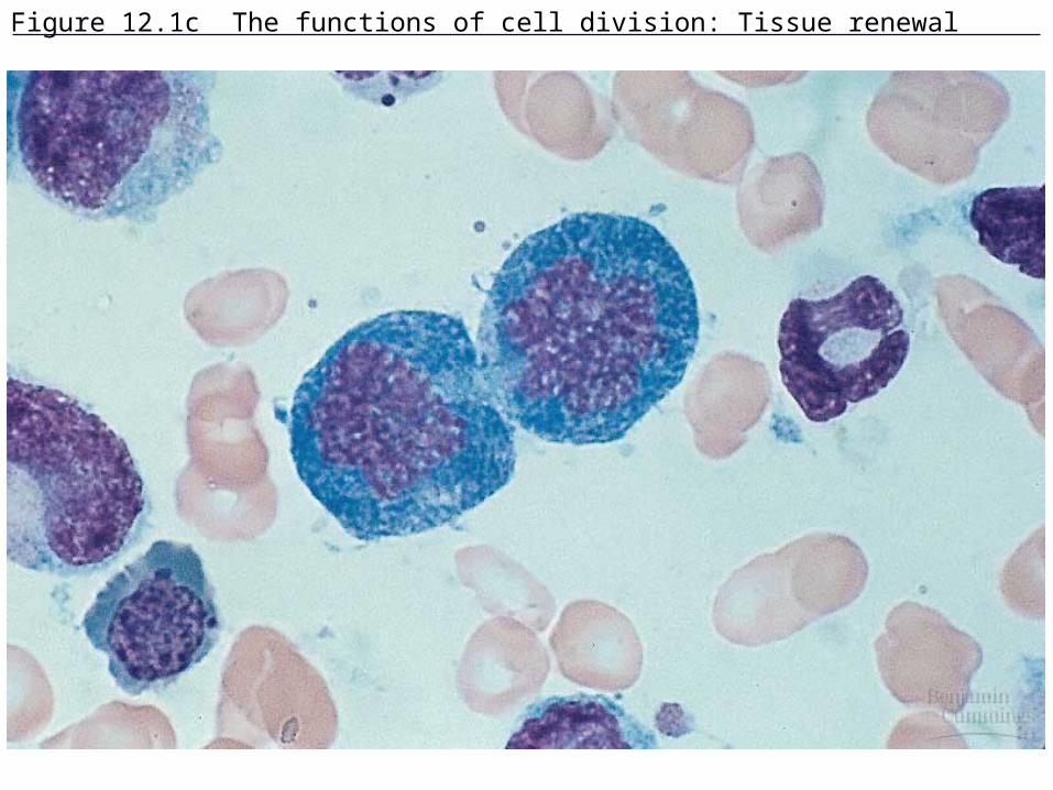
Figure 12.1c The functions of cell division: Tissue renewal
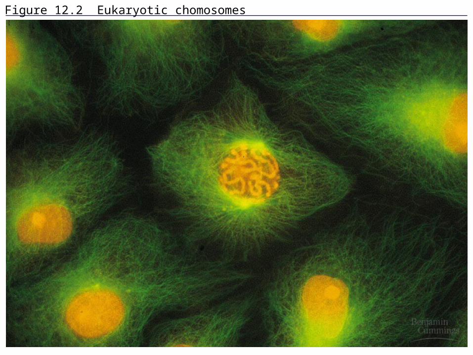
Figure 12.2 Eukaryotic chomosomes

Vocabulary Chromatin – long, thin fibers of
DNA wrapped around proteins Chromosome – one long DNA
molecule; condensed and clearly visible during cell division
Chromatid – two identical DNA molecules attached by a centromere (sister chromatids)

NEW VOCABULARY Centrosome – microtubule organizing
center which includes a pair of centrioles Centrosomes replicate in interphase and
move to opposite poles in prophase Centromere – region where 2 chromatids
are attached to one another Kinetochore – specialized region of
centromere where spindle fibers attach

Figure 12.3 Chromosome duplication and distribution during mitosis
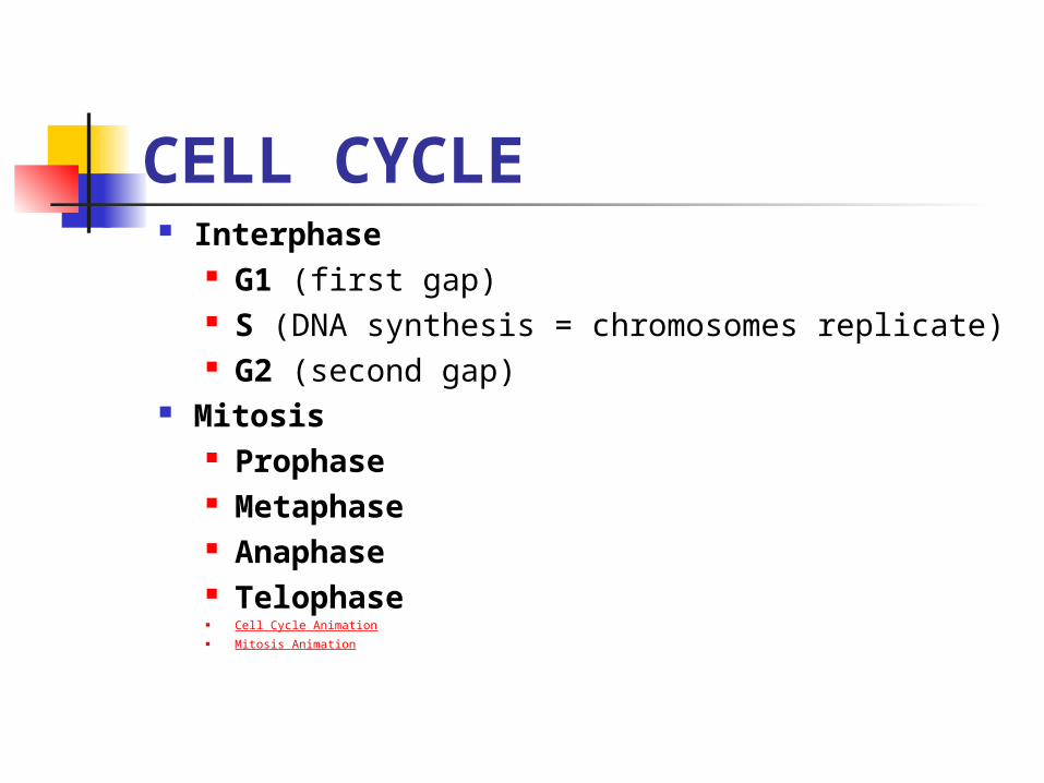
CELL CYCLE Interphase
G1 (first gap) S (DNA synthesis = chromosomes replicate) G2 (second gap)
Mitosis Prophase Metaphase Anaphase Telophase Cell Cycle Animation Mitosis Animation

Figure 12.4 The cell cycle

Prophase Chromosomes visible Centrosomes move towards
opposite poles and begin making spindle fiber
Nuclear membrane, nucleus, and nucleolus disintegrate
Spindle fiber form and some attach to the kinetochores of the centromeres
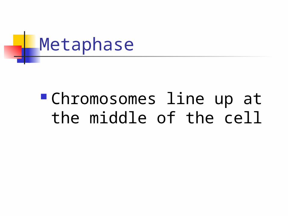
Metaphase
Chromosomes line up at the middle of the cell

Figure 12.6 The mitotic spindle at metaphase

Figure 12.5 The stages of mitotic cell division in an animal cell: G2 phase; prophase; prometaphase

Anaphase Sister chromatids are pulled
apart and move toward opposite ends of the cell by the spindle fiber
Nonkinetochore spindle help elongate the cell
Cell plate begins to form in plant cells (immature cell wall)

Telophase Events are opposite those of prophase Nuclear membranes, nuclei, and
nucleoli form in each new cell Cytokinesis occurs – (cleavage forms) Chromosomes unravel and become
chromatin again Spindle fibers disintegrate

Figure 12.5 The stages of mitotic cell division in an animal cell: metaphase; anaphase; telophase and cytokinesis.

Figure 12.5x Mitosis
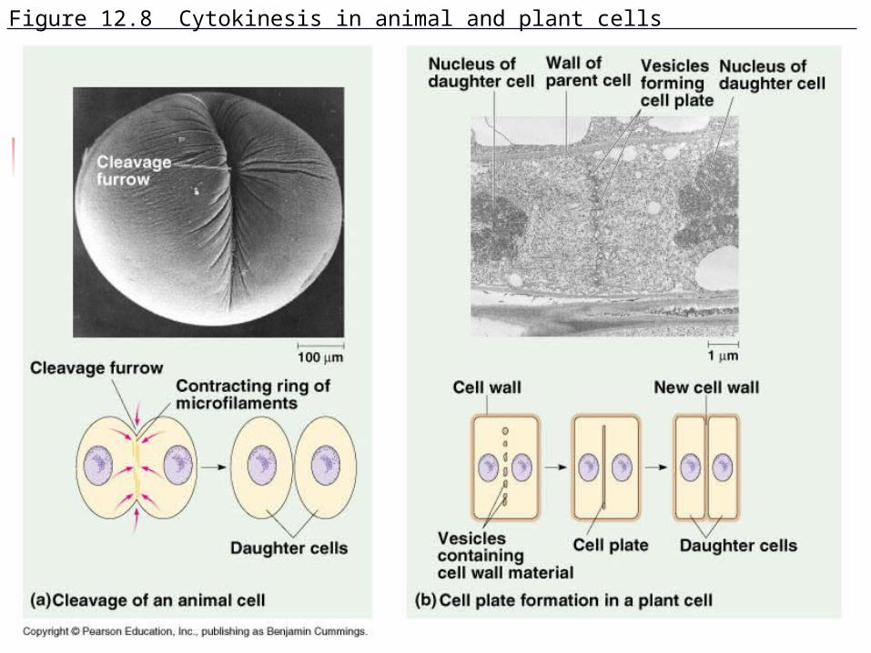
Figure 12.8 Cytokinesis in animal and plant cells

Figure 12.9 Mitosis in a plant cell
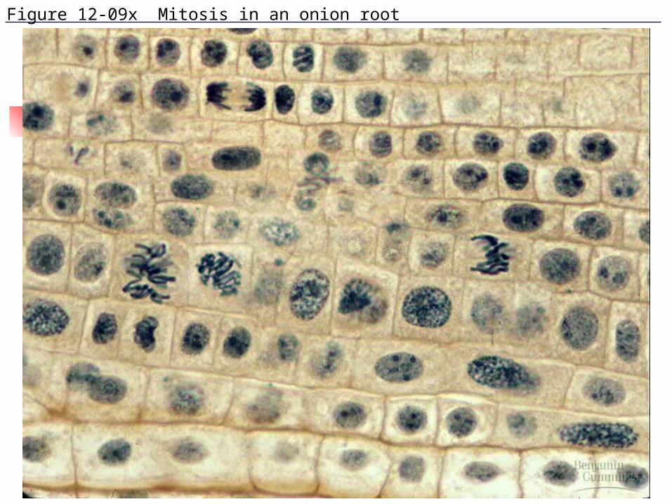
Figure 12-09x Mitosis in an onion root

BINARY FISSION Bacteria only have one
chromosome so steps of mitosis are not needed
Bacteria replicate via binary fission
DNA replicates at a specific point (origin of replication)

Figure 12.10 Bacterial cell division (binary fission) (Layer 1)

Figure 12.10 Bacterial cell division (binary fission) (Layer 2)

Figure 12.10 Bacterial cell division (binary fission) (Layer 3)

Evolution of Mitosis Prokaryotic and eukaryotic cell division share
some similar proteins that are involved in cell division
Possible intermediates: Current examples in some protists
Nuclear envelopes remain intact and replicated chromosomes attach to envelope
As nucleus elongates, chromosome separate
Spindle forms inside nucleus

REGULATION OF CELL CYCLE
Checkpoint – critical point in cell cycle where process can stop or go ahead according to signals
Kinases – enzymes that can activate or inactivate something via phosphorylation

Figure 12.13 Mechanical analogy for the cell cycle control system
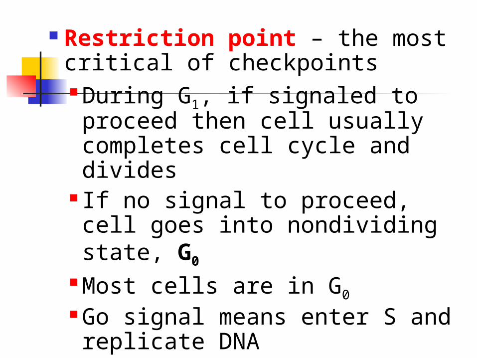
Restriction point – the most critical of checkpoints During G1, if signaled to proceed then cell usually completes cell cycle and divides
If no signal to proceed, cell goes into nondividing state, G0
Most cells are in G0
Go signal means enter S and replicate DNA

Cyclin is a protein that activates kinases that are called cyclin-dependent kinases or Cdks
MPF (maturation promoting factor) – combination of Cdks and cyclin
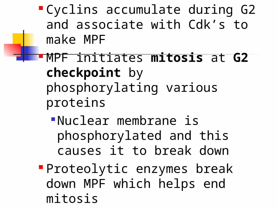
Cyclins accumulate during G2 and associate with Cdk’s to make MPF
MPF initiates mitosis at G2 checkpoint by phosphorylating various proteins
Nuclear membrane is phosphorylated and this causes it to break down
Proteolytic enzymes break down MPF which helps end mitosis

Figure 12.14 Molecular control of the cell cycle at the G2 checkpoint

M Phase Checkpoint M phase (metaphase checkpoint) Kinetochores not attached yet to
spindle send delay signals to prevent anaphase from starting too early.
Why must the cell wait for all of the chromosomes to line up in the middle of metaphase before proceeding to anaphase?

OTHER SIGNALS A signal that delays
anaphase so that right number of chromosomes end up in each new cell
Growth factors – external signals that can stimulate cell division

Density-dependent inhibition – cells stop dividing when crowded
Anchorage-dependent – most animal cells must be attach to substratum

Figure 12.16 Density-dependent inhibition of cell division
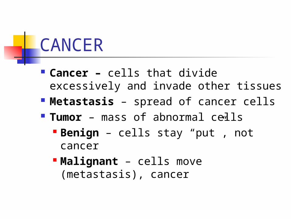
CANCER Cancer – cells that divide
excessively and invade other tissues Metastasis – spread of cancer cells Tumor – mass of abnormal cells
Benign – cells stay “put”, not cancer
Malignant – cells move (metastasis), cancer

Figure 12.17 The growth and metastasis of a malignant breast tumor
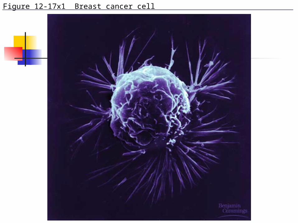
Figure 12-17x1 Breast cancer cell

Figure 12-17x2 Mammogram: normal (left) and cancerous (right)
Related Documents