RESEARCH Open Access Cell-associated bacteria in the human lung microbiome Robert P Dickson 1 , John R Erb-Downward 1 , Hallie C Prescott 1 , Fernando J Martinez 1 , Jeffrey L Curtis 1,2 , Vibha N Lama 1 and Gary B Huffnagle 1,3* Abstract Background: Recent studies have revealed that bronchoalveolar lavage (BAL) fluid contains previously unappreciated communities of bacteria. In vitro and in vivo studies have shown that host inflammatory signals prompt bacteria to disperse from cell-associated biofilms and adopt a virulent free-living phenotype. The proportion of the lung microbiota that is cell-associated is unknown. Results: Forty-six BAL specimens were obtained from lung transplant recipients and divided into two aliquots: ‘whole BAL’ and ‘acellular BAL,’ the latter processed with a low-speed, short-duration centrifugation step. Both aliquots were analyzed via bacterial 16S rRNA gene pyrosequencing. The BAL specimens represented a wide spectrum of lung health, ranging from healthy and asymptomatic to acutely infected. Bacterial signal was detected in 52% of acellular BAL aliquots, fewer than were detected in whole BAL (96%, p ≤ 0.0001). Detection of bacteria in acellular BAL was associated with indices of acute infection [BAL neutrophilia, high total bacterial (16S) DNA, low community diversity, p < 0.01 for all] and, independently, with low relative abundance of specific taxonomic groups (p< 0.05). When whole and acellular aliquots from the same bronchoscopy were directly compared, acellular BAL contained fewer bacterial species (p < 0.05); whole and acellular BAL similarity was positively associated with evidence of infection and negatively associated with relative abundance of several prominent taxa (p< 0.001). Acellular BAL contained decreased relative abundance of Prevotella spp. (p< 0.05) and Pseudomonas fluorescens (p< 0.05). Conclusions: We present a novel methodological and analytical approach to the localization of lung microbiota and show that prominent members of the lung microbiome are cell-associated, potentially via biofilms, cell adhesion, or intracellularity. Keywords: Lung microbiome, Bronchoalveolar lavage, 16S, Pyrosequencing, Pneumonia Background Novel techniques of culture-independent microbial identification have revealed that bronchoalveolar lavage (BAL) fluid acquired from healthy and diseased subjects contains diverse communities of previously unappreci- ated bacteria [1-4]. The location of these bacteria within the various compartments of the respiratory tract is unknown. Recent in vitro and in vivo studies have demonstrated that some biofilm-associated bacteria, when stimulated by inflammation-associated host signals (fever, norepinephrine, free ATP), disperse from bio- films and adopt a planktonic phenotype with increased virulence [5]. Most lung microbiome studies to date have employed pyrosequencing of 16S rRNA gene amplicons obtained from whole BAL specimens [1], while others have used acellular BAL obtained via a low-speed, short-duration centrifugation step for eukaryotic cell removal [6,7]. This use of acellular BAL may exclude bacteria that are cell- associated via biofilms, cell-adhesion appendages, or intra- cellularity, though to date no published study has directly compared whole BAL to acellular BAL microbiota. In this study, we sought to determine which members of the lung microbiome are predominantly cell-associated * Correspondence: [email protected] 1 Division of Pulmonary and Critical Care Medicine, Department of Internal Medicine, University of Michigan Medical School, Ann Arbor, MI 48109, USA 3 Department of Microbiology and Immunology, University of Michigan Medical School, Ann Arbor, MI 48109, USA Full list of author information is available at the end of the article © 2014 Dickson et al.; licensee BioMed Central Ltd. This is an Open Access article distributed under the terms of the Creative Commons Attribution License (http://creativecommons.org/licenses/by/4.0), which permits unrestricted use, distribution, and reproduction in any medium, provided the original work is properly credited. The Creative Commons Public Domain Dedication waiver (http://creativecommons.org/publicdomain/zero/1.0/) applies to the data made available in this article, unless otherwise stated. Dickson et al. Microbiome 2014, 2:28 http://www.microbiomejournal.com/content/2/1/28

Welcome message from author
This document is posted to help you gain knowledge. Please leave a comment to let me know what you think about it! Share it to your friends and learn new things together.
Transcript
Dickson et al. Microbiome 2014, 2:28http://www.microbiomejournal.com/content/2/1/28
RESEARCH Open Access
Cell-associated bacteria in the human lungmicrobiomeRobert P Dickson1, John R Erb-Downward1, Hallie C Prescott1, Fernando J Martinez1, Jeffrey L Curtis1,2,Vibha N Lama1 and Gary B Huffnagle1,3*
Abstract
Background: Recent studies have revealed that bronchoalveolar lavage (BAL) fluid contains previously unappreciatedcommunities of bacteria. In vitro and in vivo studies have shown that host inflammatory signals prompt bacteria todisperse from cell-associated biofilms and adopt a virulent free-living phenotype. The proportion of the lungmicrobiota that is cell-associated is unknown.
Results: Forty-six BAL specimens were obtained from lung transplant recipients and divided into two aliquots:‘whole BAL’ and ‘acellular BAL,’ the latter processed with a low-speed, short-duration centrifugation step. Bothaliquots were analyzed via bacterial 16S rRNA gene pyrosequencing. The BAL specimens represented a widespectrum of lung health, ranging from healthy and asymptomatic to acutely infected. Bacterial signal was detectedin 52% of acellular BAL aliquots, fewer than were detected in whole BAL (96%, p ≤ 0.0001). Detection of bacteriain acellular BAL was associated with indices of acute infection [BAL neutrophilia, high total bacterial (16S) DNA,low community diversity, p < 0.01 for all] and, independently, with low relative abundance of specific taxonomicgroups (p < 0.05). When whole and acellular aliquots from the same bronchoscopy were directly compared,acellular BAL contained fewer bacterial species (p < 0.05); whole and acellular BAL similarity was positivelyassociated with evidence of infection and negatively associated with relative abundance of several prominenttaxa (p < 0.001). Acellular BAL contained decreased relative abundance of Prevotella spp. (p < 0.05) andPseudomonas fluorescens (p < 0.05).
Conclusions: We present a novel methodological and analytical approach to the localization of lung microbiotaand show that prominent members of the lung microbiome are cell-associated, potentially via biofilms, celladhesion, or intracellularity.
Keywords: Lung microbiome, Bronchoalveolar lavage, 16S, Pyrosequencing, Pneumonia
BackgroundNovel techniques of culture-independent microbialidentification have revealed that bronchoalveolar lavage(BAL) fluid acquired from healthy and diseased subjectscontains diverse communities of previously unappreci-ated bacteria [1-4]. The location of these bacteria withinthe various compartments of the respiratory tract isunknown. Recent in vitro and in vivo studies havedemonstrated that some biofilm-associated bacteria,
* Correspondence: [email protected] of Pulmonary and Critical Care Medicine, Department of InternalMedicine, University of Michigan Medical School, Ann Arbor, MI 48109, USA3Department of Microbiology and Immunology, University of MichiganMedical School, Ann Arbor, MI 48109, USAFull list of author information is available at the end of the article
© 2014 Dickson et al.; licensee BioMed CentraCommons Attribution License (http://creativecreproduction in any medium, provided the orDedication waiver (http://creativecommons.orunless otherwise stated.
when stimulated by inflammation-associated host signals(fever, norepinephrine, free ATP), disperse from bio-films and adopt a planktonic phenotype with increasedvirulence [5].Most lung microbiome studies to date have employed
pyrosequencing of 16S rRNA gene amplicons obtainedfrom whole BAL specimens [1], while others have usedacellular BAL obtained via a low-speed, short-durationcentrifugation step for eukaryotic cell removal [6,7]. Thisuse of acellular BAL may exclude bacteria that are cell-associated via biofilms, cell-adhesion appendages, or intra-cellularity, though to date no published study has directlycompared whole BAL to acellular BAL microbiota.In this study, we sought to determine which members
of the lung microbiome are predominantly cell-associated
l Ltd. This is an Open Access article distributed under the terms of the Creativeommons.org/licenses/by/4.0), which permits unrestricted use, distribution, andiginal work is properly credited. The Creative Commons Public Domaing/publicdomain/zero/1.0/) applies to the data made available in this article,
Dickson et al. Microbiome 2014, 2:28 Page 2 of 10http://www.microbiomejournal.com/content/2/1/28
and which are free-living within the respiratory tract. Wehypothesized that removal of eukaryotic cells from BALfluid would alter the composition of the microbial com-munities identified by pyrosequencing, reflecting the se-lective removal of cell-associated bacteria. We furtherhypothesized that the predominance of free-living bacteriawould be associated with indices of acute infection. Totest these hypotheses, we designed an analysis of 46 clinic-ally obtained BAL specimens, each analyzed for bacterialcommunity membership using both whole and acellularBAL. All BAL specimens were obtained from lung trans-plant recipients, which represented a wide spectrum oflung health (ranging from healthy and asymptomatic toacutely infected). The respiratory pathogen profile in thisgroup is similar both to that observed in healthcare-associated pneumonia as well as pneumonia in other im-munocompromised states [8-10]. We present a novelmethodological and analytical approach to the localizationof lung microbiota and demonstrate that prominent mem-bers of the lung microbiome are cell-associated.
ResultsFactors associated with detection of bacteria in acellularBALAs described in the ‘Methods’ section, 46 BAL specimenswere obtained from lung transplant recipients, with 46%collected for an acute clinical indication (dyspnea, cough,radiographic infiltrate, or decline in lung function) and theremaining 54% performed as surveillance bronchoscopieson asymptomatic patients. As we have previously reported[11], the microbiological profile of respiratory pathogensidentified in BAL obtained from symptomatic patients inour study strongly resembled that of healthcare-associatedpneumonia/hospital-acquired pneumonia as well as pneu-monia in other immunocompromised states [8,9], domi-nated by Staphylococcus aureus, Pseudomonas aeruginosa,and other non-fermenting gram negative rods. Each BALspecimen was divided into two equal aliquots, andeukaryotic cells were removed from one aliquot (‘acellularBAL’) by short, low-speed centrifugation. Both acellularand whole BAL aliquots were then centrifuged at highspeed for 30 min to pellet the bacteria in the sample forDNA preparation, V3–V5 16S rRNA gene amplicon li-brary generation, and 454 pyrosequencing and subsequentbioinformatics, as outlined in the ‘Methods’ section.Bacterial communities were detected in 96% of whole
BAL aliquots, while only 52% of acellular BAL aliquotscontained detectable bacteria via pyrosequencing (p ≤0.0001) (as assessed by the presence or absence of bandscorresponding to the 16S rRNA gene after the amplifica-tion step of our pyrosequencing protocol). Given thismarked difference, we compared BAL specimens based onwhether their acellular aliquots contained detectable bac-terial signal (Figure 1). Anticipating that total bacterial
burden would be an important factor, we confirmed thatsamples with detectable bacteria in acellular BAL hadhigher bacterial (16S) DNA burden as measured by quan-titative polymerase chain reaction (qPCR) of the wholeBAL (p < 0.0001). Consistent with this, the same BALspecimens had higher BAL neutrophilia (p < 0.01) andlower whole BAL community diversity (p < 0.01), indicat-ing that the presence of detectable bacteria in acellularBAL is associated with indices of acute infection.To compare the bacterial community membership of
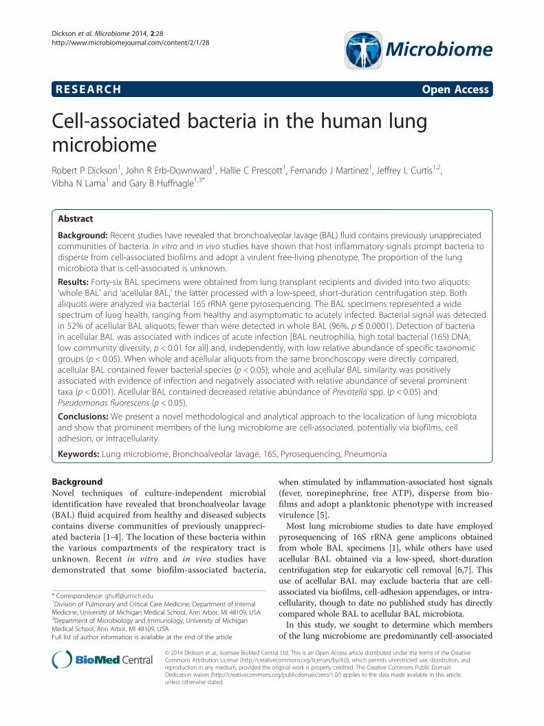
BAL specimens based on whether they had bacteria de-tectable in acellular BAL, we used the data visualizationtechnique of unconstrained ordination, with whole BALaliquots labeled according to whether bacteria weredetectable in their corresponding acellular aliquots(Figure 2). This demonstrated a spatial separation ofBAL specimens based on the presence of bacteria intheir acellular aliquot, implying collective differences inthe microbiota. This difference was confirmed as statisti-cally significant using multiple methods of hypothesistesting [constrained ordination (redundancy analysis,RDA) and PERMANOVA (adonis), p < 0.05 both], evenwhen controlled for total bacterial (16S) DNA (p < 0.05both). These multiple analyses confirmed that the detec-tion of bacteria in acellular specimens is independentlyinfluenced by bacterial community membership, notmerely by total bacterial (16S) DNA.To better define the influence of community member-
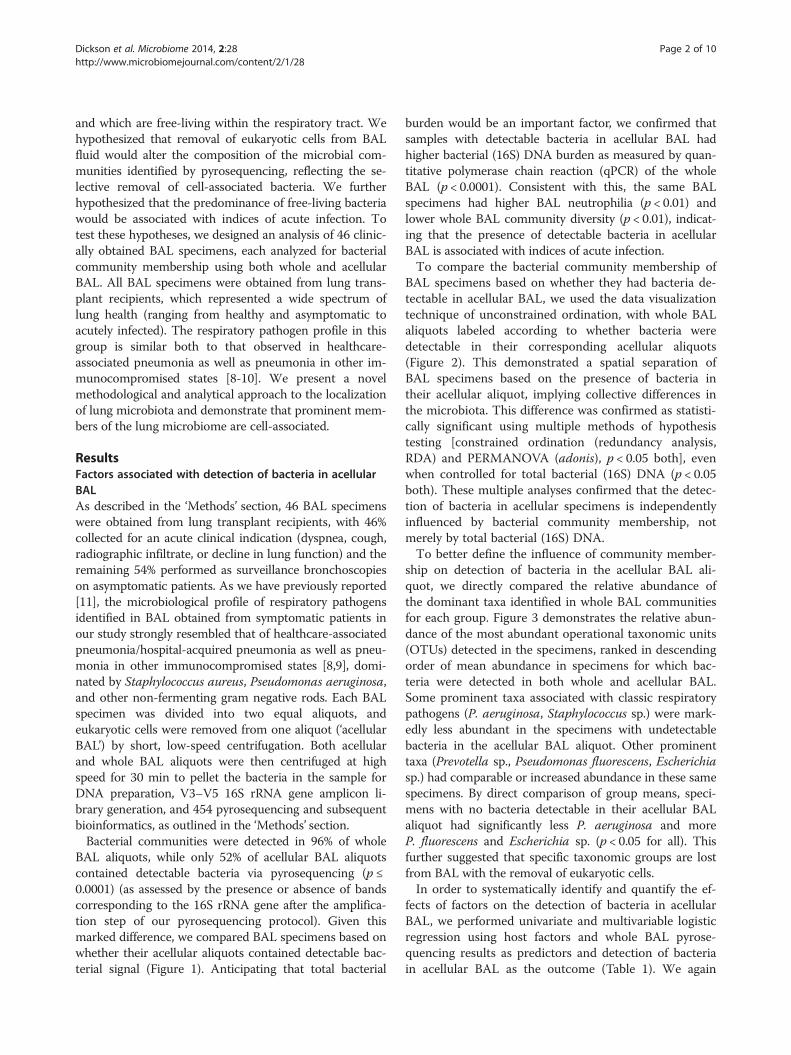
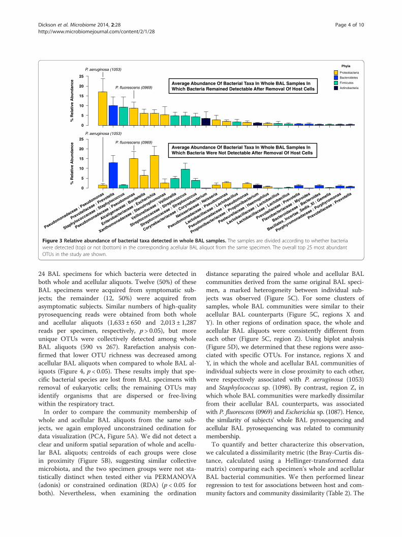
ship on detection of bacteria in the acellular BAL ali-quot, we directly compared the relative abundance ofthe dominant taxa identified in whole BAL communitiesfor each group. Figure 3 demonstrates the relative abun-dance of the most abundant operational taxonomic units(OTUs) detected in the specimens, ranked in descendingorder of mean abundance in specimens for which bac-teria were detected in both whole and acellular BAL.Some prominent taxa associated with classic respiratorypathogens (P. aeruginosa, Staphylococcus sp.) were mark-edly less abundant in the specimens with undetectablebacteria in the acellular BAL aliquot. Other prominenttaxa (Prevotella sp., Pseudomonas fluorescens, Escherichiasp.) had comparable or increased abundance in these samespecimens. By direct comparison of group means, speci-mens with no bacteria detectable in their acellular BALaliquot had significantly less P. aeruginosa and moreP. fluorescens and Escherichia sp. (p < 0.05 for all). Thisfurther suggested that specific taxonomic groups are lostfrom BAL with the removal of eukaryotic cells.In order to systematically identify and quantify the ef-
fects of factors on the detection of bacteria in acellularBAL, we performed univariate and multivariable logisticregression using host factors and whole BAL pyrose-quencing results as predictors and detection of bacteriain acellular BAL as the outcome (Table 1). We again
Bacteria Detected in Acellular BAL
Bacteria Not Detected in Acellular BAL
0
50
100
% N
eutr
ophi
ls
Bacteria Detected in Acellular BAL
Bacteria Not Detected in Acellular BAL
2
4
6
8
16S
DN
Alo
g(co
pies
per
5 m
L of
BA
L)
Bacteria Detected in Acellular BAL
Bacteria Not Detected in Acellular BAL
0
1
2
3
4
5
Sha
non
Div
ersi
ty In
dex
LOD
A. C.B.
p < 0.01 p < 0.0001 p < 0.01
Figure 1 Comparison of whole BAL specimens by whether bacterial DNA was detected in acellular specimens. When whole BALspecimens were analyzed according to whether bacterial DNA was detected in their acellular fraction, specimens with bacterial DNA in theacellular fraction had more associated neutrophilia (A), more bacterial (16S) DNA as measured by qPCR (B), and lower community diversity asmeasured by the Shannon Index (C). LOD, level of detection.
Dickson et al. Microbiome 2014, 2:28 Page 3 of 10http://www.microbiomejournal.com/content/2/1/28
found that the detection of bacteria in acellular BALby pyrosequencing was positively associated with indicesof acute infection [total bacterial (16S) DNA, BAL neutro-philia and low community diversity (p < 0.01)]. Detection ofbacteria in acellular BAL was negatively associated with rela-tive abundance of the Enterobacteriaceae family (p < 0.05)and the OTU classified as Escherichia sp. (1087) (p < 0.05).Both of these taxonomic associations remained significanteven when controlled via multivariable logistic regressionfor total bacterial (16S) DNA. Thus, detection of bacteria
-6 -4 -2
-4-2
02
46
8
PC1 (23
PC
2 (1
7.4%
Exp
lain
ed)
Bacteria Not Detectable in Acellular BAL
Bacteria Detectable in Acellular BAL
Figure 2 Ordination analyses of bacterial communities detected in whof bacterial communities in unfractionated whole BAL aliquots, as determinlabeled by whether bacteria could be detected in the acellular BAL aliquot
in acellular BAL aliquots was strongly associated with evi-dence of acute infection but also independently and nega-tively associated with relative abundance of specificprominent taxonomic groups, implying cell association ofthese types of microbes.
Influence of eukaryotic cell removal on BAL bacterialcommunitiesTo determine the direct effect of host cell removal ondetecting bacterial communities, we then focused on the
0 2 4 6
.9% Explained)
ole BAL samples. Unsupervised principal component analysis (PCA)ed by pyrosequencing of V3–V5 16S rRNA gene amplicon libraries,s of the same samples.
% R
elat
ive
Ab
un
dan
ce
0
5
10
15
20
25%
Rel
ativ
e A
bu
nd
ance
0
5
10
15
20
25
Pseudomonadaceae : Pseudomonas
Prevotellaceae : Prevotella
Staphylococcaceae : Staphylococcus
Pseudomonadaceae : Pseudomonas
Alcaligenaceae : Bordetella
Enterobacteriaceae : E
scherichia
Xanthomonadaceae : Stenotro
phomonas
Veillonellaceae : V
eillonella
Streptococcaceae : S
treptococcus
Streptococcaceae : S
treptococcus
Corynebacteriaceae : C
orynebacterium
Neisseriaceae : N
eisseria
Pseudomonadaceae : Pseudomonas
Lactobacillaceae : L
actobacillus
Pseudomonadaceae : Pseudomonas
Propionibacteriaceae : P
ropionibacterium
Pasteurellaceae : Haemophilus
Lactobacillaceae : L
actobacillus
Lactobacillaceae : L
actobacillus
Prevotellaceae : Prevotella
Flavobacteriaceae : M
yroides
Bacteroidaceae : Bacteroides
Bacillales_Incerta
e_Sedis_XI : Gemella
Porphyromonadaceae : Porphyromonas
Prevotellaceae : Prevotella
Average Abundance Of Bacterial Taxa In Whole BAL Samples In Which Bacteria Remained Detectable After Removal Of Host Cells
Firmicutes
Phyla
Proteobacteria
Bacteroidetes
Actinobacteria
P. aeruginosa (1053)
P. fluorescens (0969)
P. aeruginosa (1053)
P. fluorescens (0969)Average Abundance Of Bacterial Taxa In Whole BAL Samples In Which Bacteria Were Not Detectable After Removal Of Host Cells
Figure 3 Relative abundance of bacterial taxa detected in whole BAL samples. The samples are divided according to whether bacteriawere detected (top) or not (bottom) in the corresponding acellular BAL aliquot from the same specimen. The overall top 25 most abundantOTUs in the study are shown.
Dickson et al. Microbiome 2014, 2:28 Page 4 of 10http://www.microbiomejournal.com/content/2/1/28
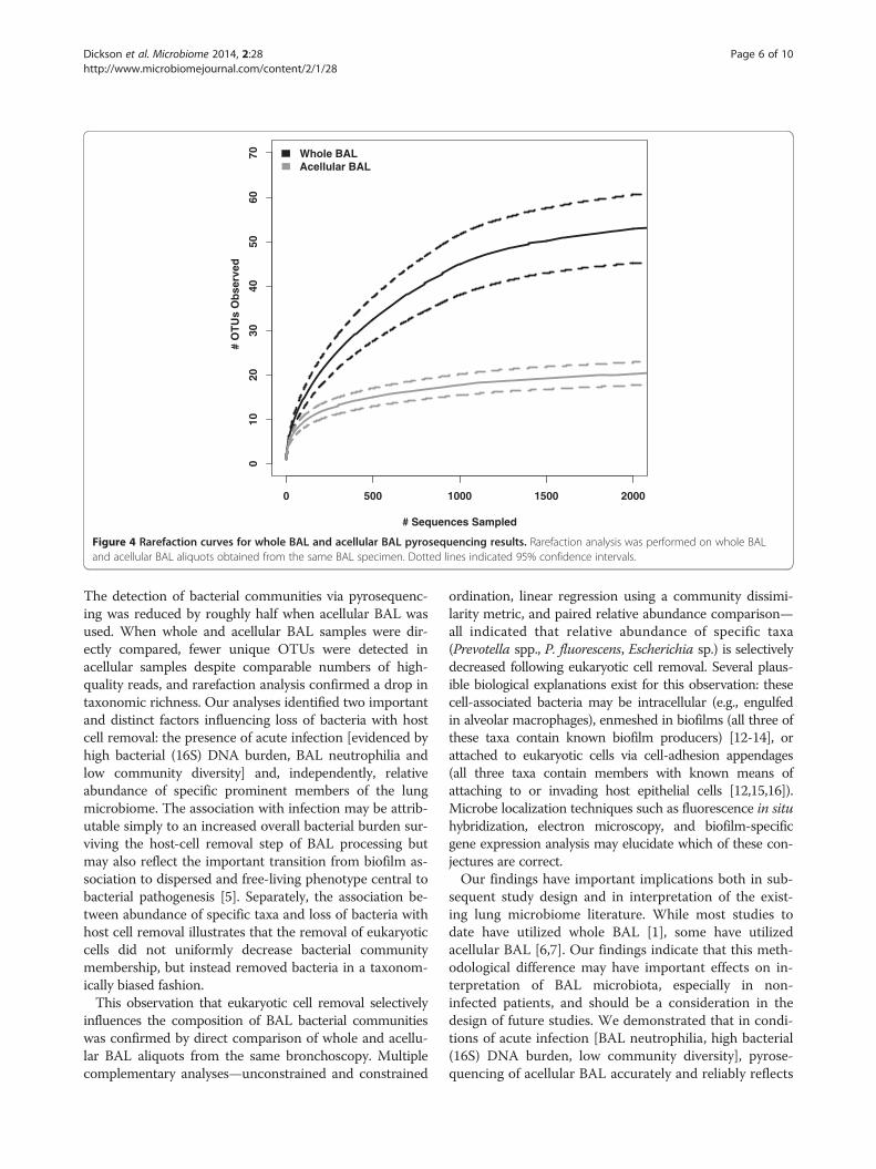
24 BAL specimens for which bacteria were detected inboth whole and acellular aliquots. Twelve (50%) of theseBAL specimens were acquired from symptomatic sub-jects; the remainder (12, 50%) were acquired fromasymptomatic subjects. Similar numbers of high-qualitypyrosequencing reads were obtained from both wholeand acellular aliquots (1,633 ± 650 and 2,013 ± 1,287reads per specimen, respectively, p > 0.05), but moreunique OTUs were collectively detected among wholeBAL aliquots (590 vs 267). Rarefaction analysis con-firmed that lower OTU richness was decreased amongacellular BAL aliquots when compared to whole BAL al-iquots (Figure 4, p < 0.05). These results imply that spe-cific bacterial species are lost from BAL specimens withremoval of eukaryotic cells; the remaining OTUs mayidentify organisms that are dispersed or free-livingwithin the respiratory tract.In order to compare the community membership of
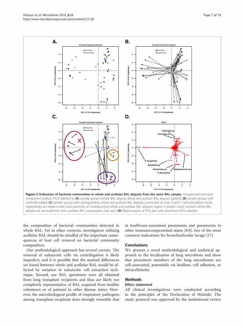
whole and acellular BAL aliquots from the same sub-jects, we again employed unconstrained ordination fordata visualization (PCA, Figure 5A). We did not detect aclear and uniform spatial separation of whole and acellu-lar BAL aliquots; centroids of each groups were closein proximity (Figure 5B), suggesting similar collectivemicrobiota, and the two specimen groups were not sta-tistically distinct when tested either via PERMANOVA(adonis) or constrained ordination (RDA) (p < 0.05 forboth). Nevertheless, when examining the ordination
distance separating the paired whole and acellular BALcommunities derived from the same original BAL speci-men, a marked heterogeneity between individual sub-jects was observed (Figure 5C). For some clusters ofsamples, whole BAL communities were similar to theiracellular BAL counterparts (Figure 5C, regions X andY). In other regions of ordination space, the whole andacellular BAL aliquots were consistently different fromeach other (Figure 5C, region Z). Using biplot analysis(Figure 5D), we determined that these regions were asso-ciated with specific OTUs. For instance, regions X andY, in which the whole and acellular BAL communities ofindividual subjects were in close proximity to each other,were respectively associated with P. aeruginosa (1053)and Staphylococcus sp. (1098). By contrast, region Z, inwhich whole BAL communities were markedly dissimilarfrom their acellular BAL counterparts, was associatedwith P. fluorescens (0969) and Escherichia sp. (1087). Hence,the similarity of subjects' whole BAL pyrosequencing andacellular BAL pyrosequencing was related to communitymembership.To quantify and better characterize this observation,
we calculated a dissimilarity metric (the Bray-Curtis dis-tance, calculated using a Hellinger-transformed datamatrix) comparing each specimen's whole and acellularBAL bacterial communities. We then performed linearregression to test for associations between host and com-munity factors and community dissimilarity (Table 2). The
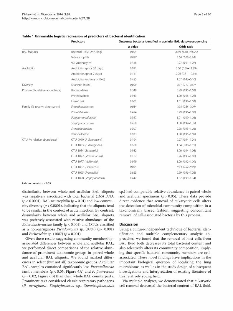
Table 1 Univariable logistic regression of predictors of bacterial identification
Predictors Outcome: bacteria identified in acellular BAL via pyrosequencing
p value Odds ratio
BAL features Bacterial (16S) DNA (log) 0.004 26.93 (4.58–476.29)
% Neutrophils 0.027 1.06 (1.02–1.14)
% Lymphocytes 0.318 0.97 (0.91–1.02)
Antibiotics Antibiotics (prior 30 days) 0.091 3.00 (0.86–11.29)
Antibiotics (prior 7 days) 0.111 2.76 (0.81–10.14)
Antibiotics (at time of BAL) 0.425 1.67 (0.48–6.10)
Diversity Shannon Index 0.009 0.31 (0.11–0.67)
Phylum (% relative abundance) Bacteroidetes 0.349 0.99 (0.95–1.02)
Proteobacteria 0.933 1.00 (0.98–1.02)
Firmicutes 0.601 1.01 (0.98–1.03)
Family (% relative abundance) Enterobacteriaceae 0.034 0.93 (0.86–0.99)
Prevotellaceae 0.494 0.99 (0.96–1.02)
Pseudomonadaceae 0.367 1.01 (0.99–1.03)
Staphylococcaceae 0.450 1.08 (0.99–1.39)
Streptococcaceae 0.307 0.98 (0.93–1.02)
Veillonellaceae 0.933 1.00 (0.91–1.09)
OTU (% relative abundance) OTU 0969 (P. fluorescens) 0.194 0.97 (0.94–1.01)
OTU 1053 (P. aeruginosa) 0.168 1.04 (1.00–1.19)
OTU 1054 (Bordetella) 0.932 1.00 (0.94–1.06)
OTU 1072 (Streptococcus) 0.172 0.96 (0.90–1.01)
OTU 1077 (Veillonella) 0.999 1.00 (0.92–1.09)
OTU 1087 (Escherichia) 0.035 0.93 (0.87–0.99)
OTU 1095 (Prevotella) 0.625 0.99 (0.96–1.02)
OTU 1098 (Staphylococcus) 0.442 1.07 (0.99–1.34)
Italicized results: p < 0.05.
Dickson et al. Microbiome 2014, 2:28 Page 5 of 10http://www.microbiomejournal.com/content/2/1/28
dissimilarity between whole and acellular BAL aliquotswas negatively associated with total bacterial (16S) DNA(p < 0.0001), BAL neutrophilia (p = 0.01) and low commu-nity diversity (p < 0.0001), indicating that the aliquots tendto be similar in the context of acute infection. By contrast,dissimilarity between whole and acellular BAL aliquotswas positively associated with relative abundance of theEnterobacteriaceae family (p < 0.001) and OTUs classifiedas a non-aeruginosa Pseudomonas sp. (0969) (p < 0.001)and Escherichia sp. (1087) (p = 0.001).Given these results suggesting community membership-
associated differences between whole and acellular BAL,we performed direct comparisons of the relative abun-dance of prominent taxonomic groups in paired wholeand acellular BAL aliquots. We found marked differ-ences in select (but not all) taxonomic groups. AcellularBAL samples contained significantly less Prevotellaceaefamily members (p < 0.05, Figure 6A) and P. fluorescens(p = 0.02, Figure 6B) than their whole BAL counterparts.Prominent taxa considered classic respiratory pathogens(P. aeruginosa, Staphylococcus sp., Stenotrophomonas
sp.) had comparable relative abundance in paired wholeand acellular specimens (p > 0.05). These data providedirect evidence that removal of eukaryotic cells altersthe detection of microbial community composition in ataxonomically biased fashion, suggesting concomitantremoval of cell-associated bacteria by this process.
DiscussionUsing a culture-independent technique of bacterial iden-tification and multiple complementary analytic ap-proaches, we found that the removal of host cells fromBAL fluid both decreases its total bacterial content andalso selectively alters its community composition, imply-ing that specific bacterial community members are cell-associated. These novel findings have implications in theimportant biological question of localizing the lungmicrobiome, as well as in the study design of subsequentinvestigations and interpretation of existing literature ofthis relatively young field.Via multiple analyses, we demonstrated that eukaryotic
cell removal decreased the bacterial content of BAL fluid.
# O
TU
s O
bse
rved
0 500 1000 1500 2000
010
2030
4050
6070
# Sequences Sampled
Whole BALAcellular BAL
Figure 4 Rarefaction curves for whole BAL and acellular BAL pyrosequencing results. Rarefaction analysis was performed on whole BALand acellular BAL aliquots obtained from the same BAL specimen. Dotted lines indicated 95% confidence intervals.
Dickson et al. Microbiome 2014, 2:28 Page 6 of 10http://www.microbiomejournal.com/content/2/1/28
The detection of bacterial communities via pyrosequenc-ing was reduced by roughly half when acellular BAL wasused. When whole and acellular BAL samples were dir-ectly compared, fewer unique OTUs were detected inacellular samples despite comparable numbers of high-quality reads, and rarefaction analysis confirmed a drop intaxonomic richness. Our analyses identified two importantand distinct factors influencing loss of bacteria with hostcell removal: the presence of acute infection [evidenced byhigh bacterial (16S) DNA burden, BAL neutrophilia andlow community diversity] and, independently, relativeabundance of specific prominent members of the lungmicrobiome. The association with infection may be attrib-utable simply to an increased overall bacterial burden sur-viving the host-cell removal step of BAL processing butmay also reflect the important transition from biofilm as-sociation to dispersed and free-living phenotype central tobacterial pathogenesis [5]. Separately, the association be-tween abundance of specific taxa and loss of bacteria withhost cell removal illustrates that the removal of eukaryoticcells did not uniformly decrease bacterial communitymembership, but instead removed bacteria in a taxonom-ically biased fashion.This observation that eukaryotic cell removal selectively
influences the composition of BAL bacterial communitieswas confirmed by direct comparison of whole and acellu-lar BAL aliquots from the same bronchoscopy. Multiplecomplementary analyses—unconstrained and constrained
ordination, linear regression using a community dissimi-larity metric, and paired relative abundance comparison—all indicated that relative abundance of specific taxa(Prevotella spp., P. fluorescens, Escherichia sp.) is selectivelydecreased following eukaryotic cell removal. Several plaus-ible biological explanations exist for this observation: thesecell-associated bacteria may be intracellular (e.g., engulfedin alveolar macrophages), enmeshed in biofilms (all three ofthese taxa contain known biofilm producers) [12-14], orattached to eukaryotic cells via cell-adhesion appendages(all three taxa contain members with known means ofattaching to or invading host epithelial cells [12,15,16]).Microbe localization techniques such as fluorescence in situhybridization, electron microscopy, and biofilm-specificgene expression analysis may elucidate which of these con-jectures are correct.Our findings have important implications both in sub-
sequent study design and in interpretation of the exist-ing lung microbiome literature. While most studies todate have utilized whole BAL [1], some have utilizedacellular BAL [6,7]. Our findings indicate that this meth-odological difference may have important effects on in-terpretation of BAL microbiota, especially in non-infected patients, and should be a consideration in thedesign of future studies. We demonstrated that in condi-tions of acute infection [BAL neutrophilia, high bacterial(16S) DNA burden, low community diversity], pyrose-quencing of acellular BAL accurately and reliably reflects
A. B.
C. D.
-8 -6 -4 -2 0 2 4
-8-6
-4-2
02
4
PC1 (17.3% Explained)
PC
2 (1
2.9%
Exp
lain
ed)
Principal Component Analysis
Whole BAL
Acellular BAL
-8 -6 -4 -2 0 2 4
-8-6
-4-2
02
4
PC1 (17.3% Explained)
PC
2 (1
2.9%
Exp
lain
ed)
Whole BAL
Acellular BAL
Principal Component Analysis
Whole BAL
Acellular BAL
-8 -6 -4 -2 0 2 4
-8-6
-4-2
02
4
PC1 (17.3% Explained)
PC
2 (1
2.9%
Exp
lain
ed)
Principal Component Analysis
X
Z
Y
Whole BAL
Acellular BAL
-8 -6 -4 -2 0 2 4
-8-6
-4-2
02
46
PC1 (17.3% Explained)
PC
2 (1
2.9%
Exp
lain
ed)
Otu0024Otu0035Otu0262Otu0357Otu0445Otu0493Otu0514Otu0571Otu0582Otu0641Otu0661Otu0680Otu0708Otu0715Otu0723Otu0740Otu0741Otu0745Otu0746Otu0749Otu0756Otu0766Otu0767Otu0773
Otu0776
Otu0779Otu0802Otu0804Otu0806Otu0807Otu0824Otu0827Otu0847Otu0852Otu0863Otu0878Otu0881Otu0886Otu0887Otu0888Otu0889Otu0893Otu0895Otu0896Otu0897Otu0898Otu0899Otu0901Otu0902Otu0903Otu0904Otu0905Otu0907Otu0908Otu0913Otu0914Otu0917
Otu0921
Otu0926Otu0928Otu0929Otu0930Otu0931Otu0934Otu0939Otu0940Otu0942Otu0943Otu0944Otu0945Otu0953Otu0956Otu0958Otu0959Otu0960Otu0961
Otu0962Otu0966Otu0967Otu0968
Otu0969
Otu0970Otu0981Otu0993Otu0996Otu0997Otu0998Otu1001Otu1003Otu1005
Otu1007Otu1019Otu1024
Otu1029
Otu1032Otu1033
Otu1039
Otu1042
Otu1045
Otu1053
Otu1054
Otu1057
Otu1059
Otu1060Otu1062Otu1063Otu1066Otu1067
Otu1072Otu1075
Otu1077
Otu1082
Otu1083
Otu1087
Otu1092Otu1094
Otu1095
Otu1096Otu1097
Otu1098
Otu1099
Biplot
P. aeruginosa
Bordetella sp.
Escherichia sp.
P. fluorescens
Staphylococcus sp.
Veillonella sp.
Prevotella sp.
Figure 5 Ordination of bacterial communities in whole and acellular BAL aliquots from the same BAL sample. Unsupervised principalcomponent analysis (PCA) labeled as (A) sample groups [whole BAL aliquots (blue) and acellular BAL aliquots (green)]; (B) sample groups withcentroids added; (C) sample groups with corresponding whole and acellular BAL aliquots connected via lines. X and Y (red and yellow circles,respectively) are regions with close proximity of corresponding whole and acellular BAL aliquots; region Z (purple circle) contains whole BALaliquots far removed from their acellular BAL counterparts. (See text.) (D) Biplot analysis of PCA plot with prominent OTUs labeled.
Dickson et al. Microbiome 2014, 2:28 Page 7 of 10http://www.microbiomejournal.com/content/2/1/28
the composition of bacterial communities detected inwhole BAL. Yet in other contexts, investigators utilizingacellular BAL should be mindful of the important conse-quences of host cell removal on bacterial communitycomposition.Our methodological approach has several caveats. The
removal of eukaryotic cells via centrifugation is likelyimperfect, and it is possible that the marked differenceswe found between whole and acellular BAL would be af-fected by variation in eukaryotic cell extraction tech-nique. Second, our BAL specimens were all obtainedfrom lung transplant recipients and thus are likely notcompletely representative of BAL acquired from healthyvolunteers or of patients in other disease states. How-ever, the microbiological profile of respiratory pathogensamong transplant recipients does strongly resemble that
in healthcare-associated pneumonia and pneumonia inother immunocompromised states [8,9], two of the mostcommon indications for bronchoalveolar lavage [17].
ConclusionsWe present a novel methodological and analytical ap-proach to the localization of lung microbiota and showthat prominent members of the lung microbiome arecell-associated, potentially via biofilms, cell adhesion, orintracellularity.
MethodsEthics statementAll clinical investigations were conducted accordingto the principles of the Declaration of Helsinki. Thestudy protocol was approved by the institutional review
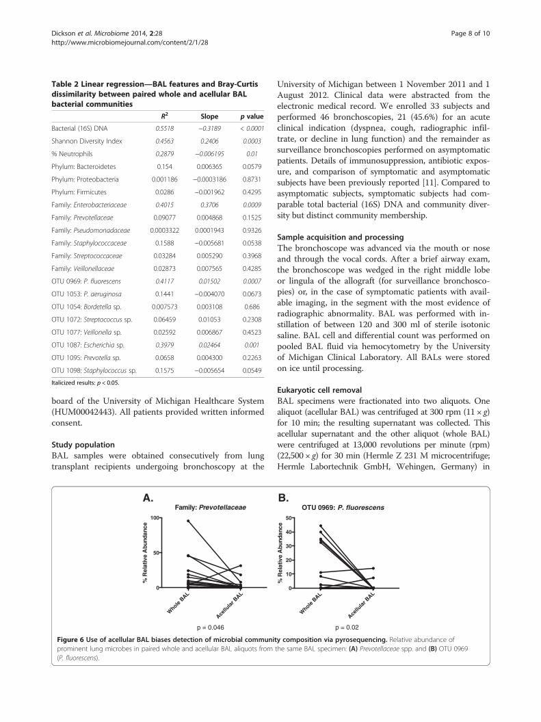
Table 2 Linear regression—BAL features and Bray-Curtisdissimilarity between paired whole and acellular BALbacterial communities
R2 Slope p value
Bacterial (16S) DNA 0.5518 −0.3189 < 0.0001
Shannon Diversity Index 0.4563 0.2406 0.0003
% Neutrophils 0.2879 −0.006195 0.01
Phylum: Bacteroidetes 0.154 0.006365 0.0579
Phylum: Proteobacteria 0.001186 −0.0003186 0.8731
Phylum: Firmicutes 0.0286 −0.001962 0.4295
Family: Enterobacteriaceae 0.4015 0.3706 0.0009
Family: Prevotellaceae 0.09077 0.004868 0.1525
Family: Pseudomonadaceae 0.0003322 0.0001943 0.9326
Family: Staphylococcaceae 0.1588 −0.005681 0.0538
Family: Streptococcaceae 0.03284 0.005290 0.3968
Family: Veillonellaceae 0.02873 0.007565 0.4285
OTU 0969: P. fluorescens 0.4117 0.01502 0.0007
OTU 1053: P. aeruginosa 0.1441 −0.004070 0.0673
OTU 1054: Bordetella sp. 0.007573 0.003108 0.686
OTU 1072: Streptococcus sp. 0.06459 0.01053 0.2308
OTU 1077: Veillonella sp. 0.02592 0.006867 0.4523
OTU 1087: Escherichia sp. 0.3979 0.02464 0.001
OTU 1095: Prevotella sp. 0.0658 0.004300 0.2263
OTU 1098: Staphylococcus sp. 0.1575 −0.005654 0.0549
Italicized results: p < 0.05.
Dickson et al. Microbiome 2014, 2:28 Page 8 of 10http://www.microbiomejournal.com/content/2/1/28
board of the University of Michigan Healthcare System(HUM00042443). All patients provided written informedconsent.
Study populationBAL samples were obtained consecutively from lungtransplant recipients undergoing bronchoscopy at the
Whole
BAL
Acellu
lar B
AL0
50
100
% R
elat
ive
Abu
ndan
ce
Family: Prevotellaceae
p = 0.046
A.
Figure 6 Use of acellular BAL biases detection of microbial communitprominent lung microbes in paired whole and acellular BAL aliquots from(P. fluorescens).
University of Michigan between 1 November 2011 and 1August 2012. Clinical data were abstracted from theelectronic medical record. We enrolled 33 subjects andperformed 46 bronchoscopies, 21 (45.6%) for an acuteclinical indication (dyspnea, cough, radiographic infil-trate, or decline in lung function) and the remainder assurveillance bronchoscopies performed on asymptomaticpatients. Details of immunosuppression, antibiotic expos-ure, and comparison of symptomatic and asymptomaticsubjects have been previously reported [11]. Compared toasymptomatic subjects, symptomatic subjects had com-parable total bacterial (16S) DNA and community diver-sity but distinct community membership.
Sample acquisition and processingThe bronchoscope was advanced via the mouth or noseand through the vocal cords. After a brief airway exam,the bronchoscope was wedged in the right middle lobeor lingula of the allograft (for surveillance bronchosco-pies) or, in the case of symptomatic patients with avail-able imaging, in the segment with the most evidence ofradiographic abnormality. BAL was performed with in-stillation of between 120 and 300 ml of sterile isotonicsaline. BAL cell and differential count was performed onpooled BAL fluid via hemocytometry by the Universityof Michigan Clinical Laboratory. All BALs were storedon ice until processing.
Eukaryotic cell removalBAL specimens were fractionated into two aliquots. Onealiquot (acellular BAL) was centrifuged at 300 rpm (11 × g)for 10 min; the resulting supernatant was collected. Thisacellular supernatant and the other aliquot (whole BAL)were centrifuged at 13,000 revolutions per minute (rpm)(22,500 × g) for 30 min (Hermle Z 231 M microcentrifuge;Hermle Labortechnik GmbH, Wehingen, Germany) in
Whole
BAL0
10
20
30
40
50
% R
elat
ive
Abu
ndan
ce
OTU 0969: P. fluorescens
p = 0.02
Acellu
lar B
AL
B.
y composition via pyrosequencing. Relative abundance ofthe same BAL specimen: (A) Prevotellaceae spp. and (B) OTU 0969
Dickson et al. Microbiome 2014, 2:28 Page 9 of 10http://www.microbiomejournal.com/content/2/1/28
dolphin-nosed Eppendorf tubes and the pellets for eachstored at −80°C until the time of DNA extraction.
DNA isolation, quantitative polymerase chain reaction,and 454 pyrosequencingGenomic DNA was extracted from BAL pellets, resus-pended in 360 μl ATL buffer (Qiagen DNeasy Blood &Tissue kit; Qiagen, Venlo, Limburg, the Netherlands)and homogenized in UltraClean fecal DNA bead tubes(MO-BIO, Carlsbad, CA, USA) using a modified protocolpreviously demonstrated to isolate bacterial DNA [18].Quantification of bacterial 16S rDNA was performed onwhole BAL specimens by real-time polymerase chain reac-tion (PCR) utilizing TaqMan hydrolysis probes on a Roche480 LightCycler (Roche Diagnostics GmbH, Mannheim,Germany), as described previously [11,19-21]. Ampliconlibraries were generated as previously described [11] andsequenced using a Roche 454 GS Junior according toestablished protocols [22]. The V3–V5 hypervariable re-gions of the bacterial 16S rRNA gene were sequenced inthe V5–V3 direction using barcoded primer sets corre-sponding to 357 F and 926R [11]. Pre-procedure bron-choscopy rinse controls, reagent water controls, and mockcommunity standards were sequenced and analyzed asquality controls. The data set supporting the results of thisarticle has been posted to the NIH Sequence Read Archive(SRA:SRP041659).
Data analysisSequence data were processed and analyzed using thesoftware mothur v.1.27.0 according to the Standard Oper-ating Procedure for 454 sequence data (http://www.mothur.org) using a minimum sequence length of 250base pairs [23]. A shared community file and a phylotyped(genus-level grouping) file were generated using OTUsbinned at 97% identity. OTUs detected in reagent watercontrols were removed from all BAL specimens prior toanalysis. OTU numbers were arbitrarily assigned in thebinning process and are referred to throughout the manu-script in association with their most specified level of tax-onomy. Classification of OTUs was carried out using themothur implementation of the Ribosomal Database Pro-ject (RDP) Classifier and the RDP taxonomy training set(http://rdp.cme.msu.edu). Using multiple complementarytechniques (culture, microbe-specific PCR, NCBI BLAST,phylogenetic tree generation), we have previously identi-fied two prominent Pseudomonas-classified OTUs in thisdataset as P. aeruginosa (0153) and P. fluorescens (0969)[11]. Comparison of group proportions was performedusing Fisher's exact test. Odds ratios were determinedusing univariable and multivariable logistic regression inR [24]. Group means were compared using Student'st test. Ordination and PERMANOVA (adonis) testing was
performed using the vegan package 2.0-4 in R [24,25]. Allanalyses were performed in R and GraphPad Prism 6.
AbbreviationsATP: adenosine triphosphate; BAL: bronchoalveolar lavage;DNA: deoxyribonucleic acid; OTU: operational taxonomic units; PCA: principalcomponents analysis; PCR: polymerase chain reaction; RDA: redundancyanalysis; RDP: Ribosomal Database Project; rpm: revolutions per minute;rRNA: ribosomal ribonucleic acid.
Competing interestsThe authors declare that they have no competing interests.
Authors’ contributionsRPD conceived of the study design, participated in data acquisition,executed the analysis and drafted and revised the manuscript. JREparticipated in study design, data acquisition and bioinformatic analysis andrevised the manuscript. HCP participated in statistical analysis and revisedthe manuscript. FJM participated in data acquisition, interpreted the dataand revised the manuscript. JLC interpreted the data and revised themanuscript. VNL participated in study design and data acquisition,interpreted the data and revised the manuscript. GBH participated in studydesign, data acquisition, bioinformatic analysis and manuscript revision. Allauthors read and approved this version of the manuscript.
AcknowledgementsThe authors thank Natalie Walker, Zachary Britt, Nicole Falkowski, DayanaRojas and Brittan Scales for assistance in tissue processing. The authors thankRick Bushman and his lab at the University of Pennsylvania for providing the16S qPCR protocol.Funding provided by NIH grants T32HL00774921 (RPD, HCP), U01HL098961(JLC, GBH), R01HL094622 (VNL), R01HL114447 (GBH, FJM), and the BiomedicalLaboratory and Clinical Science Research & Development Services,Department of Veterans Affairs (JLC). Support provided by the HostMicrobiome Initiative of the University of Michigan.
Author details1Division of Pulmonary and Critical Care Medicine, Department of InternalMedicine, University of Michigan Medical School, Ann Arbor, MI 48109, USA.2Pulmonary and Critical Care Medicine Section, Medical Service, VA AnnArbor Healthcare System, Ann Arbor, MI 48105, USA. 3Department ofMicrobiology and Immunology, University of Michigan Medical School, AnnArbor, MI 48109, USA.
Received: 28 April 2014 Accepted: 26 June 2014Published: 18 August 2014
References1. Dickson RP, Erb-Downward JR, Huffnagle GB: The role of the bacterial
microbiome in lung disease. Expert Rev Respir Med 2013, 7:245–257.2. Erb-Downward JR, Thompson DL, Han MK, Freeman CM, McCloskey L,
Schmidt LA, Young VB, Toews GB, Curtis JL, Sundaram B, Martinez FJ,Huffnagle GB: Analysis of the lung microbiome in the "healthy" smokerand in COPD. PLoS One 2011, 6:e16384.
3. Hilty M, Burke C, Pedro H, Cardenas P, Bush A, Bossley C, Davies J, Ervine A,Poulter L, Pachter L, Moffatt MF, Cookson WO: Disordered microbialcommunities in asthmatic airways. PLoS One 2010, 5:e8578.
4. Morris A, Beck JM, Schloss PD, Campbell TB, Crothers K, Curtis JL, Flores SC,Fontenot AP, Ghedin E, Huang L, Jablonski K, Kleerup E, Lynch SV,Sodergren E, Twigg H, Young VB, Bassis CM, Venkataraman A, Schmidt TM,Weinstock GM: Comparison of the respiratory microbiome in healthynonsmokers and smokers. Am J Respir Crit Care Med 2013, 187:1067–1075.
5. Marks LR, Davidson BA, Knight PR, Hakansson AP: Interkingdom signalinginduces Streptococcus pneumoniae biofilm dispersion and transitionfrom asymptomatic colonization to disease. MBio 2013, 4, doi:10.1128/mBio.00438-13.
6. Lozupone C, Cota-Gomez A, Palmer BE, Linderman DJ, Charlson ES,Sodergren E, Mitreva M, Abubucker S, Martin J, Yao G, Campbell TB, FloresSC, Ackerman G, Stombaugh J, Ursell L, Beck JM, Curtis JL, Young VB, LynchSV, Huang L, Weinstock GM, Knox KS, Twigg H, Morris A, Ghedin E, BushmanFD, Collman RG, Knight R, Fontenot AP: Widespread colonization of the
Dickson et al. Microbiome 2014, 2:28 Page 10 of 10http://www.microbiomejournal.com/content/2/1/28
lung by Tropheryma whipplei in HIV infection. Am J Respir Crit Care Med2013, 187:1110–1117.
7. Segal LN, Alekseyenko AV, Clemente JC, Kulkarni R, Wu B, Chen H, Berger KI,Goldring RM, Rom WN, Blaser MJ, Weiden MD: Enrichment of lungmicrobiome with supraglottic taxa is associated with increasedpulmonary inflammation. Microbiome 2013, 1:19.
8. Aguilar-Guisado M, Givalda J, Ussetti P, Ramos A, Morales P, Blanes M,Bou G, de la Torre-Cisneros J, Roman A, Borro JM, Lama R, Cisneros JM:Pneumonia after lung transplantation in the RESITRA Cohort: amulticenter prospective study. Am J Transplant 2007, 7:1989–1996.
9. Chan CC, Abi-Saleh WJ, Arroliga AC, Stillwell PC, Kirby TJ, Gordon SM,Petras RE, Mehta AC: Diagnostic yield and therapeutic impact of flexiblebronchoscopy in lung transplant recipients. J Heart Lung Transplant 1996,15:196–205.
10. Society AT: Guidelines for the management of adults with hospital-acquired,ventilator-associated, and healthcare-associated pneumonia. Am J Respir CritCare Med 2005, 171:388–416.
11. Dickson RP, Erb-Downward JR, Freeman CM, Walker N, Scales BS, Beck JM,Martinez FJ, Curtis JL, Lama VN, Huffnagle GB: Changes in the lungmicrobiome following lung transplantation include the emergence oftwo distinct Pseudomonas species with distinct clinical associations.PLoS One 2014, 9:e97214.
12. Pratt LA, Kolter R: Genetic analysis of Escherichia coli biofilm formation:roles of flagella, motility, chemotaxis and type I pili. Mol Microbiol 1998,30:285–293.
13. Vandecandelaere I, Matthijs N, Van Nieuwerburgh F, Deforce D, Vosters P,De Bus L, Nelis HJ, Depuydt P, Coenye T: Assessment of microbial diversityin biofilms recovered from endotracheal tubes using culture dependentand independent approaches. PLoS One 2012, 7:e38401.
14. O’Toole GA, Kolter R: Initiation of biofilm formation in Pseudomonasfluorescens WCS365 proceeds via multiple, convergent signallingpathways: a genetic analysis. Mol Microbiol 1998, 28:449–461.
15. Dorn BR, Leung K-P, Progulske-Fox A: Invasion of human oral epithelialcells by Prevotella intermedia. Infect Immun 1998, 66:6054–6057.
16. Madi A, Lakhdari O, Blottière HM, Guyard-Nicodème M, Le Roux K, GroboillotA, Svinareff P, Doré J, Orange N, Feuilloley MG: The clinical Pseudomonasfluorescens MFN1032 strain exerts a cytotoxic effect on epithelialintestinal cells and induces interleukin-8 via the AP-1 signaling pathway.BMC Microbiol 2010, 10:215.
17. Wang K-P, Mehta AC, Turner JF Jr: Flexible Bronchoscopy. Chichester:Wiley-Blackwell; 2012:74.
18. Mason KL, Erb Downward JR, Mason KD, Falkowski NR, Eaton KA, Kao JY,Young VB, Huffnagle GB: Candida albicans and bacterial microbiotainteractions in the cecum during recolonization following broad-spectrum antibiotic therapy. Infect Immun 2012, 80:3371–3380.
19. Charlson ES, Bittinger K, Haas AR, Fitzgerald AS, Frank I, Yadav A, BushmanFD, Collman RG: Topographical continuity of bacterial populations inthe healthy human respiratory tract. Am J Respir Crit Care Med 2011,184:957–963.
20. Hill DA, Hoffmann C, Abt MC, Du Y, Kobuley D, Kirn TJ, Bushman FD, Artis D:Metagenomic analyses reveal antibiotic-induced temporal and spatialchanges in intestinal microbiota with associated alterations in immunecell homeostasis. Mucosal Immunol 2009, 3:148–158.
21. Wilmotte A, Van der Auwera G, De Wachter R: Structure of the 16 Sribosomal RNA of the thermophilic cyanobacterium Chlorogloeopsis HTF(‘Mastigocladus laminosus HTF’) strain PCC7518, and phylogeneticanalysis. FEBS Lett 1993, 317:96–100.
22. Daigle D, Simen BB, Pochart P: High-throughput sequencing of PCRproducts tagged with universal primers using 454 life sciences systems.Curr Protoc Mol Biol 2011, Chapter 7:Unit 7.5. doi:10.1002/0471142727.mb0705s96.
23. Schloss PD, Westcott SL, Ryabin T, Hall JR, Hartmann M, Hollister EB,Lesniewski RA, Oakley BB, Parks DH, Robinson CJ, Sahl JW, Stres B,Thallinger GG, Van Horn DJ, Weber CF: Introducing mothur: open-source,platform-independent, community-supported software for describingand comparing microbial communities. Appl Environ Microbiol 2009,75:7537–7541.
24. R Core Team: R: a language and environment for statistical computing.Vienna, Austria: R Foundation for Statistical Computing; http://www.R-project.org.
25. Oksanen JF, Blanchet G, Kindt R, Legendre P, Minchin PR, O’Hara RB,Simpson GL, Solymos P, Stevens MHH, Wagner H: vegan: CommunityEcology Package. R package version 2.0-4. http://CRAN.R-project.org/package=vegan.
doi:10.1186/2049-2618-2-28Cite this article as: Dickson et al.: Cell-associated bacteria in the humanlung microbiome. Microbiome 2014 2:28.
Submit your next manuscript to BioMed Centraland take full advantage of:
• Convenient online submission
• Thorough peer review
• No space constraints or color figure charges
• Immediate publication on acceptance
• Inclusion in PubMed, CAS, Scopus and Google Scholar
• Research which is freely available for redistribution
Submit your manuscript at www.biomedcentral.com/submit
Related Documents