Celastrol Blocks Interleukin-6 Gene Expression via Downregulation of NF-kB in Prostate Carcinoma Cells Kun-Chun Chiang 1. , Ke-Hung Tsui 2. , Li-Chuan Chung 3 , Chun-Nan Yeh 4 , Wen-Tsung Chen 5 , Phei-Lang Chang 2 , Horng-Heng Juang 3 * 1 Department of General Surgery, Chang Gung Memorial Hospital, Keelung, Taiwan, ROC, 2 Department of Urology, Chang Gung Memorial Hospital, Kwei-Shan, Tao-Yuan, Taiwan, ROC, 3 Department of Anatomy, School of Medicine, Chang Gung University, Kwei-Shan, Tao-Yuan, Taiwan, ROC, 4 Department of General Surgery, Chang Gung Memorial Hospital, Kwei-Shan, Tao-Yuan, Taiwan, ROC, 5 National Kaohsiung University of Hospitality and Tourism, Hsiao-Kang, Kaohsiung Taiwan, ROC Abstract Interleukin-6 (IL-6), a multifunctional cytokine, contributes to proliferation or differentiation of prostate carcinoma cells in a highly cell type-specific manner. Celastrol (3-hydroxy-24-nor-2oxo-1(10),3,5,7-friedelatetrane-29-oic acid), also named as tripterine, is extracted from root of Chinese traditional herb Tripterygiumwilfordii Hook f with potent anti-inflammatory and anti-cancer activities. In this study, we evaluated the molecular mechanisms of celastrol on cell proliferation and IL-6 gene expression in prostate carcinoma cells. 3 H-thymidine incorporation and flow cytometric analysis indicated that celastrol treatments arrested the cell cycle at the G0/G1 phase, thus attenuating cell proliferation in prostate carcinoma PC-3 cells; moreover, celastrol induced cell apoptosis at higher dosage. Knockdown of IL-6 attenuated the anti-proliferative effect of celastrol on PC-3 cells. Results from ELISA and 5’-deletion transient gene expression assays indicated that celastrol treatment decreased IL-6 secretion and gene expression, and this effect is dependent on the NF-kB response element within IL-6 promoter area since mutation of the NF-kB response element from AAATGTCCCATTTTCCC to AAATGTTACATTTTCCC by site-directed mutagenesis abolished the inhibition of celastrol on the IL-6 promoter activity. Celastrol also attenuated the activation of PMA and TNFa on the gene expression and secretion of IL-6 in PC-3 cells. Immunoblot assays revealed that celastrol treatment downregulated the expressions of IKKa, p50 and p65, supporting the 5’-deletion transient gene expression assay result that celastrol blocked IL-6 expression through the NF-kB pathway in PC-3 cells. For the first time, our results concluded that celastrol attenuates PC-3 cell proliferation via downregulation of IL-6 gene expression through the NF-kB-dependent pathway. Citation: Chiang K-C, Tsui K-H, Chung L-C, Yeh C-N, Chen W-T, et al. (2014) Celastrol Blocks Interleukin-6 Gene Expression via Downregulation of NF-kB in Prostate Carcinoma Cells. PLoS ONE 9(3): e93151. doi:10.1371/journal.pone.0093151 Editor: Zoran Culig, Innsbruck Medical University, Austria Received November 6, 2013; Accepted March 3, 2014; Published March 24, 2014 Copyright: ß 2014 Chiang et al. This is an open-access article distributed under the terms of the Creative Commons Attribution License, which permits unrestricted use, distribution, and reproduction in any medium, provided the original author and source are credited. Funding: This Research was supported by Chang Gung Memorial Hospital (CMRP-D190543, -D190613, -G3B1891, D1D0201, and -G3D0311) and Taiwan National Science Council (101-2314-B-182A-099-MY3 and 102-2320-B-182-003 -MY3). The funders had no role in study design, data collection and analysis, decision to publish, or preparation of the manuscript. Competing Interests: The authors have declared that no competing interests exist. * E-mail: [email protected] . These authors contributed equally to this work. Introduction Prostate cancer is the second most common solid tumor for men in United States with 28,170 patients dying of this disease in 2012 [1]. Although the early diagnosis is more feasible due to the recent improvement of prostate-specific antigen (PSA) measurement, which improves the overall survival of prostate cancer patients, however, for the 15% of prostate cancer patients categorized as high-risk prostate cancer, 30–60% of them at around 10 years would eventually have metastasis with 10–25% patients dying of metastasis. [2,3]. Currently, no consensus on the optimal management of high-risk patients is available. Multimodal approaches seem to have better outcome than the single-modality treatment. Under this bleak background, development of a new therapeutic regimen to treat prostate cancer should be prioritized. Recently, to discover new potent anti-tumor compounds with less-toxic characteristics from Chinese natural medicine is getting popular. Among these compounds, celastrol (or tripterine), a quinine methidetriterpenoid, is derived from the root of Trypter- igiumwilfordii (also known as Thunder of God Vine) [4,5]. Celastrol has been implicated with potent anti-inflammation and anti-tumor effects in ample studies. So far, celastrol has been shown to have beneficial effects on a variety of cancers in vitro and in vivo, such as breast cancer, melanoma, squamous cell cancer, and prostate cancer [6–9]. Interleukin 6 (IL-6) is a glycoprotein consisting of 184 amino acids. IL-6 is first identified as a T-cell-derived regulation factor controlling B cell differentiation. IL-6 is now to be known to have multi-functions in a variety of cells and tissues [10]. Since the cloning of IL-6 cDNA, IL-6 has been proved to be produced in varied kinds of cells, including cancer cells, in addition to T- cell. IL-6 has been shown to involve in a number of important biological activities, including immune modulation, pro-inflam- mation, oncogenesis, and pro- or de- differentiation, in a highly cell- or tissue- specific way. In terms of prostate cancer, IL-6 has been demonstrated to be able to induce androgen receptor expression and promote tumor progression [11,12], thus deemed as a growth factor for most prostate cancer cells in vitro. Some transcriptional factors have been reported to involve in the PLOS ONE | www.plosone.org 1 March 2014 | Volume 9 | Issue 3 | e93151

Welcome message from author
This document is posted to help you gain knowledge. Please leave a comment to let me know what you think about it! Share it to your friends and learn new things together.
Transcript
Celastrol Blocks Interleukin-6 Gene Expression viaDownregulation of NF-kB in Prostate Carcinoma CellsKun-Chun Chiang1., Ke-Hung Tsui2., Li-Chuan Chung3, Chun-Nan Yeh4, Wen-Tsung Chen5,
Phei-Lang Chang2, Horng-Heng Juang3*
1 Department of General Surgery, Chang Gung Memorial Hospital, Keelung, Taiwan, ROC, 2 Department of Urology, Chang Gung Memorial Hospital, Kwei-Shan, Tao-Yuan,
Taiwan, ROC, 3 Department of Anatomy, School of Medicine, Chang Gung University, Kwei-Shan, Tao-Yuan, Taiwan, ROC, 4 Department of General Surgery, Chang Gung
Memorial Hospital, Kwei-Shan, Tao-Yuan, Taiwan, ROC, 5 National Kaohsiung University of Hospitality and Tourism, Hsiao-Kang, Kaohsiung Taiwan, ROC
Abstract
Interleukin-6 (IL-6), a multifunctional cytokine, contributes to proliferation or differentiation of prostate carcinoma cells in ahighly cell type-specific manner. Celastrol (3-hydroxy-24-nor-2oxo-1(10),3,5,7-friedelatetrane-29-oic acid), also named astripterine, is extracted from root of Chinese traditional herb Tripterygiumwilfordii Hook f with potent anti-inflammatory andanti-cancer activities. In this study, we evaluated the molecular mechanisms of celastrol on cell proliferation and IL-6 geneexpression in prostate carcinoma cells. 3H-thymidine incorporation and flow cytometric analysis indicated that celastroltreatments arrested the cell cycle at the G0/G1 phase, thus attenuating cell proliferation in prostate carcinoma PC-3 cells;moreover, celastrol induced cell apoptosis at higher dosage. Knockdown of IL-6 attenuated the anti-proliferative effect ofcelastrol on PC-3 cells. Results from ELISA and 5’-deletion transient gene expression assays indicated that celastrol treatmentdecreased IL-6 secretion and gene expression, and this effect is dependent on the NF-kB response element within IL-6promoter area since mutation of the NF-kB response element from AAATGTCCCATTTTCCC to AAATGTTACATTTTCCC bysite-directed mutagenesis abolished the inhibition of celastrol on the IL-6 promoter activity. Celastrol also attenuated theactivation of PMA and TNFa on the gene expression and secretion of IL-6 in PC-3 cells. Immunoblot assays revealed thatcelastrol treatment downregulated the expressions of IKKa, p50 and p65, supporting the 5’-deletion transient geneexpression assay result that celastrol blocked IL-6 expression through the NF-kB pathway in PC-3 cells. For the first time, ourresults concluded that celastrol attenuates PC-3 cell proliferation via downregulation of IL-6 gene expression through theNF-kB-dependent pathway.
Citation: Chiang K-C, Tsui K-H, Chung L-C, Yeh C-N, Chen W-T, et al. (2014) Celastrol Blocks Interleukin-6 Gene Expression via Downregulation of NF-kB inProstate Carcinoma Cells. PLoS ONE 9(3): e93151. doi:10.1371/journal.pone.0093151
Editor: Zoran Culig, Innsbruck Medical University, Austria
Received November 6, 2013; Accepted March 3, 2014; Published March 24, 2014
Copyright: � 2014 Chiang et al. This is an open-access article distributed under the terms of the Creative Commons Attribution License, which permitsunrestricted use, distribution, and reproduction in any medium, provided the original author and source are credited.
Funding: This Research was supported by Chang Gung Memorial Hospital (CMRP-D190543, -D190613, -G3B1891, D1D0201, and -G3D0311) and Taiwan NationalScience Council (101-2314-B-182A-099-MY3 and 102-2320-B-182-003 -MY3). The funders had no role in study design, data collection and analysis, decision topublish, or preparation of the manuscript.
Competing Interests: The authors have declared that no competing interests exist.
* E-mail: [email protected]
. These authors contributed equally to this work.
Introduction
Prostate cancer is the second most common solid tumor for men
in United States with 28,170 patients dying of this disease in 2012
[1]. Although the early diagnosis is more feasible due to the recent
improvement of prostate-specific antigen (PSA) measurement,
which improves the overall survival of prostate cancer patients,
however, for the 15% of prostate cancer patients categorized as
high-risk prostate cancer, 30–60% of them at around 10 years
would eventually have metastasis with 10–25% patients dying of
metastasis. [2,3]. Currently, no consensus on the optimal
management of high-risk patients is available. Multimodal
approaches seem to have better outcome than the single-modality
treatment. Under this bleak background, development of a new
therapeutic regimen to treat prostate cancer should be prioritized.
Recently, to discover new potent anti-tumor compounds with
less-toxic characteristics from Chinese natural medicine is getting
popular. Among these compounds, celastrol (or tripterine), a
quinine methidetriterpenoid, is derived from the root of Trypter-
igiumwilfordii (also known as Thunder of God Vine) [4,5].
Celastrol has been implicated with potent anti-inflammation and
anti-tumor effects in ample studies. So far, celastrol has been
shown to have beneficial effects on a variety of cancers in vitro and
in vivo, such as breast cancer, melanoma, squamous cell cancer,
and prostate cancer [6–9].
Interleukin 6 (IL-6) is a glycoprotein consisting of 184 amino
acids. IL-6 is first identified as a T-cell-derived regulation factor
controlling B cell differentiation. IL-6 is now to be known to have
multi-functions in a variety of cells and tissues [10]. Since the
cloning of IL-6 cDNA, IL-6 has been proved to be produced in
varied kinds of cells, including cancer cells, in addition to T- cell.
IL-6 has been shown to involve in a number of important
biological activities, including immune modulation, pro-inflam-
mation, oncogenesis, and pro- or de- differentiation, in a highly
cell- or tissue- specific way. In terms of prostate cancer, IL-6 has
been demonstrated to be able to induce androgen receptor
expression and promote tumor progression [11,12], thus deemed
as a growth factor for most prostate cancer cells in vitro. Some
transcriptional factors have been reported to involve in the
PLOS ONE | www.plosone.org 1 March 2014 | Volume 9 | Issue 3 | e93151
modulation of IL-6 gene expression and have binding sites within
the IL-6 promoter area, including AP-1, cAMP, NF-kB, etc [13].
Previously, celastrol has been shown to possess anti-growth
effect on prostate cancer in vitro and in vivo [8,14,15]. In this current
study, we aimed to investigate the underlying mechanism whereby
celastrol inhibits prostate cancer growth. We found for the first
time that celastrol inhibited IL-6 secretion and expression in PC-3
cells, one kind of prostate cancer cells, which partly contributed to
the anti-proliferative effect of celastrol on PC-3 cell. The secretion
and expression of IL-6 in DU-145 cells was also repressed by
celastrol. Further, we also provided the first laboratory evidence
that celastrol repressed IL-6 secretion and expression in a NF-kB-
dependent pathway in PC-3 cells.
Materials and Methods
Materials, cell lines, and cell culturePC-3 and DU145 cells were obtained and maintained as
described previously [16]. Celastrol and phorbol 12-myristate 13-
acetate (PMA) were purchased from Sigma (St. Louis, MO). The
stock celastrol (10 mM) was dissolved in DMSO. TNFa was
purchase from PeproTech (Rebovot, Israel). The culture media
were purchased from Life Technologies (Rockville, MD), and fetal
calf serum (FCS) was from the HyClone (Logan, Utah). PC-3 and
DU145 cells were cultured in RPMI 1640 medium with 10% FCS.
The control groups in this experiment were treated with DMSO.
Cell proliferation assayCell proliferation in response to celastrol was measured using a
3H-thymidine incorporation assay as previously described [16].
Flow cytometryCells were serum starved for 24 hours and then cultured in
RPMI 1640 medium with 10% FCS and with or without different
concentrations of celastrol for another 48 hours. The cells were
collected and stained with propidium iodide. Cell cycle analysis
was performed using the FACS-Calibur cytometer and CellQuest-
Pro software (BD Biosciences, San Jose, CA); the data were
analyzed using ModFit LT Mac 3.0 software as previously
described [17].
Immunoblot AssayCells were incubated in the RPMI-1640 medium with 10% FCS
and different treatments for a period of 24 hours. The nuclear and
cytoplasmic fractions were extracted by NE-PER Nuclear and
Cytoplasmic Extraction Reagent Kit (Thermo Scientific, Rock-
ford, IL). Equal quantities of cell extract were loaded onto a 10%
sodium dodecyl sulfate polyacrylamide (SDS) gel and analyzed by
the electrochemiluminescent detection system. The blotting
membranes were probed with 1:1000 diluted IkB kinase a (IKKa)
antiserum, 1:1000 NFkBp50 antiserum, 1:1000 diluted NFkBp65
antiserum (Merck Millipore, Darmstadt, Germany), 1:1000 diluted
PARP (Cell Signaling, Danvers, MA), 1:200 diluted NFkB-
inducing kinase (NIK) antiserum, 1:1000 diluted IkB antiserum,
1:200 diluted Lamin B antiserum, or 1:3000 diluted b-actin
antiserum (Santa Cruz Biotechnology, Santa Cruz, CA). The
intensity of different bands was recorded and analyzed by
GeneTools of ChemiGenius (Syngene, Cambridge, UK).
Tunnel assayAfter one day of treatment, cellular DNA was stained by
TumorTACS in Situ Apoptosis Detection kit (TREVIGEN
#4815-30-K). The assay was performed according to the
manufacturer’s instructions.
Knockdown of IL-6The pSMc2 retroviral vectors containing the IL-6 short hairpin
RNA (shRNA; V2HS-111640) and the GFP shRNA (RHS1764-
9394112) were purchased from Open Biosystems (Huntsville, AL).
The IL-6 shRNA and GFP shRNA vectors were introduced into
PC-3 cells by electroporation using a singles 70-msec pulse of 180
V, and the transfections were selected using 2 mg/ml puromycin
dihydrochloride. The IL-6-knockdown PC-3 cells were designated
PC-IL6si cells and GFP-knockdown PC-3 cells were designated
PC-COLsi cells as described previously [18].
ELISA of IL-6The IL-6 protein level in the culture medium was measured by
ELISA (cat. No. 2107; Bio Scientific Corporation, Austin, TX).
The relative mass of the IL6 protein present in each sample was
determined based on the total protein concentration of the whole
cell extract as described previously [19].
Report Vector ConstructsThe MMTV reporter vector was constructed as previously
described [20]. The NF-kB reporter vector was purchase from
Clontech (Moutain View, CA). The DNA fragment containing the
enhancer/promoter of human IL-6 was isolated from the BAC
clone (RPI11-240H8) and the reporter vectors containing the
different fragments of 5’-flanking region of human IL-6 gene and
mutant NF-kB response element were constructed as previously
described [21].
Luciferase and b-Galactosidasea assayCells were seeded onto 24-well plates at 16104 cells/well 1 day
prior to transfection. The cells were transiently transfected using
TransFast transfection reagent as described previously [19]. The
media containing the liposome-DNA complex was removed and
replaced with RPMI 1640 medium with 10% FCS for overnight.
The media were replaced with RPMI 1640 medium with 10%
FCS with different concentrations of celastrol, TNFa, or PMA as
indicated for further 24 hours. Cells were harvested for activities of
luciferase and b-galactosidase as described by the manufacturer
instructions (Promega Bioscience, Madison, WI,).
Statistical analysisResults are expressed as the mean 6 S.E. of at least three
independent replication of each experiment. Statistical significance
was determined by one way ANOVA and pair-t test analysis with
program of SigmaStat for Window version 2.03 (SPSS Inc,
Chicago, IL).
Results
Cell proliferation in the PC-3 cells was measured by 3H-
thymidine incorporation assay. Results indicated cell proliferation
decreased 37% when cells were treated with 1 mM of celastrol and
80% cell proliferation inhibition was observed as treated by 3 mM
celastrol for 48 hours (Figure 1A). Immunoblot assay revealed that
3 mM of celastrol induced cleaved form of PARP (c-PARP)
expression in PC-3 cells, indicating apoptosis induction (Figure 1B).
To confirm apoptosis induction by high dose of celastrol, we
further conducted tunnel assay. As shown in Figure 1C, after one
day of treatment, 3 mM celastrol induced obvious apoptosis in PC-
3 cells with an apoptosis index ratio of 2163.2. Therefore, we used
the proapoptosis (# 1 mM) dosage of celastrol for further studies
below. Results from flow cytometric analysis revealed that celastrol
induced cell cycle arrest at G0/G1 phase in PC-3 cells dose-
dependently after 48 hours treatment with 1 mM of celastrol
Celastrol Blocks IL-6 via NF-kB in Prostate Cancer
PLOS ONE | www.plosone.org 2 March 2014 | Volume 9 | Issue 3 | e93151

inducing 16% increase in G0/G1 phase cells together with a
decrease in S phase cells (Figure 1D).
In vitro studies revealed that knockdown of IL-6 significantly
(P = 0.0217) attenuated the blocking effect of celastrol on cell
proliferation in PC-3 cells as determined by the 3H-thymidine
incorporation assay. As shown in figure 2, 48 hours 1, 3, and 6 mM
celastrol treatments induced 32.6%, 77.4%, and 83.3% growth
inhibition, respectively, in PC-COLsi cells. In the contrast, the
same dosage of celastrol treatments repressed PC-IL6si cells
growth by only 0%, 44.1%, and 61.8%, respectively (Figure 2).
Figure 1. Celastrol inhibits PC-3 cell growth through cell cycle arrest at G0/G1 and apoptosis induction. (A) PC-3 cells were treated withindicated concentrations of celastrol for 48 hours and the cell proliferation was determined by the H3-thymidine incorporation. (B) PC-3 cells weretreated with indicated concentrations of celastrol for48 hours. Cells were lysed and expressions of PARP, cleaved PARP (c-PARP) were determined byimmunoblotting assay. (C) After one day of treatment, the apoptotic index of PC-3 cells treated with different concentrations of celastrol wascalculated. Each value is a mean 6 SE of 3 determinations. (D) PC-3 cells were serum starved for 24 hours and then were treated with 0 – 1 mM ofcelastrol as indicated for 48 hours. The cells were stained with PI, and the cell cycle distribution was analyzed by flow cytometry. Each box representsthe mean 6 SE (n = 6). (* p,0.05; **p,0.01).doi:10.1371/journal.pone.0093151.g001
Celastrol Blocks IL-6 via NF-kB in Prostate Cancer
PLOS ONE | www.plosone.org 3 March 2014 | Volume 9 | Issue 3 | e93151

Results from ELISA indicated that celastrol blocked IL-6
secretion of PC-3 and DU145 cells in a dosage-dependent
manner. 1 mM celastrol treatment blocked 62% of IL-6 secretion
(Figure 3A). Further ELISA revealed that PMA (40 nM) and
TNFa (10 ng/ml) increased 8.2- and 20.5-fold, respectively, of IL-
6 secretion. However, 1 mM celastrol attenuated the activation of
both PMA and TNFa on IL-6 secretion of PC-3 cells (Figure 3B).
Transient gene expression assays using the human IL-6 reporter
vector showed similar results. 1 mM celastrol treatment blocked
55% and 40%, respectively, of IL-6 promoter activity in PC-3 and
DU145 cells (Figure 4A). In order to evaluate the effect of celastrol
on the NF-kB activity, we conducted the transient gene expression
assays using the NF-kB specific reporter vector containing four
NF-kB response elements. Our results indicated that celastrol
blocked NF-kB activity in PC-3 cells in a dosage-dependent
manner (Figure 4B) but did not affect the promoter activity of the
MMTV reporter vector which was derived by the promoter of
mouse mammary tumor virus.
Further transient gene expression assay also indicated the PMA
and TNFa enhanced the promoter activity of IL-6 (Figure 5A) and
NF-kB (Figure 5B) reporter vectors in PC-3 cells, while these
effects were blocked by celastrol. Immunblot assays revealed that
celastrol treatments not only decreased the expression of IKKa in
the cytoplasm but also the p50 and p65 in the nucleus of PC-3
cells. However, celastrol did not affect the expression of NF-kappa-
B-inducing kinase (NIK) but enhanced the protein levels of IkB
(Figure 6A and 6B). The results from the 5’-deletion reporter
assays indicated the response element for the effects of celastrol on
IL-6 promoter activity was located at 2149 to +8 of the 5’-flanking
of the human IL-6 gene (Figure 6C). Further transient gene
expression assay indicated that celastrol did not affect the
promoter activity of the mutant IL-6 reporter vector, in which
the NF-kB binding site was mutated from AAATGTGG-
GATTTTTCCC to AAATGTTACATTTTCCC by site-directed
mutagenesis (Figure 6C). Combined with the results shown in
figure 5, we thus concluded that the effect of celastrol on IL-6 gene
expression depends on the NFkB pathway (Figure 6C).
Discussion
There are numerous studies showing positive effect of celastrol
on cancer growth and metastasis in a variety of cancers, such as
pancreatic cancer, lung cancer, and breast cancer [22]. In terms of
prostate cancer, celastrol has been demonstrated to repress
prostate cancer cell proliferation and induce apoptosis with
downregulation of androgen receptor expression. Moreover,
celastrol has also been found to exert antitumor effect on prostate
cancer in vivo without obvious side effect [8,14,15]. Thus,
Figure 2. Knockdown of interleukin-6 attenuates the growth-inhibitorty effect ofcelastrol onPC-3 cells. PC-COLsi cells (opencircle) and PC-IL6si cells (close circle) were treated with variousconcentrations of celastrol, as indicated, for 48 hours. The cellproliferation was determined by the 3H-thymidine incorporation. Dataare presented as mean percentage 6 SE (n = 6). (* p,0.05; **p,0.01).doi:10.1371/journal.pone.0093151.g002
Figure 3. Celastrol Modulates interleukin-6 secretion in PC-3cells. (A) PC-3 (black bars) and DU145 (white bars) cells were treatedwith various concentrations of celastrol, as indicated, for 24 hours. (B)PC-3 cells were treated with 1 mM celastrol, 40 nM PMA, and/or 10 ng/ml TNFa for 24 hours. IL-6 levels in the conditioned media weredetermined by ELISA. Data are expression as mean percentagestimulation 6 SE of 6 preparation induced by different treatmentsrelative to the control solvent treatment. (*p,0.05; **p,0.01).doi:10.1371/journal.pone.0093151.g003
Celastrol Blocks IL-6 via NF-kB in Prostate Cancer
PLOS ONE | www.plosone.org 4 March 2014 | Volume 9 | Issue 3 | e93151

application of celastrol to treat prostate cancer seems to be a
promising alternative regimen. In this current study, we demon-
strated that celastrol repressed PC-3 cell growth dose-dependently
(Figure 1A) through cell cycle arrest at G0/G1 phase as indicated
by increased G0/G1 phase cells (lower dose, Figure 1D) and
apoptosis induction as indicated by increasing c-PARP expression,
which is also supported by the result of tunnel assay (higher dose,
Figure 1B, 1C), in line with previous reports [8,14,15].
IL-6, a multifunctional cytokine, has been shown to play a vital
role in lots of important biologic activities in a cell- or tissue-
specific manner and to be produced by a variety of cells, including
cancer cells [10]. IL-6 belongs to a cytokine family comprising of
IL-11, oncostain M, cardiotropin-1, etc [10]. IL-6 exerts its
function through binding with a cell surface type 1 cytokine
receptor complex which contains two components, i.e. the ligant-
binding component (CD126) and the signal-transducing compo-
nent (CD130). IL-6 has deemed as a growth factor through
activation of JAK-STAT3, RAS, MAPK, Cox-2, PI3K/AKT, and
Wnt pathways [10,23–25]. Most cancers have been found to
overexpress IL-6 and have an aberrant IL-6 signaling pathway
[26–28]. Moreover, ample clinical studies have implicated that
higher serum IL-6 concentrations in cancer patients are associated
with advanced tumor stages and poor survival. Thus, blocking IL-
6 signaling seems to be a rational direction to repress cancer
growth [10]. Regarding prostate cancer, IL-6 expression is
detectable in both epithelium and stroma of human prostates,
with increased IL-6 expression in epithelium as the prostate tissues
are getting transformation toward malignancy [29]. It has been
shown that IL-6 is a growth factor for most prostate cancer cells
and anti-IL-6 monoclonal antibody has been proven to effectively
inhibit xenografted prostate cancer cells growth [30]. In addition,
Figure 4. Celastrol downregulates interleukin-6 and NF-kBreporter activity in PC-3 cells. (A) Luciferase activity of IL-6 reportervector (pIL6-SX)-transfected PC-3 (black bars) and DU145 (white bars)cells treated with different concentrations of celastrol as indicated. (B)Luciferase activity of NFkB reporter vectors (black bars)- and MMTVreporter vector (white bars)-transfected PC-3 cells treated with differentconcentrations of celastrol as indicated. Data are presented as the meanpercentage 6 SE (n = 6) of the reporter activities induced by celastroltreatments in relation to the control solvent-treated group. (*p,0.05;**p,0.01).doi:10.1371/journal.pone.0093151.g004
Figure 5. Celastrol blocks the activation of TNFa and PMA oninterleukin-6 and NF-kB promoter activity. Luciferase activity ofIL-6 reporter vector- (A) and NF-kB reporter vectors- (B) transfected PC-3cells treated with 1 mM celastrol (Cel), 40 nM PMA, or/and 10 ng/mlTNFa. Data are presented as the mean percentage 6 SE (n = 6) of thereporter activities induce by different treatments in relation to thecontrol solvent-treated group. (*p,0.05; **p,0.01).doi:10.1371/journal.pone.0093151.g005
Celastrol Blocks IL-6 via NF-kB in Prostate Cancer
PLOS ONE | www.plosone.org 5 March 2014 | Volume 9 | Issue 3 | e93151
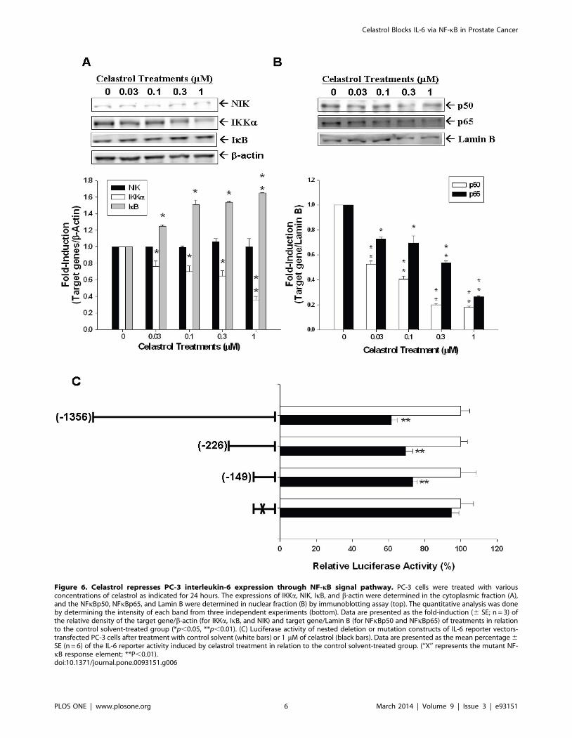
Figure 6. Celastrol represses PC-3 interleukin-6 expression through NF-kB signal pathway. PC-3 cells were treated with variousconcentrations of celastrol as indicated for 24 hours. The expressions of IKKa, NIK, IkB, and b-actin were determined in the cytoplasmic fraction (A),and the NFkBp50, NFkBp65, and Lamin B were determined in nuclear fraction (B) by immunoblotting assay (top). The quantitative analysis was doneby determining the intensity of each band from three independent experiments (bottom). Data are presented as the fold-induction (6 SE; n = 3) ofthe relative density of the target gene/b-actin (for IKKa, IkB, and NIK) and target gene/Lamin B (for NFkBp50 and NFkBp65) of treatments in relationto the control solvent-treated group (*p,0.05, **p,0.01). (C) Luciferase activity of nested deletion or mutation constructs of IL-6 reporter vectors-transfected PC-3 cells after treatment with control solvent (white bars) or 1 mM of celastrol (black bars). Data are presented as the mean percentage 6SE (n = 6) of the IL-6 reporter activity induced by celastrol treatment in relation to the control solvent-treated group. (‘‘X’’ represents the mutant NF-kB response element; **P,0.01).doi:10.1371/journal.pone.0093151.g006
Celastrol Blocks IL-6 via NF-kB in Prostate Cancer
PLOS ONE | www.plosone.org 6 March 2014 | Volume 9 | Issue 3 | e93151
serum IL-6 level has been deemed as a prognostic marker in
metastatic hormone-refractory prostate cancer patients [31]. In
this study, we demonstrated for the first time that celastrol-
mediated antitumor effect on PC-3 cells is IL-6-dependently as
knockdown of IL-6 blunted the anti-proliferative effect of celastrol
on PC-3 cells (Figure 2). Since IL-6 could be produced by PC-3
cells and acts in an autocrine or paracrine manner to stimulate
cancer growth, we next measured whether the secretion of IL-6 by
PC-3 cells is affected by celastrol treatment. As shown in figure 3A,
a dose-dependent manner of downregulation of IL-6 secretion in
PC-3 and DU145 cells by celastrol was observed as measured by
ELISA. In addition, PMA- and TNFa-induced IL-6 secretion was
also blocked by celastrol in PC-3 cells (Figure 3B). Transient gene
reporter assay showed the similar result indicating that celastrol
repressed IL-6 gene promoter activity in PC-3 and DU145 cells
(Figure 4A). Collectively, we concluded that celastrol repressed IL-
6 gene expression and secretion and inhibited prostate carcinoma
cell growth IL-6-dependently.
The IKK/NF-kB signaling is an important pathway with
aberrant NF-kB regulation existing in a myriad of cancers [32–
34]. The NF-kB protein family comprises RelA (p65), RelB, c-Rel,
p50 (p105 precursor), and p52 (p100 precursor) [33]. In the latent
state, NF-kBs are bound to their inhibitor IkB (inhibitor of NF-kB)
proteins and, thus, sequestered in the cytosol. Once receiving
stimulation, IKK (IkB kinase), consisting of IKKa, IKKb (two
catalytic subunits), and NEMO/IKKc (regulatory subunit), is
activated to phosphorylate IkB, which, in turn, leads to
proteasomal degradation of phosphorylated IkB and the release
of NF-kB with subsequent nuclear translocation for gene
expression modulation [35]. There are some transcriptional
factors, such as AP-1, CCAAT enhancer binding protein, cAMP
response element binding protein, as well as NF-kB reported to
have potential binding sites within the human IL-6 gene promoter
area and, thus, could interfere IL-6 gene expression in prostate
cancer cells [21,36]. NF-kB signaling pathway has also previously
been shown to be one of the celastrol-targeted anticancer
pathways [37]. As shown in Figure 4A and 4B, celastrol reduced
the promoter activity of IL-6 reporter vector and NF-kB reporter
vector, which contains the four repeats consensus NF-kB response
elements, in PC-3 cells. PMA and TNFa both upregualted IL-6
and NF-kB promoter activity in PC-3 cells, however, this effect
was blocked by celastrol (Figure 5A and 5B). Moreover, as
determined by western blot assays, expression of IKKa in the
cytoplasm and p50 and p65 in the nucleus of PC-3 cells were all
inhibited by celastrol, while IkB expression was upregulated
(Figure 6A, B). To the contrary, NIK , encoded by MAP3K14
gene in human and functioning as an alternative NF-kB pathway
stimulator as binding with TRAF2 [38], was not repressed by
celastrol treatment in PC-3 cells (Figure 6A and 6B), indicating
celastrol seemed to repress NF-kB pathway directly in PC-3 cells.
To further verify how celastrol regulates IL-6 gene expression in
PC-3 cells, we conducted 5’-deletion reporter assays. As shown in
figure 6C, our results indicated that the celastrol response element
in IL-6 promoter area was located at 2149 to +8 of the 5’-flanking
of the human IL-6 gene, which also contains the NF-kB response
element [21]. As we mutated NF-kB binding site from
AAATGTGGGATTTTTCCC to AAATGTTACATTTTCCC
by site-directed mutagenesis, the celastrol-mediated downregula-
tion of IL-6 promoter activity was abolished. Taken together,
based on our result, we concluded that celastrol inhibits IL-6 gene
expression via the NF-kB pathway in PC-3 cells.
In conclusion, celastrol, one kind of active compound extracted
from Chinese herbal, possesses potent anti-growth effect on
prostate cancer through cell cycle arrest at G0/G1 and apoptosis
induction. The growth inhibition of celastrol against prostate
carcinoma cells depends on IL-6 pathway since knockdown of IL-6
blunts the growth inhibition induced by celastrol. Further, celastrol
represses IL-6 gene expression and secretion in prostate carcinoma
cells via the NF-kB signaling pathway.
Author Contributions
Conceived and designed the experiments: KCC HHJ KHT. Performed the
experiments: KCC HHJ KHT WTC. Analyzed the data: CNY LCC PLC.
Contributed reagents/materials/analysis tools: CNY LCC PLC. Wrote the
paper: KCC HHJ.
References
1. Siegel R, Naishadham D, Jemal A (2012) Cancer statistics, 2012. CA
Cancer J Clin 62: 10–29.
2. Han M, Partin AW, Pound CR, Epstein JI, Walsh PC (2001) Long-term
biochemical disease-free and cancer-specific survival following anatomic radical
retropubic prostatectomy. The 15-year Johns Hopkins experience. Urol Clin
North Am 28: 555–565.
3. D’Amico AV, Cote K, Loffredo M, Renshaw AA, Schultz D (2002)
Determinants of prostate cancer-specific survival after radiation therapy for
patients with clinically localized prostate cancer. J Clin Oncol 20: 4567–4573.
4. Gullett NP, Ruhul Amin AR, Bayraktar S, Pezzuto JM, Shin DM, et al. (2010)
Cancer prevention with natural compounds. Semin Oncol 37: 258–281.
5. Salminen A, Lehtonen M, Paimela T, Kaarniranta K (2010) Celastrol:
Molecular targets of Thunder God Vine. Biochem Biophys Res Commun
394: 439–442.
6. Zhu H, Liu XW, Ding WJ, Xu DQ, Zhao YC, et al. (2010) Up-regulation of
death receptor 4 and 5 by celastrol enhances the anti-cancer activity of TRAIL/
Apo-2L. Cancer Lett 297: 155–164.
7. Sung B, Park B, Yadav VR, Aggarwal BB (2010) Celastrol, a triterpene,
enhances TRAIL-induced apoptosis through the down-regulation of cell survival
proteins and up-regulation of death receptors. J Biol Chem 285: 11498–11507.
8. Yang H, Chen D, Cui QC, Yuan X, Dou QP (2006) Celastrol, a triterpene
extracted from the Chinese "Thunder of God Vine," is a potent proteasome
inhibitor and suppresses human prostate cancer growth in nude mice. Cancer
Res 66: 4758–4765.
9. He D, Xu Q, Yan M, Zhang P, Zhou X, et al. (2009) The NF-kappa B inhibitor,
celastrol, could enhance the anti-cancer effect of gambogic acid on oral
squamous cell carcinoma. BMC Cancer 9: 343.
10. Guo Y, Xu F, Lu T, Duan Z, Zhang Z (2012) Interleukin-6 signaling pathway in
targeted therapy for cancer. Cancer Treat Rev 38: 904–910.
11. Edwards J, Bartlett JM (2005) The androgen receptor and signal-transduction
pathways in hormone-refractory prostate cancer. Part 2: Androgen-receptor
cofactors and bypass pathways. BJU Int 95: 1327–1335.
12. Jia L, Choong CS, Ricciardelli C, Kim J, Tilley WD, et al. (2004) Androgen
receptor signaling: mechanism of interleukin-6 inhibition. Cancer Res 64: 2619–
2626.
13. Xiao W, Hodge DR, Wang L, Yang X, Zhang X, et al. (2004) Co-operative
functions between nuclear factors NFkappaB and CCAT/enhancer-binding
protein-beta (C/EBP-beta) regulate the IL-6 promoter in autocrine human
prostate cancer cells. Prostate 61: 354–370.
14. Pang X, Yi Z, Zhang J, Lu B, Sung B, et al. (2010) Celastrol suppresses
angiogenesis-mediated tumor growth through inhibition of AKT/mammalian
target of rapamycin pathway. Cancer Res 70: 1951–1959.
15. Dai Y, DeSano JT, Meng Y, Ji Q, Ljungman M, et al. (2009) Celastrol
potentiates radiotherapy by impairment of DNA damage processing in human
prostate cancer. Int J Radiat Oncol Biol Phys 74: 1217–1225.
16. Juang HH, Chung LC, Sung HC, Feng TH, Lee YH, et al. (2013)
Metallothionein 3: An androgen-upregulated gene enhances cell invasion and
tumorigenesis of prostate carcinoma cells. Prostate 73: 1495–1506.
17. Chung LC, Tsui KH, Feng TH, Lee SL, Chang PL, et al. (2012) L-Mimosine
blocks cell proliferation via upregulation of B-cell translocation gene 2 and N-
myc downstream regulated gene 1 in prostate carcinoma cells. Am J Physiol Cell
Physiol 302: C676–685.
18. Tsui KH, Chang YL, Feng TH, Chung LC, Lee TY, et al. (2012) Growth
differentiation factor-15 upregulates interleukin-6 to promote tumorigenesis of
prostate carcinoma PC-3 cells. J Mol Endocrinol 49: 153–163.
19. Tsui KH, Wang SW, Chung LC, Feng TH, Lee TY, et al. (2013) Mechanisms
by which interleukin-6 attenuates cell invasion and tumorigenesis in human
bladder carcinoma cells. Biomed Res Int 2013: 791212.
Celastrol Blocks IL-6 via NF-kB in Prostate Cancer
PLOS ONE | www.plosone.org 7 March 2014 | Volume 9 | Issue 3 | e93151
20. Tsui KH, Feng TH, Chung LC, Chao CH, Chang PL, et al. (2008) Prostate
specific antigen gene expression in androgen insensitive prostate carcinomasubculture cell line. Anticancer Res 28: 1969–1976.
21. Tsui KH, Feng TH, Hsieh WC, Chang PL, Juang HH (2008) Expression of
interleukin-6 is downregulated by 17-(allylamino)-17-demethoxygeldanamycinin human prostatic carcinoma cells. Acta Pharmacol Sin 29: 1334–1341.
22. Kannaiyan R, Shanmugam MK, Sethi G (2011) Molecular targets of celastrolderived from Thunder of God Vine: potential role in the treatment of
inflammatory disorders and cancer. Cancer Lett 303: 9–20.
23. Imada K, Leonard WJ (2000) The Jak-STAT pathway. Mol Immunol 37: 1–11.24. Ara T, Declerck YA (2010) Interleukin-6 in bone metastasis and cancer
progression. Eur J Cancer 46: 1223–1231.25. Jee SH, Chu CY, Chiu HC, Huang YL, Tsai WL, et al. (2004) Interleukin-6
induced basic fibroblast growth factor-dependent angiogenesis in basal cellcarcinoma cell line via JAK/STAT3 and PI3-kinase/Akt pathways. J Invest
Dermatol 123: 1169–1175.
26. Bellone S, Watts K, Cane S, Palmieri M, Cannon MJ, et al. (2005) High serumlevels of interleukin-6 in endometrial carcinoma are associated with uterine
serous papillary histology, a highly aggressive and chemotherapy-resistantvariant of endometrial cancer. Gynecol Oncol 98: 92–98.
27. Conze D, Weiss L, Regen PS, Bhushan A, Weaver D, et al. (2001) Autocrine
production of interleukin 6 causes multidrug resistance in breast cancer cells.Cancer Res 61: 8851–8858.
28. Cozen W, Gill PS, Ingles SA, Masood R, Martinez-Maza O, et al. (2004) IL-6levels and genotype are associated with risk of young adult Hodgkin lymphoma.
Blood 103: 3216–3221.29. Cardillo MR, Ippoliti F (2006) IL-6, IL-10 and HSP-90 expression in tissue
microarrays from human prostate cancer assessed by computer-assisted image
analysis. Anticancer Res 26: 3409–3416.
30. Steiner H, Cavarretta IT, Moser PL, Berger AP, Bektic J, et al. (2006)
Regulation of growth of prostate cancer cells selected in the presence of
interleukin-6 by the anti-interleukin-6 antibody CNTO 328. Prostate 66: 1744–
1752.
31. George DJ, Halabi S, Shepard TF, Sanford B, Vogelzang NJ, et al. (2005) The
prognostic significance of plasma interleukin-6 levels in patients with metastatic
hormone-refractory prostate cancer: results from cancer and leukemia group B
9480. Clin Cancer Res 11: 1815–1820.
32. Basseres DS, Baldwin AS (2006) Nuclear factor-kappaB and inhibitor of kappaB
kinase pathways in oncogenic initiation and progression. Oncogene 25: 6817–
6830.
33. Napetschnig J, Wu H (2013) Molecular basis of NF-kappaB signaling. Annu Rev
Biophys 42: 443–468.
34. Baud V, Karin M (2009) Is NF-kappaB a good target for cancer therapy? Hopes
and pitfalls. Nat Rev Drug Discov 8: 33–40.
35. Pomerantz JL, Baltimore D (2002) Two pathways to NF-kappaB. Mol Cell 10:
693–695.
36. Keller ET, Chang C, Ershler WB (1996) Inhibition of NFkappaB activity
through maintenance of IkappaBalpha levels contributes to dihydrotestosterone-
mediated repression of the interleukin-6 promoter. J Biol Chem 271: 26267–
26275.
37. Lee JH, Koo TH, Yoon H, Jung HS, Jin HZ, et al. (2006) Inhibition of NF-
kappa B activation through targeting I kappa B kinase by celastrol, a quinone
methide triterpenoid. Biochem Pharmacol 72: 1311–1321.
38. Malinin NL, Boldin MP, Kovalenko AV, Wallach D (1997) MAP3K-related
kinase involved in NF-kappaB induction by TNF, CD95 and IL-1. Nature 385:
540–544.
Celastrol Blocks IL-6 via NF-kB in Prostate Cancer
PLOS ONE | www.plosone.org 8 March 2014 | Volume 9 | Issue 3 | e93151
Related Documents