CD44v6 Regulates Growth of Brain Tumor Stem Cells Partially through the AKT-Mediated Pathway Mayumi Jijiwa 1 , Habibe Demir 1 , Snehalata Gupta 1 , Crystal Leung 2 , Kaushal Joshi 1 , Nicholas Orozco 3 , Tiffany Huang 4 , Vedat O. Yildiz 5 , Ichiyo Shibahara 3 , Jason A. de Jesus 4 , William H. Yong 4 , Paul S. Mischel 4 , Soledad Fernandez 5 , Harley I. Kornblum 2,3 , Ichiro Nakano 1,6 * 1 Department of Neurological Surgery, The Ohio State University, Columbus, Ohio, United States of America, 2 Jonsson Comprehensive Cancer Center, David Geffen School of Medicine, University of California Los Angeles, Los Angeles, California, United States of America, 3 Department of Molecular and Medical Pharmacology, David Geffen School of Medicine, University of California Los Angeles, Los Angeles, California, United States of America, 4 Department of Pathology, David Geffen School of Medicine, University of California Los Angeles, Los Angeles, California, United States of America, 5 Center for Biostatistics, The Ohio State University, Columbus, Ohio, United States of America, 6 James Comprehensive Cancer Center, The Ohio State University, Columbus, Ohio, United States of America Abstract Identification of stem cell-like brain tumor cells (brain tumor stem-like cells; BTSC) has gained substantial attention by scientists and physicians. However, the mechanism of tumor initiation and proliferation is still poorly understood. CD44 is a cell surface protein linked to tumorigenesis in various cancers. In particular, one of its variant isoforms, CD44v6, is associated with several cancer types. To date its expression and function in BTSC is yet to be identified. Here, we demonstrate the presence and function of the variant form 6 of CD44 (CD44v6) in BTSC of a subset of glioblastoma multiforme (GBM). Patients with CD44 high GBM exhibited significantly poorer prognoses. Among various variant forms, CD44v6 was the only isoform that was detected in BTSC and its knockdown inhibited in vitro growth of BTSC from CD44 high GBM but not from CD44 low GBM. In contrast, this siRNA-mediated growth inhibition was not apparent in the matched GBM sample that does not possess stem-like properties. Stimulation with a CD44v6 ligand, osteopontin (OPN), increased expression of phosphorylated AKT in CD44 high GBM, but not in CD44 low GBM. Lastly, in a mouse spontaneous intracranial tumor model, CD44v6 was abundantly expressed by tumor precursors, in contrast to no detectable CD44v6 expression in normal neural precursors. Furthermore, overexpression of mouse CD44v6 or OPN, but not its dominant negative form, resulted in enhanced growth of the mouse tumor stem-like cells in vitro. Collectively, these data indicate that a subset of GBM expresses high CD44 in BTSC, and its growth may depend on CD44v6/AKTpathway. Citation: Jijiwa M, Demir H, Gupta S, Leung C, Joshi K, et al. (2011) CD44v6 Regulates Growth of Brain Tumor Stem Cells Partially through the AKT-Mediated Pathway. PLoS ONE 6(9): e24217. doi:10.1371/journal.pone.0024217 Editor: Maciej S. Lesniak, The University of Chicago, United States of America Received August 12, 2010; Accepted August 8, 2011; Published September 6, 2011 Copyright: ß 2011 Jijiwa et al. This is an open-access article distributed under the terms of the Creative Commons Attribution License, which permits unrestricted use, distribution, and reproduction in any medium, provided the original author and source are credited. Funding: This work was financially supported by the American Cancer Society (MRSG-08-108-01), Vincent J. Sgro/The American Brain Tumor Association, National Institutes of Health (NIH) grant (1R21CA135013-01A1) for IN, and NIH grant (R01: NS052563) for HK. The funders had no role in study design, data collection and analysis, decision to publish, or preparation of the manuscript. Competing Interests: The authors have declared that no competing interests exist. * E-mail: [email protected] Introduction Glioblastoma multiforme (GBM) is one of the most lethal types of human cancers, with a median patient survival of 12–15 months [1,2]. Current therapy, including surgery followed by chemother- apy and radiation, is generally only palliative and does not result in marked improvement in patient survival [2,3]. Although the initial treatment regimen generally shrinks the tumor mass, recurrence of the tumor is virtually inevitable, suggesting that at least a subset of tumor cells is resistant to therapy [2]. Emerging evidence indicates that at least some of this resistance is mediated by brain tumor stem-like cells (BTSC) [4,5]. Therefore, identification of BTSC inhibitors is a high priority for the development of effective GBM therapies. However, the development of therapies directed against BTSC is complicated partly due to the fact that they are heterogeneous, lacking a definitive marker set, even within tumors of the same histopathological types [6,7,8]. CD44 is a cell surface protein expressed in multiple types of tumors. It is also expressed in certain normal tissues where it functions in the regulation of cell proliferation, cell migration, transmission of survival signals, and other cell-cell and cell-matrix interactions [9,10,11,12,13] demonstrated that CD44 antagonists attenuate in vivo growth of mouse tumors derived from glioma cell lines, suggesting that CD44 is a potential therapeutic target for GBM. Further, Anido et al. [14] recently reported that GBM tumor initiation is attenuated by targeting TGF-b and its receptor CD44 in vivo. Recent studies, however, have been inconclusive regarding which isoforms of CD44 are the key molecules in BTSC. CD44 exists as a large family of isoforms, produced by the alternative splicing of up to 20 exons, which generate different binding sites for the molecule [9,15]. Exons 1–5 and 16–19 are spliced together to form the transcript for CD44s (s for standard form), which is expressed in a wide range of normal tissues as well as in tumors of ectodermal origin [15]. Exons 6–15 are alternatively spliced into the mRNA to form the variable exons v1–v10 [11,15, and 16]. These variant isoforms are expressed in many different organs and have been strongly linked to tumor progression behaviors in PLoS ONE | www.plosone.org 1 September 2011 | Volume 6 | Issue 9 | e24217

Welcome message from author
This document is posted to help you gain knowledge. Please leave a comment to let me know what you think about it! Share it to your friends and learn new things together.
Transcript
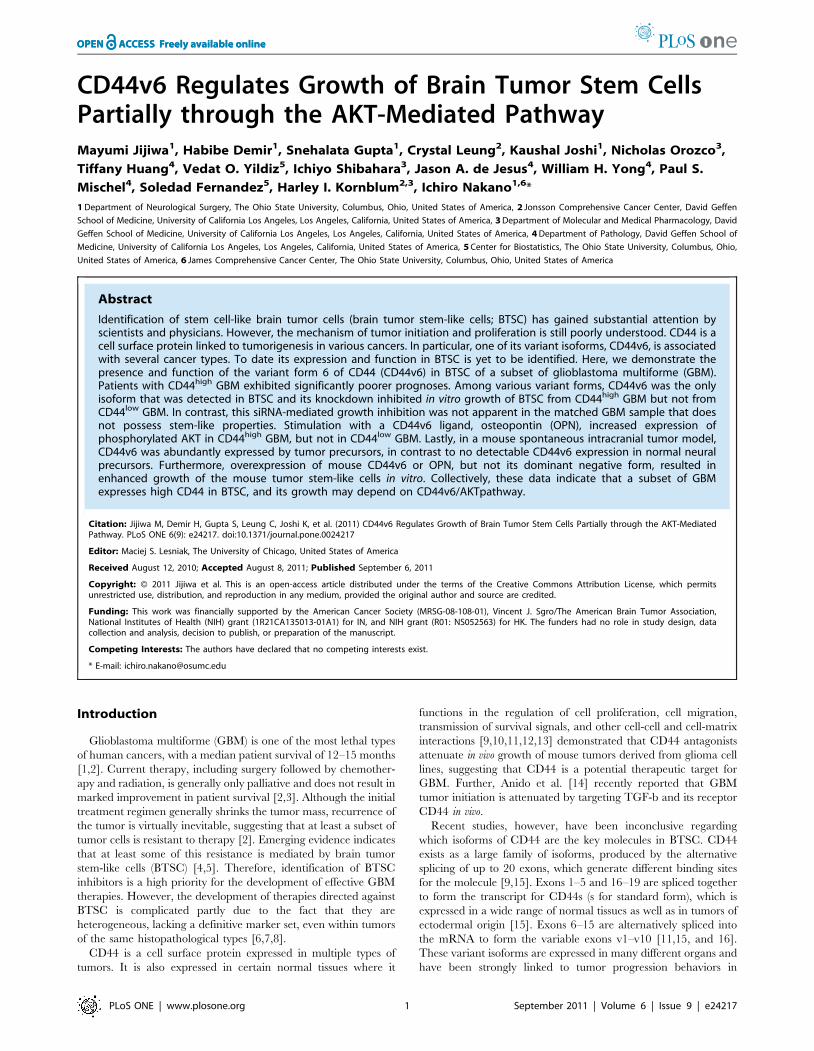
CD44v6 Regulates Growth of Brain Tumor Stem CellsPartially through the AKT-Mediated PathwayMayumi Jijiwa1, Habibe Demir1, Snehalata Gupta1, Crystal Leung2, Kaushal Joshi1, Nicholas Orozco3,
Tiffany Huang4, Vedat O. Yildiz5, Ichiyo Shibahara3, Jason A. de Jesus4, William H. Yong4, Paul S.
Mischel4, Soledad Fernandez5, Harley I. Kornblum2,3, Ichiro Nakano1,6*
1 Department of Neurological Surgery, The Ohio State University, Columbus, Ohio, United States of America, 2 Jonsson Comprehensive Cancer Center, David Geffen
School of Medicine, University of California Los Angeles, Los Angeles, California, United States of America, 3 Department of Molecular and Medical Pharmacology, David
Geffen School of Medicine, University of California Los Angeles, Los Angeles, California, United States of America, 4 Department of Pathology, David Geffen School of
Medicine, University of California Los Angeles, Los Angeles, California, United States of America, 5 Center for Biostatistics, The Ohio State University, Columbus, Ohio,
United States of America, 6 James Comprehensive Cancer Center, The Ohio State University, Columbus, Ohio, United States of America
Abstract
Identification of stem cell-like brain tumor cells (brain tumor stem-like cells; BTSC) has gained substantial attention byscientists and physicians. However, the mechanism of tumor initiation and proliferation is still poorly understood. CD44 is acell surface protein linked to tumorigenesis in various cancers. In particular, one of its variant isoforms, CD44v6, is associatedwith several cancer types. To date its expression and function in BTSC is yet to be identified. Here, we demonstrate thepresence and function of the variant form 6 of CD44 (CD44v6) in BTSC of a subset of glioblastoma multiforme (GBM).Patients with CD44high GBM exhibited significantly poorer prognoses. Among various variant forms, CD44v6 was the onlyisoform that was detected in BTSC and its knockdown inhibited in vitro growth of BTSC from CD44high GBM but not fromCD44low GBM. In contrast, this siRNA-mediated growth inhibition was not apparent in the matched GBM sample that doesnot possess stem-like properties. Stimulation with a CD44v6 ligand, osteopontin (OPN), increased expression ofphosphorylated AKT in CD44high GBM, but not in CD44low GBM. Lastly, in a mouse spontaneous intracranial tumor model,CD44v6 was abundantly expressed by tumor precursors, in contrast to no detectable CD44v6 expression in normal neuralprecursors. Furthermore, overexpression of mouse CD44v6 or OPN, but not its dominant negative form, resulted inenhanced growth of the mouse tumor stem-like cells in vitro. Collectively, these data indicate that a subset of GBMexpresses high CD44 in BTSC, and its growth may depend on CD44v6/AKTpathway.
Citation: Jijiwa M, Demir H, Gupta S, Leung C, Joshi K, et al. (2011) CD44v6 Regulates Growth of Brain Tumor Stem Cells Partially through the AKT-MediatedPathway. PLoS ONE 6(9): e24217. doi:10.1371/journal.pone.0024217
Editor: Maciej S. Lesniak, The University of Chicago, United States of America
Received August 12, 2010; Accepted August 8, 2011; Published September 6, 2011
Copyright: � 2011 Jijiwa et al. This is an open-access article distributed under the terms of the Creative Commons Attribution License, which permitsunrestricted use, distribution, and reproduction in any medium, provided the original author and source are credited.
Funding: This work was financially supported by the American Cancer Society (MRSG-08-108-01), Vincent J. Sgro/The American Brain Tumor Association,National Institutes of Health (NIH) grant (1R21CA135013-01A1) for IN, and NIH grant (R01: NS052563) for HK. The funders had no role in study design, datacollection and analysis, decision to publish, or preparation of the manuscript.
Competing Interests: The authors have declared that no competing interests exist.
* E-mail: [email protected]
Introduction
Glioblastoma multiforme (GBM) is one of the most lethal types
of human cancers, with a median patient survival of 12–15 months
[1,2]. Current therapy, including surgery followed by chemother-
apy and radiation, is generally only palliative and does not result in
marked improvement in patient survival [2,3]. Although the initial
treatment regimen generally shrinks the tumor mass, recurrence of
the tumor is virtually inevitable, suggesting that at least a subset of
tumor cells is resistant to therapy [2]. Emerging evidence indicates
that at least some of this resistance is mediated by brain tumor
stem-like cells (BTSC) [4,5]. Therefore, identification of BTSC
inhibitors is a high priority for the development of effective GBM
therapies. However, the development of therapies directed against
BTSC is complicated partly due to the fact that they are
heterogeneous, lacking a definitive marker set, even within tumors
of the same histopathological types [6,7,8].
CD44 is a cell surface protein expressed in multiple types of
tumors. It is also expressed in certain normal tissues where it
functions in the regulation of cell proliferation, cell migration,
transmission of survival signals, and other cell-cell and cell-matrix
interactions [9,10,11,12,13] demonstrated that CD44 antagonists
attenuate in vivo growth of mouse tumors derived from glioma cell
lines, suggesting that CD44 is a potential therapeutic target for
GBM. Further, Anido et al. [14] recently reported that GBM
tumor initiation is attenuated by targeting TGF-b and its receptor
CD44 in vivo.
Recent studies, however, have been inconclusive regarding
which isoforms of CD44 are the key molecules in BTSC. CD44
exists as a large family of isoforms, produced by the alternative
splicing of up to 20 exons, which generate different binding sites
for the molecule [9,15]. Exons 1–5 and 16–19 are spliced together
to form the transcript for CD44s (s for standard form), which is
expressed in a wide range of normal tissues as well as in tumors of
ectodermal origin [15]. Exons 6–15 are alternatively spliced into
the mRNA to form the variable exons v1–v10 [11,15, and 16].
These variant isoforms are expressed in many different organs and
have been strongly linked to tumor progression behaviors in
PLoS ONE | www.plosone.org 1 September 2011 | Volume 6 | Issue 9 | e24217
various cancers [15,17,18,19]. The variant 6 isoform (CD44v6), in
particular, is associated with several cancer types but not in
somatic cells [17,20,21]. CD44v6 and its ligand, osteopontin
(OPN) are highly expressed in breast cancer [20,22] as well as
leukemia [23] and gastric cancer [24], and regulate malignant
transformation by inducing tumor cell proliferation and control-
ling migration [20,21].
Expression of CD44 is widely identified in cancer stem cells in
various organs, such as breast, colon, and pancreas [25,26,27]. In
these tumors, tumor-forming cells in vivo are restricted to CD44-
positive tumor cells [25,27]. However, little is known about which
isoforms are specifically associated with cancer stem cells. A recent
study demonstrated that CD44v6 is likely expressed by bladder
carcinoma stem cells, suggesting that this isoform may be of
particular link to cancer stem cells [28]. To our knowledge, no study
has identified the specific CD44 isoforms that are present in BTSC.
Here, we demonstrate that a subset of BTSC in GBM express
CD44 and its variant form 6 (CD44v6) plays a positive role in their
growth in vitro. CD44-neutralizing antibody inhibits the growth of
BTSC derived from GBM samples that contain cells with high
CD44 (CD44high GBM), while it has no significant effect on the
BTSC from GBM containing population of cells with low
expression of CD44 (CD44low GBM). BTSC derived from
CD44high GBM depend on CD44v6 to maintain proliferation.
Targeting CD44v6 has, therefore, potential impact in eradicating
therapy-resistant BTSC in GBM.
Materials and Methods
EthicsExperiments using human tissue-derived materials were carried
out under the approved institutional review board at UCLA.
Informed consent was obtained in written form from all human
subjects and families of autopsy patients to publication of their case
details prior to the experiments performed in this study. All animal
experimentation was performed with the approval of the UCLA
Animal Research Committee, following NIH guidelines, using
animal protocol number 93-285. The mice were experimentally
used in accordance with the Institutional Animal Care and Use
Committee guidelines at UCLA.
Tissue cultureTumors derived from mice deficient in both p53 and patched
were kindly provided by Dr. James A. Waschek at UCLA [29].
Neurosphere (NS) cultures were prepared as previously described
[30]. Briefly, small GBM samples were dissociated with a fire-
polished glass pipette and resuspended at 50,000 cells/ml in
neurosphere medium, containing Dulbecco’s modified Eagle
medium (DMEM)/F12 medium (GIBCO, Invitrogen, Carlsbad,
CA) supplemented with B27 (final concentration 2%, GIBCO,
Invitrogen, Carlsbad, CA), basic fibroblast growth factor (bFGF)
(20 ng/ml, Peprotech, Rocky Hill, NJ), epidermal growth factor
(EGF) (50 ng/ml, Peprotech), penicillin/streptomycin (1%,
GIBCO, Invitrogen, Carlsbad, CA), and heparin (5 mg/ml, Sigma
Aldrich, St. Louis, MO). To differentiate the BTSC, spheres were
dissociated into single cells, added to poly-L-Lysine coated dishes
containing Neurobasal medium (GIBCO, Invitrogen, Carlsbad,
CA) with B27, and maintained for up to five days, followed by
culture in serum containing medium.
RT-PCRTotal RNA was isolated with TRIzol (GIBCO, North Andover,
MA) from GBM specimens and adjacent normal brains of autopsy
samples. One mg of each total RNA was reverse transcripted to
cDNA with ImProm-II Reverse Transcriptase (Invitrogen for
conventional polymerase chain reaction (PCR), Promega, Madi-
son, WI, for quantitative PCR), according to the manufacturer’s
protocol. Reverse transcription-polymerase chain reaction (RT-
PCR) with primers for the glyceraldehyde-3-phosphate-dehydro-
genase (GAPDH) gene served as an internal control. After correc-
ting for GAPDH signals by electrophoresis, reverse transcripted
cDNA was subjected to quantitative PCR analysis using the gene
specific primers. The primers for the CD44; forward: TTTGC-
ATTGCAGTCAACAGTC and reverse: GTTACACCCCAAT-
CTTCATGTCCAC, for the CD44v6; forward: GAAGAAACA-
GCTACCCAGAAGGAACAG and reverse: GCCAAGAGGGA-
TGCCAAGATG and for the GAPDH; forward: AAGGTGA-
AGGTCGGAGTCAA and reverse: AATGAAGGGGTCATT-
GATGG were constructed based upon GenBank accession
number NM000610. The protocol for the thermal cycler was
described previously [30]. Control experiments excluded reverse
transcriptase and/or template cDNA. Each reaction was visual-
ized after electrophoresis with 2% agarose gel. Relative quanti-
fication for quantitative real-time polymerase chain reaction
(qRT-PCR) was determined with the LightCycler Relative
Quantification Software (Roche Diagnostics, Indianapolis, IN).
Western blotTotal lysate of brain tissue were prepared from GBM specimens
and adjacent normal brains of autopsy samples using sodium
dodecyl sulfate (SDS) sample buffer. Whole-cell lysates were
prepared in lysis buffer containing Protease Inhibitor CockTail
(P8340, Sigma Aldrich, St. Louis, MO) and protein concentrations
determined by bicinconic acid (BCA) protein assay kit (Thermo
SCIENTIFIC, Rockford, IL) according to the manufacturer’s
protocol. Equal amounts of proteins were fractionated on sodium
dodecyl sulfate-polyacrylamide gel electrophoresis and transferred
to polyvinylidene fluoride (PVDF) membrane (Invitrogen, Carls-
bad, CA). The membrane was incubated with AKT (rabbit,
1:1000, Cell Signaling Technology, Danvers, MA), phospho-AKT
(rabbit, 1:1000, Cell Signaling Technology, Danvers, MA), and
GAPDH antibody (rabbit, 14C10, Cell Signaling Technology),
followed by signal amplification with anti-rabbit immunoglobulin
G (1:250, GE Healthcare, Pataskala, OH) and detection with
enhanced chemiluminescence.
Tissue MicroarrayTissue microarray (TMA) consisting of three to six representa-
tive 0.6-mm cores from formalin-fixed, paraffin-embedded tissue
blocks was generated at the Department of Pathology and
Laboratory Medicine at UCLA, under the protocols approved
by the UCLA Institutional Review Board. The tissue samples were
collected either from autopsies of patients with GBM within
24 hours of death or from patients who underwent surgery at
UCLA Medical Center. After immunohistochemistry, tissues too
small and/or crushed were eliminated, and 64 samples from 37
patients were introduced to further analysis. All samples were
diagnosed as high grade glioma (corresponding to glioblastoma
and anaplastic astrocytoma) or low grade glioma by nuclear atypia
and cell density, or tumor-free region. CD44 expression was
analyzed by two neuropathologists in a blind protocol, and
staining patterns were determined according to the immunoreac-
tive site as cell surface and process. Overall staining intensity was
scored as 2 (negative), + (weak), ++ (moderate to strong). For the
characterization of TMA samples and appropriate patients,
highest grade tissues of patients showing multiple features were
adopted. Overall survival period was defined as from initial
diagnosis to decease.
CD44v6 in Brain Tumor Stem Cells
PLoS ONE | www.plosone.org 2 September 2011 | Volume 6 | Issue 9 | e24217
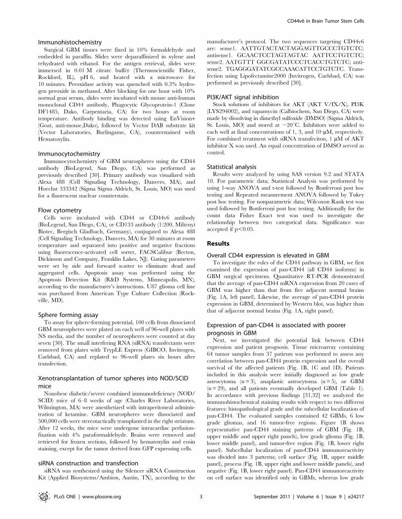
ImmunohistochemistrySurgical GBM tissues were fixed in 10% formaldehyde and
embedded in paraffin. Slides were deparaffinized in xylene and
rehydrated with ethanol. For the antigen retrieval, slides were
immersed in 0.01 M citrate buffer (Thermoscientific Fisher,
Rockford, IL), pH 6, and heated with a microwave for
10 minutes. Peroxidase activity was quenched with 0.3% hydro-
gen peroxide in methanol. After blocking for one hour with 10%
normal goat serum, slides were incubated with mouse anti-human
monoclonal CD44 antibody, Phagocytic Glycoprotein-1 (Clone
DF1485, Dako, Carpentaria, CA) for two hours at room
temperature. Antibody binding was detected using EnVision+(Goat, anti-mouse,Dako), followed by Vector DAB substrate kit
(Vector Laboratories, Burlingame, CA), counterstained with
Hematoxylin.
ImmunocytochemistryImmunocytochemistry of GBM neurospheres using the CD44
antibody (BioLegend, San Diego, CA) was performed as
previously described [30]. Primary antibody was visualized with
Alexa 488 (Cell Signaling Technology, Danvers, MA), and
Hoechst 333342 (Sigma Sigma Aldrich, St. Louis, MO) was used
for a fluorescent nuclear counterstain.
Flow cytometryCells were incubated with CD44 or CD44v6 antibody
(BioLegend, San Diego, CA), or CD133 antibody (1:200, Miltenyi
Biotec, Bergisch Gladbach, Germany), conjugated to Alexa 488
(Cell Signaling Technology, Danvers, MA) for 30 minutes at room
temperature and separated into positive and negative fractions
using fluorescence-activated cell sorter, FACSCalibur (Becton,
Dickinson and Company, Franklin Lakes, NJ). Gating parameters
were set by side and forward scatter to eliminate dead and
aggregated cells. Apoptosis assay was performed using the
Apoptosis Detection Kit (R&D Systems, Minneapolis, MN),
according to the manufacturer’s instructions. U87 glioma cell line
was purchased from American Type Culture Collection (Rock-
ville, MD).
Sphere forming assayTo assay for sphere-forming potential, 100 cells from dissociated
GBM neurospheres were plated on each well of 96-well plates with
NS media, and the number of neurospheres were counted at day
seven [30]. The small interfering RNA (siRNA) transfectants were
removed from plates with TrypLE Express (GIBCO, Invitrogen,
Carlsbad, CA) and replated to 96-well plates six hours after
transfection.
Xenotransplantation of tumor spheres into NOD/SCIDmice
Nonobese diabetic/severe combined immunodeficiency (NOD/
SCID) mice of 6–8 weeks of age (Charles River Laboratories,
Wilmington, MA) were anesthetized with intraperitoneal adminis-
tration of ketamine. GBM neurospheres were dissociated and
500,000 cells were stereotactically transplanted in the right striatum.
After 12 weeks, the mice were undergone intracardiac perfusion-
fixation with 4% paraformaldehyde. Brains were removed and
retrieved for frozen sections, followed by hematoxylin and eosin
staining, except for the tumor derived from GFP expressing cells.
siRNA construction and transfectionsiRNA was synthesized using the Silencer siRNA Construction
Kit (Applied Biosystems/Ambion, Austin, TX), according to the
manufacturer’s protocol. The two sequences targeting CD44v6
are: sense1. AATTGTACTACTAGGAGTTGCCCTGTCTC;
antisense1. GCAACTCCTAGTAGTAC AATTCCTGTCTC;
sense2. AATGTTT GGCGATATCCCTCACCTGTCTC; anti-
sense2. TGAGGGATATCGCCAAACATTCCTGTCTC. Trans-
fection using Lipofectamine2000 (Invitrogen, Carlsbad, CA) was
performed as previously described [30].
PI3K/AKT signal inhibitionStock solutions of inhibitors for AKT (AKT V/IX/X), PI3K
(LYS294002), and rapamycin (Calbiochem, San Diego, CA) were
made by dissolving in dimethyl sulfoxide (DMSO) (Sigma Aldrich,
St. Louis, MO) and stored at 220uC. Inhibitors were added to
each well at final concentrations of 1, 3, and 10 mM, respectively.
For combined treatment with siRNA transfeciton, 1 mM of AKT
inhibitor X was used. An equal concentration of DMSO served as
control.
Statistical analysisResults were analyzed by using SAS version 9.2 and STATA
10. For parametric data; Statistical Analysis was performed by
using 1-way ANOVA and t-test followed by Bonferroni post hoc
testing and Repeated measurement ANOVA followed by Tukey
post hoc testing. For nonparametric data; Wilcoxon Rank test was
used followed by Bonferroni post hoc testing. Additionally for the
count data Fisher Exact test was used to investigate the
relationship between two categorical data. Significance was
accepted if p,0.05.
Results
Overall CD44 expression is elevated in GBMTo investigate the roles of the CD44 pathway in GBM, we first
examined the expression of pan-CD44 (all CD44 isoforms) in
GBM surgical specimens. Quantitative RT-PCR demonstrated
that the average of pan-CD44 mRNA expression from 20 cases of
GBM was higher than that from five adjacent normal brains
(Fig. 1A, left panel). Likewise, the average of pan-CD44 protein
expression in GBM, determined by Western blot, was higher than
that of adjacent normal brains (Fig. 1A, right panel).
Expression of pan-CD44 is associated with poorerprognosis in GBM
Next, we investigated the potential link between CD44
expression and patient prognosis. Tissue microarray containing
64 tumor samples from 37 patients was performed to assess any
correlation between pan-CD44 protein expression and the overall
survival of the affected patients (Fig. 1B, 1C and 1D). Patients
included in this analysis were initially diagnosed as low grade
astrocytoma (n = 3), anaplastic astrocytoma (n = 5), or GBM
(n = 29), and all patients eventually developed GBM (Table 1).
In accordance with previous findings [31,32] we analyzed the
immunohistochemical staining results with respect to two different
features: histopathological grade and the subcellular localization of
pan-CD44. The evaluated samples contained 42 GBMs, 6 low
grade gliomas, and 16 tumor-free regions. Figure 1B shows
representative pan-CD44 staining patterns of GBM (Fig. 1B,
upper middle and upper right panels), low grade glioma (Fig. 1B,
lower middle panel), and tumor-free region (Fig. 1B, lower right
panel). Subcellular localization of pan-CD44 immunoreactivity
was divided into 3 patterns; cell surface (Fig. 1B, upper middle
panel), process (Fig. 1B, upper right and lower middle panels), and
negative (Fig. 1B, lower right panel). Pan-CD44 immunoreactivity
on cell surface was identified only in GBMs, whereas low grade
CD44v6 in Brain Tumor Stem Cells
PLoS ONE | www.plosone.org 3 September 2011 | Volume 6 | Issue 9 | e24217
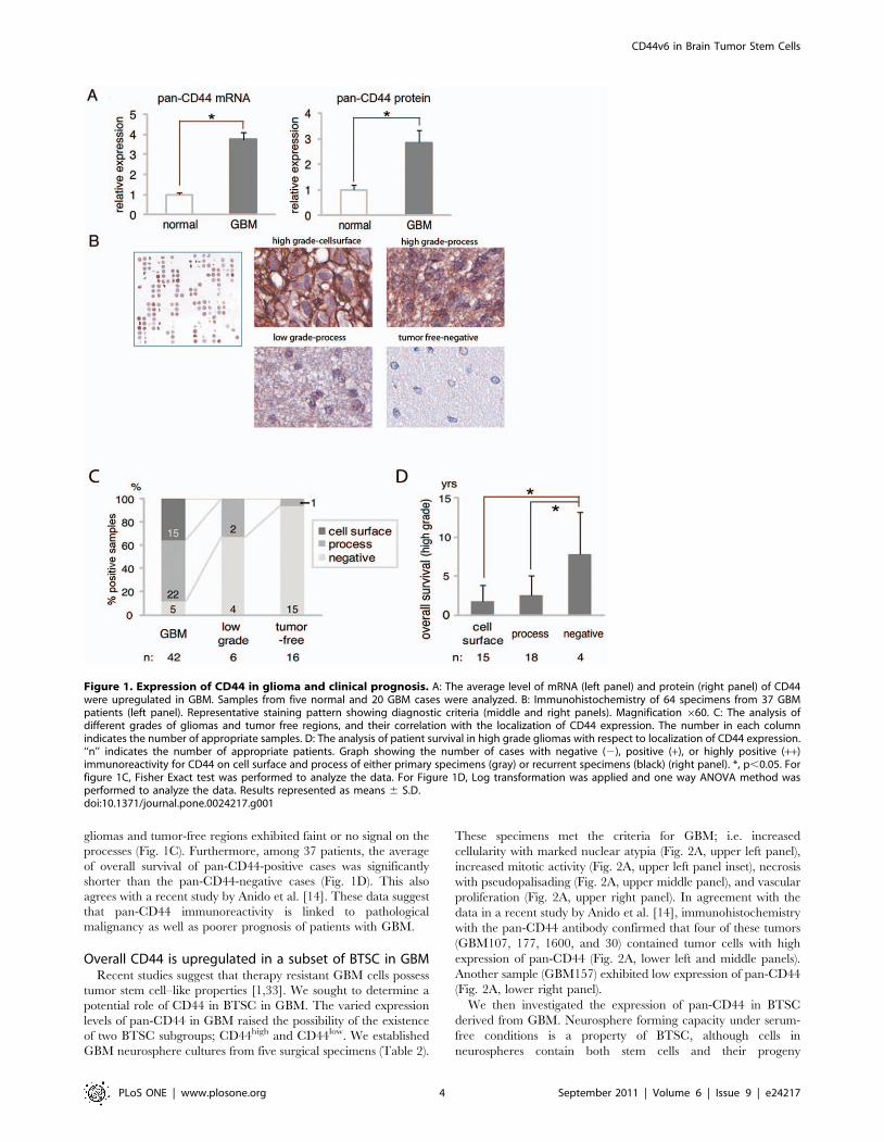
gliomas and tumor-free regions exhibited faint or no signal on the
processes (Fig. 1C). Furthermore, among 37 patients, the average
of overall survival of pan-CD44-positive cases was significantly
shorter than the pan-CD44-negative cases (Fig. 1D). This also
agrees with a recent study by Anido et al. [14]. These data suggest
that pan-CD44 immunoreactivity is linked to pathological
malignancy as well as poorer prognosis of patients with GBM.
Overall CD44 is upregulated in a subset of BTSC in GBMRecent studies suggest that therapy resistant GBM cells possess
tumor stem cell–like properties [1,33]. We sought to determine a
potential role of CD44 in BTSC in GBM. The varied expression
levels of pan-CD44 in GBM raised the possibility of the existence
of two BTSC subgroups; CD44high and CD44low. We established
GBM neurosphere cultures from five surgical specimens (Table 2).
These specimens met the criteria for GBM; i.e. increased
cellularity with marked nuclear atypia (Fig. 2A, upper left panel),
increased mitotic activity (Fig. 2A, upper left panel inset), necrosis
with pseudopalisading (Fig. 2A, upper middle panel), and vascular
proliferation (Fig. 2A, upper right panel). In agreement with the
data in a recent study by Anido et al. [14], immunohistochemistry
with the pan-CD44 antibody confirmed that four of these tumors
(GBM107, 177, 1600, and 30) contained tumor cells with high
expression of pan-CD44 (Fig. 2A, lower left and middle panels).
Another sample (GBM157) exhibited low expression of pan-CD44
(Fig. 2A, lower right panel).
We then investigated the expression of pan-CD44 in BTSC
derived from GBM. Neurosphere forming capacity under serum-
free conditions is a property of BTSC, although cells in
neurospheres contain both stem cells and their progeny
Figure 1. Expression of CD44 in glioma and clinical prognosis. A: The average level of mRNA (left panel) and protein (right panel) of CD44were upregulated in GBM. Samples from five normal and 20 GBM cases were analyzed. B: Immunohistochemistry of 64 specimens from 37 GBMpatients (left panel). Representative staining pattern showing diagnostic criteria (middle and right panels). Magnification 660. C: The analysis ofdifferent grades of gliomas and tumor free regions, and their correlation with the localization of CD44 expression. The number in each columnindicates the number of appropriate samples. D: The analysis of patient survival in high grade gliomas with respect to localization of CD44 expression.‘‘n’’ indicates the number of appropriate patients. Graph showing the number of cases with negative (2), positive (+), or highly positive (++)immunoreactivity for CD44 on cell surface and process of either primary specimens (gray) or recurrent specimens (black) (right panel). *, p,0.05. Forfigure 1C, Fisher Exact test was performed to analyze the data. For Figure 1D, Log transformation was applied and one way ANOVA method wasperformed to analyze the data. Results represented as means 6 S.D.doi:10.1371/journal.pone.0024217.g001
CD44v6 in Brain Tumor Stem Cells
PLoS ONE | www.plosone.org 4 September 2011 | Volume 6 | Issue 9 | e24217
[4,5,33,34]. When the GBM neurospheres were established from
the above 5 samples, both CD44high GBM and CD44low GBM
formed neurospheres with no significant differences in their
growth rate (data not shown). Immunocytochemical staining
revealed that GBM107, 177, 1600 and 30 neurospheres expressed
pan-CD44, whereas GBM157 neurospheres were negative for
pan-CD44 (Fig. 2B and Figure S1). Thus, these neurospheres
samples were found to retain a similar expression pattern of pan-
CD44 to the parental tumors.
CD44 plays a role in the growth of a subset of BTSCCD44 is known to play a key role in the self-renewal of cancer
stem cells in several cancer types including breast cancer,
pancreatic cancer, and acute myeloid leukemia [25,27,35]. In
addition, two recent studies demonstrated the presence of pan-
CD44 in GBM and their BTSC as well as its positive role in their
growth [14,36]. Similar to these observations, cell sorting
experiments demonstrated that only pan-CD44-positive GBM
cells were capable of forming neurospheres in the CD44high GBM
samples (GBM107 and 1600) (Figure S 2, left panel), whereas no
significant difference in neurosphere-forming potential was
exhibited between equal numbers of pan-CD44-positive and -
negative cells seeded in serum-free media from the CD44low GBM
sample (GBM157) (Figure S2, right panel). Further, treatment of
dissociated GBM cells with a monoclonal antibody for pan-CD44
(clone IM7; ABcam), which is widely used to block the pan-CD44
signals in vitro [37,38] resulted in abraded neurosphere formation
in the CD44high samples (GBM107 and 1600) (Figure S. 3A, left
panel). To exclude non-specific effect caused by incubation with
this antibody, we confirmed no significant inhibitory effect on the
CD44low sample (GBM157) (Figure S3A, right panel).
To discriminate the effect on either arrest of cell proliferation,
induced cell death, and/or differentiation, we analyzed cell growth
(Fig. S3B), apoptosis (Fig. S3C) and differentiation (Fig. S3D).
Treatment of GBM1600 cells in neurospheres with the CD44-
blocking antibody inhibited cell growth (Fig. S3B) without
increasing propidium iodide (PI)/AnnexinV positive apoptotic
cells (Figure S 3C) or decreasing the proportion of CD133 positive
undifferentiated cells (Figure S3D). Taken together, these results
suggest that CD44 plays a role in the growth of BTSC in CD44high
GBM.
Presence of CD44v6 in neurospheres derived fromCD44high GBM
CD44 has multiple isoforms; the standard form and the variant
forms. In human cancer, splice variants of CD44 were frequently
identified in advanced stages of tumorigenesis [15,17,18]. To
determine if any variant forms of CD44 are expressed in BTSC,
we performed RT-PCR with a primer set that amplified the
standard form (CD44s) and the variant form (CD44v) as different
sizes (Fig. 2C, upper panel). GBM neurospheres, but not normal
human astrocytes (NHA), yielded only two detectable PCR
products, and DNA sequencing demonstrated that the amplified
PCR products are CD44s (Fig. 2C, lower left panel, arrow) and
CD44v6 (Fig. 2C, lower left panel, arrowhead). Further, by
designing a specific primer set to amplify CD44v6 (Fig. 2C, upper
panel), we confirmed that neurospheres derived from CD44high
GBM, but not from CD44low GBM, expressed CD44v6 (Fig. 2C,
lower right panel). We then examined both pan-CD44 and
CD44v6 expression in our five GBM neurosphere samples with
flow cytometry. All four CD44high GBM neurospheres (GBM107,
177, 1600, and 30) contained a subpopulation of tumor cells
expressing CD44v6 (Fig. 2D). In contrast, the CD44low GBM
sample (GBM157) exhibited no detectable level of CD44v6-
positive cells (Fig. 2D).
Inhibition of neurosphere formation from CD44high BTSCby targeting CD44v6
To determine the specific role of CD44v6 in CD44high BTSC,
we designed two siRNA constructs that target CD44v6, and
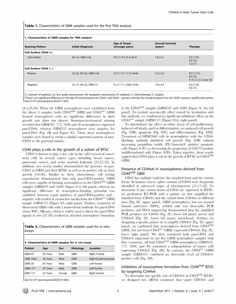
Table 1. Characteristics of GBM samples used for the first TMA analysis.
1. Characteristics of GBM samples for TMA analysis
Staining Pattern Initial DiagnosisAge of Onset(average years)
Overall Survival(years) Therapy
Cell Surface CD44 (+)
Cell Surface AA [1], GBM [14] 43.7616.9 (5.4–66.3) 1.862.1 R,C,S [9];R,S [5];C,S [1]
Cell Surface CD44 (2)
Process LA [2], AA [2], GBM [14] 57.7617.1 (17.2–84.8) 2.562.5 R,C,S [11];R,S [2];R [1]; S [1]; none [3]
Negative LA [1], AA [2], GBM [1] 41.2611.1 (28.8–54.8) 7.865.4 R,C,S [3];R,S [1];
[ ]: number of patients; LA: low grade astrocytoma; AA: anaplastic astrocytoma; R: radiation; C: chemotherapy; S: surgery.\There is no significant difference in the age of onset between the CD44+ and CD442 groups, whereas the overall prognosis for the CD44+ group is significantly poorer.doi:10.1371/journal.pone.0024217.t001
Table 2. Characteristics of GBM samples used for in vitroassays.
2. Characteristics of GBM samples for in vitro assay
Patient Age Sex Histology Location
GBM107 63 Years Male GBM Right Frontal
GBM 1600 34 Years Male GBM Right frontal temporal
GBM 30 65 Years Male GBM Left frontal
GBM 177 47 Years Male GBM Left frontal
GBM 177 54 Years Female GBM Right frontal
doi:10.1371/journal.pone.0024217.t002
CD44v6 in Brain Tumor Stem Cells
PLoS ONE | www.plosone.org 5 September 2011 | Volume 6 | Issue 9 | e24217

CD44v6 in Brain Tumor Stem Cells
PLoS ONE | www.plosone.org 6 September 2011 | Volume 6 | Issue 9 | e24217

verified their effects on three different GBM cell samples;
neurospheres derived from CD44high GBM1600 and CD44low
GBM157, and serum-propagated cells derived from GBM1600.
The phenotypic difference between neurospheres and serum-
propagated cells was highlighted by their tumorigenic capacity in
the xenograft model. When implanted into immunodeficient mice
brains, GBM neurospheres from CD44high GBM were capable of
forming GBM-like tumors (Fig. 3A, left panel, tumor formation
incidence: 3/3). On the other hand, GBM cells from the same
tumor that were propagated in serum-containing medium did not
possess tumorigenic potential (Fig. 3A, middle panel, tumor
formation incidence: 0/3), implying a loss of tumor initiating cells
under this condition, in consistence with previous observations
[39,40]. The CD44v6 specific siRNA, when transfected into these
two GBM spheres and serum-propagated cells, reduced CD44v6
expression levels, while CD44s expression was not affected in
either condition (Fig. 3B, left and middle panels). Knockdown of
CD44v6 in CD44high GBM neurospheres resulted in a subsequent
reduction in neurosphere formation (Fig. 3C, left panel), whereas
growth of the serum-propagated cells was not significantly affected
(Fig. 3C, middle panel). As expected, the expression of CD44v6
was undetectable by RT-PCR in the CD44low GBM cells (Fig. 3B,
right panel), and siRNA treatment did not affect their neurosphere
formation (Fig. 3C, right panel).
A CD44v6 ligand, osteopontin, activates AKT pathway ina subset of GBM
To further gain insight into their signaling pathway, we sought
to identify their downstream targets and to characterize the
distinct mechanism of CD44v6 in BTSC growth. OPN is a ligand
for CD44v6, and, in some hematopoietic malignancies, activation
of CD44v6 by OPN contributes to tumor cell survival via the
PI3K/AKT pathway [41]. Therefore, we investigated whether
OPN triggers AKT activity in neurospheres derived from GBM30
and GBM1600 (both CD44high), as well as GBM157 (CD44low)
(Fig. 4A, upper left panel). Incubation with OPN resulted in an
appreciable increase in phosphorylated AKT in spheres derived
from both GBM30 and GBM1600 spheres but not in the
GBM157 spheres (Fig. 4A, upper left panel). As expected, OPN
treatment also activated S6R, a downstream target of AKT, in
both GBM30 and GBM1600 spheres (Fig. 4A, upper left panel).
To elucidate the signaling pathways in CD44v6-expressing GBM
cells, we knocked down CD44v6 with siRNA in GBM1600 cells
(Fig. 4A, lower right panel). Untreated cells showed activation of
the AKT and S6R pathway without ligand stimulation (Fig. 4A,
upper right panel). With CD44v6 knockdown, both phosphory-
lated AKT and S6R were under detectable level and OPN failed
to activate AKT and S6R, while EGF substantially increased both
molecules (Fig. 4A, upper right panel). These observations
prompted the question of whether sensitivity to the PI3K/AKT
inhibitors is different between CD44v6-expressing BTSC and
CD44v6-negativeBTSC. To address this question, we performed
neurosphere-forming assay using 3 GBM samples (Fig. 4B). For
inhibition of the PI3K/AKT pathway, we used five different small
molecules. In both GBM177 cells and GBM1600 cells, a dose-
dependent inhibition for neurosphere formation was observed for
all five inhibitors (Fig. 4B, upper left and right panels). In contrast,
GBM157 cells were relatively resistant to the treatment (Fig. 4B,
lower left panel).
We also addressed an assumption that if AKT is the major
downstream target of CD44v6, the phenotype of CD44v6
knockdown in GBM cells should be masked by inhibition of
AKT. Therefore, we combined transfection of siRNA for CD44v6
and AKT inhibitor X treatment in GBM 1600 cells (Fig. 4C).
AKT inhibitor X-treated GBM1600 cells did not yield significant
difference in the number of neurospheres with or without
knockdown of CD44v6. These data suggest that a subset of BTSC
in GBM may depend on CD44v6 and the action of CD44v6 is
mediated through AKT.
CD44v6 in normal stem cells and brain tumor stem cellsin the murine model
Recent studies have suggested that overlapping genes andsignal-
ing pathways regulate the proliferation of both tumor stem cells
and normal stem cells [42,43]. Targeting pathways that are
essential for proliferation of both tumor and normal stem cells may
result in the same outcome for both. To determine whether
CD44v6 might be a potential target in BTSC without affecting the
maintenance of proliferating normal stem cells, we used a mouse
model of brain tumor. Mice heterozygous for a mutation in
the gene encoding the sonic hedgehog receptor patched 1 (Ptc),
known to form spontaneous intracranial malignant tumors, were
bred with mice heterozygous for a tumor suppressor gene, p53
(Ptc+/2, p53+/2). Diamandis et al. [44] have demonstrated that
neurospheres derived from these mouse tumors are enriched with
multipotent self-renewing brain tumor stem cells. We, therefore,
used these mice to further characterize CD44v6 in BTSC and
their normal counterpart. To enrich for normal neural stem cells,
we cultured neurospheres from the subventricular zone cells using
the wild type mice at the gestation age of 11.5 (E11.5). RT-PCR
detected mouse CD44v6 in tumor neurospheres, but not in normal
mouse brain samples (Fig. 5A). Flow cytometry analysis exhibited
similar results (Fig. 5B and C). Although neurospheres derived
from the E11.5 cortices had 14.9% of the cells labeled with the
pan-CD44 antibody, these normal progenitors did not express
appreciable CD44v6 (0.3% compared to the negative control
samples) (Fig. 5B). In contrast, tumor specimens in the p53+/2,
Ptc+/2 mice had a much greater fraction of the CD44v6-
expressing cells (8.3%) (Fig. 5C, upper panel). Interestingly, when
these tumor cells were cultured in serum-free medium to form
neurospheres, the fraction of CD44v6-positive cells increased over
the period of one month (8.3% vs. 52.1%), suggesting the
preferential proliferation of CD44v6-positive cells in conditions
that enriched for BTSC (Fig. 5C middle and lower panels). This
result was consistent with the increased expression of stem cell-
associated proteins, including Nestin and Sox2 in these cultures
Figure 2. CD44v6 is upregulated in a subset of BTSC in GBM. A: Histopathology of parental tumor of established GBM neurospheres. All GBMsamples showed increased cellularity with marked nuclear atypia (upper left panel), magnification 640, increased mitotic activity (inset), necrosis(upper middle panel),magnification 620, pseudopalisading (black arrowhead) and vascular proliferation (upper right panel), magnification 640.GBM107 showed high immunoreactivity with CD44 antibody on process (lower left panel), magnification 640. GBM177 showed highimmunoreactivity with CD44 antibody on cell surface (lower middle panel, white arrowhead) as well as on process. Magnification 640. GBM157showed minimal immunoreactivity on process (lower right panel). Magnification 640. B: GBM 1600 cells revealed CD44 immunostaining (green) (leftpanel), however, GBM 157 cells were not stained (left panel). Nuclei were counterstained with Hoechst. Magnification640. C: A schematic diagram ofthe CD44 gene with position of primers (upper panel). An expected size of CD44s (arrow) and a longer product (arrowhead) were amplified fromGBM107 (lower left panel). GBM107 exhibited CD44s and CD44v6 specific bands (lower right panel). D: Flow cytometry showing the ratio of CD44-expressing cells or CD44v6-expressing cells in each GBM sphere sample.doi:10.1371/journal.pone.0024217.g002
CD44v6 in Brain Tumor Stem Cells
PLoS ONE | www.plosone.org 7 September 2011 | Volume 6 | Issue 9 | e24217

(data not shown). We then investigated the function of CD44v6
and its ligand, OPN, in these mouse tumor spheres (Fig. 5D and
E). Both overexpressions of CD44v6 and OPN, but not the
dominant negative form of OPN, significantly increased sphere
formation derived from the tumor cells (Fig. 5E). These data
suggests that the OPN-CD44v6 axis plays a positive role in
proliferation of BTSC in a subset of GBM cases.
Discussion
An accumulating body of evidence suggests that tumor
heterogeneity exists in various types of cancers, including GBM
[4,6,12]. However, differential regulatory molecules and pathways
specific to each tumor type are poorly understood. In agreement
with a recent study by Anido et al. [14], we identified a
subpopulation of GBM in which CD44 expression was upregu-
lated (CD44high GBM). CD44high GBM demonstrated correlation
with poorer clinical prognosis. Xu et al. [13] showed combined
treatment of mouse intracranial tumors derived from a glioma cell
line with CD44 antagonist and the current first line chemotherapy,
temozolomide, prolonged survival of mice. Temozolomide is
known to preferentially kill non-stem GBM cells [14]. Collectively,
these data raise a possibility that CD44-expressing GBM cells are
relatively therapy resistant and likely a reasonable therapeutic
target, especially in recurrent GBM tumors that survived over the
current therapies. However, the case number of our comparison is
still limited and a definite conclusion should be drawn with more
accumulated data set in the future.
Figure 3. CD44v6 is required for the growth of BTSC in CD44high GBM. A: Brain sections with tumors originated from transplanted GBMneurosphere/serum-propagated cells in mice brains. Neurospheres from CD44high GBM formed remarkable tumor (left panel, surrounded by whitedots), while serum-propagated cells formed tiny region (middle panel, surrounded by white dots). Neurospheres from CD44low GBM (GBM157)formed a large tumor (right panel). Magnification 61. B: Treatment with siRNA decreased the expression of CD44v6 in both neurosphere (left panel)and serum-propagated CD44high GBM (middle panel). C. The numbers of neurospheres (left panel, right panel) or total cells (middle panel) grownafter treatment with CD44v6-siRNA. Neurospheres propagated from CD44high GBM neurosphere cells showed statistically significant differencebetween siRNA and siGFP treated groups (left panel). No statistical difference was seen in growth of serum-propagated cells (middle panel) andneurospheres propagated from CD44low GBM neurosphere cells (right panel). All the experiments were performed in triplicates. *, p,0.05. Forneurospheres, Wilcoxon rank test was performed to analyze the data and Bonferroni adjustment was used for pairwise comparison. For serum-propagated cells, Repeated Measurement ANOVA was performed to analyze the trends over time data and Tukey adjustment was used for pairwisecomparisons. Results represented as means 6 SEM.doi:10.1371/journal.pone.0024217.g003
CD44v6 in Brain Tumor Stem Cells
PLoS ONE | www.plosone.org 8 September 2011 | Volume 6 | Issue 9 | e24217
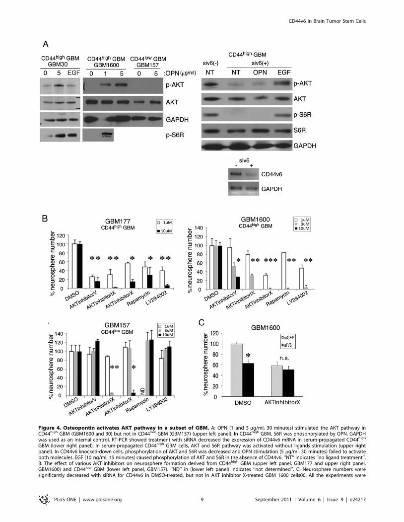
Figure 4. Osteopontin activates AKT pathway in a subset of GBM. A: OPN (1 and 5 mg/ml, 30 minutes) stimulated the AKT pathway inCD44high GBM (GBM1600 and 30) but not in CD44low GBM (GBM157) (upper left panel). In CD44high GBM, S6R was phosphorylated by OPN. GAPDHwas used as an internal control. RT-PCR showed treatment with siRNA decreased the expression of CD44v6 mRNA in serum-propagated CD44high
GBM (lower right panel). In serum-propagated CD44high GBM cells, AKT and S6R pathway was activated without ligands stimulation (upper rightpanel). In CD44v6 knocked-down cells, phosphorylation of AKT and S6R was decreased and OPN stimulation (5 mg/ml, 30 minutes) failed to activateboth molecules. EGF (10 ng/ml, 15 minutes) caused phosphorylation of AKT and S6R in the absence of CD44v6. ‘‘NT’’ indicates ‘‘no ligand treatment’’.B: The effect of various AKT inhibitors on neurosphere formation derived from CD44high GBM (upper left panel, GBM177 and upper right panel,GBM1600) and CD44low GBM (lower left panel, GBM157). ‘‘ND’’ in (lower left panel) indicates ‘‘not determined’’. C: Neurosphere numbers weresignificantly decreased with siRNA for CD44v6 in DMSO-treated, but not in AKT inhibitor X-treated GBM 1600 cells00. All the experiments were
CD44v6 in Brain Tumor Stem Cells
PLoS ONE | www.plosone.org 9 September 2011 | Volume 6 | Issue 9 | e24217

Here, we provide the first evidence for the presence of CD44v6 in
BTSC derived from CD44high GBM. Khan et al. [20] suggested
that CD44v6 regulates the aggressiveness of breast cancer cells. We
found that both CD44v6 overexpression and OPN overexpression
increased sphere forming ability of mouse intracranial tumor cells.
In turn, knockdown of CD44v6 resulted in reduced growth of
human BTSC derived from CD44high GBM but not from CD44low
GBM in vitro. On the other hand, the effect on serum-propagated
cells from the matched CD44high GBM was less prominent and not
statistically significant. However, these data need to be carefully
interpreted, as serum-propagated human GBM cells do express
CD44v6 (Fig. 5B). It is possible that targeting CD44v6 may reduce
the growth of both BTSC and non-stem GBM cells with different
potency. Future study is needed to address this question.
Interestingly, CD44v6 was not detected in normal mouse brains
or neural progenitors (Fig. 5A and B). The clear difference of
CD44v6 expression between normal neural cells and glioma cells
may indicate a potential therapeutic target molecule in GBM. The
data in this study suggest that, in a subset of GBM, CD44v6 may
preferentially target BTSC in GBM and such a treatment may not
significantly affect the normal cells in the brain.
Several studies have demonstrated that elevated AKT expres-
sion in GBM correlates with poor clinical prognosis [45,46,47].
Recently, Gallia et al. [48] exhibited some data suggesting that
inhibition of the AKT pathway eliminates the growth of GBM and
GBM stem-like cells, implicating a role for AKT in BTSC survival
and proliferation. Additionally, Eyler et al. [49] provided evidence
that treatment of BTSC with AKT inhibitors induces apoptosis,
decreases motility and invasiveness of BTSC in vitro, and inhibits
tumor growth in vivo in a xenograft model. In colon cancers, action
of CD44v6 is likely mediated through the AKT pathway [17].
Consistent with these findings, our data suggest that downstream
targets of the CD44v6 action in BTSC include the AKT-mediated
signaling pathway (Fig. 4A). Knockdown of CD44v6 eliminated in
vitro growth of BTSC in CD44high GBM (Fig. 3C). In addition, a
ligand for CD44v6, OPN, phosphorylated AKT in these cells
(Fig. 4A). These data may indicate that the activity of the AKT-
mediated pathway may, at least in part, depend on the OPN-
Figure 5. The OPN-CD44v6 axis plays a positive role in growth of stem-like tumor cells in p53/Ptc double heterozygous mice. A: RT-PCR detection of CD44v6 in indicated samples. B: Flow cytometry using CD44v6 antibody with E11 cortical progenitors. C: Flow cytometry usingCD44v6 antibody with tumor cells in p53/Ptc double heterozygous mouse brains. D: Bands represent CD44v6, as detected by RT-PCR. E: Graphsindicate the effect of overexpression of CD44v6 or OPN on tumor neurosphere formation. All the experiments were performed in triplicates. *,P,0.05, ANOVA followed by post-hoc t test. Results represented as means +/2 SEM. Abbreviations: DN-OPN: Dominant negative form of OPN, ptc:sonic hedgehog receptor patched, E11.5: Gestation age of 11.5.doi:10.1371/journal.pone.0024217.g005
performed in triplicates. ‘‘n.s’’ indicates ‘‘not significant’’. *, p,0.05. Two sample t-test with bonferroni adjustment was performed to compare thegroups. Results represented as means 6 SEM.doi:10.1371/journal.pone.0024217.g004
CD44v6 in Brain Tumor Stem Cells
PLoS ONE | www.plosone.org 10 September 2011 | Volume 6 | Issue 9 | e24217
CD44v6 status. The experiments using PI3K/AKT inhibitors
exhibited that various inhibition of AKT affected the neurosphere
formation in CD44high GBM cells, while CD44low GBM cells
appeared to be relatively less dependent on the AKT pathway
(Fig. 4B). Collectively, these results prompted a speculation that
CD44v6-mediated AKT pathway plays a role in proliferation,
specifically in CD44high BTSC.
Another question still remains open. Both CD44high and
CD44low GBM cells formed neurospheres without significant
difference in their growth rate. Neurospheres derived from
GBM157 (CD44low) had similar tumorigenic potential in compar-
ison to CD44high neurosphere samples. These data suggest that
CD44 and CD44v6 are not universally expressed by sphere-
forming tumorigenic stem-like GBM cells. To determine what
extent of GBMs are dependent on the CD44v6/AKT pathway
and the mechanisms underlying the interaction between CD44v6
and AKT, future studies with larger numbers of GBM specimens
and other ligands of CD44v6 will be required.
In conclusion, we identified that CD44high GBM relied on their
variant form 6 for proliferation and conferred a shorter survival
period on the patients. Our data suggested that the mechanism of
the CD44v6 action on BTSC proliferation is mediated, at least in
part, through its interactions with OPN and the subsequent
activation of the AKT pathway. Collectively, targeting the
CD44v6 pathway through inhibition of CD44v6 itself or its
ligands appears to be a promising strategy for future therapeutic
development for patients with CD44high GBM.
Supporting Information
Figure S1 CD44 is expressed by a subset of patient-derived GBM sphere samples. Immunocytochemistry indi-
cates CD44 signals (green) in GBM samples. Hoechst is used for
nuclear staining.
(TIF)
Figure S2 CD44-expressing GBM cells have highersphere-forming ability in a subset of GBM samples.CD44-positive cells sorted from CD44high GBM sphere showed
statistically significant increase of sphere formation than CD44-
negative cells (lower left panel). Cells from CD44low GBM sphere
showed no statistical difference (lower right panel). All the
experiments were performed in triplicates. *, p,0.05, one way
analysis of variance followed by post-hoc t test. Results represented
as means 6 SEM.
(TIF)
Figure S3 CD44 plays a key role in the growth of asubset of BTSC. A: Inhibition of CD44 by anti-CD44
neutralizing antibody. Neutralized GBM sphere cells from
CD44high GBM decreased the sphere formation (upper left panel).
Cells from CD44low GBM showed no difference (upper right
panel). B: Neutralized cells from CD44high GBM decreased the cell
growth. C, D: Neutralized cells from CD44high GBM did not show
the shift of Propidium Iodide (PI)/AnnexinV staining pattern (C)
and CD133-positive undifferentiated cell ratio (D). All the
experiments were performed in triplicates. *, p,0.05, one way
analysis of variance followed by post-hoc t test. Results represented
as means 6 SEM.
(TIF)
Acknowledgments
The authors appreciate Dan R. Laks for the editorial support.
Author Contributions
Conceived and designed the experiments: IN. Performed the experiments:
MJ HD SG CL KJ NO TH IS JdJ. Analyzed the data: VY PM SF HK IN.
Contributed reagents/materials/analysis tools: WY IN. Wrote the paper:
WY IN.
References
1. Bao S, Wu Q, McLendon RE, Hao Y, Shi Q, et al. (2006) Glioma stem cells
promote radioresistance by preferential activation of the DNA damage response.
Nature 444: 756–760.
2. Sathornsumetee S, Rich JN (2006) New treatment strategies for malignant
gliomas. Expert Rev Anticancer Ther 6: 1087–1104.
3. Boudreau CR, Yang I, Liau LM (2005) Gliomas: advances in molecular analysis
and characterization. Surg Neurol 64: 286–294; discussion 294.
4. Singh SK, Clarke ID, Terasaki M, Bonn VE, Hawkins C, et al. (2003) Identification
of a cancer stem cell in human brain tumors. Cancer Res 63: 5821–5828.
5. Galli R, Binda E, Orfanelli U, Cipelletti B, Gritti A, et al. (2004) Isolation and
characterization of tumorigenic, stem-like neural precursors from human
glioblastoma. Cancer Res 64: 7011–7021.
6. Nakano I, Saigusa K, Kornblum HI (2008) BMPing off glioma stem cells.
Cancer Cell 13: 3–4.
7. Singh SK, Hawkins C, Clarke ID, Squire JA, Bayani J, et al. (2004)
Identification of human brain tumour initiating cells. Nature 432: 396–401.
8. Clement V, Sanchez P, de Tribolet N, Radovanovic I, Ruiz i Altaba A (2007)
HEDGEHOG-GLI1 signaling regulates human glioma growth, cancer stem cell
self-renewal, and tumorigenicity. Curr Biol 17: 165–172.
9. Goodison S, Urquidi V, Tarin D (1999) CD44 cell adhesion molecules. Mol
Pathol 52: 189–196.
10. Gotte M, Yip GW (2006) Heparanase, hyaluronan, and CD44 in cancers: a
breast carcinoma perspective. Cancer Res 66: 10233–10237.
11. Marhaba R, Zoller M (2004) CD44 in cancer progression: adhesion, migration
and growth regulation. J Mol Histol 35: 211–231.
12. Gladson CL (1999) The extracellular matrix of gliomas: modulation of cell
function. J Neuropathol Exp Neurol 58: 1029–1040.
13. Xu Y, Stamenkovic I, Yu Q (2010) CD44 attenuates activation of the hippo
signaling pathway and is a prime therapeutic target for glioblastoma. Cancer Res
70: 2455–2464.
14. Anido J, Saez-Borderias A, Gonzalez-Junca A, Rodon L, Folch G, et al. (2010)
TGF-beta Receptor Inhibitors Target the CD44(high)/Id1(high) Glioma-
Initiating Cell Population in Human Glioblastoma. Cancer Cell 18: 655–668.
15. Naor D, Sionov RV, Ish-Shalom D (1997) CD44: structure, function, and
association with the malignant process. Adv Cancer Res 71: 241–319.
16. Iczkowski KA, Bai S, Pantazis CG (2003) Prostate cancer overexpresses CD44
variants 7–9 at the messenger RNA and protein level. Anticancer Res 23:
3129–3140.
17. Georgolios A, Batistatou A, Charalabopoulos A, Manolopoulos L,
Charalabopoulos K (2006) The role of CD44 adhesion molecule in oral cavity
cancer. Exp Oncol 28: 94–98.
18. Bates RC, Edwards NS, Burns GF, Fisher DE (2001) A CD44 survival pathway
triggers chemoresistance via lyn kinase and phosphoinositide 3-kinase/Akt in
colon carcinoma cells. Cancer Res 61: 5275–5283.
19. Hovinga KE, Shimizu F, Wang R, Panagiotakos G, Van Der Heijden M, et al.
(2010) Inhibition of notch signaling in glioblastoma targets cancer stem cells via
an endothelial cell intermediate. Stem Cells 28: 1019–1029.
20. Khan SA, Cook AC, Kappil M, Gunthert U, Chambers AF, et al. (2005)
Enhanced cell surface CD44 variant (v6, v9) expression by osteopontin in breast
cancer epithelial cells facilitates tumor cell migration: novel post-transcriptional,
post-translational regulation. Clin Exp Metastasis 22: 663–673.
21. Christofori G (2003) : Changing neighbours, changing behaviour: cell adhesion
molecule-mediated signalling during tumour progression. EMBO J 22:
2318–2323.
22. Saleh F, Reno W (2008) Invasive cribriform breast carcinomas in patients with
grade 1 and stage IIA (T2 N0 M0) breast cancer strongly express the v3 and v6,
but not the v4 isoforms of the metastatic marker CD44. Neoplasma 55: 246–255.
23. Akisik E, Bavbek S, Dalay N (2002) CD44 variant exons in leukemia and
lymphoma. Pathol Oncol Res 8: 36–40.
24. Lee JL, Wang MJ, Sudhir PR, Chen GD, Chi CW, et al. (2007) Osteopontin
promotes integrin activation through outside-in and inside-out mechanisms:
OPN-CD44V interaction enhances survival in gastrointestinal cancer cells.
Cancer Res 67: 2089–2097.
25. Dontu G, Liu S, Wicha MS (2005) Stem cells in mammary development and
carcinogenesis: implications for prevention and treatment. Stem Cell Rev 1:
207–213.
CD44v6 in Brain Tumor Stem Cells
PLoS ONE | www.plosone.org 11 September 2011 | Volume 6 | Issue 9 | e24217

26. Beier D, Rohrl S, Pillai DR, Schwarz S, Kunz-Schughart LA, et al. (2008)
Temozolomide preferentially depletes cancer stem cells in glioblastoma. CancerRes 68: 5706–5715.
27. Lee CJ, Dosch J, Simeone DM (2008) Pancreatic cancer stem cells. J Clin Oncol
26: 2806–2812.28. Yang YM, Chang JW (2008) Bladder cancer initiating cells (BCICs) are among
EMA-CD44v6+ subset: novel methods for isolating undetermined cancer stem(initiating) cells. Cancer Invest 26: 725–733.
29. Nakano I, Masterman-Smith M, Saigusa K, Paucar AA, Horvath S, et al. (2008)
Maternal embryonic leucine zipper kinase is a key regulator of the proliferationof malignant brain tumors, including brain tumor stem cells. J Neurosci Res 86:
48–60.30. Nakano I, Paucar AA, Bajpai R, Dougherty JD, Zewail A, et al. (2005) Maternal
embryonic leucine zipper kinase (MELK) regulates multipotent neuralprogenitor proliferation. J Cell Biol 170: 413–427.
31. Ylagan LR, Quinn B (1997) CD44 expression in astrocytic tumors. Mod Pathol
10: 1239–1246.32. Ranuncolo SM, Ladeda V, Specterman S, Varela M, Lastiri J, et al. (2002)
CD44 expression in human gliomas. J Surg Oncol 79: 30–35; discussion 35–36.33. Nakano I, Joshi K, Visneyi K, Hu B, Watanabe M, et al. (2011) Siomycin A
targets brain tumor stem cells partially through a MELK-mediated pathway.
Neuro Oncology. pp 622–34.34. Vescovi AL, Galli R, Reynolds BA (2006) Brain tumour stem cells. Nat Rev
Cancer 6: 425–436.35. Jin L, Hope KJ, Zhai Q, Smadja-Joffe F, Dick JE (2006) Targeting of CD44
eradicates human acute myeloid leukemic stem cells. Nat Med 12: 1167–1174.36. Xu Y, Stamenkovic I, Yu Q (2010) CD44 attenuates activation of the hippo
signaling pathway and is a prime therapeutic target for glioblastoma. Cancer Res
70: 2455–2464.37. Wiranowska M, Ladd S, Moscinski LC, Hill B, Haller E, et al. (2010)
Modulation of hyaluronan production by CD44 positive glioma cells.Int J Cancer 127: 532–542.
38. Katoh S, Matsumoto N, Kawakita K, Tominaga A, Kincade PW, et al. (2003) A
role for CD44 in an antigen-induced murine model of pulmonary eosinophilia.J Clin Invest 111: 1563–1570.
39. Lee J, Kotliarova S, Kotliarov Y, Li A, Su Q, et al. (2006) Tumor stem cells
derived from glioblastomas cultured in bFGF and EGF more closely mirror the
phenotype and genotype of primary tumors than do serum-cultured cell lines.
Cancer Cell 9: 391–403.
40. Laks DR, Masterman-Smith M, Visnyei K, Angenieux B, Orozco NM, et al.
(2009) Neurosphere formation is an independent predictor of clinical outcome in
malignant glioma. Stem Cells 27: 980–987.
41. Lin YH, Yang-Yen HF (2001) The osteopontin-CD44 survival signal involves
activation of the phosphatidylinositol 3-kinase/Akt signaling pathway. J Biol
Chem 276: 46024–46030.
42. Dirks PB Brain tumor stem cells (2010): the cancer stem cell hypothesis writ
large. Mol Oncol 4: 420–430.
43. Dirks P Cancer stem cells (2010): Invitation to a second round. Nature 466:
40–41.
44. Diamandis P, Wildenhain J, Clarke ID, Sacher AG, Graham J, et al. (2007)
Chemical genetics reveals a complex functional ground state of neural stem cells.
Nat Chem Biol 3: 268–273.
45. Sonoda Y, Ozawa T, Aldape KD, Deen DF, Berger MS, et al. (2001) Akt
pathway activation converts anaplastic astrocytoma to glioblastoma multiforme
in a human astrocyte model of glioma. Cancer Res 61: 6674–6678.
46. Lagadec C, Vlashi E, Della Donna L, Meng Y, Dekmezian C, et al. (2010)
Survival and self-renewing capacity of breast cancer initiating cells during
fractionated radiation treatment. Breast Cancer Res 12: R13.
47. Chakravarti A, Zhai G, Suzuki Y, Sarkesh S, Black PM, et al. (2004) The
prognostic significance of phosphatidylinositol 3-kinase pathway activation in
human gliomas. J Clin Oncol 22: 1926–1933.
48. Gallia GL, Tyler BM, Hann CL, Siu IM, Giranda VL, et al. (2009) Inhibition of
Akt inhibits growth of glioblastoma and glioblastoma stem-like cells. Mol Cancer
Ther 8: 386–393.
49. Eyler CE, Foo WC, LaFiura KM, McLendon RE, Hjelmeland AB, et al. (2008)
Brain cancer stem cells display preferential sensitivity to Akt inhibition. Stem
Cells 26: 3027–3036.
CD44v6 in Brain Tumor Stem Cells
PLoS ONE | www.plosone.org 12 September 2011 | Volume 6 | Issue 9 | e24217
Related Documents