CD44 is a Marker for the Outer Pillar Cells in the Early Postnatal Mouse Inner Ear RONNA HERTZANO, 1 CHANDRAKALA PULIGILLA, 2 SIAW-LIN CHAN, 1 CAROLINE TIMOTHY , 1 DIDIER A. DEPIREUX, 3 ZUBAIR AHMED, 4,5 JEFFREY WOLF , 1 DAVID J. EISENMAN, 1 THOMAS B. FRIEDMAN, 6 SHEIKH RIAZUDDIN, 7 MATTHEW W. KELLEY , 2 AND SCOTT E. STROME 1 1 Department of Otorhinolaryngology–Head and Neck Surgery, University of Maryland, 16 South Eutaw Street, Suite 500, Baltimore, MD 21201, USA 2 Section on Developmental Neuroscience, National Institute on Deafness and other Communication Disorders, National Institutes of Health, Bethesda, MD 20892, USA 3 Institute for Systems Research, School of Engineering, University of Maryland, College Park, MD 20742, USA 4 Division of Pediatric Ophthalmology, Cincinnati Children’s Hospital Research Foundation, Cincinnati, OH 45229, USA 5 Department of Ophthalmology, University of Cincinnati, Cincinnati, OH 45229, USA 6 Section on Human Genetics, Laboratory of Molecular Genetics, National Institute on Deafness and Other Communication Disorders, National Institutes of Health, Rockville, MD 20850, USA 7 National Centre of Excellence in Molecular Biology, Punjab University, Lahore, Pakistan Received: 18 December 2009; Accepted: 24 February 2010; Online publication: 13 April 2010 ABSTRACT Cluster of differentiation antigens (CD proteins) are classically used as immune cell markers. However, their expression within the inner ear is still largely undefined. In this study, we explored the possibility that specific CD proteins might be useful for defining inner ear cell populations. mRNA expression profiling of microdissected auditory and vestibular sensory epithelia revealed 107 CD genes as expressed in the early postnatal mouse inner ear. The expression of 68 CD genes was validated with real-time RT-PCR using RNA extracted from microdissected sensory epithelia of cochleae, utricles, saccules, and cristae of newborn mice. Specifically, CD44 was identified as preferen- tially expressed in the auditory sensory epithelium. Immunohistochemistry revealed that within the early postnatal organ of Corti, the expression of CD44 is restricted to outer pillar cells. In order to confirm and expand this finding, we characterized the expression of CD44 in two different strains of mice with loss- and gain-of-function mutations in Fgfr3 which encodes a receptor for FGF8 that is essential for pillar cell development. We found that the expression of CD44 is abolished from the immature pillar cells in homo- zygous Fgfr3 knockout mice. In contrast, both the outer pillar cells and the aberrant Deiters’ cells in the Fgfr3 P244R/+ mice express CD44. The deafness pheno- type segregating in DFNB51 families maps to a linkage interval that includes CD44. To study the potential role of CD44 in hearing, we characterized the auditory system of CD44 knockout mice and sequenced the entire open reading frame of CD44 of affected members of DFNB51 families. Our results suggest that CD44 does not underlie the deafness phenotype of the DFNB51 families. Finally, our study reveals multiple potential new cell type-specific markers in the mouse inner ear and identifies a new marker for outer pillar cells. Keywords: CD44, FGFR3, cochlea, outer pillar cells, deafness Correspondence to : Ronna Hertzano & Department of Otorhinolaryng- ology–Head and Neck Surgery & University of Maryland & 16 South Eutaw Street, Suite 500, Baltimore, MD 21201, USA. Telephone: +1- 410-3286467; fax: +1-410-3286912; email: [email protected] land.edu Electronic supplementary material The online version of this article (doi:10.1007/s10162-010-0211-x) contains supplementary material, which is available to authorized users. JARO 11: 407–418 (2010) DOI: 10.1007/s10162-010-0211-x D 2010 Association for Research in Otolaryngology 407 JARO Journal of the Association for Research in Otolaryngology

Welcome message from author
This document is posted to help you gain knowledge. Please leave a comment to let me know what you think about it! Share it to your friends and learn new things together.
Transcript
CD44 is a Marker for the Outer Pillar Cells in the EarlyPostnatal Mouse Inner Ear
RONNA HERTZANO,1 CHANDRAKALA PULIGILLA,2 SIAW-LIN CHAN,1 CAROLINE TIMOTHY,1DIDIER A. DEPIREUX,3 ZUBAIR AHMED,4,5 JEFFREY WOLF,1 DAVID J. EISENMAN,1 THOMAS B. FRIEDMAN,6SHEIKH RIAZUDDIN,7 MATTHEW W. KELLEY,2 AND SCOTT E. STROME
1
1Department of Otorhinolaryngology–Head and Neck Surgery, University of Maryland, 16 South Eutaw Street, Suite 500,Baltimore, MD 21201, USA2Section on Developmental Neuroscience, National Institute on Deafness and other Communication Disorders, NationalInstitutes of Health, Bethesda, MD 20892, USA3Institute for Systems Research, School of Engineering, University of Maryland, College Park, MD 20742, USA4Division of Pediatric Ophthalmology, Cincinnati Children’s Hospital Research Foundation, Cincinnati, OH 45229, USA5Department of Ophthalmology, University of Cincinnati, Cincinnati, OH 45229, USA6Section on Human Genetics, Laboratory of Molecular Genetics, National Institute on Deafness and Other CommunicationDisorders, National Institutes of Health, Rockville, MD 20850, USA7National Centre of Excellence in Molecular Biology, Punjab University, Lahore, Pakistan
Received: 18 December 2009; Accepted: 24 February 2010; Online publication: 13 April 2010
ABSTRACT
Cluster of differentiation antigens (CD proteins) areclassically used as immune cell markers. However,their expression within the inner ear is still largelyundefined. In this study, we explored the possibilitythat specific CD proteins might be useful for defininginner ear cell populations. mRNA expression profilingof microdissected auditory and vestibular sensoryepithelia revealed 107 CD genes as expressed in theearly postnatal mouse inner ear. The expression of 68CD genes was validated with real-time RT-PCR usingRNA extracted from microdissected sensory epitheliaof cochleae, utricles, saccules, and cristae of newbornmice. Specifically, CD44 was identified as preferen-tially expressed in the auditory sensory epithelium.Immunohistochemistry revealed that within the early
postnatal organ of Corti, the expression of CD44 isrestricted to outer pillar cells. In order to confirm andexpand this finding, we characterized the expressionof CD44 in two different strains of mice with loss- andgain-of-function mutations in Fgfr3 which encodes areceptor for FGF8 that is essential for pillar celldevelopment. We found that the expression of CD44is abolished from the immature pillar cells in homo-zygous Fgfr3 knockout mice. In contrast, both theouter pillar cells and the aberrant Deiters’ cells in theFgfr3P244R/+ mice express CD44. The deafness pheno-type segregating in DFNB51 families maps to a linkageinterval that includes CD44. To study the potentialrole of CD44 in hearing, we characterized theauditory system of CD44 knockout mice andsequenced the entire open reading frame of CD44 ofaffected members of DFNB51 families. Our resultssuggest that CD44 does not underlie the deafnessphenotype of the DFNB51 families. Finally, our studyreveals multiple potential new cell type-specificmarkers in the mouse inner ear and identifies a newmarker for outer pillar cells.
Keywords: CD44, FGFR3, cochlea, outer pillar cells,deafness
Correspondence to: Ronna Hertzano & Department of Otorhinolaryng-ology–Head and Neck Surgery & University of Maryland & 16 SouthEutaw Street, Suite 500, Baltimore, MD 21201, USA. Telephone: +1-410-3286467; fax: +1-410-3286912; email: [email protected]
Electronic supplementary material The online version of this article(doi:10.1007/s10162-010-0211-x) contains supplementary material,which is available to authorized users.
JARO 11: 407–418 (2010)DOI: 10.1007/s10162-010-0211-xD 2010 Association for Research in Otolaryngology
407
JAROJournal of the Association for Research in Otolaryngology

INTRODUCTION
The auditory sensory epithelium consists of two typesof mechanosensory hair cells and at least six types ofsupporting cells. The single row of inner hair cells isseparated from the first row of outer hair cells bysingle rows of inner and outer pillar cells. These cellsalso form the boundaries of a unique fluid-filledstructure called the tunnel of Corti, which extendsalong the length of the cochlear spiral (Raphael andAltschuler 2003). In addition, each outer hair cell isseparated from neighboring hair cells by Deiters’supporting cells. The cell bodies of Deiters’ cells arelocated directly adjacent to the basement membrane,and each cell extends a process that interdigitatesbetween the lumenally positioned outer hair cells.Similarly, inner hair cells are surrounded by interdi-gitations from inner phalangeal cells. Finally, Claudiuscells are located on the lateral edge of the last row ofouter hair cells and Deiters’ cells. All of these celltypes are either known or assumed to be essential forhearing (Dror and Avraham 2009).
The molecular factors that regulate hair cell differ-entiation have been the focus of many studies, andthus far, multiple hair cell-specific markers have beenidentified. In contrast, although supporting cells havebeen shown to function in ion recycling, patterning ofthe organ of Corti, and formation of a cytoskeletalframework for hair cells (Doetzlhofer et al. 2009;Forge and Wright 2002; McKenzie et al. 2004), thespecific roles of the individual supporting cells are lesswell understood. In addition, relatively few supportingcell-specific markers have been characterized. There-fore, the aim of this study was to identify new cellsurface proteins that could be used as cell type-specific markers in the mouse inner ear.
Following the advent of fluorescence-activated cellsorting (FACS) in the late 1960s, proteins expressedon leukocytes and identified by at least two differentmonoclonal antibodies were designated as cluster ofdifferentiation antigens (CD proteins; Beare et al.2008; Herzenberg and De Rosa 2000). We exploredthe possibility that specific CD proteins might beuseful for defining inner ear cell populations. Resultsof microarray analyses comparing the transcriptomesof early postnatal mouse auditory and vestibularsensory epithelia were interrogated for the expressionof CD genes. We identified and confirmed theexpression of 68 CD genes in the early postnatalmouse inner ear using real-time RT-PCR. CD44 wasselected for further validation based on a preferentialexpression in the auditory sensory epithelium andidentified as a unique marker for the outer pillar cellsin the early postnatal organ of Corti. In order toconfirm our findings, we next characterized theexpression of CD44 in mice with a deletion or a
gain-of-function mutation of Fgfr3 (Hayashi et al.2007; Mansour et al. 2009), a gene that is involved inpillar cell development. Finally, in order to deter-mine the functional significance of CD44, we inves-tigated in humans and mice a possible role of CD44in hearing.
METHODS
Animals and genotyping
All procedures involving animals were carried out inaccordance with the National Institutes of Health Guide forthe Care and Use of Laboratory Animals and have beenapproved by the Animal Care Committee at theUniversity of Maryland, Baltimore. B6.Cg-Cd44tm1Hbg/Jmice were obtained from the Jackson Laboratory (BarHarbor, ME) and were maintained and genotypedfollowing the Jackson Laboratory guidelines (Protin etal. 1999). A multiplex PCR using oIMR0013-5′-CTTGGG TGG AGA GGC TAT TC-3′, oIMR0014-5′-AGGTGA GAT GAC AGG AGA TC-3′, oIMR1432-5′-GGCGAC TAG ATC CCT CCG TT-3′, and oIMR1433-5′-ACCCAG AGG CATACC AGC TG-3′ was used to distinguishbetween the wild-type and mutant alleles (125 and280 bp, respectively). Inner ears from P6 Fgfr3+/+ andFgfr3−/− were obtained as previously described (Puligillaet al. 2007). Inner ears from P7 Fgfr3+/+ and Fgfr3P244R/+
were obtained from Dr. S. Mansour (University of Utah,Salt Lake City, UT) after genotyping as previouslydescribed (Mansour et al. 2009). Wild-type ICR micewere obtained as time-mated animals from CharlesRiver Laboratories (Germantown, MD).
Gene expression analysis
For microarray expression analysis, auditory andvestibular sensory epithelia from 2-day-old wild-typeC3H mice were dissected and collected separately.Each RNA pool consisted of either cochlear orvestibular sensory epithelia collected from 10 to 12inner ears. The cochlear sensory epithelia includedits underlying mesenchyme, as would be dissected fora regular explant culture. The vestibular sensoryepithelia consisted of the saccule, utricle, and two ofthe three cristae ampullaris (anterior and horizontal)with their surrounding mesenchyme. Total RNA wasextracted, processed, and hybridized to mouseGenome 430 version 2.0 Affymetrix microarrays, aspreviously described (Hertzano et al. 2004), with theexception of adding an amplification step using theAffymetrix two-cycle amplification kit. For PCR andRT-PCR reactions, four separate batches of sensoryepithelia of cochleae, utricles, saccules, and cristaewere collected from at least 12 ears of P0–P2 mice. Weused a thermolysin-assisted dissection to separate the
408 HERTZANO ET AL.: CD44 is an Outer Pillar Cells Marker
epithelium from the underlying cells (Montcouquioland Corwin 2001). Total RNA was then extractedusing the RNeasy Plus Micro Kit (Qiagen, Valencia,CA) after homogenization with QIAshredder columns(Qiagen). The RNA was reverse-transcribed usingHigh-Capacity cDNA Reverse Transcription Kit(Applied Biosystems, Foster City, CA) and evaluatedusing the RNA 6000 Nano Assay (Agilent Technolo-gies, Santa Clara, CA). All RNA samples used had anRNA integrity number of above 8 (maximal score is10). To control for possible DNA contamination, theRNA gels were visually screened for high-molecular-weight DNA, and PCR primers were designed to spanat least one intron. For real-time RT-PCR, weemployed a custom-made Taq Man low-density arrayfor 75 genes (for probe names, see Electronic supple-mentary material (ESM) Table S1). Relative quantifica-tion was performed using Pgk1 as an endogenouscontrol (Applied Biosystems). All reactions were per-formed as four independent replicates using an ABI7900HT real-time RT-PCR instrument. A two-tailedStudent’s t test was used to compare the relativeabundance of the transcripts. For amplification ofCD44 isoforms from P0 sensory epithelia, forwardand reverse primers were designed in the constantareas of the gene CD44-exon 1F: 5'-CTT CCG TTGGCT GCT TAG TC-3', and CD44-exon19R: 5'-AGCTTT TTC TTC TGC CCA CA-3' PCR products wererun on an agarose gel, excised, gel-purified, andcloned into a TOPO TA Cloning® vector (pCR®2.1-TOPO®; Invitrogen, Carlsbad, CA). Each insert wasconfirmed by direct sequencing using the vector T7and M13 primers as well as an internal primerCD44_700F: 5'-AGC CCC TCC TGA AGA AGA CT-3' at the Biopolymer/Genomics Core Facility, Uni-versity of Maryland, Baltimore.
Immunofluorescence and immunohistochemistry
Protein detection using immunofluorescence andperoxidase immunostaining was performed on wholemounted cochleae or inner ear sections as previouslydescribed (Dabdoub et al. 2008; Hertzano et al. 2004).All primary antibodies were diluted in PBS with 0.1%Tween-20. For detection of CD44, purified rat anti-mouse CD44 antibody (BD Pharmigen, San Jose, CA,catalogue no. 550538) was used at 1:200. Secondarydetection was performed with a goat anti-rat Alexa-fluor546 conjugated antibody (Invitrogen) at 1:1,000 or witha biotinylated anti-rat IgG antibody (Vector Laborato-ries, Inc., Burlingame, CA) followed by reaction with theElite ABC kit PK-6100 and ImmPACT DAB (VectorLaboratories, Inc.) following the manufacturer’s proto-col. Images were acquired using a Nikon Eclipse E600fluorescent microscope (Nikon Instruments Inc., Mel-ville, NY) with a SPOT camera and image acquisition
software or with a LSM510 (Zeiss, Thornwood, NY)confocal microscope.
Determination of auditory brainstem evokedpotentials
Auditory brainstem responses (ABR) were recordedfrom 6-, 12-, and 20-week-old mice after induction ofanesthesia using an intraperitoneal injection of 15 µl/gof 2.5% Avertin (2,2,2 tribromoethanol, Sigma-Aldrich,St. Louis, MO) reconstituted in sterile water. Hearingthresholds were determined at 8, 16, and 32 kHz usingan ABR recording system (Tucker-Davis Technologies,Alachua, FL). Recording electrodes were attached tothe superior postauricular area of the stimulated ear (−)and the superior postauricular area of the non-stimu-lated ear (+). A ground electrode was attached to theleg. Eight hundred sweeps of 5-ms-long bursts (shapedwith 1-ms onset and offset sinusoidal ramps) werepresented to the mouse ear at varying intensities begin-ning at 94-dB sound pressure level (SPL) and proceedingin 5-dB decrements down to 25-dB SPL. Electrical signalswere recorded for 10 ms. Hearing threshold wasdetermined as the lowest intensity at which a definiteABR response pattern could be identified at eachfrequency. All recordings were performed in a sound-proof box (IAC, Industrial Acoustics, The Bronx, NY).
Mutation screening of DFNB51 families
Primers used for PCR amplification and subsequentsequencing of CD44 were designed from the flankingregion of each exon of CD44 using Primer3 web utility(http://frodo.wi.mit.edu/cgi-bin/primer3/primer3_www.cgi/). The amplification, sequencing, and muta-tion analysis were carried out as previously described(Ahmed et al. 2001).
RESULTS
Multiple CD genes are expressed in the mouseinner ear
In order to identify new cochlear cell type-specificmarkers, we analyzed a microarray-based datasetcomparing the transcriptomes of auditory and vestib-ular sensory epithelia dissected from early postnatalmice for the expression of CD genes. We hypothesizedthat the majority of the genes preferentially expressedin the cochlea are likely to represent supporting cell-specific markers as some of the supporting cells arecochlea-specific. In particular, the pillar cells areunique to the organ of Corti and are essential forhearing (Colvin et al. 1996; Mansour et al. 2009;Puligilla et al. 2007). Out of the 309 mouse CD genesdefined at the time of the analysis, 261 had probes on
HERTZANO ET AL.: CD44 is an Outer Pillar Cells Marker 409

the Affymetrix microarray and 107 were detected asexpressed based on the Affymetrix MAS 5.0 software(Table 1). Next, 75 genes were selected for valida-tion with real-time RT-PCR based on their expres-sion levels and availability of commercial antibodies.We dissected sensory epithelia from cochleae,utricles, saccules, and cristae of newborn mice. Tospecifically quantify the relative expression of thegenes in the sensory epithelium, the underlyingcells, primarily composed of mesenchymal cells,were separated and removed from the epitheliaprior to RNA extraction. This was done using athermolysin-assisted dissection (Montcouquiol andCorwin 2001). The expression of 68 genes wasconfirmed by real-time RT-PCR (Fig. 1). For valida-tion by real-time RT-PCR, genes were considered aspresent if their expression could be detected at acycle threshold lower than 30.
Three genes had mRNA levels that were signifi-cantly higher in the cochlea compared with thevestibular system (defined as greater than twofold inthe microarray analysis). These included CD44, Fgfr3(the mouse ortholog of human FGFR3 also known asCD333), and Jag1 (the mouse ortholog of JAG1 alsoknown as human CD339), which were 6.2-, 11.6-, and2.7-fold enriched in the cochlea, respectively(Table 1). Fgfr3 is recognized to be expressed in thedeveloping pillar and Deiters’ cells (Peters et al.1993), while Jag1 expression is restricted to most ofthe supporting cells in the developing auditory andvestibular systems (Morrison et al. 1999). However,while CD44 was detected by in situ hybridization in theE15 mouse inner ear, it was not studied in later stagesof development or associated with specific cell types inthe auditory or vestibular sensory epithelia (Yu andToole 1997). Our real-time RT-PCR results validatedthe microarray results for all three genes. Specifically,the expression of CD44 was found to be 14- to 40-foldhigher in the auditory sensory epithelium comparedwith the different vestibular sensory epithelia (p G 0.001,Fig. 1).
Multiple isoforms of CD44 are preferentiallyexpressed in the mouse auditory sensoryepithelium
CD44 is encoded by a gene that encompasses over90 kb of genomic DNA and consists of 19 exons(RefSeq NM_009851.2). The first five exons and thelast four exons are constant and flank ten alternativelyspliced exons. A short isoform of CD44 (CD44s) is themost common transcript of this gene and consists onlyof the nine constant exons. The alternatively splicedexons are named based on the exons that are spliced in;for example, if alternative exons 8–10 are included, theisoform is named v8–10. To further characterize the
expression of different isoforms of CD44 in the newbornauditory sensory epithelium, CD44 was amplified usingforward and reverse primers from constant exons 1 and19. Gel electrophoresis of the PCR products revealedfour predominant splice isoforms (Fig. 2A). Cloningand direct sequencing of the PCR products identifiedCD44s (1,155 bp), CD44v6-v10 (1,834 bp), CD44v8-v10(1,549 bp), and CD44v9-v10 (1,447 bp) as predom-inantly cochlea-specific (Fig. 2B). The intensity of thePCR band obtained by amplification of CD44s wassignificantly stronger than the bands for the otherisoforms, indicating that the short isoform is mostprobably the most prevalent one in the newborn mouseinner ear.
CD44 expression in the developing inner ear
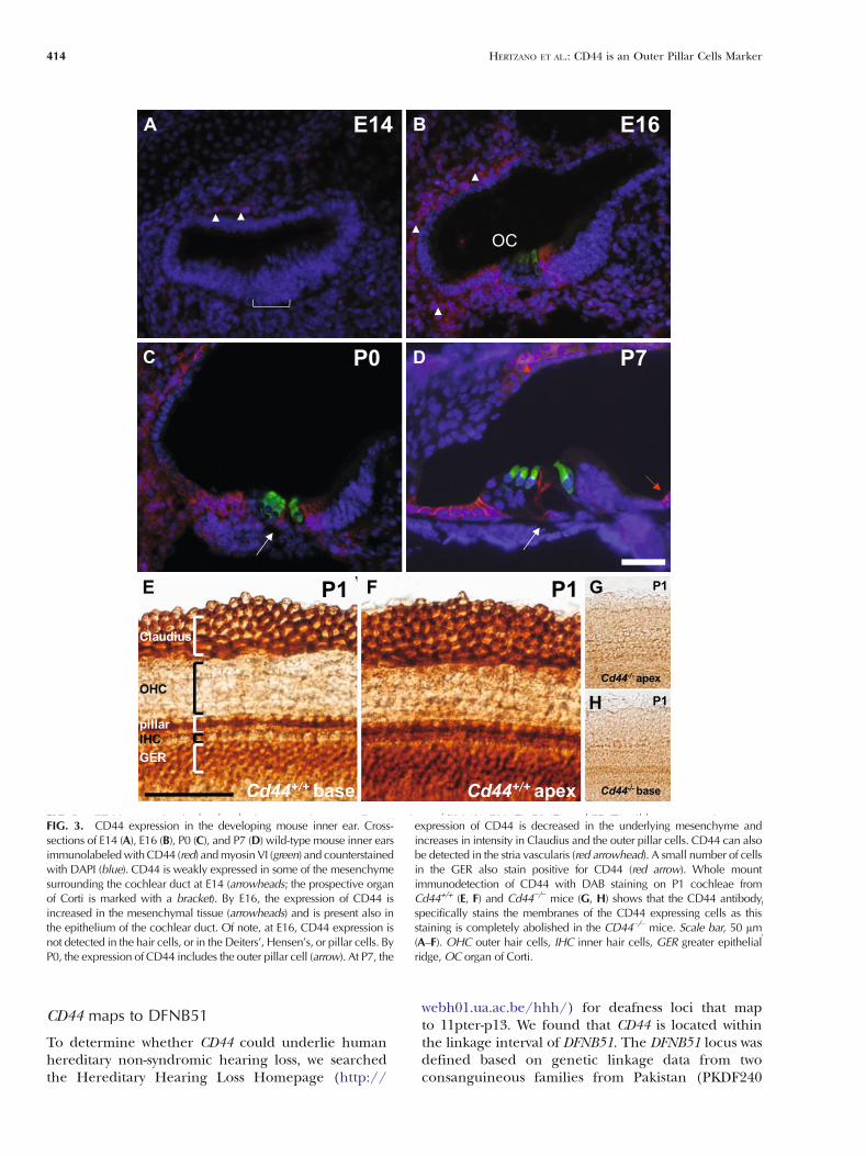
To study the expression of CD44 in the developingmouse inner ear, cross-sections of inner ears fromwild-type mice were stained with a CD44 antibodyalong with the inner ear hair cell-specific antibodies,myosin VI, or myosin VIIa and counter-labeled with4′,6-diamidino-2-phenylindole (DAPI) to detect thecell nuclei and overall tissue architecture (Fig. 3). Alow level of CD44 expression was detected in somecells of the inner ear mesenchyme and surroundingotic capsule as early as E14 both in the auditory(Fig. 3A) and the vestibular systems (data not shown).By E16, CD44 expression was detected at a higherlevel in the mesenchymal cells surrounding thecochlear duct. CD44 was also expressed in theepithelium of the cochlear duct at this time point,medially in the regions of the greater epithelial ridgeand laterally in the area of Claudius cells. Neither thehair cells nor the pillar, Deiters, or Hensen cellsstained positive for CD44 at E16 (Fig. 3B). At P0,expression of CD44 was detected in the outer pillarcells throughout the cochlear duct, with a strongerexpression in the base compared to that in apex(Fig. 3C and data not shown). By P7, CD44 expressionwas seen in fewer mesenchymal cells. Interestingly, theexpression of CD44 increased in intensity in the outerpillar cells and in Claudius cells throughout thelength of the cochlear duct. A small number ofepithelial cells from the cells medial to the organ ofCorti, known as greater epithelial ridge (GER) cells,expressed CD44. In addition, a weak level ofexpression was also observed in the stria vascularis(Fig. 3D). In the vestibular system, CD44 expressionwas limited to the mesenchymal cells and oticcapsule and was absent in the epithelial layer of thevestibular system at all time points tested (data notshown). Finally, by P24, the expression of CD44persisted in the epithelial cells medial and lateral tothe organ of Corti, but was abolished from the outerpillar cells (ESM Fig. S1).
410 HERTZANO ET AL.: CD44 is an Outer Pillar Cells Marker

To confirm the specificity of CD44 immunolabel-ing, cochleae from P1 CD44+/+ and CD44−/− mice werereacted with the antibody for CD44 and expressionwas detected with enzymatic labeling using a perox-idase-based assay (Fig. 3E–H). We found that CD44
TABLE 1
Microarray results for the relative expression of mouse CDgenes from early postnatal auditory and vestibular sensory
epithelia
CD Symbol Cochlea Vestibule FC
CD1d1 CD1d1 548 826 0.66CD1d2 CD1d2 110 140 0.79CD2ap CD2ap 404 403 1CD2bp2 CD2bp2 420 463 0.91CD9 CD9 1,843 3,245 0.57CD10 Mme 2,428 1,320 1.84CD14 CD14 95 206 0.46CD16 Fcr3 130 470 0.28CD24 CD24a 3,620 2,741 1.32CD26 Dpp4 600 900 0.67CD29 Itgb1 490 460 1.07CD32 Fcgr2b 227 330 0.69CD34 CD34 252 436 0.58CD38 CD38 111 317 0.35CD41 Itga2b 158 204 0.77CD42c Gp1bb 76 48 1.58CD44 CD44 671 108 6.21CD45 Ptprc 63 189 0.33CD47 Itgp 550 544 1.01CD49c Itga3 237 275 0.86CD49f Itga6 1,532 1,433 1.07CD51 Itgav 1,442 1,672 0.86CD53 CD53 48 108 0.44CD56 Ncam 694 720 0.96CD59 CD59a 372 519 0.72CD66a Ceacam1 86 158 0.54CD68 CD68 98 400 0.25CD73 Nt5 648 626 1.04CD81 CD81 3,924 6,401 0.61CD82 Kai 436 446 0.98CD83 CD83 230 178 1.29CD86 CD86 42 109 0.39CD91 Lrp1 1,127 1,413 0.8CD97 CD97 166 207 0.8CD98 Slc3a2 803 1,658 0.48CD99I2 CD99I2 326 328 0.99CD107b Lamp2 4,494 5,225 0.86CD108 Sema7a 180 184 0.98CD109 CD109 43 105 0.41CD110 Mpl 87 89 0.98CD116 Csf2ra 126 158 0.8CD117 Kit 254 380 0.67CD118 Ifnar 315 328 0.96CD119 Ifngr 217 282 0.77CD120a Tnfrsf1a 834 1,123 0.74CD121a Il1r1 144 100 1.44CD124 Il4ra 97 101 0.96CD126 Il6ra 79 74 1.07CD130 Il6st/gp130 867 1,292 0.67CD133 Prom1 572 732 0.78CD134 Tnfrsf4 92 83 1.11CD140a Pdgfra 2,857 5,310 0.54CD140b Pdgfrb 1000 976 1.02CD141 Thbd 373 646 0.58CD142 F3 101 385 0.26CD143 Ace 225 114 1.97CD146 Mcam 280 329 0.85CD147 Bsg 1,482 2,171 0.68CD151 CD151 1,378 1,574 0.88CD156b Adam17 865 863 1
TABLE 1
(continued)
CD Symbol Cochlea Vestibule FC
CDw156c Adam10 2,394 2,405 1CD163 CD163 31 141 0.22CD164 CD164 3,824 4,612 0.83CD164l2 CD164l2 275 394 0.7CD166 Alcam 250 512 0.49CD171 I1cam 101 54 1.87CD172a Ptpns1 336 365 0.92CD176 Trf, Tfn 177 872 0.2CD199 Cmkbr9 1,233 1,043 1.18CD200 Ox2 427 354 1.21CD201 Procr 99 110 0.9CD202 Tek 236 242 0.98CD203c Enpp1 248 133 1.86CD206 Mrc1 468 2,317 0.2CD210 Il10ra/b 463 466 0.99CD222 Igf2r 218 183 1.19CD228 Mfi2 1,571 2,579 0.61CD230 Prnp 1,656 3,673 0.45CD232 Plxnc1 365 410 0.89CD236r Gypc 235 350 0.67CD247 CD3z 285 298 0.96CD248 CD248 379 347 1.09CD256 Tnfsf13 137 238 0.58CD271 Ngfrap1 4,870 5,019 0.97CD276 CD276 342 314 1.09CD280 Mrc2 585 755 0.77CD282 Tlr2 177 227 0.78CD292 Bmpr1a 2,224 2,037 1.09CD298 Atp1b3 1,613 3,049 0.53CD302 CD302 1,160 688 1.69CD304 Nrp1 799 1,015 0.79CD309 Kdr 133 1,046 0.13CD315 Ptgfrn 1,092 1,224 0.89CD316 Igsf8 753 763 0.99CD318 Cdcp1 244 301 0.81CD320 CD320 465 524 0.89CD322 Jam2 312 625 0.5CD324 Cdh1 1,512 1,153 1.31CDw325 Cdh2 521 454 1.15CD326 Tacstd1 4553 6,913 0.66CDw293 Bmpr1b 232 547 0.42CD331 Fgfr1 638 686 0.93CD332 Fgfr2 883 1,608 0.55CD333 Fgfr3 1,081 93 11.62CD334 Fgfr4 475 715 0.66CD338 Abcg2 678 779 0.87CD339 Jag1 1,579 586 2.69
Cells that contain genes with a greater than two-fold mRNA enrichment inthe cochlea are marked in bold italics
CD assigned CD for the gene, Symbol gene symbol, FC fold change
HERTZANO ET AL.: CD44 is an Outer Pillar Cells Marker 411

specifically stained cells in the greater epithelial ridge,outer pillar cells, and Claudius cells throughout thelength of the cochlear duct (Fig. 3E, F). This patternof expression is consistent with the results that wereobtained in sections of P0 mice. No expression wasdetected in the ears of theCD44−/−mice, confirming thespecificity of the antibody and staining (Fig. 3G, H).
CD44 is a marker for outer pillar cells
To confirm and expand our results, we studied theexpression of CD44 in mice with mutations that affectpillar cell development. Pillar cells develop adjacentto the lateral edge of inner and medial edge of outerhair cells. Previous studies have shown that Fgfr3 plays
FIG. 1. Relative quantification of 68 CD genes in the newborn mouse inner ear using real-time RT-PCR. All results were quantified relative to themean expression level in the samples from the cochlear sensory epithelium. The expression of CD44 and CD333 (marked with red circles) wasgreater than tenfold higher in the auditory sensory epithelium compared with the individual vestibular sensory epithelia. Error bars represent onestandard deviation. The numerical values for the relative expression results are available in ESM Table S2.
FIG. 1. Relative quantification of 68 CD genes in the newbornmouse inner ear using real-time RT-PCR. All results were quantifiedrelative to the mean expression level in the samples from the cochlearsensory epithelium. The expression of CD44 and CD333 (marked with
red circles) was greater than tenfold higher in the auditory sensoryepithelium compared with the individual vestibular sensory epithelia.Error bars represent one standard deviation. The numerical values forthe relative expression results are available in ESM Table S2.
412 HERTZANO ET AL.: CD44 is an Outer Pillar Cells Marker
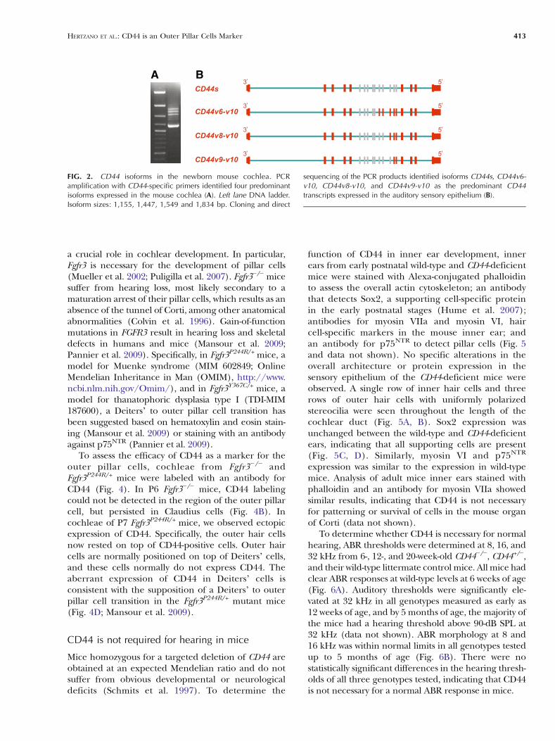
a crucial role in cochlear development. In particular,Fgfr3 is necessary for the development of pillar cells(Mueller et al. 2002; Puligilla et al. 2007). Fgfr3−/− micesuffer from hearing loss, most likely secondary to amaturation arrest of their pillar cells, which results as anabsence of the tunnel of Corti, among other anatomicalabnormalities (Colvin et al. 1996). Gain-of-functionmutations in FGFR3 result in hearing loss and skeletaldefects in humans and mice (Mansour et al. 2009;Pannier et al. 2009). Specifically, in Fgfr3P244R/+ mice, amodel for Muenke syndrome (MIM 602849; OnlineMendelian Inheritance in Man (OMIM), http://www.ncbi.nlm.nih.gov/Omim/), and in Fgfr3Y367C/+ mice, amodel for thanatophoric dysplasia type I (TDI-MIM187600), a Deiters’ to outer pillar cell transition hasbeen suggested based on hematoxylin and eosin stain-ing (Mansour et al. 2009) or staining with an antibodyagainst p75NTR (Pannier et al. 2009).
To assess the efficacy of CD44 as a marker for theouter pillar cells, cochleae from Fgfr3−/− andFgfr3P244R/+ mice were labeled with an antibody forCD44 (Fig. 4). In P6 Fgfr3−/− mice, CD44 labelingcould not be detected in the region of the outer pillarcell, but persisted in Claudius cells (Fig. 4B). Incochleae of P7 Fgfr3P244R/+ mice, we observed ectopicexpression of CD44. Specifically, the outer hair cellsnow rested on top of CD44-positive cells. Outer haircells are normally positioned on top of Deiters’ cells,and these cells normally do not express CD44. Theaberrant expression of CD44 in Deiters’ cells isconsistent with the supposition of a Deiters’ to outerpillar cell transition in the Fgfr3P244R/+ mutant mice(Fig. 4D; Mansour et al. 2009).
CD44 is not required for hearing in mice
Mice homozygous for a targeted deletion of CD44 areobtained at an expected Mendelian ratio and do notsuffer from obvious developmental or neurologicaldeficits (Schmits et al. 1997). To determine the
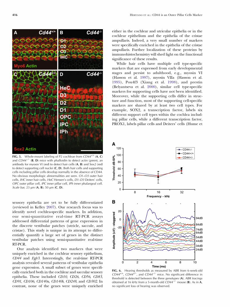
function of CD44 in inner ear development, innerears from early postnatal wild-type and CD44-deficientmice were stained with Alexa-conjugated phalloidinto assess the overall actin cytoskeleton; an antibodythat detects Sox2, a supporting cell-specific proteinin the early postnatal stages (Hume et al. 2007);antibodies for myosin VIIa and myosin VI, haircell-specific markers in the mouse inner ear; andan antibody for p75NTR to detect pillar cells (Fig. 5and data not shown). No specific alterations in theoverall architecture or protein expression in thesensory epithelium of the CD44-deficient mice wereobserved. A single row of inner hair cells and threerows of outer hair cells with uniformly polarizedstereocilia were seen throughout the length of thecochlear duct (Fig. 5A, B). Sox2 expression wasunchanged between the wild-type and CD44-deficientears, indicating that all supporting cells are present(Fig. 5C, D). Similarly, myosin VI and p75NTR
expression was similar to the expression in wild-typemice. Analysis of adult mice inner ears stained withphalloidin and an antibody for myosin VIIa showedsimilar results, indicating that CD44 is not necessaryfor patterning or survival of cells in the mouse organof Corti (data not shown).
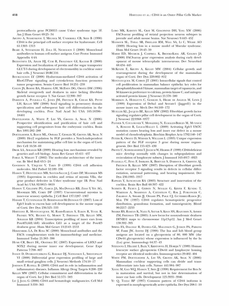
To determine whether CD44 is necessary for normalhearing, ABR thresholds were determined at 8, 16, and32 kHz from 6-, 12-, and 20-week-old CD44−/−, CD44+/−,and their wild-type littermate control mice. All mice hadclear ABR responses at wild-type levels at 6 weeks of age(Fig. 6A). Auditory thresholds were significantly ele-vated at 32 kHz in all genotypes measured as early as12 weeks of age, and by 5 months of age, the majority ofthe mice had a hearing threshold above 90-dB SPL at32 kHz (data not shown). ABR morphology at 8 and16 kHz was within normal limits in all genotypes testedup to 5 months of age (Fig. 6B). There were nostatistically significant differences in the hearing thresh-olds of all three genotypes tested, indicating that CD44is not necessary for a normal ABR response in mice.
BA
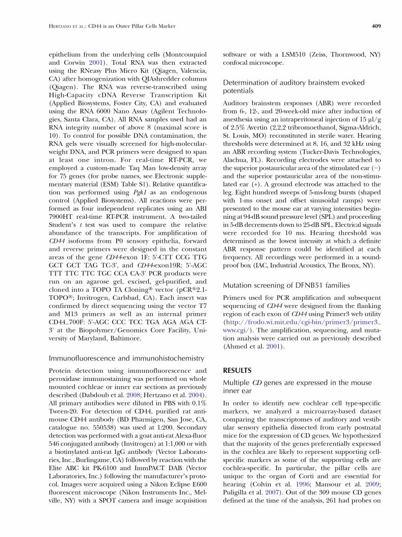
FIG. 2. CD44 isoforms in the newborn mouse cochlea. PCR amplification with CD44-specific primers identified four predominant isoformsexpressed in the mouse cochlea (A). Left lane 100-kb marker. Isoform sizes: 1,155, 1,447, 1,549 and 1,834 bp. Cloning and direct sequencing ofthe PCR products identified isoforms CD44s, CD44v6-v10, CD44v8-v10, and CD44v9-v10 as the predominant CD44 transcripts expressed in theauditory sensory epithelium (B).
FIG. 2. CD44 isoforms in the newborn mouse cochlea. PCRamplification with CD44-specific primers identified four predominantisoforms expressed in the mouse cochlea (A). Left lane DNA ladder.Isoform sizes: 1,155, 1,447, 1,549 and 1,834 bp. Cloning and direct
sequencing of the PCR products identified isoforms CD44s, CD44v6-v10, CD44v8-v10, and CD44v9-v10 as the predominant CD44transcripts expressed in the auditory sensory epithelium (B).
HERTZANO ET AL.: CD44 is an Outer Pillar Cells Marker 413
CD44 maps to DFNB51
To determine whether CD44 could underlie humanhereditary non-syndromic hearing loss, we searchedthe Hereditary Hearing Loss Homepage (http://
webh01.ua.ac.be/hhh/) for deafness loci that mapto 11pter-p13. We found that CD44 is located withinthe linkage interval of DFNB51. The DFNB51 locus wasdefined based on genetic linkage data from twoconsanguineous families from Pakistan (PKDF240
FIG. 3. CD44 expression in the developing mouse inner ear. Cross-sections of E14 (A), E16 (B), P0 (C), and P7 (D) wild-type mouse inner earsimmunolabeled with CD44 (red) and myosin VI (green) and counterstained with DAPI (blue). CD44 is weakly expressed in some of themesenchyme surrounding the cochlear duct at E14 (arrowheads; the prospective organ of Corti is marked with a bracket). By E16, the expressionof CD44 is increased in the mesenchymal tissue (arrowheads) and is present also in the epithelium of the cochlear duct. Of note, at E16, CD44expression is not detected in the hair cells, or in the Deiters’, Hensen’s, or pillar cells. By P0, the expression of CD44 includes the outer pillar cell(arrow). At P7, the expression of CD44 is decreased in the underlying mesenchyme and increases in intensity in Claudius and the outer pillarcells. CD44 can also be detected in the stria vascularis (red arrowhead). A small number of cells in the GER also stain positive for CD44 (redarrow). Whole mount immunodetection of CD44 with DAB staining on P1 cochleae from Cd44+/+ (E, F) and Cd44−/− mice (G, H) shows that theCD44 antibody specifically stains the membranes of the CD44 expressing cells as this staining is completely abolished in the CD44−/− mice. Scalebar, 50 µm (A–F). OHC outer hair cells, IHC inner hair cells, GER greater epithelial ridge, OC organ of Corti.
FIG. 3. CD44 expression in the developing mouse inner ear. Cross-sections of E14 (A), E16 (B), P0 (C), and P7 (D) wild-type mouse inner earsimmunolabeledwithCD44 (red) andmyosin VI (green) and counterstainedwith DAPI (blue). CD44 is weakly expressed in some of the mesenchymesurrounding the cochlear duct at E14 (arrowheads; the prospective organof Corti is marked with a bracket). By E16, the expression of CD44 isincreased in the mesenchymal tissue (arrowheads) and is present also inthe epithelium of the cochlear duct. Of note, at E16, CD44 expression isnot detected in the hair cells, or in the Deiters’, Hensen’s, or pillar cells. ByP0, the expression of CD44 includes the outer pillar cell (arrow). At P7, the
expression of CD44 is decreased in the underlying mesenchyme andincreases in intensity in Claudius and the outer pillar cells. CD44 can alsobe detected in the stria vascularis (red arrowhead). A small number of cellsin the GER also stain positive for CD44 (red arrow). Whole mountimmunodetection of CD44 with DAB staining on P1 cochleae fromCd44+/+ (E, F) and Cd44−/− mice (G, H) shows that the CD44 antibodyspecifically stains the membranes of the CD44 expressing cells as thisstaining is completely abolished in the CD44−/− mice. Scale bar, 50 µm(A–F). OHC outer hair cells, IHC inner hair cells, GER greater epithelialridge, OC organ of Corti.
414 HERTZANO ET AL.: CD44 is an Outer Pillar Cells Marker

and PKDF407) that segregate recessively inherited,profound congenital deafness (Shaikh et al. 2005).
All of the coding exons of CD44 were sequenced in adeaf and in an unaffected individual from each of the twoDFNB51 families. In family PKDF407, we found homo-zygosity for a nucleotide change of a G to C resulting inan arginine to proline substitution (p.R46P) in constantexon 2 of CD44 in all three affected individuals. Thischange was not identified in family PKDF240. Wesequenced 394 alleles and found two heterozygouschanges in two normal individual controls. Both individ-uals are from the same caste as family PKDF407,consistent with p.R46P being a polymorphism. Furtherinvestigation showed that p.R46P represents a polymor-phism known also as the uncommon Indian (In) bloodgroup antigen allele Ina (Spring et al. 1988). Homo-zygosity for this allele is rare and has been associated withproduction of alloantibodies to the common variant ofCD44 (Inb) after transfusion or pregnancy.
DISCUSSION
Cell type-specific markers offer advantages that rangefrom identification of cells by immunolabeling, abilityto cell sort and enrich a particular cell type from aheterogenous population, to functional studies ofthese proteins using mouse models. Specifically, dueto their wide use in FACS, there is a large variety ofcommercially available fluorochrome-conjugated anti-bodies against CD proteins that can be used for cellsorting or cell tracking in vivo (Beare et al. 2008).Using a combination of microarray data analysis,comparisons of the expression profiles of auditoryand vestibular sensory epithelia from newborn mice,followed by validation with real-time RT-PCR, we haveestablished the expression of 68 CD genes in thenewborn mouse inner ear. We focused our study onears of early postnatal mice. By P0, most of the celltypes of the mouse inner ear are present; however, the
BA
DC
FIG. 4. CD44 specifically stains the outer pillar cells. A, B Cross-sections of P6 Fgfr3+/+ (A) and Fgfr3−/− cochleae (B) immunolabeled withantibodies against CD44 (red) and phalloidin (green). The CD44 staining is completely lost from the outer pillar cell, but not from the lesserepithelial ridge cells (LER) in the Fgfr3−/− cochlea. C, D Cross-sections of P7 Fgfr3+/+ (C) and Fgfr3P244R/+ cochleae (D) immunolabeled withantibodies against CD44 (red) and myoVIIa (green) to mark the hair cells and DAPI to mark the cell nuclei. In the wild-type mice, every outer haircell is positioned over a Deiters’ cell (dotted arrows) (C). In the Fgfr3P244R/+ mice, the Deiters’ cells convert to cells that resemble the outer pillarcells (two left solid arrows) (D) and stain positive for CD44. Scale bar, 20 µm.
FIG. 4. CD44 specifically stains the outer pillar cells. A, B Cross-sections of P6 Fgfr3+/+ (A) and Fgfr3−/− cochleae (B) immunolabeledwith antibodies against CD44 (red) and phalloidin (green). The CD44staining is completely lost from the outer pillar cell, but not from thelesser epithelial ridge cells (LER) in the Fgfr3−/− cochlea. C, D Cross-sections of P7 Fgfr3+/+ (C) and Fgfr3P244R/+ cochleae (D) immunola-
beled with antibodies against CD44 (red) and myoVIIa (green) to markthe hair cells and DAPI to mark the cell nuclei. In the wild-type mice,every outer hair cell is positioned over a Deiters’ cell (dotted arrows)(C). In the Fgfr3P244R/+ mice, the Deiters’ cells convert to cells thatresemble the outer pillar cells (two left solid arrows) (D) and stainpositive for CD44. Scale bar, 20 µm.
HERTZANO ET AL.: CD44 is an Outer Pillar Cells Marker 415
sensory epithelia are yet to be fully differentiated(reviewed in Kelley 2007). Our research focus was toidentify novel cochlea-specific markers. In addition,our semi-quantitative real-time RT-PCR assaysaddressed differential patterns of gene expression inthe discrete vestibular patches (utricle, saccule, andcristae). This study is unique in its attempt to differ-entially quantify a large set of genes in the distinctvestibular patches using semi-quantitative real-timeRT-PCR.
Our analysis identified two markers that wereuniquely enriched in the cochlear sensory epithelium,CD44 and Fgfr3. Interestingly, the real-time RT-PCRanalysis revealed several patterns of vestibular epitheliagene expression. A small subset of genes were specifi-cally enriched both in the cochlear and saccular sensoryepithelia. These included CD10, CD26, CD56, CD83,CD91, CD106, CD140a, CD140b, CD248, and CD302. Incontrast, none of the genes were uniquely enriched
either in the cochlear and utricular epithelia or in thecochlear epithelium and the epithelia of the cristaeampullaris. Indeed, a very small number of markerswere specifically enriched in the epithelia of the cristaeampullaris. Further localization of these proteins byimmunohistochemistry will shed light on the functionalsignificance of these results.
While hair cells have multiple cell type-specificmarkers that are expressed from early developmentalstages and persist to adulthood, e.g., myosin VI(Hasson et al. 1997), myosin VIIa (Hasson et al.1995), Pou4f3 (Xiang et al. 1998), and prestin(Belyantseva et al. 2000), similar cell type-specificmarkers for supporting cells have not been identified.Moreover, while the supporting cells differ in struc-ture and function, most of the supporting cell-specificmarkers are shared by at least two cell types. Forexample, SOX2, a transcription factor, labels sixdifferent support cell types within the cochlea includ-ing pillar cells, while a different transcription factor,PROX1, labels pillar cells and Deiters’ cells (Hume et
BA
DC
FIG. 5. Whole-mount labeling of P2 cochleae from CD44+/+ (A, C)and CD44−/− (B, D) mice with phalloidin to detect actin (green), anantibody for myosin VI (red) to detect hair cells (A, B) and Sox2 (red)to detect supporting cell nuclei (C, D). Both hair cells and supportingcells including pillar cells develop normally in the absence of CD44.No obvious morphologic abnormalities are seen. O1–O3 outer haircells, IHC inner hair cells, HeC Hensen’s cells, D1–D3 Deiters’ cells,OPC outer pillar cell, IPC inner pillar cell, IPh inner phalangeal cell.Scale bar, 23 μm (A, B); 50 μm (C, D).
0 2 4 6 8 10
-20
0
20
94dB
Time (ms)
Am
plit
ud
e (u
V)
- st
agg
ered
84dB
74dB64dB
54dB44dB
34dB24dB
B
A
FIG. 6. Hearing thresholds as measured by ABR from 6-week-oldCD44+/+, CD44+/−, and CD44−/− mice. No significant difference inthreshold is detected between the three genotypes (A). ABR tracingsobtained at 16 kHz from a 5-month-old CD44−/− mouse (B). As in A,no significant loss of hearing was observed.
416 HERTZANO ET AL.: CD44 is an Outer Pillar Cells Marker

al. 2007). Previous studies have also identified FGFR3,a cell surface marker, as expressed by developingpillar and Deiters’ cells (Mueller et al. 2002).
We have identified CD44 as the first outer pillar cell-specific marker in the early postnatal mouse inner ear.Like many other supporting cell markers, the expres-sion of CD44 is not limited to the outer pillar cells.However, the lack of CD44 staining in other cellsimmediately adjacent to the outer pillar cells withinthe sensory epithelium, makes it an ideal marker for theouter pillar cells in the early postnatal period. CD44 isan integral cell membrane glycoprotein with a diverserange of suggested functions (MIM 107269). Theprincipal ligand of CD44 is hyaluronic acid, an extrac-ellular matrix protein (Aruffo et al. 1990). CD44 hasbeen primarily studied as a receptor expressed onactivated T cells and is known as a lymphocyte homingreceptor (Aruffo et al. 1990; Stefanova et al. 1989). Bybinding hyaluronic acid, CD44 can facilitate extravasa-tion of lymphocytes at sites of inflammation. Otheridentified ligands include laminin, fibronectin, colla-gens, serglycin, and osteoponin (reviewed in Goodisonet al. 1999). CD44 consists of extracellular, transmem-brane, and intracellular domains. The mouse CD44consists of nine constant exons that flank ten variableexons. The transmembrane and intracellular domainsof CD44 are encoded by the constant exons, while thealternatively spliced exons affect the structure of theextracellular domain (Goodison et al. 1999). Specificsplice variants of the protein have since been implicatedin malignant transformation, cancer metastasis, andinflammatory diseases (reviewed in Bourguignon 2008;Johnson and Ruffell 2009; Liu and Jiang 2006). Theexpression of CD44 has also been described in multiplemorphogenetically active epithelia as well as in Mullercell apical microvili in the retina (Stefanova et al. 1989;Yu and Toole 1997). Using in situ hybridization, CD44v4-7, v4-5, v6-7, v8-10, and the short isoform weredetected in E15 inner ears (Yu and Toole 1997). Ourresults show that by P0, the expression of the shortisoform of CD44 as well as CD44 v8-10 persists. CD44 v6-10 and v9-10 could also be detected. Other isoformswere not found, and it is possible that they are expressedonly at earlier developmental stages.
CD44 KO mice are viable and do not displayobvious developmental defects, but do suffer fromspecific alterations in their lymphocyte-dependentimmune responses (Protin et al. 1999; Schmits et al.1997). Our results show that in the absence of CD44,the mouse inner ear develops normally and that ABRhearing thresholds are comparable to wild-type andheterozygous littermates. In addition, specific block-ing experiments of cochlear explant cultures har-vested at E14.5 and incubated with a blockingantibody for CD44 did not result in any discernablephenotype until a toxic dose was reached (data not
shown). Therefore, the specific role of CD44 and itsisoforms in the mouse inner ear remains to bedetermined. Furthermore, as we could not identifymutations in CD44 among the human DFNB51families, mutations of CD44 appear not to have a rolein human hereditary hearing loss.
Finally, the mammalian auditory and vestibularsensory epithelia are highly complex and composed ofa variety of tightly organized cell types. Recently, micewith transgenic expression of a green fluorescentprotein (GFP) under the regulation of general support-ing cell-specific (p27kip1) or hair cell-specific (Atoh1)promoters were used to isolate these mixed cellpopulations, respectively (Doetzlhofer et al. 2006; Whiteet al. 2006). Similarly, mice with cell type-specific GFPexpression have been used to isolate and characterizethe transcriptomes of specific cell populations fromthe brain and the retina (Ivanov et al. 2008; Lobo et al.2006; Marsh et al. 2008). Antibodies to CD proteinsare routinely used for FACS analysis of leukocytes,routine diagnostics of hematologic diseases, and forstaining pathology slides for cancer diagnostics. Ourresults indicate that multiple CD genes are expressedin the mouse auditory and vestibular sensory epithe-lia, and suggest that at least some of these proteinsmay have differential expression patterns in the innerear, similar to CD44. By further characterizing theexpression pattern of other CD genes in the mouseinner ear, it may be possible to devise protocols for celltype-specific sorting from wild-type mice. This wouldallow for comparative analysis of inner ear cell type-specific expression profiles of wild-type and mutantmice and could assist in the identification of newdeafness genes.
ACKNOWLEDGEMENTS
We are grateful to Janice K. Babus, Yadong Ji, Amiel A. Drorand Dr. Weise Chang for technical assistance, Dr. SuziMansour and Chaoying Li for sending us the Fgfr3-P244Rmutant mice. We thank Dr. Karen B. Avraham for sharingunpublished data, and Dr. Tomoko Makishima for criticallyreviewing this manuscript. Finally, we thank Dr. Strome’slaboratory members for their technical help and advice.This research was supported by a resident research grantprovided by the American Academy of Otolaryngology-Head and Neck Surgery Foundation (R.H.), a DeafnessResearch Foundation grant (R.H.) and by funds from theIntramural Program at NIDCD 1-Z01-000070 (M.W.K), and1-Z01-000039 (T.B.F).
REFERENCES
AHMED ZM, RIAZUDDIN S, BERNSTEIN SL, AHMED Z, KHAN S, GRIFFITH AJ,MORELL RJ, FRIEDMAN TB, WILCOX ER (2001) Mutations of the
HERTZANO ET AL.: CD44 is an Outer Pillar Cells Marker 417
protocadherin gene PCDH15 cause Usher syndrome type 1F.Am J Hum Genet 69:25–34
ARUFFO A, STAMENKOVIC I, MELNICK M, UNDERHILL CB, SEED B (1990)CD44 is the principal cell surface receptor for hyaluronate. Cell61:1303–1313
BEARE A, STOCKINGER H, ZOLA H, NICHOLSON I (2008) Monoclonalantibodies to human cell surface antigens. Curr Protoc ImmunolAppendix 4:4A
BELYANTSEVA IA, ADLER HJ, CURI R, FROLENKOV GI, KACHAR B (2000)Expression and localization of prestin and the sugar transporterGLUT-5 during development of electromotility in cochlear outerhair cells. J Neurosci 20:RC116
BOURGUIGNON LY (2008) Hyaluronan-mediated CD44 activation ofRhoGTPase signaling and cytoskeleton function promotestumor progression. Semin Cancer Biol 18:251–259
COLVIN JS, BOHNE BA, HARDING GW, MCEWEN DG, ORNITZ DM (1996)Skeletal overgrowth and deafness in mice lacking fibroblastgrowth factor receptor 3. Nat Genet 12:390–397
DABDOUB A, PULIGILLA C, JONES JM, FRITZSCH B, CHEAH KS, PEVNY
LH, KELLEY MW (2008) Sox2 signaling in prosensory domainspecification and subsequent hair cell differentiation in thedeveloping cochlea. Proc Natl Acad Sci USA 105:18396–18401
DOETZLHOFER A, WHITE P, LEE YS, GROVES A, SEGIL N (2006)Prospective identification and purification of hair cell andsupporting cell progenitors from the embryonic cochlea. BrainRes 1091:282–288
DOETZLHOFER A, BASCH ML, OHYAMAT, GESSLER M, GROVES AK, SEGIL N(2009) Hey2 regulation by FGF provides a Notch-independentmechanism for maintaining pillar cell fate in the organ of Corti.Dev Cell 16:58–69
DROR AA, AVRAHAM KB (2009) Hearing loss: mechanisms revealed bygenetics and cell biology. Annu Rev Genet 43:411–437
FORGE A, WRIGHT T (2002) The molecular architecture of the innerear. Br Med Bull 63:5–24
GOODISON S, URQUIDI V, TARIN D (1999) CD44 cell adhesionmolecules. Mol Pathol 52:189–196
HASSON T, HEINTZELMAN MB, SANTOS-SACCHI J, COREY DP, MOOSEKER MS(1995) Expression in cochlea and retina of myosin VIIa, thegene product defective in Usher syndrome type 1B. Proc NatlAcad Sci USA 92:9815–9819
HASSON T, GILLESPIE PG, GARCIA JA, MACDONALD RB, ZHAO Y, YEE AG,MOOSEKER MS, COREY DP (1997) Unconventional myosins ininner-ear sensory epithelia. J Cell Biol 137:1287–1307
HAYASHI T, CUNNINGHAM D, BERMINGHAM-MCDONOGH O (2007) Loss ofFgfr3 leads to excess hair cell development in the mouse organof Corti. Dev Dyn 236:525–533
HERTZANO R, MONTCOUQUIOL M, RASHI-ELKELES S, ELKON R, YUCEL R,FRANKEL WN, RECHAVI G, MOROY T, FRIEDMAN TB, KELLEY MW,AVRAHAM KB (2004) Transcription profiling of inner ears fromPou4f3(ddl/ddl) identifies Gfi1 as a target of the Pou4f3deafness gene. Hum Mol Genet 13:2143–2153
HERZENBERG LA, DE ROSA SC (2000) Monoclonal antibodies and theFACS: complementary tools for immunobiology and medicine.Immunol Today 21:383–390
HUME CR, BRATT DL, OESTERLE EC (2007) Expression of LHX3 andSOX2 during mouse inner ear development. Gene ExprPatterns 7:798–807
IVANOV D, DVORIANTCHIKOVA G, BARAKAT DJ, NATHANSON L, SHESTOPALOV
VI (2008) Differential gene expression profiling of large andsmall retinal ganglion cells. J Neurosci Methods 174:10–17
JOHNSON P, RUFFELL B (2009) CD44 and its role in inflammation andinflammatory diseases. Inflamm Allergy Drug Targets 8:208–220
KELLEY MW (2007) Cellular commitment and differentiation in theorgan of Corti. Int J Dev Biol 51:571–583
LIU J, JIANG G (2006) CD44 and hematologic malignancies. Cell MolImmunol 3:359–365
LOBO MK, KARSTEN SL, GRAY M, GESCHWIND DH, YANG XW (2006)FACS-array profiling of striatal projection neuron subtypes injuvenile and adult mouse brains. Nat Neurosci 9:443–452
MANSOUR SL, TWIGG SR, FREELAND RM, WALL SA, LI C, WILKIE AO(2009) Hearing loss in a mouse model of Muenke syndrome.Hum Mol Genet 18:43–50
MARSH ED, MINARCIK J, CAMPBELL K, BROOKS-KAYAL AR, GOLDEN JA(2008) FACS-array gene expression analysis during early devel-opment of mouse telencephalic interneurons. Dev Neurobiol68:434–445
MCKENZIE E, KRUPIN A, KELLEY MW (2004) Cellular growth andrearrangement during the development of the mammalianorgan of Corti. Dev Dyn 229:802–812
MONTCOUQUIOL M, CORWIN JT (2001) Intracellular signals that controlcell proliferation in mammalian balance epithelia: key roles forphosphatidylinositol-3 kinase, mammalian target of rapamycin, andS6 kinases in preference to calcium, protein kinase C, andmitogen-activated protein kinase. J Neurosci 21:570–580
MORRISON A, HODGETTS C, GOSSLER A, HRABE DE ANGELIS M, LEWIS J(1999) Expression of Delta1 and Serrate1 (Jagged1) in themouse inner ear. Mech Dev 84:169–172
MUELLER KL, JACQUES BE, KELLEY MW (2002) Fibroblast growth factorsignaling regulates pillar cell development in the organ of Corti.J Neurosci 22:9368–9377
PANNIER S, COULOIGNER V, MESSADDEQ N, ELMALEH-BERGES M, MUNNICH
A, ROMAND R, LEGEAI-MALLET L (2009) Activating Fgfr3 Y367Cmutation causes hearing loss and inner ear defect in a mousemodel of chondrodysplasia. Biochim Biophys Acta 1792:140–147
PETERS K, ORNITZ D, WERNER S, WILLIAMS L (1993) Unique expressionpattern of the FGF receptor 3 gene during mouse organo-genesis. Dev Biol 155:423–430
PROTIN U, SCHWEIGHOFFERT, JOCHUMW,HILBERG F (1999) CD44-deficientmice develop normally with changes in subpopulations andrecirculation of lymphocyte subsets. J Immunol 163:4917–4923
PULIGILLA C, FENG F, ISHIKAWA K, BERTUZZI S, DABDOUB A, GRIFFITH AJ,FRITZSCH B, KELLEY MW (2007) Disruption of fibroblast growthfactor receptor 3 signaling results in defects in cellular differ-entiation, neuronal patterning, and hearing impairment. DevDyn 236:1905–1917
RAPHAEL Y, ALTSCHULER RA (2003) Structure and innervation of thecochlea. Brain Res Bull 60:397–422
SCHMITS R, FILMUS J, GERWIN N, SENALDI G, KIEFER F, KUNDIG T,WAKEHAM A, SHAHINIAN A, CATZAVELOS C, RAK J, FURLONGER C,ZAKARIAN A, SIMARD JJ, OHASHI PS, PAIGE CJ, GUTIERREZ-RAMOS JC,MAK TW (1997) CD44 regulates hematopoietic progenitordistribution, granuloma formation, and tumorigenicity. Blood90:2217–2233
SHAIKH RS, RAMZAN K, NAZLI S, SATTAR S, KHAN SN, RIAZUDDIN S, AHMED
ZM, FRIEDMAN TB (2005) A new locus for nonsyndromic deafnessDFNB51 maps to chromosome 11p13-p12. Am J Med Genet138:392–395
SPRING FA, DALCHAU R, DANIELS GL, MALLINSON G, JUDSON PA, PARSONS
SF, FABRE JW, ANSTEE DJ (1988) The Ina and Inb blood groupantigens are located on a glycoprotein of 80, 000 MW (theCDw44 glycoprotein) whose expression is influenced by the In(Lu) gene. Immunology 64:37–43
STEFANOVA I, HILGERT I, BAZIL V, KRISTOFOVA H, HOREJSI V (1989) Humanleucocyte surface glycoprotein CDw44 and lymphocyte homingreceptor are identical molecules. Immunogenetics 29:402–404
WHITE PM, DOETZLHOFER A, LEE YS, GROVES AK, SEGIL N (2006)Mammalian cochlear supporting cells can divide and trans-differentiate into hair cells. Nature 441:984–987
XIANG M, GAO WQ, HASSON T, SHIN JJ (1998) Requirement for Brn-3cin maturation and survival, but not in fate determination ofinner ear hair cells. Development 125:3935–3946
YU Q, TOOLE BP (1997) Common pattern of CD44 isoforms isexpressed in morphogenetically active epithelia. Dev Dyn 208:1–10
418 HERTZANO ET AL.: CD44 is an Outer Pillar Cells Marker
Related Documents