CD39/Adenosine Pathway Is Involved in AIDS Progression Maria Nikolova 1,2. , Matthieu Carriere 1. , Mohammad-Ali Jenabian 1. , Sophie Limou 1,3 , Mehwish Younas 1 , Ayrin Ko ¨k 1 , Sophie Hue ¨ 1 , Nabila Seddiki 1 , Anne Hulin 4 , Olivier Delaneau 3 , Hanneke Schuitemaker 5 , Joshua T. Herbeck 6 , James I. Mullins 6 , Maria Muhtarova 2 , Armand Bensussan 7 , Jean- Franc ¸ois Zagury 1,3 , Jean-Daniel Lelievre 1,8,9 , Yves Le ´vy 1,8,9 * 1 INSERM, Unite U955, Creteil, France, 2 National Center of Infectious and Parasitic Diseases, Sofia, Bulgaria, 3 Chaire de Bioinformatique, Conservatoire National des Arts et Me ´ tiers, Paris, France, 4 AP-HP, Groupe Henri-Mondor Albert-Chenevier, Laboratory of Pharmacology and Toxicology, Creteil, France, 5 Department of Experimental Immunology, Sanquin Research, Landsteiner Laboratory, Center for Infectious Diseases and Immunity Amsterdam (CINIMA) Academic Medical Center, University of Amsterdam, Amsterdam, The Netherlands, 6 University of Washington, School of Medicine, Department of Microbiology, Seattle, Washington, United States of America, 7 INSERM UMR 976 and Universite Denis-Diderot-Paris 7, Hopital Saint-Louis, Paris, France, 8 Universite ´ Paris Est Cre ´ teil, Faculte ´ de Me ´ decine, Creteil, France, 9 AP-HP, Groupe Henri-Mondor Albert-Chenevier, Immunologie clinique, Creteil, France Abstract HIV-1 infection is characterized by a chronic activation of the immune system and suppressed function of T lymphocytes. Regulatory CD4+ CD25 high FoxP3+CD127 low T cells (Treg) play a key role in both conditions. Here, we show that HIV-1 positive patients have a significant increase of Treg-associated expression of CD39/ENTPD1, an ectoenzyme which in concert with CD73 generates adenosine. We show in vitro that the CD39/adenosine axis is involved in Treg suppression in HIV infection. Treg inhibitory effects are relieved by CD39 down modulation and are reproduced by an adenosine-agonist in accordance with a higher expression of the adenosine A2A receptor on patients’ T cells. Notably, the expansion of the Treg CD39+ correlates with the level of immune activation and lower CD4+ counts in HIV-1 infected patients. Finally, in a genetic association study performed in three different cohorts, we identified a CD39 gene polymorphism that was associated with down-modulated CD39 expression and a slower progression to AIDS. Citation: Nikolova M, Carriere M, Jenabian M-A, Limou S, Younas M, et al. (2011) CD39/Adenosine Pathway Is Involved in AIDS Progression. PLoS Pathog 7(7): e1002110. doi:10.1371/journal.ppat.1002110 Editor: Ronald C. Desrosiers, Harvard University, United States of America Received November 19, 2010; Accepted April 23, 2011; Published July 7, 2011 Copyright: ß 2011 Nikolova et al. This is an open-access article distributed under the terms of the Creative Commons Attribution License, which permits unrestricted use, distribution, and reproduction in any medium, provided the original author and source are credited. Funding: AK and MY were granted from Sidaction. This work was performed with fundings from the ANRS (Agence Nationale de Recherche contre le SIDA et les he ´patites virales), Sidaction and the Partnership Hubert Curien 2009 (RILA 4). The funders had no role in study design, data collection and analysis, decision to publish, or preparation of the manuscript. Competing Interests: The authors have declared that no competing interests exist. * E-mail: [email protected] . These authors contributed equally to this work. Introduction HIV-1 infection is characterized by chronic immune activation which, in combination with the progressive depletion of CD4+ T cells, profoundly perturbs antigen-specific T cell responses [1]. The population of CD4+CD25 high FoxP3+ regulatory T cells (Treg) suppresses antigen-specific T cell responses and controls inappropriate or exaggerated immune activation induced by pathogens, thereby influencing the outcome of various infections [2,3]. In particular, these cells suppress in vitro HIV-1-specific CD4+ and CD8+ effector T-cell responses [2,4]. We, and others, have reported an HIV-1-driven expansion of Treg expression in chronic and acute HIV-1 infection [5,6], including a relationship between the expansion of Treg, the level of cellular immune activation and the depletion of CD4+ T cells in acute HIV infection [5]. The molecular mechanisms by which Treg mediate their suppressive activity remain poorly understood. In humans, the Treg population exhibits considerable diversity. Phenotypically and functionally distinct subsets of Treg can mediate suppression through distinct mechanisms from secretion of IL-10, TGF-ß, IL- 35, Granzyme B, perforin, to CTLA-4 and GITR interactions [7,8,9]. Recently, it has been reported that CD39 is expressed on human and murine Treg, while CD73 is found only on the surface of murine Treg [10,11,12]. CD39, a member of the ectonucleotidase triphosphate diphosphohydrolase family (ENTPD), also referred to as ENTPD-1 (EC 3.6.1.5), is the dominant immune system ectonucleotidase that hydrolyses extracellular ATP and adenosine diphosphate (ADP) into adenosine monophosphate (AMP) at the sites of immune activation. CD73 is an ecto-59-nucleotidase (59NT) that exists in a soluble or membrane-bound form and catalyzes the dephos- phorylation of AMP to adenosine [13,14,15]. Adenosine is a critical regulator of innate and adaptive immune responses [16,17], inhibiting T lymphocyte proliferation and the secretion of inflammatory cytokines including IL-2, TNFa, and IFN-c [13,14,15]. These effects are mediated through A2A receptors stimulating the generation of cAMP, and are mimicked by adenosine agonists [18]. CD39 has also been described as an activation marker of lymphoid cells [19]. Therefore, the CD39/ Adenosine pathway may be important to the balance between activation and regulation of effector immune responses. PLoS Pathogens | www.plospathogens.org 1 July 2011 | Volume 7 | Issue 7 | e1002110

Welcome message from author
This document is posted to help you gain knowledge. Please leave a comment to let me know what you think about it! Share it to your friends and learn new things together.
Transcript

CD39/Adenosine Pathway Is Involved in AIDSProgressionMaria Nikolova1,2., Matthieu Carriere1., Mohammad-Ali Jenabian1., Sophie Limou1,3, Mehwish
Younas1, Ayrin Kok1, Sophie Hue1, Nabila Seddiki1, Anne Hulin4, Olivier Delaneau3, Hanneke
Schuitemaker5, Joshua T. Herbeck6, James I. Mullins6, Maria Muhtarova2, Armand Bensussan7, Jean-
Francois Zagury1,3, Jean-Daniel Lelievre1,8,9, Yves Levy1,8,9*
1 INSERM, Unite U955, Creteil, France, 2 National Center of Infectious and Parasitic Diseases, Sofia, Bulgaria, 3 Chaire de Bioinformatique, Conservatoire National des Arts et
Metiers, Paris, France, 4 AP-HP, Groupe Henri-Mondor Albert-Chenevier, Laboratory of Pharmacology and Toxicology, Creteil, France, 5 Department of Experimental
Immunology, Sanquin Research, Landsteiner Laboratory, Center for Infectious Diseases and Immunity Amsterdam (CINIMA) Academic Medical Center, University of
Amsterdam, Amsterdam, The Netherlands, 6 University of Washington, School of Medicine, Department of Microbiology, Seattle, Washington, United States of America,
7 INSERM UMR 976 and Universite Denis-Diderot-Paris 7, Hopital Saint-Louis, Paris, France, 8 Universite Paris Est Creteil, Faculte de Medecine, Creteil, France, 9 AP-HP,
Groupe Henri-Mondor Albert-Chenevier, Immunologie clinique, Creteil, France
Abstract
HIV-1 infection is characterized by a chronic activation of the immune system and suppressed function of T lymphocytes.Regulatory CD4+ CD25high FoxP3+CD127low T cells (Treg) play a key role in both conditions. Here, we show that HIV-1positive patients have a significant increase of Treg-associated expression of CD39/ENTPD1, an ectoenzyme which inconcert with CD73 generates adenosine. We show in vitro that the CD39/adenosine axis is involved in Treg suppression inHIV infection. Treg inhibitory effects are relieved by CD39 down modulation and are reproduced by an adenosine-agonist inaccordance with a higher expression of the adenosine A2A receptor on patients’ T cells. Notably, the expansion of the TregCD39+ correlates with the level of immune activation and lower CD4+ counts in HIV-1 infected patients. Finally, in a geneticassociation study performed in three different cohorts, we identified a CD39 gene polymorphism that was associated withdown-modulated CD39 expression and a slower progression to AIDS.
Citation: Nikolova M, Carriere M, Jenabian M-A, Limou S, Younas M, et al. (2011) CD39/Adenosine Pathway Is Involved in AIDS Progression. PLoS Pathog 7(7):e1002110. doi:10.1371/journal.ppat.1002110
Editor: Ronald C. Desrosiers, Harvard University, United States of America
Received November 19, 2010; Accepted April 23, 2011; Published July 7, 2011
Copyright: � 2011 Nikolova et al. This is an open-access article distributed under the terms of the Creative Commons Attribution License, which permitsunrestricted use, distribution, and reproduction in any medium, provided the original author and source are credited.
Funding: AK and MY were granted from Sidaction. This work was performed with fundings from the ANRS (Agence Nationale de Recherche contre le SIDA et leshepatites virales), Sidaction and the Partnership Hubert Curien 2009 (RILA 4). The funders had no role in study design, data collection and analysis, decision topublish, or preparation of the manuscript.
Competing Interests: The authors have declared that no competing interests exist.
* E-mail: [email protected]
. These authors contributed equally to this work.
Introduction
HIV-1 infection is characterized by chronic immune activation
which, in combination with the progressive depletion of CD4+ T
cells, profoundly perturbs antigen-specific T cell responses [1].
The population of CD4+CD25high FoxP3+ regulatory T cells
(Treg) suppresses antigen-specific T cell responses and controls
inappropriate or exaggerated immune activation induced by
pathogens, thereby influencing the outcome of various infections
[2,3]. In particular, these cells suppress in vitro HIV-1-specific
CD4+ and CD8+ effector T-cell responses [2,4]. We, and others,
have reported an HIV-1-driven expansion of Treg expression in
chronic and acute HIV-1 infection [5,6], including a relationship
between the expansion of Treg, the level of cellular immune
activation and the depletion of CD4+ T cells in acute HIV
infection [5].
The molecular mechanisms by which Treg mediate their
suppressive activity remain poorly understood. In humans, the
Treg population exhibits considerable diversity. Phenotypically
and functionally distinct subsets of Treg can mediate suppression
through distinct mechanisms from secretion of IL-10, TGF-ß, IL-
35, Granzyme B, perforin, to CTLA-4 and GITR interactions
[7,8,9]. Recently, it has been reported that CD39 is expressed on
human and murine Treg, while CD73 is found only on the
surface of murine Treg [10,11,12]. CD39, a member of the
ectonucleotidase triphosphate diphosphohydrolase family
(ENTPD), also referred to as ENTPD-1 (EC 3.6.1.5), is the
dominant immune system ectonucleotidase that hydrolyses
extracellular ATP and adenosine diphosphate (ADP) into
adenosine monophosphate (AMP) at the sites of immune
activation. CD73 is an ecto-59-nucleotidase (59NT) that exists in
a soluble or membrane-bound form and catalyzes the dephos-
phorylation of AMP to adenosine [13,14,15]. Adenosine is a
critical regulator of innate and adaptive immune responses
[16,17], inhibiting T lymphocyte proliferation and the secretion
of inflammatory cytokines including IL-2, TNFa, and IFN-c[13,14,15]. These effects are mediated through A2A receptors
stimulating the generation of cAMP, and are mimicked by
adenosine agonists [18]. CD39 has also been described as an
activation marker of lymphoid cells [19]. Therefore, the CD39/
Adenosine pathway may be important to the balance between
activation and regulation of effector immune responses.
PLoS Pathogens | www.plospathogens.org 1 July 2011 | Volume 7 | Issue 7 | e1002110

Here we tested the hypothesis that the CD39/adenosine
pathway is involved in the pathogenesis of HIV-1 disease. First,
we investigated the phenotype and the function of Treg-expressing
CD39 molecules in a cohort of chronically HIV-positive patients
and determined whether these characteristics are associated with
clinical outcomes. Second, to assess our hypothesis in an in vivo
context, we investigated whether CD39 genetic polymorphisms
were associated with rates of HIV-1 disease progression in three
independent cohorts.
Results
CD4+CD25high Treg, but not CD4+CD25low activated Tcells, from HIV-positive subjects express high density ofcell membrane CD39 molecules
In order to discriminate between Treg and activated T cells, we
further characterized Treg population as gated T cells expressing
CD4+CD25high FoxP3+high and CD127low (gating strategy is shown
in Fig. S1). These cells are designated thereafter as Treg cells while
CD4+CD25lowCD127high T cells are designated as activated
CD4+CD25low T cells (T act). First, we confirmed a significant
increase in the percentages of Treg cells in a cohort of HIV-positive
individuals, receiving either a combination of antiretroviral drugs (c-
ART+, n = 39) or not (c-ART2, n = 39), as compared to healthy
controls (n = 25) (mean 5.8% and 6.2% respectively vs 2.4%,
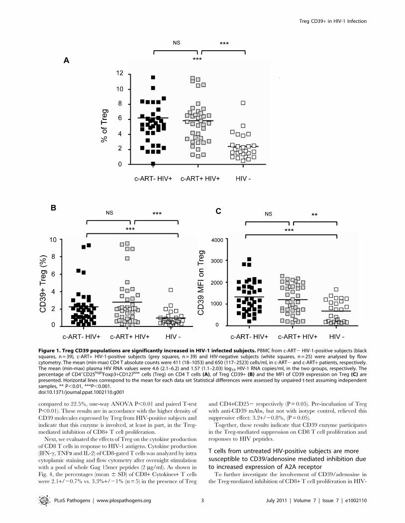
P,0.0001) (Fig. 1a). As shown in Fig. 1b and 1c, percentages of
Treg expressing CD39+ (Treg CD39+) were significantly higher in
both c-ART+ and c-ART2 patients, as compared to healthy controls
(mean 2.79% and 2.26% vs 0.97%, P,0.001, Fig. 1b). Moreover,
Treg from both c-ART2 and c-ART+ subjects expressed a higher
density of CD39 molecules as compared to those from HIV-1
negative controls (mean fluorescence intensity (MFI) 1327 and 1203,
respectively, vs. 652, P,0.001 and P,0.01) (Fig. 1c).
Phenotypic analyses were performed in 16 HIV-1 positive
patients before and 12 months following c-ART initiation. Among
them, 9 patients experienced a good response to c-ART (group A;
undetectable plasma viral load at month 12), while in 7 patients
(group B) viral replication remained detectable (above 50 copies/
ml). No significant decrease of CD39 expression was observed in
group A: % Treg CD39+ (mean 6 SD): 2.461.2 vs.1.861.0 at
baseline; TregCD39+ MFI (mean 6 SD): 15576360 vs. 12616656
at baseline, (P.0.05 for both). Moreover, in patients with on-going
viral replication %Treg CD39+ increased significantly in spite of
ART (6.162.4 versus 3.462.3 at baseline; P = 0.043).
CD39 has also been described as an activation marker of
lymphoid cells [19]. Therefore, we looked at the percentages of
Tact in HIV-1 positive patients and controls. As expected, the
frequency of activated CD4+CD25low T cells was significantly
higher in both populations of patients as compared to controls
(Fig.S1b). Consequently, percentages of CD4+CD25lowCD39+were significantly higher in HIV-1 positive patients as compared to
controls (Fig.S1c). In contrast to Treg, CD4+CD252 T cells from
both HIV-positive subjects and controls did not express CD39 (not
shown). Thus, an expansion of CD39+CD4+ T cells in both Treg
and T act T cell populations, which persist in patients with
controlled viral load under c-ART, is observed in HIV-1 positive
patients. In HIV-positive subjects and in HIV-negative controls,
Treg cells were mostly of CD45RA2CD28+ memory phenotype
(mean 75%). CD45RA2CD28+ Treg contained a higher
percentage of CD39+ cells as compared to CD45RA+CD28+Treg cells (mean 65% vs. 28%, respectively, P,0.05) (Fig. S2).
Down-modulation of CD39 expression on Treg relievesTreg-mediated inhibition of CD8 T cell proliferation andHIV specific responses
We next investigated whether down-modulation of the CD39
enzyme can impact Treg function. First, by exposing cells to a
blocking anti-CD39 (BY40) mAb, we induced a down-modulation
of CD39 expression at the surface of the YT2C2 NK line cells
(Fig. S3a). Next, BY40 mAb down-modulated the expression of
CD39 on ex-vivo purified peripheral blood Treg from HIV-
negative controls as compared to untreated cells or cells treated
with an IgG1 control mAb (% of positive cells (mean 6 SD):
32611% vs 44613%, and 42614%, respectively) (Fig. 2a,b). In
these experiments, CD39 expression following in vitro incubation
with BY40 mAb was assessed using a commercial PE anti-CD39
(clone TU66) which has been previously checked to be non-
competitive with BY40 (Fig. S4). Finally, we found that this down
modulation effect of BY40 was associated with decreased CD39
ATPase activity on primary monocytes (Fig. S3b).
The functional consequences of CD39 down-modulation were
investigated in co-culture assays developed to evaluate the
suppressive effects of Treg on T cell proliferation [5,6,20]. As
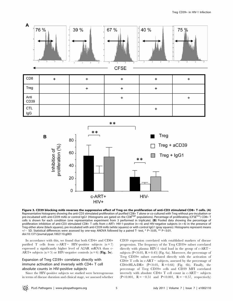
shown in Fig. 3a and b (for one representative experiment and
pooled data from 6 HIV-positive subjects), the Treg-mediated
inhibition of anti-CD3 induced CD8 T cell proliferation was
significantly higher in HIV-positive subjects (n = 6) as compared to
HIV-negative controls (n = 6), (mean inhibition 56% vs 22.5%;
P,0.01) (Fig. 3b). Pre-incubation with anti-CD39 BY40 mAb
reversed by ,50% the suppressive effect of Treg from HIV-positive
subjects (average suppression rate of 28% in the presence of Treg
pre-treated with BY40 as compared to 56% and 57% for Treg pre-
treated or not with IgG1 control mAb, (P = 0.01; one-way ANOVA
and paired T-test P = 0.01 for group by group comparisons).
Interestingly, although the suppression mediated by Treg from
HIV-negative controls was less significant, a similar effect of anti-
CD39 BY40 mAb was noted (average inhibition 12.3% as
Author Summary
HIV-1 infection is characterized by a chronic activation ofthe immune system. Regulatory T cells (Treg) represent apopulation of lymphocytes that controls inappropriate orexaggerated immune activation induced by pathogens,thereby influencing the outcome of various infections.Several studies have shown that Treg are expanded in HIVinfected patients. However, the mechanisms of Tregimmune-modulator functions are not clearly known.CD39 is an ectonucleotidase which converts the proin-flammatory ATP signal into AMP and the immunosuppres-sive adenosine in concert with CD73. A critical role of CD39has been described for Treg in general but few studieshave analyzed its role in HIV infection. We report here anexpansion of Treg expressing CD39 in a cohort of HIV-infected patients. In vitro these cells exerted a strongsuppressive effect on the effector CD8 T cells. Treginhibitory effects were relieved by CD39 down-modulationusing an anti-CD39 monoclonal antibody. Treg suppressiveeffects were reproduced by an adenosine agonist inaccordance with a higher expression of the adenosineA2A receptor on patients’ T cells. From a clinical standpoint, we show also a correlation between Treg CD39+expansion and both immune activation and CD4+ T celldepletion in patients. Finally, by genetic analysis of threedifferent cohorts of patients, we found that a CD39 genepolymorphism associated with a lower CD39 expressioncorrelated with a slower progression to AIDS. Thus, ourresults contribute to elucidate the mechanisms by whichTreg suppression occurs during HIV infection.
Treg CD39+ in HIV-1 Infection
PLoS Pathogens | www.plospathogens.org 2 July 2011 | Volume 7 | Issue 7 | e1002110
compared to 22.5%, one-way ANOVA P,0.01 and paired T-test
P,0.01). These results are in accordance with the higher density of
CD39 molecules expressed by Treg from HIV-positive subjects and
indicate that this enzyme is involved, at least in part, in the Treg-
mediated inhibition of CD8+ T cell proliferation.
Next, we evaluated the effects of Treg on the cytokine production
of CD8 T cells in response to HIV-1 antigens. Cytokine production
(IFN-c, TNFa and IL-2) of CD8-gated T cells was analyzed by intra
cytoplamic staining and flow cytometry after overnight stimulation
with a pool of whole Gag 15mer peptides (2 mg/ml). As shown in
Fig. 4, the percentages (mean 6 SD) of CD8+ Cytokines+ T cells
were 2.1+/20.7% vs. 3.3%+/21% (n = 5) in the presence of Treg
and CD4+CD252 respectively (P = 0.05). Pre-incubation of Treg
with anti-CD39 mAbs, but not with isotype control, relieved this
suppressive effect: 3.2+/20.8%, (P = 0.05).
Together, these results indicate that CD39 enzyme participates
in the Treg-mediated suppression on CD8 T cell proliferation and
responses to HIV peptides.
T cells from untreated HIV-positive subjects are moresusceptible to CD39/adenosine mediated inhibition dueto increased expression of A2A receptor
To further investigate the involvement of CD39/adenosine in
the Treg-mediated inhibition of CD8+ T cell proliferation in HIV-
Figure 1. Treg CD39 populations are significantly increased in HIV-1 infected subjects. PBMC from c-ART2 HIV-1-positive subjects (blacksquares, n = 39), c-ART+ HIV-1-positive subjects (grey squares, n = 39) and HIV-negative subjects (white squares, n = 25) were analysed by flowcytometry. The mean (min-max) CD4 T absolute counts were 411 (18–1053) and 650 (117–2523) cells/ml, in c-ART2 and c-ART+ patients, respectively.The mean (min-max) plasma HIV RNA values were 4.6 (2.1–6.2) and 1.57 (1.1–2.03) log10 HIV-1 RNA copies/ml, in the two groups, respectively. Thepercentage of CD4+CD25highFoxp3+CD127low cells (Treg) on CD4 T cells (A), of Treg CD39+ (B) and the MFI of CD39 expression on Treg (C) arepresented. Horizontal lines correspond to the mean for each data set Statistical differences were assessed by unpaired t-test assuming independentsamples, ** P,0.01, ***P,0.001.doi:10.1371/journal.ppat.1002110.g001
Treg CD39+ in HIV-1 Infection
PLoS Pathogens | www.plospathogens.org 3 July 2011 | Volume 7 | Issue 7 | e1002110

1 positive subjects, we studied the effects of the A2AR agonist
CGS21680 on proliferation of anti-CD3 stimulated T cells. The
mean (6SD) inhibition of CD4+ T cells was 47% (611) and 57%
(68.3) in the presence of 0.1 and 1 mM of CGS, respectively in c-
ART2 HIV positive patients. Similarly, the same doses of CGS
inhibited by 47% and 65% the proliferation of anti-CD3 activated
CD8+ T cells from c-ART2 HIV-positive subjects (P,0.05)
(Fig. 5a,b). In contrast, the proliferation of CD4+ and CD8+ T
cells from HIV-negative controls and c-ART+ HIV-positive
subjects was much lower and below 20% at the highest dose of
CGS21680 (1 mM) (Fig. 5a,b) (P = 0.015 and P = 0.027 respec-
tively; one-way ANOVA and P,0.05 unpaired T-test for
comparison between c-ART2HIV-positive patients and the two
other groups (Fig. 5a,b).
Figure 2. Preincubation of Treg with anti-CD39 BY40 mAb down-modulates CD39 expression on Treg. (A) Representative experimentshowing the expression of CD39 on Treg from an HIV-negative donor, preincubated in medium alone (left histogram) or either with IgG1 isotypecontrol (middle histogram) or anti-CD39 mAb BY40 (right histogram), and co-cultured for 18 h with anti-CD3-activated autologous CD8 T cells.Expression of CD39 was then assessed using a BY40 non-competitive anti-CD39 mAb on gated CD4+CD25highCD127lowFoxp3+ cells. (B) Pooled datafrom 3 independent experiments show the percentage of CD39+ cells among Treg after co-culture as in (A). Bars represent mean +/2 SD. Statisticaldifferences were assessed by a paired Student t-test, * P,0.05.doi:10.1371/journal.ppat.1002110.g002
Treg CD39+ in HIV-1 Infection
PLoS Pathogens | www.plospathogens.org 4 July 2011 | Volume 7 | Issue 7 | e1002110
In accordance with this, we found that both CD4+ and CD8+purified T cells from c-ART2 HIV-positive subjects (n = 7)
expressed a significantly higher level of A2AR mRNA than c-
ART+ subjects (n = 5) or HIV-negative controls (n = 6) (Fig. 5c).
Expansion of Treg CD39+ correlates directly withimmune activation and inversely with CD4+ T cellabsolute counts in HIV-positive subjects
Since the HIV-positive subjects we studied were heterogeneous
in terms of disease duration and clinical stage, we assessed whether
CD39 expression correlated with established markers of disease
progression. The frequency of the Treg CD39+ subset correlated
directly with plasma HIV-1 viral load in the group of c-ART2
subjects (P,0.05, R = 0.45) (Fig. 6a). Moreover, the percentage of
Treg CD39+ subset correlated directly with the activation of
CD4+ T cells in c-ART2 subjects, assessed by the percentage of
CD4+HLA-DR+ (P,0.05, R = 0.66) (Fig. 6b). Finally, the
percentage of Treg CD39+ cells and CD39 MFI correlated
inversely with absolute CD4+ T cell count in c-ART2 subjects
(P,0.001, R = 20.51 and P,0.001, R = 20.57, respectively)
Figure 3. CD39 blocking mAb reverses the suppressive effect of Treg on the proliferation of anti-CD3 stimulated CD8+ T cells. (A)Representative histograms showing the anti-CD3 stimulated proliferation of purified CD8+ T alone or co-cultured with Treg without pre-incubation orpre-incubated with anti-CD39 mAb or control IgG1 (Histograms are gated on the CD8high populations). Percentage of proliferating (CFSElow) CD8+ Tcells is shown for each condition (one representative experiment from 3 performed in triplicate). (B) Pooled data showing the percentage ofproliferation inhibition of anti-CD3 stimulated CD8+ T cells from c-ART+ HIV-1-positive (n = 6) and HIV-negative subjects (n = 6) in the presence ofTreg either alone (black squares), pre-incubated with anti-CD39 mAb (white squares) or with control IgG1 (gray squares). Histograms represent means+/2 SD. Statistical differences were assessed by one-way ANOVA followed by a paired T- test, * P,0.05, ** P,0.01.doi:10.1371/journal.ppat.1002110.g003
Treg CD39+ in HIV-1 Infection
PLoS Pathogens | www.plospathogens.org 5 July 2011 | Volume 7 | Issue 7 | e1002110

(Fig. 6 c,d) as well as in c-ART+ subjects (P,0.001 , R = 20.57
and P,0.01, R = 20.43) (Fig. 6 e,f).
The independent prognostication value of CD39 expression on
Treg for CD4 T cell counts was studied in c-ART2 and in c-
ART+ patients, using multiple linear regression models (SPSS
v.17.0). The frequencies of Treg, Treg CD39+, Tact, Tact
CD39+, and viral load (for c-ART2 group only) were included as
predictors of CD4 absolute count. For c-ART+HIV-positive
patients, in a full model (R2 = 0,398, ANOVA P = 0.02) the
percentage of Treg CD39+ had the most important partial
predictive effect (partial correlation coefficient 20.479), confirmed
by sequential multiple regression analysis of the same set of
variables (partial correlation coefficient 20.612 vs. 0.360 for Tact,
ANOVA P = 0.001).
For c-ART2HIV-positive patients, in a full model (R2 = 0,392,
ANOVA sig. = 0.045), again the percentage of TregCD39+ had
the most important contribution as predictor for CD4 absolute
count, followed by CD39Tact (partial correlation coefficients
20.375, and 0. 265 respectively).
These results indicate that the frequency of Treg CD39+ is an
independent predictive factor for CD4 cell count variability.
A genetic variant of CD39 associated with a lower geneexpression is involved in slower progression to AIDS
Our results highly suggest that the frequency of Treg CD39+cells, as well as the density of the enzyme molecule at the surface of
those cells, predict disease progression. Recently, CD39 gene
polymorphisms associated with the level of enzyme expression
have been shown to be associated with susceptibility to Crohn’s
disease [21]. In order to assess the role of CD39 on HIV-1 disease
progression, we investigated whether CD39 gene polymorphisms
could be associated with clinical outcomes. For that, we exploited
the GRIV cohort, comprising subjects exhibiting extreme profiles
of AIDS progression (LTNP, long-term non-progressors and RP,
rapid progressors) [20,22,23]. We thus performed a genetic case-
control association study on the candidate gene CD39 using the
genotype data collected from our previous genome-wide associa-
tion studies [22,23] (see Methods).
Fourteen SNPs were identified in the CD39 gene. No
polymorphism was significantly associated with rapid progression,
whereas four SNPs were significantly associated with LTNP:
rs10882665 (P = 1.3361022), rs3181123 (P = 1.3861022),
rs1933166 (P = 1.7661022), and rs11188513 (P = 3.6061022)
(Fig. S5). Of note, rs10882665 and rs3181123 are in full linkage
disequilibrium (r2 = 1). To eliminate a potential association with
HIV-1 infection rather than with LTNP, we compared the allelic
frequency of each of these SNPs in the RP population. The
frequency observed in the RP group was similar to the frequency
observed in the control group, confirming that this was an
association with LTNP.
To confirm these results, we used two additional independent
Caucasian cohorts that examined AIDS progression phenotype:
the ACS and the MACS cohorts (see Methods). The rs11188513
SNP (whose frequency in LTNP and control groups were,
respectively, 39% and 34%, P = 3.6061022, (Fig. 7a) was the
only polymorphism also associated with disease progression both
Figure 4. CD39 blocking mAb reverses the suppressive effect of Treg on the cytokine production of Gag-stimulated CD8+ T cells.Pooled data showing the percentage of CD8+ T cells from c-ART2 HIV-1-positive patients (n = 5) producing cytokines (IL2/IFN-g/TNF-a) afterovernight stimulation with Gag peptides. CD8+ T cells were cultured in the presence or not of Treg (ratio J) and in the presence or not of antiCD39Mab or isotype control (see M&M). Histograms represent means +/2 SD. Statistical differences were assessed by a paired t-test, * P,0.05.doi:10.1371/journal.ppat.1002110.g004
Treg CD39+ in HIV-1 Infection
PLoS Pathogens | www.plospathogens.org 6 July 2011 | Volume 7 | Issue 7 | e1002110

Treg CD39+ in HIV-1 Infection
PLoS Pathogens | www.plospathogens.org 7 July 2011 | Volume 7 | Issue 7 | e1002110

in ACS (P = 2.6461022) and MACS (P = 2.0761022) (Fig. 7b,cand Table S1). The P values compute the probability that an
association is due to chance and the combined P value for
rs11188513 over the three cohorts was significant after Bonferroni
corrections, P = 6.1161023. Importantly, as shown in Fig. 7, the
rs11188513-C allele favoured slower progression of HIV infection
in all three cohorts. This association was independent from the
CCR5 polymorphisms (P1 and Delta32) also located in chromo-
some 3, since the p value was not modified by using the CCR5
variants as covariates.
To further explore this association, we examined the Genevar
[24] and the Dixon [25] mRNA expression databases, and found a
correlation (P = 3.2661025 and P = 1.9610214, respectively)
between the rs11188513-C allele and lower expression of the
CD39 gene. Thus, the genetic association study combined with the
mRNA expression database information demonstrate that the
rs11188513-C allele is associated both with a slower progression to
AIDS and with a lower expression of CD39 gene.
Discussion
We show here the involvement of the CD39/adenosine
pathway in the Treg-mediated suppressive effect on HIV-1-
infected subjects’ T cell functions. We demonstrate that HIV-
positive subjects exhibit both a higher frequency of Treg CD39+and a higher in vitro sensitivity of effector T cells to the suppressive
effect of adenosine, due to a higher expression of its predominant
A2A receptor. Expansion of Treg CD39+ correlates inversely with
CD4 T cell counts in HIV infection independently of plasma viral
loads and T cell activation. Finally, in a genetic association study
conducted in three different HIV-positive cohorts we show that
the level of CD39 gene expression can indeed impact the course of
disease progression.
Recent data have shown that mouse Treg constitutively express
CD39 [26], while the proportion of Treg CD39+ cells appears
highly variable in healthy human controls [10]. Therefore, in
contrast to mice, CD39 expression might delineate a subpopula-
tion of human Treg [10,27]. However, studies on human Treg
CD39+ cells are scarce. Few studies have analyzed the expression
of CD39 in HIV disease [28]. Leal et al. have shown an increased
nucleotidase activity related to enhanced CD39 expression on
lymphocytes of HIV-positive subjects [28]. More recently, and in
accordance with results presented here, an increase in the
frequency of Treg expressing CD39 has been shown in different
cohorts of HIV infected patients [29]. However, these observations
warrant further investigations on the role of CD39 and the clinical
relevance of these findings. Our results reinforce these observa-
tions and provide new insights about the biological mechanisms
involving the CD39/adenosine axis. The demonstration that
blocking of CD39 with BY40 mAb relieved, although not
completely, the suppressive effect of Treg on effector T cells
opens the way to new therapeutic interventions aimed to modulate
Treg functions [29]. Moreover, we found that Treg CD39+ inhibit
cytokine production by HIV-specific CD8 T cells, an effect
partially relieved by pre-incubation of Treg CD39+ with anti-
CD39 mAb. These results demonstrate that CD39 enzymatic
pathway is responsible, at least in part, for the inefficiency of CD8
T cells responses in chronic HIV-1 infection. In contrast, the
CD39 pathway seemed to be less predominant in coculture studies
performed with cells purified from HIV negative controls.
However, we cannot rule out that down-modulation of CD39
enzymatic activity may also interfere with other suppressive
pathways.
Our results are similar to those reported in cancer and HIV
patients in whom the purified Treg CD39+ subset mediated a
higher suppression as compared to control patients [27]. From a
clinical stand-point, it is interesting to note the persistence of a
higher frequency of Treg CD39+ cells in HIV-positive subjects
with controlled viral load, as compared to HIV-negative controls.
Likely, this may reflect ongoing chronic immune activation. We
show here that the frequency of TregCD39+ is correlated
positively to the percentages of activated CD4+ T cells expressing
HLA-DR (Fig. 6b) and a higher frequency of conventional T cells
(CD4+CD252) expressing CCR5 (not shown) which may partly
explain CD4+ T cell depletion. Alternatively, since the Treg
CD39+ subset is mostly confined to the memory CD4 T cell
compartment, this population may represent HIV-inducible Treg,
as previously reported [5,6]. Recently, an expansion of suppressive
FoxP3+CD39+ CD8 regulatory T cells associated with poor
antiviral response has been reported in HIV-infected patients [30].
In our study, we have checked that expression of CD39 molecule
on other blood subsets (B, NK and monocytes) did not vary
significantly between patients’ groups (Fig S6).
Altogether these results support the conclusion that the Treg
subset expressing a high density of both CD25 and CD39
molecules represents a highly-enriched population of suppressor T
cells in HIV-1 infected patients.
Adenosine is formed in tissue microenvironments under
inflammatory insult [16,31,32,33]. Several studies have shown
that adenosine plays an important non-redundant role in the
regulation of T cell activation [18,34,35]. Using the dose-
dependant inhibitory effect of the adenosine receptor agonist
CGS21680 [18], we confirmed the involvement of CD39/
adenosine pathway in the Treg-mediated inhibition of T cell
proliferation in HIV-1 infected patients. It is noteworthy that
CD39/adenosine inhibition affected both CD8 and CD4 T cells,
and was significantly more important in c-ART-naıve HIV
positive subjects. This latter difference was due to a significantly
higher level of A2AR expression. We found that CGS21680 did
not inhibit the proliferation of T cells from c-ART treated patients.
However, as we did not evaluate CGS21680 effects on other T cell
functions, we cannot rule out that A2AR agonists may also impair
T cell cytotoxicity and production of cytokines such as IL-2 and
IFN-g rather than cell proliferation, as recently demonstrated
[36,37].
Our data provide clues to the suppressive mechanisms of Treg
in the context of chronic immune activation. CD39 expression by
Treg is important for the extracellular removal of ATP and allows
Figure 5. T cells from c-ART2 HIV-1 positive patients are more susceptible to the inhibitory effects of the adenosine agonist CGS-21680 and express a high density of A2A receptor. (A) PBMC from one representative control (upper panel) and one c-ART2 HIV-1-positivesubject (lower panel) were labelled with CFSE and activated using anti-CD3 mAb. CGS-21680 was added at day 0 and histograms of CFSE staining ofgated CD8+ T cells are from day 5. (B) Pooled data (n = 4) from c-ART2 (black squares) and c-ART+ (grey squares) HIV-positive, and from HIV-negative(white squares) subjects showing the dose-dependent effect of CGS-21680 on the proliferation of anti-CD3 activated CD4+ (left panel) and CD8+(right panel) T cells treated as in (A). (C) CD4+ and CD8+ T cells were purified from the blood of c-ART2 subjects (black squares, n = 5), c-ART+subjects (grey squares, n = 7), and HIV-negative subjects (white squares, n = 6). A2AR mRNA expression was assessed using qPCR. Results werestandardized using the expression of the S14 mRNA house-keeping gene. Horizontal lines correspond to the mean for each data set, statisticaldifferences were assessed by one-way ANOVA and unpaired t-test assuming independent samples, * P,0.05., * P,0.05.doi:10.1371/journal.ppat.1002110.g005
Treg CD39+ in HIV-1 Infection
PLoS Pathogens | www.plospathogens.org 8 July 2011 | Volume 7 | Issue 7 | e1002110

Treg CD39+ in HIV-1 Infection
PLoS Pathogens | www.plospathogens.org 9 July 2011 | Volume 7 | Issue 7 | e1002110

Treg infiltration of inflamed tissues, resulting in an increase of
local extracellular adenosine concentration by ATP catabolism
[11,38]. Extracellular ATP depletion may also increase Treg
survival and favour the local accumulation of Treg, since high
levels of ATP have been shown to be a pro-apoptotic factor
[39,40]. On the other hand, this microenvironment represents a
self-protective mechanism against immune attacks [16,41] by
inducing a rapid tolerization of activated cells, as demonstrated in
cancer models [42]. Recent data in a mice model has shown that
tissue-derived adenosine promotes peripheral tolerance by induc-
ing T cell anergy and Treg differentiation [37]. Altogether, these
studies show that initiation of T cell activation in inflamed tissue
and/or tumour microenvironments might result in the induction
of T cell unresponsiveness by an A2AR-dependent mechanism.
These observations may explain the reports of HIV infection in
which Treg coexist in tissues infiltrated with HIV-specific T cells
that are poorly capable of controlling local HIV replication
[43,44]. Of note, our study was limited to peripheral blood.
Whether, the involvement of CD39/adenosine pathway plays also
a key role in secondary lymphoid organs or in mucosa deserves
further studies.
Treg CD39+ expansion may help establish the relationship
between immune activation and Treg-mediated suppression in
HIV-1 infection. Increased ATP and adenine nucleotides in
inflamed sites may serve as substrates for Treg-expressed
nucleotidases but also may exert direct Treg-activating effects
[45]. Thus, the ATP-Treg balance might be crucial for the
regulation of inflammation. However, in the long term, CD39-
mediated inhibition of T cell proliferation might exert an adverse
effect not only on the immediate generation of T-cell immune
responses, but also on the maintenance and restoration of the T-
cell pool, thus contributing to disease progression. We also showed
that despite efficient c-ART, the percentage of Treg CD39+remains higher in c-ART+ HIV-1 subjects as compared to
controls. Although T cells from these individuals express low levels
of A2AR, we found that Treg still exert a significant inhibitory
effect that was relieved by anti-CD39 blocking antibodies. This
observation corroborates the observation of an inverse relationship
between the frequency of Treg CD39+ and CD4+ T cell counts in
patients (Fig. 6). Although the role of Treg in HIV-1 infection
remains unclear, the identification of a novel Treg subset
participating in Treg suppression may be useful to discriminate
between a ‘‘friend or foe’’ role of Treg in HIV-1 infection.
Through a candidate gene association study, we identified a
CD39 gene variant associated with down-modulation of CD39
expression that impacts the course of disease progression, a finding
that was replicated in three different cohorts. Such high P values
for the association of this variant and CD39 expression in both
Genevar and Dixon databases are extremely rare. Since the SNP
identified is in high linkage disequilibrium (r2.0.9) with several
other SNPs within the CD39 gene, further studies are warranted to
determine which of them is a causal variant. It is important to note
that, according to the HapMap database, this SNP exists at a
allelic frequency of ,30% in the African population and at ,70%
in the Asian population, suggesting that this genetic variant may
be an important determinant of disease progression in both
populations. Overall, the genetic association study confirms in vivo
the hypotheses put forward by our experimental work: subjects
carrying the CD39-C allele are likely to exhibit a lower CD39
expression, which could impact the control of T cell immune
responses, and in turn slow down HIV-1 disease progression.
Our data show that the CD39/adenosine axis might be a novel
pathway involved in the Treg-mediated suppression in HIV
infection through both an expansion of Treg strongly expressing
the ectonucleotidase CD39, and an increased sensitivity of
patients’ T cells to adenosine. In this context, the possibility to
revert Treg-mediated inhibition using CD39-blocking mAb or by
modifying the adenosine turnover with specific drugs seems an
attractive approach for the design of novel treatments to enhance
T lymphocyte restoration and effector T cell responses.
Materials and Methods
Patients and cell populationsBlood samples were collected from HIV-1-positive subjects
either naive from treatment (c-ART–, n = 39, CD4+ T cells counts
(mean 6 SD): 3876242 cells/ml; viral load (mean 6 SD): 4,261,1
log HIV RNA copies /ml or stable under c-ART for more than 6
months (c-ART+, n = 39, CD4+ T cells counts (mean 6 SD):
4856440 cells/ml ; viral load ,1,6 log copies /ml), at the Hospital
of Infectious Diseases, Sofia, Bulgaria and Henri Mondor
Hospital, Creteil, France. Blood from 25 HIV-negative donors
was obtained at the Regional Blood Transfusion Centre, Creteil,
France. CD8+ and CD4+ T cells were purified using RosetteSep
enrichment antibody cocktails (StemCell Technologies, Vancou-
ver, BC, Canada) according to the manufacturer’s instructions.
CD4+CD25hi cells were further isolated with CD25 magnetic
beads and two passages on MS columns (Miltenyi Biotec,
Bergisch-Gladbach, Germany). The positive fraction contained
.80% Treg expressing high levels of FoxP3 transcription factor as
verified by flow cytometry (data not shown).
Proliferation and intra-cellular cytokine production assaysCD8+ T cells were stained with 0.5 mM CFSE (Molecular
probes, Eugene OR, US) as previously described [46]. CFSE-
labelled CD8+ T cells were cultivated in 96-well U-bottom plates,
coated with 5 mg/mL anti-CD3 mAb (UCHT1; Beckman
Coulter, Villepinte, France) in the presence or absence of Treg
(total cell concentration 1.256105/ml and final volume 200 ml
and the Treg/Effector ratio was 1/4 as determined in previous
studies [43,44]). In some experiments, Treg were pre-incubated
with 10 mg/ml of anti-CD39 (BY40, IgG1) or isotype control mAb
for 15 min at 37uC, and added to CD8+ T cells without a washing
step.
The effects of BY40 mAb on CD39 expression and inhibition of
ATPase activity were evaluated using YT2C2 NK cell line (flow
cytometry) and fresh monocytes using malachite green phosphate
detection kit (R&D System, Minneapolis, USA), according to
manufacturer’s instruction (See methods in the legend of Fig. S3).
To assess the effect of adenosine analogue CGS 21680, PBMC
were pre-incubated for 1 h with different concentrations of either
CGS 21680 (Sigma-Aldrich, Lyon, France) or DMSO as control.
Figure 6. CD39 expression on Treg correlates positively with viral load and T cell activation and negatively with CD4+ T cell countin HIV-1 positive subjects. Data from c-ART2 (A, n = 31, B, n = 11, C, n = 39, D, n = 38 and, black squares) and c-ART+ HIV-1 subjects (E, n = 39, F,n = 37, gray squares) are shown. In c-ART2HIV-1 positive subjects, the percentage of Treg CD39+ correlates directly with HIV-1 viral load (A) and theexpression of the activation marker HLA-DR on CD4+ T cells (B). The percentage of Treg CD39+ (C, E) and the MFI of CD39 expression on Treg (D, F)correlates inversely with the absolute CD4+ T cell count in both c-ART+ and c-ART2 groups. Correlations were assessed using Spearman’s rank ordertest.doi:10.1371/journal.ppat.1002110.g006
Treg CD39+ in HIV-1 Infection
PLoS Pathogens | www.plospathogens.org 10 July 2011 | Volume 7 | Issue 7 | e1002110

Cells were then stimulated with anti-CD3 for 5 days as described
above. At day 2 of culture, DMSO and CGS 21680 were added in
identical concentrations.
For intracellular staining (ICS), CD8+ T cells were stimulated in
the presence or absence of Treg (Treg/effector ratio:1/4)
overnight with a pool of whole Gag 15-mer peptides (2 mg/ml)
supplemented with anti-CD28 and anti-CD49d antibodies (1 mg/
ml of each). Brefeldine A (10 mg/ml) was added 1 h after the
peptide stimulation. Cells were surface stained with anti-CD8
mAb and ICS was performed with PE-Cy7-conjugated IFN-c,
TNFa and IL-2 antibodies. When indicated, Treg were pre-
incubated with 10 mg/ml of anti-CD39 mAb or isotype control for
15 min at 37uC, and added to CD8+ T cells without a washing
step.
A2AR mRNA quantificationTotal RNA was isolated from purified CD4+ and CD8+ T cells
and RT-PCR was performed by the ABI Prism 7500 Sequence
Detection System (Applied Biosystems, Courtaboeuf, France) in
50 mL reaction with Platinum SYBR Green qPCR SuperMix-
Figure 7. Effect of rs11188513 in the GRIV, ACS and MACS study groups. (A) Allelic frequency of rs11188513-C in the GRIV LTNP population(LTNP, n = 275) and the control group (CTR, n = 697). (B) Kaplan-Meier survival curve derived from the ACS cohort (n = 404) for time to AIDS-relateddeath. Genotypes CC (black), CT (dark grey) and TT (light grey). (C) Kaplan-Meier survival curve derived from the MACS cohort (n = 156) for time toclinical AIDS. Genotypes CC (black), CT (dark grey) and TT (light grey). P-values were computed by regression in an additive model including ascovariates the 10 principal Eigenstrat components. The GRIV cohort comprised 275 LTNP and 86 RP French HIV-1 seropositive individuals ofCaucasian descent. The control group comprised 697 French HIV-1 seronegative individuals of Caucasian descent from the D.E.S.I.R. program. In theACS cohort, 417 HIV-1 subjects were collected on the course of HIV-1 infection using AIDS-related death as an endpoint. In the MACS cohort, 156 HIV-1 Caucasian homosexual men were included, using time to clinical AIDS as an endpoint.doi:10.1371/journal.ppat.1002110.g007
Treg CD39+ in HIV-1 Infection
PLoS Pathogens | www.plospathogens.org 11 July 2011 | Volume 7 | Issue 7 | e1002110

UDG w/ROX (Invitrogen) and 0.2 mM of each primer. S14
mRNA which expression was found to be stable among the
different group of patients was used as control to normalize each
sample. Sequences of the A2AR- and S14-specific primers were
forward: CGAGGGCTAAGGGCATCATTG, reverse: CTCCT-
TTGGCTGACCGCAGTT) and forward: GGCAGACCGA-
GATGAATCCTCA, reverse: CAGGTCCAGGGGTCTTGG
TCC. The relative levels of A2AR mRNA were calculated using
the 22DDCT method.
Immunofluorescence and flow cytometryAnti-CD39-PE (clone TU66), anti-CD25-PC7, anti-CD4-FITC
or Pac.blue, anti-CD8-PerCP, anti-CD3-APC, and CD28-PerCP-
Cy5.5, were products of BD Biosciences (Le Pont de Claix,
France),CD45RA-ECD from Beckman Coulter (Villepinte,
France), and CD127-Biot/ strepta-APCCy5.5, FoxP3-Alexa 488,
CCR7-APC-Alexa 750 from ebiosciences (Montrouge, France).
Blocking anti-CD39 mAb (BY40) was produced in one of our
laboratories (A.B) by immunizing mice with the YT2C2 NK cell
line. BY40 is IgG1 monoclonal antibody, which is with BY12 mAb
unique regarding its epitope mapping as we previously reported
[47]. BY40 is not cytotoxic and it inhibits directly ATPase
activities mediated by cell membrane anchored CD39 (AB
personal data and this paper Fig. S3) Cells were analysed by
LSR II (BD Immunocytometry systems). At least 20 000 CD4 or
CD8-gated events were collected for cell surface studies.
Statistical analysisStatistically significant differences were assessed by one-way
ANOVA, followed by paired t-samples T-test, or by unpaired T-
test assuming independent samples where appropriate. Correla-
tions were assessed using Spearman’s rank order test (GraphPaduPrism 5.0 statistical software). The independent prognostication
value of CD39 expression on Treg was evaluated in multiple linear
regression models (SPSS v.17.0).
Cohorts used for the CD39 genetic association studyThe GRIV cohort. The GRIV (Genomics of Resistance to
Immunodeficiency Virus) cohort comprised 275 LTNP and 86 RP
French HIV-1 seroprevalent individuals of Caucasian descent
[20,48]. All of them are French HIV-1 seroprevalent subjects of
Caucasian descent and included on the basis of the main clinical
outcomes, CD4 T-cell count and time to disease progression: an
asymptomatic HIV-1 infection for more than 8 years, no
antiretroviral treatment and a CD4 T-cell count consistently
above 500/mm3 for LTNP; and a drop of CD4 T-cell count below
300/mm3 less than 3 years after the last seronegative test for RP.
The control group used for comparison with GRIV subjects
[22,23] comprised 697 French HIV-1 seronegative individuals of
Caucasian descent from the D.E.S.I.R. program [49]. GRIV is a
case-control study (LTNP vs. CTR or RP vs. CTR) and the
comparison of the LTNP vs. RP groups is less adequate to identify
signals for the following reasons: 1. The past experience has shown
that most signals are either linked to long-term non-progression or
to rapid progression, very rarely to both; 2. Without a control
population, the LTNP vs. RP comparison does not allow to
discriminate if a putative signal is associated to LTNP or to RP; 3.
Finally, the three groups are needed to discriminate if a signal is
linked to progression or to the acquisition phenotype.The ACS group. 417 HIV-1 seroconverter and seroprevalent
Dutch subjects were collected from the ACS (Amsterdam Cohort
Study) on the course of HIV-1 infection using AIDS-related death
as an endpoint [50]. AIDS-related death is defined as death with
AIDS-related malignancy, death with AIDS-opportunistic
infections, or death with an AIDS-related cause not specified by
the treating physician. Written informed consent was obtained
prior to data collection for the ACS, and the study was approved
by the Academic Medical Center institutional medical ethics
committee.
The MACS cohort. 156 HIV-1 seroconverter Caucasian
homosexual men were collected from the MACS (Multicenter
AIDS Cohort Study) cohort using time to clinical AIDS (CDC
1987 definition) as an endpoint [51]. This panel was enriched with
extreme AIDS progression phenotypes (rapid progressors, and
long-term non-progressors) in order to increase the power of the
study.
Genotyping methods and quality control for the CD39genetic association study
(For more details, see previously published works [23,50,51]).
For the GRIV (cases and controls) and ACS analyses, the CD39
genotyping data were obtained using the Illumina Infinium II
HumanHap300 BeadChips, when for the MACS analysis, they
were obtained using the Affymetrix GeneChip Human Mapping
500K Array. In each study, quality control filters (e.g. missingness,
low minor allele frequency, Hardy-Weinberg equilibrium devia-
tion) were applied to ensure reliable genotyping data as previously
described [23,50,51]. In each cohort, potential population
stratification was also considered using the Eigenstrat software
[52]. First, to confirm continental ancestries, the genotypes of each
participants group were combined with the genotypes from the
three HapMap reference populations. Among the initial ACS
group, 13 subjects were thus excluded from further analyses
(n = 404) to avoid spurious associations resulting from a non-
European ancestry. Then, in each study group of European
descent, the top ten most significant principal components were
identified and included as covariates in the regression models
described below. The rs11188513 SNP untyped in the MACS
group was imputed using Impute software [53] and the HapMap
release 21 phased data for the population of European descent
(CEU) as the reference panel.
Statistical analysis for the CD39 genetic association studyWe first performed a genetic case-control association analysis in
the GRIV cohort using a logistic regression and an additive model,
including as covariates the 10 principal components identified by
Eigenstrat. All SNPs found to be significant in the GRIV cohort
were tested for replication in ACS and MACS cohorts. The SNP
rs11188513 was the only polymorphism exhibiting a significant p-
value both in ACS and MACS. For the replication in the ACS and
MACS groups, we performed Kaplan-Meier survival analysis and
regression -Cox proportional regression and linear regression for
ACS and MACS respectively- in an additive model including as
covariates the 10 principal components identified by Eigenstrat.
The significant associations (P,0.05) were also retested using age,
sex, and CCR5-P1 and D32 polymorphisms as covariates and
yielded identical results.
To evaluate the combined p-value obtained over the 3 cohorts
for each SNP, we used the classical Fisher method [54].
Ethic statementsApproval and written informed consent from all subjects were
obtained before study initiation. The study was approved by the
following ethical committees : Hospital of Infectious Diseases,
Sofia, Bulgaria and CCP IX Ile de France - Henri Mondor
Hospital, Creteil, France. Ethic statements for GRIV, MACS ACS
cohorts have been already reported [23,50,51].
Treg CD39+ in HIV-1 Infection
PLoS Pathogens | www.plospathogens.org 12 July 2011 | Volume 7 | Issue 7 | e1002110

Supporting Information
Figure S1 Expression of CD39 on CD4+CD25highFoxP3+C-
D127low Treg and CD4+CD25low cells. (a) gating strategy:
Representative experiment showing the expression of CD39 on
Treg and CD4+CD25low activated T cells from an HIV-negative
donor. (b and c) PBMC from c-ART2 HIV-1 positive patients
(black squares, n = 39), c-ART+ HIV-positive subjects (grey
squares, n = 39) and HIV- negative controls (white squares,
n = 25) were analysed by flow cytometry. Percentages of
CD4+CD25low (b) CD4+CD25lowCD39+ (c) are shown. Statis-
tical differences were assessed by unpaired t-test assuming
independent samples * P,0.05, ** P,0.01, ***P,0.001).
(PPT)
Figure S2 Phenotype of Treg CD39+. CD3 T cells purified
from PBMC from c-ART2 (black histogram, n = 8) and c-ART+(grey histogram, n = 7) HIV-positive subjects, and from HIV-
negative controls (white histogram n = 7). The distribution of naıve
(CD45RA+CD282) central memory (CD45RA2CD28+) effector
memory (CD45RA2CD282) and terminal effector cells
(CD45RA+CD282) among CD4+CD25highCD127lowFoxP3+Treg CD39+, are represented in (a). The expression of CD39
on Treg CD28+CD45RA+ and Treg CD28+CD45RA2 subsets
is represented in (b). Statistical differences were assessed by
unpaired t-test assuming independent samples, * P,0.05.
(PPT)
Figure S3 Mechanism of action of CD39 mAb BY40. (a)Down-regulation of CD39 molecule induced by CD39 mAb
BY40. Histogram overlays represent CD39 expression on the
surface of YT2C2 NK line cells before (thick line) and after 2 h of
culture in the presence of BY40 (dotted line), in comparison to cells
stained with an irrelevant isotype control (filled histogram). This
result is representative of three independent experiments. (b)CD39 mAb BY40 inhibits the ATPase activity of CD39.
Monocytes were cultured alone or in the presence of BY40 or
control IgG1 mAb (10 mg/mL) for 16 h. The cells were then
washed with a phosphate-free reaction buffer (containing 0.5 mM
CaCl2, 120 mM NaCl, 5 mM KCl, 60 mM glucose, and 50 mM
Tris 2HCl buffer, pH = 8) and ATPase activity was initiated by
the addition of ATP at a concentration 100 M in 200 ml of
reaction buffer for 15 min at 37uC. The release of inorganic
phosphate was measured using the malachite green phosphate
detection kit (R&D System, Minneapolis, USA) according to the
manufacturer’s instructions. This result is representative of three
independent experiments. * P,0.05.
(PPT)
Figure S4 Competitivity test experiment between commercial
anti-CD39 (clone TU66) and BY40. Purifed CD4 T cells stained
at 4uc during 30 min. with PE anti-CD39 (clone TU66) with or
without BY40 at 10 mg/ml. CD39 expression was gated on
CD4+CD25high.
(PPT)
Figure S5 Genetic map of CD39 gene. Exons and introns are
symbolized by full and empty rectangles, respectively. The
positions of the ATG and STOP cordons are indicated by a
triangle (c) and by an asterisk (*), respectively. The significant
polymorphisms in the GRIV study are indicated by the symbol. Of
note, an alternative splicing variant has been described that differs
from the one presented in the position of the first exon.
(PPT)
Figure S6 Expression of CD39 on NK cells, B cells, monocytes
and CD8+ T cells. (a) Representative experiment showing the
expression of CD39 on NK cells, B cells, monocytes and CD8+ T
cells from an HIV-negative and a c-ART2HIV-positive patient.
(b) Cumulative data of CD39 expression on monocytes and B cells
(MFI) and CD8+ T cells and NK cells (%) from c-ART2HIV-
positive (n = 3), c-ART+HIV-positive (n = 3) and HIV negative
(n = 3) donors.
(PPT)
Table S1 Allelic frequencies in the different populations. the
French GRIV cohort (LTNP: long-term non-progressor ; CTR:
seronegative controls ; RP: rapid progressors), the Dutch ACS,
and the USA MACS. Unlike the GRIV cohort, ACS and MACS
are seroconverter cohorts containing subjects with all type of
progression profiles. The frequency in these two cohorts are thus
naturally similar to these observed in the control groups, and the
progression effect is observed through Kaplan-Meier curves (see
Fig. 7).
(DOCX)
Acknowledgments
The authors are grateful to all the patients and the medical staff who have
kindly collaborated with the genetic association studies. They are also
grateful to Sigrid Le Clerc, Danielle van Manen who performed the
genotyping of the various cohorts. The CD39 data from the D.E.S.I.R
control group were kindly provided by Philippe Froguel, Christian Dina,
and Rob Sladek.
Author Contributions
Conceived and designed the experiments: YL. Performed the experiments:
MN MC MAJ MM MY NS SL AH AK SH. Analyzed the data: MN AB
JFZ JDL YL. Contributed reagents/materials/analysis tools: JFZ HS OD
JTH JIM AB. Wrote the paper: MN JDL YL.
References
1. Appay V, Nixon DF, Donahoe SM, Gillespie GM, Dong T, et al. (2000) HIV-
specific CD8(+) T cells produce antiviral cytokines but are impaired in cytolytic
function. J Exp Med 192: 63–75.
2. Aandahl EM, Michaelsson J, Moretto WJ, Hecht FM, Nixon DF (2004) Human
CD4+CD25+ regulatory T cells control T-cell responses to human immuno-
deficiency virus and cytomegalovirus antigens. J Virol 78: 2454–2459.
3. Cabrera R, Tu Z, Xu Y, Firpi RJ, Rosen HR, et al. (2004) An
immunomodulatory role for CD4(+)CD25(+) regulatory T lymphocytes in
hepatitis C virus infection. Hepatology 40: 1062–1071.
4. Kinter AL, Hennessey M, Bell A, Kern S, Lin Y, et al. (2004) CD25(+)CD4(+)
regulatory T cells from the peripheral blood of asymptomatic HIV-infected
individuals regulate CD4(+) and CD8(+) HIV-specific T cell immune responses
in vitro and are associated with favorable clinical markers of disease status. J Exp
Med 200: 331–343.
5. Kared H, Lelievre JD, Donkova-Petrini V, Aouba A, Melica G, et al. (2008)
HIV-specific regulatory T cells are associated with higher CD4 cell counts in
primary infection. AIDS 22: 2451–2460.
6. Weiss L, Donkova-Petrini V, Caccavelli L, Balbo M, Carbonneil C, et al. (2004)
Human immunodeficiency virus-driven expansion of CD4+CD25+ regulatory T
cells, which suppress HIV-specific CD4 T-cell responses in HIV-infected
patients. Blood 104: 3249–3256.
7. Collison LW, Workman CJ, Kuo TT, Boyd K, Wang Y, et al. (2007) The inhibitory
cytokine IL-35 contributes to regulatory T-cell function. Nature 450: 566–569.
8. Paust S, Lu L, McCarty N, Cantor H (2004) Engagement of B7 on effector T
cells by regulatory T cells prevents autoimmune disease. Proc Natl Acad Sci U S A
101: 10398–10403.
9. Vignali DA, Collison LW, Workman CJ (2008) How regulatory T cells work.
Nat Rev Immunol 8: 523–532.
Treg CD39+ in HIV-1 Infection
PLoS Pathogens | www.plospathogens.org 13 July 2011 | Volume 7 | Issue 7 | e1002110
10. Borsellino G, Kleinewietfeld M, Di Mitri D, Sternjak A, Diamantini A, et al.
(2007) Expression of ectonucleotidase CD39 by Foxp3+ Treg cells: hydrolysis ofextracellular ATP and immune suppression. Blood 110: 1225–1232.
11. Deaglio S, Dwyer KM, Gao W, Friedman D, Usheva A, et al. (2007) Adenosine
generation catalyzed by CD39 and CD73 expressed on regulatory T cellsmediates immune suppression. J Exp Med 204: 1257–1265.
12. Mandapathil M, Szczepanski MJ, Szajnik M, Ren J, Lenzner DE, et al. (2009)Increased ectonucleotidase expression and activity in regulatory T cells of
patients with head and neck cancer. Clin Cancer Res 15: 6348–6357.
13. Airas L, Hellman J, Salmi M, Bono P, Puurunen T, et al. (1995) CD73 isinvolved in lymphocyte binding to the endothelium: characterization of
lymphocyte-vascular adhesion protein 2 identifies it as CD73. J Exp Med 182:1603–1608.
14. Bours MJ, Swennen EL, Di Virgilio F, Cronstein BN, Dagnelie PC (2006)Adenosine 59-triphosphate and adenosine as endogenous signaling molecules in
immunity and inflammation. Pharmacol Ther 112: 358–404.
15. Robson SC, Sevigny J, Zimmermann H (2006) The E-NTPDase family ofectonucleotidases: Structure function relationships and pathophysiological
significance. Purinergic Signal 2: 409–430.16. Sitkovsky MV, Lukashev D, Apasov S, Kojima H, Koshiba M, et al. (2004)
Physiological control of immune response and inflammatory tissue damage by
hypoxia-inducible factors and adenosine A2A receptors. Annu Rev Immunol 22:657–682.
17. Thiel M, Caldwell CC, Sitkovsky MV (2003) The critical role of adenosine A2Areceptors in downregulation of inflammation and immunity in the pathogenesis
of infectious diseases. Microbes Infect 5: 515–526.18. Huang S, Apasov S, Koshiba M, Sitkovsky M (1997) Role of A2a extracellular
adenosine receptor-mediated signaling in adenosine-mediated inhibition of T-
cell activation and expansion. Blood 90: 1600–1610.19. Maliszewski CR, Delespesse GJ, Schoenborn MA, Armitage RJ, Fanslow WC,
et al. (1994) The CD39 lymphoid cell activation antigen. Molecular cloning andstructural characterization. J Immunol 153: 3574–3583.
20. Hendel H, Caillat-Zucman S, Lebuanec H, Carrington M, O’Brien S, et al.
(1999) New class I and II HLA alleles strongly associated with opposite patternsof progression to AIDS. J Immunol 162: 6942–6946.
21. Friedman DJ, Kunzli BM, YI AR, Sevigny J, Berberat PO, et al. (2009) Fromthe Cover: CD39 deletion exacerbates experimental murine colitis and human
polymorphisms increase susceptibility to inflammatory bowel disease. Proc NatlAcad Sci U S A 106: 16788–16793.
22. Le Clerc S, Limou S, Coulonges C, Carpentier W, Dina C, et al. (2009)
Genomewide association study of a rapid progression cohort identifies newsusceptibility alleles for AIDS (ANRS Genomewide Association Study 03).
J Infect Dis 200: 1194–1201.23. Limou S, Le Clerc S, Coulonges C, Carpentier W, Dina C, et al. (2009)
Genomewide association study of an AIDS-nonprogression cohort emphasizes
the role played by HLA genes (ANRS Genomewide Association Study 02).J Infect Dis 199: 419–426.
24. Ge D, Zhang K, Need AC, Martin O, Fellay J, et al. (2008) WGAViewer:software for genomic annotation of whole genome association studies. Genome
Res 18: 640–643.25. Dixon AL, Liang L, Moffatt MF, Chen W, Heath S, et al. (2007) A genome-wide
association study of global gene expression. Nat Genet 39: 1202–1207.
26. Dwyer KM, Deaglio S, Gao W, Friedman D, Strom TB, et al. (2007) CD39 andcontrol of cellular immune responses. Purinergic Signal 3: 171–180.
27. Mandapathil M, Lang S, Gorelik E, Whiteside TL (2009) Isolation of functionalhuman regulatory T cells (Treg) from the peripheral blood based on the CD39
expression. J Immunol Methods 346: 55–63.
28. Leal DB, Streher CA, Bertoncheli Cde M, Carli LF, Leal CA, et al. (2005) HIVinfection is associated with increased NTPDase activity that correlates with
CD39-positive lymphocytes. Biochim Biophys Acta 1746: 129–134.29. Schulze Zur Wiesch J, Thomssen A, Hartjen P, Toth I, Lehmann C, et al. (2011)
Comprehensive Analysis of Frequency and Phenotype of T Regulatory Cells in
HIV Infection: CD39 expression of FoxP3+ T regulatory cells correlates withprogressive disease. J Virol 85: 1287–1297.
30. Nigam P, Velu V, Kannanganat S, Chennareddi L, Kwa S, et al. (2010)Expansion of FOXP3+ CD8 T cells with suppressive potential in colorectal
mucosa following a pathogenic Simian Immunodeficiency Virus infectioncorrelates with diminished antiviral T cell response and viral control. J Immunol
184: 1690–1701.
31. Burnstock G (2006) Pathophysiology and therapeutic potential of purinergicsignaling. Pharmacol Rev 58: 58–86.
32. Martin C, Leone M, Viviand X, Ayem ML, Guieu R (2000) High adenosine
plasma concentration as a prognostic index for outcome in patients with septic
shock. Crit Care Med 28: 3198–3202.
33. Ogura Y, Sutterwala FS, Flavell RA (2006) The inflammasome: first line of the
immune response to cell stress. Cell 126: 659–662.
34. Erdmann AA, Gao ZG, Jung U, Foley J, Borenstein T, et al. (2005) Activation ofTh1 and Tc1 cell adenosine A2A receptors directly inhibits IL-2 secretion in
vitro and IL-2-driven expansion in vivo. Blood 105: 4707–4714.
35. Ohta A, Sitkovsky M (2001) Role of G-protein-coupled adenosine receptors indownregulation of inflammation and protection from tissue damage. Nature
414: 916–920.
36. Ohta A, Ohta A, Madasu M, Kini R, Subramanian M, et al. (2009) A2Aadenosine receptor may allow expansion of T cells lacking effector functions in
extracellular adenosine-rich microenvironments. J Immunol 183: 5487–5493.
37. Zarek PE, Huang CT, Lutz ER, Kowalski J, Horton MR, et al. (2008) A2Areceptor signaling promotes peripheral tolerance by inducing T-cell anergy and
the generation of adaptive regulatory T cells. Blood 111: 251–259.
38. Kobie JJ, Shah PR, Yang L, Rebhahn JA, Fowell DJ, et al. (2006) T regulatoryand primed uncommitted CD4 T cells express CD73, which suppresses effector
CD4 T cells by converting 59-adenosine monophosphate to adenosine.
J Immunol 177: 6780–6786.
39. Aswad F, Kawamura H, Dennert G (2005) High sensitivity of CD4+CD25+regulatory T cells to extracellular metabolites nicotinamide adenine dinucleotide
and ATP: a role for P2X7 receptors. J Immunol 175: 3075–3083.
40. Dong RP, Kameoka J, Hegen M, Tanaka T, Xu Y, et al. (1996)
Characterization of adenosine deaminase binding to human CD26 on T cellsand its biologic role in immune response. J Immunol 156: 1349–1355.
41. Sitkovsky MV, Ohta A (2005) The ‘danger’ sensors that STOP the immune
response: the A2 adenosine receptors? Trends Immunol 26: 299–304.
42. Gajewski TF, Meng Y, Harlin H (2006) Immune suppression in the tumormicroenvironment. J Immunother 29: 233–240.
43. Kinter A, McNally J, Riggin L, Jackson R, Roby G, et al. (2007) Suppression of
HIV-specific T cell activity by lymph node CD25+ regulatory T cells from HIV-infected individuals. Proc Natl Acad Sci U S A 104: 3390–3395.
44. Estes JD, Li Q, Reynolds MR, Wietgrefe S, Duan L, et al. (2006) Premature
induction of an immunosuppressive regulatory T cell response during acutesimian immunodeficiency virus infection. J Infect Dis 193: 703–712.
45. Lichterfeld M, Kaufmann DE, Yu XG, Mui SK, Addo MM, et al. (2004) Loss of
HIV-1-specific CD8+ T cell proliferation after acute HIV-1 infection andrestoration by vaccine-induced HIV-1-specific CD4+ T cells. J Exp Med 200:
701–712.
46. Nikolova M, Lelievre JD, Carriere M, Bensussan A, Levy Y (2009) Regulatory Tcells differentially modulate the maturation and apoptosis of human CD8+ T-
cell subsets. Blood 113: 4556–4565.
47. Gouttefangeas C, Mansur I, Bensussan A, Boumsell L (1995) Biochemicalanalysis and epitope mapping of mAb defining CD39. In: Schlossman SF,
Boumsell L, Gilks W, Harlan JM, Kishimoto T, et al. (1995) Leucocyte TypingV. New York: Oxford University Press. pp 383–385.
48. Winkler CA, Hendel H, Carrington M, Smith MW, Nelson GW, et al. (2004)
Dominant effects of CCR2–CCR5 haplotypes in HIV-1 disease progression.J Acquir Immune Defic Syndr 37: 1534–1538.
49. Balkau B (1996) [An epidemiologic survey from a network of French Health
Examination Centres, (D.E.S.I.R.): epidemiologic data on the insulin resistancesyndrome]. Rev Epidemiol Sante Publique 44: 373–375.
50. van Manen D, Kootstra NA, Boeser-Nunnink B, Handulle MA, van’t Wout AB,
et al. (2009) Association of HLA-C and HCP5 gene regions with the clinicalcourse of HIV-1 infection. AIDS 23: 19–28.
51. Herbeck JT, Gottlieb GS, Winkler CA, Nelson GW, An P, et al. (2010)
Multistage genomewide association study identifies a locus at 1q41 associatedwith rate of HIV-1 disease progression to clinical AIDS. J Infect Dis 201:
618–626.
52. Price AL, Patterson NJ, Plenge RM, Weinblatt ME, Shadick NA, et al. (2006)Principal components analysis corrects for stratification in genome-wide
association studies. Nat Genet 38: 904–909.
53. Marchini J, Howie B, Myers S, McVean G, Donnelly P (2007) A new multipointmethod for genome-wide association studies by imputation of genotypes. Nat
Genet 39: 906–913.
54. Fisher RA, Immer FR, Tedin O (1932) The Genetical Interpretation of Statisticsof the Third Degree in the Study of Quantitative Inheritance. Genetics 17:
107–124.
Treg CD39+ in HIV-1 Infection
PLoS Pathogens | www.plospathogens.org 14 July 2011 | Volume 7 | Issue 7 | e1002110
Related Documents