doi:10.1182/blood-2009-08-238899 Prepublished online July 15, 2010; 2010 116: 3907-3922 Luigi Del Vecchio, Massimo F. Martelli and Brunangelo Falini Delia, Giorgina Specchia, Arcangelo Liso, Mauro Di Ianni, Francesco Di Raimondo, Franca Falzetti, Nicla Manes, Roberta Rossi, Linda Giunchi, Uta Oelschlägel, Lorenzo Brunetti, Marica Gemei, Mario Gionfriddo, Mezzasoma, Debora Cecchini, Roberta Pacini, Alessia Tabarrini, Raffaella Ciurnelli, Ilaria Maria Paola Martelli, Valentina Pettirossi, Christian Thiede, Elisabetta Bonifacio, Federica nucleophosmin and generate leukemia in immunocompromised mice harbor cytoplasmic mutated NPM1 cells from AML with mutated + CD34 http://bloodjournal.hematologylibrary.org/content/116/19/3907.full.html Updated information and services can be found at: (903 articles) Myeloid Neoplasia Articles on similar topics can be found in the following Blood collections http://bloodjournal.hematologylibrary.org/site/misc/rights.xhtml#repub_requests Information about reproducing this article in parts or in its entirety may be found online at: http://bloodjournal.hematologylibrary.org/site/misc/rights.xhtml#reprints Information about ordering reprints may be found online at: http://bloodjournal.hematologylibrary.org/site/subscriptions/index.xhtml Information about subscriptions and ASH membership may be found online at: Copyright 2011 by The American Society of Hematology; all rights reserved. Washington DC 20036. by the American Society of Hematology, 2021 L St, NW, Suite 900, Blood (print ISSN 0006-4971, online ISSN 1528-0020), is published weekly For personal use only. at FEDERICO NAPOLI II on March 29, 2013. bloodjournal.hematologylibrary.org From

Welcome message from author
This document is posted to help you gain knowledge. Please leave a comment to let me know what you think about it! Share it to your friends and learn new things together.
Transcript
doi:10.1182/blood-2009-08-238899Prepublished online July 15, 2010;2010 116: 3907-3922
Luigi Del Vecchio, Massimo F. Martelli and Brunangelo FaliniDelia, Giorgina Specchia, Arcangelo Liso, Mauro Di Ianni, Francesco Di Raimondo, Franca Falzetti,Nicla Manes, Roberta Rossi, Linda Giunchi, Uta Oelschlägel, Lorenzo Brunetti, Marica Gemei, Mario
Gionfriddo,Mezzasoma, Debora Cecchini, Roberta Pacini, Alessia Tabarrini, Raffaella Ciurnelli, Ilaria Maria Paola Martelli, Valentina Pettirossi, Christian Thiede, Elisabetta Bonifacio, Federica nucleophosmin and generate leukemia in immunocompromised mice
harbor cytoplasmic mutatedNPM1 cells from AML with mutated +CD34
http://bloodjournal.hematologylibrary.org/content/116/19/3907.full.htmlUpdated information and services can be found at:
(903 articles)Myeloid Neoplasia �Articles on similar topics can be found in the following Blood collections
http://bloodjournal.hematologylibrary.org/site/misc/rights.xhtml#repub_requestsInformation about reproducing this article in parts or in its entirety may be found online at:
http://bloodjournal.hematologylibrary.org/site/misc/rights.xhtml#reprintsInformation about ordering reprints may be found online at:
http://bloodjournal.hematologylibrary.org/site/subscriptions/index.xhtmlInformation about subscriptions and ASH membership may be found online at:
Copyright 2011 by The American Society of Hematology; all rights reserved.Washington DC 20036.by the American Society of Hematology, 2021 L St, NW, Suite 900, Blood (print ISSN 0006-4971, online ISSN 1528-0020), is published weekly
For personal use only. at FEDERICO NAPOLI II on March 29, 2013. bloodjournal.hematologylibrary.orgFrom
MYELOID NEOPLASIA
CD34� cells from AML with mutated NPM1 harbor cytoplasmic mutatednucleophosmin and generate leukemia in immunocompromised mice*Maria Paola Martelli,1 *Valentina Pettirossi,1 Christian Thiede,2 Elisabetta Bonifacio,1 Federica Mezzasoma,1
Debora Cecchini,1 Roberta Pacini,1 Alessia Tabarrini,1 Raffaella Ciurnelli,1 Ilaria Gionfriddo,1 Nicla Manes,1 Roberta Rossi,1
Linda Giunchi,1 Uta Oelschlagel,2 Lorenzo Brunetti,1 Marica Gemei,3 Mario Delia,4 Giorgina Specchia,4 Arcangelo Liso,5
Mauro Di Ianni,6 Francesco Di Raimondo,7 Franca Falzetti,1 Luigi Del Vecchio,3 Massimo F. Martelli,1 and Brunangelo Falini1
1Institute of Hematology and Clinical Immunology, University of Perugia, Perugia, Italy; 2Medical Department 1, University Hospital Carl Gustav Carus, Dresden,Germany; 3Centro di Ingegneria Genetica (CEINGE), Biotecnologie Avanzate di Napoli, University of Napoli Federico II, Napoli, Italy; 4Institute of Hematology,University of Bari, Bari, Italy; 5Institute of Hematology, University of Foggia, Foggia, Italy; 6Hematology, University of L’Aquila, L’Aquila, Italy; and 7Institute ofHematology, University of Catania, Catania, Italy
Acute myeloid leukemia (AML) with mu-tated NPM1 shows distinctive biologicand clinical features, including absent/low CD34 expression, the significance ofwhich remains unclear. Therefore, we an-alyzed CD34� cells from 41 NPM1-mutatedAML. At flow cytometry, 31 of 41 samplescontained less than 10% cells showinglow intensity CD34 positivity and variableexpression of CD38. Mutational analysisand/or Western blotting of purified CD34�
cells from 17 patients revealed NPM1-mutated gene and/or protein in all. Im-munohistochemistry of trephine bone
marrow biopsies and/or flow cytometryproved CD34� leukemia cells fromNPM1-mutated AML had aberrant nucleo-phosmin expression in cytoplasm. NPM1-mutated gene and/or protein was alsoconfirmed in a CD34� subfraction exhibit-ing the phenotype (CD34�/CD38�/CD123�/CD33�/CD90�) of leukemic stem cells.When transplanted into immunocom-promised mice, CD34� cells generated aleukemia recapitulating, both morphologi-cally and immunohistochemically (aber-rant cytoplasmic nucleophosmin, CD34negativity), the original patient’s disease.
These results indicate that the CD34�
fraction in NPM1-mutated AML belongs tothe leukemic clone and contains NPM1-mutated cells exhibiting properties typi-cal of leukemia-initiating cells. CD34�
cells from few cases (2/15) also showedsignificant leukemia-initiating cell poten-tial in immunocompromised mice. Thisstudy provides further evidence thatNPM1 mutation is a founder genetic le-sion and has potential implications forthe cell-of-origin and targeted therapy ofNPM1-mutated AML. (Blood. 2010;116(19):3907-3922)
Introduction
Acute myeloid leukemia (AML), with mutated nucleophosmin (NPM1)and aberrant cytoplasmic expression of nucleophosmin (NPMc�AML),1
accounts for approximately one-third ofAML. Because of its distinctivemolecular, clinical, and prognostic features.2-5 AML with mutatedNPM1 was included as a new provisional entity in the 2008 WorldHealth Organization classification of myeloid neoplasms.6
Its unique gene expression profile is characterized by up-regulation of most HOX genes and down-regulation of CD34 andCD133.7,8 Because HOX genes are involved in stem-cell phenotypemaintenance,9 gene expression profile findings strongly suggestthat NPM1-mutated AML originates from an early hematopoieticprogenitor. This view is also supported by immunohistochemistry withantibodies specific for NPM1 mutants and by mutational analysis oflaser-microdissected bone marrow cells showing that NPM1-mutatedAML frequently displays multilineage involvement,10 with exclusionof lymphoid lineage.11 Conversely, the observation that leukemiccells in most NPM1-mutated AML show down-regulation ofCD341,7,8 raises questions as to whether the NPM1 mutation occursin a CD34� multipotent hemopoietic progenitor12,13 or whether aminimal pool of CD34�/CD38� NPM1-mutated progenitors exists.
CD34�/CD38� cells usually contain the so-called leukemia-initiating cells (LICs) or leukemic stem cells (LSCs) that exhibit
long-term repopulating potential and the ability to propagate andmaintain the AML phenotype in immunocompromised mice.14,15
Engraftment capability of AML cells has been also associated withprognosis.16
CD34�/CD38� hematopoietic stem cells (HSCs) are thought tobe the cell of origin of most AML cases. Indeed, these cells werefound to carry the same genetic lesion as the more matureCD34�/CD38� and CD34� leukemic populations in various cyto-genetic AML subtypes, including those with inv(16), t(6;9), and�8,17,18 but not in acute promyelocytic leukemia, which mayderive from a more mature hemopoietic progenitor.19,20
Until the discovery of NPM1 mutation,1 the genetic andfunctional characterization of CD34�/CD38� cells in AML withnormal karyotype was difficult because of the lack of reliablemolecular markers. Molecular and/or immunohistochemical detec-tion of NPM1 mutations allows tracking of the genetic lesion inleukemic cells at different hierarchical stages in approximately60% of AML with normal cytogenetics.
Aims of this study were: (1) to search for NPM1-mutated geneand/or protein in CD34� cells (including CD38� and CD38�
subsets) purified from NPM1-mutated AML patients; (2) to deter-mine by immunohistochemistry and/or flow cytometry whether
Submitted August 21, 2009; accepted July 8, 2010. Prepublished online asBlood First Edition paper, July 15, 2010; DOI 10.1182/blood-2009-08-238899.
*M.P.M. and V.P. contributed equally to this study.
The online version of this article contains a data supplement.
The publication costs of this article were defrayed in part by page chargepayment. Therefore, and solely to indicate this fact, this article is herebymarked ‘‘advertisement’’ in accordance with 18 USC section 1734.
© 2010 by The American Society of Hematology
3907BLOOD, 11 NOVEMBER 2010 � VOLUME 116, NUMBER 19
For personal use only. at FEDERICO NAPOLI II on March 29, 2013. bloodjournal.hematologylibrary.orgFrom
these CD34� cells carried aberrant cytoplasmic NPM (a distinctivefunctional feature of NPM1-mutated AML); (3) to investigate thecapability of purified CD34� and CD34� cells from NPM1-mutatedAML to engraft in immunocompromised mice and to evaluate thenature and topographic distribution of engrafted cells; and (4) tocompare the morphologic, immunophenotypic, and molecularfeatures of murine-engrafted and patients’ primary AML cells.
We proved that the minor population of CD34� hemopoieticprogenitors in NPM1-mutated AML consistently carried the NPM1mutation, at least when CD34� cells represented more than 1% ofthe bulk cell population. In most cases, CD34�, but not CD34�,cells generated in immunocompromised mice a leukemia recapitu-lating, both morphologically and immunohistochemically (aberrantcytoplasmic NPM1 and CD34 negativity), the original patient’sdisease. As previously reported by Taussig et al,21 we also foundthat CD34� cells from a few NPM1-mutated AML patients hadsignificant LIC potential in immunocompromised mice.
Methods
Samples from AML patients
We studied 41 leukemia samples from 38 consecutive NPM1-mutated AMLpatients, including 3 cases evaluated at diagnosis and relapse (Table 1;supplemental Table 1, available on the Blood Web site; see the Supplemen-tal Materials link at the top of the online article). Patients were from theInstitutes of Hematology of the Universities of Perugia, Bari, and Catania(Italy), and Dresden (Germany). AML was defined as NPM1-mutated basedon cytoplasmic expression of nucleophosmin at immunohistochemistry orflow cytometry, which is predictive of NPM1 mutations.22,23 MutatedNPM1 protein and/or gene was also confirmed by Western blotting (WB)24
and/or mutational analysis.1 The study was approved by the local ethicalcommittees, and written informed consent for analysis of leukemic sampleswas obtained at each participating center.
Flow cytometric immunophenotyping
Immunophenotyping was performed using the following antibodies: peri-dinin chlorophyll protein complex-conjugated anti-CD45 (CD45-peridininchlorophyll protein complex), fluorescein isothiocyanate (FITC)–conjugatedanti-CD34 (CD34�FITC), phycoerythrin (PE)–conjugated, or allophyco-cyanin (APC)–conjugated anti-CD38 (CD38-PE, CD38-APC), CD33-APC, andCD123-PE (Becton Dickinson [BD] Biosciences) or phycoerythrin-TexasRed (ECD)–conjugated anti-CD45 (CD45-ECD), CD34�FITC, phyco-erythrin-cyanin 5 (PC5)–conjugated anti-CD38 (CD38-PC5), CD33-PE,phycoerythrin-cyanin 7 (PC7)–conjugated anti-CD19 (CD19-PC7), CD11b-FITC, CD90-FITC, and CD90-PC5 (Beckman Coulter). CytoplasmicNPM1 in leukemic cells was detected by flow cytometry as previouslydescribed.23 Analysis was performed on either a Cytomics FC500 cytom-eter equipped with the CXP analysis software 2.0 (Beckman Coulter) or aFACSCalibur or FACSAria flow cytometers using the CellQuest Proanalysis software 6.0 (BD Biosciences). Gates were drawn to excludenonviable cells and debris.
MACS cell sorting
All leukemia samples were subjected to CD34� cell selection by magnetic-activated cell sorting (MACS) technology, according to manufacturer’sinstruction (CD34 MicroBeads; Miltenyi Biotec). Both positive andnegative cell fractions were analyzed for CD34� cells percentage.
FACS
The CD34� cells MACS-enriched cell fraction from 1 case and leukemicbulk cells from 2 additional cases were subjected to sorting for specific cellsubpopulations by the cell sorter FACSAria (BD Biosciences) equippedwith blue, red, and violet lasers. Dead cells were excluded by analyzing
forward scatter versus side scatter dot plots. Doublets were excluded byforward scatter-H versus forward scatter-A dot plots. Data compensationand analysis were obtained using the “logicle” display method.25
Detection of NPM1 mutated protein and gene
NPM1 mutant protein was detected on lysates from 1 to 2 � 106 cells byWB analysis with a rabbit polyclonal antibody specific for the mutatedNPM1 protein.10,24 Lysate from the human leukemic cell line OCI/AML326
was used as positive control for NPM1 mutant A protein expression. Inselected cases, NPM1 mutations were analyzed by either direct sequencing8
or genomic DNA fragment analysis.27
Immunohistochemical studies
Immunohistochemistry was performed on human and mice paraffin-embedded samples fixed in B5 (Bio-Optica) for 2 hours; bone tissues werealso decalcified in ethylenediaminetetraacetic acid (Osteodec; Bio-Optica)for 5 to 6 hours. Antigen retrieval was carried out by microwaving in0.1mM ethylenediaminetetraacetic acid, pH 8.0.
Cytoplasmic nucleophosmin was revealed using a mouse anti-NPMmonoclonal antibody (mAb).1 Other antigens included: nucleolin (C23;mAb MS-3; Santa Cruz Biotechnology), myeloperoxidase (rabbit anti-myeloperoxidase antibody; Dako Denmark), macrophage-restricted CD68(mouse mAb PG-M1 generated by B.F.), CD20 (mouse mAb; L26; DakoDenmark), CD3 (rabbit mAb, SP7; Thermo Scientific), CD45 and glycoph-orin (Dako Denmark). The antibody/antigen interaction was revealed by thealkaline phosphatase anti-alkaline phosphatase (APAAP) technique.1
Double stainings for CD34/NPM and CD34/C23 were performed usinga sequential immunoperoxidase/APAAP procedure.10
Leukemia-initiating ability of CD34� versus CD34� cells fromNPM1-mutated AML in immunocompromised mice
Isolated CD34� and CD34� cells from NPM1-mutated AML were screenedfor their potential to engraft and generate leukemia in either nonobesediabetic/severe combined immunodeficient (NOD/SCID) or NOD/SCID/IL2r�null (NOG)28 mice. Mice were originally obtained from The JacksonLaboratory. Mouse colonies were maintained in the certified AnimalFacility of University of Perugia, Perugia, Italy, in accordance with nationalguidelines. They were kept in microisolator cages and fed sterile food andacidified water, containing 100 �g/mL ciprofloxacin. Mice 6 to 10 weeks ofage were subjected to 3.5 Gy �-irradiation up to 24 hours before intrave-nous (tail vein) injection of cells. Mice were killed at 3 to 21 weeks aftertransplantation; bone marrow was removed from one of the femurs andtibias by flushing with phosphate-buffered saline and analyzed for engraft-ment using a specific anti-hCD45 mAb. Other bones, including vertebralbodies, were fixed/decalcified and processed for paraffin embedding.Positive marrow samples (� 0.1% hCD45� cells) were further analyzed byimmunophenotyping, immunohistochemistry, WB, and molecular analysisfor NPM1-mutated protein and/or gene to confirm and characterize the typeof engraftment. In some mice, spleen, liver, lung, and brain were alsostudied by immunohistochemistry for leukemic infiltration. Self-renewalcapacity of cells recovered from primary recipients was assessed by serialtransplantations in mice.
CFC assay
A total of 10 to 50 � 103 cells from the CD34� or CD34� fractions ofpatients 17, 32, and 34 were plated in triplicate in 1 mL of MethoCult GFH4434 (StemCell Technologies) in 35-mm tissue-culture dishes. Colonyscoring was performed after 14 days of culture. Colonies were analyzed forNPM1 mutation by genomic DNA fragment analysis.
Statistics
Leukemia-initiating cells frequency in CD34� and CD34� cells fromNPM1-mutated AML was calculated in limiting-dilution experiments usingthe StemSoft’s L-Calc software Version 1.1.1 (StemCell Technologies).
3908 MARTELLI et al BLOOD, 11 NOVEMBER 2010 � VOLUME 116, NUMBER 19
For personal use only. at FEDERICO NAPOLI II on March 29, 2013. bloodjournal.hematologylibrary.orgFrom

Tab
le1.
Ch
arac
teri
stic
so
fsam
ple
sfr
om
21p
atie
nts
wit
hN
PM
1-m
uta
ted
AM
L
Pat
ien
tco
de
Dis
ease
stat
us
FA
BIH
C(N
PM
)W
BK
aryo
typ
eF
LT
3st
atu
sW
BC
/�L
Sam
ple
sou
rce
CD
34,*
%(o
fMN
Cs)
IHC
(CD
34),
%
Po
st-M
AC
S*
pu
rity
(%C
D34
�)
CD
34�
/CD
38�
,%(g
ated
on
CD
34�
)M
FI*
(CD
34�
/CD
38�
)
1D
iagn
osis
M1
NP
Mc�
�N
AN
A20
000
Pb
2320
-30
9325
2.62
2D
iagn
osis
M5
NP
Mc�
�46
XX
;t(2
;17)
(p22
;q25
)F
LT3-
ITD
3104
0P
b5
�90
.64
9415
2RR
elap
seM
5N
PM
c��
46X
X;t
(2;1
7)(p
22;q
25)
FLT
3-IT
D67
080
Pb
34�
9998
.418
.5
4D
iagn
osis
NC
NP
Mc�
�N
orm
alw
t26
870
Pb
6040
-50
96.3
0.16
16
7R
elap
seM
5bN
PM
c�N
AN
orm
alw
t30
170
Pb
7.4
�94
15.6
14
8RR
elap
seM
4N
PM
c��
Nor
mal
wt
127
700
Pb
1.9
�77
33.3
1.7
10D
iagn
osis
M2
NP
Mc�
�N
AF
LT3-
ITD
2500
0P
b27
Rar
e98
.570
.53.
2
11R
elap
seM
2N
PM
c��
Nor
mal
wt
6679
0P
b11
.8�
96.2
70.1
5.1
12D
iagn
osis
M1
NP
Mc�
�N
AN
A11
600
0P
b21
.1R
are
9675
.72.
8
15D
iagn
osis
M4
NP
Mc�
NA
Nor
mal
wt
2873
0P
b1.
5�
7037
7.47
17R
elap
seM
4N
PM
c��
Nor
mal
FLT
3-IT
D35
750
Pb
27R
are
98.3
705.
9
18D
iagn
osis
M4
NP
Mc�
�N
orm
alF
LT3-
ITD
6244
0P
b8.
7�
9592
.54.
5
19R
elap
seM
4N
PM
c��
Nor
mal
wt
4070
BM
1.5
�99
.7§
395.
2
22D
iagn
osis
M1
NP
Mc�
�N
orm
alw
t44
000
Pb
4.3
�86
.2�
112
22R
Rel
apse
M1
NP
Mc�
�N
Aw
t10
800
Pb
7250
-60
99.9
152.
4
23R
elap
seM
2N
PM
c��
Nor
mal
wt
1978
0P
b1.
7R
are
18.5
�45
.52
25D
iagn
osis
M5b
NP
Mc�
†N
AN
AF
LT3-
ITD
4892
0B
M2.
0N
A99
¶20
8.3
26D
iagn
osis
M2
NP
Mc�
†N
A46
XX
;46X
X;d
el(3
)(p2
1)F
LT3-
ITD
2600
BM
2.6
NA
99¶
2.2
7.0
30D
iagn
osis
M4
NP
Mc�
�47
XX
;�8
FLT
3-D
385
8477
0P
b2.
93�
7929
.44.
8an
d11
.4#
31D
iagn
osis
M4
NP
Mc�
�N
orm
alw
t23
000
Pb
4.76
�92
.445
11
32D
iagn
osis
M4
NP
Mc�
�46
XX
;t(2
;12)
FLT
3-IT
D26
400
0P
b1.
3�
81/9
2.3§
886.
2an
d12
.4#
34D
iagn
osis
M4
NA
‡�
NA
wt
150
000
Pb
5N
A96
.52.
483.
8an
d13
.6#
35D
iagn
osis
M2
NP
Mc�
�N
orm
alF
LT3-
ITD
7600
Pb
22�
96.4
684.
8
Cha
ract
eris
tics
ofot
herp
atie
nts
are
show
nin
supp
lem
enta
lTab
le1.
FAB
indi
cate
sF
renc
h-A
mer
ican
-Brit
ish
clas
sific
atio
n;IH
C,i
mm
unoh
isto
chem
istr
yan
alys
is;W
B,W
este
rnbl
otw
ithsp
ecifi
can
ti-N
PM
mut
anta
ntib
odie
s24 ;
WB
C,w
hite
bloo
dce
ll;M
FI,
mea
nflu
ores
cenc
ein
tens
ity;N
C,n
otcl
assi
fied;
NA
,not
avai
labl
e;w
t,w
ild-t
ype;
Pb,
perip
hera
lblo
od;R
,rel
apse
;–,C
D34
�ce
llsno
tdet
ecta
ble
atIH
C;a
ndB
M,b
one
mar
row
.*A
naly
zed
onC
ytom
ics
FC
500
cyto
met
ereq
uipp
edw
ithth
eC
XP
anal
ysis
softw
are
(Bec
kman
Cou
lter)
.†C
ytop
lasm
icN
PM
was
dete
cted
byflo
wcy
tom
etry
.23
‡Onl
yst
udie
dby
WB
.§A
fter2
purifi
catio
nst
eps.
�CD
34�
MA
CS
-enr
iche
dce
llfr
actio
nus
edfo
rFA
CS
sort
ing.
¶CD
34�
FAC
Sso
rted
.#T
wo
dist
inct
cell
popu
latio
ns(C
D34
low
and
CD
34br
ight
).
CD34� CELLS IN NPMc� AML HARBOR THE NPM1 MUTATION 3909BLOOD, 11 NOVEMBER 2010 � VOLUME 116, NUMBER 19
For personal use only. at FEDERICO NAPOLI II on March 29, 2013. bloodjournal.hematologylibrary.orgFrom
Results
Immunophenotypic characterization andisolation of CD34� cells in NPM1-mutated AML
Flow cytometry of the 41 NPM1-mutated AML samples showedvariable percentage of CD34� cells (range, 0.02%-75%) with 31 of41 (75.6%) samples expressing less than 10% CD34� cells (Table1; supplemental Table 1). Notably, 18 of 41 samples expressed lessthan 1% CD34� cells (supplemental Table 1). Mean fluorescenceintensity for CD34 was low (range, 1.7-18.5) in most of samples(Table 1; supplemental Table 1).
Immunohistochemistry for CD34 was carried out in bone marrowtrephines from 37 of 41 cases. No or rare CD34� cells (Table 1;supplemental Table 1) were detected in 34 of 37 samples, whereasin 3 cases (patients 1, 4, and 22R) the CD34� cells ranged between20% and 60% (Table 1). The lower percentage of CD34� cellsdetectable by immunohistochemistry compared with flow cytom-etry is probably the result of the lower sensitivity of immunohisto-chemistry for detecting low-intensity CD34 expression.
CD34� cells were purified by MACS (39 of 41 samples) orFACS (2 of 41 samples; Table 1; supplemental Table 1). TheMACS-sorting approach was chosen because it allows reliable re-covery of cells expressing CD34 at different levels, includinglow-intensity CD34� cells, which are characteristically found inNPM1-mutated AML. Indeed, MACS-purified CD34� cells showed
the same mean fluorescence intensity for CD34 as the CD34� cellsin the original sample, with low, intermediate, or bright intensitycell populations similarly represented. The CD34� cell fractionalso showed the expected heterogeneity in terms of CD38 expres-sion (Table 1; supplemental Table 1). In particular, early CD34�/CD38� hematopoietic progenitors were variously representedwithin the CD34� population (range, 0.16%-98.4%; Table 1;supplemental Table 1).
These results clearly indicate that the minor population ofCD34� cells in NPM1-mutated AML usually express the CD34molecule at low intensity and exhibit the expected heterogeneousphenotype of this cell fraction.
CD34� cells from NPM1-mutated AMLexpress mutated NPM1 gene and protein
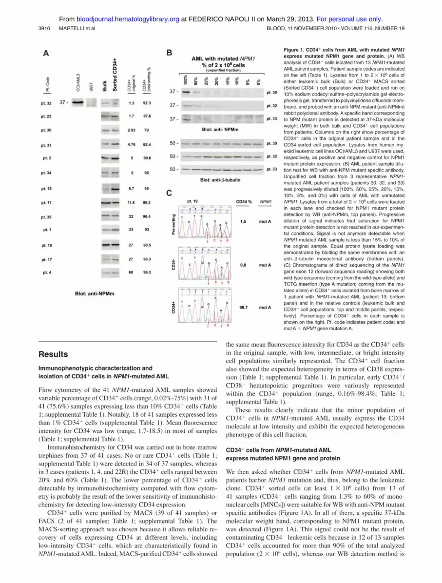
We then asked whether CD34� cells from NPM1-mutated AMLpatients harbor NPM1 mutation and, thus, belong to the leukemicclone. CD34� sorted cells (at least 1 � 106 cells) from 13 of41 samples (CD34� cells ranging from 1.3% to 60% of mono-nuclear cells [MNCs]) were suitable for WB with anti-NPM mutantspecific antibodies (Figure 1A). In all of them, a specific 37-kDamolecular weight band, corresponding to NPM1 mutant protein,was detected (Figure 1A). This signal could not be the result ofcontaminating CD34� leukemic cells because in 12 of 13 samplesCD34� cells accounted for more than 90% of the total analyzedpopulation (2 � 106 cells), whereas our WB detection method is
Figure 1. CD34� cells from AML with mutated NPM1express mutated NPM1 gene and protein. (A) WBanalysis of CD34� cells isolated from 13 NPM1-mutatedAML patient samples. Patient sample codes are indicatedon the left (Table 1). Lysates from 1 to 2 � 106 cells ofeither leukemic bulk (Bulk) or CD34� MACS sorted(Sorted CD34�) cell population were loaded and run on10% sodium dodecyl sulfate–polyacrylamide gel electro-phoresis gel, transferred to polyvinylidene difluoride mem-brane, and probed with an anti-NPM mutant (anti-NPMm)rabbit polyclonal antibody. A specific band correspondingto NPM mutant protein is detected at 37-kDa molecularweight (MW) in both bulk and CD34� cell populationsfrom patients. Columns on the right show percentage ofCD34� cells in the original patient sample and in theCD34-sorted cell population. Lysates from human my-eloid leukemic cell lines OCI/AML3 and U937 were used,respectively, as positive and negative control for NPM1mutant protein expression. (B) AML patient sample dilu-tion test for WB with anti-NPM mutant specific antibody.Unpurified cell fraction from 3 representative NPM1-mutated AML patient samples (patients 30, 32, and 33)was progressively diluted (100%, 50%, 25%, 20%, 15%,10%, 5%, and 0%) with cells of AML with unmutatedNPM1. Lysates from a total of 2 � 106 cells were loadedin each lane and checked for NPM1 mutant proteindetection by WB (anti-NPMm, top panels). Progressivedilution of signal indicates that saturation for NPM1mutant protein detection is not reached in our experimen-tal conditions. Signal is not anymore detectable whenNPM1-mutated AML sample is less than 15% to 10% ofthe original sample. Equal protein lysate loading wasdemonstrated by blotting the same membranes with ananti–�-tubulin monoclonal antibody (bottom panels).(C) Chromatograms of direct sequencing of the NPM1gene exon 12 (forward sequence reading) showing bothwild-type sequence (coming from the wild-type allele) andTCTG insertion (type A mutation; coming from the mu-tated allele) in CD34� cells isolated from bone marrow of1 patient with NPM1-mutated AML (patient 19; bottompanel) and in the relative controls (leukemic bulk andCD34� cell populations; top and middle panels, respec-tively). Percentage of CD34� cells in each sample isshown on the right. Pt. code indicates patient code; andmut A � NPM1 gene mutation A.
3910 MARTELLI et al BLOOD, 11 NOVEMBER 2010 � VOLUME 116, NUMBER 19
For personal use only. at FEDERICO NAPOLI II on March 29, 2013. bloodjournal.hematologylibrary.orgFrom
not sensitive enough to reveal NPM1 mutant protein from cells,which accounts for less than or equal to 10% (Figure 1B).Moreover, unlike that observed in AML cell dilution assay (Figure1B), the WB signal intensity was not decreased in the CD34�
purified cells compared with leukemic bulk (Figure 1A). Thus, it isconceivable that, even in the case with lower CD34� cell purity(patient 30, 79% CD34� cell purity; Table 1), positive signal couldnot be ascribed to contaminating CD34� cells.
In 1 leukopenic patient (patient 19), only 0.1 � 106 CD34�
bone marrow cells (99.7% pure) could be recovered, which werenot enough for WB analysis. In this case, NPM1 mutation A wasdetected by direct gene sequencing in purified CD34�, CD34�, andpresorting cell populations (Figure 1C). Again, detection of NPM1mutation in the CD34� cells could not be the result of the minorityof contaminating CD34� cells (0.3%) because the latter was underthe detection threshold ( 25%) of our direct gene sequencingassay (supplemental Figure 1).
These findings clearly show that the CD34� fraction fromNPM1-mutated AML with CD34� cells representing more than 1%of the bulk cell population is mutated both at gene and proteinlevel, thus indicating it belongs to the leukemic clone.
The CD34� fraction from NPM1-mutated AML containsCD34�/CD38� cells harboring NPM1 mutated gene and protein
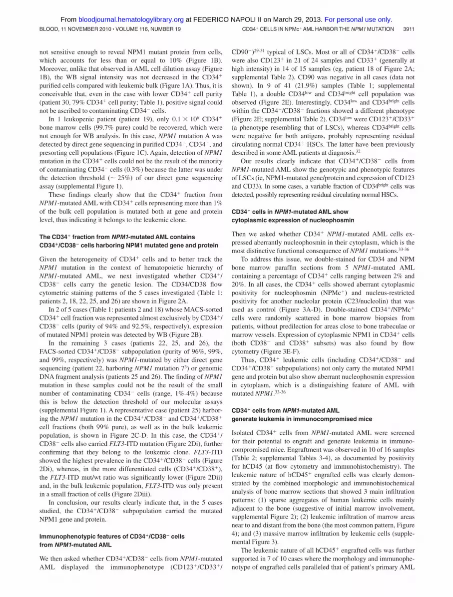
Given the heterogeneity of CD34� cells and to better track theNPM1 mutation in the context of hematopoietic hierarchy ofNPM1-mutated AML, we next investigated whether CD34�/CD38� cells carry the genetic lesion. The CD34/CD38 flowcytometric staining patterns of the 5 cases investigated (Table 1:patients 2, 18, 22, 25, and 26) are shown in Figure 2A.
In 2 of 5 cases (Table 1: patients 2 and 18) whose MACS-sortedCD34� cell fraction was represented almost exclusively by CD34�/CD38� cells (purity of 94% and 92.5%, respectively), expressionof mutated NPM1 protein was detected by WB (Figure 2B).
In the remaining 3 cases (patients 22, 25, and 26), theFACS-sorted CD34�/CD38� subpopulation (purity of 96%, 99%,and 99%, respectively) was NPM1-mutated by either direct genesequencing (patient 22, harboring NPM1 mutation 73) or genomicDNA fragment analysis (patients 25 and 26). The finding of NPM1mutation in these samples could not be the result of the smallnumber of contaminating CD34� cells (range, 1%-4%) becausethis is below the detection threshold of our molecular assays(supplemental Figure 1). A representative case (patient 25) harbor-ing the NPM1 mutation in the CD34�/CD38� and CD34�/CD38�
cell fractions (both 99% pure), as well as in the bulk leukemicpopulation, is shown in Figure 2C-D. In this case, the CD34�/CD38� cells also carried FLT3-ITD mutation (Figure 2Di), furtherconfirming that they belong to the leukemic clone. FLT3-ITDshowed the highest prevalence in the CD34�/CD38� cells (Figure2Di), whereas, in the more differentiated cells (CD34�/CD38�),the FLT3-ITD mut/wt ratio was significantly lower (Figure 2Dii)and, in the bulk leukemic population, FLT3-ITD was only presentin a small fraction of cells (Figure 2Diii).
In conclusion, our results clearly indicate that, in the 5 casesstudied, the CD34�/CD38� subpopulation carried the mutatedNPM1 gene and protein.
Immunophenotypic features of CD34�/CD38� cellsfrom NPM1-mutated AML
We then asked whether CD34�/CD38� cells from NPM1-mutatedAML displayed the immunophenotype (CD123�/CD33�/
CD90�)29-31 typical of LSCs. Most or all of CD34�/CD38� cellswere also CD123� in 21 of 24 samples and CD33� (generally athigh intensity) in 14 of 15 samples (eg, patient 18 of Figure 2A;supplemental Table 2). CD90 was negative in all cases (data notshown). In 9 of 41 (21.9%) samples (Table 1; supplementalTable 1), a double CD34low and CD34bright cell population wasobserved (Figure 2E). Interestingly, CD34low and CD34bright cellswithin the CD34�/CD38� fractions showed a different phenotype(Figure 2E; supplemental Table 2). CD34low were CD123�/CD33�
(a phenotype resembling that of LSCs), whereas CD34bright cellswere negative for both antigens, probably representing residualcirculating normal CD34� HSCs. The latter have been previouslydescribed in some AML patients at diagnosis.32
Our results clearly indicate that CD34�/CD38� cells fromNPM1-mutated AML show the genotypic and phenotypic featuresof LSCs (ie, NPM1-mutated gene/protein and expression of CD123and CD33). In some cases, a variable fraction of CD34bright cells wasdetected, possibly representing residual circulating normal HSCs.
CD34� cells in NPM1-mutated AML showcytoplasmic expression of nucleophosmin
Then we asked whether CD34� NPM1-mutated AML cells ex-pressed aberrantly nucleophosmin in their cytoplasm, which is themost distinctive functional consequence of NPM1 mutations.33-36
To address this issue, we double-stained for CD34 and NPMbone marrow paraffin sections from 5 NPM1-mutated AMLcontaining a percentage of CD34� cells ranging between 2% and20%. In all cases, the CD34� cells showed aberrant cytoplasmicpositivity for nucleophosmin (NPMc�) and nucleus-restrictedpositivity for another nucleolar protein (C23/nucleolin) that wasused as control (Figure 3A-D). Double-stained CD34�/NPMc�
cells were randomly scattered in bone marrow biopsies frompatients, without predilection for areas close to bone trabeculae ormarrow vessels. Expression of cytoplasmic NPM1 in CD34� cells(both CD38� and CD38� subsets) was also found by flowcytometry (Figure 3E-F).
Thus, CD34� leukemic cells (including CD34�/CD38� andCD34�/CD38� subpopulations) not only carry the mutated NPM1gene and protein but also show aberrant nucleophosmin expressionin cytoplasm, which is a distinguishing feature of AML withmutated NPM1.33-36
CD34� cells from NPM1-mutated AMLgenerate leukemia in immunocompromised mice
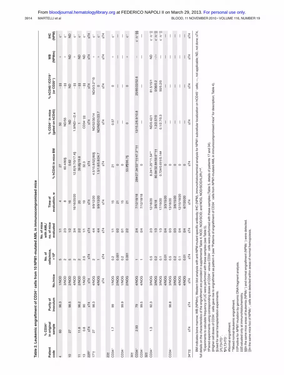
Isolated CD34� cells from NPM1-mutated AML were screenedfor their potential to engraft and generate leukemia in immuno-compromised mice. Engraftment was observed in 10 of 16 samples(Table 2; supplemental Tables 3-4), as documented by positivityfor hCD45 (at flow cytometry and immunohistochemistry). Theleukemic nature of hCD45� engrafted cells was clearly demon-strated by the combined morphologic and immunohistochemicalanalysis of bone marrow sections that showed 3 main infiltrationpatterns: (1) sparse aggregates of human leukemic cells mainlyadjacent to the bone (suggestive of initial marrow involvement,supplemental Figure 2); (2) leukemic infiltration of marrow areasnear to and distant from the bone (the most common pattern, Figure4); and (3) massive marrow infiltration by leukemic cells (supple-mental Figure 3).
The leukemic nature of all hCD45� engrafted cells was furthersupported in 7 of 10 cases where the morphology and immunophe-notype of engrafted cells paralleled that of patient’s primary AML
CD34� CELLS IN NPMc� AML HARBOR THE NPM1 MUTATION 3911BLOOD, 11 NOVEMBER 2010 � VOLUME 116, NUMBER 19
For personal use only. at FEDERICO NAPOLI II on March 29, 2013. bloodjournal.hematologylibrary.orgFrom
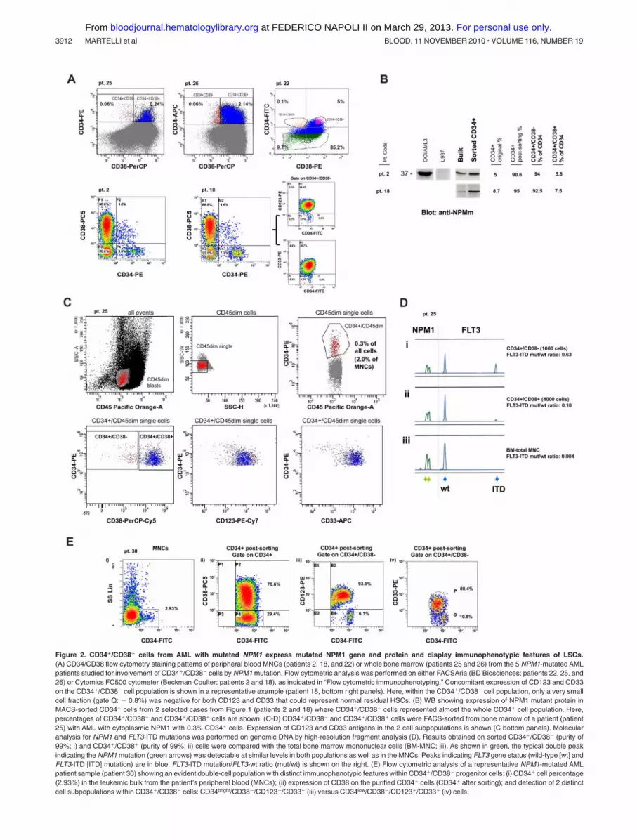
Figure 2. CD34�/CD38� cells from AML with mutated NPM1 express mutated NPM1 gene and protein and display immunophenotypic features of LSCs.(A) CD34/CD38 flow cytometry staining patterns of peripheral blood MNCs (patients 2, 18, and 22) or whole bone marrow (patients 25 and 26) from the 5 NPM1-mutated AMLpatients studied for involvement of CD34�/CD38� cells by NPM1 mutation. Flow cytometric analysis was performed on either FACSAria (BD Biosciences; patients 22, 25, and26) or Cytomics FC500 cytometer (Beckman Coulter; patients 2 and 18), as indicated in “Flow cytometric immunophenotyping.” Concomitant expression of CD123 and CD33on the CD34�/CD38� cell population is shown in a representative example (patient 18, bottom right panels). Here, within the CD34�/CD38� cell population, only a very smallcell fraction (gate Q: 0.8%) was negative for both CD123 and CD33 that could represent normal residual HSCs. (B) WB showing expression of NPM1 mutant protein inMACS-sorted CD34� cells from 2 selected cases from Figure 1 (patients 2 and 18) where CD34�/CD38� cells represented almost the whole CD34� cell population. Here,percentages of CD34�/CD38� and CD34�/CD38� cells are shown. (C-D) CD34�/CD38� and CD34�/CD38� cells were FACS-sorted from bone marrow of a patient (patient25) with AML with cytoplasmic NPM1 with 0.3% CD34� cells. Expression of CD123 and CD33 antigens in the 2 cell subpopulations is shown (C bottom panels). Molecularanalysis for NPM1 and FLT3-ITD mutations was performed on genomic DNA by high-resolution fragment analysis (D). Results obtained on sorted CD34�/CD38� (purity of99%; i) and CD34�/CD38� (purity of 99%; ii) cells were compared with the total bone marrow mononuclear cells (BM-MNC; iii). As shown in green, the typical double peakindicating the NPM1 mutation (green arrows) was detectable at similar levels in both populations as well as in the MNCs. Peaks indicating FLT3 gene status (wild-type [wt] andFLT3-ITD [ITD] mutation) are in blue. FLT3-ITD mutation/FLT3-wt ratio (mut/wt) is shown on the right. (E) Flow cytometric analysis of a representative NPM1-mutated AMLpatient sample (patient 30) showing an evident double-cell population with distinct immunophenotypic features within CD34�/CD38� progenitor cells: (i) CD34� cell percentage(2.93%) in the leukemic bulk from the patient’s peripheral blood (MNCs); (ii) expression of CD38 on the purified CD34� cells (CD34� after sorting); and detection of 2 distinctcell subpopulations within CD34�/CD38� cells: CD34bright/CD38�/CD123�/CD33� (iii) versus CD34low/CD38�/CD123�/CD33� (iv) cells.
3912 MARTELLI et al BLOOD, 11 NOVEMBER 2010 � VOLUME 116, NUMBER 19
For personal use only. at FEDERICO NAPOLI II on March 29, 2013. bloodjournal.hematologylibrary.orgFrom

(Table 2; supplemental Table 4). In these cases, all engrafted cellswere myeloid (hCD45�/CD33� and myeloperoxidase-positive;Figure 4B,E-F). In contrast, no hCD45� cells coexpressing CD19and/or CD20 (suggestive of normal engraftment) were detected inthe great majority of evaluated mice (Table 2; supplemental Table4). Conclusive evidence that all human engrafted cells belonged tothe leukemic clone came from the immunohistochemical demonstra-tion that they expressed cytoplasmic NPM/nuclear C23 (Figure4C-D), the hallmark feature of human NPM1-mutated AML cells.Moreover, in 5 of 7 cases (patients 4, 10, 17, 22R, and 23), the WBand/or molecular analysis showed a mutated NPM1 protein and/orgene (Figure 4G; Table 2; supplemental Table 4). Although CD34�
cells from 1 patient (patient 17) did not show the ability to produceAML colonies in in vitro colony-forming cell (CFC) assay (datanot shown), cells recovered from primary mice recipients demon-strated self-renewal capacity in serial transplantations (Table 3:patients 4, 10, 11, 17, and 22R).
Interestingly, in 3 of 10 cases (patients 30, 32, and 34),immunohistochemistry and flow cytometry showed either normalor mixed (normal plus leukemic) engraftment. The latter pattern ofengraftment was characterized by areas of bone marrow infiltratedby human NPMc� AML cells adjacent to areas of normal human
trilineage hematopoiesis (Table 2; Figure 5). Molecular analysis(performed on the whole cell population recovered by boneflushing) confirmed NPM1-mutation in all cases (Table 2). AMLdevelopment was usually seen later than normal human hematopoi-etic engraftment (Table 2). Interestingly, flow cytometry of originalMNCs from patients 30, 32, and 34 showed a relevant subpopula-tion of CD34bright/CD38�/CD123�/CD33� cells, which probablyrepresent normal HSCs (Figure 2E; supplemental Table 2). Further-more, in CFC assay, CD34� cells from 2 of these cases (patients 32and 34) produced mixed and erythroid colonies, which, because ofgermline NPM1 gene (data not shown), are normal in origin.
Our findings clearly indicate that the small fraction of CD34�
cells from most NPM1-mutated AML cases generate a leukemiashowing the same morphologic and immunohistochemical features(aberrant cytoplasmic NPM1) as the original patient’s AML.
Patterns of engraftment of CD34� cells fromNPM1-mutated AML in immunocompromised mice
The main aim of this study was to characterize the small fractionof CD34� cells in NPM1-mutated AML. However, there isexperimental evidence that, in some AML cases37,38 (including
Figure 3. CD34� cells in NPM1-mutated AML show cytoplasmic expression of nucleophosmin. (A-B) CD34� leukemic cells (brown) from a patient with NPM1-mutatedAML showing cytoplasmic expression of NPM (blue; A arrows) and nucleus-restricted positivity for C23/nucleolin (B arrows). (C-D) Another NPM1-mutated patient showing rareCD34� cells (C) that double stain for CD34 (brown) and cytoplasmic NPM (blue; D single arrow); endothelium of a vessel express CD34 (D double arrow). CD34� leukemic cell(brown) from the same case show nucleus-restricted positivity for C23/nucleolin (blue; D inset). (A-B,D) Sequential immunoperoxidase/APAAP staining; no counterstaining.(C) APAAP technique (hematoxylin counterstaining). (A-D) Paraffin sections from bone marrow biopsies. All images were collected using an Olympus B61 microscope and aUPlan FI 100�/1.3 NA oil objective; Camedia 4040, Dp_soft Version 3.2; and Adobe Photoshop 7.0. (E-F) Flow cytometric detection of NPM-cytoplasmic expression (patient25) in CD34� and CD34� blasts (E), and after additional gating on CD34�/CD38� or CD34�/CD38� cells, respectively (F).
CD34� CELLS IN NPMc� AML HARBOR THE NPM1 MUTATION 3913BLOOD, 11 NOVEMBER 2010 � VOLUME 116, NUMBER 19
For personal use only. at FEDERICO NAPOLI II on March 29, 2013. bloodjournal.hematologylibrary.orgFrom
Tab
le2.
Leu
kem
icen
gra
ftm
ento
fCD
34�
cells
fro
m10
NP
M1-
mu
tate
dA
ML
inim
mu
no
com
pro
mis
edm
ice
Pat
ien
tco
de
%C
D34
�
ino
rig
inal
sam
ple
Pu
rity
of
ino
culu
mN
o./m
ice
No
.of
cells
/mic
e,�
106
No
.ofm
ice
wit
hA
ML
/n
o.o
fmic
eev
alu
ated
Tim
eo
fev
alu
atio
n,w
%h
CD
45in
mic
eB
M%
CD
34�
inm
ice
(gat
edo
nh
CD
45)
%h
CD
45�
/CD
19�
(or
CD
20�
)W
B(N
PM
m)
IHC
(NP
M)
460
96.3
1/N
OD
51/
14
2750
�‡‡
�c�
3/N
OD
22/
38
60.4
/80§
ND
/55
�‡‡
�c�
1027
98.5
2/N
OD
51/
216
0.62
ND
ND
ND
ND
4/N
OD
23/
416
/16/
20/2
012
.8�/9
.7/0
/17.
4§1.
9/N
D/—
/2.4
�‡‡
�c�
1111
.896
.22/
NO
D2
2/2
2039
.8§/
19.8
1�
‡‡N
Dc�
130.
890
1/N
OD
2.5
1/1
1932
.3C
D34
�‡‡
�‡‡
ND
c�
22R
*sT
4sT
4sT
4sT
4sT
4sT
4sT
4sT
4sT
4sT
4sT
4
17*†
2798
.34/
NO
G2
4/4
9/9/
12/2
04.
5/15
.8/5
2�/8
0§N
D/5
2/35
/14
ND
/0/3
.2**
/0�
c�
4/N
OG
14/
49/
9/13
/20
1.5/
1.8/
5.8/
24.7
ND
/ND
/21/
23.7
0�
c�
sT4
sT4
sT4
sT4
sT4
sT4
sT4
sT4
sT4
23†
CD
34�
1.7
991/
NO
G0.
21/
115
210.
570
�c�
2/N
OG
0.00
10/
221
0—
——
—
CD
34�
99.9
1/N
OG
0.2
0/1
150
——
——
2/N
OG
0.00
12/
221
53.8
¶/95
.7§
00
�c�
30†
CD
34�
2.93
794/
NO
G1
2/4
7/12
/18/
1828
#/27
.3#/
33**
††/4
7.3*
*††
13/1
5.2/
6.9/
10.8
25/8
5/22
/52.
6�
c�/c
�§§
CD
34�
99.5
4/N
OG
10/
47/
12/1
8/18
0—
——
—
32†
CD
34�
1.3
92.3
3/N
OG
0.5
2/3
12/1
8/20
8.2#
/1.2
5**/
1.54
**N
D/0
.42/
181
.5/1
5/1
ND
c�/c
���
3/N
OG
0.2
1/3
3/6/
1880
.8#/
32.8
#/59
.6**
††1.
2/2.
57/0
0/36
/0.2
�/�
c�/c
���
3/N
OG
0.1
1/3
17/1
9/20
0.72
#/0.
6††/
0.14
#0.
1/2.
7/0.
350
/5.2
/0—
c�/c
���
4/N
OG
0.01
0/4
3/3/
15/2
00
——
——
CD
34�
98.8
3/N
OG
0.5
0/3
13/1
8/20
0—
——
—
3/N
OG
0.2
0/3
3/6/
15/2
00
——
——
4/N
OG
0.1
0/4
12/1
5/18
/20
0—
——
—
4/N
OG
0.01
0/4
6/7/
20/2
00
——
——
34*†
‡sT
4sT
4sT
4sT
4sT
4sT
4sT
4sT
4sT
4sT
4sT
4
BM
indi
cate
sbo
nem
arro
w;W
B(N
PM
m),
Wes
tern
blot
anal
ysis
with
anti-
NP
Mm
utan
tspe
cific
antib
ody;
IHC
(NP
M),
imm
unoh
isto
chem
ical
anal
ysis
forN
PM
1su
bcel
lula
rloc
aliz
atio
non
hCD
45�
cells
;—,n
otap
plic
able
;ND
,not
done
;sT
4,fu
llde
tails
onth
ech
arac
teris
tics
ofth
een
graf
tmen
tare
prov
ided
insu
pple
men
talT
able
4;N
OD
,NO
D/S
CID
;and
NO
G,N
OD
/SC
ID/IL
2R�
null .
*Exp
erim
ents
toca
lcul
ate
freq
uenc
yof
LIC
wer
epe
rfor
med
with
thes
esa
mpl
es(s
eeTa
ble
5).
†Pai
red
expe
rimen
tsus
ing
corr
espo
ndin
gdo
ses
ofC
D34
�ve
rsus
CD
34�
cells
wer
epe
rfor
med
with
thes
esa
mpl
es(T
able
5,de
tails
ofpa
tient
s17
and
34).
‡Hig
herc
elld
oses
ofC
D34
�ce
llsga
veris
eto
engr
aftm
enta
spa
ttern
4(s
ee“P
atte
rns
ofen
graf
tmen
tofC
D34
�ce
llsfr
omN
PM
1-m
utat
edA
ML
inim
mun
ocom
prom
ised
mic
e”fo
rdes
crip
tion;
Tabl
e4)
.§U
sed
fors
eria
ltra
nspl
anta
tion
expe
rimen
ts.
�FLT
3-IT
D�
.¶F
LT3-
ITD
�.
#Nor
mal
engr
aftm
ent.
**M
ixed
norm
al-le
ukem
icen
graf
tmen
t.††
Pos
itive
forN
PM
1m
utat
ion
byge
nom
icD
NA
frag
men
tana
lysi
s.‡‡
Eva
luat
edon
lyat
imm
unoh
isto
chem
istr
y.§§
Inth
esa
me
mic
ear
eas
ofle
ukem
ia(N
PM
c�)a
ndno
rmal
engr
aftm
ent(
NP
Mc�
)wer
ede
tect
ed.
��In
the
sam
em
ice
foci
ofN
PM
c�,c
ells
wer
ede
tect
edw
ithin
area
sof
norm
alhe
mop
oies
is.
3914 MARTELLI et al BLOOD, 11 NOVEMBER 2010 � VOLUME 116, NUMBER 19
For personal use only. at FEDERICO NAPOLI II on March 29, 2013. bloodjournal.hematologylibrary.orgFrom
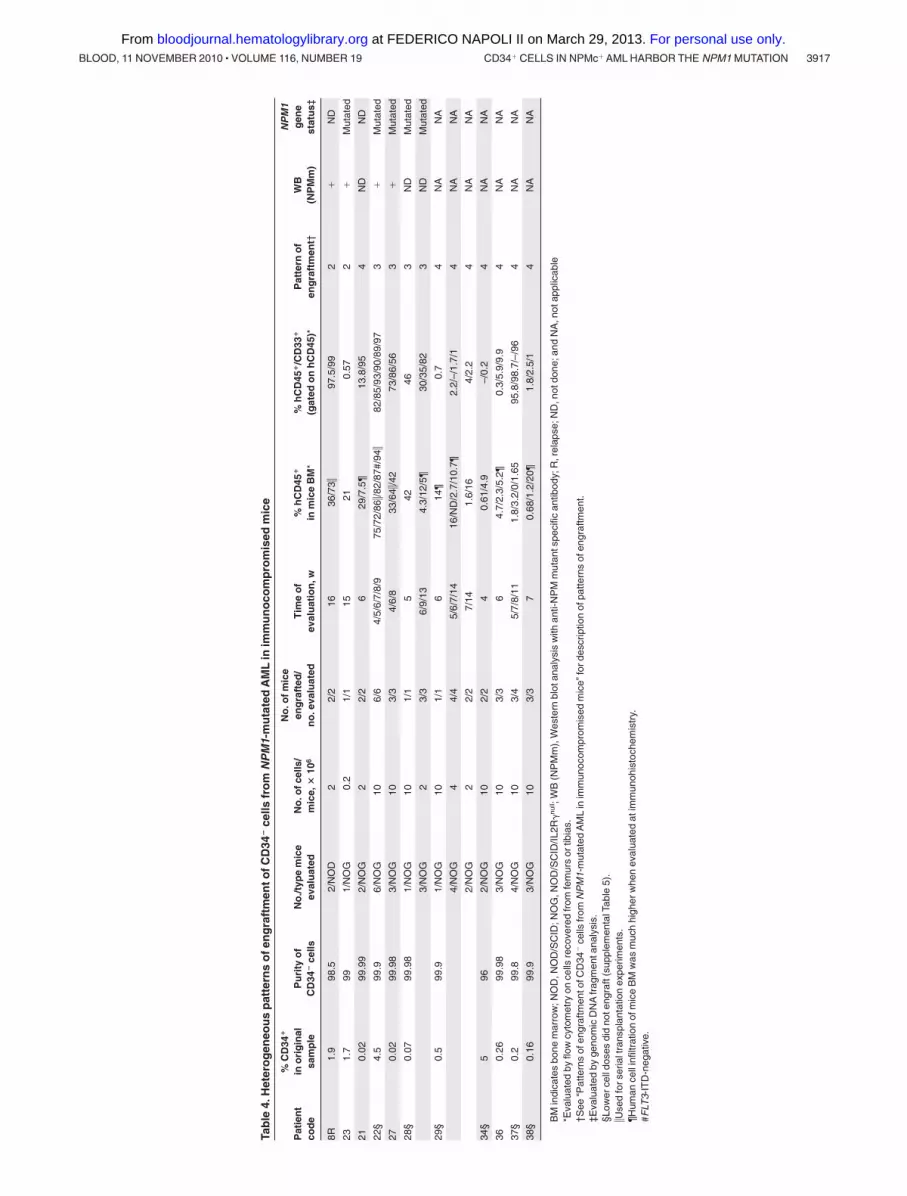
NPM1-mutated AML21), also the CD34� population may containthe LICs. Therefore, we assessed the engraftment capability ofCD34� cells from 15 NPM1-mutated AMLs (Table 4; supplemen-tal Table 5). Inoculation of these cells in immunocompromisedmice led to 4 different patterns, which varied also according to theinjected cell dose:
Pattern 1. CD34� cells from 10 patients did not engraft in micewhen inoculated at doses less than or equal to 1 � 106 (supplemen-tal Table 5). In contrast, CD34� cells from 4 of these casesgenerated typical NPMc� AML, when injected in mice at the samedoses (Table 2: patients 17, 30, 32, and 34; supplemental Table 4).
Moreover, CD34� cells from patients 17, 32, and 34 did notoutgrow into AML colonies in CFC assays (data not shown).
Pattern 2. Engraftment of CD34� as typical NPMc� AML(similar to that observed injecting mice with purified CD34� cells)was observed in 2 cases (not shown). In both of them, CD34� cellswere purified from NPM1-mutated AML at relapse (Table 4:patients 8R and 23). Self-renewal capability of CD34� cells fromthese cases was demonstrated after transfer to secondary recipients(Table 3).
Pattern 3. Inoculation of CD34� fraction (� 2 � 106 cells)from 3 cases (Table 4: patients 22, 27, and 28) resulted into
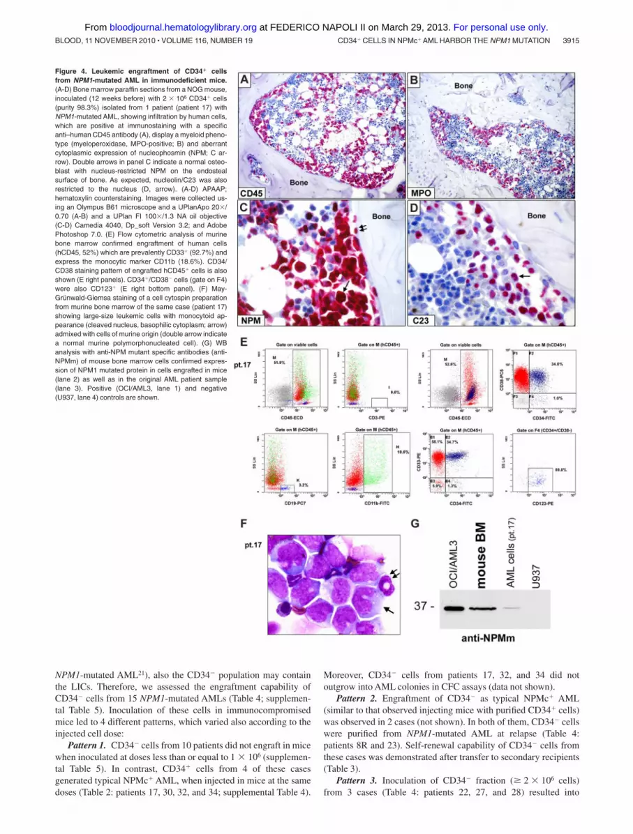
Figure 4. Leukemic engraftment of CD34� cellsfrom NPM1-mutated AML in immunodeficient mice.(A-D) Bone marrow paraffin sections from a NOG mouse,inoculated (12 weeks before) with 2 � 106 CD34� cells(purity 98.3%) isolated from 1 patient (patient 17) withNPM1-mutated AML, showing infiltration by human cells,which are positive at immunostaining with a specificanti–human CD45 antibody (A), display a myeloid pheno-type (myeloperoxidase, MPO-positive; B) and aberrantcytoplasmic expression of nucleophosmin (NPM; C ar-row). Double arrows in panel C indicate a normal osteo-blast with nucleus-restricted NPM on the endostealsurface of bone. As expected, nucleolin/C23 was alsorestricted to the nucleus (D, arrow). (A-D) APAAP;hematoxylin counterstaining. Images were collected us-ing an Olympus B61 microscope and a UPlanApo 20�/0.70 (A-B) and a UPlan FI 100�/1.3 NA oil objective(C-D) Camedia 4040, Dp_soft Version 3.2; and AdobePhotoshop 7.0. (E) Flow cytometric analysis of murinebone marrow confirmed engraftment of human cells(hCD45, 52%) which are prevalently CD33� (92.7%) andexpress the monocytic marker CD11b (18.6%). CD34/CD38 staining pattern of engrafted hCD45� cells is alsoshown (E right panels). CD34�/CD38� cells (gate on F4)were also CD123� (E right bottom panel). (F) May-Grunwald-Giemsa staining of a cell cytospin preparationfrom murine bone marrow of the same case (patient 17)showing large-size leukemic cells with monocytoid ap-pearance (cleaved nucleus, basophilic cytoplasm; arrow)admixed with cells of murine origin (double arrow indicatea normal murine polymorphonucleated cell). (G) WBanalysis with anti-NPM mutant specific antibodies (anti-NPMm) of mouse bone marrow cells confirmed expres-sion of NPM1 mutated protein in cells engrafted in mice(lane 2) as well as in the original AML patient sample(lane 3). Positive (OCI/AML3, lane 1) and negative(U937, lane 4) controls are shown.
CD34� CELLS IN NPMc� AML HARBOR THE NPM1 MUTATION 3915BLOOD, 11 NOVEMBER 2010 � VOLUME 116, NUMBER 19
For personal use only. at FEDERICO NAPOLI II on March 29, 2013. bloodjournal.hematologylibrary.orgFrom

marrow engraftment by hCD45�/hCD33� cells (Figure 6A), which,in tissue sections, consisted of 2 populations (Figure 6B-D):(1) myeloperoxidase-positive cells located close to bone trabeculaethat exhibited weak cytoplasmic NPM1 positivity (not shown); and
(2) mature CD68� histiocytes that were located in the central areaof bone marrow. In all cases, detection of NPM1 mutant proteinand gene (Figure 6 E-F) proved the leukemic nature of engraftedcells. These findings possibly reflect short-term engraftment by
Figure 5. Mixed (normal and leukemic) engraft-ment of CD34� cells from NPM1-mutated AML-immunodeficient mice. (A-F) Bone marrow paraffinsections from a vertebral body of an NOG mouse,inoculated (18 weeks before) with 1 � 106 CD34� cells(purity 79%) from patient 30 (Table 2). An area in thebottom part of panel A (square) is packed with leukemiccells expressing hCD45 (B left square), cytoplasmicNPM1 (C) and nucleus-restricted C23 (D). Another areafrom the same section shows involvement by normalhuman hemopoietic cells (square in the top part of panelA), expressing CD45 (B right square), nucleus-restrictedNPM1 and C23 (E-F single and double arrows).(A) Hematoxylin-eosin. (B-F) APAAP, hematoxylin coun-terstaining. Images were collected using an OlympusB61 microscope and a UPlanApo 20�/0.70 (A-B),UPlanApo 40�/0.85 (C-F); Olympus E330-ADU1.2�camera; and Adobe Photoshop 7.0.
Table 3. Summary of serial transplantations experiments
Patientcode
Engrafted cellfraction in
primary recipients* No./miceNo. of cells/mice, �106†
No. of micewith AML/no. of miceevaluated
Time ofevaluation, w % hCD45 in mice BM‡
4 CD34� 4/NOD 2 4/4§ 6 87/15/89/87.5
10 CD34� 1/NOD 0.6 1/1 18 94
11 CD34� 1/NOD 1.6 1/1 18 3
17� CD34� 2/NOG¶ 0.125¶ 1/2 25 0.12
17� CD34� 4/NOG# 0.125# 0/4 25 0
22R CD34� 4/NOG 0.52 4/4 11 7.1/6/8.3/7
8R CD34� (pattern 2) 3/NOD 1 3/3 6.5 6.7/1.2/0.5
23 CD34� (pattern 2) 3/NOG 4/4/2 3/3 12 60.5/79/69.5
22 CD34� (pattern 3) 2/NOG 2.5 0/2 8 0
22 CD34� (pattern 3) 2/NOG 1 0/2 12 0
27 CD34� (pattern 3) 1/NOG 0.5 0/1 12 0
R indicates relapse.*See Tables 2 and 4 for details on cases used for serial transplantations.†hCD45� cell equivalent.‡Human cells were all hCD45�/CD33� and expressed cytoplasmic NPM1 at immunohistochemical analysis.§From these mice, we obtained engraftment up to quaternary recipients.�Human cells recovered from engrafted mice were sorted in CD34� and CD34� cell fractions and inoculated separately.¶CD34�.#CD34�.
3916 MARTELLI et al BLOOD, 11 NOVEMBER 2010 � VOLUME 116, NUMBER 19
For personal use only. at FEDERICO NAPOLI II on March 29, 2013. bloodjournal.hematologylibrary.orgFrom
Tab
le4.
Het
ero
gen
eou
sp
atte
rns
ofe
ng
raft
men
tofC
D34
�ce
llsfr
om
NP
M1-
mu
tate
dA
ML
inim
mu
no
com
pro
mis
edm
ice
Pat
ien
tco
de
%C
D34
�
ino
rig
inal
sam
ple
Pu
rity
of
CD
34�
cells
No
./typ
em
ice
eval
uat
edN
o.o
fcel
ls/
mic
e,�
106
No
.ofm
ice
eng
raft
ed/
no
.eva
luat
edT
ime
of
eval
uat
ion
,w%
hC
D45
�
inm
ice
BM
*%
hC
D45
�/C
D33
�
(gat
edo
nh
CD
45)*
Pat
tern
of
eng
raft
men
t†W
B(N
PM
m)
NP
M1
gen
est
atu
s‡
8R1.
998
.52/
NO
D2
2/2
1636
/73�
97.5
/99
2�
ND
231.
799
1/N
OG
0.2
1/1
1521
0.57
2�
Mut
ated
210.
0299
.99
2/N
OG
22/
26
29/7
.5¶
13.8
/95
4N
DN
D
22§
4.5
99.9
6/N
OG
106/
64/
5/6/
7/8/
975
/72/
86�/8
2/87
#/94
�82
/85/
93/9
0/89
/97
3�
Mut
ated
270.
0299
.98
3/N
OG
103/
34/
6/8
33/6
4�/4
273
/86/
563
�M
utat
ed
28§
0.07
99.9
81/
NO
G10
1/1
542
463
ND
Mut
ated
3/N
OG
23/
36/
9/13
4.3/
12/5
¶30
/35/
823
ND
Mut
ated
29§
0.5
99.9
1/N
OG
101/
16
14¶
0.7
4N
AN
A
4/N
OG
44/
45/
6/7/
1416
/ND
/2.7
/10.
7¶2.
2/–/
1.7/
14
NA
NA
2/N
OG
22/
27/
141.
6/16
4/2.
24
NA
NA
34§
596
2/N
OG
102/
24
0.61
/4.9
–/0.
24
NA
NA
360.
2699
.98
3/N
OG
103/
36
4.7/
2.3/
5.2¶
0.3/
5.9/
9.9
4N
AN
A
37§
0.2
99.8
4/N
OG
103/
45/
7/8/
111.
8/3.
2/0/
1.65
95.8
/98.
7/–/
964
NA
NA
38§
0.16
99.9
3/N
OG
103/
37
0.68
/1.2
/20¶
1.8/
2.5/
14
NA
NA
BM
indi
cate
sbo
nem
arro
w;N
OD
,NO
D/S
CID
;NO
G,N
OD
/SC
ID/IL
2R�
null ;
WB
(NP
Mm
),W
este
rnbl
otan
alys
isw
ithan
ti-N
PM
mut
ants
peci
fican
tibod
y;R
,rel
apse
;ND
,not
done
;and
NA
,not
appl
icab
le*E
valu
ated
byflo
wcy
tom
etry
once
llsre
cove
red
from
fem
urs
ortib
ias.
†See
“Pat
tern
sof
engr
aftm
ento
fCD
34�
cells
from
NP
M1-
mut
ated
AM
Lin
imm
unoc
ompr
omis
edm
ice”
ford
escr
iptio
nof
patte
rns
ofen
graf
tmen
t.‡E
valu
ated
byge
nom
icD
NA
frag
men
tana
lysi
s.§L
ower
cell
dose
sdi
dno
teng
raft
(sup
plem
enta
lTab
le5)
.�U
sed
fors
eria
ltra
nspl
anta
tion
expe
rimen
ts.
¶Hum
ance
llin
filtr
atio
nof
mic
eB
Mw
asm
uch
high
erw
hen
eval
uate
dat
imm
unoh
isto
chem
istr
y.#F
LT3-
ITD
-neg
ativ
e.
CD34� CELLS IN NPMc� AML HARBOR THE NPM1 MUTATION 3917BLOOD, 11 NOVEMBER 2010 � VOLUME 116, NUMBER 19
For personal use only. at FEDERICO NAPOLI II on March 29, 2013. bloodjournal.hematologylibrary.orgFrom
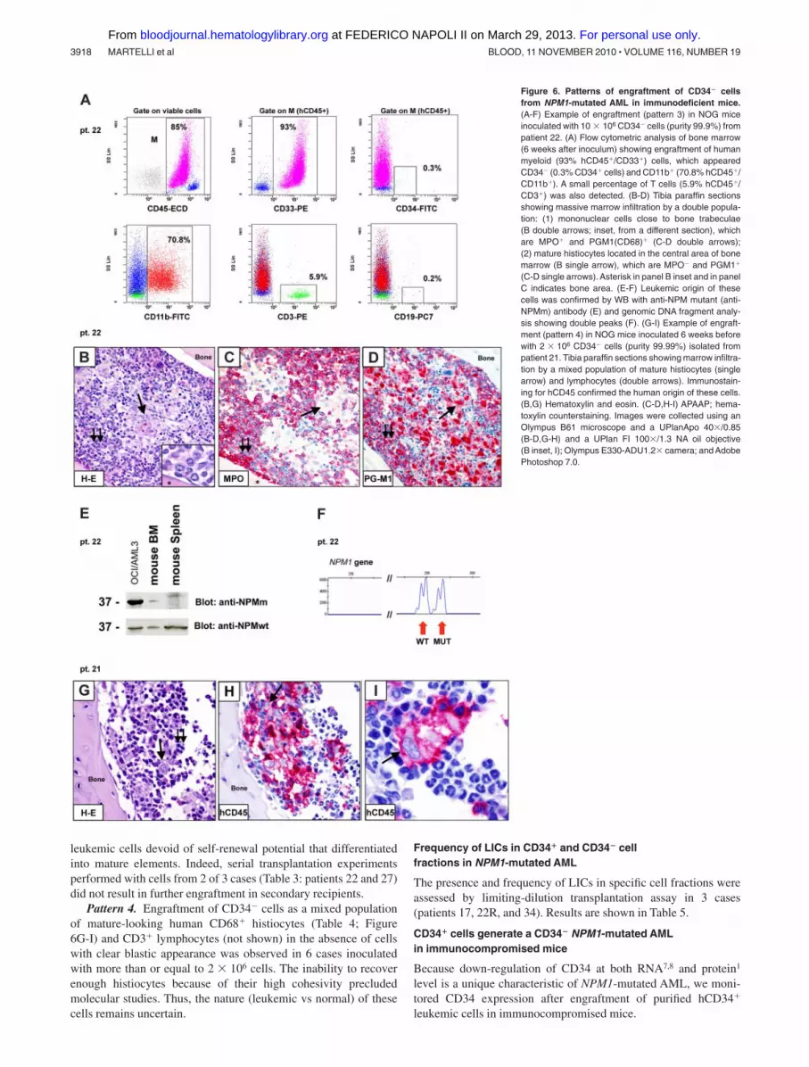
leukemic cells devoid of self-renewal potential that differentiatedinto mature elements. Indeed, serial transplantation experimentsperformed with cells from 2 of 3 cases (Table 3: patients 22 and 27)did not result in further engraftment in secondary recipients.
Pattern 4. Engraftment of CD34� cells as a mixed populationof mature-looking human CD68� histiocytes (Table 4; Figure6G-I) and CD3� lymphocytes (not shown) in the absence of cellswith clear blastic appearance was observed in 6 cases inoculatedwith more than or equal to 2 � 106 cells. The inability to recoverenough histiocytes because of their high cohesivity precludedmolecular studies. Thus, the nature (leukemic vs normal) of thesecells remains uncertain.
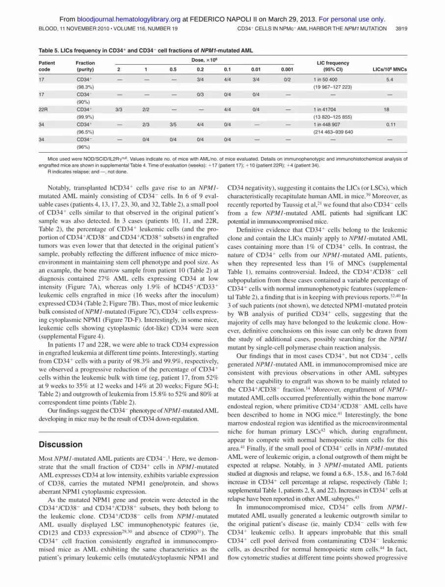
Frequency of LICs in CD34� and CD34� cellfractions in NPM1-mutated AML
The presence and frequency of LICs in specific cell fractions wereassessed by limiting-dilution transplantation assay in 3 cases(patients 17, 22R, and 34). Results are shown in Table 5.
CD34� cells generate a CD34� NPM1-mutated AMLin immunocompromised mice
Because down-regulation of CD34 at both RNA7,8 and protein1
level is a unique characteristic of NPM1-mutated AML, we moni-tored CD34 expression after engraftment of purified hCD34�
leukemic cells in immunocompromised mice.
Figure 6. Patterns of engraftment of CD34� cellsfrom NPM1-mutated AML in immunodeficient mice.(A-F) Example of engraftment (pattern 3) in NOG miceinoculated with 10 � 106 CD34� cells (purity 99.9%) frompatient 22. (A) Flow cytometric analysis of bone marrow(6 weeks after inoculum) showing engraftment of humanmyeloid (93% hCD45�/CD33�) cells, which appearedCD34� (0.3% CD34� cells) and CD11b� (70.8% hCD45�/CD11b�). A small percentage of T cells (5.9% hCD45�/CD3�) was also detected. (B-D) Tibia paraffin sectionsshowing massive marrow infiltration by a double popula-tion: (1) mononuclear cells close to bone trabeculae(B double arrows; inset, from a different section), whichare MPO� and PGM1(CD68)� (C-D double arrows);(2) mature histiocytes located in the central area of bonemarrow (B single arrow), which are MPO� and PGM1�
(C-D single arrows). Asterisk in panel B inset and in panelC indicates bone area. (E-F) Leukemic origin of thesecells was confirmed by WB with anti-NPM mutant (anti-NPMm) antibody (E) and genomic DNA fragment analy-sis showing double peaks (F). (G-I) Example of engraft-ment (pattern 4) in NOG mice inoculated 6 weeks beforewith 2 � 106 CD34� cells (purity 99.99%) isolated frompatient 21. Tibia paraffin sections showing marrow infiltra-tion by a mixed population of mature histiocytes (singlearrow) and lymphocytes (double arrows). Immunostain-ing for hCD45 confirmed the human origin of these cells.(B,G) Hematoxylin and eosin. (C-D,H-I) APAAP; hema-toxylin counterstaining. Images were collected using anOlympus B61 microscope and a UPlanApo 40�/0.85(B-D,G-H) and a UPlan FI 100�/1.3 NA oil objective(B inset, I); Olympus E330-ADU1.2� camera; and AdobePhotoshop 7.0.
3918 MARTELLI et al BLOOD, 11 NOVEMBER 2010 � VOLUME 116, NUMBER 19
For personal use only. at FEDERICO NAPOLI II on March 29, 2013. bloodjournal.hematologylibrary.orgFrom
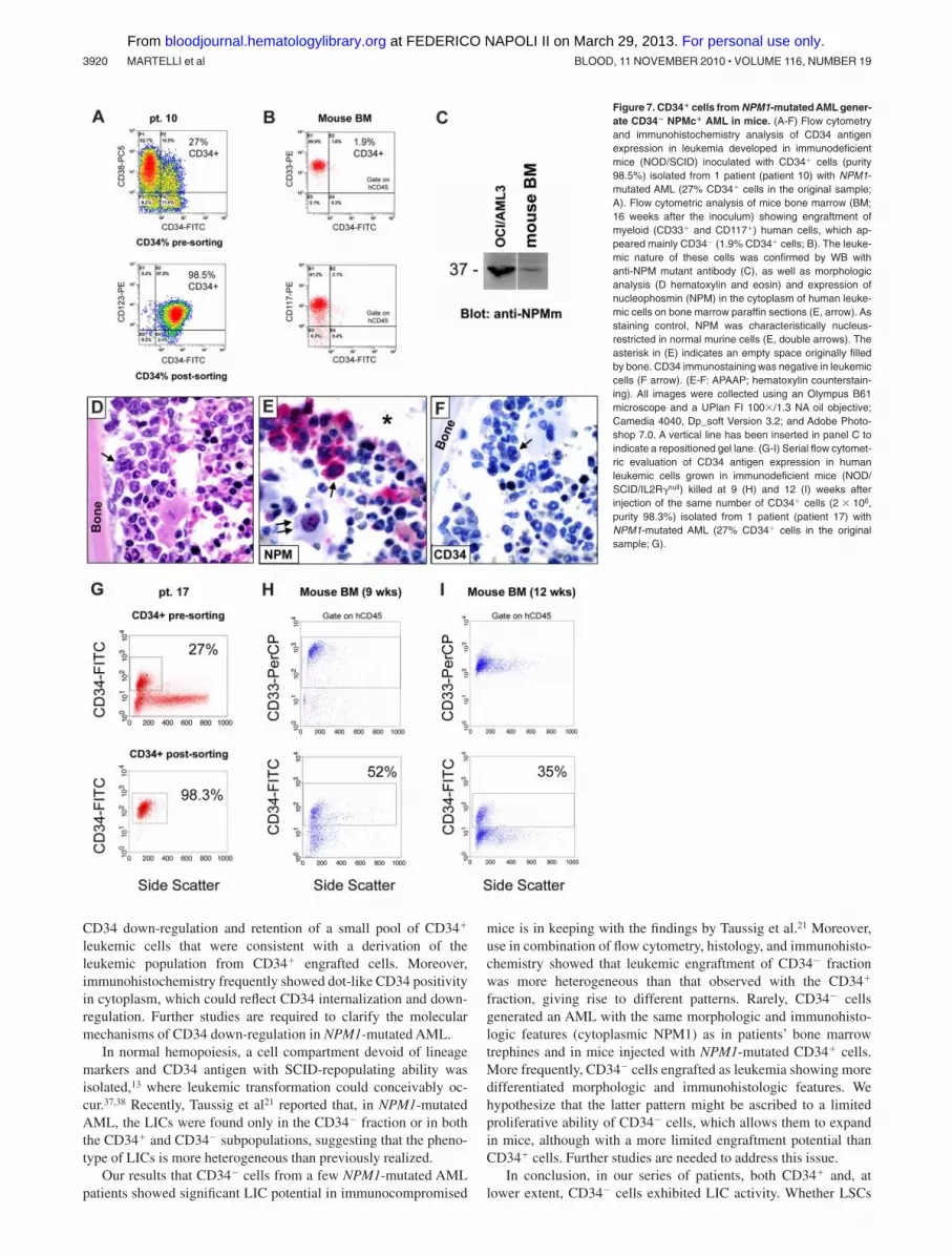
Notably, transplanted hCD34� cells gave rise to an NPM1-mutated AML mainly consisting of CD34� cells. In 6 of 9 eval-uable cases (patients 4, 13, 17, 23, 30, and 32, Table 2), a small poolof CD34� cells similar to that observed in the original patient’ssample was also detected. In 3 cases (patients 10, 11, and 22R,Table 2), the percentage of CD34� leukemic cells (and the pro-portion of CD34�/CD38� and CD34�/CD38� subsets) in engraftedtumors was even lower that that detected in the original patient’ssample, probably reflecting the different influence of mice micro-environment in maintaining stem cell phenotype and pool size. Asan example, the bone marrow sample from patient 10 (Table 2) atdiagnosis contained 27% AML cells expressing CD34 at lowintensity (Figure 7A), whereas only 1.9% of hCD45�/CD33�
leukemic cells engrafted in mice (16 weeks after the inoculum)expressed CD34 (Table 2; Figure 7B). Thus, most of mice leukemicbulk consisted of NPM1-mutated (Figure 7C), CD34� cells express-ing cytoplasmic NPM1 (Figure 7D-F). Interestingly, in some mice,leukemic cells showing cytoplasmic (dot-like) CD34 were seen(supplemental Figure 4).
In patients 17 and 22R, we were able to track CD34 expressionin engrafted leukemia at different time points. Interestingly, startingfrom CD34� cells with a purity of 98.3% and 99.9%, respectively,we observed a progressive reduction of the percentage of CD34�
cells within the leukemic bulk with time (eg, patient 17, from 52%at 9 weeks to 35% at 12 weeks and 14% at 20 weeks; Figure 5G-I;Table 2) and outgrowth of leukemia from 15.8% to 52% and 80% atcorrespondent time points (Table 2).
Our findings suggest the CD34� phenotype of NPM1-mutatedAMLdeveloping in mice may be the result of CD34 down-regulation.
Discussion
Most NPM1-mutated AML patients are CD34�.1 Here, we demon-strate that the small fraction of CD34� cells in NPM1-mutatedAML expresses CD34 at low intensity, exhibits variable expressionof CD38, carries the mutated NPM1 gene/protein, and showsaberrant NPM1 cytoplasmic expression.
As the mutated NPM1 gene and protein were detected in theCD34�/CD38� and CD34�/CD38� subsets, they both belong tothe leukemic clone. CD34�/CD38� cells from NPM1-mutatedAML usually displayed LSC immunophenotypic features (ie,CD123 and CD33 expression29,30 and absence of CD9031). TheCD34� cell fraction consistently engrafted in immunocompro-mised mice as AML exhibiting the same characteristics as thepatient’s primary leukemic cells (mutated/cytoplasmic NPM1 and
CD34 negativity), suggesting it contains the LICs (or LSCs), whichcharacteristically recapitulate human AML in mice.39 Moreover, asrecently reported by Taussig et al,21 we found that also CD34� cellsfrom a few NPM1-mutated AML patients had significant LICpotential in immunocompromised mice.
Definitive evidence that CD34� cells belong to the leukemicclone and contain the LICs mainly apply to NPM1-mutated AMLcases containing more than 1% of CD34� cells. In contrast, thenature of CD34� cells from our NPM1-mutated AML patients,when they represented less than 1% of MNCs (supplementalTable 1), remains controversial. Indeed, the CD34�/CD38� cellsubpopulation from these cases contained a variable percentage ofCD34� cells with normal immunophenotypic features (supplemen-tal Table 2), a finding that is in keeping with previous reports.32,40 In3 of such patients (not shown), we detected NPM1-mutated proteinby WB analysis of purified CD34� cells, suggesting that themajority of cells may have belonged to the leukemic clone. How-ever, definitive conclusions on this issue can only be drawn fromthe study of additional cases, possibly searching for the NPM1mutant by single-cell polymerase chain reaction analysis.
Our findings that in most cases CD34�, but not CD34�, cellsgenerated NPM1-mutated AML in immunocompromised mice areconsistent with previous observations in other AML subtypeswhere the capability to engraft was shown to be mainly related tothe CD34�/CD38� fraction.14 Moreover, engraftment of NPM1-mutated AML cells occurred preferentially within the bone marrowendosteal region, where primitive CD34�/CD38� AML cells havebeen described to home in NOG mice.41 Interestingly, the bonemarrow endosteal region was identified as the microenvironmentalniche for human primary LSCs42 which, during engraftment,appear to compete with normal hemopoietic stem cells for thisarea.41 Finally, if the small pool of CD34� cells in NPM1-mutatedAML were of leukemic origin, a clonal outgrowth of them might beexpected at relapse. Notably, in 3 NPM1-mutated AML patientsstudied at diagnosis and relapse, we found a 6.8-, 15.8-, and 16.7-foldincrease in CD34� cell percentage at relapse, respectively (Table 1;supplemental Table 1, patients 2, 8, and 22). Increases in CD34� cells atrelapse have been reported in other AML subtypes.43
In immunocompromised mice, CD34� cells from NPM1-mutated AML usually generated a leukemic outgrowth similar tothe original patient’s disease (ie, mainly CD34� cells with fewCD34� leukemic cells). It appears improbable that this smallCD34� cell pool derived from contaminating CD34� leukemiccells, as described for normal hemopoietic stem cells.44 In fact,flow cytometric studies at different time points showed progressive
Table 5. LICs frequency in CD34� and CD34� cell fractions of NPM1-mutated AML
Patientcode
Fraction(purity)
Dose, �106LIC frequency
(95% CI) LICs/106 MNCs2 1 0.5 0.2 0.1 0.01 0.001
17 CD34� — — — 3/4 4/4 3/4 0/2 1 in 50 400 5.4
(98.3%) (19 967–127 223)
17 CD34� — — — 0/3 0/4 0/4 — — —
(90%)
22R CD34� 3/3 2/2 — — 4/4 0/4 — 1 in 41704 18
(99.9%) (13 820–125 855)
34 CD34� — 2/3 3/5 4/4 0/4 — — 1 in 448 907 0.11
(96.5%) (214 463–939 640
34 CD34� — 0/4 0/4 0/4 0/4 — — — —
(96%)
Mice used were NOD/SCID/IL2R�null. Values indicate no. of mice with AML/no. of mice evaluated. Details on immunophenotypic and immunohistochemical analysis ofengrafted mice are shown in supplemental Table 4. Time of evaluation (weeks): �17 (patient 17); �10 (patient 22R); �4 (patient 34).
R indicates relapse; and —, not done.
CD34� CELLS IN NPMc� AML HARBOR THE NPM1 MUTATION 3919BLOOD, 11 NOVEMBER 2010 � VOLUME 116, NUMBER 19
For personal use only. at FEDERICO NAPOLI II on March 29, 2013. bloodjournal.hematologylibrary.orgFrom
CD34 down-regulation and retention of a small pool of CD34�
leukemic cells that were consistent with a derivation of theleukemic population from CD34� engrafted cells. Moreover,immunohistochemistry frequently showed dot-like CD34 positivityin cytoplasm, which could reflect CD34 internalization and down-regulation. Further studies are required to clarify the molecularmechanisms of CD34 down-regulation in NPM1-mutated AML.
In normal hemopoiesis, a cell compartment devoid of lineagemarkers and CD34 antigen with SCID-repopulating ability wasisolated,13 where leukemic transformation could conceivably oc-cur.37,38 Recently, Taussig et al21 reported that, in NPM1-mutatedAML, the LICs were found only in the CD34� fraction or in boththe CD34� and CD34� subpopulations, suggesting that the pheno-type of LICs is more heterogeneous than previously realized.
Our results that CD34� cells from a few NPM1-mutated AMLpatients showed significant LIC potential in immunocompromised
mice is in keeping with the findings by Taussig et al.21 Moreover,use in combination of flow cytometry, histology, and immunohisto-chemistry showed that leukemic engraftment of CD34� fractionwas more heterogeneous than that observed with the CD34�
fraction, giving rise to different patterns. Rarely, CD34� cellsgenerated an AML with the same morphologic and immunohisto-logic features (cytoplasmic NPM1) as in patients’ bone marrowtrephines and in mice injected with NPM1-mutated CD34� cells.More frequently, CD34� cells engrafted as leukemia showing moredifferentiated morphologic and immunohistologic features. Wehypothesize that the latter pattern might be ascribed to a limitedproliferative ability of CD34� cells, which allows them to expandin mice, although with a more limited engraftment potential thanCD34� cells. Further studies are needed to address this issue.
In conclusion, in our series of patients, both CD34� and, atlower extent, CD34� cells exhibited LIC activity. Whether LSCs
Figure 7. CD34� cells from NPM1-mutated AML gener-ate CD34� NPMc� AML in mice. (A-F) Flow cytometryand immunohistochemistry analysis of CD34 antigenexpression in leukemia developed in immunodeficientmice (NOD/SCID) inoculated with CD34� cells (purity98.5%) isolated from 1 patient (patient 10) with NPM1-mutated AML (27% CD34� cells in the original sample;A). Flow cytometric analysis of mice bone marrow (BM;16 weeks after the inoculum) showing engraftment ofmyeloid (CD33� and CD117�) human cells, which ap-peared mainly CD34� (1.9% CD34� cells; B). The leuke-mic nature of these cells was confirmed by WB withanti-NPM mutant antibody (C), as well as morphologicanalysis (D hematoxylin and eosin) and expression ofnucleophosmin (NPM) in the cytoplasm of human leuke-mic cells on bone marrow paraffin sections (E, arrow). Asstaining control, NPM was characteristically nucleus-restricted in normal murine cells (E, double arrows). Theasterisk in (E) indicates an empty space originally filledby bone. CD34 immunostaining was negative in leukemiccells (F arrow). (E-F: APAAP; hematoxylin counterstain-ing). All images were collected using an Olympus B61microscope and a UPlan FI 100�/1.3 NA oil objective;Camedia 4040, Dp_soft Version 3.2; and Adobe Photo-shop 7.0. A vertical line has been inserted in panel C toindicate a repositioned gel lane. (G-I) Serial flow cytomet-ric evaluation of CD34 antigen expression in humanleukemic cells grown in immunodeficient mice (NOD/SCID/IL2R�null) killed at 9 (H) and 12 (I) weeks afterinjection of the same number of CD34� cells (2 � 106,purity 98.3%) isolated from 1 patient (patient 17) withNPM1-mutated AML (27% CD34� cells in the originalsample; G).
3920 MARTELLI et al BLOOD, 11 NOVEMBER 2010 � VOLUME 116, NUMBER 19
For personal use only. at FEDERICO NAPOLI II on March 29, 2013. bloodjournal.hematologylibrary.orgFrom
in NPM1-mutated AML originate from very early progenitors orcommitted myeloid precursors45 remains to be elucidated. Ourstudies have biologic and potential clinical implications. Thefinding that CD34�/CD38� cells from NPM1-mutated AML mayharbor the same genetic lesion as the CD34� tumor bulk populationadds to the evidence that the NPM1 mutation is a founder geneticlesion defining a new leukemia entity. This evidence includes:(1) specificity of NPM1 mutation for AML among human tumors1;(2) mutual exclusion of NPM1 mutation with other AML recurrentcytogenetic abnormalities46; (3) secondary nature of chromosomalaberrations in 15% of NPM1-mutated AML47; (4) association ofNPM1-mutated AML with distinctive gene expression7,8 andmicroRNA profiles4; and (5) results of whole genomic sequencingin AML with normal karyotype.48,49
As LSCs from NPM1-mutated AML strongly express CD33 andCD123, immunotherapy with CD33 and/or CD12350 targetingdrugs combined with chemotherapy is an attractive strategy.Development of novel therapeutic approaches is important be-cause, although NPM1-mutated without FLT3-ITD is usuallycharacterized by a favorable prognosis,5 a significant number ofpatients with NPM1-mutated AML still die of their disease.
Acknowledgments
The authors thank Dr Geraldine A. Boyd for editing the paper,Claudia Tibido for secretarial assistance, Luca De Carolis,Chiara Balucani, Tiziana Zei, Roberta Iacucci, and FedericaCecchetti for their invaluable technical help, and the personnel ofthe Animal Facility of University of Perugia for their assistance.
This work was supported by the Associazione Italiana RicercaCancro, Fondazione Cassa di Risparmio di Perugia (grants2007.0099.020 and 2008.020.058), and Fondazione Cassa diRisparmio di Spoleto.
Authorship
Contribution: M.P.M. and B.F. had the original idea for the study,designed experiments, and wrote the paper; V.P. designed andperformed experiments, analyzed data, and wrote the paper;C.T. and U.O. performed FACS experiments, flow cytometry, andmolecular analyses, analyzed data, and contributed to the writing ofthe manuscript; F.M. performed mice experiments; E.B., I.G., D.C.,and L.B. performed immunophenotypic analysis; M.G. and L.D.V.performed FACS experiments; F.F., M.D.I., and R.C. were respon-sible for molecular diagnostics and analysis; N.M., R.R., and L.G.processed patient samples and performed WB studies; A.L., R.P.,and A.T. performed immunohistochemical studies; and M.D., G.S.,F.D.R., and M.F.M. provided patient samples and clinical informa-tion and contributed to discussion of data.
Conflict-of-interest disclosure: B.F. applied for a patent onclinical use of NPM1 mutants. The remaining authors declare nocompeting financial interests.
Correspondence: Brunangelo Falini, Institute of Hematology,University of Perugia, Strada Sant’Andrea della Fratte, Perugia,06132, Italy; e-mail: [email protected]; and Maria Paola Martelli,Institute of Hematology, University of Perugia, Strada Sant’Andreadella Fratte, Perugia, 06132, Italy; e-mail: [email protected].
References
1. Falini B, Mecucci C, Tiacci E, et al. Cytoplasmicnucleophosmin in acute myelogenous leukemiawith a normal karyotype. N Engl J Med. 2005;352(3):254-266.
2. Falini B, Sportoletti P, Martelli MP. Acute myeloidleukemia with mutated NPM1: diagnosis progno-sis and therapeutic perspectives. Curr OpinOncol. 2009;21(6):573-581.
3. Falini B, Nicoletti I, Martelli MF, Mecucci C. Acutemyeloid leukemia carrying cytoplasmic/mutatednucleophosmin (NPMc� AML): biologic and clini-cal features. Blood. 2007;109(3):874-885.
4. Garzon R, Garofalo M, Martelli MP, et al. Distinc-tive microRNA signature of acute myeloid leuke-mia bearing cytoplasmic mutated nucleophosmin.Proc Natl Acad Sci U S A. 2008;105(10):3945-3950.
5. Schlenk RF, Dohner K, Krauter J, et al. Mutationsand treatment outcome in cytogenetically normalacute myeloid leukemia. N Engl J Med. 2008;358(18):1909-1918.
6. Arber D, Brunning RD, Le Beau MM, et al. Acutemyeloid leukaemia with recurrent genetic abnor-malities. In: Swerdlow SE, Campo E, Harris NL,et al, eds. WHO Classification of Tumours ofHaematopoietic and Lymphoid Tissues. Lyon,France: International Agency for Research onCancer; 2008:110-123.
7. Alcalay M, Tiacci E, Bergomas R, et al. Acute my-eloid leukemia bearing cytoplasmic nucleophos-min (NPMc� AML) shows a distinct gene expres-sion profile characterized by up-regulation ofgenes involved in stem-cell maintenance. Blood.2005;106(3):899-902.
8. Verhaak RG, Goudswaard CS, van Putten W,et al. Mutations in nucleophosmin (NPM1) inacute myeloid leukemia (AML): association withother gene abnormalities and previously estab-lished gene expression signatures and their fa-
vorable prognostic significance. Blood. 2005;106(12):3747-3754.
9. Magli MC, Barba P, Celetti A, De Vita G, Cillo C,Boncinelli E. Coordinate regulation of HOX genesin human hematopoietic cells. Proc Natl Acad SciU S A. 1991;88(14):6348-6352.
10. Pasqualucci L, Liso A, Martelli MP, et al. Mutatednucleophosmin detects clonal multilineage in-volvement in acute myeloid leukemia: impacton WHO classification. Blood. 2006;108(13):4146-4155.
11. Martelli MP, Manes N, Pettirossi V, et al. Absenceof nucleophosmin leukaemic mutants in B andT cells from AML with NPM1 mutations: impli-cations for the cell of origin of NPMc� AML.Leukemia. 2008;22(1):195-198.
12. Engelhardt M, Lubbert M, Guo Y. CD34(�) orCD34(-): which is the more primitive? Leukemia.2002;16(9):1603-1608.
13. Bhatia M, Bonnet D, Murdoch B, Gan OI,Dick JE. A newly discovered class of humanhematopoietic cells with SCID-repopulatingactivity. Nat Med. 1998;4(9):1038-1045.
14. Lapidot T, Sirard C, Vormoor J, et al. A cell initiat-ing human acute myeloid leukaemia after trans-plantation into SCID mice. Nature. 1994;367(6464):645-648.
15. Warner JK, Wang JC, Hope KJ, Jin L, Dick JE.Concepts of human leukemic development.Oncogene. 2004;23(43):7164-7177.
16. Pearce DJ, Taussig D, Zibara K, et al. AML en-graftment in the NOD/SCID assay reflects theoutcome of AML: implications for our understand-ing of the heterogeneity of AML. Blood. 2006;107(3):1166-1173.
17. Haase D, Feuring-Buske M, Konemann S, et al.Evidence for malignant transformation in acutemyeloid leukemia at the level of early hematopoi-
etic stem cells by cytogenetic analysis of CD34�subpopulations. Blood. 1995;86(8):2906-2912.
18. Mehrotra B, George TI, Kavanau K, et al. Cytoge-netically aberrant cells in the stem cell compart-ment (CD34�lin�) in acute myeloid leukemia.Blood. 1995;86(3):1139-1147.
19. Turhan AG, Lemoine FM, Debert C, et al. Highlypurified primitive hematopoietic stem cells arePML-RARA negative and generate nonclonal pro-genitors in acute promyelocytic leukemia. Blood.1995;85(8):2154-2161.
20. Guibal FC, Alberich-Jorda M, Hirai H, et al. Identi-fication of a myeloid committed progenitor as thecancer-initiating cell in acute promyelocytic leuke-mia. Blood. 2009;114(27):5415-5425.
21. Taussig DC, Vargaftig J, Miraki-Moud F, et al.Leukemia-initiating cells from some acute my-eloid leukemia patients with mutated nucleophos-min reside in the CD34(-) fraction. Blood. 2010;115(10):1976-1984.
22. Falini B, Martelli MP, Bolli N, et al. Immunohisto-chemistry predicts nucleophosmin (NPM) muta-tions in acute myeloid leukemia. Blood. 2006;108(6):1999-2005.
23. Oelschlaegel U, Koch S, Schaich M, et al. Rapidflow cytometric detection of aberrant cytoplasmiclocalization of nucleophosmin (NPMc) indicatingmutant NPM1 gene in acute myeloid lekemia.Leukemia. 24(10):1813-1816.
24. Martelli MP, Manes N, Liso A, et al. A westernblot assay for detecting mutant nucleophosmin(NPM1) proteins in acute myeloid leukaemia.Leukemia. 2008;22(12):2285-2288.
25. Parks DR, Roederer M, Moore WA. A new“Logicle” display method avoids deceptive effectsof logarithmic scaling for low signals and compen-sated data. Cytometry A. 2006;69(6):541-551.
26. Quentmeier H, Martelli MP, Dirks WG, et al. Cell
CD34� CELLS IN NPMc� AML HARBOR THE NPM1 MUTATION 3921BLOOD, 11 NOVEMBER 2010 � VOLUME 116, NUMBER 19
For personal use only. at FEDERICO NAPOLI II on March 29, 2013. bloodjournal.hematologylibrary.orgFrom
line OCI/AML3 bears exon-12 NPM gene muta-tion-A and cytoplasmic expression of nucleo-phosmin. Leukemia. 2005;19(10):1760-1767.
27. Thiede C, Koch S, Creutzig E, et al. Prevalenceand prognostic impact of NPM1 mutations in1485 adult patients with acute myeloid leukemia(AML). Blood. 2006;107(10):4011-4020.
28. Ito M, Hiramatsu H, Kobayashi K, et al. NOD/SCID/gamma(c)(null) mouse: an excellent recipi-ent mouse model for engraftment of human cells.Blood. 2002;100(9):3175-3182.
29. Hauswirth AW, Florian S, Printz D, et al. Expres-sion of the target receptor CD33 in CD34�/CD38�/CD123� AML stem cells. Eur J ClinInvest. 2007;37(1):73-82.
30. Taussig DC, Pearce DJ, Simpson C, et al.Hematopoietic stem cells express multiplemyeloid markers: implications for the origin andtargeted therapy of acute myeloid leukemia.Blood. 2005;106(13):4086-4092.
31. Blair A, Hogge DE, Ailles LE, Lansdorp PM,Sutherland HJ. Lack of expression of Thy-1(CD90) on acute myeloid leukemia cells withlong-term proliferative ability in vitro and in vivo.Blood. 1997;89(9):3104-3112.
32. Pearce DJ, Taussig D, Simpson C, et al. Charac-terization of cells with a high aldehyde dehydro-genase activity from cord blood and acute my-eloid leukemia samples. Stem Cells. 2005;23(6):752-760.
33. Falini B, Bolli N, Shan J, et al. Both carboxy-terminus NES motif and mutated tryptophan(s)are crucial for aberrant nuclear export of nucleo-phosmin leukemic mutants in NPMc� AML.Blood. 2006;107(11):4514-4523.
34. Bolli N, Nicoletti I, De Marco MF, et al. Born to be
exported: COOH-terminal nuclear export signalsof different strength ensure cytoplasmic accu-mulation of nucleophosmin leukemic mutants.Cancer Res. 2007;67(13):6230-6237.
35. Bolli N, De Marco MF, Martelli MP, et al. A dose-dependent tug of war involving the NPM1 leukae-mic mutant, nucleophosmin, and ARF. Leukemia.2009;23(3):501-509.
36. Falini B, Bolli N, Liso A, et al. Altered nucleophos-min transport in acute myeloid leukaemia withmutated NPM1: molecular basis and clinicalimplications. Leukemia. 2009;23(10):1731-1743.
37. Terpstra W, Prins A, Ploemacher RE, et al. Long-term leukemia-initiating capacity of a CD34�subpopulation of acute myeloid leukemia. Blood.1996;87(6):2187-2194.
38. Sutherland HJ, Blair A, Zapf RW. Character-ization of a hierarchy in human acute myeloidleukemia progenitor cells. Blood. 1996;87(11):4754-4761.
39. Bonnet D, Dick JE. Human acute myeloid leuke-mia is organized as a hierarchy that originatesfrom a primitive hematopoietic cell. Nat Med.1997;3(7):730-737.
40. van der Pol MA, Feller N, Roseboom M, et al.Assessment of the normal or leukemic nature ofCD34� cells in acute myeloid leukemia with lowpercentages of CD34 cells. Haematologica.2003;88(9):983-993.
41. Ishikawa F, Yoshida S, Saito Y, et al. Chemo-therapy-resistant human AML stem cells home toand engraft within the bone-marrow endostealregion. Nat Biotechnol. 2007;25(11):1315-1321.
42. Lapidot T, Goichberg P, Lapid K, Avigdor A,Kollet O. The endosteum region keeps humanleukemic stem cells alive. Cell Stem Cell. 2007;1(5):483-484.
43. Baer MR, Stewart CC, Dodge RK, et al. Highfrequency of immunophenotype changes inacute myeloid leukemia at relapse: implicationsfor residual disease detection (Cancer and Leu-kemia Group B Study 8361). Blood. 2001;97(11):3574-3580.
44. Nakamura Y, Ando K, Chargui J, et al. Ex vivogeneration of CD34(�) cells from CD34(�) he-matopoietic cells. Blood. 1999;94(12):4053-4059.
45. Krivtsov AV, Twomey D, Feng Z, et al. Transfor-mation from committed progenitor to leukaemiastem cell initiated by MLL-AF9. Nature. 2006;442(7104):818-822.
46. Falini B, Mecucci C, Saglio G, et al. NPM1 muta-tions and cytoplasmic nucleophosmin are mutu-ally exclusive of recurrent genetic abnormalities:a comparative analysis of 2562 patients withacute myeloid leukemia. Haematologica. 2008;93(3):439-442.
47. Haferlach C, Mecucci C, Schnittger S, et al. AMLwith mutated NPM1 carrying a normal or aberrantkaryotype show overlapping biologic, pathologic,immunophenotypic, and prognostic features.Blood. 2009;114(14):3024-3032.
48. Ley TJ, Mardis ER, Ding L, et al. DNA sequencingof a cytogenetically normal acute myeloid leukae-mia genome. Nature. 2008;456(7218):66-72.
49. Mardis ER, Ding L, Dooling DJ, et al. Recurringmutations found by sequencing an acute myeloidleukemia genome. N Engl J Med. 2009;361(11):1058-1066.
50. Jin L, Lee EM, Ramshaw HS, et al. Monoclonalantibody-mediated targeting of CD123, IL-3 re-ceptor alpha chain, eliminates human acute my-eloid leukemic stem cells. Cell Stem Cell. 2009;5(1):31-42.
3922 MARTELLI et al BLOOD, 11 NOVEMBER 2010 � VOLUME 116, NUMBER 19
For personal use only. at FEDERICO NAPOLI II on March 29, 2013. bloodjournal.hematologylibrary.orgFrom
Related Documents


![complexes with nucleophosmin (Npm1) to control embryonic ...€¦ · in addition, different sets of transcription factors with this effect have been identified [3-6]. Dedifferentiation](https://static.cupdf.com/doc/110x72/605bff25d988cd755b6e9e04/complexes-with-nucleophosmin-npm1-to-control-embryonic-in-addition-different.jpg)