CD1d expression in renal cell carcinoma is associated with higher relapse rates, poorer cancer-specific and overall survival Tsung Wen Chong, 1 Fera Yiqian Goh, 1 Mei Yi Sim, 1 Hong Hong Huang, 1 Daw Aye Aye Thike, 2 Weng Khong Lim, 3,4 Bin Tean Teh, 3,4,5 Puay Hoon Tan 2 1 Department of Urology, Singapore General Hospital, Singapore, Singapore 2 Department of Pathology, Singapore General Hospital, Singapore, Singapore 3 Laboratory of Cancer Epigenome, Division of Medical Sciences, National Cancer Centre, Singapore, Singapore 4 Division of Cancer and Stem Cell Biology, Duke-NUS Graduate Medical School, Singapore, Singapore 5 Cancer Science Institute of Singapore, National University of Singapore, Singapore, Singapore Correspondence to Dr Tsung Wen Chong, Department of Urology, Singapore General Hospital, 20 College Road, Singapore 169856, Singapore; [email protected] Received 23 October 2014 Accepted 19 November 2014 Published Online First 4 December 2014 To cite: Chong TW, Goh FY, Sim MY, et al. J Clin Pathol 2015;68: 200–205. ABSTRACT Aims We hypothesised that CD1d expression in renal cell carcinoma (RCC) may play a role in modifying the host immune response. Our aims were to investigate the expression of CD1d and to correlate this with histopathology and clinical outcomes in a cohort study of patients with RCC. Methods Gene expression and tissue microarray studies on a panel of RCC tissue were performed. Clinicopathological correlation was analysed using χ 2 / Fisher’s exact test. Relapse-free survival, cancer-specific survival and overall survival were calculated for both CD1d high and low expressors. Survival outcomes were estimated with the Kaplan–Meier method and compared using Cox regression analysis. Results Gene expression microarray showed significant expression of CD1d in RCC versus normal renal tissue. By immunohistochemistry, we found that CD1d expression significantly associated with tumour stage/ grade, higher relapse rates, poorer cancer-specific and overall survival. Conclusions CD1d expression on RCC correlated with aggressive disease and poorer clinical outcomes. INTRODUCTION There is ample evidence that renal cell carcinoma (RCC) is an immunogenic tumour. Rare spontan- eous remission of metastatic RCC has been attribu- ted to an immune response, 1 and high-dose interleukin 2 remains an effective immunotherapy with durable clinical responses. 2 Notwithstanding, the path to successful immunotherapy for RCC is paved with many obstacles. Besides the presence of inhibitory cells such as regulatory T cells (Tregs) and myeloid-derived suppressor cells (MDSCs), there are tumour-associated molecules that directly suppress the host immune response. B7-H1 (PD-L1) has been demonstrated to independently predict patients with RCC at risk for progression. 3 A phase I trial using anti-PD-L1 antibody therapy has demonstrated objective responses in patients with RCC. 4 B7-H3 and H4 have also been shown to be associated with aggressive tumour behaviour in RCC. 5 6 Glycolipids are yet another class of molecules expressed by RCC that have long been associated with worse clinical outcomes. 7 Das et al 8 described the synergy between tumour-derived tumour necrosis factor α and RCC-associated gan- glioside GM1 in inducing T-cell apoptosis. Shedding of other tumour-associated gangliosides, GM2, GD2 and GD3, have also been linked with immune dysfunction in RCC. 9 10 Another ganglioside DSGb5 was shown to inhibit natural killer (NK) cell cytotoxicity against RCC via inhibi- tory receptor Siglec-7. 11 CD1d are non-polymorphic major histocompatibil- ity (MHC)-like molecules that are mainly expressed by antigen-presenting cells (APC) including mono- cytes, macrophages, dendritic cells (DCs) and B cells. CD1d molecules present glycolipids to NKT cells that express both the NK cell marker CD161 and a T-cell receptor (TCR). NKT cells play a pivotal role at the interface between the innate and adaptive immune response. The largest group of NKT cells (invariant or iNKT) express the semi-invariant TCR Vα24-Jα18 paired with Vβ11 and display antitumour immune responses when activated by the synthetic glycosphin- golipid, α-galactosylceramide (αGalCer). 12 Diverse NKT cells are non-reactive to αGalCer and have been found to suppress anti-tumour activity in animal models. CD1d expression has been demonstrated in human cancers, 13–15 and pulsing of CD1d-positive tumour cells with αGalCer rendered them sensitive to direct NKT cell killing. 16 Conversely, in ovarian cancer-associated ascites, the presence of GD3 inhib- ited αGalCer-induced NKT cell activation. 17 The abundance of glycolipids contributing to the ‘clear cell’ histology of RCC (the large majority of RCC) led us to hypothesise that endogenous lipid antigens presented on CD1d in RCC may play an important role in modulating the host immune response. Our objectives were to study CD1d expression in RCC tissue and to correlate this with histopathological features and clinical outcomes. MATERIALS AND METHODS Gene expression microarray Publicly available Affymetrix GeneChip Human Genome U133 Plus 2.0 microarray data from 138 clear cell RCC samples and 22 normal kidney tissue samples were downloaded from the Gene Expression Omnibus (http://www.ncbi.nlm.nih.gov/ geo, accession ID: GSE17895) and processed using the R statistical software (http://www.r-project.org) and open-source BioConductor (http://www. bioconductor.org) packages. Differential expression of genes between normal kidney and RCC samples was analysed using an empirical Bayes moderated t test provided by the limma package. Genes were considered differential expressed if upregulated or downregulated more than twofold with a Benjamini–Hochberg false-discovery rate <0.05. Genes involved in immune response were anno- tated by obtaining genes associated with the IMMUNE_RESPONSE GO (Gene Ontology) term. Open Access Scan to access more free content 200 Chong TW, et al. J Clin Pathol 2015;68:200–205. doi:10.1136/jclinpath-2014-202735 Original article

Welcome message from author
This document is posted to help you gain knowledge. Please leave a comment to let me know what you think about it! Share it to your friends and learn new things together.
Transcript

CD1d expression in renal cell carcinoma isassociated with higher relapse rates, poorercancer-specific and overall survivalTsung Wen Chong,1 Fera Yiqian Goh,1 Mei Yi Sim,1 Hong Hong Huang,1
Daw Aye Aye Thike,2 Weng Khong Lim,3,4 Bin Tean Teh,3,4,5 Puay Hoon Tan2
1Department of Urology,Singapore General Hospital,Singapore, Singapore2Department of Pathology,Singapore General Hospital,Singapore, Singapore3Laboratory of CancerEpigenome, Division of MedicalSciences, National CancerCentre, Singapore, Singapore4Division of Cancer and StemCell Biology, Duke-NUSGraduate Medical School,Singapore, Singapore5Cancer Science Institute ofSingapore, National Universityof Singapore, Singapore,Singapore
Correspondence toDr Tsung Wen Chong,Department of Urology,Singapore General Hospital, 20College Road, Singapore169856, Singapore;[email protected]
Received 23 October 2014Accepted 19 November 2014Published Online First4 December 2014
To cite: Chong TW,Goh FY, Sim MY, et al. JClin Pathol 2015;68:200–205.
ABSTRACTAims We hypothesised that CD1d expression in renalcell carcinoma (RCC) may play a role in modifying thehost immune response. Our aims were to investigate theexpression of CD1d and to correlate this withhistopathology and clinical outcomes in a cohort study ofpatients with RCC.Methods Gene expression and tissue microarraystudies on a panel of RCC tissue were performed.Clinicopathological correlation was analysed using χ2/Fisher’s exact test. Relapse-free survival, cancer-specificsurvival and overall survival were calculated for bothCD1d high and low expressors. Survival outcomes wereestimated with the Kaplan–Meier method and comparedusing Cox regression analysis.Results Gene expression microarray showed significantexpression of CD1d in RCC versus normal renal tissue.By immunohistochemistry, we found that CD1dexpression significantly associated with tumour stage/grade, higher relapse rates, poorer cancer-specific andoverall survival.Conclusions CD1d expression on RCC correlated withaggressive disease and poorer clinical outcomes.
INTRODUCTIONThere is ample evidence that renal cell carcinoma(RCC) is an immunogenic tumour. Rare spontan-eous remission of metastatic RCC has been attribu-ted to an immune response,1 and high-doseinterleukin 2 remains an effective immunotherapywith durable clinical responses.2 Notwithstanding,the path to successful immunotherapy for RCC ispaved with many obstacles. Besides the presence ofinhibitory cells such as regulatory T cells (Tregs)and myeloid-derived suppressor cells (MDSCs),there are tumour-associated molecules that directlysuppress the host immune response. B7-H1(PD-L1) has been demonstrated to independentlypredict patients with RCC at risk for progression.3
A phase I trial using anti-PD-L1 antibody therapyhas demonstrated objective responses in patientswith RCC.4 B7-H3 and H4 have also been shownto be associated with aggressive tumour behaviourin RCC.5 6 Glycolipids are yet another class ofmolecules expressed by RCC that have long beenassociated with worse clinical outcomes.7 Das et al8
described the synergy between tumour-derivedtumour necrosis factor α and RCC-associated gan-glioside GM1 in inducing T-cell apoptosis.Shedding of other tumour-associated gangliosides,GM2, GD2 and GD3, have also been linked withimmune dysfunction in RCC.9 10 Another
ganglioside DSGb5 was shown to inhibit naturalkiller (NK) cell cytotoxicity against RCC via inhibi-tory receptor Siglec-7.11
CD1d are non-polymorphic major histocompatibil-ity (MHC)-like molecules that are mainly expressedby antigen-presenting cells (APC) including mono-cytes, macrophages, dendritic cells (DCs) and B cells.CD1d molecules present glycolipids to NKTcells thatexpress both the NK cell marker CD161 and a T-cellreceptor (TCR). NKT cells play a pivotal role at theinterface between the innate and adaptive immuneresponse. The largest group of NKTcells (invariant oriNKT) express the semi-invariant TCR Vα24-Jα18paired with Vβ11 and display antitumour immuneresponses when activated by the synthetic glycosphin-golipid, α-galactosylceramide (αGalCer).12 DiverseNKTcells are non-reactive to αGalCer and have beenfound to suppress anti-tumour activity in animalmodels. CD1d expression has been demonstrated inhuman cancers,13–15 and pulsing of CD1d-positivetumour cells with αGalCer rendered them sensitive todirect NKT cell killing.16 Conversely, in ovariancancer-associated ascites, the presence of GD3 inhib-ited αGalCer-induced NKTcell activation.17
The abundance of glycolipids contributing to the‘clear cell’ histology of RCC (the large majority ofRCC) led us to hypothesise that endogenous lipidantigens presented on CD1d in RCC may play animportant role in modulating the host immuneresponse. Our objectives were to study CD1dexpression in RCC tissue and to correlate this withhistopathological features and clinical outcomes.
MATERIALS AND METHODSGene expression microarrayPublicly available Affymetrix GeneChip HumanGenome U133 Plus 2.0 microarray data from 138clear cell RCC samples and 22 normal kidneytissue samples were downloaded from the GeneExpression Omnibus (http://www.ncbi.nlm.nih.gov/geo, accession ID: GSE17895) and processed usingthe R statistical software (http://www.r-project.org)and open-source BioConductor (http://www.bioconductor.org) packages. Differential expressionof genes between normal kidney and RCC sampleswas analysed using an empirical Bayes moderated ttest provided by the limma package. Genes wereconsidered differential expressed if upregulated ordownregulated more than twofold with aBenjamini–Hochberg false-discovery rate <0.05.Genes involved in immune response were anno-tated by obtaining genes associated with theIMMUNE_RESPONSE GO (Gene Ontology) term.
Open AccessScan to access more
free content
200 Chong TW, et al. J Clin Pathol 2015;68:200–205. doi:10.1136/jclinpath-2014-202735
Original article
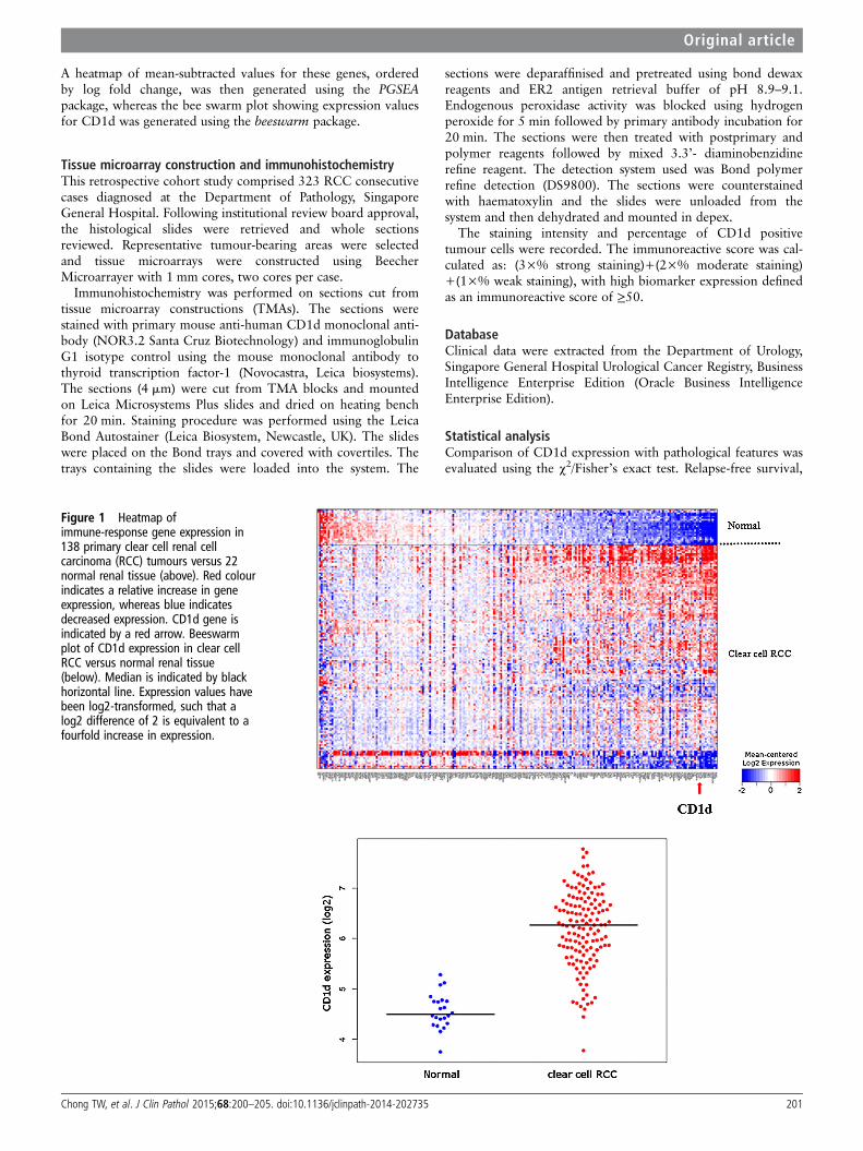
A heatmap of mean-subtracted values for these genes, orderedby log fold change, was then generated using the PGSEApackage, whereas the bee swarm plot showing expression valuesfor CD1d was generated using the beeswarm package.
Tissue microarray construction and immunohistochemistryThis retrospective cohort study comprised 323 RCC consecutivecases diagnosed at the Department of Pathology, SingaporeGeneral Hospital. Following institutional review board approval,the histological slides were retrieved and whole sectionsreviewed. Representative tumour-bearing areas were selectedand tissue microarrays were constructed using BeecherMicroarrayer with 1 mm cores, two cores per case.
Immunohistochemistry was performed on sections cut fromtissue microarray constructions (TMAs). The sections werestained with primary mouse anti-human CD1d monoclonal anti-body (NOR3.2 Santa Cruz Biotechnology) and immunoglobulinG1 isotype control using the mouse monoclonal antibody tothyroid transcription factor-1 (Novocastra, Leica biosystems).The sections (4 mm) were cut from TMA blocks and mountedon Leica Microsystems Plus slides and dried on heating benchfor 20 min. Staining procedure was performed using the LeicaBond Autostainer (Leica Biosystem, Newcastle, UK). The slideswere placed on the Bond trays and covered with covertiles. Thetrays containing the slides were loaded into the system. The
sections were deparaffinised and pretreated using bond dewaxreagents and ER2 antigen retrieval buffer of pH 8.9–9.1.Endogenous peroxidase activity was blocked using hydrogenperoxide for 5 min followed by primary antibody incubation for20 min. The sections were then treated with postprimary andpolymer reagents followed by mixed 3.3’- diaminobenzidinerefine reagent. The detection system used was Bond polymerrefine detection (DS9800). The sections were counterstainedwith haematoxylin and the slides were unloaded from thesystem and then dehydrated and mounted in depex.
The staining intensity and percentage of CD1d positivetumour cells were recorded. The immunoreactive score was cal-culated as: (3×% strong staining)+(2×% moderate staining)+(1×% weak staining), with high biomarker expression definedas an immunoreactive score of ≥50.
DatabaseClinical data were extracted from the Department of Urology,Singapore General Hospital Urological Cancer Registry, BusinessIntelligence Enterprise Edition (Oracle Business IntelligenceEnterprise Edition).
Statistical analysisComparison of CD1d expression with pathological features wasevaluated using the χ2/Fisher’s exact test. Relapse-free survival,
Figure 1 Heatmap ofimmune-response gene expression in138 primary clear cell renal cellcarcinoma (RCC) tumours versus 22normal renal tissue (above). Red colourindicates a relative increase in geneexpression, whereas blue indicatesdecreased expression. CD1d gene isindicated by a red arrow. Beeswarmplot of CD1d expression in clear cellRCC versus normal renal tissue(below). Median is indicated by blackhorizontal line. Expression values havebeen log2-transformed, such that alog2 difference of 2 is equivalent to afourfold increase in expression.
Chong TW, et al. J Clin Pathol 2015;68:200–205. doi:10.1136/jclinpath-2014-202735 201
Original article

cancer-specific survival and overall survival were calculated forboth CD1d high and low expressors. Survival outcomes wereestimated with the Kaplan–Meier method and compared usingCox regression analysis. OR was calculated with 95% CIs. Datawere further adjusted for the Mayo clinic SSIGN score (stage,size, grade and necrosis). Statistical significance was taken atp<0.05. Software for statistical analyses was SPSS V.17.0.
RESULTSWe examined the expression of CD1d in a gene expressionmicroarray of 138 clear cell RCC compared with 22 normalrenal tissue from a publicly available database, looking particu-larly at genes associated with immune responses. Figure 1 showsthe heatmap generated with the CD1d gene highlighted by thered arrow. Gene expression microarray studies showed signifi-cant upregulation of CD1d in clear cell RCC compared withnormal tissue. The upregulation was highly significant with afalse-discovery rate of 1.47E−16.
We then examined the expression of CD1d in various tissuesincluding normal and malignant renal histology. Tonsillar tissuewas used as positive control. Figure 2A shows positive stain ofCD1d on lymphocytes and APC in the mantle zones withnotable absence within the germinal centres of the tonsil.Expression of CD1d is seen in tubules within normal renal
epithelium in figure 2B. Figure 2C, D shows the expression ofCD1d in low-grade and high-grade clear cell RCC, respectively.There was minimal CD1d stain in low-grade disease, with exten-sive staining seen in high-grade disease.
We looked at RCC tissue from patients for whom a nephrec-tomy was performed with curative intent (ie, excluding pallia-tive or cytoreductive nephrectomy). A tissue array of 323resected RCC tumours was constructed and stained for CD1d.Exclusion of 52 cases was made based on incompleteness ofclinical data (n=17), presence of metastases at the time ofsurgery (n=31) or unclear pathological grading (n=4). Finalanalysis was performed on 271 cases. Two hundred andforty-eight cases (91.5%) were of clear cell pathology, with themajority of remainder being of the papillary type. Specimenswere segregated according to the immunoreactivity score of low(<50) or high (≥50) expression of CD1d and correlated withclinicopathological features.
Table 1 shows that CD1d high expressors were significantlyassociated with tumour stage (American Joint Committee onCancer 2002 classification), tumour grade, presence of sarcoma-toid change (p=0.04) and lymphovascular invasion (p=0.048),the latter two features well known to be associated with aggres-sive disease. There was CD1d expression found in both clearcell and papillary pathologies, but the numbers were too few to
Figure 2 Immunohistochemistry ofCD1d (stained brown) on left column,with isotype controls on correspondingtissue on right column. (A) Tonsil withmantle zone and germinal centrelabelled accordingly. (B) Normal renaltissue. Isolated tubular cells positivefor CD1d labelled with black arrow. (C)Low-grade clear cell renal cellcarcinoma (RCC). (D) High-grade clearcell RCC. All magnifications at ×20.
202 Chong TW, et al. J Clin Pathol 2015;68:200–205. doi:10.1136/jclinpath-2014-202735
Original article

discern any significant differences in CD1d expression betweenhistological types.
At last follow-up, 41 of the 271 patients studied had died,including 22 patients who died from RCC at a median of2.5 years after surgery (range 0.6–9.7 years). Among the 230patients remaining (85%), the median duration of follow-upwas 4 years (range 0–12.9 years). Figure 3A shows the relapse-free survival, figure 3B shows the cancer-specific survival andfigure 3C the overall survival between CD1d high and lowexpressors. There was a significant difference in survivalbetween low and high expressors (OR 1.997 (1.041 to 3.830),p=0.037; OR 2.819 (1.038 to 7.654), p=0.042; OR 2.849(1.355 to 5.990) p=0.006, respectively). Because tumour stageand grade were strongly associated with CD1d expression, we
Table 1 Clinical and pathological features by CD1dimmunoreactivity score
Feature
Immunoreactivity score, n (%)
p ValueLow (<50), n=126 High (≥50), n=145
Age at surgery (years)<65 90 (71.4) 98 (67.6) 0.512≥65 36 (28.6) 47 (32.4)
Sex
Female 47 (37.3) 49 (33.8) 0.611Male 79 (62.7) 96 (66.2)
Symptoms at presentationYes 66 (52.4) 63 (43.4) 0.146No 60 (47.6) 82 (56.6)
2002 primary tumour classificationpT1a 50 (39.7) 40 (27.6) 0.011pT1b 34 (27.0) 27 (18.6)pT2 10 (7.9) 23 (15.9)pT3a 18 (14.3) 37 (25.5)pT3b 13 (10.3) 15 (10.3)pT3c 0 (0.0) 3 (2.1)pT4 1 (0.8) 0 (0.0)
Regional lymph node involvementpNX/pN0 124 (98.4) 141 (97.2) 0.689pN1/pN2 2 (1.6) 4 (2.8)
2002 TNM stage groupingsI 83 (65.9) 67 (46.2) 0.011II 10 (7.9) 23 (15.9)III 31 (24.6) 52 (35.9)IV 2 (1.6) 3 (2.1)
Nuclear grade1 28 (22.2) 11 (7.6) 0.0002 83 (65.9) 75 (51.7)3 11 (8.7) 42 (29.0)4 4 (3.2) 17 (11.7)
Coagulative tumour necrosisYes 24 (19.0) 41 (28.3) 0.880No 102 (81.0) 104 (71.7)
Sarcomatoid differentiationYes 1 (0.8) 8 (5.5) 0.040No 125 (99.2) 137 (94.5)
Lymphovascular invasion
Yes 8 (6.3) 20 (13.8) 0.048No 118 (93.7) 125 (86.2)
The staining intensity and percentage of positive tumour cells were recorded. Theimmunoreactive score was calculated as described in the Materials and methodssection.TNM, tumour, node and metastases.
Figure 3 (A) Relapse-free survival of CD1d low and high expressorsfrom 271 patients with renal cell carcinoma (RCC). Unadjusted OR was1.997 (1.041 to 3.830), p=0.037. After adjustment for the Mayo ClinicSSIGN (stage, size, grade and necrosis) score, OR was 1.191 (0.602 to2.356), p=0.616. (B) Cancer-specific survival of CD1d low and highexpressors from 271 patients with RCC. Unadjusted OR was 2.819(1.038 to 7.654), p=0.042. After adjustment for the Mayo Clinic SSIGNscore, OR was 1.735 (0.613 to 4.911), p=0.299. (C) Overall survival ofCD1d low and high expressors from 271 patients with RCC. UnadjustedOR was 2.849 (1.355 to 5.990), p=0.006. After adjustment for theMayo Clinic SSIGN score, OR was 1.982 (0.921 to 4.265), p=0.08.
Chong TW, et al. J Clin Pathol 2015;68:200–205. doi:10.1136/jclinpath-2014-202735 203
Original article

sought to determine whether CD1d expression was an inde-pendent predictor of survival. After further stratification usingthe SSIGN score for RCC tumour SSIGN (Mayo clinic), thedata showed the same trend but did not attain statistical signifi-cance (OR 1.191 (0.602 to 2.356), p=0.616; OR 1.735 (0.613to 4.911), p=0.299; OR 1.982 (0.921 to 4.265), p=0.08,respectively).
DISCUSSIONUnlike classical MHC molecules, CD1d does not map to thehuman leucocyte antigen locus on chromosome 6 and displaysrestricted tissue distribution. Canchis et al18 examined the tissuedistribution of CD1d in a variety of tissues. Apart from lymph-atic tissue of the thymus and extrathymic cells of haematopoi-etic lineage, CD1d expression was described in thegastrointestinal tract, breast, skin and kidneys, with preferentialexpression in epithelial cells, vascular smooth muscle and someparenchymal cells in the liver and pancreas. In normal kidneytissue, expression was confined to the tubules and muscularcomponents of the blood vessels, similar to our findings. Themajority of non-haematological solid tumours are CD1d nega-tive, and no study to our knowledge has studied CD1d expres-sion in RCC. The presence of CD1d has been correlated withan increased antitumour response. CD1d-positive gliomas aresusceptible to ex vivo-activated NKT cell killing.19 CD1d wasshown to be stage specific in myelomonocytic leukaemias andcould provide a target for NKT cell-mediated immunotherapy.14
Our data suggest the contrary, in that the expression of CD1d inRCC was associated with a more aggressive phenotype, withhigher relapse rates, poorer cancer-specific survival and overallsurvival. However, this was not an independent predictor ofoutcome when stratified by stage and grade.
CD1d can present endogenous and exogenous lipids to NKTcells, a heterogeneous group of readily activated immune cellsthat have the ability to skew the subsequent immune responsesvia massive cytokine release (Th1-like or Th2-like). NKT cellsalso interact with DCs, Tregs20 and MDSC.21 DC-based vac-cines have been studied in phase I trials in RCC.22 Tregs arewell-described inhibitors of immunity towards self-antigens andplay a controversial inhibitory role in RCC immunity, with somestudies correlating the presence of peripheral Tregs in patientswith RCC with worse outcome,23 24 and others finding no rela-tion with intratumoural Tregs.25 MDSCs have been associatedwith suppression of tumour immunity in RCC,26 and theremoval of these suppressive cells has been proposed to be amechanism of anti-tumour response by targeted therapies suchas Sunitinib, in current use for metastatic RCC.27
Diverse NKT cells interacting with CD1d and endogenouslipid ligands expressed on RCC tumour cells could explain theadverse outcome seen in our data. We propose that the higherexpression of CD1d on more aggressive RCC tumours tilts thebalance towards immune suppression via a combination of sup-pressive cytokines (Th2-type) and immune cells (Tregs, MDSC)in the tumour micro-environment. We also hypothesise a thera-peutic window of opportunity in harnessing the potent abilityof iNKT cells to reverse immune suppression and augment anti-tumour immunity by judicious use of exogenous activating lipidligands. We are currently exploring CD1d-expressing RCCpulsed ex vivo with αGalCer analogues as autologous tumourvaccines.
Our study was limited by its retrospective nature and com-prised mainly RCC of the clear cell variety (91.5%). We areextending our efforts to non-clear cell types of RCC.
In conclusion, our data suggest that the expression of CD1din RCC is associated with an aggressive tumour phenotype,higher relapse rates, poorer cancer-specific survival and overallsurvival.
Take home messages
▸ CD1d expression is increased in renal cell carcinoma.▸ CD1d expression is associated with more aggressive disease
and poorer clinical outcomes in patients with renal cellcarcinoma.
▸ The CD1d-natural killer T cell axis may be potentiallyexploited to modulate tumour immunity.
Acknowledgements The authors would like to thank Dr Lucia Mori (SingaporeImmunology Network, A*STAR, Singapore) for her expert advice in the preparationof the manuscript.
Contributors All authors are aware of and have agreed to this submission andhave individually contributed to the work as described. Collection of samples:FYG and DAAT. Performed experiments: FYG, MYS and DAAT. Clinical data andstatistical analyses: HHH and WKL. Writing of paper: TWC and FYG. Experimentaldesign: TWC, BTT and PHT. Overall guarantor: TWC.
Funding This work was not specifically funded, but was carried out within thefacilities of the Singapore General Hospital and National Cancer Centre, Singaporewith Institutional Research Board approval.
Competing interests None.
Ethics approval Institutional Review Board, Singhealth.
Provenance and peer review Not commissioned; internally peer reviewed.
Open Access This is an Open Access article distributed in accordance with theCreative Commons Attribution Non Commercial (CC BY-NC 4.0) license, whichpermits others to distribute, remix, adapt, build upon this work non-commercially,and license their derivative works on different terms, provided the original work isproperly cited and the use is non-commercial. See: http://creativecommons.org/licenses/by-nc/4.0/
REFERENCES1 Snow RM, Schellhammer PF. Spontaneous regression of metastatic renal cell
carcinoma. Urology 1982;20:177–81.2 Rosenberg SA. Interleukin 2 for patients with renal cancer. Nat Clin Pract Oncol
2007;4:497.3 Thompson RH, Kuntz SM, Leibovich BC, et al. Tumor B7-H1 is associated with poor
prognosis in renal cell carcinoma patients with long-term follow-up. Cancer Res2006;66:3381–5.
4 Brahmer JR, Tykodi SS, Chow LQM, et al. Safety and activity of anti–PD-L1 antibodyin patients with advanced cancer. New Engl J Med 2012;366:2455–65.
5 Krambeck AE, Thompson RH, Dong H, et al. B7-H4 expression in renal cellcarcinoma and tumor vasculature: associations with cancer progression and survival.Proc Natl Acad Sci USA 2006;103:10391–6.
6 Crispen PL, Sheinin Y, Roth TJ, et al. Tumor cell and tumor vasculature expressionof B7-H3 predict survival in clear cell renal cell carcinoma. Clin Cancer Res2008;14:5150–7.
7 Saito T, Yamagata T, Takahashi T, et al. ICSAT overexpression is not sufficient tocause adult T-cell leukemia or multiple myeloma. Biochem Biophys Res Commun1999;260:329–31.
8 Das T, Sa G, Hilston C, et al. GM1 and tumor necrosis factor-alpha, overexpressedin renal cell carcinoma, synergize to induce T-cell apoptosis. Cancer Res2008;68:2014–23.
9 Biswas S, Biswas K, Richmond A, et al. Elevated levels of select gangliosides in Tcells from renal cell carcinoma patients is associated with T cell dysfunction.J Immunol 2009;183:5050–8.
10 Sa G, Das T, Moon C, et al. GD3, an overexpressed tumor-derived ganglioside,mediates the apoptosis of activated but not resting T cells. Cancer Res2009;69:3095–104.
11 Kawasaki Y, Ito A, Withers DA, et al. Ganglioside DSGb5, preferred ligand forSiglec-7, inhibits NK cell cytotoxicity against renal cell carcinoma cells. Glycobiology2010;20:1373–9.
204 Chong TW, et al. J Clin Pathol 2015;68:200–205. doi:10.1136/jclinpath-2014-202735
Original article
12 Kawano T, Cui J, Koezuka Y, et al. CD1d-restricted and TCR-mediated activation ofvalpha14 NKT cells by glycosylceramides. Science 1997;278:1626–9.
13 Fais F, Tenca C, Cimino G, et al. CD1d expression on B-precursor acutelymphoblastic leukemia subsets with poor prognosis. Leukemia 2005;19:551–6.
14 Metelitsa LS, Weinberg KI, Emanuel PD, et al. Expression of CD1d bymyelomonocytic leukemias provides a target for cytotoxic NKT cells. Leukemia2003;17:1068–77.
15 Xu C, de Vries R, Visser L, et al. Expression of CD1d and presence of invariant NKTcells in classical Hodgkin lymphoma. Am J Hematol 2010;85:539–41.
16 Fais F, Morabito F, Stelitano C, et al. CD1d is expressed on B-chronic lymphocyticleukemia cells and mediates alpha-galactosylceramide presentation to natural killer Tlymphocytes. Int J Cancer 2004;109:402–11.
17 Webb TJ, Li X, Giuntoli RL II, et al. Molecular identification of GD3 as a suppressorof the innate immune response in ovarian cancer. Cancer Res 2012;72:3744–52.
18 Canchis PW, Bhan AK, Landau SB, et al. Tissue distribution of the non-polymorphic majorhistocompatibility complex class I-like molecule, CD1d. Immunology 1993;80:561–5.
19 Dhodapkar KM, Cirignano B, Chamian F, et al. Invariant natural killer T cells arepreserved in patients with glioma and exhibit antitumor lytic activity followingdendritic cell-mediated expansion. Int J Cancer 2004;109:893–9.
20 Terabe M, Berzofsky JA. The immunoregulatory role of type I and type II NKT cellsin cancer and other diseases. Cancer Immunol Immunother 2014;63:199–213.
21 Mussai F, De Santo C, Cerundolo V. Interaction between invariant NKT cells andmyeloid-derived suppressor cells in cancer patients: evidence and therapeuticopportunities. J Immunother 2012;35:449–59.
22 Berntsen A, Geertsen PF, Svane IM. Therapeutic dendritic cell vaccination ofpatients with renal cell carcinoma. Eur Urol 2006;50:34–43.
23 Polimeno M, Napolitano M, Costantini S, et al. Regulatory T cells, interleukin (IL)-6,IL-8, vascular endothelial growth factor (VEGF), CXCL10, CXCL11, epidermalgrowth factor (EGF) and hepatocyte growth factor (HGF) as surrogate markers ofhost immunity in patients with renal cell carcinoma. BJU Int 2013;112:686–96.
24 Griffiths RW, Elkord E, Gilham DE, et al. Frequency of regulatory T cells in renal cellcarcinoma patients and investigation of correlation with survival. Cancer ImmunolImmunother 2007;56:1743–53.
25 Siddiqui SA, Frigola X, Bonne-Annee S, et al. Tumor-infiltrating Foxp3-CD4+CD25+T cells predict poor survival in renal cell carcinoma. Clin Cancer Res2007;13:2075–81.
26 Ochoa AC, Zea AH, Hernandez C, et al. Arginase, prostaglandins, andmyeloid-derived suppressor cells in renal cell carcinoma. Clin Cancer Res2007;13:721s–26s.
27 Ko JS, Zea AH, Rini BI, et al. Sunitinib mediates reversal of myeloid-derivedsuppressor cell accumulation in renal cell carcinoma patients. Clin Cancer Res2009;15:2148–57.
Chong TW, et al. J Clin Pathol 2015;68:200–205. doi:10.1136/jclinpath-2014-202735 205
Original article
Related Documents