4/5/2014 1 CCRN/PCCN Review Multisystem Prepared by: Erin L. Hallinan MSN, RN, APRN, FNP-BC, CCRN Revised by: Cynthia Bautista, PhD, RN, CNRN, SCRN, CCNS, ACNS-BC Nursing Brains, LLC [email protected] Copyright Nursing Brains, LLC Test Plan CCRN 8% (8 questions) Asphyxia Shock states SIRS Multisystem trauma Toxic exposure PCCN 5% (5 questions) Infectious disease Shock states SIRS Copyright Nursing Brains, LLC Asphyxia (CCRN) Copyright Nursing Brains, LLC Asphyxia Definition Extreme decrease in oxygen concentration Increased carbon dioxide concentration Leads to loss of consciousness or death Traumatic Causes Suicide: Drowning & Hanging Choking: internal FBO or external Compressive/restraint : Healthcare, Police, Children Electrocution Auto-erotic Perinatal Drug overdose Co-sleeping/overlay Physiologic Causes Aspiration: EtOH/Drugs/PNA Angioedema: anaphylaxis Laryngeal Edema Isocapneic Hypoxia Cerebral Hypoxia Disease Processes ◦ End stage COPD, ◦ Pulmonary Fibrosis, ◦ Obstructive Sleep Apnea ◦ Pickwickian

Welcome message from author
This document is posted to help you gain knowledge. Please leave a comment to let me know what you think about it! Share it to your friends and learn new things together.
Transcript

4/5/2014
1
CCRN/PCCN Review
Multisystem
Prepared by:
Erin L. Hallinan MSN, RN, APRN, FNP-BC, CCRN
Revised by:
Cynthia Bautista, PhD, RN, CNRN, SCRN, CCNS, ACNS-BC
Nursing Brains, LLC
[email protected] Copyright Nursing Brains, LLC
Test Plan
CCRN
8% (8 questions)
Asphyxia
Shock states
SIRS
Multisystem
trauma
Toxic exposure
PCCN
5% (5 questions)
Infectious disease
Shock states
SIRS
Copyright Nursing Brains, LLC
Asphyxia
(CCRN)
Copyright Nursing Brains, LLC
Asphyxia Definition
Extreme decrease in oxygen concentration
Increased carbon dioxide concentration
Leads to loss of consciousness or death
Traumatic Causes
Suicide: Drowning & Hanging Choking: internal FBO or external Compressive/restraint : Healthcare,
Police, Children Electrocution Auto-erotic Perinatal Drug overdose Co-sleeping/overlay
Physiologic Causes
Aspiration: EtOH/Drugs/PNA Angioedema: anaphylaxis Laryngeal Edema Isocapneic Hypoxia Cerebral Hypoxia Disease Processes ◦ End stage COPD,
◦ Pulmonary Fibrosis,
◦ Obstructive Sleep Apnea
◦ Pickwickian
4/5/2014
2
Chemical/Toxic Inhalation
Causes
CO2 Propane Helium Argon Nitrogen Other: ◦ Altitude/Vacuum (mining accidents)
◦ Burns
Asphyxia Clinical Presentation
Early Tachypnea
Tachycardia
Normo/Hypertensive
SNS: Fight or Flight
Late Confusion/Lethargy/Unresponsive
Insufficient Breathing
Hypotension
Bradycardia
Coma/Death
Asphyxia Treatment
Identify the Cause
Neutralize the cause if possible
Establish Duration
ABC’s Supportive Care Prevention/Monitoring for indirect injury
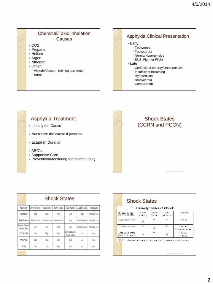
Shock States
(CCRN and PCCN)
Copyright Nursing Brains, LLC
Shock States Shock States
Copyright Nursing Brains, LLC

4/5/2014
3
Shock States
Copyright Nursing Brains, LLC
SIRS
(CCRN and PCCN)
Systemic
Inflammatory
Response
System
Copyright Nursing Brains, LLC
SIRS Definition
Widespread inflammation
Due to a variety of severe clinical insults
◦ Pancreatitis
◦ Ischemia and/ or reperfusion
◦ Multiple trauma and tissue injury
◦ Hemorrhagic shock
◦ Immune-mediated organ injury
Secondary diagnosis of the activation of the complement pathway, cytokine production, and endothelium activating chemical mediators
Manifested by two or more
Hyper/ Hypothermia (Temperature > 38 or < 36) (Temperature >100.4 or <96.8)
Tachycardia: HR> 90 beats/minute
Hyperpnea: RR> 20 breaths/minute or PaC02 < 32 mm Hg or mechanical ventilation
WBC > 12, 000 or < 4,000 OR 10% mature neutrophils
Sepsis Definition
SIRS resulting from infection (bacterial, viral, fungal, or parasitic)
Known or suspected clinical infection with two or more of the SIRS criteria
Bacterial infection of the blood
Can be a secondary complication to injury & illness
Associated with generalized inflammatory response leading to abnormal clotting and bleeding in the presence of infection
Infection
140,000 cases of gram negative sepsis annually
Mortality is still 40-60% with timely therapy
Mortality is 90-95% with delayed or ineffective treatment

4/5/2014
4
Gram-Negative
25% of all cases
E coli Klebsiella Pseudomonas Serratia Haemophilius Enterobacter
Gram-Positive
50% of cases
Staph aureus Staph epi Strep pneumoniae Clostridia Pneumococcus
Viral and Fungi
Viral ◦ HIV ◦ Herpes
Fungi: (10% of cases) ◦ Candida
Most Common Sources
Exogenous
Hospitals & Staff
Endogenous
Skin
GI Tract
Lungs
GU Tract
Copyright Nursing Brains, LLC
Sepsis Signs & Symptoms
◦ Chills
◦ Alteration in temperature
◦ Tachypnea
◦ Change in mental status
◦ Nausea/ Vomiting
◦ Diarrhea
◦ Tachycardia
◦ Low Blood Pressure
◦ Altered WBC, Bandemia
◦ Thrombocytopenia
◦ Decreased perfusion: mottling, poor capillary refill
◦ Increased blood sugar
◦ Petechiae/ Purpura
Severe Sepsis Defined
Sepsis with either hypotension or systemic manifestations of hypoperfusion
OR
Sepsis + dysfunction of at least one organ

4/5/2014
5
Signs & Symptoms…Early
Acute alteration in Mental Status
Confusion
Tachypnea
Tachycardia
Oliguria
Warm extremities with bounding pulses
Signs & Symptoms…Late
Lactic acidosis > 4
Oliguria <1/2ml/kg/hr, Anuria, Elevated Creatinine
Acute alterations in Mental Status/ Confusion/ Psychosis
Chills
Tachypnea: PaO2 < 70 mmHg, SaO2 < 90%, PaO2/FiO2 <300
Signs & Symptoms…Late (con’t)
Tachycardia: SPB < 90 mmHg, decreased CVP & PAOP
Altered WBCs, elevated immature neutrophils, decreased platelet count, increased PT/PTT, increased D-Dimer, decreased protein C, increased FDP/FSP
Increased LFTs, jaundice, hypoglycemia, decreased albumin
Cool extremities, mottling, poor capillary refill, petechiae
Septic Shock Defined
Sepsis induced state Hypotension despite adequate fluid
resuscitation With physiologic evidence of abnormal
perfusion may include, but are not limited to….. ◦ Lactic acidosis, oliguria, mental status
changes, hypotension and reduced perfusion to vital organs
Septic Shock Treatment Early goal directed therapy in first 6 hours
Early resuscitation
◦ Decreases mortality by maximizing preload, afterload, & contractility to balance O2 delivery with demand
Follow sepsis pathways and protocols
All treatments should include basic nursing care, pain management, nutrition, and emotional support of the patient and family
Septic Shock
Initial Resuscitation
Begin resuscitation immediately in patients with hypotension or elevated serum lactate >4 mmol/L
Do not delay ICU admission…
Resuscitation goals:
◦ CVP: 8–12 mmHg
◦ Mean arterial pressure: 65 mmHg
◦ Urine output 0.5 mL/kg/hr
◦ Central venous (superior vena cava) oxygen saturation 70% or mixed venous 65%
If venous oxygen saturation target is not achieved
◦ Consider further fluid
◦ Transfuse packed red blood cells if required to hematocrit of >30% and/or
◦ Start Dobutamine infusion, maximum 20 mg/kg/min

4/5/2014
6
Sepsis Diagnosis
Obtain appropriate cultures before starting antibiotics (provided this does not significantly delay antimicrobial administration)
Obtain two or more BCs
One or more BCs should be percutaneous
One BC from each vascular access device in place >48 hrs
Culture other sites as clinically indicated
Perform imaging studies promptly to confirm and sample any source of infection (if safe to do so)
Antibiotic Therapy
Begin IV antibiotics as early as possible within the first hour of recognizing severe sepsis and septic shock
Broad-spectrum: one or more agents active against likely bacterial/fungal pathogens and with good penetration into presumed source
Antibiotic Therapy (con’t)
Reassess antimicrobial regimen daily to optimize efficacy, prevent resistance, avoid toxicity, and minimize costs
◦ Consider combination therapy in Pseudomonas infections
◦ Consider combination empiric therapy in neutropenic patients
◦ Combination therapy < 3–5 days and de-escalation following susceptibilities
◦ Duration of therapy typically limited to 7–10 days; longer if response is slow or there are undrainable foci of infection or immunologic deficiencies
Stop antimicrobial therapy if cause is found to be noninfectious
Source Identification & Control
Establish site of infection within first 6 hours
Evaluate for focus of infection (e.g. abscess drainage, tissue debridement)
Implement measures as soon as possible following successful initial resuscitation (exception: infected pancreatic necrosis, where surgical intervention is best delayed)
Choose measure with maximum efficacy and minimal physiologic upset
Remove intravascular access devices if potentially infected
Fluid Therapy
Fluid-resuscitate using crystalloids or colloids
Target CVP of 8 mmHg ◦ 12 mm Hg if mechanically ventilated
Use a fluid challenge technique ◦ Give fluid challenges of 1000 mL of crystalloids or
300–500 mL of colloids over 30 minutes
More rapid and larger volumes may be required
Rate of fluid administration should be reduced if cardiac filling pressures increase
Vasopressors
Maintain MAP >65 mmHg
Initial choice - Norepinephrine and Dopamine centrally administered
◦ Epinephrine, Phenylephrine, or Vasopressin should not be administered as initial choice
◦ Vasopressin 0.03 units/min may be subsequently added to norepinephrine
◦ Use Epinephrine as the first alternative agent when blood pressure is poorly responsive to Norepinephrine or Dopamine

4/5/2014
7
Vasopressors (con’t)
Do not use low-dose Dopamine for renal protection
In patients requiring vasopressors, insert an arterial catheter as soon as practical
Inotropic therapy
◦ Use Dobutamine in patients with myocardial dysfunction as supported by elevated cardiac filling pressures and low cardiac output
◦ Do not increase cardiac index to predetermined supranormal levels
Steroids
Consider IV hydrocortisone when hypotension responds poorly to adequate fluid resuscitation and vasopressors
◦ ACTH stimulation test is not recommended
◦ Hydrocortisone is preferred to dexamethasone
◦ Fludrocortisone (50 mcg orally once a day) may be included if an alternative to hydrocortisone is being used that lacks significant mineralocorticoid activity
◦ Fludrocortisone if optional if hydrocortisone is used
◦ Steroid therapy may be weaned once vasopressors are no longer required
Hydrocortisone dose should be <300 mg/day IV
Do not use corticosteroids to treat sepsis in the absence of shock unless the patient’s endocrine or corticosteroid history warrants it
Recombinant
Human Activated Protein C
Consider Xigris if APEX present: ◦ Antibiotic ◦ vasoPressor…consider ◦ Evaluating for… ◦ Xigris
Adult patients with severe sepsis and low risk
of death (typically one organ failure) should not receive rhAPC
Must monitor patient for BLEEDING
Recombinant
Human Activated Protein C
Consider if there are no contraindications:
◦ Active internal bleeding
◦ Recent (within 3 months) hemorrhagic stroke
◦ Recent (within 2 months) intracranial/intraspinal surgery, or severe head trauma
◦ Trauma with risk of life-threatening bleeding
◦ Presence of an epidural catheter
◦ Intracranial neoplasm/mass lesion/evidence of cerebral herniation
Blood Product Administration
Give RBCs when hemoglobin <7.0 g/dL (<70 g/L) to target a hemoglobin of 7.0–9.0 g/dL
A higher hemoglobin level may be required in special circumstances (e.g., myocardial ischemia, severe hypoxemia, acute hemorrhage, cyanotic heart disease, or lactic acidosis)
◦ Do not use erythropoietin to treat sepsis-related anemia
◦ Do not use FFP to correct clotting abnormalities unless there is bleeding or planned invasive procedures
Blood Product Administration
Do not use antithrombin therapy
◦ Administer platelets when counts are <5,000/mm3 regardless of bleeding
◦ Counts are 5,000–30,000/mm3 and there is significant bleeding risk
◦ Higher platelet counts (>50,000/mm3 ) are required for surgery or invasive procedures

4/5/2014
8
Mechanical Ventilation
Target a tidal volume of 6 mL/kg (predicted) body weight in patients with ALI/ARDS
Target an initial upper limit plateau pressure <30 cm H2O. Consider chest wall compliance when assessing plateau pressure
Allow PaCO2 to increase above normal, if needed, to minimize plateau pressures and tidal volumes
Mechanical Ventilation (con’t) Set PEEP to avoid extensive lung collapse at
end-expiration
◦ Use prone position for ARDS patients requiring potentially injurious levels of FIO2 or plateau pressure, provided they are not put at risk from positional changes
Maintain in a semirecumbent position (head of the bed raised to 45°) unless contraindicated
Noninvasive ventilation may be considered in the minority of ALI/ARDS patients with mild to moderate hypoxemic respiratory failure
Mechanical Ventilation(con’t)
Use a weaning protocol & SBT regularly
SBT options include a low level of pressure support with continuous positive airway pressure 5 cm H2O or a T piece
Before the SBT, patients should be:
◦ Arousable
◦ Hemodynamically stable
◦ No new potentially serious conditions
◦ Low ventilatory & end-expiratory pressure requirement
◦ FIO2 levels that can be safely delivered with a face mask or nasal cannula
Mechanical Ventilation (con’t)
Do not use a pulmonary artery catheter for the routine monitoring of patients with ALI/ARDS
Use a conservative fluid strategy for patients with established ALI who do not have evidence of tissue hypoperfusion
Sedation, Analgesia
Neuromuscular Blockade
Use sedation protocols with a sedation goal
Provide daily interruption/lightening to produce awakening
Re-titrate if necessary
Avoid neuromuscular blockers
Monitor depth of block with train-of-four when using continuous infusions
Glucose Control Use IV insulin to control hyperglycemia Keep blood glucose <150 mg/dL
(8.3 mmol/L) using a validated protocol for insulin dose adjustment
Provide glucose calorie source
Monitor blood glucose values every 1–2 hrs (4 hrs when stable) when receiving IV insulin
POC T may overestimate arterial blood or plasma glucose values

4/5/2014
9
Other Considerations
Renal replacement
◦ Intermittent hemodialysis and CVVH are considered equivalent
◦ CVVH offers easier management in hemodynamically unstable patients
Bicarbonate therapy
◦ Do not use bicarbonate therapy for the purpose of improving hemodynamics or reducing vasopressor requirements when treating hypoperfusion induced lactic acidemia with pH >7.15
Deep Vein Thrombosis
Prophylaxis
Use either low-dose UFH or LMWH, unless contraindicated
Use a mechanical prophylactic device, such as compression stockings or an intermittent compression device, when heparin is contraindicated
Use a combination of pharmacologic and mechanical therapy for patients who are at very high risk for deep vein thrombosis
In patients at very high risk, LMWH should be used rather than UFH
Other Considerations
Stress ulcer prophylaxis
◦ Provide stress ulcer prophylaxis using H2 blocker or proton pump inhibitor
◦ Benefits of prevention of upper gastrointestinal bleed must be weighed against the potential for development of ventilator-acquired pneumonia
Consideration for limitation of support
◦ Discuss advance care planning with patients and families
◦ Describe likely outcomes and set realistic expectations
Multisystem Organ Dysfunction
Syndrome (MODS): Defined
Presence of altered organ function in an acutely ill patient such that homeostasis cannot be maintained without intervention
Primary MODS is the direct result of a well-defined insult in which organ dysfunction occurs early and can be directly attributable to the insult itself
Secondary MODS develops as a consequence of a host response and is identified within the context of SIRS
Clinical factors associated with progression to MODS:
◦ Inadequate initial resuscitation
◦ Persistent infection
◦ Systemic inflammation in the absence of infection
Organ Failure…
Central Nervous System
Lethargy Fever Hepatic encephalopathy GCS <15 or decreased by 1 point “Brain Failure” (confusion, agitation,
psychosis)
Organ Failure…Cardiovascular
Hyperdynamic i PAOP/ Wedge i SVR/PVR i CVP/RAP i LVSWI hO2Consumption
/delivery h C.O./C.I. Tachycardia Hypotension
Hypodynamic
h SVR/PVR h CVP/RAP h LVSWI i O2
Consumption /delivery
i C.O./C.I.
Copyright Nursing Brains, LLC

4/5/2014
10
Organ Failure…Pulmonary
ARDS/ ALI
Bilateral infiltrates on x-ray
Wedge <18 mmHg
Unexplained hypoxemia… PaO2/FiO2 <175-250 mmHg
ABG deterioration from baseline
Tachypnea
Dyspnea
Pulmonary HTN
Organ Failure…Gastrointestinal
Paralytic ileus
Intolerance of GI feeding for > 5 days
GIB
Stress ulcers
Decrease bowel sounds
Abdominal distension
Organ Failure…Renal
◦ Oliguria <0.5ml/kg/hr
◦ Serum creatinine up to 2-3 mg/dL (normal renal function)
◦ Urine Na <40 mmol/L (normal renal function)
◦ Serum creatinine up by 2.0 mg/dL (chronic renal failure patients)
Organ Failure…Hepatobiliary
LFTs elevated to twice baseline
Bilirubin above 2.0 mg/dL
PT twice normal time
Decreased albumin
Jaundice
Increased ammonia
Organ Failure…Coagulation
Hematologic
Decrease in platelets by 25% Thrombocytopenia Bleeding Elevated PT/PTT to 125% of normal DIC:
<Platelets: <100,000/mm2
>PTT: >60-90 seconds
>PT: >15 seconds
< Fibrinogen: <200mg/100ml
>FDP/FSP elevated: >10mg/ml but<100
>D-Dimer elevated: <2mg/L abn>2mg/L
<Antithrombin III (nml 80-120%, abn <70%)
Multisystem Trauma
(CCRN)
Copyright Nursing Brains, LLC

4/5/2014
11
Extent of Injury
Determined by
◦ The type of energy applied
◦ How quickly the energy is applied
◦ To what part of the body the energy is applied
◦ Mechanical energy can cause damage to:
Epithelial tissue: Skin, trachea, mucous membranes
Connective tissue: Cartilage, bone, joint structures
Muscle tissue: Cardiac, skeletal, blood vessels
Nerve tissue: Neurons and supporting cells
Mass vs. Velocity
Double the Mass= Double the Energy
Double the Velocity= Quadruple the Energy
Descriptors of Injury
Blunt An injury produced by the wounding
forces of compression and change of speed (shearing), which may disrupt tissue
Direct compression or pressure on a structure
The most common type of force MVC’s and Falls
Descriptors of Injury
Penetrating An injury that causes lacerations, cuts,
puncture, piercing, and amputation…any interruption in skin and integrity (avulsion/degloving)
Firearms, knives, and other items can cause penetration injuries
Most important to leave penetration device in place until surgical removal!!
Descriptors of Injury
Acceleration
An injury event that occurs when the victim is slow moving or stationary and in acted upon by increasing amounts of energy and velocity.
Example: A slow moving car struck
from behind by a fast moving car.
Descriptors of Injury
Deceleration
Force that stops or decreases the velocity of a moving victim.
Motor vehicle collision: ◦ The vehicle strikes an object
◦ The occupant collides with the inside of the car
◦ The internal organs collide inside the body
◦ Tensile stress (tissue cells are separated i.e. splenic capsule)
◦ Compressive stress (tissue and structures pressed together i.e. comminuted bone fracture)
◦ Shearing stress (stress results from a tangential force i.e. ligamentum arteriosum & ligament of Treitz)
Descriptors of Injury
Direct: Initial injury/point of impact, injury resulting from a dynamic energy load
◦ Examples: Brain contusion or concussion, Long bone fracture, Shattered pelvis, Asphyxia from drowning, Electrical burn
Indirect: secondary injury that is a result of direct injury
◦ Examples: cerebral edema, ischemia & bleeding, Fat Emboli, Retroperitoneal bleed, ARDS , Renal failure from Myoglobinuria, Sepsis…

4/5/2014
12
Class 1 Hemorrhage
15% loss (up to 750ml)
Pulse: less than 100
BP: normal
Pulse Pressure: Normal or increased
LOC: Slightly anxious
RR: 14-20
U.O: >30ml/hr
Treatment: Crystalloids
Class 2 Hemorrhage
15-30% loss (750ml- 1500ml)
Pulse: >100
BP: normal
Pulse Pressure: decreased
LOC: Mildly anxious
RR: 20-30
U.O: 20-30 ml/hr
Treatment: Crystalloids & Blood Products
Class 3 Hemorrhage
30-40% loss (1500ml- 2000ml)
Pulse: >120
BP: decreased
Pulse Pressure: decreased
LOC: Anxious, Confused
RR: 30-40
U.O: 5-15 ml/hr
Treatment: Blood Products
Class 4 Hemorrhage
>40% loss (> 2000ml)
Pulse: >140
BP: decreased
Pulse Pressure: decreased
LOC: Confused, Lethargic
RR: >35
U.O: Minimal
Treatment: Blood Products, Surgery
Rules of Resuscitation Two large bore catheters or central line
Warm fluids for massive resuscitation to maintain body temp…Level I Rapid Infuser
Monitor and expect edema …protect airway, watch for pulmonary edema
Maintain adequate Hgb/Hct
◦ Sat >94%, Hgb >7 g/dl, PaO2> 60 mm Hg
Rules of Resuscitation
For every unit of PRBCs, you anticipate Hct increasing by 3 pts.
Repeat CBC should be done 2-3 hours AFTER last unit transfused
For every four units of PRBCs, give one amp of Ca Gluconate
For every unit of PRBCs, you should anticipate administering FFP for Factor V-VII
Replacement of blood loss/resuscitation: PRBC:Plasma:Platelets/ 1:1:1

4/5/2014
13
Osmolality of Solutions
Isotonic ◦ Expand intravascular compartment
◦ Do Not give LR to pts with hepatic compromise
Hypotonic ◦ Cellular hydration…fluid moves into cells
◦ Do Not give to neuro, burn, trauma, malnourished or liver diseased pts.
Hypertonic ◦ Cellular “shrinking” fluid move into
intravascular space
◦ Watch for CHF, fluid overload, fluid shifts
Copyright Nursing Brains, LLC
Endpoints…
When Enough is Enough
Hemodynamic Parameters WNL MAP >65 U.O > 0.5ml/kg/hr Lactate level WNL <2 Cleared Base Deficit +/- 2 pH 7.35-7.45 Compensated SvO2 65-75% pAO2 35-45 on mixed venous gas
Toxic Exposure
(CCRN)
Copyright Nursing Brains, LLC
Burns & Chemical Exposure
Severity of chemical exposure injuries depends on
◦ Strength or concentration of chemical
◦ Length of contact with skin
◦ Quantity of chemical
◦ Extent of tissue penetration
◦ Mode of action of chemical
Burns & Chemical Exposure
Cellular dehydration, denaturation (discoloring), Oxidation (tissue degeneration), Chemical coagulation of protein, and Protoplasmic poisoning can be caused by:
◦ Desiccants (strong acids): sulfuric, muriatic, and hydrofluoric acid
◦ Alkalis: Lime, ammonia, caustics
◦ Corrosives: Phenol, Lye, white phospohorus
◦ Oxidizing Agents: Chromic Acid, Potassium permanganate
◦ Vesicants: chemical warfare agents
◦ Protoplasmic Poisons: hydrochloric acid, tannic acid, formic acid
Burns & Chemical Exposure
Systemic response to injury = 20% of TBSA
SIRS and release of mediators Release of vasoactive substances Possible bacterial translocation Release of stress hormones
4/5/2014
14
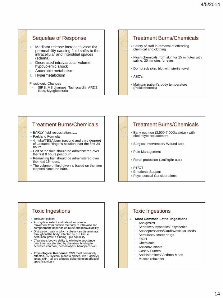
Sequelae of Response
1. Mediator release increases vascular permeability causing fluid shifts to the intracellular and interstitial spaces (edema)
2. Decreased intravascular volume = hypovolemic shock
3. Anaerobic metabolism 4. Hypermetabolism
Physiologic Changes
◦ SIRS, MS changes, Tachycardia, ARDS, Ileus, Myoglobinuria
Treatment Burns/Chemicals
Safety of staff in removal of offending chemical and clothing
Flush chemicals from skin for 15 minutes with saline, 30 minutes for eyes
Do not rub skin, blot with sterile towel
ABC’s
Maintain patient’s body temperature (Poikilothermia)
Treatment Burns/Chemicals
EARLY fluid resuscitation:…..
Parkland Formula
4 ml/kg/TBSA burn (second and third degree) of Lactated Ringer's solution over the first 24 hours.
Half of the fluid should be administered over the first 8 hours post burn
Remaining half should be administered over the next 16 hours.
The volume of fluid given is based on the time elapsed since the burn.
Treatment Burns/Chemicals
Early nutrition (3,500-7,000kcal/day) with electrolyte replacement
Surgical Intervention/ Wound care
Pain Management
Renal protection (1ml/kg/hr u.o.)
PT/OT
Emotional Support
Psychosocial Considerations
Toxic Ingestions
Toxicant: poison
Absorption: extent and rate of substance movement from outside the body to intravascular compartment; depends on route and bioavailability
Distribution: way in which substances disseminate throughout the body, affected by pH, tissue perfusion, protein binding, lipid solubility
Clearance: body’s ability to eliminate substance over time; accelerated by chelation, binding to activated charcoal, hemodialysis, hemoperfusion
Physiological Response: CNS most commonly affected, CV system, blood & spleen, liver, kidneys, lungs, skin…all are affected depending on effect of specific toxicant
Toxic Ingestions
Most Common Lethal Ingestions
◦ Analgesics
◦ Sedatives/ hypnotics/ psychotics
◦ Antidepressants/Cardiovascular Meds
◦ Stimulants/ street drugs
◦ EtOH
◦ Chemicals
◦ Anticonvulsants
◦ Gases/ Fumes
◦ Antihistamines/ Asthma Meds
◦ Muscle relaxants

4/5/2014
15
Signs & Symptoms
of Selected Toxins
Acetaminophen: N/V, low BP, diaphoresis, pallor, hepatotoxicity
Sedatives: Low HR, low Temp, low BP, low RR, HA, Nystagmus, depressed DTR
Beta-Blockers: Bradycardia, heart blocks, RBBB, Low BP, heart failure, Cardiogenic shock, Arrest, low RR, seizures, decreased LOC, Hyper or hypoglycemia
Cocaine: High HR, dysrrhythmias, M.I., hyper/hypo-tension & RR, pallor, cyanosis, Hyperexcitability, high temp, diaphoresis, N/V, Confusion, delirium, seizures, Respiratory arrest, coma
Signs & Symptoms
of Selected Toxins
Ethanol: affective alterations, alcohol odor on breath, hypoglycemia, seizures, metabolic acidosis
Ethylene Glycol: “drunk” with odor of alcohol, N/V, seizures, coma, nystagmus, metabolic acidosis with anion gap, ARF, pulmonary edema, HF
Salicylates: decreased LOC, high temp, high RR (respiratory alkalosis), tinnitus, diaphoresis, thirst, metabolic acidosis with anion gap, low BP, deafness
Toxic Ingestions - Treatment ABC’s
Call Poison Control!!!
100ml of 50g dextrose in water
Thiamine 100mg IV to prevent
Wernicke-Korsakoff Syndrome
Naloxone 2mg IV for narcotics
Provide Antidote…
Toxic Ingestions: Antidotes Agent
1. Acetaminophen
2. Amphetamines/ Arsenic/ Barbituates/ Cocaine/ Ethanol/ Ethylene Glycol (antifreeze)/ Isopropyl alcohol/ Lithium/ PCP/ Salicylates (ASA)/ Theophylline
3. Benzodiazepines
4. Cyanide
Antidote
1. Mucomyst: 140mg/kg then 70mg/kg for 17 doses
2. No Known Antidote: Supportive therapies as outlined on previous slide
3. Flumazenil
4. Sodium Thiosulfate/ Amyl Nitrate/ Hydroxocobalamin
Toxic Ingestions: Antidotes
Agent:
1. Cyclic Antidepressants
2. Isoniazid (INH)
3. Opioids
4. Methemoglobinemia (nitrates, sulfa drugs)
Antidote
1. Sodium Bicarbonate
2. Pyridoxine 1 G for every gram of INH
3. Naloxone 2mg IV/ 30 minute half life, repeat up to 10mg
4. Metheylene Blue 2 mg/kg over 5min IVP, repeat 1mg/kg after 30min.
Toxic Ingestions- Treatment
Do not give routine doses of flumazenil, analeptics, or physostigmine
Noncaustic agents - orogastric lavage
Caustic agents - dilute with milk or water
Emetics
Activated Charcoal: 1G/kg…sometimes given with cathartic like Magnesium citrate

4/5/2014
16
Toxic Ingestions - Treatment
Whole Bowel Irrigation ◦ Polyethylene glycol and electrolytes, 2 L/hr until
clear…good for sustained release meds, paint chips, cocaine, heroin (smuggling)
Hemodialysis/ Hemoperfusion/ Hemofiltration
Surgical intervention for body stuffing and packing (drug mules)
Hemodynamic and physiological support
Salicylate Toxicity
Metabolic acidosis Respiratory alkalosis Urine alkalinization increases
salicylate elimination ◦ IV sodium bicarbonate infusion
Potassium replacement – monitor levels
Hemodialysis for severe cases
Copyright Nursing Brains, LLC
Beta-Blocker Toxicity
Hypotension, bradycardia Hypoglycemia ◦ Decrease glucose formation in liver
◦ Enhance hypoglycemic action of insulin
Give glucagon to increase myocardial contractility, heart rate, and AV conduction – non-beta mechanism
Dose 3-10mg IV bolus Infusion of 2-5mg/hr
Copyright Nursing Brains, LLC
Excited Delirium Syndrome
Extreme hypermetabolic state Related to illicit stimulant use Mehtamphetamine, cocaine, PCP Bizarre, aggressive behavior Paranoid, panic, violence Rhabdomyolysis - consequence 33% experience acute renal failure Serial CPK monitoring to identify
rhabdomyolysis
Copyright Nursing Brains, LLC
Related Documents


