ORIGINAL ARTICLE CCL2 (pM Levels) as a Therapeutic Agent in Inflammatory Bowel Disease Models in Mice N. Maharshak, MD,* , † G. Hart, PhD,* E. Ron, MSc, ‡ E. Zelman, MSc,* A. Sagiv, MSc,* N. Arber, MD, PhD, ‡ E. Brazowski, MD, § R. Margalit,* E. Elinav, MD, PhD, † and I. Shachar, PhD* Background: Chemokines regulate the pathways that restrict homing of specific subsets of immune cells, and thereby fine tune the immune response at specific lymphoid and peripheral tissues. CCL2 is a chemokine that induces migration of monocytes, mem- ory T cells, and dendritic cells. Previously, we demonstrated that pM levels of CCL2 dramatically inhibit migration of T cells. The aim was to test whether subphysiological doses of CCL2 can ameliorate murine colitis and inflammation-induced colorectal cancer. Methods: TNBS (2,4,6 trinitrobenzene sulfonic acid) colitis and dextran sodium sulfate (DSS) colitis were induced in Balb/c and C57BL/6 mice, respectively. Mice were treated daily with intra- peritoneal CCL2 injections. Disease activity was assessed clini- cally, histologically, and by measuring inflammatory cytokine levels. In addition, an inflammatory cancer model was induced by azoxymethane-DSS (AOM-DSS) in Balb/c mice. Mice were treated daily with CCL2 for 11 weeks and then assessed for num- ber of tumors in the colons. Results: Daily administration of CCL2 (60–120 ng) significantly decreased the development of TNBS- and DSS-induced colitis. In a DSS-AOM model, CCL2-treated mice developed significantly fewer tumors (P < 0.005) at 11 weeks. Chronic inflammation in the CCL2-treated mice was significantly less pronounced as com- pared to phosphate-buffered saline-treated mice. Conclusions: Administration of pM levels of CCL2 significantly inhibits migration of T cells in amelioration of TNBS and DSS colitis and inhibits development of colorectal cancer in an AOM- DSS colitis model in mice. Thus, pM levels of CCL2 may be clinically beneficial as an antiinflammatory agent in IBD. (Inflamm Bowel Dis 2010;16:1496–1504) Key Words: CCL2, inflammatory bowel disease, murine colitis models, colorectal cancer I nflammatory bowel disease (IBD) is a generic term for a group of inflammatory disorders of the gastrointestinal tract characterized by intestinal inflammation and mucosal damage. Crohn’s disease (CD) and ulcerative colitis (UC) are the two major forms of IBD. UC primarily affects the mucosal lining of the colon and rectum, whereas CD pri- marily affects all intestinal wall layers and may potentially extend to any part of the gastrointestinal tract. A disregulated activation of the intestinal immune system plays a pivotal role in the pathogenesis of IBD. It has been established that inflammatory mediators such as tumor necrosis factor-a (TNF-a), interferon-c (IFN-c), and interleukin (IL)-12 produced by infiltrating CD4þ T cells and macrophages have a key role in the pathogenesis of disease exacerbation. 1,2 However, the etiologies of both CD and UC still remain largely unclear, and probably result from an aberrant immune response to an environ- mental trigger in a genetically susceptible host. 3 IBDs pose a major therapeutic challenge, as their course is chronic-relapsing with significant damage to the quality of life of patients. None of the available therapies result in complete remission in all patients. Moreover, IBD colitis significantly increases the risk of colorectal cancer (CRC) compared to the general population. 4–6 A better understanding of the pathophysiology of these diseases and the development of new therapeutic options can favorably affect a sizable portion of the population suffering from IBD. None of the existing IBD models constitutes a faith- ful reproduction of the human diseases. Therefore, it is essential to evaluate the effect of any drug or treatment in several animal IBD models. One of the widely used animal models is dextran sodium sulfate (DSS) colitis, induced by DSS administration through drinking water, leading to Received for publication January 6, 2010; Accepted January 11, 2010. From the *Department of Immunology, the Weizmann Institute of Science, Rehovot, Israel, † Department of Gastroenterology and Liver Diseases, Tel Aviv Sourasky Medical Center, affiliated with the Sackler Faculty of Medicine, Tel Aviv University, Tel Aviv, Israel, ‡ Integrated Cancer Prevention Center, Tel-Aviv Sourasky Medical Center, affiliated with the Sackler Faculty of Medicine, Tel Aviv University, Tel Aviv, Israel, § Department of Pathology, Tel Aviv Sourasky Medical Center, affiliated with the Sackler Faculty of Medicine, Pathology Institute, Tel Aviv University, Tel Aviv, Israel. Supported by the Marguerite Stolz Research Fellowship Fund and by the Israel Science Foundation (Morasha), the Horowitz Foundation, and the Kirk Center for Childhood Diseases. Reprints: I. Shachar, PhD, Department of Immunology, Weizmann Institute of Science, Rehovot, Israel 76100 (e-mail: idit.shachar@ weizmann.ac.il) Copyright V C 2010 Crohn’s & Colitis Foundation of America, Inc. DOI 10.1002/ibd.21254 Published online 10 March 2010 in Wiley Online Library (wileyonlinelibrary.com). Inflamm Bowel Dis Volume 16, Number 9, September 2010 1496

Welcome message from author
This document is posted to help you gain knowledge. Please leave a comment to let me know what you think about it! Share it to your friends and learn new things together.
Transcript
ORIGINAL ARTICLE
CCL2 (pM Levels) as a Therapeutic Agent in InflammatoryBowel Disease Models in Mice
N. Maharshak, MD,*,† G. Hart, PhD,* E. Ron, MSc,‡ E. Zelman, MSc,* A. Sagiv, MSc,* N. Arber, MD, PhD,‡
E. Brazowski, MD,§ R. Margalit,* E. Elinav, MD, PhD,† and I. Shachar, PhD*
Background: Chemokines regulate the pathways that restrict
homing of specific subsets of immune cells, and thereby fine tune
the immune response at specific lymphoid and peripheral tissues.
CCL2 is a chemokine that induces migration of monocytes, mem-
ory T cells, and dendritic cells. Previously, we demonstrated that
pM levels of CCL2 dramatically inhibit migration of T cells. The
aim was to test whether subphysiological doses of CCL2 can
ameliorate murine colitis and inflammation-induced colorectal
cancer.
Methods: TNBS (2,4,6 trinitrobenzene sulfonic acid) colitis and
dextran sodium sulfate (DSS) colitis were induced in Balb/c and
C57BL/6 mice, respectively. Mice were treated daily with intra-
peritoneal CCL2 injections. Disease activity was assessed clini-
cally, histologically, and by measuring inflammatory cytokine
levels. In addition, an inflammatory cancer model was induced by
azoxymethane-DSS (AOM-DSS) in Balb/c mice. Mice were
treated daily with CCL2 for 11 weeks and then assessed for num-
ber of tumors in the colons.
Results: Daily administration of CCL2 (60–120 ng) significantly
decreased the development of TNBS- and DSS-induced colitis. In
a DSS-AOM model, CCL2-treated mice developed significantly
fewer tumors (P < 0.005) at 11 weeks. Chronic inflammation in
the CCL2-treated mice was significantly less pronounced as com-
pared to phosphate-buffered saline-treated mice.
Conclusions: Administration of pM levels of CCL2 significantly
inhibits migration of T cells in amelioration of TNBS and DSS
colitis and inhibits development of colorectal cancer in an AOM-
DSS colitis model in mice. Thus, pM levels of CCL2 may be
clinically beneficial as an antiinflammatory agent in IBD.
(Inflamm Bowel Dis 2010;16:1496–1504)
Key Words: CCL2, inflammatory bowel disease, murine colitismodels, colorectal cancer
I nflammatory bowel disease (IBD) is a generic term for a
group of inflammatory disorders of the gastrointestinal
tract characterized by intestinal inflammation and mucosal
damage. Crohn’s disease (CD) and ulcerative colitis (UC)
are the two major forms of IBD. UC primarily affects the
mucosal lining of the colon and rectum, whereas CD pri-
marily affects all intestinal wall layers and may potentially
extend to any part of the gastrointestinal tract.
A disregulated activation of the intestinal immune
system plays a pivotal role in the pathogenesis of IBD. It
has been established that inflammatory mediators such as
tumor necrosis factor-a (TNF-a), interferon-c (IFN-c), andinterleukin (IL)-12 produced by infiltrating CD4þ T cells
and macrophages have a key role in the pathogenesis of
disease exacerbation.1,2 However, the etiologies of both
CD and UC still remain largely unclear, and probably
result from an aberrant immune response to an environ-
mental trigger in a genetically susceptible host.3
IBDs pose a major therapeutic challenge, as their
course is chronic-relapsing with significant damage to the
quality of life of patients. None of the available therapies
result in complete remission in all patients. Moreover, IBD
colitis significantly increases the risk of colorectal cancer
(CRC) compared to the general population.4–6 A better
understanding of the pathophysiology of these diseases and
the development of new therapeutic options can favorably
affect a sizable portion of the population suffering from IBD.
None of the existing IBD models constitutes a faith-
ful reproduction of the human diseases. Therefore, it is
essential to evaluate the effect of any drug or treatment in
several animal IBD models. One of the widely used animal
models is dextran sodium sulfate (DSS) colitis, induced by
DSS administration through drinking water, leading to
Received for publication January 6, 2010; Accepted January 11, 2010.
From the *Department of Immunology, the Weizmann Institute of
Science, Rehovot, Israel, †Department of Gastroenterology and Liver
Diseases, Tel Aviv Sourasky Medical Center, affiliated with the Sackler
Faculty of Medicine, Tel Aviv University, Tel Aviv, Israel, ‡Integrated
Cancer Prevention Center, Tel-Aviv Sourasky Medical Center, affiliated
with the Sackler Faculty of Medicine, Tel Aviv University, Tel Aviv, Israel,§Department of Pathology, Tel Aviv Sourasky Medical Center, affiliated
with the Sackler Faculty of Medicine, Pathology Institute, Tel Aviv
University, Tel Aviv, Israel.
Supported by the Marguerite Stolz Research Fellowship Fund and by
the Israel Science Foundation (Morasha), the Horowitz Foundation, and
the Kirk Center for Childhood Diseases.
Reprints: I. Shachar, PhD, Department of Immunology, Weizmann
Institute of Science, Rehovot, Israel 76100 (e-mail: idit.shachar@
weizmann.ac.il)
CopyrightVC 2010 Crohn’s & Colitis Foundation of America, Inc.
DOI 10.1002/ibd.21254
Published online 10 March 2010 in Wiley Online Library
(wileyonlinelibrary.com).
Inflamm Bowel Dis � Volume 16, Number 9, September 20101496
many of the events presumed to initiate and sustain human
IBD. This model allows generation of variable diseases of
acute or chronic nature, depending on the mouse strain and
the dose and frequency of DSS administration. It is gener-
ally believed that DSS is directly toxic to gut epithelial
cells of the basal crypts and affects the integrity of the mu-
cosal barrier.
Another model of colitis is achieved by intrarectal
instillation of the haptenizing substance 2,4,6 trinitroben-
zene sulfonic acid (TNBS) in ethanol. Ethanol is required
to break the mucosal barrier, whereas TNBS is believed to
haptenize colonic autologous or microbial proteins, render-
ing them immunogenic to the host immune system. It is
thought that this model resembles CD because of the
resulting of Th1 response but it was also shown to com-
prise a Th2 component.7
The intensity of the inflammatory response in IBD is
determined both by the local expression of growth factors
and proinflammatory cytokines within the mucosa, and by
a coordinated mechanisms of cellular recruitment, involv-
ing the upregulation of both vascular adhesion molecules
and chemokine expression.8 Chemokines play a major role
in the maintenance of inflammatory processes, and the final
composition of leukocytes present in the inflamed intestine
is most likely due to both secreted chemokines and the rel-
ative expression of specific chemokine cell surface recep-
tors on different cell types. The production of chemokines
within the intestine establishes a chemotactic gradient capa-
ble of increasing the migration of monocytes/macrophages,
granulocytes, and lymphocytes from the bloodstream
through the endothelium into both the mucosa and submu-
cosa during chronic IBD.
Surveillance of the body for foreign antigens is a critical
function of the immune system. Lymphocytes migrate from
the blood into tissues and secondary lymphoid organs and
return to the blood via lymphatic vessels and the thoracic
duct. The majority of lymphocytes are capable of tissue selec-
tive trafficking (homing), recognizing organ-specific adhesion
molecules on specialized endothelial cells. We were the first
to characterize the pathway that negatively regulate homing of
lymphocytes to the lymph nodes. We demonstrated that pM
levels of CCL2 exert strong inhibition of T- and B-cell migra-
tion and their homing to lymph nodes.9,10
In this study we examined whether administration of
pM levels of CCL2 to mice can ameliorate development of
TNBS and DSS colitis, and further evaluated whether CCL2
may inhibit development inflammation-induced CRC in mice.
MATERIALS AND METHODS
MiceSpecific pathogen-free Balb/c and C57BL/6 male
mice, age 8 weeks, were purchased from Harlan (Indianap-
olis, IN), weighing 22–25 g; mice were maintained in
standard wire cages and allowed free access to food and
water. All experiments were approved by the Institutional
Animal Care and Use Committee.
Induction of TNBS ColitisTNBS colitis was induced in Balb/c mice as previ-
ously described.11 In brief, mice were anesthetized. Next,
100 lL of TNBS (55% volume of 50% ethanol mixed with
45% volume of TNBS solution (trinitrobenzene sulfonic
acid; Sigma-Aldrich, St. Louis, MO) was infused into the
colonic lumen via a 1-mL syringe attached to a feeding
needle.
Assessment of ColitisIn all mice body weight, rectal bleeding, and survival
were monitored daily.
Macroscopic Assessment of ColitisA person blinded to the identity of the groups per-
formed the scoring of the severity of disease. After the
mice were sacrificed the colon was examined under �5
magnification to evaluate the macroscopic lesions accord-
ing to the Wallace criteria. The Wallace score ranks macro-
scopic colon lesions on a scale from 0 to 16 based on crite-
ria reflecting inflammation, such as hyperemia, thickening
of the bowel, and the extent of ulceration.12
Induction of DSS ColitisDSS (MP Biomedicals, Solon, OH) colitis was
induced in C57BL/6 mice as previously described.13 In
brief, DSS 2% was mixed with the drinking water for 5
days. Thereafter, mice were rested for another 5 days with
access to DSS-free water. Mice were sacrificed on day 10
of the experiment.
Treatment with CCL2The mice were divided into six groups with 10 mice
in each CCL2 group. Each group was intraperitoneal (i.p.)
injected with different CCL2 concentrations in 200 lL of
PBS from day 0 (immediately after TNBS or DSS induc-
tion) to day 6 (TNBS) or 9 (DSS), daily.
Histological Assessment of DSS ColitisInflammation Score
Colons were fixed in 4% paraformaldehyde for histol-
ogy with hematoxylin and eosin (H&E). The degree of his-
tological damage and inflammation was graded in a blinded
fashion. The following manifestations were included in the
evaluation: distribution of lesions (0–4), extent of epithelial
damage (0–4), and layers involved (0–4). The overall histo-
logical score represented the sum of the three manifesta-
tions (maximal score of 12).1
Inflamm Bowel Dis � Volume 16, Number 9, September 2010 CCL2 (pM Levels) in IBD Mouse Models
1497
Azoxymethane-DSS CRC ModelBalb/c mice weighing 18–20 g at 7 weeks of age
were injected i.p. with a single dose (7.4 mg/kg) of azoxy-
methane (AOM) followed by 3% DSS in drinking water
for 1 week, then 2 weeks of drinking water without DSS.
On the fourth week mice were again treated with a 1-week
course of 1.5% DSS in their water.
The mice were divided into two groups. Each group
was treated daily with i.p. injections of either 200 lL phos-
phate-buffered saline (PBS) (control) or CCL2.
On week 11 mice were sacrificed and each colon was
cut longitudinally, cleansed with PBS, and the distal half
of each colon was rinsed by methylene blue; thereafter,
tumors were counted and measured.
Colons were fixed in 4% paraformaldehyde for histol-
ogy and stained with H&E. Specimens were cut longitudi-
nally into five different sections and carefully assessed for
number of tumors, adenomas, and microadenomas by a pa-
thologist (E.B.) blinded to the treatment received.
Enzyme-linked Immunosorbent Assay (ELISA)In each colitis experiment at least two colons were
used for ELISA. For preparation of colon tissue samples,
colon tissue samples in PBS containing a cocktail of prote-
ase inhibitors (1 lL to 20 mg of tissues according to the
manufacturer’s protocol) were homogenized using a poly-
tron homogenizer and centrifuged at 12,000g for 30
minutes. The supernatants were subjected to ELISA. Tissue
levels of the inflammatory cytokines, TNF-a, IL-12, and
IFN-c, were assessed using an ELISA kit (e-Bioscience,
San Diego, CA) according to the manufacturer’s protocol.
Isolation of Lamina Propria Lymphocytes (LPLs)LPLs were isolated using a modification of the
method described previously.14 Colons were washed with
PBS until all content was removed. Colons were then
opened lengthwise. The gut epithelium was removed from
the lamina propria by incubation with 1 mM DTT and
1 mM EDTA in PBS for 30 minutes. The remaining tissues
were digested by collagenase type VIII (Sigma) and 5 U/
mL DNAse (Roche, Nutley, NJ) at 37�C for 2 hours in
RPMI. In order to further purify LPL, cells were centri-
fuged on a discontinuous Percoll gradient for 20 minutes.
Viable cells at the 40%/70% interface were collected and
used in the LPL migration assay.
Isolation of BM MonocytesBM cells were harvested from the femora and tibiae
of C57BL/6 mice, enriched for mononuclear cells on a
Ficoll density gradient, and then isolated by MACS enrich-
ment using biotinylated anti-CD115 antibodies and strepta-
vidin-coupled magnetic beads (Miltenyi Biotec, Auburn,
CA) according to the manufacturer’s protocol.
Transwell Migration Assay
LymphocytesLPL cells were incubated with 1 ng/mL CCL2 for 30
minutes or left untreated. Chemotaxis toward CXCL12
(100 ng/mL) was assayed using Transwell chambers as pre-
viously described.15 Migrating cells retrieved from the
lower chambers were counted by fluorescence activated
cell sorting (FACS).
MonocytesMonocytes were suspended in 0.5% bovine serum al-
bumin (BSA) in RPMI and incubated with either CCL2 or
left untreated for 30 minutes at 37�C. Chemotaxis was
assayed using a Transwell chamber (Corning Inc, Corning,
NY). Approximately 5 � 106 monocytic cells were placed
in the upper chamber of the Transwell plate apparatus.
Next, 600 lL of medium containing 0.5% BSA with or
without 50 ng/mL mouse CXCL12 (R&D Systems, Minne-
apolis, MN) was placed in the bottom chamber. The migra-
tion toward the chemokine CXCL12, added to the lower
part of the apparatus, was analyzed after 2.5 hours by
FACS.
Murine ColonoscopyA high-resolution mouse video endoscopic system
(‘‘Coloview system’’) previously described for murine en-
doscopic procedures,16 was used. This system consists of a
miniature endoscope (scope 1.9 mm outer diameter), a xe-
non light source, a triple chip camera, and an air pump to
achieve regulated inflation of the mouse peritoneal cavity
(Karl Storz, Tuttlingen, Germany). The endoscopic proce-
dure was viewed on a color monitor and digitally recorded
on tape. For methylene blue staining, 100 lL of 1:5 diluted
methylene blue was intrarectally administered to anesthe-
tized mice. After 6 minutes the colon was washed in tap
water and colonoscopic examination was performed. Mice
underwent murine colonoscopy on week 7 after disease
induction.
StatisticsData are presented as the mean 6 standard error. The
results were analyzed statistically using Student’s t-test.
RESULTS
Low Levels of CCL2 Inhibit the InflammatoryResponse to TNBS Colitis
The powerful inhibitory effect of CCL2 on chemo-
kine triggered migration and integrin-dependent adhesion
of T-lymphocytes in vitro and in vivo10 suggested that this
Inflamm Bowel Dis � Volume 16, Number 9, September 2010Maharshak et al
1498
chemokine might serve as an anti-inflammatory mediator at
pM levels in IBD models.
We therefore evaluated the effect of various low dos-
ages of CCL2 (30, 60, 120, 180 ng/mL) on development of
TNBS colitis. In each group 10 mice were evaluated.
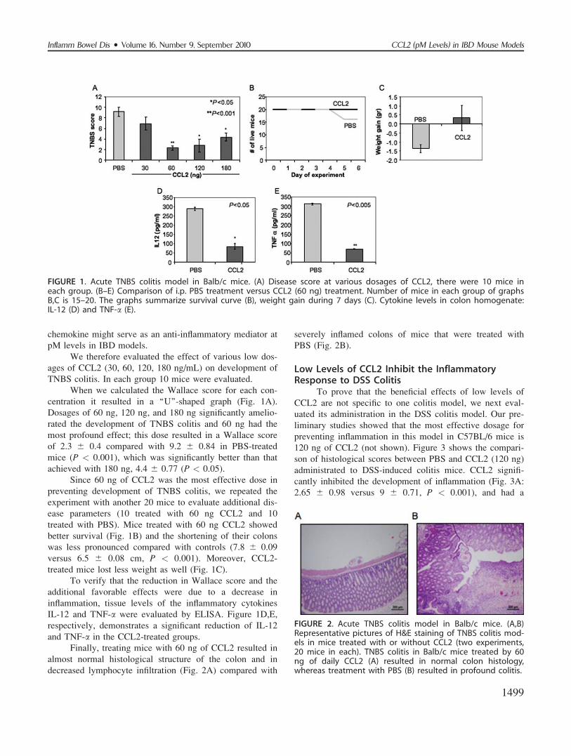
When we calculated the Wallace score for each con-
centration it resulted in a ‘‘U’’-shaped graph (Fig. 1A).
Dosages of 60 ng, 120 ng, and 180 ng significantly amelio-
rated the development of TNBS colitis and 60 ng had the
most profound effect; this dose resulted in a Wallace score
of 2.3 6 0.4 compared with 9.2 6 0.84 in PBS-treated
mice (P < 0.001), which was significantly better than that
achieved with 180 ng, 4.4 6 0.77 (P < 0.05).
Since 60 ng of CCL2 was the most effective dose in
preventing development of TNBS colitis, we repeated the
experiment with another 20 mice to evaluate additional dis-
ease parameters (10 treated with 60 ng CCL2 and 10
treated with PBS). Mice treated with 60 ng CCL2 showed
better survival (Fig. 1B) and the shortening of their colons
was less pronounced compared with controls (7.8 6 0.09
versus 6.5 6 0.08 cm, P < 0.001). Moreover, CCL2-
treated mice lost less weight as well (Fig. 1C).
To verify that the reduction in Wallace score and the
additional favorable effects were due to a decrease in
inflammation, tissue levels of the inflammatory cytokines
IL-12 and TNF-a were evaluated by ELISA. Figure 1D,E,
respectively, demonstrates a significant reduction of IL-12
and TNF-a in the CCL2-treated groups.
Finally, treating mice with 60 ng of CCL2 resulted in
almost normal histological structure of the colon and in
decreased lymphocyte infiltration (Fig. 2A) compared with
severely inflamed colons of mice that were treated with
PBS (Fig. 2B).
Low Levels of CCL2 Inhibit the InflammatoryResponse to DSS Colitis
To prove that the beneficial effects of low levels of
CCL2 are not specific to one colitis model, we next eval-
uated its administration in the DSS colitis model. Our pre-
liminary studies showed that the most effective dosage for
preventing inflammation in this model in C57BL/6 mice is
120 ng of CCL2 (not shown). Figure 3 shows the compari-
son of histological scores between PBS and CCL2 (120 ng)
administrated to DSS-induced colitis mice. CCL2 signifi-
cantly inhibited the development of inflammation (Fig. 3A:
2.65 6 0.98 versus 9 6 0.71, P < 0.001), and had a
FIGURE 1. Acute TNBS colitis model in Balb/c mice. (A) Disease score at various dosages of CCL2, there were 10 mice ineach group. (B–E) Comparison of i.p. PBS treatment versus CCL2 (60 ng) treatment. Number of mice in each group of graphsB,C is 15–20. The graphs summarize survival curve (B), weight gain during 7 days (C). Cytokine levels in colon homogenate:IL-12 (D) and TNF-a (E).
FIGURE 2. Acute TNBS colitis model in Balb/c mice. (A,B)Representative pictures of H&E staining of TNBS colitis mod-els in mice treated with or without CCL2 (two experiments,20 mice in each). TNBS colitis in Balb/c mice treated by 60ng of daily CCL2 (A) resulted in normal colon histology,whereas treatment with PBS (B) resulted in profound colitis.
Inflamm Bowel Dis � Volume 16, Number 9, September 2010 CCL2 (pM Levels) in IBD Mouse Models
1499
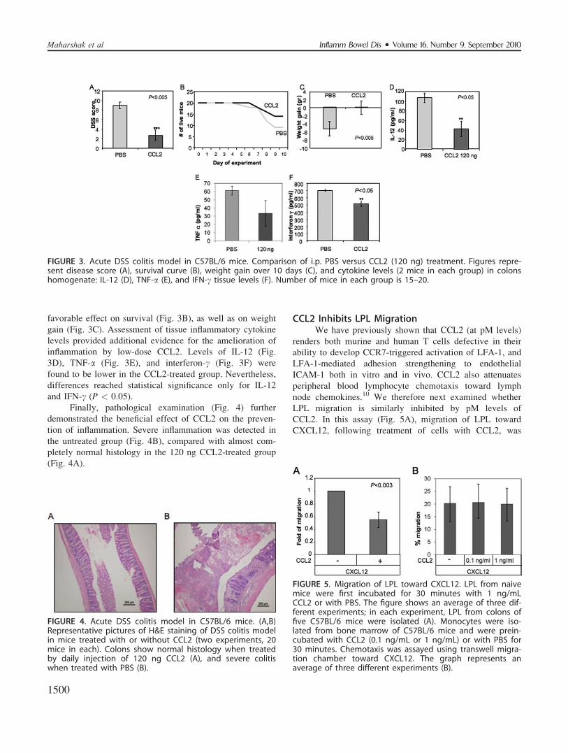
favorable effect on survival (Fig. 3B), as well as on weight
gain (Fig. 3C). Assessment of tissue inflammatory cytokine
levels provided additional evidence for the amelioration of
inflammation by low-dose CCL2. Levels of IL-12 (Fig.
3D), TNF-a (Fig. 3E), and interferon-c (Fig. 3F) were
found to be lower in the CCL2-treated group. Nevertheless,
differences reached statistical significance only for IL-12
and IFN-c (P < 0.05).
Finally, pathological examination (Fig. 4) further
demonstrated the beneficial effect of CCL2 on the preven-
tion of inflammation. Severe inflammation was detected in
the untreated group (Fig. 4B), compared with almost com-
pletely normal histology in the 120 ng CCL2-treated group
(Fig. 4A).
CCL2 Inhibits LPL MigrationWe have previously shown that CCL2 (at pM levels)
renders both murine and human T cells defective in their
ability to develop CCR7-triggered activation of LFA-1, and
LFA-1-mediated adhesion strengthening to endothelial
ICAM-1 both in vitro and in vivo. CCL2 also attenuates
peripheral blood lymphocyte chemotaxis toward lymph
node chemokines.10 We therefore next examined whether
LPL migration is similarly inhibited by pM levels of
CCL2. In this assay (Fig. 5A), migration of LPL toward
CXCL12, following treatment of cells with CCL2, was
FIGURE 3. Acute DSS colitis model in C57BL/6 mice. Comparison of i.p. PBS versus CCL2 (120 ng) treatment. Figures repre-sent disease score (A), survival curve (B), weight gain over 10 days (C), and cytokine levels (2 mice in each group) in colonshomogenate: IL-12 (D), TNF-a (E), and IFN-c tissue levels (F). Number of mice in each group is 15–20.
FIGURE 4. Acute DSS colitis model in C57BL/6 mice. (A,B)Representative pictures of H&E staining of DSS colitis modelin mice treated with or without CCL2 (two experiments, 20mice in each). Colons show normal histology when treatedby daily injection of 120 ng CCL2 (A), and severe colitiswhen treated with PBS (B).
FIGURE 5. Migration of LPL toward CXCL12. LPL from naivemice were first incubated for 30 minutes with 1 ng/mLCCL2 or with PBS. The figure shows an average of three dif-ferent experiments; in each experiment, LPL from colons offive C57BL/6 mice were isolated (A). Monocytes were iso-lated from bone marrow of C57BL/6 mice and were prein-cubated with CCL2 (0.1 ng/mL or 1 ng/mL) or with PBS for30 minutes. Chemotaxis was assayed using transwell migra-tion chamber toward CXCL12. The graph represents anaverage of three different experiments (B).
Inflamm Bowel Dis � Volume 16, Number 9, September 2010Maharshak et al
1500
inhibited by 45% compared to control LPL preincubated in
PBS (P < 0.003), suggesting local migration of LPL within
the gut wall compartment may be inhibited, and indicating
an additional possible mechanism by which CCL2 inhibits
inflammation.
Monocytes/macrophages play an important role dur-
ing inflammation. To determine whether pM levels of
CCL2 inhibit the inflammatory response in these IBD mod-
els by attenuating monocyte migration, we evaluated
whether exposing monocytes to low levels of CCL2 inhib-
its monocyte migration by analyzing their migration in a
Transwell assay toward CXCL12. No difference was
observed in migration of monocytes that were first incu-
bated in low levels of CCL2 (0.1 ng/mL or 1 ng/mL)
versus those that were not treated by CCL2 (Fig. 5B), sug-
gesting that the effect of CCL2 is not monocyte-dependent.
pM Levels of CCL2 Have Favorable Effects on aCRC Model
We have shown that administration of low doses of
CCL2 has a favorable effect on colitis. We further inquired
whether these effects are sustained during long-term
administration in prevention of inflammation-related CRC
using the AOM-DSS CRC model.17 In this model, cancer
is induced by i.p. injection of the genotoxic carcinogen
AOM. The progression of cancer is accelerated by induc-
tion of inflammation by administration of DSS through the
drinking water. Balb/c mice were shown to be sensitive to
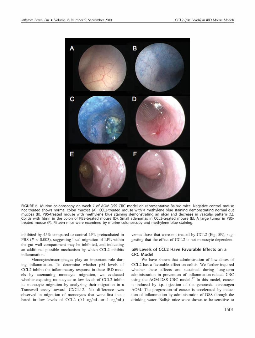
FIGURE 6. Murine colonoscopy on week 7 of AOM-DSS CRC model on representative Balb/c mice. Negative control mousenot treated shows normal colon mucosa (A). CCL2-treated mouse with a methylene blue staining demonstrating normal gutmucosa (B). PBS-treated mouse with methylene blue staining demonstrating an ulcer and decrease in vascular pattern (C).Colitis with fibrin in the colon of PBS-treated mouse (D). Small adenomas in CCL2-treated mouse (E). A large tumor in PBS-treated mouse (F). Fifteen mice were examined by murine colonoscopy and methylene blue staining.
Inflamm Bowel Dis � Volume 16, Number 9, September 2010 CCL2 (pM Levels) in IBD Mouse Models
1501
development of tumors in this model.17 At week 7 we
assessed inflammatory changes and development of tumors
by murine colonoscopy on some of the mice (Fig. 6). In
the CCL2-treated mice we found a decrease in inflamma-
tory changes (Fig. 6B) and in number and size of tumors
(Fig. 6E) compared to the inflammation (Fig. 6C,D) and
tumors (Fig. 6F) observed in PBS-treated mice.
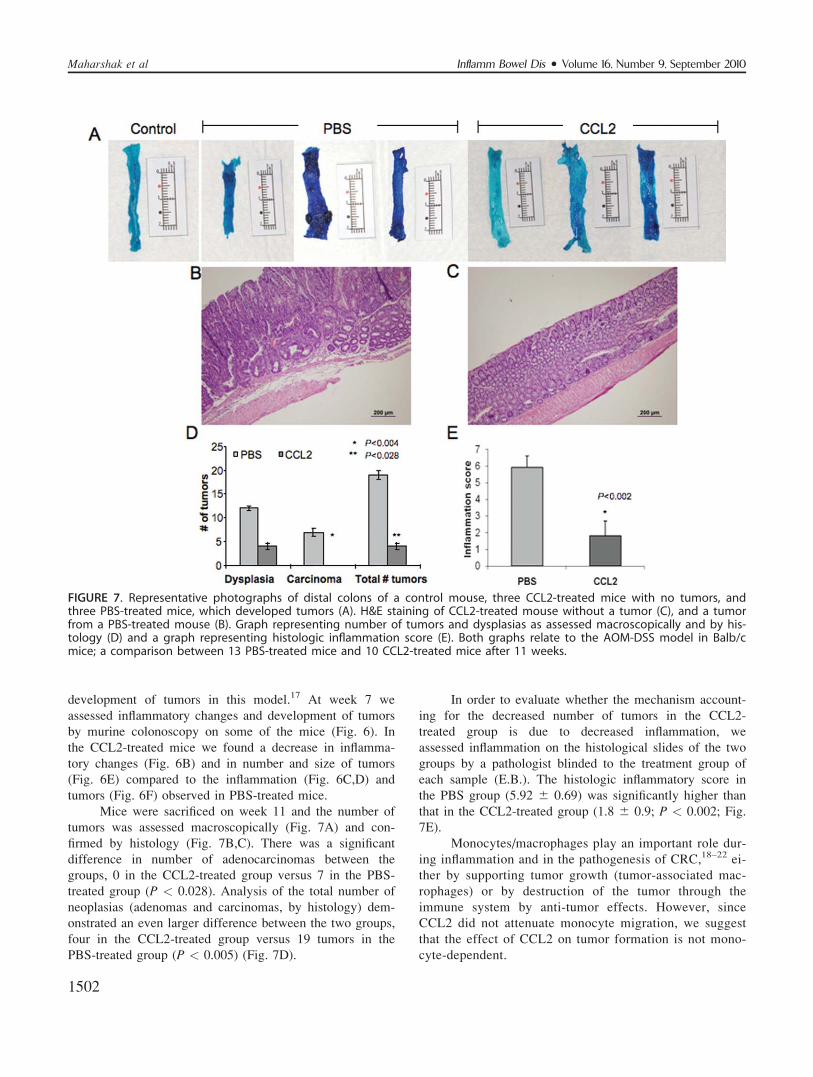
Mice were sacrificed on week 11 and the number of
tumors was assessed macroscopically (Fig. 7A) and con-
firmed by histology (Fig. 7B,C). There was a significant
difference in number of adenocarcinomas between the
groups, 0 in the CCL2-treated group versus 7 in the PBS-
treated group (P < 0.028). Analysis of the total number of
neoplasias (adenomas and carcinomas, by histology) dem-
onstrated an even larger difference between the two groups,
four in the CCL2-treated group versus 19 tumors in the
PBS-treated group (P < 0.005) (Fig. 7D).
In order to evaluate whether the mechanism account-
ing for the decreased number of tumors in the CCL2-
treated group is due to decreased inflammation, we
assessed inflammation on the histological slides of the two
groups by a pathologist blinded to the treatment group of
each sample (E.B.). The histologic inflammatory score in
the PBS group (5.92 6 0.69) was significantly higher than
that in the CCL2-treated group (1.8 6 0.9; P < 0.002; Fig.
7E).
Monocytes/macrophages play an important role dur-
ing inflammation and in the pathogenesis of CRC,18–22 ei-
ther by supporting tumor growth (tumor-associated mac-
rophages) or by destruction of the tumor through the
immune system by anti-tumor effects. However, since
CCL2 did not attenuate monocyte migration, we suggest
that the effect of CCL2 on tumor formation is not mono-
cyte-dependent.
FIGURE 7. Representative photographs of distal colons of a control mouse, three CCL2-treated mice with no tumors, andthree PBS-treated mice, which developed tumors (A). H&E staining of CCL2-treated mouse without a tumor (C), and a tumorfrom a PBS-treated mouse (B). Graph representing number of tumors and dysplasias as assessed macroscopically and by his-tology (D) and a graph representing histologic inflammation score (E). Both graphs relate to the AOM-DSS model in Balb/cmice; a comparison between 13 PBS-treated mice and 10 CCL2-treated mice after 11 weeks.
Inflamm Bowel Dis � Volume 16, Number 9, September 2010Maharshak et al
1502
DISCUSSIONIn this study we demonstrated that low doses of
CCL2 inhibit development of colitis in two models of IBD,
induced by DSS and by TNBS. We found that administra-
tion of pM levels of CCL2 almost completely inhibits the
development of colitis as assessed by clinical score, histo-
logically, and as reflected by tissue cytokine levels. More-
over, CCL2 treatment improved survival in the treated
groups. Disease scores of the PBS-treated mice indicate
that the induced colitis was severe. The effect of CCL2
was dose-dependent; while the lowest concentration (30
ng) had only minimal effect on colitis score, dosages of
60–120 ng optimally obliterated colitis.
Intriguingly, long-term administration of low-dose
CCL2 in an inflammation-enhanced carcinoma model
(AOM-DSS model) almost completely prevented develop-
ment of tumors compared with PBS-treated mice.
Treatment of colitis is a clinical challenge. Research
efforts have been aimed at all levels of known mecha-
nisms, targeting antigens, lymphocytes and dendritic cells,
cell trafficking, and cytokines.23 In addition, targeting
proinflammatory cytokines has proved to be an effective
strategy in the treatment of IBD. One of the most important
advances in the last decade in the treatment of IBD is the
use of anti-TNF-a agents, which were found to be effective
against both UC and CD.24,25 Other emerging treatments
that have already shown their effectiveness in animal mod-
els, and in preliminary human trials target IL-12 as well as
IFN-c.26
In this study we show that treatment with a low dose
of CCL2 ameliorates levels of these cytokines as well as
preventing colitis progression. Our studies suggest that
CCL2 has a dual role in the inflammatory process. We
have previously shown that CCL2 exerts inhibitory effects
on B and T cells, both in vivo and in vitro.9,10 Here we
show that CCL2 has a similar inhibitory effect on LPL iso-
lated from the colon. Inhibition of local migration of LPL
may represent an additional mechanism by which exposure
of lymphocytes to pM CCL2 levels interferes with the
inflammatory process in the gut wall. CCL2 downregulates
homing of naı̈ve T cells to the lymph nodes, and thereby
reduces the exposure of these T cells to antigen, and as a
consequence their activation is prevented. In addition,
CCL2 dramatically inhibits migration of effector cells,
probably to sites of inflammation where they exert their
function. Thus, exposure of T cells to pM levels of CCL2
probably inhibits both the sensitization of naı̈ve T cells and
migration of effector cells, which together dramatically
attenuate the inflammatory response.
A major threat to IBD patients is the risk of CRC. It
has been suggested that there is a correlation between the
severity of colitis and CRC risk.27,28 Use of 5-aminosalicy-
late (5-ASA) preparations has an apparently protective
effect against CRC in UC patients,29,30 despite only a
minor effect on inflammatory score in this disease. Thus,
the mechanisms responsible for cancer in the inflammatory
state are incompletely understood. However, it was recently
shown that in a chronic DSS colitis model inflammation
has a central role in cancer development by inducing oxi-
dative stress, resulting in genetic mutations and DNA
damage.31
We suggested that administration of low-dose CCL2
may prevent the development of CRC by at least two
mechanisms, primarily by decreasing the severity of
inflammation through downregulation of migration of
effector lymphocytes to the colon, CCL2 could decrease
tumerogenic mechanisms, such as oxidative stress. Second,
by affecting migration of monocytes, as well, either
through inhibition18,19,21,22 of monocyte migration to the
inflamed gut, thereby decreasing the number of potential
tumor-associated macrophages that are crucial for the sup-
port of tumor development, or by regulating chemotaxis of
antitumor monocytes.20 Indeed, CCL2 treatment dramati-
cally reduced the number of tumors in the CCL2-treated
mice and, similar to the acute colitis models, inflammation
was significantly attenuated in those mice as well. How-
ever, we found that low levels of CCL2 had no effect on
migration of monocytes and, thus, we believe that the prin-
cipal mechanism for inhibition of tumor development is
due to the inhibition of inflammatory colitis.
Our results demonstrate that CCL2, although a key
inflammatory chemokine, does not always augment inflam-
matory processes. Rather, pM levels of circulating CCL2
can exert global suppressive effects on murine inflamma-
tory colitis and may be clinically effective as an antiinflam-
matory and tumor-suppressing agent in acute and chronic
colitis models in mice. Further research is needed to under-
stand these effects in murine colitis genetic models and
perhaps, in the future, in humans.
ACKNOWLEDGMENTSI.S. is the incumbent of the Dr. Morton and Ann
Kleiman Professorial Chair. Author contributions: N.M.
designed and performed most of the experiments, analyzed
results, and wrote the article; G.H., U.R., E.Z., A.S., R.M.,
E.E., designed and performed some of the experiments and
analyzed the results; N.A. obtained funding and critically
revised the article; E.B. analyzed and interpreted some of
the results. I.S. analyzed the results, designed the experi-
ment and wrote the paper.
REFERENCES1. Aharoni R, Kayhan B, Brenner O, et al. Immunomodulatory therapeu-
tic effect of glatiramer acetate on several murine models of inflamma-tory bowel disease. J Pharmacol Exp Ther. 2006;318:68–78.
Inflamm Bowel Dis � Volume 16, Number 9, September 2010 CCL2 (pM Levels) in IBD Mouse Models
1503
2. Huibregtse IL, van Lent AU, van Deventer SJ. Immunopathogenesisof IBD: insufficient suppressor function in the gut? Gut. 2007;56:584–592.
3. Sartor RB. Pathogenesis and immune mechanisms of chronic inflam-matory bowel diseases. Am J Gastroenterol. 1997;92(12 Suppl):5S–11S.
4. Ekbom A, Helmick C, Zack M, et al. Ulcerative colitis and colorectalcancer. A population-based study. N Engl J Med. 1990;323:1228–1233.
5. Ekbom A, Helmick C, Zack M, et al. Increased risk of large-bowelcancer in Crohn’s disease with colonic involvement. Lancet. 1990;336:357–359.
6. Itzkowitz SH, Yio X. Inflammation and cancer IV. Colorectal cancerin inflammatory bowel disease: the role of inflammation. Am J PhysiolGastrointest Liver Physiol. 2004;287:G7–17.
7. te Velde AA, Verstege MI, Hommes DW. Critical appraisal of thecurrent practice in murine TNBS-induced colitis. Inflamm Bowel Dis.2006;12:995–999.
8. Puleston J, Cooper M, Murch S, et al. A distinct subset of chemokinesdominates the mucosal chemokine response in inflammatory boweldisease. Aliment Pharmacol Ther. 2005;21:109–120.
9. Flaishon L, Becker-Herman S, Hart G, et al. Expression of the chemo-kine receptor CCR2 on immature B cells negatively regulates theircytoskeletal rearrangement and migration. Blood. 2004;104:933–941.
10. Flaishon L, Hart G, Zelman E, et al. Anti-inflammatory effects of aninflammatory chemokine: CCL2 inhibits lymphocyte homing by mod-ulation of CCL21-triggered integrin-mediated adhesions. Blood. 2008;112:5016–5025.
11. Dohi T, Ejima C, Kato R, et al. Therapeutic potential of follistatin forcolonic inflammation in mice. Gastroenterology. 2005;128:411–423.
12. Reuter BK, Asfaha S, Buret A, et al. Exacerbation of inflammation-associated colonic injury in rat through inhibition of cyclooxygenase-2. J Clin Invest. 1996;98:2076–2085.
13. Ohkawara T, Nishihira J, Takeda H, et al. Amelioration of dextransulfate sodium-induced colitis by anti-macrophage migration inhibitoryfactor antibody in mice. Gastroenterology. 2002;123:256–270.
14. Weigmann B, Tubbe I, Seidel D, et al. Isolation and subsequent analy-sis of murine lamina propria mononuclear cells from colonic tissue.Nat Protoc. 2007;2:2307–2311.
15. Flaishon L, Lantner F, Hershkoviz R, et al. Low levels of IFN-gammadown-regulate the integrin-dependent adhesion of B cells by activatinga pathway that interferes with cytoskeleton rearrangement. J BiolChem. 2001;276:46701–46706.
16. Becker C, Fantini MC, Wirtz S, et al. In vivo imaging of colitis andcolon cancer development in mice using high resolution chromoendo-scopy. Gut. 2005;54:950–954.
17. Suzuki R, Kohno H, Sugie S, et al. Strain differences in the suscepti-bility to azoxymethane and dextran sodium sulfate-induced coloncarcinogenesis in mice. Carcinogenesis. 2006;27:162–169.
18. Bailey C, Negus R, Morris A, et al. Chemokine expression is associ-ated with the accumulation of tumour associated macrophages(TAMs) and progression in human colorectal cancer. Clin Exp Metas-tasis. 2007;24:121–130.
19. Barbera-Guillem E, Nyhus JK, Wolford CC, et al. Vascular endothe-lial growth factor secretion by tumor-infiltrating macrophages essen-tially supports tumor angiogenesis, and IgG immune complexespotentiate the process. Cancer Res. 2002;62:7042–7049.
20. Kagaya T, Nakamoto Y, Sakai Y, et al. Monocyte chemoattractantprotein-1 gene delivery enhances antitumor effects of herpes simplexvirus thymidine kinase/ganciclovir system in a model of colon cancer.Cancer Gene Ther. 2006;13:357–366.
21. Lewis CE, Pollard JW. Distinct role of macrophages in different tu-mor microenvironments. Cancer Res. 2006;66:605–612.
22. Mantovani A, Bottazzi B, Colotta F, et al. The origin and function oftumor-associated macrophages. Immunol Today. 1992;13:265–270.
23. Sartor RB. Mechanisms of disease: pathogenesis of Crohn’s diseaseand ulcerative colitis. Nat Clin Pract Gastroenterol Hepatol. 2006;3:390–407.
24. Hanauer SB, Feagan BG, Lichtenstein GR, et al. Maintenance inflixi-mab for Crohn’s disease: the ACCENT I randomised trial. Lancet.2002;359:1541–1549.
25. Rutgeerts P, Sandborn WJ, Feagan BG, et al. Infliximab for inductionand maintenance therapy for ulcerative colitis. N Engl J Med. 2005;353:2462–2476.
26. Peluso I, Pallone F, Monteleone G. Interleukin-12 and Th1 immuneresponse in Crohn’s disease: pathogenetic relevance and therapeuticimplication. World J Gastroenterol. 2006;12:5606–5610.
27. Gupta RB, Harpaz N, Itzkowitz S, et al. Histologic inflammation is a riskfactor for progression to colorectal neoplasia in ulcerative colitis: acohort study. Gastroenterology. 2007;133:1099–1105; quiz 1340–1341.
28. Rutter M, Saunders B, Wilkinson K, et al. Severity of inflammation isa risk factor for colorectal neoplasia in ulcerative colitis. Gastroenter-ology. 2004;126:451–459.
29. Ullman T, Croog V, Harpaz N, et al. Progression to colorectal neopla-sia in ulcerative colitis: effect of mesalamine. Clin Gastroenterol Hep-atol. 2008;6:1225–1230; quiz 1177.
30. Velayos FS, Terdiman JP, Walsh JM. Effect of 5-aminosalicylate use oncolorectal cancer and dysplasia risk: a systematic review and metaanaly-sis of observational studies. Am J Gastroenterol. 2005;100:1345–1353.
31. Westbrook AM, Wei B, Braun J, et al. Intestinal mucosal inflamma-tion leads to systemic genotoxicity in mice. Cancer Res. 2009;69:4827–4834.
1504
Inflamm Bowel Dis � Volume 16, Number 9, September 2010Maharshak et al
Related Documents