doi:10.1182/blood-2003-05-1643 Prepublished online October 30, 2003; Marcus Groettrup Elke Scandella, Ying Men, Daniel F Legler, Silke Gillessen, Ladislav Prikler, Burkhard Ludewig and cells require prostaglandin E2 CCL19/CCL21-triggered signal transduction and migration of dendritic (1930 articles) Signal Transduction (973 articles) Phagocytes (5022 articles) Immunobiology (564 articles) Chemokines, Cytokines, and Interleukins (790 articles) Cell Adhesion and Motility Articles on similar topics can be found in the following Blood collections http://bloodjournal.hematologylibrary.org/site/misc/rights.xhtml#repub_requests Information about reproducing this article in parts or in its entirety may be found online at: http://bloodjournal.hematologylibrary.org/site/misc/rights.xhtml#reprints Information about ordering reprints may be found online at: http://bloodjournal.hematologylibrary.org/site/subscriptions/index.xhtml Information about subscriptions and ASH membership may be found online at: articles must include the digital object identifier (DOIs) and date of initial publication. priority; they are indexed by PubMed from initial publication. Citations to Advance online prior to final publication). Advance online articles are citable and establish publication yet appeared in the paper journal (edited, typeset versions may be posted when available Advance online articles have been peer reviewed and accepted for publication but have not Copyright 2011 by The American Society of Hematology; all rights reserved. Washington DC 20036. by the American Society of Hematology, 2021 L St, NW, Suite 900, Blood (print ISSN 0006-4971, online ISSN 1528-0020), is published weekly For personal use only. by guest on June 8, 2013. bloodjournal.hematologylibrary.org From

Welcome message from author
This document is posted to help you gain knowledge. Please leave a comment to let me know what you think about it! Share it to your friends and learn new things together.
Transcript
doi:10.1182/blood-2003-05-1643Prepublished online October 30, 2003;
Marcus GroettrupElke Scandella, Ying Men, Daniel F Legler, Silke Gillessen, Ladislav Prikler, Burkhard Ludewig and cells require prostaglandin E2CCL19/CCL21-triggered signal transduction and migration of dendritic
(1930 articles)Signal Transduction � (973 articles)Phagocytes �
(5022 articles)Immunobiology � (564 articles)Chemokines, Cytokines, and Interleukins �
(790 articles)Cell Adhesion and Motility �Articles on similar topics can be found in the following Blood collections
http://bloodjournal.hematologylibrary.org/site/misc/rights.xhtml#repub_requestsInformation about reproducing this article in parts or in its entirety may be found online at:
http://bloodjournal.hematologylibrary.org/site/misc/rights.xhtml#reprintsInformation about ordering reprints may be found online at:
http://bloodjournal.hematologylibrary.org/site/subscriptions/index.xhtmlInformation about subscriptions and ASH membership may be found online at:
articles must include the digital object identifier (DOIs) and date of initial publication. priority; they are indexed by PubMed from initial publication. Citations to Advance online prior to final publication). Advance online articles are citable and establish publicationyet appeared in the paper journal (edited, typeset versions may be posted when available Advance online articles have been peer reviewed and accepted for publication but have not
Copyright 2011 by The American Society of Hematology; all rights reserved.Washington DC 20036.by the American Society of Hematology, 2021 L St, NW, Suite 900, Blood (print ISSN 0006-4971, online ISSN 1528-0020), is published weekly
For personal use only. by guest on June 8, 2013. bloodjournal.hematologylibrary.orgFrom

CCL19/CCL21 triggered signal transduction and migration of dendritic
cells requires prostaglandin E2
Elke Scandella1, Ying Men1, Daniel F. Legler2, Silke Gillessen3, Ladislav Prikler4,
Burkhard Ludewig1, and Marcus Groettrup1,2
Departments of 1Research, 3Oncology, and 4Urology, Cantonal Hospital St. Gallen, CH-
9007 St. Gallen, Switzerland; 2Department of Immunology, Faculty of Biology,
University of Constance, D-78457 Konstanz, Germany
running title: PGE2 enables signal transduction / migration of DC
scientific heading: Chemokines
corresponding authors:
Elke Scandella, Kantonsspital St. Gallen, LFA, Haus 09, CH-9007 St. Gallen Switzerland,
Tel.: +41 71 4941097, FAX: +41 71 4946321, e-mail: [email protected]
Marcus Groettrup, Universität Konstanz, P1101, Universitätsstrasse 10, D-78457 Konstanz,
Germany, Tel.: +49 7531 882130, FAX: +49 7531 883102, e-mail: Marcus.Groettrup@uni-
konstanz.de, (communicates with editorial and production offices)
word count: 5304
abstract: 182
This work was supported by the Cancer League St. Gallen-Appenzell, the Swiss Cancer
League, Foundation Propter Homines Vaduz Liechtenstein, the Cancer Research Insitute,
CaPCURE Foundation, Roche Research Foundation, Rentenanstalt Jubiläumsstiftung, and
German Research Foundation (DFG, TR-SFB 11).
Copyright (c) 2003 American Society of Hematology
Blood First Edition Paper, prepublished online October 30, 2003; DOI 10.1182/blood-2003-05-1643 For personal use only. by guest on June 8, 2013. bloodjournal.hematologylibrary.orgFrom
- 2 -
Abstract
The control of dendritic cell (DC) migration is pivotal for the initiation of cellular immune
responses. Upon activation with inflammatory stimuli, the chemokine receptor CCR7 is
upregulated on DCs. Activated DCs home to lymphoid organs where the CCR7 ligands
CCL19 and CCL21 are expressed. We previously found that human monocyte-derived DCs
(MoDCs) exclusively migrated towards CCL19 and CCL21 when matured in the presence of
prostaglandin (PG) E2. Since PGE2 did not alter CCR7 cell surface expression we examined
whether PGE2 may exert its effect by coupling CCR7 to signal transduction modules. Indeed,
stimulation with CCR7 ligands led to enhanced phosphatidylinositol-3-kinase mediated
phosphorylation of protein kinase B when MoDCs were matured in the presence of PGE2.
Moreover, CCL19/CCL21 - induced intracellular calcium-mobilization in MoDCs occurred
only when PGE2 was present during maturation. MoDC migration towards CCL19 and
CCL21 was dependent on phospholipase C and intracellular calcium flux but not on
phosphatidylinositol-3 kinase. Hence our data provide insight into CCL19/CCL21 - triggered
signal transduction pathways and identify a novel function for PGE2 in controlling the
migration of mature MoDC by facilitating CCR7 signal transduction.
abbreviations: CCR, CC chemokine receptor; DAG, diacylglycerol; DC, dendritic cell; Erk,
extracellular signal-regulated kinase; PI 3-kinase, phosphatidylinositol 3-kinase; PK, protein
kinase; PLC, phospholipase C
keywords: calcium mobilization / CC chemokine receptor / CCR7 / chemotaxis / protein
kinase B
For personal use only. by guest on June 8, 2013. bloodjournal.hematologylibrary.orgFrom
- 3 -
Introduction
Dendritic cells (DCs) are professional antigen presenting cells capable of antigen transport
and presentation in secondary lymphoid organs which is crucial for the initiation and
maintenance of T-cell mediated immune responses1. They reside in the periphery in an
immature state taking up pathogens through pinocytosis or receptor mediated endocytosis
which leads to the induction of DC maturation. Mature DCs undergo phenotypical and
functional changes including up-regulation of the chemokine receptor CCR72. The
expression of CCR7 on mature DCs, as well as on naïve and central memory T-cells, is
essential for their coordinated migration to the T-cell area of draining lymph nodes, since this
migration is guided by CCL19 (EBI1-ligand chemokine (ELC), Macrophage inflammatory
protein (MIP)-3β) and CCL21 (secondary lymphoid-tissue chemokine (SLC), 6-Ckine), the
two ligands for CCR7. Both chemokines are expressed by stromal cells in the T-cell area of
secondary lymphoid organs. The essential role of CCR7 and its ligands for the migration of
mature DCs to lymph nodes was demonstrated in CCR7-deficient mice3 and plt/plt mice
which lack the ligands for CCR74.
Chemokine receptors are transmembrane receptors of the class A rhodopsin-like
family, which span the membrane sevenfold and transduce their signals via G proteins, mainly
of the Gαi subclass5. Receptor stimulation leads to inhibition of adenyl cyclases through Gαi-
and Gβγ-mediated activation of phospholipase C (PLC) followed by diacylglycerol-mediated
activation of protein kinase C (PKC) and the release of calcium from intracellular stores6.
Furthermore, Gβγ subunits that are released from Gαi proteins transiently activate
phosphatidylinositol 3-kinases (PI3Ks) leading to the activation of protein kinase B (PKB,
Akt) as well as extracellular signal-regulated kinase (Erk)-27. However, most data on the
signal transduction of chemokine receptors has been obtained with CXCR1, CCR2, CCR5,
For personal use only. by guest on June 8, 2013. bloodjournal.hematologylibrary.orgFrom

- 4 -
and CXCR4 but little information is currently available about CCR7 signal transduction.
Recently, Rho associated kinases have been shown to be required for CCR7 mediated
polarization and chemotaxis of T lymphocytes8. Another study suggests that CCR7 signaling
involves the activation of a Jak kinase9. It is still a matter of debate, however, whether the
JAK/STAT pathway which would require chemokine receptor dimerization contributes to
chemokine-mediated signal transduction10. In addition, most studies have been performed
with neutrophils, peritoneal macrophages and T-cells but not with DCs.
Recently, we and others observed that the maturation-induced upregulation of CCR7
expression on human monocyte-derived DCs (MoDCs) was not sufficient to allow MoDC
migration towards CCL19 and CCL2111,12. Human MoDCs matured either with soluble
CD40L (sCD40L) or polyI:C markedly enhanced surface expression of CCR7 but were,
nevertheless, not at all or only poorly responsive to CCL19 and CCL21. Interestingly, MoDC
migration towards CCL19 and CCL21 was readily observed upon maturation in the presence
of the pro-inflammatory mediator prostaglandin (PG)E2, although PGE2 did not change the
expression level of CCR7 on mature MoDCs, providing evidence for an alternative effect of
PGE2. The importance of PGE2 for DC migration in vivo has recently been shown in Ptger4-/-
mice lacking the PGE2 receptor EP4. These mice display impaired migration of Langerhans
cells as well as reduced skin immune responses13. However, other DC types as for instance
CD1b/c+ peripheral blood DCs do not require an additional PGE2 stimulus for effective
CCR7 mediated migration12.
In this study we investigated the hypothesis that PGE2 may facilitate CCL19/CCL21
directed migration by coupling the cognate chemokine receptor CCR7 to its signal
transduction modules. Indeed, we found that PGE2 was required during sCD40L stimulated
maturation of MoDCs to activate PKB and to mobilize intracellular free calcium as
For personal use only. by guest on June 8, 2013. bloodjournal.hematologylibrary.orgFrom

- 5 -
downstream signaling events of CCR7. Moreover, we demonstrate that PLC mediated
cytoplasmic Ca2+-mobilization was a prerequisite for the chemotaxis of MoDCs towards
CCL19 and CCL21. Our results hence provide evidence that PGE2 is required to facilitate
CCR7 mediated signal transduction and migration of MoDC.
For personal use only. by guest on June 8, 2013. bloodjournal.hematologylibrary.orgFrom

- 6 -
Materials and methods
Tissue culture media and reagents
AIM V was purchased from Invitrogen (Groningen, The Netherlands). IL-4 was obtained from
Strathmann (Hamburg, Germany) and GM-CSF from Novartis (Leukomax, Basel,
Switzerland). Soluble CD40L (sCD40L) was kindly provided by Immunex Corporation
(Seattle, WA). Prostaglandin E2 was purchased from Pharmacia & Upjohn (Prostin E2,
Dübendorf, Switzerland). Human CCL19 and CCL21 were provided by R&D Systems
(Wiesbaden-Nordenstadt, Germany), and CXCL-12 was purchased from PromoCell
(Heidelberg). Staurosporine, U-73122, H-89, wortmannin, Ly-294002, Y-27632 and 1,2-
bis(o-aminophenoxy)ethane-N,N,N',N'-tetraacetic acid-tetra(acetomethyl) ester (BAPTA-AM)
were bought from Calbiochem (Lucerne, Switzerland). Phorbol-myristate-acetate (PMA) and
ionomycin was purchased from Sigma (Buchs, Switzerland).
Antibodies
Horseradish peroxidase (HRP) conjugated anti-mouse antibody was obtained from DAKO
(Zug, Switzerland) and anti-rabbit antibody from Milan (La Roche, Switzerland). Antibodies
against Akt/PKB, phospho-Akt/PKB (Ser473), p42/44 MAP kinase/Erk-1/2 and phospho-
p44/42 MAP kinase/Erk-1/2 (Thr202/Tyr204) were from Cell Signaling Technology (Frankfurt,
Germany).
Generation of human monocyte-derived DCs (MoDCs)
MoDCs were generated from human PBMCs as described previously11. Briefly, PBMCs were
separated by density gradient centrifugation on Ficoll-Paque (Pharmacia, Uppsala, Sweden),
resuspended at 4 x 10⁶ cells/ml in AIM V medium and were allowed to adhere to plastic for 1
hour at 37°C. Non-adherent cells were removed and remaining cells were cultured in AIM V
For personal use only. by guest on June 8, 2013. bloodjournal.hematologylibrary.orgFrom
- 7 -
medium supplemented with GM-CSF (50 ng/ml) and IL-4 (1000 U/ml). MoDCs derived form
the adherence step were only used when the content of remaining B and T cells was below
2%. Alternatively, monocytes were purified from PBMCs by positive selection using anti-
CD14 conjugated magnetic microbeads (Miltenyi, Bergisch Gladbach, Germany) and
cultivated at 1 x 10⁶ cells/ml. On day 6, nonadherent and loosely adherent cells were
harvested and recultured (5 x 10⁵ cells/ml) for additional 48 hours in cytokine containing
medium in the absence or presence of 1 µg/ml sCD40L to induce maturation. Where
indicated, PGE2 (1 µg/ml) or H-89 was added during stimulation.
Cell stimulation and western blot analysis
MoDCs were washed twice with PBS and resuspended in AIM V medium without additives
(2 x 10⁷ cells/ml). Aliquots of 2 x 10⁶ cells were incubated for 10 min at 37°C and stimulated
either with CCL19 or CCL21 (250 ng/ml) for different periods of time. Incubations were
terminated by the addition of trichloroacetic acid (TCA) to a final concentration of 10%.
Protein pellets were washed twice with ice cold acetone and dissolved by boiling in 1x
Laemmli SDS-loading buffer containing 5% 2-mercaptoethanol. Proteins were separated on
10% SDS-PAGE and transferred to nitrocellulose membranes. The membranes were blocked
with 5% low fat dry milk in 1x TBS-buffer and incubated with the respective antibodies
overnight on a rocking plate at 4°C. After washing, HRP-conjugated secondary antibodies
were bound and detected using enhanced chemiluminescence (Pierce, Socochim, Lausanne,
Switzerland). Thereafter membranes were stripped with 0,2 M NaOH for 5 min, followed by
washing and reprobing with control antibodies.
For personal use only. by guest on June 8, 2013. bloodjournal.hematologylibrary.orgFrom
- 8 -
Chemotaxis assay
Chemotaxis of MoDCs was measured by migration through a polycarbonate filter with 5 µm
pore size in 24-well transwell chambers (Corning Costar, Cambridge, MA). AIM V (600 µl)
containing indicated doses of CCL19, CCL21, CXCL-12 or medium alone as a control for
spontaneous migration were filled into the lower chamber. 1 x 10⁵ DCs (100 µl) were added
to the upper chamber and incubated for 3 hours at 37°C. A 500 µl aliquot of the cells that
migrated to the bottom chamber was counted by flow cytometry in a FACScan acquiring
events for a fixed time period of 60s using CellQuest software (Becton Dickinson, Basel).
Each experiment was performed in duplicates. The mean number of spontaneously migrated
cells was subtracted from the total number of migrated cells. Values are given as percent
migrated cells ± SEM. Where indicated, cells were incubated for 30 min at 37°C with various
drugs and washed twice prior to the migration assay.
Intracellular free Ca2+-mobilization
MoDCs (1 x 10⁶/ml) were loaded with 4 µM fluo-3-actetomethylester (fluo-3-AM) in the
presence of 1 µM pluronic F-127 (Molecular Probes, Leiden, Netherlands) in loading-buffer
(145 mM NaCl, 5 mM KCl, 1 mM Na2HPO4, 1 mM MgCl2, 5mM Glucose, 1 mM CaCl2 and
10 mM HEPES, pH 7.5) for 30 min at RT. Where indicated BAPTA-AM was loaded
simultaneously into MoDCs. Subsequently, cells were washed twice with loading-buffer or
Ca2+-free loading-buffer containing 2 mM EGTA and stored on ice. For aquisition, cells were
incubated for 10 min at 37°C before stimulation with CCL19, CCL21 (250 ng/ml) or
ionomycin (1 µg/ml) and fluorescence was recorded over time by flow cytometry.
For personal use only. by guest on June 8, 2013. bloodjournal.hematologylibrary.orgFrom

- 9 -
Results
MoDC migration towards CCL19 and CCL21 requires activation of protein kinase A
and Rho associated kinases
Previously, we and others demonstrated that human MoDCs required PGE2 during maturation
to migrate towards CCL19 and CCL21 effectively11,12. Earlier studies showing PGE2-
induced migration of MoDCs were performed in the presence of high PGE2 concentrations (in
the micromolar range), thus at concentrations far above physiological levels found in
extracellular fluids at least under non-inflammatory conditions14. For a better understanding
on the effect of PGE2 to enhance MoDC migration, we first performed trans-well chemotaxis
assays with MoDCs matured with sCD40L and graded concentrations of PGE2. The
maturation with CD40L resulted in mature MoDCs with homogeneous surface expression of
CCR7 irrespective of the addition of PGE2 (Figure 1A)11. However, CCL19 and CCL21
induced migration of sCD40L matured MoDCs required PGE2 and was dose dependent being
already significant at a PGE2 concentration of 15 ng/ml (Figure 1A). These PGE2
concentrations are expected to occur at sites of inflammation but not in healthy tissues.
Furthermore, PGE2 was needed throughout the maturation process of MoDCs, since a one
hour pre-incubation of mature MoDCs with PGE2 was not sufficient to induce MoDC
migration in response to CCL19 and CCL21 (data not shown).
PGE2 signaling occurs through the prostaglandin receptors EP2 and EP4 which are
expressed on MoDCs and leads to increased cAMP levels and the activation of the cAMP-
dependent protein kinase A (PKA) in the cytosol11. Therefore we investigated whether MoDC
migration in the presence of PGE2 is dependent on the activation of PKA. To this end,
MoDCs were stimulated with sCD40L and PGE2 in the presence of the PKA specific
inhibitor H-89. Treatment of MoDCs with H-89 at a concentration of 10 µM had no effect on
For personal use only. by guest on June 8, 2013. bloodjournal.hematologylibrary.orgFrom
- 10 -
MoDC maturation, CCR7 expression, and viability (Fig. 1B and data not shown). However,
the addition of H-89 for the entire period of maturation reduced the migration of MoDCs in
response to CCL19 and CCL21 by about 50% (Figure 1C). In contrast, blocking activation of
PKA during the migration assay had no significant effect on MoDC migration, indicating that
PGE2-induced activation of PKA during maturation of MoDCs may provide a signal which
allows them to migrate.
Very recently, the Rho family of GTPases was shown to be involved in CCR7
mediated migration of T lymphocytes8. To test the involvement of Rho kinase, mature
MoDCs were pretreated with graded doses of Y-27632, a specific inhibitor of Rho associated
kinases. Blocking Rho associated kinases resulted in a significant reduction of CCL21
mediated migration of MoDCs (Figure 1D) confirming the findings observed in T
lymphocytes.
PGE2 does not affect chemokine-mediated Erk-1/2 activation
Since PGE2 did not alter cell surface expression of CCR7 on sCD40L-matured MoDCs, we
investigated whether PGE2 may facilitate migration by coupling CCR7 to its signal
transduction pathway. Signal transduction of CCR7 in DCs has not been addressed so far. An
earlier study on T cells, however, demonstrated that CCL19 stimulation resulted in the
activation of the MAP-kinase cascade leading to a transient activation of Erk-2 by
phosphorylation at threonine- and tyrosin residues7. We matured MoDCs with sCD40L in the
presence or absence of PGE2, stimulated them with CCL19 for different time periods and
investigated Erk activation by western blotting. Using an antibody detecting Erk-1 and Erk-2
exclusively when phosphorylated at Thr202 and Tyr204 we found a rapid and transient Erk-1/2
activation after 2 to 7 min of CCL19 stimulation (Figure 2A). However, there was no
For personal use only. by guest on June 8, 2013. bloodjournal.hematologylibrary.orgFrom

- 11 -
significant difference in Erk-1/2 activation and duration between MoDCs matured with
sCD40L in the presence or absence of PGE2.
PGE2 enhances CCL19 mediated protein kinase B activation
A second major pathway triggered by chemokines is the stimulation of phosphatidyl-inositol
3-kinase (PI3-kinase) leading to the formation of phosphatidyl-3,4,5,-trisphosphate (PIP3) and
the activation of protein kinase B (PKB)7. Hence, we investigated the activation of PKB in
MoDCs upon CCR7 triggering. CCL19 induced a very rapid and transient phosphorylation of
PKB in MoDCs as demonstrated by western blotting using phospho Ser473-specific PKB
antibodies (Figure 2B). Interestingly, PKB activation was much stronger and prolonged in
MoDCs matured in the presence of PGE2 as compared to MoDCs matured with sCD40L
alone. PKB phosphorylation peaked after 2 min of CCL19 triggering and remained detectable
after 15 min in MoDCs matured in the presence of PGE2. In contrast, maturation of MoDCs
without PGE2 allowed only a weak phosphorylation of PKB after 2 min which disappeared
after 5 min of chemokine stimulation (Figure 2B). In some experiments activation of PKB in
MoDCs matured in the absence of PGE2 was even below the detection limit (data not shown).
Migration of MoDCs towards CCL19 and CCL21 is not dependent on PKB activation
We next analyzed whether inefficient chemokine-mediated activation of PKB in MoDCs
matured in the absence of PGE2 was responsible for their reduced migration. Hence, MoDCs
matured in the presence of PGE2 were incubated for 10 min with the specific PI 3-kinase
inhibitors wortmannin or Ly-249002 which block the downstream activation of PKB.
Unexpectedly, wortmannin and Ly-249002 had no effect on MoDC migration towards CCL19
and CCL21 (Figure 3A and B). To rule out ineffective inhibition of PKB phosphorylation by
wortmannin, MoDC lysates were subjected to western blot analysis. Pre-incubation of MoDCs
For personal use only. by guest on June 8, 2013. bloodjournal.hematologylibrary.orgFrom
- 12 -
with 100 nM wortmannin prevented Ser473 phosphorylation of PKB almost completely,
whereas activation of Erk-1/2 was not affected by this treatment (Figure 3C), indicating that
inhibition by wortmannin was effective.
PGE2 is required for CCL19/CCL21-mediated Ca2+-mobilization
Another consequence of chemokine receptor stimulation is the activation of G-protein
sensitive phospholipase C (PLC) isoforms, resulting in the generation of diacylglycerol
(DAG) and inositol 3,4,5-triphosphate which leads to the release of Ca2+ from intracellular
stores. Hence, we analyzed CCL19/CCL21-induced Ca2+-mobilization in MoDCs matured in
the presence or absence of PGE2 by flow cytometry. Triggering MoDCs with CCL19 or
CCL21 resulted in a rapid rise in intracellular free Ca2+-concentrations only upon maturation
in the presence (Figure 4C, D), but not in the absence of PGE2 (Figure 4A, B). Ca2+-
mobilization was substantially reduced when PGE2 treated MoDCs were pre-incubated with
the specific PLC inhibitor U73122 (Figure 5C). In contrast, rises in cytosolic free Ca2+-
concentrations were less affected by the chelation of extracellular Ca2+-concentrations by 2
mM EGTA (Figure 5B). The combination of both, inhibition of PLC with U73122 and the
presence of 2 mM EGTA, abrogated the Ca2+-signal completely (Figure 5D) thus indicating
that both intracellular and extracellular calcium stores contributed to the cytoplasmic calcium
flux. Migration of MoDCs in response to CCL21 was not reduced in the presence of 2mM
EGTA in the assay buffer (Figure 6A) which suggests that calcium mobilization from
intracellular stores suffices to maintain the migratory capacity.
PLC activation is required for MoDC migration towards CCL19, CCL21, and CXCL-12
To assess whether activation of PLC is required for migration, MoDCs were incubated with
increasing amounts of the PLC inhibitor U73122. As depicted in Figure 6B, MoDC migration
For personal use only. by guest on June 8, 2013. bloodjournal.hematologylibrary.orgFrom

- 13 -
in response to CCL19 and CCL21 was completely blocked after treatment with 200 nM
U73122. Half maximal inhibition was observed at 50 nM of U73122.
Ca2+ activates, in conjunction with DAG, various isoforms of protein kinase C (PKC).
Additionally, PKC can be activated through signal transduction events initiated by the
stimulation of the PI3-kinase pathway. The contribution of PKC to MoDC migration was
investigated using either the PKC inhibitor staurosporine or the PKC activator PMA.
Staurosporine is a rather unspecific protein kinase inhibitor and inhibits PKC (IC50 = 3nM),
protein kinase A (IC50 = 7nM) as well as Ca2+/calmodulin dependent (CaM) kinase II (IC50 =
20 nM)15,16. Pretreatment of MoDCs matured in the presence of PGE2 with staurosporine at
effective concentrations to block PKC (4 nM) did not affect MoDC migration towards CCL19
and CCL21 (Figure 6B). However, staurosporine significantly reduced migration of MoDCs
in response to CCL19 and CCL21 at concentrations that interfere with the activation of other
protein kinases such as CaM kinases (Figure 6D). Interestingly, migration of MoDCs towards
CCL19 and CCL21 was abrogated after pretreatment with graded concentrations of PMA
implicating that heterologous activation of PKC impedes chemokine-induced signaling
(Figure 6C).
In order to investigate whether the dependence of MoDC migration on PGE2 was also
valid for another chemokine receptor of MoDCs, we examined the CXCR4 directed migration
of MoDC towards SDF-1/CXCL12 (Figure 6E). Remarkably, the migration of MoDC towards
CXCL12 in trans well assays occurred likewise only when MoDCs were matured in the
presence of PGE2 in agreement with a recent report by Luft et al.12. Also the migration
towards CXCL12 was inhibited with the PLC inhibitor U73122 but not with the PI3K
inhibitor wortmannin.
Intracellular Ca2+-flux is required for MoDC migration
For personal use only. by guest on June 8, 2013. bloodjournal.hematologylibrary.orgFrom

- 14 -
To test whether Ca2+-mobilization is essential for migration, the cell permeable Ca2+-chelator
BAPTA-AM was used to trap liberated Ca2+ in the cytoplasm after chemokine stimulation of
PGE2 treated MoDCs (Figure 7B). Chelation of intracellular free Ca2+ by BAPTA-AM
inhibited migration of MoDCs towards both CCL19 and CCL21 in a dose dependent manner
(Figure 7A), thus indicating that Ca2+-mobilization is required for MoDC migration. Taken
together, MoDCs require PGE2 during maturation for CCL19/CCL21-induced activation of
PKB and Ca2+-mobilization. In contrast to the PI3-kinase mediated activation of PKB,
activation of the PLC pathway leading to the liberation of intracellular free Ca2+ is essential
for MoDC migration.
For personal use only. by guest on June 8, 2013. bloodjournal.hematologylibrary.orgFrom

- 15 -
Discussion
Up-regulation of the chemokine receptor CCR7 on DCs is crucial for their homing to
secondary lymphoid organs where the CCR7 ligands CCL19 and CCL21 are expressed. Two
recent studies demonstrated that CCR7, although present on mature MoDCs, failed under
certain conditions to mediate migration towards CCL19 and CCL21, suggesting that CCR7
can exist on the cell surface in an inactive form11,12. One of the factors required for MoDC
migration towards lymph node derived chemokines was identified to be the inflammatory
mediator PGE2 that transmits its effect through the elevation of intracellular cAMP. As we
have demonstrated, the amount of PGE2 and the duration of PGE2 stimulation during
maturation seemed to be critical for CCR7 mediated MoDC migration. PGE2 concentrations
at inflammatory sites were reported to be between 0,2 nM and 1,69 µM14,17,18. Here, we
demonstrate that at a physiological concentration of PGE2, MoDCs are capable to migrate in
response to CCL19 and CCL21. It is also very likely that the microenvironment of DCs at
sites of inflammation contains quite high levels of PGE2 because they are surrounded by other
immune cells such as monocytes which produce large amounts of this mediator once they are
stimulated19.
Increased cytosolic cAMP levels following PGE2 stimulation and subsequent
activation of the cAMP-dependent PKA seem to play a key role in the activation of the
migratory capacity of MoDCs. We showed that inhibition of the cAMP-dependent PKA
dramatically reduces MoDCs chemokine-directed migration. Furthermore, a recent study
shows that stimulation of MoDCs with ATP, which also leads to an increase in intracellular
cAMP levels, likewise enhances migration of MoDCs towards CCR7 ligands20.
CCL19/CCL21-induced migration of MoDCs is pertussis toxin sensitive indicating
that the responding chemokine receptor CCR7 is coupled to G proteins of the Gαi type11. In
For personal use only. by guest on June 8, 2013. bloodjournal.hematologylibrary.orgFrom

- 16 -
this study we show that CCR7 stimulation with CCL19 and CCL21 involves the activation of
at least three main pathways in PGE2-treated MoDCs, including PI3-kinase and PKB
phosphorylation, PLC activation and calcium release, as well as the MAP-kinase pathway.
The requirement for PI3-kinase and PLC activation in chemokine-induced migration is still a
matter of debate and seems to vary with the cell type as well as the kind of chemokine
receptor analyzed. While migration of human neutrophils towards formyl peptides,
complement components like C5a or C3a, and CXCL8 (IL-8) was independent of PI3-kinase
activation, studies in mice lacking the PI3-kinase-γ isoform showed reduced neutrophil
migration towards these chemoattractants21-23. In addition it has been reported that- in
contrast to monocytes - the migration of human T lymphocytes towards CCL5 was sensitive
to the specific PI3-kinase inhibitor wortmannin24. We show that MoDC chemotaxis towards
CCL19 and CCL21 is independent of the activation of PI3-kinase because migration was
insensitive to the PI3-kinase inhibitors wortmannin and Ly294002. This is in accordance with
a recent report that treatment of freshly isolated mouse lymphocytes with wortmannin or
Ly294002 had only minor effects on lymphocyte chemotaxis towards CCL219.
The requirement of PLC activation for chemotaxis was mainly reported for murine
neutrophils. Neutrophils from PLC-β2- and PLC–β3 deficient mice were normal with respect
to chemotaxis towards formyl peptides or CXCL821 thus indicating that the Ca2+-signal is not
elementary for chemotaxis in these cells. MoDC migration towards CCL19 and CCL21, in
contrast, was strictly dependent on the activation of PLC and the liberation of intracellular
free Ca2+ as shown in our experiments with the PLC specific inhibitor U73122 and by
chelating intracellular Ca2+. One possible role of chemokine induced Ca2+-mobilization in
MoDCs could be the activation of Ca2+/calmodulin-dependent kinases (CaMKs) as reported
for CXCL8-mediated migration of human neutrophils25. Consistent with this notion,
For personal use only. by guest on June 8, 2013. bloodjournal.hematologylibrary.orgFrom

- 17 -
treatment of MoDCs with staurosporine at concentrations that affect CaMK activation,
significantly reduced the migration of MoDCs towards CCL19 and CCL21 (Figure 6D).
Interestingly, chemokine-induced activation of Erk-1/2 was similar in MoDCs matured
in the presence or absence of PGE2, thus suggesting that MAP-kinase activation per se does
not mediate CCL19 and CCL21 migration of MoDCs. Indeed, there is not much evidence that
Erk activation is required for cell migration as shown for neutrophils and monocytes26,27.
Although Erk activation relies on G proteins, it depends on the chemokine receptor or cell
type whether Erk-1/2 phosphorylation occurs directly via G protein activated ras or an indirect
pathway involving activation of PI3-kinase and subsequent stimulation of ras through PKC.
The involvement of a tyrosine kinase in CCR7 mediated chemotaxis has been shown by Stein
et al. for primary lymphocytes using the janus kinase inhibitor Tyrphostin (AG490)9. We have
also observed a partial inhibition of MoDC migration towards CCL21 using the same
concentration (100µM) of AG490 but the outcome of these experiments was too variable to
make a definitive statement (data not shown).
Our results clearly show that PGE2 is essential for CCR7-mediated Ca2+-mobilization
and subsequent migration of MoDCs. Thus, we provide evidence that CCR7 can act as a
functional decoy receptor on MoDCs in the absence of PGE2. The expression of chemokine
receptors that fail to generate active intracellular signaling following ligand engagement has
been reported in several cell types. For instance, inflammatory chemokine receptors on
monocytes and MoDCs treated with IL-10 in combination with an inflammatory stimulus are
not down-regulated, but also fail to respond to their respective ligands28. Moreover, germinal
center B cells are unable to activate chemotaxis in response to CXCL12 in spite of their
surface expression of CXCR429.
Chemokine receptor inactivation can occur through phosphorylation of either the
receptor itself or the downstream effector PLC-β30. In accordance, receptor inactivation was
For personal use only. by guest on June 8, 2013. bloodjournal.hematologylibrary.orgFrom
- 18 -
mediated by heterologous activation of PKC following ligand binding to other
chemoattractant receptors and hence this mechanism is referred to as cross-desensitization.
The fact that MoDC migration towards CCL19 and CCL21 can be inhibited through
heterologous activation of PKC by PMA (Figure 6C) or ATP20, suggests that CCR7 may be
inactivated by cross-desensitization. Correspondingly, CXCR4 inactivation and failure of
monocyte migration in response to CCL3 (MIP-1α), CCL5, formyl peptides and CCL2
(monocyte chemoattractant protein (MCP)-1) were reported after PMA treatment30. However,
other mechanisms for chemokine receptor inactivation, such as the up- regulation of the
suppressors of cytokine signaling proteins that were shown to bind to CXCR4 and block
JAK/STAT and Gαi pathways have been described31.
In conclusion, we show that MoDCs require environmental instruction by PGE2 to
couple CCR7 expression to signal transduction pathways and migration in vitro. Since also
the CXCR4 mediated migration of MoDC towards CXCL-12 depended on the presence of
PGE2 during maturation, this phenomenon of receptor coupling may be valid for other
chemokine receptors as well (Fig. 6E)12. This puts a new focus onto PGE2 and its role in the
regulation of immune responses13. We provide evidence that CCL19 and CCL21 induced
migration of MoDCs requires activation of the PLC pathway and the liberation of intracellular
free Ca2+, but is independent of PI3-kinase activation and subsequent phosphorylation of
PKB. These findings could open up therapeutic opportunities for immune diseases,
transplantation medicine as well as for the development of MoDC-based vaccines.
Acknowledgements
We thank Immunex Corporation for providing sCD40L and Wolfhart Seelentag, Hans
Schiefer, and Markus Arn for the irradiation of cells. Dr. Markus Fopp and the personnel of
For personal use only. by guest on June 8, 2013. bloodjournal.hematologylibrary.orgFrom

- 19 -
St. Gallen blood bank are acknowledged for supplying blood products. We thank Edith Uetz
von Allmen for help with some of the experiments.
For personal use only. by guest on June 8, 2013. bloodjournal.hematologylibrary.orgFrom

- 20 -
References
1. Banchereau J, Steinman RM. Dendritic cells and the control of immunity. Nature.
1998;392:245-252.
2. Sallusto F, Schaerli P, Loetscher P, et al. Rapid and coordinated switch in chemokine
receptor expression during dendritic cell maturation. Eur J Immunol. 1998;28:2760-2769.
3. Förster R, Schubel A, Breitfeld D, et al. CCR7 coordinates the primary immune response
by establishing functional microenvironments in secondary lymphoid organs. Cell.
1999;99:23-33.
4. Luther SA, Tang HL, Hyman PL, Farr AG, Cyster JG. Coexpression of the chemokines
ELC and SLC by T zone stromal cells and deletion of the ELC gene in the plt/plt mouse.
Proc Natl Acad Sci USA. 2000;97:12694-12699.
5. Mellado M, Rodriguez-Frade JM, Manes S, Martinze-A C. Chemokine signalling and
functional reponses: the role of receptor dimerization and TK pathway activation. Annu
Rev Immunol. 2001;19:397-421.
6. Wu D, Huang CK, Jiang H. Roles of phospholipid signaling in chemoattractant-induced
responses. J Cell Sci. 2000;113:2935-2940.
7. Tilton B, Ho L, Oberlin E, et al. Signal transduction by CXC chemokine receptor 4:
stromal cell-derived factor 1 stimulates prolonged protein kinase B and extracellular
signal-regulated kinase 2 activation in T lymphocytes. J Exp Med. 2000;192:313-324.
8. Bardi G, Niggli V, Loetscher P. Rho kinase is required for CCR7-mediated polarization
and chemotaxis of T lymphocytes. FEBS Lett. 2003;542:79-83.
9. Stein JV, Soriano SF, M'rini C, et al. CCR7-mediated physiological lymphocyte homing
involves activation of a tyrosine kinase pathway. Blood. 2003;101:38-44.
10. Thelen M, Baggiolini M. Is dimerization of chemokine receptors functionally relevant?
Sci STKE. 2001;2001:E34.
For personal use only. by guest on June 8, 2013. bloodjournal.hematologylibrary.orgFrom

- 21 -
11. Scandella E, Men Y, Gillessen S, Förster R, Groettrup M. Prostaglandin E2 is a key factor
for CCR7 surface expression and migration of monocyte-derived dendritic cells. Blood.
2002;100:1354-1361.
12. Luft T, Jefford M, Luetjens P, et al. Functionally distinct dendritic cell (DC) populations
induced by physiologic stimuli: prostaglandin E2 regulates the migratory capacity of
specific DC subsets. Blood. 2002;100:1362-1372.
13. Kabashima K, Sakata D, Nagamachi M, Miyachi Y, Inaba K, Narumiya S. Prostaglandin
E2-EP4 signaling initiates skin immune responses by promoting migration and maturation
of Langerhans cells. Nat Med. 2003;9:744-749.
14. Anderson GD, Hauser SD, McGarity KL, Bremer ME, Isakson PC, Gregory SA. Selective
inhibition of cyclooxygenase (COX)-2 reverses inflammation and expression of COX-2
and interleukin 6 in rat adjuvant arthritis. J Clin Invest. 1996;97:2672-2679.
15. Ruegg UT, Burgess GM. Staurosporine, K-252 and UCN-01: potent but nonspecific
inhibitors of protein kinases. Trends Pharmacol Sci. 1989;10:218-220.
16. Yanagihara N, Tachikawa E, Izumi F, Yasugawa S, Yamamoto H, Miyamoto E.
Staurosporine: an effective inhibitor for Ca2+/calmodulin-dependent protein kinase II. J
Neurochem. 1991;56:294-298.
17. Hirata I, Murano M, Nitta M, et al. Estimation of mucosal inflammatory mediators in rat
DSS-induced colitis. Possible role of PGE(2) in protection against mucosal damage.
Digestion. 2001;63 Suppl 1:73-80.
18. Hinson RM, Williams JA, Shacter E. Elevated interleukin 6 is induced by prostaglandin
E2 in a murine model of inflammation: possible role of cyclooxygenase-2. Proc Natl
Acad Sci U S A. 1996;93:4885-4890.
For personal use only. by guest on June 8, 2013. bloodjournal.hematologylibrary.orgFrom
- 22 -
19. Penglis PS, Cleland LG, Demasi M, Caughey GE, James MJ. Differential regulation of
prostaglandin E2 and thromboxane A2 production in human monocytes: implications for
the use of cyclooxygenase inhibitors. J Immunol. 2000;165:1605-1611.
20. Schnurr M, Toy T, Stoitzner P, et al. ATP gradients inhibit the migratory capacity of
specific human dendritic cell types: implications for P2Y11 receptor signaling. Blood.
2003;102:613-620.
21. Li Z, Jiang H, Xie W, Zhang Z, Smrcka AV, Wu D. Roles of PLC-beta2 and -beta3 and
PI3Kgamma in chemoattractant-mediated signal transduction. Science. 2000;287:1046-
1049.
22. Hirsch E, Katanaev VL, Garlanda C, et al. Central role for G protein-coupled
phosphoinositide 3-kinase gamma in inflammation. Science. 2000;287:1049-1053.
23. Sasaki T, Irie-Sasaki J, Jones RG, et al. Function of PI3Kgamma in thymocyte
development, T cell activation, and neutrophil migration. Science. 2000;287:1040-1046.
24. Turner SJ, Domin J, Waterfield MD, Ward SG, Westwick J. The CC Chemokine
monocyte chemotactic peptide-1 activates both the class I p85/p110 phosphatidylinositol
3-kinase and the class II PI3K-C2α. J. Biol. Chem. 1998;273:25987-25995.
25. Verploegen S, van Leeuwen CM, van Deutekom HW, Lammers JW, Koenderman L,
Coffer PJ. Role of Ca2+/calmodulin regulated signaling pathways in chemoattractant
induced neutrophil effector functions. Comparison with the role of phosphotidylinositol-3
kinase. Eur J Biochem. 2002;269:4625-4634.
26. Cambien B, Pomeranz M, Millet MA, Rossi B, Schmid-Alliana A. Signal transduction
involved in MCP-1-mediated monocytic transendothelial migration. Blood. 2001;97:359-
366.
27. Knall C, Worthen GS, Johnson GL. Interleukin 8-stimulated phosphatidylinositol-3-
kinase activity regulates the migration of human neutrophils independent of extracellular
For personal use only. by guest on June 8, 2013. bloodjournal.hematologylibrary.orgFrom
- 23 -
signal-regulated kinase and p38 mitogen-activated protein kinases. Proc Natl Acad Sci U
S A. 1997;94:3052-3057.
28. D'Amico G, Frascaroli G, Bianchi G, et al. Uncoupling of inflammatory chemokine
receptors by IL-10: generation of functional decoys. Nature Immunol. 2000;1:387-391.
29. Bleul CC, Schultze JL, Springer TA. B lymphocyte chemotaxis regulated in association
with microanatomic localization, differentiation state, and B cell receptor engagement. J
Exp Med. 1998;187:753-762.
30. Ali H, Richardson RM, Haribabu B, Snyderman R. Chemoattractant receptor cross-
desensitization. J Biol Chem. 1999;274:6027-6030.
31. Soriano SF, Hernanz-Falcon P, Rodriguez-Frade JM, et al. Functional inactivation of
CXC chemokine receptor 4-mediated responses through SOCS3 up-regulation. J Exp
Med. 2002;196:311-321.
For personal use only. by guest on June 8, 2013. bloodjournal.hematologylibrary.orgFrom

- 24 -
Figure Legends, and Figures
0
30
60
90
120
1000 250 15 0.4 0.1 0
PGE2 [ng/ml]
% m
igra
ted
cel
ls
0
30
60
90
% C
CR
7+ c
ells
CCL19 CCL21 CCR7+ cells
A
H-89 [µµµµM] - 10 10 20
0
20
40
60
80
% m
igra
ted
cel
ls
*
C
Figure.1
maturation migration
B70%/38
80%/5286%/59
70%/43
- H-89 + H-89
CCR7
CD83
log fluorescence
cou
nts
0
5
10
15
20
25
0 0.1 1 10
Y-27632 [µµµµM]
% m
igra
ted
cel
ls
**
D
Figure 1. PGE2 induced MoDC migration towards CCL19 and CCL21 is PKA and Rho
kinase dependent. (A) Titration of PGE2; immature MoDCs were stimulated with sCD40L
and titrated concentrations of PGE2. Subsequently, MoDCs were tested for CCL19- and
For personal use only. by guest on June 8, 2013. bloodjournal.hematologylibrary.orgFrom

- 25 -
CCL21-triggered migration in transwell assays (left axis) and analyzed by flowcytometry for
the expression of CCR7 (right axis). (B) MoDCs were matured with sCD40L and PGE2 in the
presence or absence of the PKA inhibitor H-89 for 48h. Subsequently, MoDCs were analyzed
by flow cytometry for the expression of CD83 and CCR7. (C) MoDCs were matured with
sCD40L and PGE2 in the presence (black bar) or absence (hatched bar) of the PKA inhibitor
H-89, or MoDCs matured with sCD40L and PGE2 were preincubated with H-89 for 30 min
(grey bars) before they were allowed to migrate in response to CCL21. (D) MoDCs matured
with sCD40L and PGE2 were left either untreated (hatched bar) or pretreated with the Rho
kinase inhibitor Y-27632 (grey bars) and analyzed in a migration assay in response to CCL21.
A representative experiment out of three is shown.
For personal use only. by guest on June 8, 2013. bloodjournal.hematologylibrary.orgFrom

- 26 -
0 2 5 7 10 m in
p-Erk
p-Erk
t-Erk
CD40L
CD40L + PG E2
t-Erk
A
t-PKB
0 2 5 7 10 15 m in
CD40L
CD40L + PG E2
p-PKB
t-PKB
p-PKB
B
CCL21
CCL21
Figure.2
Figure 2. CCL21 mediated activation of Erk-1/2 and PKB in MoDCs. MoDCs were
matured in the presence or absence of PGE2 and subsequently stimulated with CCL21
(250ng/ml) for the indicated time periods. Whole cell lysates were separated on SDS-PAGE
and transferred to nitrocellulose membranes. (A) Membranes were stained with an antibody
against phosphorylated (Thr202/Tyr204) Erk-1/2 (p-Erk) or total Erk-1/2 (t-Erk). (B)
Membranes were probed with an antibody against phophorylated (S473) PKB (p-PKB) or total
PKB (t-PKB). A representative experiment out of four independent experiments is shown.
For personal use only. by guest on June 8, 2013. bloodjournal.hematologylibrary.orgFrom

- 27 -
0
3 0
6 0
9 0
1 2 0
0 1 0 1 0 0 3 0 0
W o rtm a n n in [n M ]
% m
igra
ted
cel
ls
C C L 1 9 C C L 2 1
p -E rk
t-E rk
p -P K B
W o rtm a n n in - + - + - +
CC
L21
CC
L19
A
C
med
ium
F ig u re .3
0
1 0
2 0
3 0
4 0
1 0 1 0 .1 0
L y -2 9 4 0 0 2 [ µµµµ M ]
% m
igra
ted
cel
ls
B
Figure 3. Activation of PKB is not required for migration of MoDCs in response to
CCL19 and CCL21. MoDCs matured in the presence of PGE2 were incubated for 30 min
with graded doses of (A) wortmannin or (B) Ly-294002 and tested for migration towards
CCL21 and CCL19 in a transwell assay. (C) To confirm the effectiveness of wortmannin,
MoDCs treated with or without wortmannin were stimulated with CCL19 or CCL21 for 2
min. Whole cell lysates were separated on SDS-PAGE and activated PKB or Erk-1/2 were
detected with an antibody against PKB phosphorylated at S473 (p-PKB) or Erk-1/2 dually
phosphorylated at Thr202 and Tyr204 (p-Erk). Reprobing the membranes with an antibody
reacting with total Erk-1/2 (t-Erk) was performed to confirm equal protein loading. A
representative experiment out of three using different MoDC preparations is shown.
For personal use only. by guest on June 8, 2013. bloodjournal.hematologylibrary.orgFrom

- 28 -
C C L 1 9 C C L 1 9I o n o m y c i nA C
I o n o m y c i nC C L 2 1 C C L 2 1B D
FL
1
FL
1
FL
1
FL
1
0 4 0 8 0 1 6 0t i m e ( s e c )
0 4 0 8 0 1 6 0t i m e ( s e c )
0 4 0 8 0t i m e ( s e c )
0 4 0 8 0t i m e ( s e c )
F i g u r e . 4
Figure 4. Chemokine-induced Ca2+-mobilization differs in MoDCs matured in the
presence or absence of PGE2. MoDCs stimulated with sCD40L in the presence (C, D) or
absence (A, B) of PGE2 were loaded with fluo-3-AM and chemokine-induced Ca2+-
mobilization was analyzed by flow cytometry. A baseline was established for 30 sec before
chemokines (250 ng/ml) were added as indicated by an arrow. Ionomycin (1µg/ml) was added
in panels a and b to ensure proper fluo-3AM loading of MoDCs. Representative results
obtained from MoDCs of one single donor are shown. At least 5 independent experiments
with MoDCs from different donors yielded similar results.
For personal use only. by guest on June 8, 2013. bloodjournal.hematologylibrary.orgFrom

- 29 -
0 4 0 8 0 1 6 0t i m e ( s e c )
0 4 0 8 0 1 6 0t i m e ( s e c )
0 4 0 8 0 1 6 0t i m e ( s e c )
0 4 0 8 0 1 6 0t i m e ( s e c )
FL
1
FL
1
FL
1
FL
1
C C L 2 1 C C L 2 1A C
C C L 2 1 C C L 2 1B D
F i g u r e . 5
Figure 5. CCL21 mediates Ca2+-mobilization from intracellular stores. MoDCs
stimulated with sCD40L and PGE2 were loaded with fluo-3-AM and chemokine-induced
Ca2+-mobilization was analyzed by flow cytometry (A). Experiments shown in (B) and (D)
were performed in Ca2+-free buffer in the presence of 2 mM EGTA. In (C) and (D) the release
of Ca2+ from intracellular stores was inhibited by the addition of the PLC inhibitor U73122
(10 µM). A baseline was established for 30 sec before CCL21 (250 ng/ml) was added as
indicated by an arrow. Representative results obtained from MoDCs of one single donor are
shown. At least 3 independent experiments with MoDCs from different donors gave similar
results.
For personal use only. by guest on June 8, 2013. bloodjournal.hematologylibrary.orgFrom

- 30 -
Figure.6
Figure 6. Phospholipase C pathway is involved in MoDC migration towards CCL19,
CCL21, and CXCL12. (A) MoDC matured with sCD40L and PGE2 were analyzed in a
migration assay in response to CCL21 in the absence (hatched bar) or presence (grey bar) of 2
mM EGTA in the assay buffer. MoDC matured with sCD40L and PGE2 were incubated with
For personal use only. by guest on June 8, 2013. bloodjournal.hematologylibrary.orgFrom
- 31 -
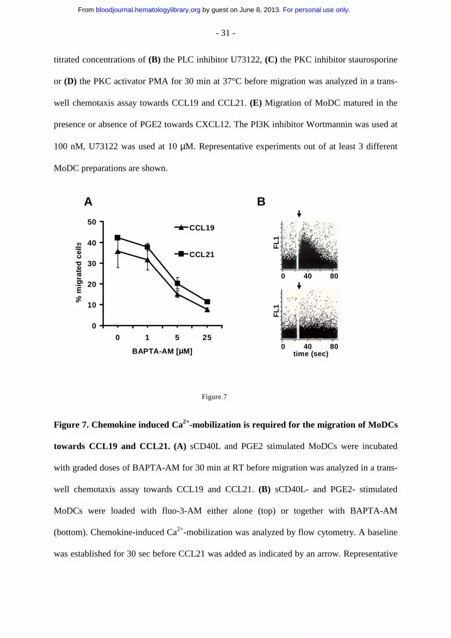
titrated concentrations of (B) the PLC inhibitor U73122, (C) the PKC inhibitor staurosporine
or (D) the PKC activator PMA for 30 min at 37°C before migration was analyzed in a trans-
well chemotaxis assay towards CCL19 and CCL21. (E) Migration of MoDC matured in the
presence or absence of PGE2 towards CXCL12. The PI3K inhibitor Wortmannin was used at
100 nM, U73122 was used at 10 µM. Representative experiments out of at least 3 different
MoDC preparations are shown.
FL
1
0 40 80
FL
10 40 80
time (sec)
0
10
20
30
40
50
0 1 5 25
BAPTA-AM [µµµµM]
% m
igra
ted
cel
ls
CCL19
CCL21
A B
Figure.7
Figure 7. Chemokine induced Ca2+-mobilization is required for the migration of MoDCs
towards CCL19 and CCL21. (A) sCD40L and PGE2 stimulated MoDCs were incubated
with graded doses of BAPTA-AM for 30 min at RT before migration was analyzed in a trans-
well chemotaxis assay towards CCL19 and CCL21. (B) sCD40L- and PGE2- stimulated
MoDCs were loaded with fluo-3-AM either alone (top) or together with BAPTA-AM
(bottom). Chemokine-induced Ca2+-mobilization was analyzed by flow cytometry. A baseline
was established for 30 sec before CCL21 was added as indicated by an arrow. Representative
For personal use only. by guest on June 8, 2013. bloodjournal.hematologylibrary.orgFrom

- 32 -
results obtained from MoDCs of one single donor are shown. Three independent experiments
with MoDCs from different donors yielded similar results.
For personal use only. by guest on June 8, 2013. bloodjournal.hematologylibrary.orgFrom
Related Documents


![RoleofPGE inAsthmaandNonasthmatic EosinophilicBronchitis2) by COXs, and metabolism of prostaglandin H 2 to prostaglandin E 2 via prostaglandin E synthase [12]. There are three enzymes](https://static.cupdf.com/doc/110x72/60d522031e41432a8f254505/roleofpge-inasthmaandnonasthmatic-eosinophilicbronchitis-2-by-coxs-and-metabolism.jpg)