ISSN 1344-1159 微生物遺伝資源利用マニュアル(17) “ 植物病原菌の同定と保存” 2004. 12 農 業 生 物 資 源 研 究 所

Welcome message from author
This document is posted to help you gain knowledge. Please leave a comment to let me know what you think about it! Share it to your friends and learn new things together.
Transcript

ISSN 1344-1159
微生物遺伝資源利用マニュアル(17)
“植物病原菌の同定と保存”
2004. 12
農 業 生 物 資 源 研 究 所
- 1 -

とりまとめ
独立行政法人
農業生物資源研究所 ジーンバンク
微生物資源研究チーム 佐藤 豊三・永井 利郎 富岡 啓介・竹内 香純
TEL 029-838-7053
- 2 -
目 次
ページ MAFF ジーンバンクのシステム ············································································· 1
農業生物資源研究所 ジーンバンク
微生物資源研究チーム 佐藤豊三
植物病原菌の分子同定のためのDNA シークエンス解析 ············································ 5
農業生物資源研究所 ジーンバンク
微生物資源研究チーム 竹内香純・長尾英幸
植物病原菌類の保存法 ······················································································· 23
農業生物資源研究所 ジーンバンク
微生物資源研究チーム・遺伝資源管理課 富岡啓介・永井利郎・飯田元子・佐藤豊三
植物病原細菌の保存法························································································ 33
農業生物資源研究所 ジーンバンク
微生物資源研究チーム・遺伝資源管理課 永井利郎・飯田元子
- 3 -

MAFF Microorganism Genetic Resources Manual
No. 17
Contents
Page MAFF Genebank system····················································································· 1
To ozo SATO y
s o
s o
Microorganism Genetic Resources Laboratory, Genebank, National Institute of Agrobiological Sciences
DNA sequencing for molecular identification of plant pathogens······························· 5
Kasumi TAKEUCHI and Hideyuki NAGAO Microorganism Genetic Resources Laboratory,
Genebank, National Institute of Agrobiological Sciences Preservation of plant pathogenic fungi ······························································· 23
Keisuke TOMIOKA, To hirou NAGAI Motoko IIDA and T yozo SATO Microorganism Genetic Resources Laboratory and Genetic Resources Management Section,
Genebank, National Institute of Agrobiological Sciences Preservation of plant pathogenic bacteria ···························································· 33
To hirou NAGAI and Motok IIDA Microorganism Genetic Resources Laboratory and Genetic Resources Management Section,
Genebank, National Institute of Agrobiological Sciences
- 4 -

微生物遺伝資源利用マニュアル (17) : 1–4 (2004)
MAFF ジーンバンクのシステム
佐藤豊三
農業生物資源研究所 ジーンバンク 微生物資源研究チーム
1985 年以前、農林水産省の試験研究機関で個々に行われていた遺伝資源研究と事業は、1985 年
に全国的なネットワークを有する「農林水産省ジーンバンク事業」として発足しました。農業生物
資源研究所は、植物、微生物 (Fig. 1)、動物の 3 部門のセンターバンクとして、国内外の遺伝資源
を収集、保存、配布し、成果を挙げてきました。1993 年には、バイオテクノロジー・ゲノム研究に
必要なDNA やDNA 情報を収集・蓄積・提供するDNA 部門の運営を開始しました。2001 年から
は、国立試験研究機関の独立行政法人化に伴い、旧農業生物資源研究所、旧蚕糸・昆虫農業技術研
究所などが 1 機関に統合されました。また、その際、林木、水産関係の遺伝資源事業が独立しまし
たが、大部分の基本的な事業の枠組みを変えることなく、独立行政法人農業生物資源研究所が実施
主体となり新たな体制の下で「農業生物資源ジーンバンク事業」の運営を図っています。 農業生物資源ジーンバンク微生物部門では、国の内外から微生物遺伝資源を探索・収集し、分類・
同定を行うとともに、特性評価を実施し、これらを増殖・保存しています。このようにして得られ
た微生物遺伝資源は、国内外の研究者に提供され、広く活用されています。これらの微生物遺伝資
源に付随する情報は、データベース化し、インターネット上で公開されています (http://www.gene.affrc. go.jp/micro/, Figs. 2, 3, 4)。 同部門では、中央農業総合研究センター、果樹研究所、花き研究所、野菜茶業研究所、畜産草地
研究所、動物衛生研究所、九州沖縄農業研究センター、農業生物資源研究所基盤研究部門、農業生
物資源研究所昆虫・動物生命科学研究部門、農業環境技術研究所、食品総合研究所、国際農林水産
業研究センターがサブバンクとなり、細菌、放線菌、動物マイコプラズマ、ファイトプラズマ、リ
ケッチア、酵母、糸状菌、動物ウイルス、植物ウイルス、バクテリオファージ、ウイロイド、原虫、
線虫、細胞融合微生物および昆虫培養細胞を収集・保存・配布しています (Figs. 5, 6)。また、セン
ターバンクでは、主に細菌、放線菌、酵母、糸状菌、植物ウイルスを上記サブバンクや森林総合研
究所、大学、都道府県の試験研究機関などより受入れ、増殖・保存・配布しています。 農業生物資源ジーンバンク微生物部門に登録保存されている微生物の多くは植物病原微生物であ
る。糸状菌、植物ウイルス、昆虫培養細胞を液体窒素の気相(-165ºC)で、また、細菌、放線菌、
酵母および糸状菌の胞子を真空凍結乾燥法により、さらに植物ウイルスは感染葉のL-乾燥と超低温
保存(-80ºC)により長期保存を行っており、定期的にそれら保存株の活性と雑菌混入の有無、さら
に糸状菌株では分類学的特性に関して検査を行っている。
- 1 -

MAFF Genebank system
Toyozo SATO
Microorganism Genetic Resources Laboratory, Genebank, National Institute of Agrobiological Sciences
1) The Genebank Project, consists the managements of the genetic resources of Plant,
Microorganism (Fig. 1), Animal, Forest tree, Aquatic Organism and DNA, supported by the Ministry of Agriculture, Forestry and Fisheries (MAFF) of Japan. The National Institute of Agrobiological Sciences (NIAS), one of the Independent Administrative Institutions, manages the Plant, Microorganism, Animal genetic resources and DNA as a central Genebank in collaboration with sub-banks. The Genebank involves the conservation of genetic resources. This includes both domestic and foreign exploration, propagation of genetic resources, characterization, evaluation, preservation, quality control, rejuvenation and documentation. An important component of the Genebank is the genetic resources which are used by scientists in the public and private sectors nationwide and the information it makes available to scientists worldwide (http://www.gene.affrc.go.jp/micro/, Figs. 2, 3, 4). NIAS genetic resources scientists are actively collaborating with in traditional and national research institutes worldwide.
2) In 2001, NIAS, was established as a Independent Administrative Institution to study the
biotechnical aspects of plants, animals and microorganisms and genetic resources based on the National Institute of Agrobiological Resources (NIAR) and the National Institute of Sericultural and Entomological Sciences (NISES). Prior to 1985, genetic resources work was independently conducted by different institutes across Japan. In 1985 an integrated system for the whole country was developed, in which the NIAR became the central coordinating institute for plant, microorganism and animal genetic resources. NIAS has a base and active collection and different institutes across Japan act as sub-banks with active collection for plants, microorganisms and animals adopted to their locations. In 1993 a DNA bank was initiated to preserve molecular materials and information. In Microorganisms Section of the Genebank, the National Research Institute of Fisheries Science and the Forestry and Forest Products Research Institute contract out of the
- 2 -
Genebank network in 2001. 3) The sub-banks of the microorganisms section of the Genebank are as follows: Genome and
Biodiversity Research Center and Division of Insect and Animal Science of NIAS, National Agricultural Research Center, National Institute of Fruit Tree Science, National Institute of Floricultural Science, National Institute of Vegetables and Tea Science, National Institute of Livestock and Grassland Science, National Institute of Animal Health, National Agricultural Research Center for Kyushu Okinawa Region, National Institute for Agro-Environmental Sciences, National Food Research Institute, and Japan International Research Center for Agricultural Sciences. The Forestry and Forest Products Research Institute is one of the main collaborative institutes of the Genebank microorganisms section because the institute has deposited numerous mushroom and toadstool strains to the section.
4) The Genebank microorganisms section collects, propagates, preserves and provides
bacteria including Actinomycetes, fungi including yeasts, plant viruses, viroids, animal viruses, bacteriophages, phytoplasmas, mycoplasmas, protozoa, nematodes, fusants (fused yeast cells) and cultured insect cells (Figs. 5, 6). The central bank of the section deals with those microorganisms except for viroids, animal viruses, phytoplasmas, mycoplasmas, protozoa and nematodes. The acronym “MAFF” is preposed accession numbers of our microorganisms strains. Our collections are mainly characterized by plant pathogenic microorganisms such as bacteria, fungi and plant viruses. In the central bank, bacterial, Actinomycetous and yeast strains are preserved by freeze-dry technique, and fungal strains are stored by cryopreservation in vapor phase of liquid nitrogen at ca. -165 ºC. These long-term-preserved strains are periodically checked for their viability and taxonomic identity.
- 3 -

Fungi 11,628 Bacteria 7,507
Yeasts 585
Viruses 493 Cells 58 Misc. 10
Protozoa 191
Total 20,472
Fig. 5 The number of preserved strainsin the central bank and the total numberof deposited strains in the MAFF Genebank
1985
1986
1987
1988
1989
1990
1991
1992
1993
1994
1995
1996
1997
1998
1999
2000
2001
2002
2003
Num
ber
Year
0
5
10
15
20
25x1000
Total
Central bank
Fig. 2 Computer Servers of the GenebankFig. 1 The central bank of microorganisms section
Fig.3 The catalogue database of MAFF strains
Fig. 6 Details of MAFF strains in 2003
Fig.4 A terminal of the database system
- 4 -
微生物遺伝資源利用マニュアル (17) : 5–22 (2004)
植物病原菌の分子同定のための DNA シークエンス解析
竹内香純・長尾英幸
農業生物資源研究所 ジーンバンク 微生物資源研究チーム
1. はじめに
MAFF ジーンバンク微生物部門では、多様な植物病原菌類の維持および保存を行っている。多く
の菌類は、低温(4℃)での維持や超低温(-80℃)での保存が可能であるが、一部の菌類では胞子
形成能の低下がしばしばみられる。菌類を同定する上で、分生子と分生子柄の形態ならびに形成の
様式は必要不可欠な情報であるので、上記のような問題が生じると形態的特徴による培養株の同定
が困難となる。近年、DNA シークエンス解析などの分子生物学的技法の導入により、無胞子性培
養株および無胞子不完全菌類の培養株が同定可能となった。また菌株によっては系統学的な関係が
決定されるようになった。こうして、病原菌の同定が迅速かつ正確に行えるようになったものの、
検索対象となる DNA データベースは各自のボランティアにより構築されているものであり、デー
タ蓄積が進行中であるため、登録されたデータには偏りがある。たとえば、Mycosphaerella は ITS(Internal transcribed spacer)、18S および 28S リボゾーマルRNA をコードする遺伝子(rDNA)、クチナーゼ、 キシラナーゼ、およびマイクロサテライト DNA の遺伝子が登録されているが、
Pyrenochaeta に関しては登録情報が全くない。また、任意の菌株が系統学的に同一のデータに必
ずしもヒットするわけではなく、形態学的な特徴に関するデータが利用できない場合、系統学的な
関連を裏付けるためには少なくとも2種類の領域の比較が必要である。 この項では、植物病原菌の同定方法として ITS 領域のシークエンス解析の利用法を紹介する。
rDNA は病原菌の同定に適した特徴を有しており、非常に安定し、また、ゲノム中保存された領域
(5S、18S、28S)と多様性に富む領域(ITS)が混在している。ITS 領域では特異的プライマーを
用いることにより近縁種の菌株同士を区別することができる(Bryan et al., 1995)。 ABI オートシークエンサーによるDNA 塩基配列の決定 この方法は、プラスミドの挿入配列やPCR産物などのDNA二重鎖を解読するのに用いられる。
シークエンス反応はジデオキシ法に基づき、Big Dye® Terminator Cycle Sequencing Kit (Applied Biosystems, Foster city, CA, USA)を使用して行う。これは 4 種類のジデオキシヌクレ
オチド(ddNTPs)それぞれが、別々の色の蛍光で標識されており、伸長反応時に最後に取り込ま
れ、それらをオートシークエンサーにより解読することで塩基配列が決定される。 所要時間: 2~3 日
- 5 -
2. 使用する機器
オートシークエンサー (ABI PRISM® 3100-Av nt DNA Sequencer、Fig. 1)、サーマルサイクラー a 3. 試薬
BigDye® Terminator Cycle Sequencing Kit 適当なプライマー Hi-DiTM Formamide Genetic Analyzer buffer with EDTA [10 ×]
4. プロトコル
Nucleic acid extraction and PCR amplification → Cycling for sequencing → Sample Preparation → Sequencing 1) Nucleic acid extraction and PCR amplification 各DNA 断片を direct PCR method (Suyama et al., 1996)の変法により抽出および増幅する。
PCR はHotstartTaq master Mix (Qiagen, Hilden, Germany)を用いて行う。 (1) サンプルチューブに以下の溶液を混合する:
HotstartTaq master Mix 12.5 µl プライマー(ITS1b など) 5 pmol(各々) テンプレート 2.5 µl 滅菌水を加え、 25 µl とする
(2) ピペッティングで混合した後、95ºC に保温しておいたサーマルサイクラーにセットし、下記
のプログラムにより反応を行う。非特異的なアニーリングを防ぐため、あらかじめ 95ºC に
しておき、ホットスタートを行うこと。 95ºC 15 min. (ホットスタート) その後、 95ºC 30 sec. 55ºC 1 min. 72ºC 1 min. 以上を 35 サイクル行う: 72ºC 10 min.(最終的な伸長反応)、 その後 4ºC にて保存
増幅後、PCR 産物を Microspin columns S-400HR (Amersham Biosciences Corp., Piscataway, NJ, USA)を用いて精製する。
- 6 -

2) Cycling for sequencing (1) サンプルチューブに各反応溶液を準備する:
Ready reaction mix 8 µl テンプレートDNA (PCR 産物) 3 µl プライマー(ITS1b など) 1.6 pmol 滅菌水を加え、10 µl とする
(2) ピペッティングで混合した後、96ºC に保温しておいたサーマルサイクラーにセットし、下記
のプログラムにより反応を行う。非特異的なアニーリングを防ぐため、あらかじめ 96ºC に
しておき、ホットスタートを行うこと。 96ºC 3 min.(ホットスタート) その後、
96ºC 10 sec. 50ºC 15 sec. 60ºC 4 min. 以上を 25 サイクル行った後、 4ºC で保存
反応終了後、AutoSeq G-50(Amersham Biosciences Corp., Piscataway, NJ, USA)に供
し、余剰の蛍光ターミネーターを除去する。その後、ABI3100 automated DNA sequencer により解析を行う。
3) Sample Preparation
(1) シークエンス反応済み(精製済み)サンプルチューブにHi-DiTM Formamideを 10~15 µl加える。
(2) ボルテックスで十分混合し、遠心器でスピンダウンする。 (3) キャップをして 95ºC で 2 分間加熱することで変性させる。 (4) すぐに氷上に置き、5 分以上冷却する。 (5) 遠心器でスピンダウンする。 (6) サンプルを 96-Well サンプルプレートに移す。気泡が入らないように注意する。 (7) プレートアッセンブリをセットする。
↓サンプルプレート上にセプタでしっかり蓋をする。 ↓プレートベースにサンプルプレートを置く。 ↓プレートリテイナーでサンプルプレートとセプタをプレートベースに固定する。
- 7 -
4) Sequencing Run の前に、ポリマーが必要量あるか確認する。ポリマーの充填はオリジナルのプロトコルに従
って行うこと。バッファーは、10 X Genetic Analyzer buffer 2.5 ml に対し、イオン交換水で 25 mlにメスアップしたものを準備して用いる。 以下の操作は、オリジナルのプロトコルに従って行うこと。
・プレートレコードの作成 ・プレートの設定とリンク ・Run セットアップの確認とRun スタート
5. データ解析 得られた解析データの塩基配列はSequencing Analysis™ (ABI) およびAutoAssembler™ (ABI)
により調整する。 シークエンス解析は対象により以下の 2 つの方法(A およびB)のいずれかによって行う。
A) 無胞子性培養株および無胞子不完全菌類の場合 (1) DDBJ のweb サイト(http://ddbj.nig.ac.jp)(Fig. 2)より、 BLAST (basic local alignment
search tool)を選択。このサイトでは、データベース中の類似性の高い配列が示される。サ
イト中の”query box”(Fig. 3)に対象とする配列をペーストする。 (2) 提示された結果の中から、関心のあるアクセッション番号をクリックし、分類学的な情報と
登録されている塩基配列を得る(Figs. 4 and 5)。 (3) 特定の属を調べる場合は、DDBJ (Fig. 2)中にある TXSearch (Fig. 6)をクリックする。
学名を入力し(属名のみの入力が便利である)、“matching”レベルと分類学ランクを選択す
る(Genus または species)。 “Search”をクリックする。 (4) 提示された“Detail taxonomy information you specified”ウィンドウ中の “GO” をクリック
し、関連した分類単位(taxon)のシークエンスを得る(Fig. 6)。 (5) このような手順で登録された属次第で種レベルの複数のデータが得られる(Fig. 7)。DDBJ
accession number をクリックすると登録データ(entry)の詳細が表示される(Fig. 8)。 (6) 系統学的な比較
• Clustal X にデータベースから得られたシークエンス(ex. Fig. 8)または解析したシー
クエンスをペーストする。 • 解析 • “complete align”が終了したらアライメントをGDE format で保存。アライメントはさ
らにSe-Al ver. 2.0 を用いて手作業で曖昧なポジションを削除し、NEXUS format で保
存。系統樹の作成はneighbor joining(NJ)method(Kimura 2-parameter distance calculation)および maximum parsimony (MP)using the heuristic search(random
- 8 -
addition with 600 replicates)with TBR-branch swapping を適用してPAUP 4.0b10(Swofford, 1998)で行う。Relative support for the branches was estimated with for NJ と MP 解析による系統樹の信頼性は 1000 回の bootstrap 値(%)によって判定
する(Felsenstein 1985)。一般に、50% 以上の値で “support”となる。その中でもレ
ベルが分かれており、50-70% で “poor support”、70-80% で “support”、90% 以上で “well support” とされる(Farr et al., 2002)。このような結果に基づき近縁な分類単位
の推測や同定が可能となる。
B) 分類学的には既知の菌類であるが、病原性あるいは培養性状が異なる場合 • A の項目(5)から解析を始める。
6. 引用文献 Bryan GT, Daniels MJ, Osbourn AE (1995) Comparison of fungi within the
Gaeumannomyces-Phialophora complex by analysis of ribosomal DNA sequence. Appl. Environ. Microbiol. 61: 681-689.
Farr DF, Castleburry LA, Rossman AY (2002) Morphological and molecular characterization of Phomopsis vaccinii and additional isolates of Phomopsis from blueberry and cranberry in the eastern United States. Mycologia 94: 494-504.
Felsenstein J (1985) Confidence limits on phylogenies: an approach using the bootstrap. Evolution 6: 227-242.
Hibbett DS (1992) Ribosomal RNA and fungal systematics. Trans. Mycol. Soc. 33: 533-556. Suyama Y, Kawamuro K, Kinoshita I, Yoshimura K, Tsumura Y, Takahara H (1996) DNA
sequence from a fossil pollen of Abies spp. from Pleistocene peat. Genes Genet Syst 71: 145-149.
Swofford DL (1998) PAUP*4.0. Phylogenetic analysis using parsimony. Sunderland, Massachusetts: Sinauer Associates.
White TJ, Bruns T, Lee SB, Taylor J (1990) Amplification and direct sequencing of fungal ribosomal RNA genes for phylogenetics. In: Gelfand M, Sninsky D, White T (eds) PCR protocols: a guide to methods and applications. Academic Press, San Diego, pp 315-322.
- 9 -
DNA sequencing for molecular identification of plant pathogens
Kasumi TAKEUCHI and Hideyuki NAGAO
Microorganism Genetic Resources Laboratory, Genebank, National Institute of Agrobiological Sciences
1. Principle
Microorganisms section of the MAFF Genebank has maintained and preserved different kinds of plant pathogenic fungi. While many fungi can be maintained at low temperature (4ºC) or preserved at ultra-low temperature (-80ºC), some fungi often decreased or lost their ability of spore formation. This makes it difficult to identify the culture by the morphological characterization because the morphology of conida including the shape of conidiophore and conidiogenesis is essential for the identification of fungi. Recently, introducing the molecular techniques such as DNA sequence analysis, asporogenic and/or agonomycete culture have been identified. In some fungi, the phylogenetic relationship could be determined. This has allowed us to identify pathogens rapidly and precisely. However, the nucleotide database is constructed by voluntary contributions and is still on-going. So the recorded data have been biased; ITS (Internal transcribed spacer), 18s and 28s ribosomal RNA genes (rDNA), genes for cutinase and xylanase, and microsatellite DNA for M cosphaerella, otherwise none for Pyrenochaeta. Arbitary isolates may not be necessarily hit on the phylogenetically identical data. Unless morphological characterization is useful, it is needed to compare at least two sequences from different regions for referring the phylogenetic relatedness.
y
cer
In this article, we describe the application of sequence analysis of PCR-amplified ITS for phytopathogenic fungal detection. rDNA possess characteristics that are suitable for detecting pathogens. These rDNA are highly stable and exhibit a mosaic of conserved and diverse regions within the genome (Hibbett, 1992). ITS regions have been used successfully to generate specific primers capable of differentiating closely related fungal species (Bryan et al., 1995). • DNA sequencing by ABI autosequen
This method is used to sequence double stranded DNA, such as a plasmid insert or purified
PCR product based on dideoxy chain termination method with a BigDye® Terminator Cycle
- 10 -

Sequencing Kit (Applied Biosystems, Foster city, CA, USA). The dideoxy chain termination method of DNA sequencing involves the in vitro synthesis of a DNA strand by a modified DNA polymerase using a single stranded DNA template. Synthesis is initiated at a specific site on the sequencing template, as determined by the annealed sequencing oligonucleotide primer. The synthesis reaction is terminated by adding a dideoxynucleotide analog (ddNTP) that will not accept further elongation of the synthesized strand. Each ddNTP is labeled with distinct fluorescence to be detected by ABI autosequencer. Time required: 2-3 days 2. Instruments
Autosequencer (ABI PRISM® 3100-Av nt, Fig. 1), thermal cycler a 3. Chemicals
BigDye® Terminator Cycle Sequencing Kit Primers Hi DiTM Formamide Genetic Analyzer buffer with EDTA [10 ×]
4. Protocol
Nucleic acid extraction and PCR amplification → Cycling for sequencing → Sample preparation → Sequencing 1) Nucleic acid extraction and PCR amplification
Each DNA fragment is extracted and amplified by a method modified from the direct PCR method (Suyama et al., 1996). PCR is performed using a HotstartTaq master Mix (Qiagen, Hilden, Germany).
(1) Prepare the following reaction mixture in a microcentrifuge tube: HotstartTaq master Mix 12.5 µl Primer (ITS1b etc.) 5 pmol each Template 2.5 µl Sterilized water to a final volume of 25 µl
- 11 -

(2) Mix briefly by pipetting up and down. Place the reaction tubes in a thermal cycler that has been preheated to 95ºC and start the cycling program as follows. It is important to preheat the thermal cycler to 95ºC to prevent nonspecifically annealed primers from being extended.
95ºC for 15 min. (hot start), then: 95ºC 30 sec. 55ºC 1 min. 72ºC 1 min. 35 cycles total, then: 72ºC 10 min. (for a final extension period) , then 4ºC (hold)
Following amplification, the PCR products are purified with Microspin columns S-400HR (Amersham Biosciences Corp., Piscataway, NJ, USA)
2) Cycling for sequencing
(1) Prepare the following reaction mixture in a microcentrifuge tube: Ready reaction mix 8 µl Template DNA (PCR product) 3 µl Primer (ITS1b etc.) 1.6 pmol Sterilized water to a final volume of 10 µl
(2) Mix briefly by pipetting up and down. Place the reaction tubes in a thermal cycler that
has been preheated to 96ºC and start the cycling program as follows. It is important to preheat the thermal cycler to 96ºC to prevent nonspecifically annealed primers from being extended.
96ºC for 3 min. (hot start), then: 96ºC 10 sec. 50ºC 15 sec. 60ºC 4 min. 25 cycles total, then 4ºC (hold)
Amplified products are sequenced with the BigDye® terminator kit on an ABI 3100 autosequencer using the following primers: ITS1b, ITS4, NL1, NL4 (White et al., 1990). AutoSeq G-50 (Amersham Biosciences Corp., Piscataway, NJ, USA) is used to remove excess fluorescent dye-terminators from sequencing reactions prior to analysis on autosequencer.
- 12 -
3) Sample preparation for the ABI PRISM 3100 Genetic Analyzer (1) Add 10-15 µl of HI-Di formamide into each tube. (2) Vortex and quick spin. (3) Denature the samples with caps by placing them at 95ºC for 2 minutes. (4) Place immediately on ice for at least 5 minutes. (5) Quick spin. (6) Deliver samples in a Microamp 96-Well Reaction Plate. Remove air bubbles. (7) Prepare a plate assembly
↓Secure a clean and dry septa strip on the sample plate ↓Place the sample plate into the plate base ↓Snap the plate retainer onto the plate and plate base
4) Sequencing
Before running, check if the volume of polymer is enough and replace the buffer and deionized water. To refill polymer, see the original manual. To prepare 25 ml of 1 X Genetic Analyzer buffer with EDTA, add 2.5 ml of 10 X Genetic Analyzer buffer into a graduated cylinder, add deionized water to bring the total volume up to 25 ml and mix well.
For the following steps, please see the user reference guide. • Creating a Plate R rd eco
tr
• Linking a Pla e to a Plate Record • Sta ting and Monitoring the Run
5. Analyzing Data
The resulting sequence was edited using Sequencing Analysis™ (ABI) and AutoAssembler™ (ABI).
There are two ways (A and B) to conduct the resulting sequence. A) For asporogenic and/or agonomycete culture
(1) Via web site (DDBJ/http://ddbj.nig.ac.jp) (Fig. 2), BLAST (basic local alignment search tool) is useful to pick up the similar sequence data in the database. Copy and paste your query in “query box” (Fig. 3).
(2) After getting the result, click and check its taxonomical information and sequence data (Figs. 4 and 5).
(3) If your interest is in a certain genus, proceed to the other site in DDBJ (http://ddbj.nig.ac.jp)(Fig. 2), and click TXSearch (Fig. 6). Input the taxonomic name (Genus is better) and select “matching” level and taxonomic rank (choose Genus or species). Then click “Search”.
- 13 -

(4) In the window of “Detail taxonomy information you specified”, click “GO” to get sequence(s) related to this taxon (Fig. 6).
(5) Then you can search the multiple data on the species level, depending on the contributed genus (Fig. 7). Click the DDBJ accession number to obtain the detail information about its entry. You can find recorded sequence data (Fig. 8).
(6) Phylogenetic comparison • Copy and paste these data from the database (ex. Fig. 8) or your original sequence
data to Clustal X. • Analyze • After finishing “complete align”, save the alignment in GDE format.
Alignments are manually adjusted using Se-Al ver. 2.0 and ambiguously aligned positions are excluded from the analyses (save as NEXUS format). Trees are inferred using PAUP 4.0b10 (Swofford, 1998) with the following methods: the neighbor joining (NJ) method (Kimura 2-parameter distance calculation), maximum parsimony (MP) using the heuristic search (random addition with 600 replicates) with TBR-branch swapping. All molecular characters are unordered and given equal weigh during analyses. Relative support for the branches is estimated with 1000 bootstrap replications (Felsenstein 1985) for NJ and MP analyses. The bootstrap value is shown as a percentage and its confidential range is not determined statically. In general, 50% or more is called “support”. For example, the range 50-70% is “poor support”, 70-80% is “support”, and more than 90% is “well support” (Farr et al., 2002). You can suppose or identify the closest taxon.
B) For taxonomically known fungus but different in pathogenicity or cultural
characteristics • Begin analysis from (5) in A).
6. References Bryan GT, Daniels MJ, Osbourn AE (1995) Comparison of fungi within the
Gaeumannomyces-Phialophora complex by analysis of ribosomal DNA sequence. Appl. Environ. Microbiol. 61: 681-689.
Farr DF, Castleburry LA, Rossman AY (2002) Morphological and molecular characterization of Phomopsis vaccinii and additional isolates of Phomopsis from blueberry and cranberry in the eastern United States. Mycologia 94: 494-504.
Felsenstein J (1985) Confidence limits on phylogenies: an approach using the bootstrap. Evolution 6: 227-242.
- 14 -

Hibbett DS (1992) Ribosomal RNA and fungal systematics. Trans. Mycol. Soc. 33: 533-556. Suyama Y, Kawamuro K, Kinoshita I, Yoshimura K, Tsumura Y, Takahara H (1996) DNA
sequence from a fossil pollen of Abies spp. from Pleistocene peat. Genes Genet Syst 71: 145-149.
Swofford DL (1998) PAUP*4.0. Phylogenetic analysis using parsimony. Sunderland, Massachusetts: Sinauer Associates.
White TJ, Bruns T, Lee SB, Taylor J (1990) Amplification and direct sequencing of fungal ribosomal RNA genes for phylogenetics. In: Gelfand M, Sninsky D, White T (eds) PCR protocols: a guide to methods and applications. Academic Press, San Diego, pp 315-322.
- 15 -

Fig. 1. ABI PRISM® 3100-Avant (above) and its inside view (below).
- 16 -
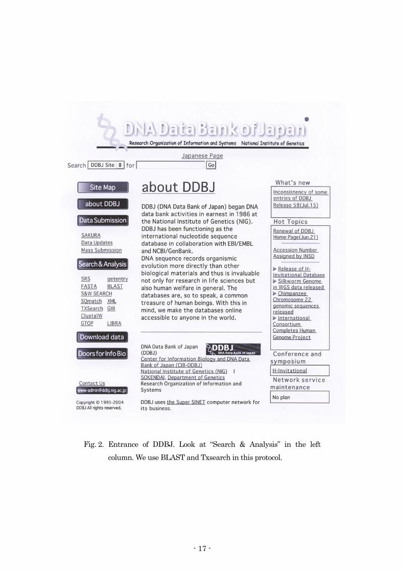
Fig. 2. Entrance of DDBJ. Look at “Search & Analysis” in the left column. We use BLAST and Txsearch in this protocol.
- 17 -

Fig. 3. Window of BLAST. Select “clear-all” in DIVISION and check again “Plants”. Copy and paste your query in query box (left: blank box). Example of pasted query (right).
- 18 -

Fig. 4. Result of query submission. It shows the similarity ranking of candidates. For ITS of Exobasidium, there is no record of known Exobasidium.
Fig. 5. Result of query submission. It shows the similarity ranking of candidates. For rDNA of Phomopsis, you can find some records of known Phomopsis.
- 19 -
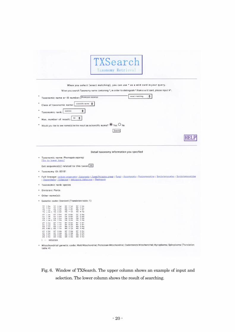
Fig. 6. Window of TXSearch. The upper column shows an example of input and
selection. The lower column shows the result of searching.
- 20 -

Fig. 7. A list of recorded sequences.
- 21 -
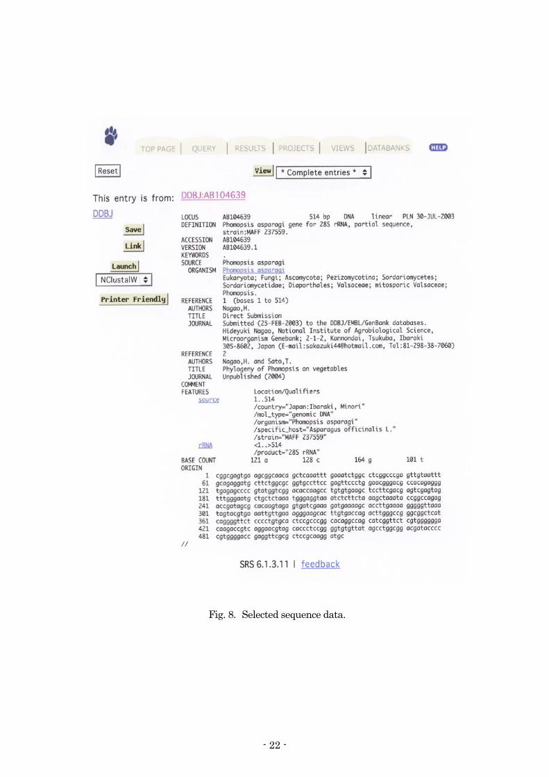
Fig. 8. Selected sequence data.
- 22 -

微生物遺伝資源利用マニュアル (17) : 23–32 (2004)
植物病原菌類の保存法
富岡啓介・永井利郎・飯田元子・佐藤豊三
農業生物資源研究所 ジーンバンク 微生物資源研究チーム・遺伝資源管理課
多種多様な菌類の保存には各種菌群に応じた方法が採られる。それらの中で最も適用範囲が広く、
菌株の諸性質を長期に渡って安定的に維持できる最適な方法が、液体窒素の液相(-196ºC)もしく
は気相(-165ºC)内での超低温凍結保存法である。本法は、菌株の代謝を休止させることから遺伝
的変異の心配がない。したがって、植物病原性等の諸特性の維持が要求される菌株の保存には、未
だ例外があるものの、可能な限りこの方法を適用することが望ましい。ここでは、植物病原菌類の
超低温凍結保存法[気相(-165ºC)]について、NIAS ジーンバンクでの保存法を例に概説する。
1. 準備 1) 器具 液体窒素気相-165ºC 貯蔵槽(Fig. 1, 大陽日酸(株))、プログラムフリーザー(Fig. 2, (株)日
本フリーザー製, TNP-87S-DX)、-80~-40ºC 冷凍庫(Fig. 3, (株)サンヨー製, MDF-382)、4~40ºC 恒温器(Fig. 4, (株)平澤製作所製, CP シリーズ, Te-Her Digital Low Temp Incubator)、フ
リージングコンテナ(Fig. 5, ナルジェヌンクインターナショナル(株)製, Mr.フロスティー)、50ºCウォーターバス(Fig. 6, タイテック(株)製, Personal 11)、オートクレーブ(Fig. 7, (株)トミ
ー製, BS-325)、クリーンベンチ(Fig. 8, (株)日立製, SCV)、顕微鏡(Fig. 9, (株)ニコン製, マイクロフォト, ライカ製, MZ125)、近紫外線照射機((株)ナショナル製, ブラックライトブルー蛍
光灯 FL20S/BL-B)、クライオチューブ(住友ベークライト(株)製, セラムチューブ)、シャーレ
((株)栄研器材製)、その他菌株の無菌操作に用いる一般的な器具(Fig. 10) 2) 培地 菌株培養に使用する培地は菌株による。NIAS ジーンバンクで一般的に使用している植物病原菌
類用培地は主に以下の8種類。いずれも121ºC で15分間高圧蒸気滅菌したものを用いる(Fig. 11)。
① Water agar[WA: 2% 寒天抹]
・・・ 主に雑菌除去、単胞子分離、単菌糸分離に使用
- 23 -

② Potato dextrose agar[PDA: ジャガイモ(200~300 g/l)煮沸浸出液, 2% ブドウ糖, 1.8% 寒天抹]
・・・ 大半の菌に適用
③ Modified Weitzman-Silva-Hutner agar[mWSH: 0.1% NaNO3, 0.1% MgSO4·7H2O, 0.1%
KH2PO4, 1% 粉砕オートミール, 2% 寒天抹] ・・・ 大半の菌に適用
④ Potato carrot agar[PCA: ジャガイモ(20 g/l)・ニンジン(20 g/l)煮沸浸出液, 2% 寒天抹]
・・・ 主にColletotrichum 属菌に適用 ⑤ Synthetic low nutrient agar[SNA: 0.1% KH2PO4, 0.1% KNO3, 0.05% MgSO4·7H2O, 0.05%
KCl, 0.02% ブドウ糖, 0.02% ショ糖, 2% 寒天抹, 0.024 g/l NaOH] ・・・ 主にFusarium 属菌に適用
⑥ Oat meal agar[OMA: オートミール(30 g/l)煮沸浸出液, 0.5% ショ糖, 1.8% 寒天抹]
・・・ 主にPyricularia 属菌に適用 ⑦ V-8 juice agar[V-8A: 4.5 gのCaCO3を添加したV-8 野菜ジュース 400 mlの低速遠心(4,000
rpm, 20 min.)上清 200 ml/l、1.8% 寒天抹] ・・・ 主に卵菌類に適用
⑧ Hemp seed agar[HSA: 粉砕アサ種子(20 g/l)煮沸浸出液, 1.5% 寒天抹]
・・・ 主に卵菌類に適用
3) 凍害防止用保護剤
NIAS ジーンバンクで一般的に使用している凍害防止用保護剤は主に以下の 3 種類である。20% グリセリン、20% スキムミルクおよび 20% ジメチルスルホキシドをその都度混合して作成する。
20% グリセリンは 121ºC で 15 分間高圧蒸気滅菌する。20% スキムミルクは 115ºC で 15 分間高
圧蒸気滅菌し、一晩置いた後、110ºC で 10 分間高圧蒸気滅菌する。 ① 10% グリセリン ・・・大半の菌に適用 ② 10% グリセリン + 10% スキムミルク ・・・主に卵菌に適用 ③ 10% グリセリン + 10% ジメチルスルホキシド ・・・主に卵菌に適用
- 24 -

2. 操作手順 1) 保存 液体窒素[気相(-165ºC)]を用いた超低温凍結保存のための操作の概要をFig. 12 に示す。 ① 菌株を平板培地や斜面培地で培養する。培養条件は菌株による。なお、近紫外線照射下での培
養によって胞子形成が促進される場合がある。 ② 培養菌叢から菌体(含菌培地片もしくは生殖・耐久器官である胞子や菌核など)を採取する
(Fig. 13)。 ③ 得られた菌体をクライオチューブ内で凍害防止用保護剤と混和する(Fig. 14)。 ④ 過冷却後凍結ショック(細胞内での氷結晶による細胞破壊)による菌株の生残率低下を防止す
るため、緩慢凍結を基本とする予備凍結を行う。予備凍結はプログラムフリーザーを用いて実
施するか、4ºC 恒温器と-80ºC 冷凍庫を用いて実施し、この過程を経て菌株を液体窒素気相
-165ºC 貯蔵槽で保存する。NIAS ジーンバンクでは、プログラムフリーザーを用いる場合、
「5ºC まで-3ºC/分→-50ºC まで-1ºC/分→-80ºC まで-5ºC /分」を基本的な制御条件としている。
4ºC 恒温器と-80ºC 冷凍庫を用いる場合は、「4ºC・3 日間→-80ºC 冷凍庫で 3 日間以上」を基
本条件としている。なお、特に卵菌についてはプログラムフリーザーを用いている。また、プ
ログラムフリーザーの代わりにフリージングコンテナを用いて緩慢凍結を行うこともできる。
菌体と凍結防止剤の入ったクライオチューブをフリージングコンテナの内蓋に空いた穴に 1本ずつ入れ、内蓋と外蓋との間にイソプロピルアルコールを満たして外蓋を閉め、4ºC に 1日置き、-70ºC のディープフリーザーに入れて 2 日間保つ。その後、液体窒素気相内にチュー
ブを移す。 2) 復元 ① 液体窒素気相-165ºC 貯蔵槽から取り出した菌株(クライオチューブ)を 50ºC ウォーターバス
に 1~1.5 分間浸して解凍する(Fig. 6)。菌株数が多い場合などは、ウォーターバスでの処理
までに、一度、-80~-40ºC 冷凍庫に保管しても良い(Fig. 15)。高い生残率を得るためには、
できるだけ急速に解凍することが重要である。 ② 解凍した菌株を平板培地や斜面培地で培養する(Fig. 16, 17)。培養条件は菌株による。 ③ なお、NIAS ジーンバンクでは、液体窒素気相-165ºC 貯蔵槽に保存した菌株が雑菌の混入な
しに生きているか否かを保存1ヶ月後と1年後に復元培養して検査している(Fig. 18)。また、
配布時にも同様に検査した上で配布している。植物病原菌類の場合、保存 1 年後と配布時に
病原性のチェックも行う場合がある。
- 25 -

Preservation of plant pathogenic fungi
Keisuke TOMIOKA, Toshirou NAGAI, Motoko IIDA and Toyozo SATO
Microorganism Genetic Resources Laboratory and Genetic Resources Management Section, Genebank, National Institute of Agrobiological Sciences
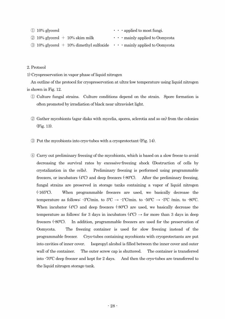
The preservation of fungi requires different methods for each fungal group. Cryopreservation at ultra-low temperature in the liquid (-196ºC) or vapor phase (-165ºC) of liquid nitrogen has the widest scope, and is also the most suitable for stable maintenance of many fungal characters over a long period of time. This method stops the metabolism of fungi, and does not cause hereditary variation. Therefore, using this method as much as possible is hopeful for storage of fungi, on which maintenance of characteristics such as phytopathogenicity are required, though there are still exceptions. Here, we describe a protocol of the cryopreservation of plant pathogenic fungi in a vapor phase of liquid nitrogen (-165ºC) in our laboratory. 1. Preparation 1) Implements
Storage tanks containing a vapor of liquid nitrogen (-165ºC) (Fig. 1, TAIYO-NISSAN Co. Ltd.), programmable freezers (Fig. 2, NIHON FREEZER Co. Ltd., TNP-87S-DX), deep freezers (-80~-40ºC) (Fig. 3, SANYO Co. Ltd., MDF-382), incubators (4~40ºC) (Fig. 4, HIRASAWA WORKS Co. Ltd., CP series, Te-Her Digital Low Temp Incubator), freezing containers (Fig. 5, NALGE NUNK INTERNATIONAL Co. Ltd., Cryo 1ºC Freezing Container), water baths (50ºC) (Fig. 6, TAITECH Co. Ltd., Personal 11), autoclaves (Fig. 7, TOMY Co. Ltd., BS-325), clean benches (Fig. 8, HITACHI Co. Ltd., SCV), microscopes (Fig. 9, NIKON Co. Ltd., Microphoto, LEICA Co. Ltd., MZ125), black lights (NATIONAL Co. Ltd., FL20S/BL-B), cryo-tubes (SUMITOMO BAKELITE Co. Ltd., Serum-tube), petri dishes (EIKEN KIZAI Co. Ltd.) and other usual tools for aseptic handling (Fig. 10). 2) Culture media
Fungal culture media depend on strains. The following eight media are usually used in our laboratory. All of them are used after being autoclaved at 121ºC for 15 min. (Fig. 11).
- 26 -
① Water agar [WA: 2% agar] ・・・ mainly used for removal of contaminants, single spore isolation and single
hyphal isolation. ② Potato dextrose agar [PDA: potato extract (200~300 g/l) + 2% dextrose + 1.8% agar]
・・・ applied to most fungi.
③ Modified Weitzman-Silva-Hutner agar [WSH: 0.1% NaNO3 + 0.1% MgSO4·7H2O + 0.1%
KH2PO4 + 1% oat meal + 2% agar] ・・・ applied to most fungi.
④ Potato carrot agar [PCA: potato 20 g/l extract + carrot 20 g/l extract + 2% agar]
・・・ mainly applied to Colletotri hum spp. c ⑤ Synthetic low nutrient agar [SNA: 0.1% KH2PO4 + 0.1% KNO3 + 0.05% MgSO4·7H2O +
0.05% KCl + 0.02% dextrose + 0.02% sucrose + 2% agar + 0.024 g/l NaOH] ・・・ mainly applied to Fusarium spp.
⑥ Oat meal agar [OMA: oat meal 30 g/l + 0.5% sucrose + 1.8% agar]
・・・ mainly applied to Pyricularia. spp. ⑦ V-8 juice agar [V-8A: Supernatants (4,000 rpm, 20 min., 200 ml/l) from V-8 juice (400 ml)
mixed with 4.5 g CaCO3 + 1.8% agar] ・・・ mainly applied to Oomycota.
⑧ Hemp seed agar [HSA: crashed hemp seed extract (20 g/l) + 1.5% agar]
・・・ mainly applied to Oomycota.
3) Cryoprotectants
The following three cryoprotectants are usually used in our laboratory. The cryoprotectants are produced by mixture or dilution of 20% glycerol, 20% skim milk and/or 20% dimethyl sulfoxide, each time. 20% glycerol is used after being autoclaved at 121ºC for 15 min. 20% skim milk is used after being autoclaved as follows: at 115ºC for 15 min. → overnight → at 110ºC for 10 min.
- 27 -
① 10% glycerol ・・・applied to most fungi. ② 10% glycerol + 10% skim milk ・・・mainly applied to Oomycota ③ 10% glycerol + 10% dimethyl sulfoxide ・・・mainly applied to Oomycota
2. Protocol 1) Cryopreservation in vapor phase of liquid nitrogen
An outline of the protocol for cryopreservation at ultra-low temperature using liquid nitrogen is shown in Fig. 12. ① Culture fungal strains. Culture conditions depend on the strain. Spore formation is
often promoted by irradiation of black near ultraviolet light. ② Gather mycobionts (agar disks with mycelia, spores, sclerotia and so on) from the colonies
(Fig. 13). ③ Put the mycobionts into cryo-tubes with a cryoprotectant (Fig. 14). ④ Carry out preliminary freezing of the mycobionts, which is based on a slow freeze to avoid
decreasing the survival rates by excessive-freezing shock (Destruction of cells by crystalization in the cells). Preliminary freezing is performed using programmable freezers, or incubators (4ºC) and deep freezers (-80ºC). After the preliminary freezing, fungal strains are preserved in storage tanks containing a vapor of liquid nitrogen (-165ºC). When programmable freezers are used, we basically decrease the temperature as follows: -3ºC/min. to 5ºC → -1ºC/min. to -50ºC → -5ºC /min. to -80ºC. When incubator (4ºC) and deep freezers (-80ºC) are used, we basically decrease the temperature as follows: for 3 days in incubators (4ºC) → for more than 3 days in deep freezers (-80ºC). In addition, programmable freezers are used for the preservation of Oomycota. The freezing container is used for slow freezing instead of the programmable freezer. Cryo-tubes containing mycobionts with cryoprotectants are put into cavities of inner cover. Isopropyl alcohol is filled between the inner cover and outer wall of the container. The outer screw cap is shuttered. The container is transferred into -70ºC deep freezer and kept for 2 days. And then the cryo-tubes are transferred to the liquid nitrogen storage tank.
- 28 -
2) Revival culture ① Take out fungal strains from the storage tanks, and then thaw the frozen strains at 50ºC
for 1~1.5 min. in water baths (Fig. 6). When many strains are treated, they may be kept in deep freezers (-80~-40ºC) before thawing in water baths (Fig. 15). It is important to thaw quickly in order to obtain a high revival rate.
② Culture the thawed strains (Fig. 16, 17). Culture conditions depend on the strain. ③ In addition, we determine whether fungal strains preserved in the storage tanks are alive
or not without contamination, after 1-month and 1-year of preservation (Fig. 18). These inspections are also carried out when the strains are distributed to users. As for phytopathogenic fungi, their pathogenicity may also be checked after 1-year of preservation and/or at the time of distribution to users.
- 29 -
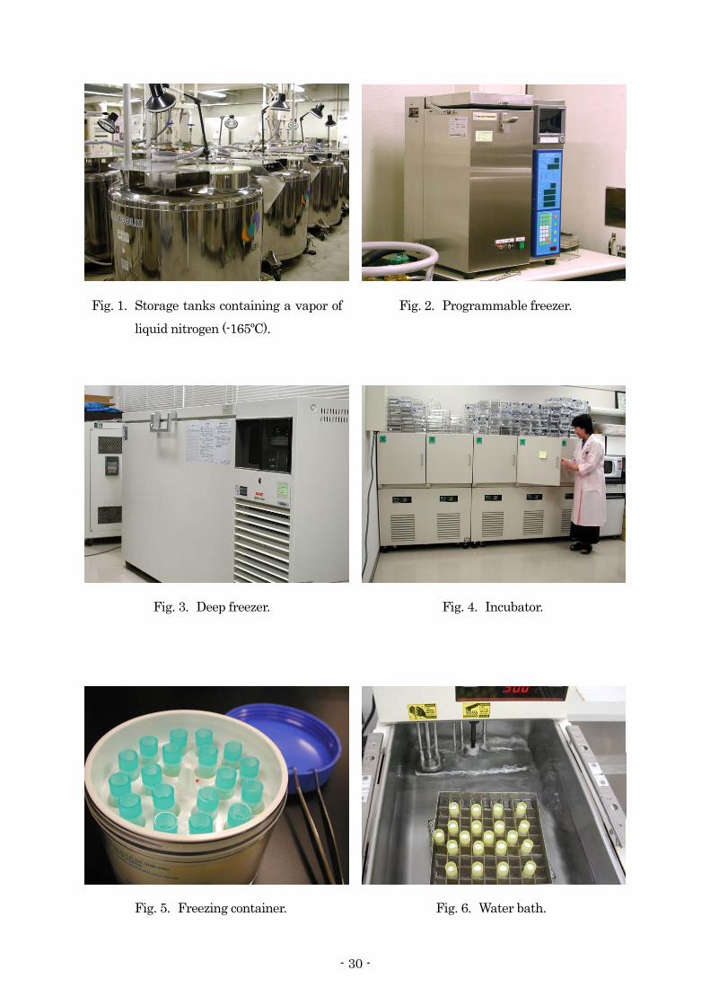
Fig. 4. Incubator. Fig. 3. Deep freezer.
Fig. 2. Programmable freezer. Fig. 1. Storage tanks containing a vapor of liquid nitrogen (-165ºC).
Fig. 5. Freezing container. Fig. 6. Water bath.
- 30 -

Fungal culture
Programmable freezer
(to -80ºC)
Mixture of mycobionts and cryoprotectants
Obtaining mycobionts
Liquid nitrogen storage tanks (-165ºC)
Incubator (4ºC)
Deep freezer (-80ºC)
①
②
③
④
Fungal culture
Programmable freezer
(to -80ºC)
Mixture of mycobionts and cryoprotectants
Obtaining mycobionts
Liquid nitrogen storage tanks (-165ºC)
Incubator (4ºC)
Deep freezer (-80ºC)
①
②
③
④
Fig. 11. Preparation of culture media.
Fig. ing.10. Other usual tools for aseptic handlFig. 9. Microscopes.
Fig. 8. Clean benches. Fig. 7. Autoclaves.
Fig. 12. An outline of cryopreservation at ultra- low temperature using liquid nitrogen.
- 31 -
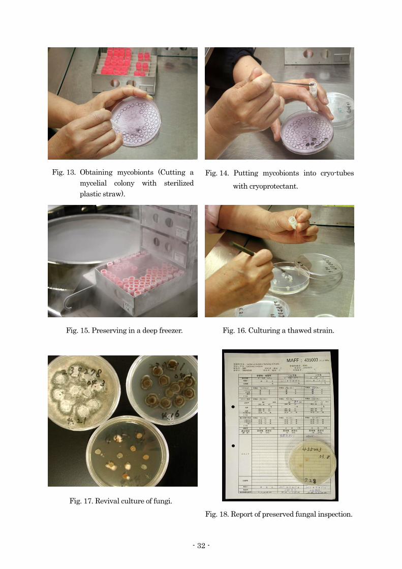
Fig. 17. Revival culture of fungi.
Fig. 16. Culturing a thawed strain. Fig. 15. Preserving in a deep freezer.
Fig. 14. Putting mycobionts into cryo-tubes with cryoprotectant.
Fig. 13. Obtaining mycobionts (Cutting a mycelial colony with sterilized plastic straw).
Fig. 18. Report of preserved fungal inspection.
- 32 -

微生物遺伝資源利用マニュアル (17) : 33–40 (2004)
植物病原細菌の保存法
永井利郎・飯田元子
農業生物資源研究所 ジーンバンク 微生物資源研究チーム・遺伝資源管理課
通常、細菌は凍結乾燥法により保存が可能である。凍結乾燥法は、アンプルに細菌と保護剤を混
ぜたものを少量入れ、凍結の後に真空凍結乾燥機を用いて乾燥させる方法である。凍結乾燥標品に
は、冷蔵庫で保存が可能であり、取り扱いや郵送が簡単であるという長所がある。細菌を保存する
他の方法には、L-乾燥法、凍結法などがある。例えば、細菌を液体培養したものと 30%グリセロー
ルを 1:1 に混合し、そのままディープフリーザ(-80ºC)に保存することも行われている (グリセロー
ルストック)。ここでは、NIAS ジーンバンクでの保存法を例に引きながら凍結乾燥法を概説する。
凍結乾燥法は、放線菌、酵母、糸状菌(胞子)、ウイルス、バクテリオファージにも適用できる。 1. 必要品 器具類:ガラス管 (アンプル用、8 mm × 150 mm、Pyrex、Fig. 1)、ガラス管用シリコン栓 (Fig.
1, (株)信越化学)、シリンジ (滅菌済み、2.5 ml、テルモ、東京) 及びニードル(1.2 mm × 150 mm、
吸口 90°)、滅菌シャーレ(栄研器材(株))、アンプルカッター (またはヤスリ)、試験管、スクリ
ューキャップ付き試験管、パスツールピペット、ニップル、白金線、クリーンベンチ、分注器 (IC3100 [KDS100]、Kd Scientific、PA、USA、Fig. 2)、凍結乾燥機 (Dura-DryµP、FTF SYSTEMS Inc.、NY、Fig. 3)、ガスバーナー (Fig. 4 および 6)、フリーザー、冷蔵庫、インキュベーター、テスラコ
イル (WTC-100、若井田理学 (株)、東京、Fig. 7c) シリコン栓を付けたガラス管は乾熱滅菌 (160ºC、3 時間) する。 ニードル、ニップルを付けたパスツールピペットをオートクレーブ滅菌 (121ºC、15 分) する。
試薬:10% スキムミルク-1.5% グルタミン酸ナトリウム キャップ付き試験管に分注し、115ºC
15 分オートクレーブ、一晩おいた後、再度 110ºC 10 分間オートクレーブする。冷蔵庫に保存する。 培地:培地や培養条件は菌株による。NIAS ジーンバンクでは多くの細菌について標準寒天培地
(日水製薬株式会社、東京) を使用している。
- 33 -
2. 真空凍結乾燥保存法 1) の植菌および 2)~4) はクリーンベンチ内で無菌的に行う。 1) 菌体を斜面培地に塗布し培養する。 2) 菌体が斜面培地を覆うまでに増殖したら、10%スキムミルク-1.5% グルタミン酸ナトリウム
を 2 ml 加える。 3) 菌体を懸濁し、懸濁液をシリンジで吸い取る。 4) シリンジを分注器にセットし、ガラス管に懸濁液を 100 µl ずつ分注する (Fig. 2)。分注器が
ない場合、パスツールピペットを用いて手動で分注する。残りの懸濁液はそのまま-40ºC 以下
で保存が可能である。
5) 懸濁液を一晩-40ºC で凍結する。 6 ガラス管を凍結乾燥機にセットし、一晩真空凍結乾燥する (Fig. 3)。 7) 真空 (<10 mT) に達したら、ガラス管を封入する。ガラス管の中程をバーナーを回転させな
がら加熱し (Fig. 4)、ガラスが柔らかくなったらバーナーをガラス管から遠ざける。ガラス管
の底を持ち、すぐにガラス管を 30cm ほど引き延ばす (Fig. 5)。手に持ったアンプル部分の上
部をバーナーで熱し切断した後、机の上で転がして先端部が机に当たらないように先端部の形
を整える (Fig. 6)。 8) テスラコイルで真空度 (真空の場合、アンプルの中で放電が起きる) を確かめ、真空が保たれ
ているものを冷蔵庫 (5ºC) に保存する (Fig. 7)。 3. 復元培養法 操作は、クリーンベンチ内で無菌的に行う。 1) アンプルカッターまたはヤスリで、アンプルの中程に軽く傷を付ける (Fig. 8)。 2) アンプルを 70%エタノールで消毒する (Fig. 9)。
3) 滅菌した紙タオルまたはガーゼでアンプル包み、アンプルを折る (Fig. 10)。
- 34 -

4) アンプルに液体培地または滅菌水を 50-100 µl 入れる (Fig. 11)。 5) ピペットで懸濁液を少量吸い取り、プレートに滴下する (Fig. 12)。
6) 図に示すように、画線する (Fig. 12)。最初と 2 番目の画線の後は、白金耳を火炎殺菌する。 7) プレートをインキュベーターで培養する。
- 35 -

Preservation of plant pathogenic bacteria
Toshirou NAGAI and Motoko IIDA
Microorganism Genetic Resources Laboratory and Genetic Resources Management Section, Genebank, National Institute of Agrobiological Sciences
Bacteria can be preserved successfully by a freeze-drying (lyophilization) technique, in which bacterial cells are mixed with a cryoprotectant and freeze-dried by a vacuum freeze dryer after freezing. Freeze-dried cells in an ampoule have an advantage of easy handling and sending. Other methods for preserving bacteria include L-drying, cryopreservation, and so on. For example in a glycerol stock technique, a bacterial culture is preserved by adding it to one volume of 30% glycerol and placing it in a deep freezer (-80ºC). In this manual, we describe the freeze-drying technique routinely employed in the NIAS Genebank. This technique can be also applied to the preservation of actinomycetes, yeasts, fungal spores, viruses and bacteriophages. 1. Required items
Apparatuses: glass tubes (for ampoules, 8 mm × 150 mm, Pyrex, Fig. 1), silicone caps for a glass tube (Fig. 1, SHINETSU CHEMICAL Co. Ltd.), syringes (sterilized, 2.5 ml, TERUMO, Tokyo, Japan) and 90° square tip needles (1.2 mm × 150 mm), plastic plates (sterilized, EUKEN KIZAI Co. Ltd.), an ampoule cutter (or a file), test tubes, test tubes with a screw cap, Pasteur pipettes, nipples, a platinum loop, a clean bench, an automatic dispenser (IC3100 [KDS100], Kd Scientific、PA、USA、Fig. 2), a vacuum freeze dryer (Dura-DryµP, FTF SYSTEMS Inc., NY, Fig. 3), a gas burner (Fig. 4 and 6), a freezer, a refrigerator, an incubator and a Tesla coil (WTC-100,WAKAIDA SCIENCE CORP., Tokyo, Japan, Fig. 7c)
Glass tubes with a silicone cap are sterilized in a dry heat sterilizer (160ºC, 3 h). Needles and Pasteur pipettes with a nipple are autoclaved at 121ºC for 15 min.
Reagents: 10% skim milk-1.5% monosodium glutamate. This cryoprotectant is dispensed to
test tubes with a screw cap, autoclaved at 115ºC for 15 min, kept in the autoclave overnight, and then autoclaved again at 110ºC for 10 min. Keep in a refrigerator.
- 36 -

Media: The media and growth conditions used depend on the strain or species. In the NIAS Genebank, standard agar (Nissui Pharmaceutical Co., LTD, Tokyo, Japan) is used to grow most bacteria. 2. How to freeze-dry bacterial cells
Inoculation in procedure 1 and procedures 2, 3 and 4 should be carried out aseptically in a clean bench.
1) Inoculate cells onto a slant and incubate them. 2) Grow the cells until they cover the surface of the slant, and add 2 ml of 10% skim
milk-1.5% monosodium glutamate. 3) Suspend the cells, and remove the suspension using a syringe with a needle. 4) Set the syringe to a dispenser and dispense the suspension to glass tubes, 100 µl per tube
(Fig. 2). If no dispensers are available, use a Pasteur pipette instead and dispense the suspension by hand. The rest of the suspension can be preserved at -40ºC or lower.
5) Freeze the suspension at -40ºC overnight. 6) Attach the glass tubes to a vacuum freeze dryer (Fig. 3), and freeze-dry the cell
suspension overnight. 7) Seal the glass tubes if the dryer's vacuum becomes less than 10 mT. Heat the middle of
the glass tube by rotating the burner around it (Fig. 4), and remove the burner from the tube if the glass tube softens. Stretch the glass tube to about 30 cm while holding the bottom of the tube (Fig. 5). Cut the tube with heating the tip of the ampoule you are holding, and arrange the tip (Fig. 6) to prevent it from coming into contact with the surface of a tabletop when the ampoule is rolled on it.
8) Check the vacuum in the ampoule with a Tesla coil (Fig. 7). If it maintains the vacuum,
then electrical discharge will occur. Preserve the ampoule maintaining the vacuum in a refrigerator at 5ºC.
- 37 -

3. How to revive freeze-dried cells All procedures should be carried out aseptically in a clean bench.
1) Scratch about the middle of the ampoule with an ampoule cutter or a file (Fig. 8). 2) Sterilize the surface of the ampoule with absorbent cotton wetted with 70% ethanol (Fig. 9). 3) Wrap the ampoule in a sterilized paper towel or sheet of gauze and snap it (Fig. 10). 4) Add 50-100 µl of liquid media or sterilized water to the ampoule (Fig. 11). 5) Remove a small volume of the suspension using a Pasteur pipette and drip it on a plate
(Fig. 12). 6) Streak the suspension as illustrated in Fig. 12. The loop was flamed after the 1st and 2nd
streak. 7) Incubate the plate.
- 38 -
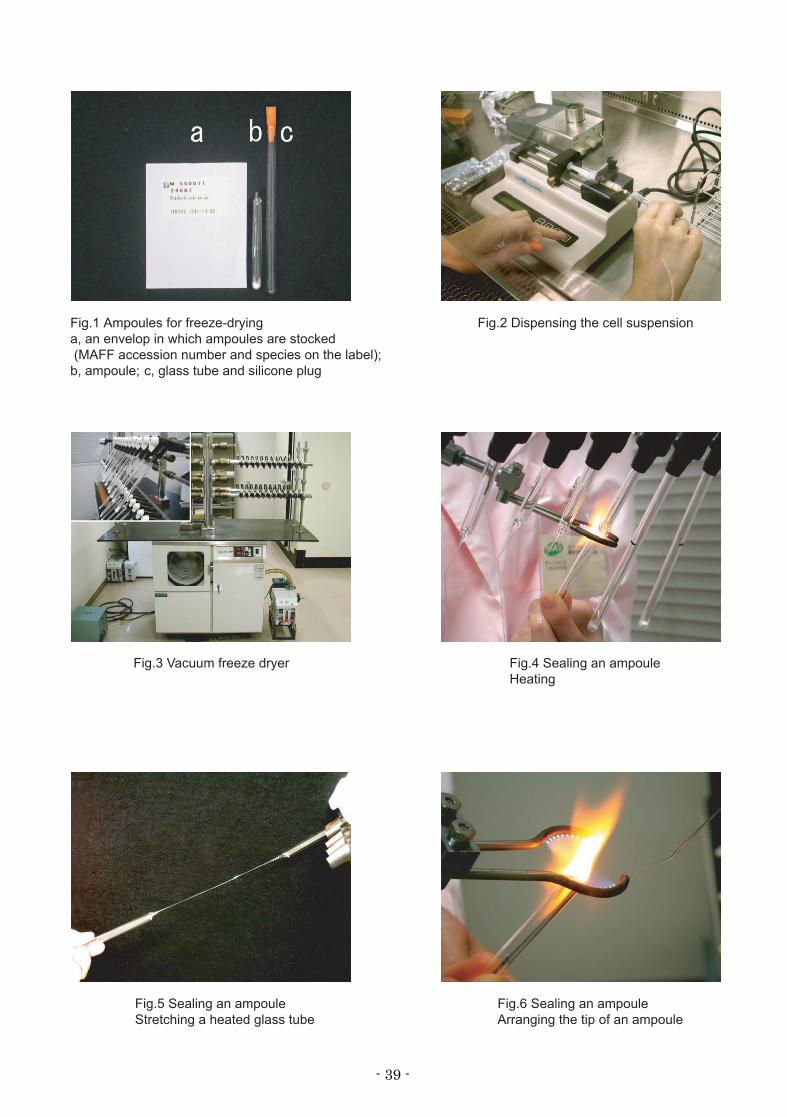
Fig.1 Ampoules for freeze-dryinga, an envelop in which ampoules are stocked (MAFF accession number and species on the label); b, ampoule; c, glass tube and silicone plug
Fig.2 Dispensing the cell suspension
Fig.3 Vacuum freeze dryer Fig.4 Sealing an ampouleHeating
Fig.5 Sealing an ampouleStretching a heated glass tube
Fig.6 Sealing an ampouleArranging the tip of an ampoule
- 39 -

Fig.7 Vacuum testa, off; b, on; c, Tesla coil
Fig.8 Scratching an ampoule
Fig.9 Sterilizing an ampoule Fig.10 Opening an ampoule
Fig.11 Suspending freeze-dried cells Fig.12 Streaking
- 40 -

生 物 研 資 料
平成 16 年 12 月
微生物遺伝資源利用マニュアル (17)
2004 年 12 月 24 日 印刷 2004 年 12 月 31 日 発行
独立行政法人農業生物資源研究所
National Institute of Agrobiological Sciences
〒305-8602 茨城県つくば市観音台2-1-2
編集兼 発行者
- 41 -

MAFF Microorganism Genetic Resources Manual
No. 17
“Identification and preservation of plant pathogens”
December 2004
National Institute of Agrobiological Sciences
- 42 -
Related Documents