Caveolin-1 Is a Negative Regulator of Caveolae-mediated Endocytosis to the Endoplasmic Reticulum* Received for publication, November 26, 2001 Published, JBC Papers in Press, November 27, 2001, DOI 10.1074/jbc.M111240200 Phuong U. Le‡§, Ginette Guay‡, Yoram Altschuler¶, and Ivan R. Nabi‡ From the ‡Department of Pathology and Cell Biology, Universite ´ de Montre ´al, Montreal, Quebec H3C 3J7, Canada and the ¶Department of Pharmacology, Hebrew University of Jerusalem, Jerusalem 91120, Israel Caveolae are flask-shaped invaginations at the plasma membrane that constitute a subclass of detergent-resis- tant membrane domains enriched in cholesterol and sphingolipids and that express caveolin, a caveolar coat protein. Autocrine motility factor receptor (AMF-R) is stably localized to caveolae, and the cholesterol extract- ing reagent, methyl--cyclodextrin, inhibits its inter- nalization to the endoplasmic reticulum implicating caveolae in this distinct receptor-mediated endocytic pathway. Curiously, the rate of methyl--cyclodextrin- sensitive endocytosis of AMF-R to the endoplasmic re- ticulum is increased in ras- and abl-transformed NIH- 3T3 cells that express significantly reduced levels of caveolin and few caveolae. Overexpression of the dy- namin K44A dominant negative mutant via an adenovi- rus expression system induces caveolar invaginations sensitive to methyl--cyclodextrin extraction in the transformed cells without increasing caveolin expres- sion. Dynamin K44A expression further inhibits AMF-R- mediated endocytosis to the endoplasmic reticulum in untransformed and transformed NIH-3T3 cells. Adeno- viral expression of caveolin-1 also induces caveolae in the transformed NIH-3T3 cells and reduces AMF-R-me- diated endocytosis to the endoplasmic reticulum to lev- els observed in untransformed NIH-3T3 cells. Cholester- ol-rich detergent-resistant membrane domains or glycolipid rafts therefore invaginate independently of caveolin-1 expression to form endocytosis-competent caveolar vesicles via rapid dynamin-dependent detach- ment from the plasma membrane. Caveolin-1 stabilizes the plasma membrane association of caveolae and thereby acts as a negative regulator of the caveolae- mediated endocytosis of AMF-R to the endoplasmic reticulum. Endocytosis via clathrin-coated vesicles represents the best characterized endocytic pathway, however, other clathrin-inde- pendent endocytic mechanisms also exist (1– 4). The large GTPase dynamin has been shown to regulate the fission of clathrin-coated pits, and expression of the dynamin K44A (dynK44A) 1 mutant inhibits clathrin-mediated endocytosis (5– 7). The dynK44A mutant does not affect fluid phase endocytosis or the clathrin-independent endocytic pathway defined by ricin endocytosis indicating that non-clathrin-coated cell surface in- vaginations can detach from the plasma membrane in the apparent absence of dynamin-mediated membrane fission (8 – 11). However, introduction of inhibitory antibodies to dynamin into hepatocytes resulted in the accumulation of both clathrin- coated vesicles and smooth caveolar invaginations and inhib- ited the endocytosis of cholera toxin (12). In endothelial cells, caveolae budding from isolated membranes was shown to be dynamin-dependent, and caveolae were shown to contain the molecular machinery necessary for vesicle budding (13, 14). Regulation of caveolae budding by dynamin identifies caveolae as endocytosis-competent cell surface invaginations. Caveolae or smooth plasmalemmal vesicles were first iden- tified in endothelial cells and are morphologically identifiable as smooth flask shaped invaginations of the plasma membrane (15–18). Caveolae are rich in cholesterol and sphingolipids, disrupted by cholesterol extracting agents, and insoluble in Triton X-100 and are therefore considered to form a subclass of cholesterol-rich detergent-resistant membrane domains or gly- colipid rafts (19 –22). The caveolins (caveolin-1, -2, and -3) are cholesterol binding proteins that form a spiral coat on the cytoplasmic surface of caveolar invaginations and represent caveolae markers (23–25). Caveolar invaginations are not pres- ent in cells that express little or no caveolin, and the reintro- duction of caveolin-1 into such cells has been shown to induce the formation of caveolae implicating caveolin in the invagina- tion of glycolipid raft microdomains (26 –28). Caveolin-1 ex- pression is inversely proportional to cell transformation, and caveolin-1 has been characterized as a tumor suppressor gene (27, 29 –31). Caveolae have long been proposed to be involved in transc- ytosis across the endothelial cell (18, 32–34). Caveolae- or raft-mediated endocytosis has been reported for cholera toxin- bound GM1 ganglioside, sphingolipids, glycosylphosphatid- ylinositol-anchored proteins, SV40, and bacteria, as well as the endothelin, growth hormone, interleukin-2, and autocrine mo- tility factor (AMF) receptors (35– 46). Autocrine motility factor receptor (AMF-R) is a seven-transmembrane domain receptor localized at steady state to caveolae and the smooth endoplas- mic reticulum (ER) that follows an endocytic pathway sensi- tive to cholesterol extraction with methyl--cyclodextrin (mCD) via caveolae to the smooth ER (43, 44, 47– 49). Using AMF as a marker for this caveolae-mediated endocytic path- way, we show that caveolar invaginations and caveolar vesicles * This study was supported in part by a grant from the Canadian Institutes for Health Research (CIHR). The costs of publication of this article were defrayed in part by the payment of page charges. This article must therefore be hereby marked “advertisement” in accordance with 18 U.S.C. Section 1734 solely to indicate this fact. § Recipient of a Graduate Student Award from the Cancer Research Society Inc. Recipient of a CIHR Investigator award. To whom correspondence should be addressed: De ´pt. de Pathologie et Biologie Cellulaire, Uni- versite ´ de Montre ´al, C. P. 6128, Succursale A, Montre ´al, Que ´bec H3C 3J7, Canada. Tel.: 514-343-6291; Fax: 514-343-2459; E-mail: ivan. [email protected]. 1 The abbreviations used are: dynK44A, dynamin-1 K44A mutant; AMF, autocrine motility factor; AMF-R, autocrine motility factor recep- tor; bAMF, biotinylated autocrine motility factor; ER, endoplasmic re- ticulum; mCD, methyl--cyclodextrin; tTA, tetracycline-regulatable chimeric transcription activator; HA, hemagglutinin; PBS, phosphate- buffered saline; FACS, fluorescence-activated cell sorting; MVB, mul- tivesicular bodies; EM, electron microscopy. THE JOURNAL OF BIOLOGICAL CHEMISTRY Vol. 277, No. 5, Issue of February 1, pp. 3371–3379, 2002 © 2002 by The American Society for Biochemistry and Molecular Biology, Inc. Printed in U.S.A. This paper is available on line at http://www.jbc.org 3371 by guest on April 9, 2016 http://www.jbc.org/ Downloaded from

Welcome message from author
This document is posted to help you gain knowledge. Please leave a comment to let me know what you think about it! Share it to your friends and learn new things together.
Transcript

Caveolin-1 Is a Negative Regulator of Caveolae-mediatedEndocytosis to the Endoplasmic Reticulum*
Received for publication, November 26, 2001Published, JBC Papers in Press, November 27, 2001, DOI 10.1074/jbc.M111240200
Phuong U. Le‡§, Ginette Guay‡, Yoram Altschuler¶, and Ivan R. Nabi‡�
From the ‡Department of Pathology and Cell Biology, Universite de Montreal, Montreal, Quebec H3C 3J7, Canada andthe ¶Department of Pharmacology, Hebrew University of Jerusalem, Jerusalem 91120, Israel
Caveolae are flask-shaped invaginations at the plasmamembrane that constitute a subclass of detergent-resis-tant membrane domains enriched in cholesterol andsphingolipids and that express caveolin, a caveolar coatprotein. Autocrine motility factor receptor (AMF-R) isstably localized to caveolae, and the cholesterol extract-ing reagent, methyl-�-cyclodextrin, inhibits its inter-nalization to the endoplasmic reticulum implicatingcaveolae in this distinct receptor-mediated endocyticpathway. Curiously, the rate of methyl-�-cyclodextrin-sensitive endocytosis of AMF-R to the endoplasmic re-ticulum is increased in ras- and abl-transformed NIH-3T3 cells that express significantly reduced levels ofcaveolin and few caveolae. Overexpression of the dy-namin K44A dominant negative mutant via an adenovi-rus expression system induces caveolar invaginationssensitive to methyl-�-cyclodextrin extraction in thetransformed cells without increasing caveolin expres-sion. Dynamin K44A expression further inhibits AMF-R-mediated endocytosis to the endoplasmic reticulum inuntransformed and transformed NIH-3T3 cells. Adeno-viral expression of caveolin-1 also induces caveolae inthe transformed NIH-3T3 cells and reduces AMF-R-me-diated endocytosis to the endoplasmic reticulum to lev-els observed in untransformed NIH-3T3 cells. Cholester-ol-rich detergent-resistant membrane domains orglycolipid rafts therefore invaginate independently ofcaveolin-1 expression to form endocytosis-competentcaveolar vesicles via rapid dynamin-dependent detach-ment from the plasma membrane. Caveolin-1 stabilizesthe plasma membrane association of caveolae andthereby acts as a negative regulator of the caveolae-mediated endocytosis of AMF-R to the endoplasmicreticulum.
Endocytosis via clathrin-coated vesicles represents the bestcharacterized endocytic pathway, however, other clathrin-inde-pendent endocytic mechanisms also exist (1–4). The largeGTPase dynamin has been shown to regulate the fission ofclathrin-coated pits, and expression of the dynamin K44A(dynK44A)1 mutant inhibits clathrin-mediated endocytosis (5–
7). The dynK44A mutant does not affect fluid phase endocytosisor the clathrin-independent endocytic pathway defined by ricinendocytosis indicating that non-clathrin-coated cell surface in-vaginations can detach from the plasma membrane in theapparent absence of dynamin-mediated membrane fission (8–11). However, introduction of inhibitory antibodies to dynamininto hepatocytes resulted in the accumulation of both clathrin-coated vesicles and smooth caveolar invaginations and inhib-ited the endocytosis of cholera toxin (12). In endothelial cells,caveolae budding from isolated membranes was shown to bedynamin-dependent, and caveolae were shown to contain themolecular machinery necessary for vesicle budding (13, 14).Regulation of caveolae budding by dynamin identifies caveolaeas endocytosis-competent cell surface invaginations.
Caveolae or smooth plasmalemmal vesicles were first iden-tified in endothelial cells and are morphologically identifiableas smooth flask shaped invaginations of the plasma membrane(15–18). Caveolae are rich in cholesterol and sphingolipids,disrupted by cholesterol extracting agents, and insoluble inTriton X-100 and are therefore considered to form a subclass ofcholesterol-rich detergent-resistant membrane domains or gly-colipid rafts (19–22). The caveolins (caveolin-1, -2, and -3) arecholesterol binding proteins that form a spiral coat on thecytoplasmic surface of caveolar invaginations and representcaveolae markers (23–25). Caveolar invaginations are not pres-ent in cells that express little or no caveolin, and the reintro-duction of caveolin-1 into such cells has been shown to inducethe formation of caveolae implicating caveolin in the invagina-tion of glycolipid raft microdomains (26–28). Caveolin-1 ex-pression is inversely proportional to cell transformation, andcaveolin-1 has been characterized as a tumor suppressor gene(27, 29–31).
Caveolae have long been proposed to be involved in transc-ytosis across the endothelial cell (18, 32–34). Caveolae- orraft-mediated endocytosis has been reported for cholera toxin-bound GM1 ganglioside, sphingolipids, glycosylphosphatid-ylinositol-anchored proteins, SV40, and bacteria, as well as theendothelin, growth hormone, interleukin-2, and autocrine mo-tility factor (AMF) receptors (35–46). Autocrine motility factorreceptor (AMF-R) is a seven-transmembrane domain receptorlocalized at steady state to caveolae and the smooth endoplas-mic reticulum (ER) that follows an endocytic pathway sensi-tive to cholesterol extraction with methyl-�-cyclodextrin(m�CD) via caveolae to the smooth ER (43, 44, 47–49). UsingAMF as a marker for this caveolae-mediated endocytic path-way, we show that caveolar invaginations and caveolar vesicles
* This study was supported in part by a grant from the CanadianInstitutes for Health Research (CIHR). The costs of publication of thisarticle were defrayed in part by the payment of page charges. Thisarticle must therefore be hereby marked “advertisement” in accordancewith 18 U.S.C. Section 1734 solely to indicate this fact.
§ Recipient of a Graduate Student Award from the Cancer ResearchSociety Inc.
� Recipient of a CIHR Investigator award. To whom correspondenceshould be addressed: Dept. de Pathologie et Biologie Cellulaire, Uni-versite de Montreal, C. P. 6128, Succursale A, Montreal, Quebec H3C3J7, Canada. Tel.: 514-343-6291; Fax: 514-343-2459; E-mail: [email protected].
1 The abbreviations used are: dynK44A, dynamin-1 K44A mutant;
AMF, autocrine motility factor; AMF-R, autocrine motility factor recep-tor; bAMF, biotinylated autocrine motility factor; ER, endoplasmic re-ticulum; m�CD, methyl-�-cyclodextrin; tTA, tetracycline-regulatablechimeric transcription activator; HA, hemagglutinin; PBS, phosphate-buffered saline; FACS, fluorescence-activated cell sorting; MVB, mul-tivesicular bodies; EM, electron microscopy.
THE JOURNAL OF BIOLOGICAL CHEMISTRY Vol. 277, No. 5, Issue of February 1, pp. 3371–3379, 2002© 2002 by The American Society for Biochemistry and Molecular Biology, Inc. Printed in U.S.A.
This paper is available on line at http://www.jbc.org 3371
by guest on April 9, 2016
http://ww
w.jbc.org/
Dow
nloaded from
mediate AMF-R endocytosis in ras- and abl-transformed NIH-3T3 cells that express little caveolin and few caveolae.Adenoviral expression of the dominant negative dynK44A mu-tant or of caveolin-1 has allowed us to demonstrate that: 1)
even when caveolin levels are significantly reduced or absent,caveolae form and rapidly bud from the plasma membrane toform caveolar vesicles that target the ER; and 2) caveolin-1regulates this endocytic pathway by stabilizing caveolae ex-pression at the plasma membrane thereby slowing down theinternalization of caveolar vesicles.
EXPERIMENTAL PROCEDURES
Antibodies, Reagents, and Cells—Monoclonal rat antibody againstAMF-R was used in the form of concentrated hybridoma supernatant(50). Rabbit anti-caveolin antibody was purchased from TransductionLaboratories (Mississauga, Ontario, Canada), mouse anti-c-Myc fromSanta Cruz Biotechnologies (Santa Cruz, CA), and mouse anti-hemag-glutinin (HA) was a gift from Luc Desgroseillers (Department of Bio-chemistry, Universite de Montreal). Horseradish peroxidase, fluores-cein isothiocyanate, and gold-conjugated secondary antibodies wereobtained from Jackson ImmunoResearch Laboratories (West Grove,
FIG. 1. AMF is internalized to the ER in ras- and abl-transformedNIH-3T3 cells. NIH-3T3 (A), NIH-ras (B), and NIH-abl (C) cells werepulse labeled with biotinylated AMF (bAMF) for 60 min at 37 °C.Postembedding labeling with nanogold-streptavidin and silver amplifi-cation revealed bAMF localization to ER tubules as well as to MVBs.Arrowheads, ER tubules; arrows, MVBs. Bar � 0.2 �m.
FIG. 2. Endocytosed bAMF colocalizes with AMF-R in ER tu-bules. NIH-3T3 cells (A, B) as well as ras (C, D)-transformed and abl(E, F)-transformed NIH-3T3 cells were pulse-labeled with bAMF for 60min at 37 °C. bAMF was first revealed by nanogold-streptavidin andsilver amplification following by the labeling of AMF-R tubules withanti-AMF-R antibodies and 12-nm gold-conjugated anti-rat IgM second-ary antibodies. Arrowheads, bAMF labeling; arrows, AMF-R labeling.Bar � 0.2 �m.
Caveolae-mediated Endocytosis3372
by guest on April 9, 2016
http://ww
w.jbc.org/
Dow
nloaded from
PA). Rabbit phosphohexose isomerase (referred to as AMF) was pur-chased from Sigma Chemical Co. (Oakville, Ontario, Canada) and bi-otinylated with long chain N-hydroxyl-succinimido-biotin (Pierce, Rock-ford, IL) according to the manufacturer’s instructions. Nanogold-streptavidin and the HQ-Silver Enhancement kit were purchased fromNanoprobes, Inc. (Stony Brook, NY). M�CD, poly-L-lysine, and pro-pidium iodide were purchased from Sigma.
An NIH-3T3 fibroblast clone (43) and H-ras- and v-abl-transformedNIH-3T3 cells (29) were grown in complete medium consisting of Dul-becco’s modified Eagle’s medium supplemented with 10% calf serum,non-essential amino acids, vitamins, glutamine, and a penicillin-strep-tomycin antibiotic mixture (Invitrogen Canada Inc., Burlington,Ontario, Canada). Treatment of cells with 5 mM m�CD was performedas previously described (44).
Viral Infection—Recombinant adenoviruses expressing the tetracy-cline-regulatable chimeric transcription activator (tTA), HA-tagged,and dynK44A, and myc-tagged caveolin-1 under the control of thetetracycline-regulated promoter were as previously described (51–53).To enhance infection rates, viral stocks of the tTA and dynK44A orcaveolin-1 adenoviruses were diluted in 320 �l of sterile PBS andpreincubated with 72 �l of 1 �g/ml polylysine for 30 min at roomtemperature. Infection with only the tTA adenovirus was used as acontrol. 2.5 � 105 untransformed, ras-transformed or abl-transformedNIH-3T3 cells were plated onto 10-cm dishes for 10 h and rinsed oncewith PBS before addition of the adenovirus polylysine mixture in 3 mlof serum-free media for 1 h at 37 °C. After removal of the adenovirusmixture, the cells were rinsed twice with serum-free media and thenincubated for 36 h in regular culture media. For the EM studies,infection rates of the three cell lines were determined by immunofluo-rescence labeling with anti-HA or anti-MYC antibodies, and viral titerswere used that resulted in greater than 75% infection rates. The rate ofinfection was determined in parallel for each experiment and infectionwith only the tTA adenovirus was used as a control. Where indicated,cells coinfected with tTA and dynK44A adenoviruses were then treatedwith 5 mM m�CD for 90 min prior to fixation.
Electron Microscopy—All three cell types were pulsed with bAMF forthe indicated times and processed for electron microscopy, and bAMFwas revealed with nanogold-streptavidin followed by silver enhance-ment as previously described (44). For the double labeling with anti-AMF-R or polyclonal anti-caveolin antibodies, bAMF was first revealedwith nanogold-streptavidin and silver amplification, and then the sec-tions were labeled with the primary antibodies following by the appro-priate gold-conjugated secondary antibodies. The sections were con-trasted with uranyl acetate and lead citrate and examined in a Zeiss
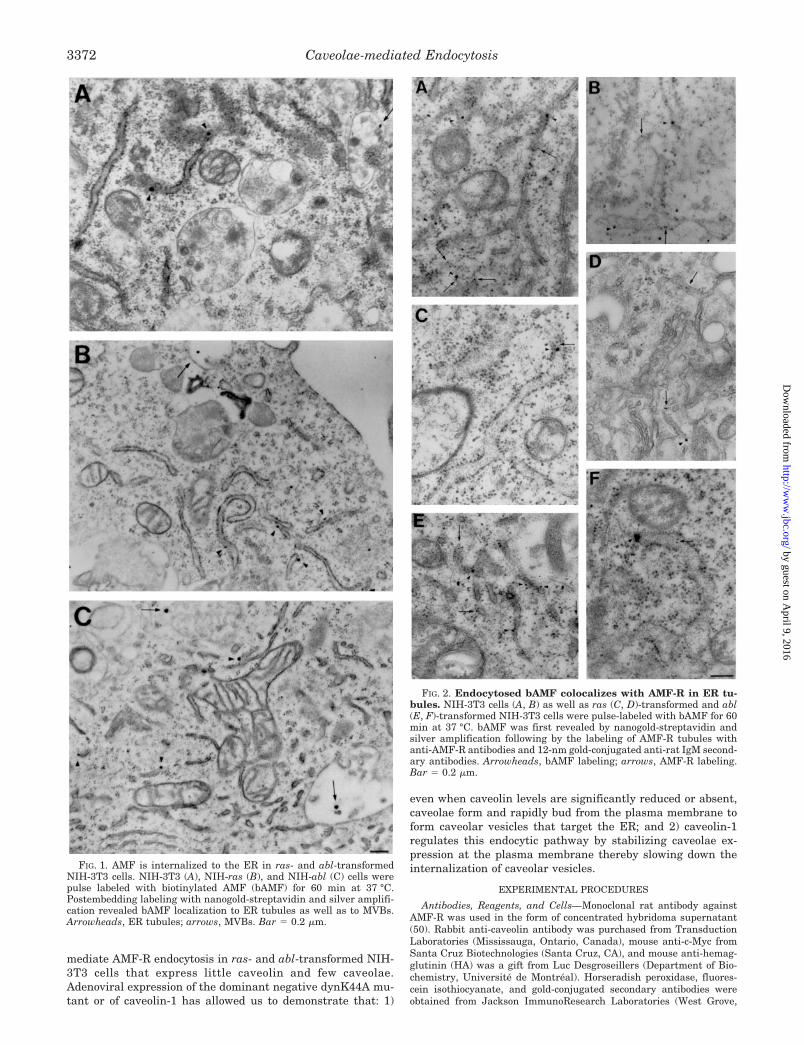
FIG. 3. Increased rate of AMF-R endocytosis to the ER in ras- and abl-transformed cells. A, NIH-3T3 (white bars) or ras (gray bars)-and abl (black bars)-transformed NIH-3T3 cells were pulsed with bAMF for 60 min at 37 °C in the absence or presence of m�CD. bAMF wasrevealed by postembedding labeling with nanogold-streptavidin and silver amplification. The numerical density of silver particles associated withthe ER, endosomes, and mitochondria was determined from 36 images (�12,000) from two different experiments for each condition. Control valuesin the absence of a bAMF pulse are presented. The results are presented as a ratio of the number of silver particles to the surface area of eachorganelle (�S.E.). The data shows that bAMF targeting to the ER is selectively inhibited by m�CD. B, quantitative analysis of AMF delivery tothe ER after 30-, 60-, and 120-min incubation with bAMF at 37 °C in NIH-3T3 (circles), NIH-ras (squares), and NIH-abl (triangles) cells.ER-associated silver particles were counted from 25 intact cell profiles, and the data represent the average per cell profile. C, FACS analysis ofAMF-R cell surface expression in untransformed and ras- and abl-transformed NIH-3T3 cells shows that AMF-R cell surface expression isdecreased following cell transformation. The data are presented as relative fluorescence intensity in percentage compared with NIH-3T3 cells.
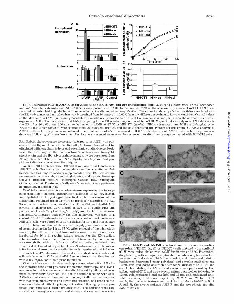
FIG. 4. bAMF and AMF-R are localized to caveolin-positivecaveolae. NIH-3T3 (A, B) or NIH-3T3 cells infected with dynK44A(C–H) were pulse-labeled with bAMF for 60 min at 37 °C. Postembed-ding labeling with nanogold-streptavidin and silver amplification firstrevealed the localization of bAMF to caveolae, and then caveolin distri-bution was determined using polyclonal anti-caveolin antibodies and12-nm gold-conjugated anti-rabbit secondary antibodies (A, C, E, andG). Double labeling for AMF-R and caveolin was performed by firstadding anti-AMF-R and anti-caveolin primary antibodies following by12-nm gold-conjugated anti-rat IgM and 18-nm gold-conjugated anti-rabbit secondary antibodies, respectively (B, D, F, and H). In A, C, E,and G, the arrows indicate caveolin and the arrowheads bAMF. In B, D,F, and H, the arrows indicate AMF-R and the arrowheads caveolin.Bars � 0.2 �m.
Caveolae-mediated Endocytosis 3373
by guest on April 9, 2016
http://ww
w.jbc.org/
Dow
nloaded from

CEM902 electron microscope. In the absence of nanogold-streptavidinlabeling, silver particles due to nonspecific silver enhancement were notobserved.
For the quantification of internalized bAMF, the number of silverparticles localized to ER, endosomes, and mitochondria were counted,and the surface area of the indicated organelles was measured using aSigma Scan measurement system. ER labeling included both ribosomesstudded rough ER profiles as well as morphologically identified smoothER (43, 47–49). To ensure that the smooth membranous organelleswere not early endosomes, only smooth membrane-bound structureswider than 75 nm and longer than 200 nm were considered to be ERtubules. Similarly, smooth caveolar invaginations and clathrin-coatedvesicles within 100 nm of the plasma membrane were counted per unitmembrane (43). The average and standard error from 36 images ob-tained from two separate experiments are presented. Alternatively,bAMF expression at the plasma membrane, in endosomes and in theER, and caveolin expression at the plasma membrane (including mem-brane invaginations) and in morphologically identifiable smooth caveo-lar invaginations were quantified from cells incubated with bAMF andlabeled with streptavidin-nanogold and silver amplification followed byanti-caveolin antibodies and 12-nm gold-conjugated anti-rabbit second-ary antibodies by postembedding immunoelectron microscopy. For eachexperiment, 25 intact cell profiles were counted.
FACS Analysis—Cells were detached from the dish with EDTA andresuspended in bicarbonate-free medium supplemented with 25 mM
Hepes and 5% calf serum. The cells were then incubated with anti-AMF-R antibodies at 4 °C for 1 h, washed three times with cold media,incubated with fluorescein isothiocyanate-conjugated anti-rat IgM at4 °C for 1 h, and then washed three more times with cold media andtwice with PBS. The cells were then incubated with 0.5 �g/ml pro-pidium iodide for 10 min at 4 °C. Cell-associated fluorescence intensitywas analyzed on a Beckman FACScan. Cells stained for propidiumiodide were discarded from the analysis, and cell surface AMF-R ex-pression was determined only on intact cells.
Immunoblot—Cells cultured at �70% confluency were scraped,lysed, and sonicated in lysis buffer containing 1% SDS, 5 mM EDTA,and protease inhibitors. Protein content was assayed using the BCAprotein assay (Pierce, Rockford, IL), and 40 �g of protein was separatedby SDS-PAGE and blotted onto nitrocellulose paper. The blots were
FIG. 5. Expression of dynK44A in-duces caveolae in ras- and abl-trans-formed NIH-3T3 cells. Ras (A, C, F, G, I,K)- and abl (B, D, E, H, J, L)-transformedNIH-3T3 cells, either uninfected (A, B) orexpressing dynK44A via adenoviral infec-tion (C–L) were pulse-labeled with bAMFfor 60 min at 37 °C. Plasma membraneprofiles show the dramatically increasedexpression of smooth caveolar invagina-tions in the dynK44A infected cells (C–I)relative to uninfected cells (A, B).DynK44A-infected ras- and abl-trans-formed NIH-3T3 cells pretreated with 5mM m�CD for 90 min prior to fixationexhibited no caveolar invaginations (K,L). Postembedding labeling with nano-gold-streptavidin and silver amplificationrevealed bAMF localization to caveolarinvaginations (E, F, G, H) and to clathrin-coated vesicles (H, J). Bar � 0.2 �m.
FIG. 6. Quantitative analysis of caveolae and clathrin-coatedvesicles at the plasma membrane following dynK44A infection.The number of morphologically identifiable caveolae (A) and clathrin-coated vesicles (B) per micron of plasma membrane length was deter-mined for untransformed and ras- and abl-transformed NIH-3T3 cellseither uninfected (white bars) or expressing dynK44A via adenoviralinfection (black bars). The increase in the number of caveolae expressedin dynK44A-infected NIH-3T3 cells was significant (p � 10�3).
Caveolae-mediated Endocytosis3374
by guest on April 9, 2016
http://ww
w.jbc.org/
Dow
nloaded from
blocked with 5% milk in PBS, incubated with rabbit anti-caveolinantibody together with mouse anti-HA, and then with horseradishperoxidase-conjugated anti-rabbit and anti-mouse secondary antibod-ies. The labeled bands were revealed by chemiluminescence and ex-posed to preflashed Kodak XRP-1 film.
RESULTS
Caveolae-mediated Endocytosis of AMF-R to the ER—InNIH-3T3 cells, AMF-R is localized to smooth caveolar invagi-nations and is endocytosed via a m�CD-sensitive caveolae-mediated pathway to the smooth ER tubules defined by AMF-Rexpression (43, 44). To specifically assess the specific role ofcaveolae in this endocytic route, endocytosis of biotinylatedAMF (bAMF) was followed in ras- and abl-transformed NIH-3T3 cells that exhibit significantly reduced expression of caveo-lae and caveolin (29). Following a 60-min bAMF pulse at 37 °C,endocytosed bAMF detected by nanogold labeling and silveramplification was localized to smooth and rough ER tubules ofNIH-3T3 and ras- and abl-transformed NIH-3T3 cells (Fig. 1).Endocytosis of bAMF to multivesicular bodies (MVBs) was alsodetected in all three cell lines (Fig. 1) as previously reported inNIH-3T3 cells (44). Double labeling of the cells for bAMF (nano-gold and silver amplification) and for AMF-R (12-nm gold) byelectron microscopy confirmed that bAMF is delivered to AMF-R-positive ER tubules in all three cell types (Fig. 2). AMF-Rlabeling of rough ER tubules appears to be qualitatively in-creased in the ras- and abl-transformed NIH-3T3 cells relativeto untransformed NIH-3T3 cells. Previous quantitative studieshave shown that the predominant distribution of AMF-R atsteady state is to smooth ER tubules in Madin-Darby caninekidney cells, NIH-3T3, and HeLa cells, although significantlabeling of rough ER tubules was observed in Madin-Darbycanine kidney cells (43, 47). For the purpose of this study therough and smooth ER were not morphologically distinguished.
Quantification of bAMF labeling of the ER, endosomes, andmitochondria (see “Experimental Procedures” for details)showed increased bAMF internalization to the ER in ras- andabl-transformed cells compared with control NIH-3T3 cells(Fig. 3A). Clathrin-dependent endocytosis to endosomal struc-tures (44) was detected at similar levels between the three celltypes. Nonspecific labeling of mitochondria and control labelingperformed in the absence of endocytosed bAMF are also pre-sented. The significantly reduced expression of caveolae and
caveolin in the transformed NIH-3T3 cell lines does not there-fore prevent endocytosis of bAMF to the ER.
Kinetic analysis of bAMF endocytosis to the ER in the threecell lines showed that the rate of accumulation of bAMF in theER was equivalent in the ras- and abl-transformed NIH-3T3 cellsand approximately 2-fold greater than that in untransformedNIH-3T3 cells (Fig. 3B). Over the 2-h time course of the experi-ment, delivery of bAMF to the ER was maintained and notsaturable in the three cell lines. FACS analysis showed that cellsurface expression of AMF-R is reduced following transformationof NIH-3T3 cells (Fig. 3C), although total AMF-R expression asdetermined by immunoblot was equivalent or increased in ras-and abl-transformed NIH-3T3 cells, respectively, compared withuntransformed NIH-3T3 cells (data not shown). The increasedrate of delivery of bAMF to the ER is therefore not a consequenceof increased receptor expression at the plasma membrane butrather due to rapid receptor recycling.
Incubation of NIH-3T3 cells with 5 mM m�CD selectivelyblocks bAMF endocytosis to the ER but not the clathrin-depend-ent endocytosis of bAMF to multivesicular endosomes (44). Ascan be seen in Fig. 3A, m�CD also blocks bAMF delivery to theER in both untransformed and ras- and abl-transformed NIH-3T3 cells without significantly affecting bAMF endocytosis toendosomes. High concentrations (10 mM) of m�CD have beenshown to block clathrin-dependent endocytosis (54, 55); however,the lack of an effect on the clathrin-dependent endocytosis ofbAMF to endosomes serves as an internal control demonstratingthat at the 5 mM concentration used, m�CD is selectively inhib-iting the caveolae-like pathway of bAMF to the ER. The ability ofm�CD to inhibit bAMF endocytosis to the ER in ras- and abl-transformed NIH-3T3 cells confirms the similar nature of thispathway in the three cell lines.
The caveolar distribution of AMF-R is based on the EMlocalization of AMF-R to smooth plasmalemmal invaginationsand its partial colocalization with caveolin by immunofluores-cence labeling (43). To ensure that the smooth invaginations towhich AMF-R is localized are indeed caveolin-positive andtherefore correspond to accepted definitions of caveolae, NIH-3T3 cells were double-labeled by postembedding EM for bAMF(nanogold-streptavidin and silver amplification) and caveolin(12-nm gold particles) (Fig. 4A) or for AMF-R (12-nm goldparticles) and caveolin (18-nm gold particles) (Fig. 4B). Both
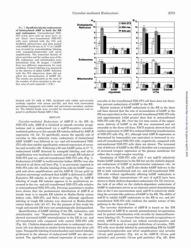
FIG. 7. DynK44A blocks endocytosisof biotinylated AMF to both the ERand endosomes. Untransformed NIH-3T3 (white bars) and ras (gray bars)- orabl (black bars)-transformed NIH-3T3cells were infected with the tTA anddynK44A adenoviruses and then pulsedwith bAMF for 60 min at 37 °C (A). bAMFwas revealed by postembedding labelingwith nanogold-streptavidin and silveramplification. The numerical density ofsilver particles and the surface area of theER, endosomes, and mitochondria weredetermined from 36 images (�12,000)from two different experiments for eachcondition. Control labeling was measuredin the absence of added bAMF. Infectionwith the tTA adenovirus alone did notaffect the internalization of bAMF (B).The results are presented as the ratio ofthe number of silver particles to the sur-face area of each organelle (�S.E.).
Caveolae-mediated Endocytosis 3375
by guest on April 9, 2016
http://ww
w.jbc.org/
Dow
nloaded from

bAMF- and AMF-R-positive invaginations are labeled forcaveolin.
Adenoviral Expression of dynK44A Induces Caveolae andInhibits AMF Endocytosis—Infection of NIH-3T3 cells with anadenovirus expressing the dynK44A mutant enhanced our abil-ity to identify double-labeled caveolae. NIH-3T3 cells express-ing this mutant show numerous caveolin-positive caveolae atthe plasma membrane (Fig. 4, C–H), including those exhibitingthe typical long neck associated with dynamin inhibition (Fig.4G) (12). Caveolae double-labeled for caveolin and either bAMF(Fig. 4, C and E) or AMF-R (Fig. 4, D, F, and H) are readilydetected. bAMF is therefore localized with its receptor to caveo-lae in NIH-3T3 cells.
ras- and abl-transformed NIH-3T3 cells exhibit significantlyfewer caveolae relative to NIH-3T3 cells (Fig. 5, A and B), asreported previously (29), and introduction of the dynK44A mu-tant by adenoviral infection (51, 52) into ras- and abl-trans-formed NIH-3T3 cells induced the expression of numeroussmooth invaginations morphologically similar to caveolae (Fig.5, C–I). Treatment of dynK44A-infected ras- and abl-trans-formed NIH-3T3 cells with m�CD prior to fixation resulted inthe complete absence of smooth caveolar invaginations (Fig. 5,K and L), as observed for uninfected cells (Fig. 5, A and B). Thecaveolar invaginations induced by dynK44A are, therefore, sen-sitive to cholesterol depletion and represent a cholesterol-richmembrane domain or class of glycolipid rafts. Quantification ofthe expression of caveolae and clathrin-coated pits in unin-fected and dynK44A-infected cells demonstrated the signifi-cantly increased expression per micron of membrane of mor-phological caveolae but not of clathrin-coated pits in all threecell types (Fig. 6).
Adenoviral expression of dynK44A blocked both the clathrin-dependent endocytosis of bAMF to endosomes and the caveo-lae-like pathway to the smooth ER (Fig. 7A). Expression of thetTA adenovirus alone did not influence either of the AMFendocytic pathways indicating that inhibition of AMF endocy-tosis is specifically due to expression of the dynK44A mutantand not to adenoviral infection (Fig. 7B). Dynamin-mediatedbudding of caveolar vesicles from the plasma membrane there-fore regulates AMF-R endocytosis to the ER.
To ensure that dynK44A expression is not inducing caveolinexpression and thereby affecting the expression of caveolae, wequantified plasma membrane-associated caveolin labeling bypostembedding immunoelectron microscopy of whole cell pro-files. As presented per micron of membrane in Fig. 6, thenumber of caveolar invaginations per cell is dramatically re-duced in ras- and abl-transformed cells, and dynK44A expres-sion induces the stable expression of a large number of smoothcaveolar invaginations (Fig. 8A). Caveolin labeling associatedwith the plasma membrane, including caveolae (Fig. 8B) orspecifically with caveolae (Fig. 8C) is significantly reduced inras- and abl-transformed NIH-3T3 cells relative to untrans-formed NIH-3T3 cells. Expression of dynK44A does not affecttotal plasma membrane-associated caveolin expression (Fig.8B) indicating that increased expression of caveolin or its in-creased recruitment to the plasma membrane is not responsi-ble for the dynK44A-mediated induction of smooth caveolarinvaginations. A slight increase in caveolin labeling of caveolarinvaginations is observed in all three cell lines (Fig. 8C) but isminimal relative to the increased number of caveolae expressed(Fig. 8A). Immunoblot analysis reveals that caveolin expres-sion in the ras and abl transformants remains significantlybelow that in NIH-3T3 cells even after adenoviral expression ofHA-tagged dynK44A (Fig. 9). Expression of dynK44A has nottherefore induced the formation of caveolar invaginations byincreasing caveolin expression levels.
Adenoviral Expression of Caveolin-1 Negatively RegulatesAMF Endocytosis to the ER—Infection of the three cell typeswith tTA and caveolin-1 adenoviruses induces increased levelsof caveolin-1 expression significantly above those in uninfectedNIH-3T3 cells (Fig. 9). As previously reported (27), the reintro-duction of caveolin-1 into ras- and abl-transformed NIH-3T3cells induces numerous caveolae at the plasma membrane (Fig.10) that are morphologically indistinguishable from the caveo-lae induced by dynK44A infection (Fig. 5). Quantitatively, adramatic increase in caveolae expression and in anti-caveolinlabeling at both the plasma membrane and in caveolae wasobserved in ras- and abl-infected cells such that caveolae andcaveolin levels were equivalent to or greater than those ofuninfected NIH-3T3 cells (Fig. 11, A–C). Infection of NIH-3T3cells with the caveolin-1 adenovirus induced lesser (1.5- to2-fold) increases in the number of caveolae and in caveolinexpression at the cell surface (Fig. 11, A–C). Caveolin-1 over-expression in the transformed cells reduced bAMF endocytosisto the ER to levels comparable to uninfected NIH-3T3 cells butdid not affect bAMF internalization to endosomes; the in-creased expression of caveolin-1 in NIH-3T3 cells also selec-tively decreased bAMF endocytosis to the ER (Fig. 11, D and
FIG. 8. Expression of caveolin at the plasma membrane is notincreased following dynK44A infection. Quantification of the num-ber of morphologically identifiable caveolae (A), caveolin labeling at theplasma membrane (including smooth caveolar invaginations) (B), andcaveolin labeling specific to smooth caveolar invaginations (C) wasdetermined for 50 complete cell profiles from anti-caveolin-labeled EMgrids for untransformed and ras- and abl-transformed NIH-3T3 cellseither uninfected (white bars) or expressing dynK44A via adenoviralinfection (black bars). The data were obtained from two distinct exper-iments and represent the average per cell profile.
Caveolae-mediated Endocytosis3376
by guest on April 9, 2016
http://ww
w.jbc.org/
Dow
nloaded from
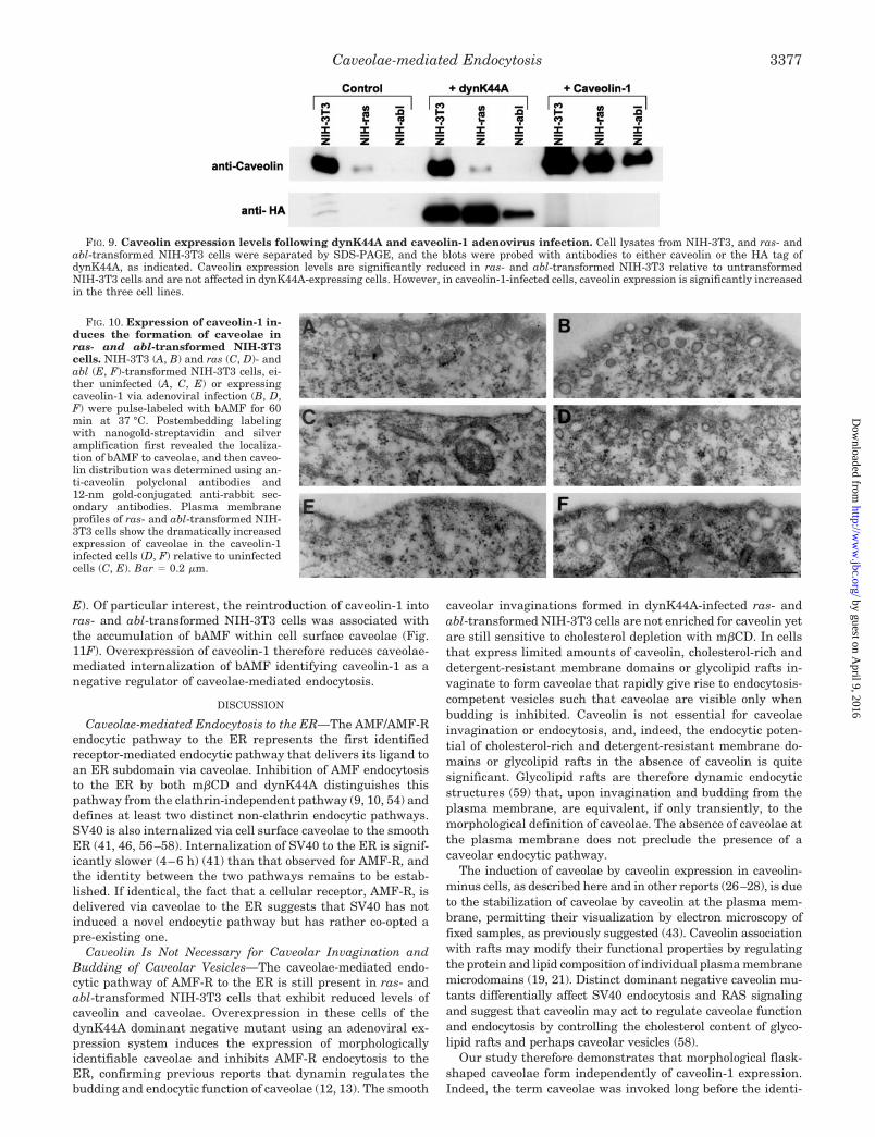
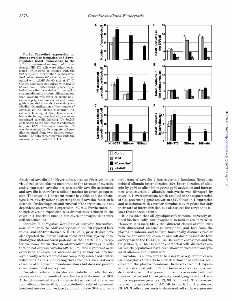
E). Of particular interest, the reintroduction of caveolin-1 intoras- and abl-transformed NIH-3T3 cells was associated withthe accumulation of bAMF within cell surface caveolae (Fig.11F). Overexpression of caveolin-1 therefore reduces caveolae-mediated internalization of bAMF identifying caveolin-1 as anegative regulator of caveolae-mediated endocytosis.
DISCUSSION
Caveolae-mediated Endocytosis to the ER—The AMF/AMF-Rendocytic pathway to the ER represents the first identifiedreceptor-mediated endocytic pathway that delivers its ligand toan ER subdomain via caveolae. Inhibition of AMF endocytosisto the ER by both m�CD and dynK44A distinguishes thispathway from the clathrin-independent pathway (9, 10, 54) anddefines at least two distinct non-clathrin endocytic pathways.SV40 is also internalized via cell surface caveolae to the smoothER (41, 46, 56–58). Internalization of SV40 to the ER is signif-icantly slower (4–6 h) (41) than that observed for AMF-R, andthe identity between the two pathways remains to be estab-lished. If identical, the fact that a cellular receptor, AMF-R, isdelivered via caveolae to the ER suggests that SV40 has notinduced a novel endocytic pathway but has rather co-opted apre-existing one.
Caveolin Is Not Necessary for Caveolar Invagination andBudding of Caveolar Vesicles—The caveolae-mediated endo-cytic pathway of AMF-R to the ER is still present in ras- andabl-transformed NIH-3T3 cells that exhibit reduced levels ofcaveolin and caveolae. Overexpression in these cells of thedynK44A dominant negative mutant using an adenoviral ex-pression system induces the expression of morphologicallyidentifiable caveolae and inhibits AMF-R endocytosis to theER, confirming previous reports that dynamin regulates thebudding and endocytic function of caveolae (12, 13). The smooth
caveolar invaginations formed in dynK44A-infected ras- andabl-transformed NIH-3T3 cells are not enriched for caveolin yetare still sensitive to cholesterol depletion with m�CD. In cellsthat express limited amounts of caveolin, cholesterol-rich anddetergent-resistant membrane domains or glycolipid rafts in-vaginate to form caveolae that rapidly give rise to endocytosis-competent vesicles such that caveolae are visible only whenbudding is inhibited. Caveolin is not essential for caveolaeinvagination or endocytosis, and, indeed, the endocytic poten-tial of cholesterol-rich and detergent-resistant membrane do-mains or glycolipid rafts in the absence of caveolin is quitesignificant. Glycolipid rafts are therefore dynamic endocyticstructures (59) that, upon invagination and budding from theplasma membrane, are equivalent, if only transiently, to themorphological definition of caveolae. The absence of caveolae atthe plasma membrane does not preclude the presence of acaveolar endocytic pathway.
The induction of caveolae by caveolin expression in caveolin-minus cells, as described here and in other reports (26–28), is dueto the stabilization of caveolae by caveolin at the plasma mem-brane, permitting their visualization by electron microscopy offixed samples, as previously suggested (43). Caveolin associationwith rafts may modify their functional properties by regulatingthe protein and lipid composition of individual plasma membranemicrodomains (19, 21). Distinct dominant negative caveolin mu-tants differentially affect SV40 endocytosis and RAS signalingand suggest that caveolin may act to regulate caveolae functionand endocytosis by controlling the cholesterol content of glyco-lipid rafts and perhaps caveolar vesicles (58).
Our study therefore demonstrates that morphological flask-shaped caveolae form independently of caveolin-1 expression.Indeed, the term caveolae was invoked long before the identi-
FIG. 9. Caveolin expression levels following dynK44A and caveolin-1 adenovirus infection. Cell lysates from NIH-3T3, and ras- andabl-transformed NIH-3T3 cells were separated by SDS-PAGE, and the blots were probed with antibodies to either caveolin or the HA tag ofdynK44A, as indicated. Caveolin expression levels are significantly reduced in ras- and abl-transformed NIH-3T3 relative to untransformedNIH-3T3 cells and are not affected in dynK44A-expressing cells. However, in caveolin-1-infected cells, caveolin expression is significantly increasedin the three cell lines.
FIG. 10. Expression of caveolin-1 in-duces the formation of caveolae inras- and abl-transformed NIH-3T3cells. NIH-3T3 (A, B) and ras (C, D)- andabl (E, F)-transformed NIH-3T3 cells, ei-ther uninfected (A, C, E) or expressingcaveolin-1 via adenoviral infection (B, D,F) were pulse-labeled with bAMF for 60min at 37 °C. Postembedding labelingwith nanogold-streptavidin and silveramplification first revealed the localiza-tion of bAMF to caveolae, and then caveo-lin distribution was determined using an-ti-caveolin polyclonal antibodies and12-nm gold-conjugated anti-rabbit sec-ondary antibodies. Plasma membraneprofiles of ras- and abl-transformed NIH-3T3 cells show the dramatically increasedexpression of caveolae in the caveolin-1infected cells (D, F) relative to uninfectedcells (C, E). Bar � 0.2 �m.
Caveolae-mediated Endocytosis 3377
by guest on April 9, 2016
http://ww
w.jbc.org/
Dow
nloaded from
fication of caveolin (15). Nevertheless, because few caveolae arevisualized at the plasma membrane in the absence of caveolin,stably expressed caveolae are necessarily caveolin-associated,and caveolin is therefore a reliable marker for caveolae expres-sion. The caveolin-1 knockout mouse is viable, and the pheno-type is relatively minor suggesting that if caveolae function isessential for development and survival of the organism, it is notdependent on caveolin-1 expression (60, 61). Furthermore, al-though caveolae expression was dramatically reduced in thecaveolin-1 knockout mice, a few caveolar invaginations werestill identified (61).
Caveolin Is a Negative Regulator of Caveolae Internaliza-tion—Similar to the AMF endocytosis to the ER reported herein ras- and abl-transformed NIH-3T3 cells, prior studies havealso reported the internalization of cholera toxin, glycosylphos-phatidylinositol-anchored proteins, or the interleukin 2 recep-tor via non-clathrin cholesterol-dependent pathways in cellsthat do not express caveolin (42, 45, 62). The significant over-expression of caveolin-1 obtained using adenoviral infectionsignificantly reduced but did not completely inhibit AMF inter-nalization (Fig. 11D) indicating that caveolin-1 stabilization ofcaveolae at the plasma membrane slows but does not preventcaveolae-mediated endocytosis.
Caveolae-mediated endocytosis in endothelial cells that ex-press significant amounts of caveolin-1 is well documented (63).Although caveolin-1 knockout mice did not exhibit altered se-rum albumin levels (61), lung endothelial cells of caveolin-1knockout mice exhibit reduced albumin uptake (64), and rein-
troduction of caveolin-1 into caveolin-1 knockout fibroblastsinduced albumin internalization (60). Internalization of albu-min by gp60 or albondin requires gp60 activation and interac-tion with caveolin-1; albumin endocytosis was disrupted bycaveolin-1 overexpression, which resulted in the sequestrationof G�i preventing gp60 activation (34). Caveolin-1 expressionand association with caveolar domains may regulate not onlytheir rate of internalization but also select the cargo that fol-lows this endocytic route.
It is possible that all glycolipid raft domains, currently de-fined biochemically, can invaginate to form caveolar vesicles.However, it is more likely that different classes of rafts existwith differential abilities to invaginate and bud from theplasma membrane and to form functionally distinct caveolarvesicles. For instance, caveolae and raft domains mediate bothendocytosis to the ER (41, 43, 44, 46) and to endosomes and theGolgi (35–37, 59, 65, 66) and in endothelial cells, distinct caveo-lar vesicle populations have been shown to mediate transcyto-sis of albumin and insulin (67).
Caveolin-1 is shown here to be a negative regulator of caveo-lar endocytosis that acts to slow detachment of caveolar vesi-cles from the plasma membrane. Reduced caveolin-1 expres-sion is associated with different forms of cancer in vivo, anddecreased caveolin-1 expression in vitro is associated with celltransformation and tumorigenicity identifying caveolin-1 as atumor suppressor gene (27, 29, 30, 53, 68–71). The increasedrate of internalization of AMF-R to the ER in transformedNIH-3T3 cells corresponds to decreased cell surface expression
FIG. 11. Caveolin-1 expression in-duces caveolae formation and down-regulates bAMF endocytosis to theER. Untransformed and ras- or abl-trans-formed NIH-3T3 cells were either not in-fected (white bars) or infected with thetTA (gray bars) or with the tTA and caveo-lin-1 adenoviruses (black bars) and thenpulsed with bAMF for 60 min at 37 °C.Control cells were not pulsed with bAMF(dotted bars). Postembedding labeling ofbAMF was first revealed with nanogold-streptavidin and silver amplification, andthen caveolin was revealed using anti-caveolin polyclonal antibodies and 12-nmgold-conjugated anti-rabbit secondary an-tibodies. Quantification of the number ofcaveolae at the plasma membrane (A),caveolin labeling at the plasma mem-brane (including caveolae) (B), caveolae-associated caveolin labeling (C), bAMFendocytosis to the ER (D) or to endosomes(E), and bAMF labeling of caveolae (F)was determined for 50 complete cell pro-files obtained from two distinct experi-ments. The data presented represents theaverage per cell profile (�S.E.).
Caveolae-mediated Endocytosis3378
by guest on April 9, 2016
http://ww
w.jbc.org/
Dow
nloaded from

of AMF-R suggesting that, in cells lacking caveolin-1, AMF-Rrapidly transits the plasma membrane. Similarly, FACS anal-ysis of B16 melanoma and K1735 fibrosarcoma metastatic vari-ants reported decreased cell surface AMF-R expression in thehigh metastatic variants (72). It is therefore conceivable thatdecreased expression of caveolin-1 results in the destabilizationof AMF-R cell surface expression and the deregulation ofAMF-R traffic. The well-characterized association of AMF-Rexpression with tumor malignancy (73–79) implicates thiscaveolae-mediated endocytic pathway in AMF-R function intumor cell motility and metastasis.
Acknowledgments—We thank Michael Lisanti and Philippe Frankfor kindly providing the ras- and abl-transformed NIH-3T3 cells andcaveolin-1 adenovirus as well as for their helpful suggestions. Wethankfully acknowledge the precious help of Anne Guenette for the EMquantification and Jean Leveille for the preparation of the figures.
REFERENCES
1. Lamaze, C., and Schmid, S. L. (1995) Curr. Opin. Cell Biol. 7, 573–5802. Mellman, I. (1996) Annu. Rev. Cell Dev. Biol. 12, 575–6263. Sandvig, K., and van Deurs, B. (1999) FEBS Lett. 452, 67–704. Nichols, B. J., and Lippincott-Schwartz, J. (2001) Trends Cell Biol. 11,
406–4125. Herskovits, J. S., Burgess, C. C., Obar, R. A., and Vallee, R. B. (1993) J. Cell
Biol. 122, 565–5786. van der Bliek, A. M., Redelmeier, T. E., Damke, H., Tisdale, E. J., Meyerowitz,
E. M., and Schmid, S. L. (1993) J. Cell Biol. 122, 553–5637. Damke, H., Baba, T., Warnock, D. E., and Schmid, S. L. (1994) J. Cell Biol.
127, 915–9348. Damke, H., Baba, T., van der Bliek, A. M., and Schmid, S. L. (1995) J. Cell Biol.
131, 69–809. Simpson, J. C., Smith, D. C., Roberts, L. M., and Lord, J. M. (1998) Exp. Cell
Res. 239, 293–30010. Llorente, A., Rapak, A., Schmid, S. L., van Deurs, B., and Sandvig, K. (1998)
J. Cell Biol. 140, 553–56311. Contamin, S., Galmiche, A., Doye, A., Flatau, G., Benmerah, A., and Boquet, P.
(2000) Mol. Biol. Cell 11, 1775–178712. Henley, J. R., Krueger, E. W., Oswald, B. J., and McNiven, M. A. (1998) J. Cell
Biol. 141, 85–9913. Oh, P., McIntosh, D. P., and Schnitzer, J. E. (1998) J. Cell Biol. 141, 101–11414. Schnitzer, J. E., Liu, J., and Oh, P. (1995) J. Biol. Chem. 270, 14399–1440415. Yamada, E. (1955) J. Biophys. Biochem. Cytol. 1, 445–44816. Palade, G. E. (1953) J. Appl. Physiol. 24, 142417. Palade, G. E. (1958) Anat. Rec. 130, 467–46818. Bruns, R. R., and Palade, G. E. (1968) J. Cell Biol. 37, 244–27619. Harder, T., and Simons, K. (1997) Curr. Opin. Cell Biol. 9, 534–54220. Anderson, R. G. (1998) Annu. Rev. Biochem. 67, 199–22521. Smart, E. J., Graf, G. A., McNiven, M. A., Sessa, W. C., Engelman, J. A.,
Scherer, P. E., Okamoto, T., and Lisanti, M. P. (1999) Mol. Cell. Biol. 19,7289–7304
22. Galbiati, F., Razani, B., and Lisanti, M. P. (2001) Cell 106, 403–41123. Rothberg, K. G., Heuser, J. E., Donzell, W. C., Ying, Y.-S., Glenney, J. R., and
Anderson, R. G. W. (1992) Cell 68, 673–68224. Murata, M., Peranen, J., Schreiner, R., Wieland, F., Kurzchalia, T. V., and
Simons, K. (1995) Proc. Natl. Acad. Sci. U. S. A. 92, 10339–1034325. Li, S., Song, K. S., and Lisanti, M. P. (1996) J. Biol. Chem. 271, 568–57326. Fra, A. M., Williamson, E., Simons, K., and Parton, R. G. (1995) Proc. Natl.
Acad. Sci. U. S. A. 92, 8655–865927. Engelman, J. A., Wykoff, C. C., Yasuhara, S., Song, K. S., Okamoto, T., and
Lisanti, M. P. (1997) J. Biol. Chem. 272, 16374–1638128. Smart, E. J., Ying, Y., Donzell, W. C., and Anderson, R. G. (1996) J. Biol.
Chem. 271, 29427–2943529. Koleske, A. J., Baltimore, D., and Lisanti, M. P. (1995) Proc. Natl. Acad. Sci.
U. S. A. 92, 1381–138530. Galbiati, F., Volonte, D., Engelman, J. A., Watanabe, G., Burk, R., Pestell,
R. G., and Lisanti, M. P. (1998) EMBO J. 17, 6633–664831. Razani, B., Schlegel, A., Liu, J., and Lisanti, M. P. (2001) Biochem. Soc. Trans.
29, 494–49932. Ghitescu, L., Fixman, A., Simionescu, M., and Simionescu, N. (1986) J. Cell
Biol. 102, 1304–131133. Schnitzer, J. E., Oh, P., Pinney, E., and Allard, J. (1994) J. Cell Biol. 127,
1217–123234. Minshall, R. D., Tiruppathi, C., Vogel, S. M., Niles, W. D., Gilchrist, A., Hamm,
H. E., and Malik, A. B. (2000) J. Cell Biol. 150, 1057–1070
35. Montesano, R., Roth, J., Robert, A., and Orci, L. (1982) Nature (Lond.) 296,651–653
36. Tran, D., Carpentier, J. L., Sawano, F., Gorden, P., and Orci, L. (1987) Proc.Natl. Acad. Sci. U. S. A. 84, 7957–7961
37. Parton, R. G., Joggerst, B., and Simons, K. (1994) J. Cell Biol. 127, 1199–121538. Lencer, W. I., Hirst, T. R., and Holmes, R. K. (1999) Biochim. Biophys. Acta
1450, 177–19039. Okamoto, Y., Ninomiya, H., Miwa, S., and Masaki, T. (2000) J. Biol. Chem.
275, 6439–644640. Shin, J. S., Gao, Z., and Abraham, S. N. (2000) Science 289, 785–78841. Pelkmans, L., Kartenbeck, J., and Helenius, A. (2001) Nat. Cell Biol. 3,
473–48342. Lamaze, C., Dujeancourt, A., Baba, T., Lo, C. G., Benmerah, A., and Dautry-
Varsat, A. (2001) Mol. Cell 7, 661–67143. Benlimame, N., Le, P. U., and Nabi, I. R. (1998) Mol. Biol. Cell 9, 1773–178644. Le, P. U., Benlimame, N., Lagana, A., Raz, A., and Nabi, I. R. (2000) J. Cell Sci.
113, 3227–324045. Orlandi, P. A., and Fishman, P. H. (1998) J. Cell Biol. 141, 905–91546. Kartenbeck, J., Stukenbrok, H., and Helenius, A. (1989) J. Cell Biol. 109,
2721–272947. Benlimame, N., Simard, D., and Nabi, I. R. (1995) J. Cell Biol. 129, 459–47148. Wang, H.-J., Benlimame, N., and Nabi, I. R. (1997) J. Cell Sci. 110, 3043–305349. Wang, H.-J., Guay, G., Pogan, L., Sauve, R., and Nabi, I. R. (2000) J. Cell Biol.
150, 1489–149850. Nabi, I. R., Watanabe, H., and Raz, A. (1990) Cancer Res. 50, 409–41451. Altschuler, Y., Barbas, S. M., Terlecky, L. J., Tang, K., Hardy, S., Mostov,
K. E., and Schmid, S. L. (1998) J. Cell Biol. 143, 1871–188152. Altschuler, Y., Liu, S., Katz, L., Tang, K., Hardy, S., Brodsky, F., Apodaca, G.,
and Mostov, K. (1999) J. Cell Biol. 147, 7–1253. Zhang, W., Razani, B., Altschuler, Y., Bouzahzah, B., Mostov, K. E., Pestell,
R. G., and Lisanti, M. P. (2000) J. Biol. Chem. 275, 20717–2072554. Rodal, S. K., Skretting, G., Garred, Ø., Vilhardt, F., van Deurs, B., and
Sandvig, K. (1999) Mol. Biol. Cell 10, 961–97455. Subtil, A., Gaidarov, I., Kobylarz, K., Lampson, M. A., Keen, J. H., and
McGraw, T. E. (1999) Proc. Natl. Acad. Sci. U. S. A. 96, 6775–678056. Anderson, H. A., Chen, Y., and Norkin, L. C. (1996) Mol. Biol. Cell 7,
1825–183457. Stang, E., Kartenbeck, J., and Parton, R. G. (1997) Mol. Biol. Cell 8, 47–5758. Roy, S., Luetterforst, R., Harding, A., Apolloni, A., Etheridge, M., Stang, E.,
Rolls, B., Hancock, J. F., and Parton, R. G. (1999) Nat. Cell Biol. 1, 98–10559. Nichols, B. J., Kenworthy, A. K., Polishchuk, R. S., Lodge, R., Roberts, T. H.,
Hirschberg, K., Phair, R. D., and Lippincott-Schwartz, J. (2001) J. Cell Biol.153, 529–541
60. Razani, B., Engelman, J. A., Wang, X. B., Schubert, W., Zhang, X. L., Marks,C. B., Macaluso, F., Russell, R. G., Li, M., Pestell, R. G., Di Vizio, D., Hou,H., Jr., Kneitz, B., Lagaud, G., Christ, G. J., Edelmann, W., and Lisanti,M. P. (2001) J. Biol. Chem. 276, 38121–38138
61. Drab, M., Verkade, P., Elger, M., Kasper, M., Lohn, M., Lauterbach, B.,Menne, J., Lindschau, C., Mende, F., Luft, F. C., Schedl, A., Haller, H., andKurzchalia, T. V. (2001) Science 293, 2449–2452
62. Verkade, P., Harder, T., Lafont, F., and Simons, K. (1999) J. Cell Biol. 148,727–739
63. Mineo, C., and Anderson, R. G. (2001) Histochem. Cell Biol. 116, 109–11864. Schubert, W., Frank, P. G., Chow, C. W., and Lisanti, M. P. (2001) J. Biol.
Chem. 276, 48619–4862265. Puri, V., Watanabe, R., Singh, R. D., Dominguez, M., Brown, J. C., Wheatley,
C. L., Marks, D. L., and Pagano, R. E. (2001) J. Cell Biol. 154, 535–54766. Gines, S., Ciruela, F., Burgueno, J., Casado, V., Canela, E. I., Mallol, J., Lluis,
C., and Franco, R. (2001) Mol. Pharmacol. 59, 1314–132367. Bendayan, M., and Rasio, E. A. (1996) J. Cell Sci. 109, 1857–186468. Lee, S. W., Reimer, C. L., Oh, P., Campbell, D. B., and Schnitzer, J. E. (1998)
Oncogene 16, 1391–139769. Racine, C., Belanger, M., Hirabayashi, H., Boucher, M., Chakir, J., and Couet,
J. (1999) Biochem. Biophys. Res. Commun. 255, 580–58670. Bender, F. C., Reymond, M. A., Bron, C., and Quest, A. F. (2000) Cancer Res.
60, 5870–587871. Razani, B., Altschuler, Y., Zhu, L., Pestell, R. G., Mostov, K. E., and Lisanti,
M. P. (2000) Biochemistry 39, 13916–1392472. Watanabe, H., Nabi, I. R., and Raz, A. (1991) Cancer Res. 51, 2699–270573. Nakamori, S., Watanabe, H., Kameyama, M., Imaoka, S., Furukawa, H.,
Ishikawa, O., Sasaki, Y., Kabuto, T., and Raz, A. (1994) Cancer 74,1855–1862
74. Otto, T., Birchmeier, W., Schmidt, U., Hinke, A., Schipper, J., Rubben, H., andRaz, A. (1994) Cancer Res. 54, 3120–3123
75. Maruyama, K., Watanabe, H., Hitoshi, S., Takayama, T., Gofuku, J., Yano, H.,Inoue, M., Tamura, S., Raz, A., and Monden, M. (1995) Int. J. Cancer 64,316–321
76. Hirono, Y., Fushida, S., Yonemura, Y., Yamamoto, H., Watanabe, H., and Raz,A. (1996) Br. J. Cancer 74, 2003–2007
77. Nagai, Y., Ishikawa, O., Miyachi, Y., and Watanabe, H. (1996) Dermatology192, 8–11
78. Takanami, I., Takeuchi, K., Naruke, M., Kodaira, S., Tanaka, F., Watanabe,H., and Raz, A. (1998) Tumour Biol. 19, 384–389
79. Taniguchi, K., Yonemura, Y., Nojima, N., Hirono, Y., Fushida, S., Fujimura, T.,Miwa, K., Endo, Y., Yamamoto, H., and Watanabe, H. (1998) Cancer 82,2112–2122
Caveolae-mediated Endocytosis 3379
by guest on April 9, 2016
http://ww
w.jbc.org/
Dow
nloaded from

Phuong U. Le, Ginette Guay, Yoram Altschuler and Ivan R. NabiEndoplasmic Reticulum
Caveolin-1 Is a Negative Regulator of Caveolae-mediated Endocytosis to the
doi: 10.1074/jbc.M111240200 originally published online November 27, 20012002, 277:3371-3379.J. Biol. Chem.
10.1074/jbc.M111240200Access the most updated version of this article at doi:
Alerts:
When a correction for this article is posted•
When this article is cited•
to choose from all of JBC's e-mail alertsClick here
http://www.jbc.org/content/277/5/3371.full.html#ref-list-1
This article cites 79 references, 52 of which can be accessed free at
by guest on April 9, 2016
http://ww
w.jbc.org/
Dow
nloaded from
Related Documents