CAUSES & FUNCTIONAL IMPLICATIONS OF VISUAL IMPAIRMENT •The eyes and associated structures must be normal in structure & function. •The neurological pathways from the retina & optic nerve to the visual cortex must be in tact. •The brain must be capable of interpreting the information received.

CAUSES & FUNCTIONAL IMPLICATIONS OF VISUAL IMPAIRMENT The eyes and associated structures must be normal in structure & function. The neurological pathways.
Dec 24, 2015
Welcome message from author
This document is posted to help you gain knowledge. Please leave a comment to let me know what you think about it! Share it to your friends and learn new things together.
Transcript
CAUSES & FUNCTIONAL IMPLICATIONS OF VISUAL
IMPAIRMENT•The eyes and associated structures must be normal in structure & function.
•The neurological pathways from the retina & optic nerve to the visual cortex must be in tact.
•The brain must be capable of interpreting the information received.

What does 20/20 mean?
• Numerator is distance patient is from chart• (usually 20 feet)• Denominator is how far away a person with
normal (emmetropic) vision can see that letter on the chart.
• Your distance 20/40 how far away a normal eye could
be
VISUAL IMPAIRMENT
• 20/200 central acuity
• Less than 20 degrees of visual field
Conditions That Result In Low Visual Acuity
• Ocular muscle disorders- eyes that are not in proper alignment

Strabismus & Amblyopia
• Defects of the eye muscle system
• Can result in loss of vision in one eye due to lack of use
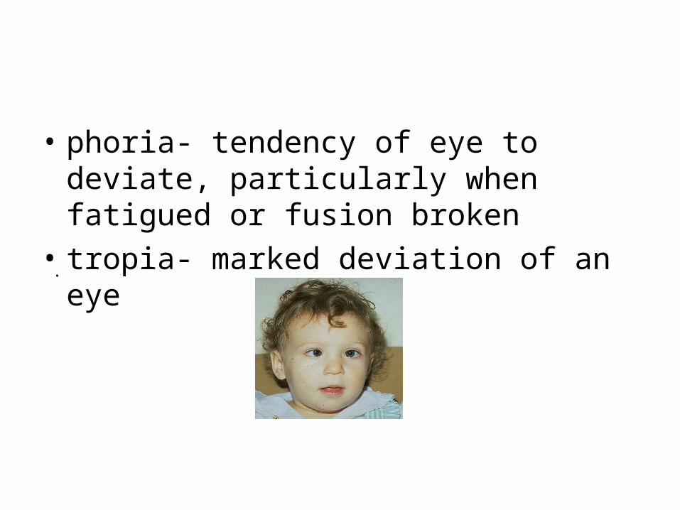
• phoria- tendency of eye to deviate, particularly when fatigued or fusion broken
• tropia- marked deviation of an eye
.
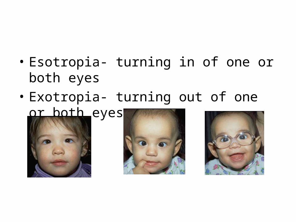
• Esotropia- turning in of one or both eyes
• Exotropia- turning out of one or both eyes

• Hypertropia- turning up of one or both eyes
• Hypotropia- turning down of one or both eyes
Nystagmus
• Null point- point of least nystagmus & best vision
• Pendular nystagmus- up-and-down movements of equal speed, amplitude & duration
• Jerk nystagmus- slower movement in one direction
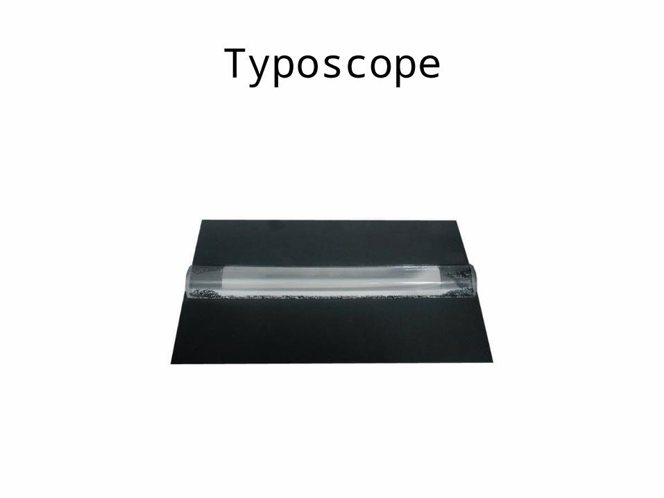
Typoscope
Disorders Relating to the Shape of the Eye
• Refractive Disorders
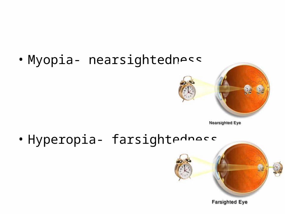
• Myopia- nearsightedness
• Hyperopia- farsightedness

• Anisometropia- more than 1 diopter difference in refractive error between both eyes
• Aniseikonia- difference in shape or image received by both eyes
• Microphthalmia- abnormally small eye
• Macrophthalmia- abnormally large eye
• Anophthalmia- underdeveloped or nondevelopment of eyes
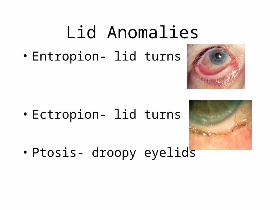
Lid Anomalies • Entropion- lid turns in
• Ectropion- lid turns out
• Ptosis- droopy eyelids

Corneal Disorders & Diseases
Astigmatism
• A. Keratoconus- cornea cone-shaped– 1 .extreme type of corneal curvature
• B. Keratitis- inflamed or ulcerated cornea• 1. ocular herpes• 2. STDs- clamidia, syphilis,
gonococcal
C. Corneal dystrophies• 1. genetic• D. Corneal scarring
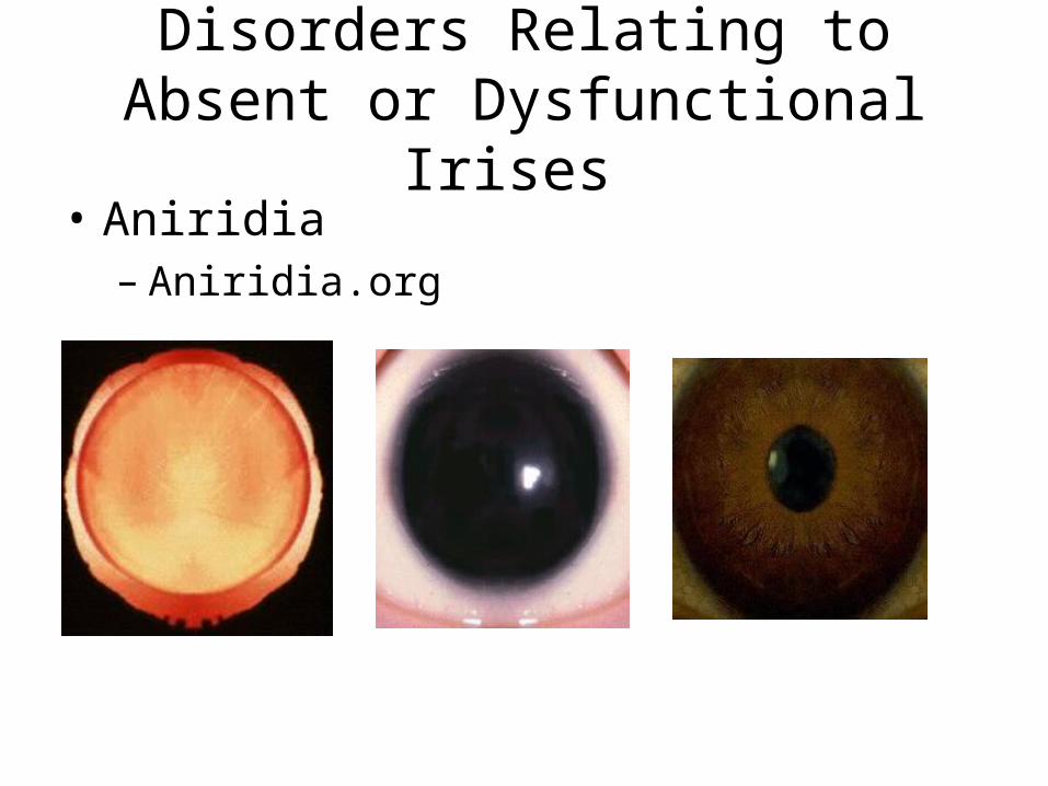
Disorders Relating to Absent or Dysfunctional Irises
• Aniridia– Aniridia.org

Dysfunctional iris- does not constrict or dilate
Coloboma or iris or choroid- keyhole-shaped iris
Iritis- inflammation of iris
Lens-Related Conditions
• Presbyopia
less flexible lens due to aging

Cataracts- opacity of the lens
• Pseudophakia- intraocular lens (IOL)
• Aphakia- no lens
• Dislocated lens

Types of Cataracts
• Juvenile
• Sutural
• Posterior subcapsular (PSC)
• Senile

Diseases of Anterior Segment
• Glaucoma-– Open angle– Closed angle
– Congenital glaucoma– Peters anomaly
Signs & Symptoms of Glaucoma
• Signs– High intraocular pressure (IOP)– Cupped disc– Increased angle of anterior segnment
• Symptoms– Pain if pressures very high– Progressive loss of visual field

Disorders Relating to Vitreous Opacities
• vitrectomy- removed of portion of vitreous due to opacity
Retinal Disorders
• 2 visual systems of eye– Central, color vision (photopic)– Peripheral, nigh, movement vision (scotopic)
Color Deficiencies
• Achromatopsia- lack of color vision– Often accompanied by photophobia (light
sensitivity) & nystagmus– Genetic abnormality of color receptors

AlbinismNOAH.org
• Lack of pigment– pink eyes– nystagmus, photophobia, strabismus, low acuity
• Ocular albinism– blue eyes– nystagmus, photophobia, strabismus, low acuity
Retinal edema
• swelling of the retina

Diabetic retinopathy
• proliferative diabetic retinopathy- vitreous pulls away from retina, vessels hemorrhage
• nonproliferative diabetic retinopathy- microaneurisms
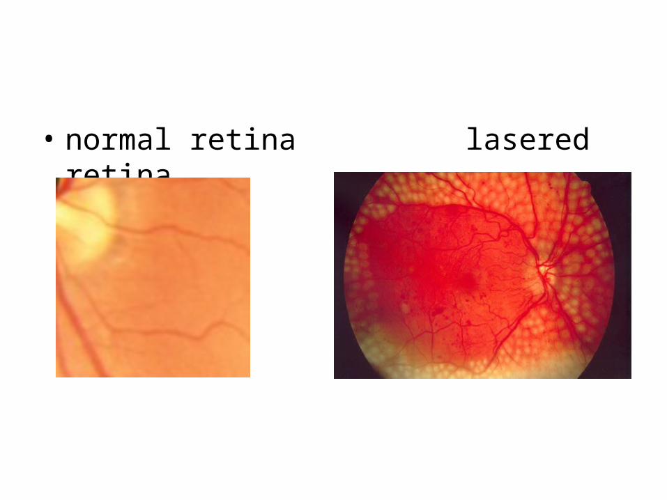
• normal retina lasered retina
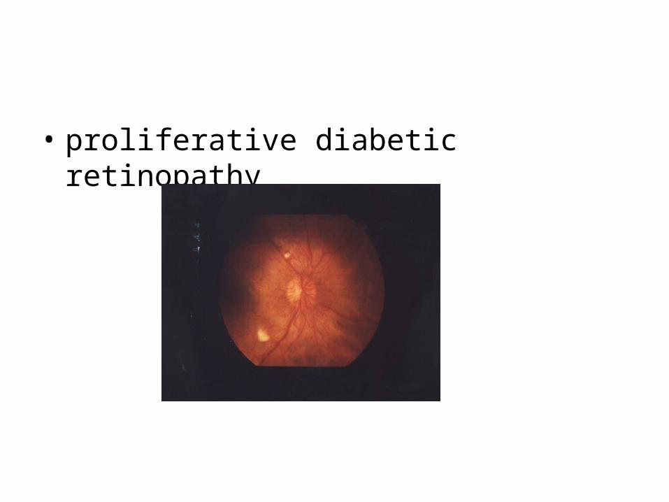
• proliferative diabetic retinopathy
Retinopathy of Prematurity
• fibrovascular proliferation into the vitreous
• refractive errors, amblyopia, strabismus, nystagmus, glaucoma, cataracts, corneal changes, retinal-vitreous abnormalities
• Treatment: laser therapy on avascular retina to anchor; scleral buckling
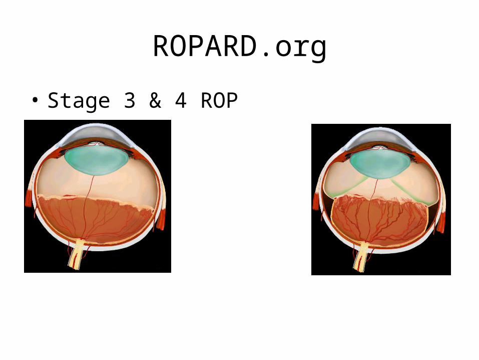
ROPARD.org
• Stage 3 & 4 ROP
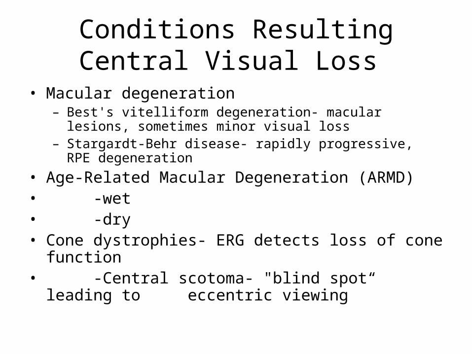
Conditions Resulting Central Visual Loss
• Macular degeneration– Best's vitelliform degeneration- macular lesions, sometimes
minor visual loss– Stargardt-Behr disease- rapidly progressive, RPE degeneration
• Age-Related Macular Degeneration (ARMD)• -wet• -dry• Cone dystrophies- ERG detects loss of cone function• -Central scotoma- "blind spot“ leading to
eccentric viewing

• Normal Dry Wet
• Macula ARMD ARMD

Central Field Loss
• ARMD, Cone dystrophy, Bests, Stargardts
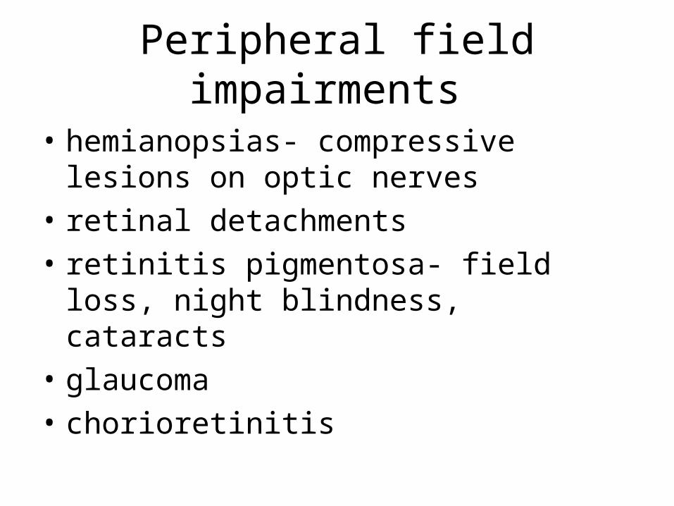
Peripheral field impairments
• hemianopsias- compressive lesions on optic nerves
• retinal detachments
• retinitis pigmentosa- field loss, night blindness, cataracts
• glaucoma
• chorioretinitis
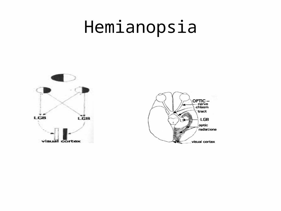
Hemianopsia
Retinal detachments
• High myopia
• Cataract surgery
• Trauma
• Retinopathy of prematurity
• Posterior vitreous detachment
• Lattice degeneration
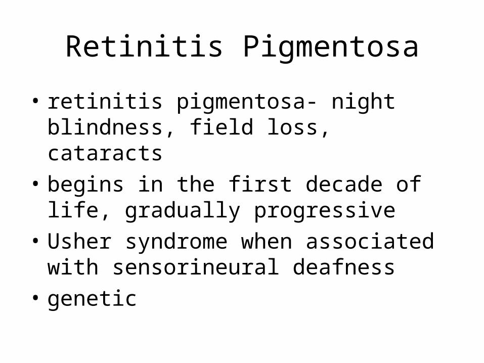
Retinitis Pigmentosa
• retinitis pigmentosa- night blindness, field loss, cataracts
• begins in the first decade of life, gradually progressive
• Usher syndrome when associated with sensorineural deafness
• genetic
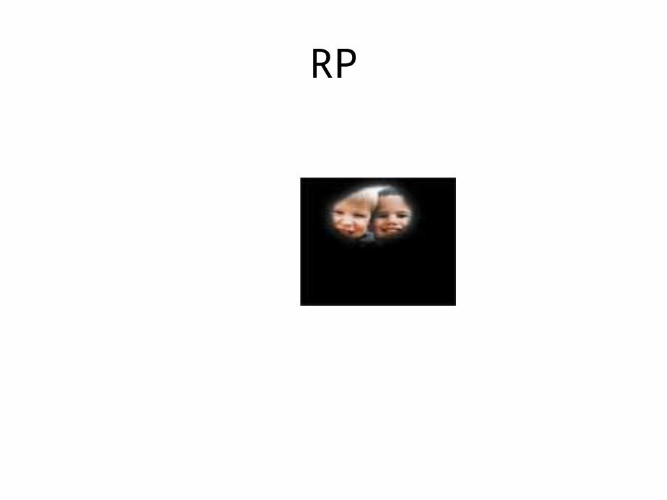
RP

Glaucoma

Retinal Eye Diseases
• Chorioretinitis
• Rubella
• Toxoplasmosis
• Cytomegalovirus
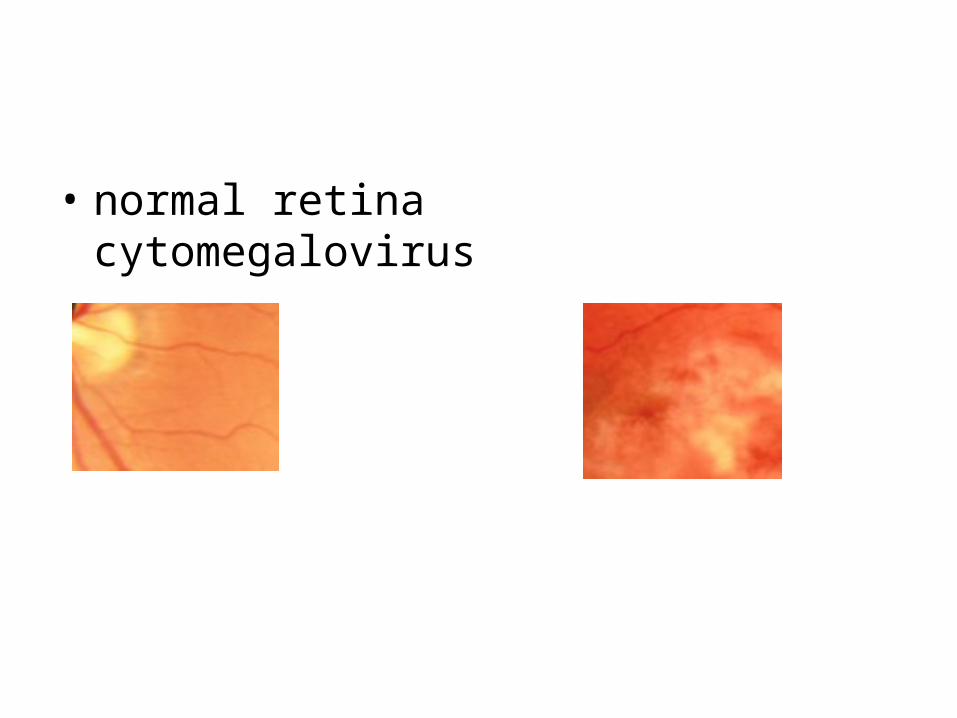
• normal retina cytomegalovirus
Chorioretinitis
• Inflammation of the uveal tract, choroid and/or retinal
• Results in scotomas (blind spots)

Combined Central & Visual Field Impairments
• Coloboma- embryonic defects resulting in incomplete formation of the lids, iris, retina and/or choroid
• Optic nerve disorders & diseases• Optic atrophy- disorder of the optic nerve
interrupting transmission of visual stimuli• Optic nerve hypoplasia & septo-optic
dysplasia- optic nerve fails to develop • Strokes

Coloboma
• CHARGE syndrome– Coloboma & cranial nerves– Heart problems– Atresia of the choanae– Retardation of growth & development– Genitourinary abnormalities– Ear & hearing abnormalities

Cortical Visual Impairment
• Disturbance of visual pathways and/or occipital cortex
• Inconsistent vision due to processing issues
• Causes can be asphyxia, ischemia, head injury, brain defects, infection or hydrocephalis
Leber’s Optic Neuropathy• This is a rare inherited condition which involves the optic nerves
with either complete or partial loss of central vision. • The optic nerve is the "information cable" joining the eye, the
"camera", to the brain. If damage occurs to the retina or the optic nerve then some of the "wires" in the optic nerve will die.
• A healthy nerve looks pink and one that has been damaged pale and is called "atrophic". In LHON the nerve can look abnormally pink and slightly swollen
• Normally males lose their eyesight between the ages of 15 and 45. Often vision is lost in one eye a few months before the other.
• There is loss of central vision and diminished colour vision. Children will have difficulty with reading and fine detail tasks especially with low contrast and small detail work.
Optic Neuritis
• Inflammation of the optic nerve.
• Eye can be painful on movement.
• Contrast vision generally permanently decreased.
• Highly correlated with multiple sclerosis
• Retrobulbar- behind the eye
Optic Atrophy
• Pale optic nerve
• Visual loss
• Congenital or adventitious
• Loss of fibers that transmit visual impulses
Optic Nerve Hypoplasia
• Congenital underdevelopment of optic nerve
• Appears small and pale
• Nystagmus is common
Conditions Caused by External and Other Factors
• Monocularity- use of one eye• Trauma
– puncture wounds– sympathetic ophthalmia– blunt trauma– burns
• Electromagnetic radiation• Disorder caused by tumors of the eye
– retinoblastoma– melanoma versus nevus
• Conjunctivitis- allergic, vernal, chemical, fungal, bacterial, viral

Genetically Determined Conditions
• autosomal recesive
• autosomal dominant
• sex-linked

Perceptual Difficulties
• Dyslexia & learning disabilities
• Scintillating scotomata/migraine
• Charles Bonnet syndrome
Cortical Vision Loss
• damage of one or both occipital lobes of visual cortex
Progressive Visual Impairment
• regular eye care & self-monitoring
• diabetes
• posterior vitreous detachment
• glaucoma
• cataract
OPTICS & LOW VISION DEVICES

Basic Optics
• The Composition of Light
• The Measurement of Light
• Refraction- the bending of visible light rays– index of refraction- speed of light passing
through various media
• Refraction & the ocular system

The Optics of Lenses
• Structure of a lens
Snell's law- the line that will travel upon exiting glass
focal point or image point- where light rays come together & converge on a point

Types of lenses
• spherical lenses– convex or plus lenses bulge outward– planoconvex- bulges on one side– chromatic aberration- light disperses as are colors in
a prism– biconcave- bulges inward on both planes– planoconcave- bulges inward on one side– cylindrical lenses– plano lenses- lens cut flat on both sides– combination of lenses– prism lenses- moves light rays into functional field

Lenses for Refractive Errors
• Myopia (nearsightedness) = biconcave (minus)
• Hyperopia (farsighted) = biconvex (plus)
• Astigmatism (irregular cornea) = cylindrical + axis (location on cornea)
• Strabismus = prisms (base in or out)

Measurement of Lenses
• focal distance- fd
• power is measured in diopters D

Types of Magnification
• Relative distance magnification
• Relative size magnification
• Angular magnification
• Projection magnification
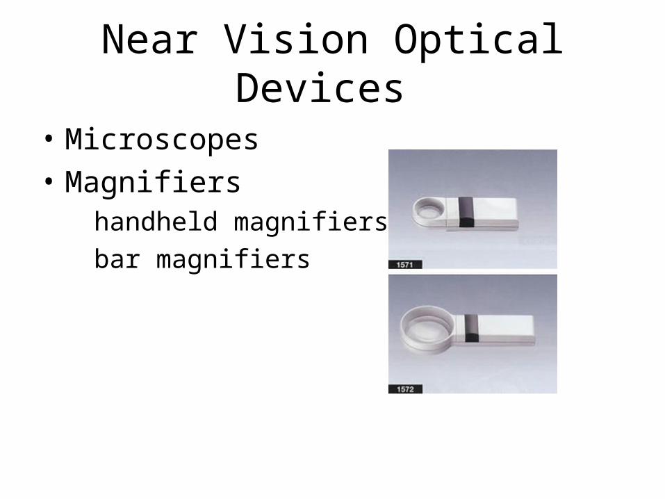
Near Vision Optical Devices
• Microscopes
• Magnifiers handheld magnifiers
bar magnifiers

Near Vision Optical Devices
• Stand magnifiers
• Illuminated magnifers
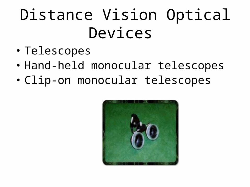
Distance Vision Optical Devices
• Telescopes• Hand-held monocular telescopes• Clip-on monocular telescopes
• Spectacle-mounted telescopes
• full-field telescope systems
• bioptic telescopes
• Contact lens telescopes
• Behind-the-lens telescopes
Non-Optical Systems
• Illumination• 1. types of light• 2. position of light• 3. adaptation of light to dark• 4. glare• Illumination control• Nonoptical magnification

Electronic Systems
• Common electronic systems
• Closed circuit TVs (CCTVs)
• Computer systems
• Other magnification systems
Field-Expansion Systems
• Bioptics
• Fresnel prisms
Related Documents