Neurobiology of Aging 29 (2008) 12–22 Cathepsin D expression is decreased in Alzheimer’s disease fibroblasts Lorena Urbanelli a , Carla Emiliani a , Carlo Massini a , Emanuele Persichetti a , Antonio Orlacchio b,c , Giuliana Pelicci d , Sandro Sorbi e , Andrej Hasilik f , Giorgio Bernardi b,c , Aldo Orlacchio a,∗ a Dipartimento di Medicina Sperimentale e Scienze Biochimiche, Universit` a di Perugia, Via del Giochetto, Perugia 06126, Italy b Laboratorio di Neurogenetica, CERC-IRCCS Santa Lucia, Rome, Italy c Dipartimento di Neuroscienze, Universit` a di Roma “Tor Vergata”, Rome, Italy d Dipartimento di Oncologia Sperimentale, Istituto Europeo di Oncologia, Milan, Italy e Dipartimento di Scienze Neurologiche e Psichiatriche, Universit` a di Firenze, Florence, Italy f Philipps-University Marburg, Marburg, Germany Received 3 November 2005; received in revised form 22 August 2006; accepted 14 September 2006 Available online 17 October 2006 Abstract Cathepsin D (CTSD), a protease detectable in different cell types whose primary function is to degrade proteins by bulk proteolysis in lysosomes, has been suggested to be involved in Alzheimer’s disease (AD). In fact, there is increasing evidence that disturbance of the normal balance and localization of cathepsins may contribute to neurodegeneration in AD [Nakanishi H. Neuronal and microglial cathepsins in aging and age-related diseases. Aging Res Rev 2003; 2(4):367–81]. Here, we provide evidence of an altered balance of CTSD in skin fibroblasts from patients affected either by sporadic or familial forms of AD. In particular, we demonstrate that CTSD is down regulated at both transcriptional and translational level and its processing is altered in AD fibroblasts. The oncogene Ras is involved in the regulation of CTSD, as high expression level of the constitutively active form of Ras in normal or AD fibroblasts induces CTSD down-regulation. p38 MAPK signalling pathway also appears to down-modulate CTSD level. Overall results reinforce the hypothesis that a lysosomal impairment may be involved in AD pathogenesis and can be detected not only in the CNS but also at a peripheral level. © 2006 Elsevier Inc. All rights reserved. Keywords: Alzheimer’s disease; Cathepsin D; Gene expression; Peripheral markers 1. Introduction Alzheimer’s disease (AD) is a progressive neurodegenera- tive dementia characterized by two pathological hallmarks in the cerebral cortex and hippocampus: senile plaques, consist- ing of deposits of -amyloid peptide (A) and neurofibrillary tangles, composed of an abnormally phosphorylated form of the cytoskeleton-associated protein tau. Even if the major- ity of AD cases appears sporadically (Sporadic Alzheimer’s Disease, SAD), a small percentage of AD cases is familial (Familial Alzheimer’s Disease, FAD) and is linked to muta- tions in three different genes: the amyloid precursor protein ∗ Corresponding author. Tel.: +39 075 5857438; fax: +39 075 5857443. E-mail address: [email protected] (A. Orlacchio). (APP), the presenilin 1 (PS1) and the presenilin 2 (PS2). The mechanism by which mutations cause FAD is not completely clear, but it was shown they are associated with abnormal APP processing [3,9,19]. Information about AD pathogenesis arises mainly from the study of these genetically transmitted forms, however the majority of AD cases are not genetically transmitted and their pathogenesis is unknown [24]. Previous studies demonstrated that neuronal populations that degenerate in AD show an up-regulation of the lysosomal system [1,5–7]. Lysosomal system is composed of a family of acidic vesicles including lysosomes, late endosomes and Golgi vesicles, containing recently synthesized enzymes, and residual bodies, containing undigested material. Many neu- rodegenerative diseases arise from lysosomal dysfunction, thus indicating the importance of these organelles for cell 0197-4580/$ – see front matter © 2006 Elsevier Inc. All rights reserved. doi:10.1016/j.neurobiolaging.2006.09.005

Welcome message from author
This document is posted to help you gain knowledge. Please leave a comment to let me know what you think about it! Share it to your friends and learn new things together.
Transcript

A
lnifibCMm©
K
1
ttittiD(t
0d
Neurobiology of Aging 29 (2008) 12–22
Cathepsin D expression is decreased in Alzheimer’s disease fibroblastsLorena Urbanelli a, Carla Emiliani a, Carlo Massini a, Emanuele Persichetti a,
Antonio Orlacchio b,c, Giuliana Pelicci d, Sandro Sorbi e, Andrej Hasilik f,Giorgio Bernardi b,c, Aldo Orlacchio a,∗
a Dipartimento di Medicina Sperimentale e Scienze Biochimiche, Universita di Perugia, Via del Giochetto, Perugia 06126, Italyb Laboratorio di Neurogenetica, CERC-IRCCS Santa Lucia, Rome, Italy
c Dipartimento di Neuroscienze, Universita di Roma “Tor Vergata”, Rome, Italyd Dipartimento di Oncologia Sperimentale, Istituto Europeo di Oncologia, Milan, Italy
e Dipartimento di Scienze Neurologiche e Psichiatriche, Universita di Firenze, Florence, Italyf Philipps-University Marburg, Marburg, Germany
Received 3 November 2005; received in revised form 22 August 2006; accepted 14 September 2006Available online 17 October 2006
bstract
Cathepsin D (CTSD), a protease detectable in different cell types whose primary function is to degrade proteins by bulk proteolysis inysosomes, has been suggested to be involved in Alzheimer’s disease (AD). In fact, there is increasing evidence that disturbance of theormal balance and localization of cathepsins may contribute to neurodegeneration in AD [Nakanishi H. Neuronal and microglial cathepsinsn aging and age-related diseases. Aging Res Rev 2003; 2(4):367–81]. Here, we provide evidence of an altered balance of CTSD in skinbroblasts from patients affected either by sporadic or familial forms of AD. In particular, we demonstrate that CTSD is down regulated atoth transcriptional and translational level and its processing is altered in AD fibroblasts. The oncogene Ras is involved in the regulation ofTSD, as high expression level of the constitutively active form of Ras in normal or AD fibroblasts induces CTSD down-regulation. p38
APK signalling pathway also appears to down-modulate CTSD level. Overall results reinforce the hypothesis that a lysosomal impairmentay be involved in AD pathogenesis and can be detected not only in the CNS but also at a peripheral level.2006 Elsevier Inc. All rights reserved.eywords: Alzheimer’s disease; Cathepsin D; Gene expression; Peripheral markers
(mcAaft
t
. Introduction
Alzheimer’s disease (AD) is a progressive neurodegenera-ive dementia characterized by two pathological hallmarks inhe cerebral cortex and hippocampus: senile plaques, consist-ng of deposits of �-amyloid peptide (A�) and neurofibrillaryangles, composed of an abnormally phosphorylated form ofhe cytoskeleton-associated protein tau. Even if the major-ty of AD cases appears sporadically (Sporadic Alzheimer’s
isease, SAD), a small percentage of AD cases is familialFamilial Alzheimer’s Disease, FAD) and is linked to muta-ions in three different genes: the amyloid precursor protein
∗ Corresponding author. Tel.: +39 075 5857438; fax: +39 075 5857443.E-mail address: [email protected] (A. Orlacchio).
soGrrt
197-4580/$ – see front matter © 2006 Elsevier Inc. All rights reserved.oi:10.1016/j.neurobiolaging.2006.09.005
APP), the presenilin 1 (PS1) and the presenilin 2 (PS2). Theechanism by which mutations cause FAD is not completely
lear, but it was shown they are associated with abnormalPP processing [3,9,19]. Information about AD pathogenesis
rises mainly from the study of these genetically transmittedorms, however the majority of AD cases are not geneticallyransmitted and their pathogenesis is unknown [24].
Previous studies demonstrated that neuronal populationshat degenerate in AD show an up-regulation of the lysosomalystem [1,5–7]. Lysosomal system is composed of a familyf acidic vesicles including lysosomes, late endosomes and
olgi vesicles, containing recently synthesized enzymes, andesidual bodies, containing undigested material. Many neu-odegenerative diseases arise from lysosomal dysfunction,hus indicating the importance of these organelles for cell
iology
vdtdmifA
mEzsmot(dtida
sNchooDls[
Dmdt�Aat[crIraDla
lWhs
obc
stilpsbsifipicir
2
2
smliBrwMSdHdspoMpowA
2
bF
L. Urbanelli et al. / Neurob
iability. Indeed, mutations that cause lysosomal enzymeeficiencies result in approximately 40 different syndromes,ermed Lysosomal Storage Disorders (LSDs). Most of theseiseases are associated with abnormal brain development andental retardation. In addition, they are characterized by
ntracellular deposition and protein aggregation, events alsoound in age-related neurodegenerative disorders, includingD [2].Cathepsin D (CTSD) (EC 3.4.23.5) is the main lysoso-
al aspartic protease. It is synthetized and translocated intondoplasmic Reticulum (ER) as a 52 kDa inactive proen-yme, that is converted in the endosomal compartment in aingle chain active form of 48 kDa [13,39]. In humans and inost species, this single chain form undergoes further prote-
lytic processing in late endosomes and lysosomes, yieldinghe mature form, composed of the light (14 kDa) and heavy32 kDa) chains, held together by disulphide bonds. Its mostlyescribed function is intracellular proteolytic catabolism inhe lysosomal compartment, but other physiological effectsnclude antigen processing and activation or inactivation ofifferent protein substrates, that results in their maturationnd secretion [14,18,47].
A naturally occurring cathepsin D mutation causes a reces-ive form of Neuronal Ceroid Lipofuscinosis (NCL) in sheep.CLs are a group of diseases, which encompasses the most
ommon reason for progressive encefalopathies of children inumans [27]. Affected sheep show an extreme brain atrophyf the cerebral cortex accompanied by a marked accumulationf storage materials in neurons [45]. Disruption of cathepsingene in mice causes ileal atrophy and loss of lymphoid cells
eading to death [40]. Moreover, affected mice show neuronaltorage with typical features of neuronal ceroid lipofuscinosis33].
Several lines of evidence suggest a role for cathepsinin Alzheimer’s disease. Initially, it was identified as a
ajor APP-processing activity [15,17,22,26,32] and tau-egradating enzyme [30]. Accumulating evidence ruled outhe involvement of cathepsin D as a critical component of-, �- and �-secretase system: amyloidogenic processing ofPP is normal in mice neurons devoid of cathepsin D [41]
nd aspartic proteases were identified that are involved inhe cleavage of APP: �-secretase seems to be due to BACE128,46,49], while �-secretase identification is still a matter ofontroversy [34,48], but invalidation of presenilin genes waseported to impair �-secretase-mediated A� production [43].n addition, cathepsin D mRNA and enzyme activity are up-egulated in AD patients’ brain and the protein is present inmyloid plaques [1,5,7,42]. Since up-regulation of cathepsin
is an early and specific observation in AD brain, it seemsikely that cathepsin D may have a role in the neurodegener-tive process [7].
There are emerging evidences of an altered balance of
ysosomal enzymes activities in AD also at peripheral level.hereas the expression of glycohydrolases, such as �-exoaminidase, �-mannosidase, �-galactosidase, is increa-ed in fibroblasts from skin biopsy of patients with familiar
t((c
of Aging 29 (2008) 12–22 13
r sporadic AD [16], the activity of cathepsin D, as evaluatedy flow cytometry, is decreased in peripheral blood lympho-ytes of AD patients [44].
To better elucidate the involvement of the lysosomalystem in AD and go more insight the role of this pro-eolytic enzyme, we characterized cathepsin D expressionn AD fibroblasts at both transcriptional and translationalevel. Cathepsin D processing was also investigated. Amongeripheral sources, fibroblasts are an easy and accessibleource to study the pathogenic mechanisms that are responsi-le for Alzheimer’s disease [9,10]. Previous studies demon-trated that Ras expression in the frontal cortex of AD patientss elevated at early stages of the disease [20,21] and that ADbroblasts are characterized by an increased content of Rasrotein and activation of p38 MAPK [16]. In addition, Rass involved in the processing and subcellular localization ofathepsin D [12]. Here, we provide evidence of the directnvolvement of Ras signalling cascades in cathepsin D down-egulation.
. Methods
.1. Materials
Nonidet P-40, H2O2, DMSO, PD098059, SB203580, ani-omycin, trypan blue, Bovine Serum Albumin (BSA), mouseonoclonal anti-�-tubulin antibody, goat anti-mouse HRP-
inked and rabbit anti-goat HRP-linked secondary antibod-es were from Sigma Chemical Co. (St. Louis, MO, USA).io-Rad protein assay reagent was from Bio-Rad Labo-
atoires (Hercules, CA, USA). Leupeptin and pepstatin Aere from Calbiochem (San Diego, CA, USA). RNeasyini kit and Taq were from Qiagen (Hilden, Germany),
uperScriptIITM RnaseH− Reverse Transcriptase and Ran-om Hexamers were from Invitrogen (Carlsbad, CA, USA).ybond-C Extra nitrocellulose and the ECL-Western blottingetection reagents were from Amersham Biosciences (Upp-ala, Sweden). Goat polyclonal anti-cathepsin D and goatolyclonal anti �-actin were from Santa Cruz Biotechnol-gy (Santa Cruz, CA, USA). Rabbit polyclonal: anti-P-p38APK, anti-p38 MAPK, anti-P-p44/42 MAPK and anti-
44/42 MAPK antibodies, goat anti-rabbit HRP-linked sec-ndary antibody and LumiGLO chemiluminescent reagentere from Cell Signaling Technology (Beverly, MA, USA).ll reagents were of analytical grade.
.2. Fibroblasts
Primary fibroblast cultures were established from skiniopsy of AD patients and controls after informed consent.AD cells were from eight familial cases, four mutated in
he PS1 gene (Leu392Val), two mutated in the PS2 geneMet146Leu) and two mutated in the APP gene (Val717Leu)four males and four females; 52.3 ± 6.5 years of age). SADells were from eight sporadic cases (three males and five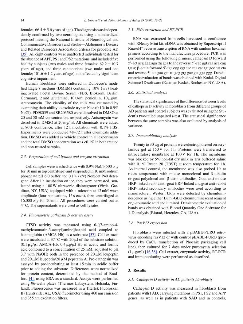
1 iology
fdpCa[thyfc
fiiGseN2daEtaa
2
fpgida14
2
mhw(a3aapffulIa
2
wRpp5taoS
2
oAdbv
2
lnwwAroHHmnob1
2
vdl(a
3
3
4 L. Urbanelli et al. / Neurob
emales; 66.4 ± 5.6 years of age). The diagnosis was indepen-ently confirmed by two neurologists using a standardizedrotocol meeting the National Institute of Neurological andommunicative Disorders and Stroke—Alzheimer’s Diseasend Related Disorders Association criteria for probable AD35]. All eight controls were unaffected individuals tested forhe absence of APP, PS1 and PS2 mutations, and included fiveealthy subjects (two males and three females; 62.2 ± 10.7ears of age), and three centenarians (two males and oneemale; 101.6 ± 1.2 years of age), not affected by significantognitive impairment.
Human fibroblasts were cultured in Dulbecco’s modi-ed Eagle’s medium (DMEM) containing 10% (v/v) heat-
nactivated Foetal Bovine Serum (FBS, Biokrom, Berlin,ermany), 2 mM glutamine, 10 U/ml penicillin, 10 �g/ml
treptomycin. The viability of the cells was estimated byxamining their ability to exclude trypan blue (0.1% in 0.9%aCl). PD98059 and SB203580 were dissolved in DMSO at0 and 50 mM concentration, respectively. Anisomycin wasissolved in DMSO at 20 mg/ml. All chemicals were addedt 80% confluence, after 12 h incubation with 0.1% FBS.xperiments were conducted 48–72 h after chemicals addi-
ion. DMSO was added as vehicle control in all experimentsnd the total DMSO concentration was <0.1% in both treatednd non-treated samples.
.3. Preparation of cell lysates and enzyme extraction
Cell samples were washed twice with 0.9% NaCl (500 × gor 10 min in top centrifuge) and suspended in 10 mM sodiumhosphate pH 6.0 buffer and 0.1% (v/v) Nonidet P40 deter-ent. After 1 h incubation on ice, they were harvested, son-cated using a 100 W ultrasonic disintegrator (Virtis, Gar-iner, NY, USA) equipped with a microtip at 12 mM wavemplitude (four sonications, 15 s each), then centrifuged at6,000 × g for 20 min. All procedures were carried out at◦C. The supernatants were used as cell lysates.
.4. Fluorimetric cathepsin D activity assay
CTSD activity was measured using 6-[(7-amino-4-ethylcoumarin-3-acetyl)amino]hexoid acid coupled to
aemoglobin (AMCA-Hb) as a substrate [37]. Cell extractsere incubated at 37 ◦C with 20 �l of the substrate solution
0.1 �g/�l AMCA-Hb, 0.4 �g/�l Hb in acetic and formiccid combined to a concentration of 25 mM, adjusted to pH.7 with NaOH) both in the presence of 20 �M leupeptinnd 20 �M leupeptin/20 �M pepstatin A. Pro-cathepsin wasssayed by pre-incubating at least 15 min in acidic bufferrior to adding the substrate. Differences were normalizedor protein content, determined by the method of Brad-ord [4], using BSA as a standard. Assays were performed
sing 96-wells plates (Thermos Labsystem, Helsinki, Fin-and). Fluorescence was measured in a Titertek FluoroskanI (Huntsville, AL, USA) fluorimeter using 460 nm emissionnd 355 nm excitation filters.pg
of Aging 29 (2008) 12–22
.5. RNA extraction and RT-PCR
RNA was extracted from cells harvested at confluenceith RNeasy Mini kit. cDNA was obtained by Superscript IInaseH− reverse transcription of RNA with random hexamerrimers according to the manufacturer procedure. PCR waserformed using the following primers: cathepsin D forward′-act acg ggg aga ttg gca tc and reverse 5′-cac ggt caa aca caggt a; �-actin forward 5′-tga cgg ggt cac cca cac tgt gcc cat ctand reverse 5′-cta gaa gca ttt gcg gtg gac gat gga ggg. Densit-metric evaluation of bands was obtained with Kodak Digitalcience 1D software (Eastman Kodak, Rochester, NY, USA).
.6. Statistical analysis
The statistical significance of the difference between levelsf cathepsin D activity in fibroblasts from different groups ofD patients and control subjects was evaluated using the Stu-ent’s two-tailed unpaired t-test. The statistical significanceetween the same samples was also evaluated by analysis ofariance.
.7. Immunoblotting analysis
Twenty to 30 �g of proteins were electrophoresed on acry-amide gel at 150 V for 1 h. Proteins were transferred toitrocellulose membrane at 100 V for 1 h. The membraneas blocked by 5% non-fat dry milk in Tris buffered salineith 0.1% Tween 20 (TBST) at room temperature for 1 h.s internal control, the membrane was also probed 1 h at
oom temperature with mouse monoclonal anti-�-tubulinr goat polyclonal anti �-actin antibodies. Goat anti-mouseRP-linked, rabbit anti-goat HRP-linked and goat anti-rabbitRP-linked secondary antibodies were used according toanufacturer. Western blots were detected by chemilumi-
escence using either Lumi-GLO chemiluminescent reagentr p-coumaric acid and luminol. Densitometric evaluation ofands was obtained with Biorad Quantity One Software for-D analysis (Biorad, Hercules, CA, USA).
.8. RasV12 expression
Fibroblasts were infected with a pBABE-PURO retro-irus encoding rasV12 or with control pBABE-PURO (pro-uced by CaCl2 transfection of Phoenix packaging celline), then cultured for 7 days under puromycin selection1 �g/ml) [16,38]. Cell extract, enzymatic activity, RT-PCRnd immunoblotting were performed as described.
. Results
.1. Cathepsin D activity in AD patients fibroblasts
Cathepsin D activity was measured in fibroblasts fromatients with FAD, carrying mutations in PS1, PS2 and APPenes, as well as in patients with SAD and in controls,

L. Urbanelli et al. / Neurobiology
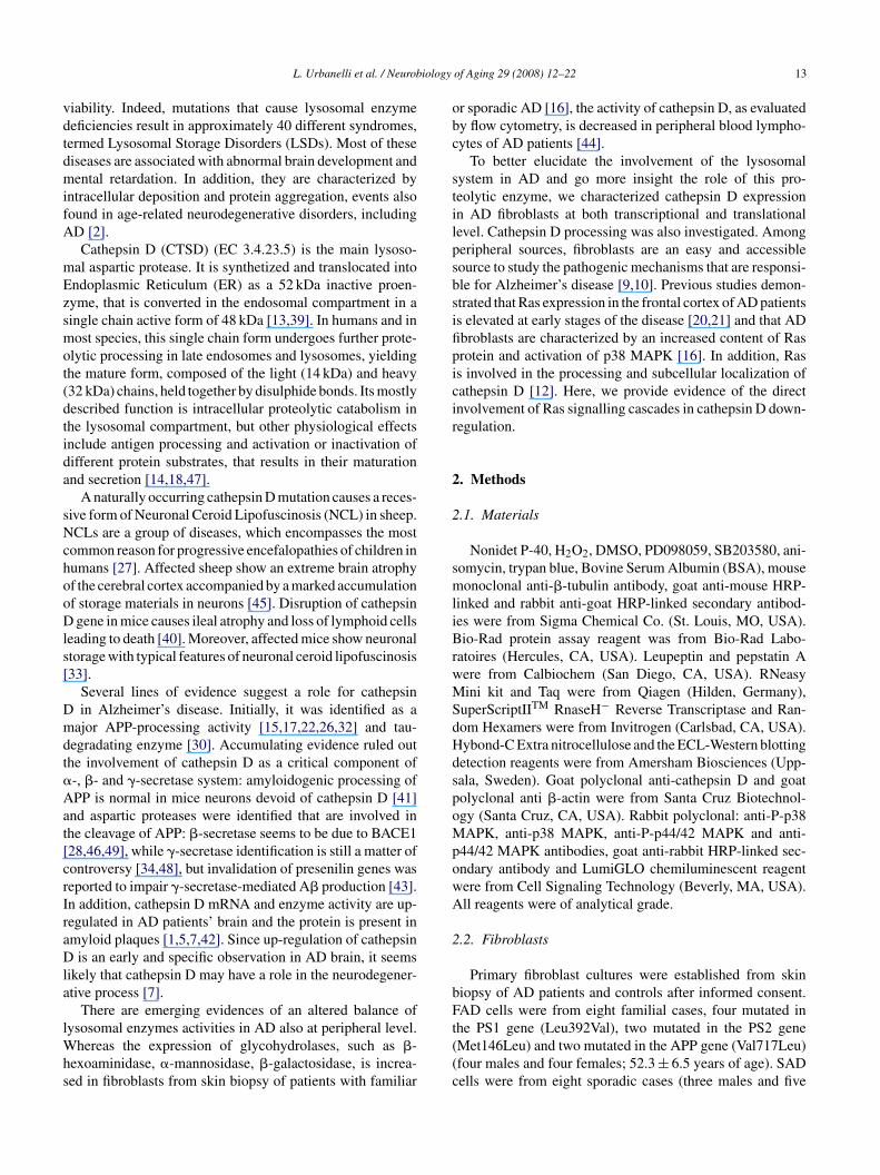
Fig. 1. Scatter plots of cathepsin D activity in fibroblasts from controls, FADaflTE
mtaaaFPgtbpsDtcddc
3p
a
pmtlsuA�wrs(sc(iiisosaaatnsrda([
3A
Dsbimr(tartiFea
nd SAD. Cell extracts, obtained from fibroblasts harvested at 80–90% con-uence, were analyzed for enzyme activity using AMCA-Hb as a substrate.he activity was expressed in Relative Fluorescence Units per mg of protein.ach value is the mean of at least three experiments, each one in duplicate.
atched for sex and age. Scatter plots (Fig. 1) show the dis-ribution of cathepsin D activity in controls as well as in FADnd SAD patients. Statistical analysis demonstrates that aver-ge level of cathepsin D activity is decreased in FAD patientss compared to controls in a significant manner (P < 0.01).AD patients considered in our study carried mutations inS1 (Leu392Val), PS2 (Met146Leu) and APP (Val717Leu)enes. These patients were characterized by an early onset ofhe disease and a strong accumulation of A� peptide in therain. Statistical analysis of fibroblasts cell lines from SADatients, all strongly affected by the disease at the time ofkin biopsy, also demonstrates that average level of cathepsin
activity is decreased significantly (P < 0.01), as comparedo controls. Comparison of FAD and SAD distribution ofathepsin D activity does not show any significant statisticalifference. Moreover, no increase of cathepsin D activity wasetected in any of the individuals examined with respect toontrols.
.2. Cathepsin D processing in fibroblasts from AD
atientsCathepsin D is synthesized as an inactive pro-enzyme withmolecular mass of 52 kDa and cleavage of its N-terminal
(aow
of Aging 29 (2008) 12–22 15
ro-peptide results in a single chain active form of 48 kDa. Inost species, this single-chain protein undergoes further pro-
eolytic processing yielding the mature form, composed of theight (14 kDa) and heavy (32 kDa) chains. To assess cathep-in D content and processing we performed immunoblotting,sing cell extracts from normal, FAD and SAD fibroblasts.s internal control, samples were also assayed with an anti--tubulin antibody. The amount of cathepsin D and �-tubulinas evaluated by densitometric analysis. The average of
atios between the intensity of cathepsin D and �-tubulinignals is reported on histograms for controls, FAD and SADFig. 2, panel a), and a representative immunoblotting is alsohown (Fig. 2, panel d). Statistical analysis confirms thatathepsin D level is decreased in FAD and SAD fibroblastsP < 0.01) and shows a difference in cathepsin D process-ng between control and AD fibroblasts, as we found anncrease in the amount of unprocessed forms (52 and 48 kDa)n FAD and SAD samples (P < 0.01). In addition, compari-on of FAD and SAD distribution of the 32 kDa form andf the 48/52 kDa forms of cathepsin D does not show anyignificant statistical difference. In the same fibroblasts, welso measured the level of Ras and the extent of p38 MAPKctivation. The amount of Ras and P-p38 MAPK was evalu-ted by densitometric analysis. The average of ratios betweenhe intensity of Ras or P-p38 MAPK and �-tubulin sig-als is reported on histograms for control, FAD and SADamples (Fig. 2, panels b and c, respectively), and a rep-esentative immunoblotting is also shown (Fig. 2, panel). We observed an increase of Ras protein as well as anctivation of p38 MAPK both in FAD and in SAD casesFig. 2), in agreement with our previously reported findings16].
.3. Levels of cathepsin D transcript in fibroblasts fromD patients
To understand whether the down-regulation of cathepsinactivity was due to reduced transcription levels, we mea-
ured cathepsin D transcript in AD and control fibroblastsy RT-PCR, using cathepsin D specific primers indicatedn Section 2.5. An amplification product of the expected
olecular weight (900 bp) was obtained and in the sameeaction, as internal control, �-actin gene was also amplified660 bp). The amount of cathepsin D and �-actin amplifica-ion products for each sample was evaluated by densitometricnalysis. The average of ratios for controls, FAD and SAD iseported on histograms in Fig. 3, panel a, while a represen-ative experiment for control and FAD fibroblasts is reportedn Fig. 3, panel b, and for control and SAD fibroblasts inig. 3, panel c. A decrease in cathepsin D transcript wasvident in FAD as well as in SAD samples, as they showverage values significantly different with respect to controls
P < 0.01). In addition, comparison of FAD and SAD aver-ge values shows no significant statistical difference. We alsobserved that the decrease of cathepsin D activity correlatesell with the decrease of cathepsin D transcript (data not
16 L. Urbanelli et al. / Neurobiology of Aging 29 (2008) 12–22
Fig. 2. Immunoblotting analysis of cathepsin D in fibroblasts from controls, FAD and SAD. Cells were lysed, then extracts were sized by SDS-PAGE andblotted as described. The membrane was probed with an anti-cathepsin D antibody, stripped and re-probed with anti-Ras and anti-P-p38 MAPK antibodies. Asinternal control, the membrane was also probed with an anti-�-tubulin. Panel a, histograms report the densitometric analysis of all experimental determinationsfor cathepsin D. Full bars represent the ratio between the signal intensity of the 32 kDa form of cathepsin D and �-tubulin, striped bars represent the ratiobetween the signal intensity of the 52/48 kDa forms of cathepsin D and �-tubulin. Panels b and c, histograms report the densitometric analysis of all experimentaldeterminations for anti-Ras and anti-P-p38 MAPK antibodies, respectively. Bars represent the ratio between the signal intensity of Ras or P-p38 MAPK and�-tubulin. *P < 0.01. Average densitometric values ± S.E. are reported. All control, FAD and SAD fibroblasts were examined. Each value is the mean of at leastthree experiments. For each experiment, the densitometric measure is the average of three independent analyses. Panel d, results obtained from cell extracts ofeight representative samples of control, FAD and SAD fibroblasts. To note that the 32 kDa form of cathepsin D is decreased in FAD and SAD samples withrespect to controls, while the 48/52 kDa forms are increased. Ras content is also increased in AD samples and P-p38 MAPK is detected only in cell extractsfrom FAD and SAD.
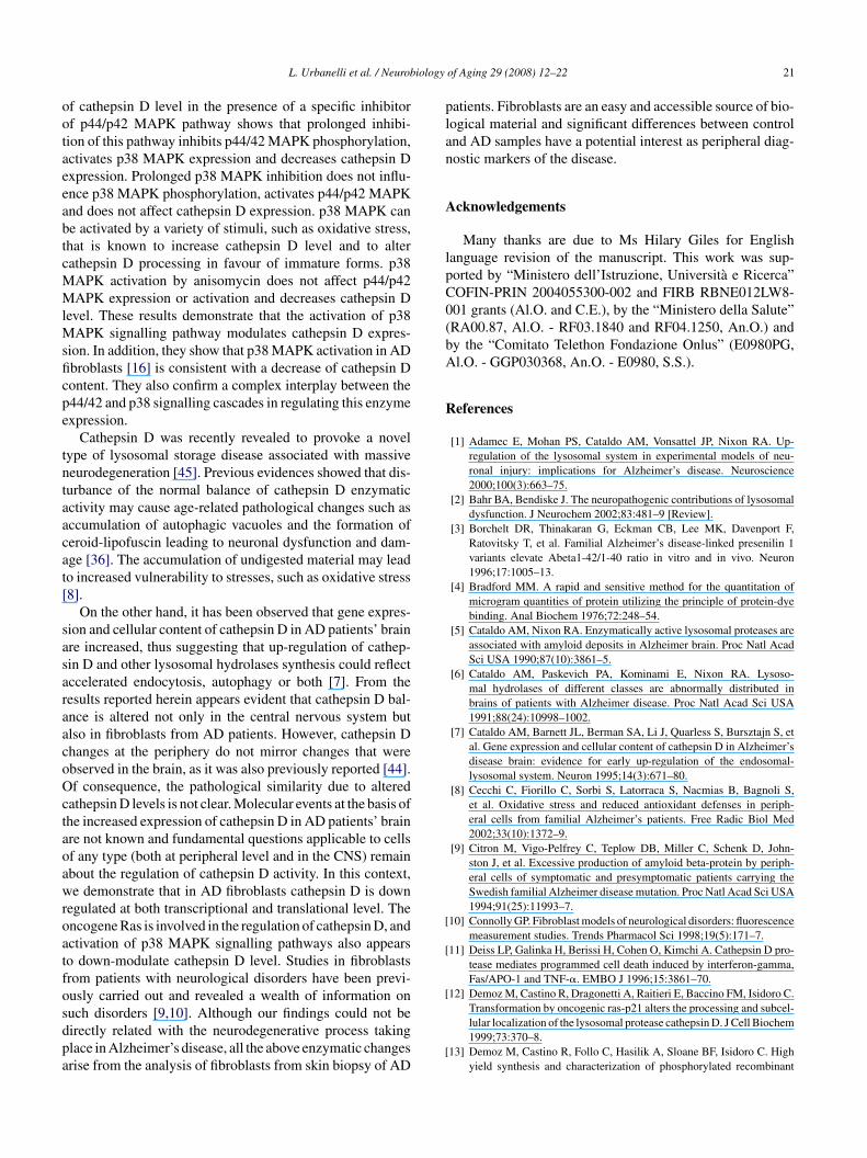
Fig. 3. Cathepsin D transcription as determined by RT-PCR in fibroblasts from controls, FAD and SAD. Total RNA was extracted from semi-confluent fibroblastsand 0.5 �g were used for RT-PCR. Ten percent of the reaction mixture was amplified by PCR and analyzed on a 1.5% agarose gel. As control, �-actin expressionwas determined for each sample in the same reaction. Panel a, histograms report the distribution of densitometric analysis results. Bars represent the average ofratio between the fluorescence intensity of cathepsin D and �-actin amplification products. *P < 0.01. Average densitometric values ± S.E. are reported. Eachvalue is the mean of at least three experiments. For each experiment, the densitometric measure is the average of three independent analyses. Panels b and c,electrophoretic analysis of the amplification products of cathepsin D and �-actin in a representative experiment for FAD and SAD fibroblasts, respectively. Thereaction yielded the bands of the expected molecular weight (900 bp for cathepsin D, 660 bp for �-actin).

L. Urbanelli et al. / Neurobiology of Aging 29 (2008) 12–22 17
Fig. 4. Cathepsin D activity and RT-PCR analysis in fibroblasts from controls and AD patients infected with Ras. Panel a, cell extracts from control, FAD andSAD fibroblasts, infected either with the retroviral construct pBABErasV12 or with the retrovirus alone, were assayed for enzyme activity using AMCA-Hbas a substrate. The activity was expressed in Relative Fluorescence Units per mg of protein. Each value is the mean ± S.D. of at least three assays, each onein duplicate. Panel b, cell extracts from control, FAD and SAD fibroblasts, infected either with the retroviral construct pBABErasV12 or with the retrovirusalone, were sized by SDS-PAGE, probed with an anti-cathepsin D antibody, stripped and re-probed with an anti-�-actin antibody, as internal control. Panelc, densitometric analysis of the experiment reported in panel b. Bars represent the ratio between the intensity of cathepsin D and �-actin signals. Eachdensitometric measure is the average ± S.D. of three independent analyses. Panel d, total RNA was extracted from fibroblasts infected with pBABErasV12 orwith the retrovirus alone and 0.5 �g were used for RT-PCR. Ten percent of the reaction mixture was amplified by PCR and analyzed on a 1.5% agarose gel. Panele, densitometric analysis of experiment reported in panel d. Bars represent the ratio between the fluorescence intensity of cathepsin D and �-actin amplificationp ndent ab .01. Refi ent for
sg
3d
3m
oorals
Foawiad
3
p
roducts. Each densitometric measure is the average ± S.D. of three indepelack bars represent cells treated with the construct pBABErasV12. *P < 0broblasts and very similar results were obtained. A representative experim
hown). Overall results are consistent with the cathepsin Dene down-regulation.
.4. Involvement of Ras signalling pathway inown-regulation of cathepsin D
.4.1. Effect of Ras overexpression on CTSD activity andRNA levelIn order to investigate if Ras is an upstream modulator
f lysosomal cathepsin D, we induced normal fibroblasts toverexpress an active form of Ras by infecting them with a
etroviral vector expressing a mutant Ras (pBABErasV12)nd with the retroviral vector alone as control [38]. Fibrob-asts from FAD and SAD patients were also treated in aimilar manner and a representative experiment for controls,taid
nalyses. Gray bars represent fibroblasts infected with the retrovirus alone,troviral infection was performed on three control, two FAD and two SADa control, a FAD and a SAD sample is reported.
AD and SAD is reported in Fig. 4. The over-expressionf active Ras induces a decrease of cathepsin D enzymaticctivity (Fig. 4, panel a). The lower activity level correlatesith a lower content of cathepsin D protein, as observed by
mmunoblotting (Fig. 4, panels b and c), and with a decreasedmount of transcript, as observed by RT-PCR (Fig. 4, panelsand e).
.4.2. Effect on CTSD activity of MAPK inhibitorsRas may mediate its action through the activation of multi-
le downstream signalling cascades. We further investigated
he role of the activation of two of them, p44/p42 MAPKnd p38 MAPK, on the expression of cathepsin D. First, wencubated control and AD fibroblasts with PD98059, whichown-modulates the activation of p44/p42 MAPK by inhibit-
18 L. Urbanelli et al. / Neurobiology of Aging 29 (2008) 12–22
Fig. 5. Immunoblotting analysis of cathepsin D, p44/42 MAPK and p38 MAPK in fibroblasts treated with PD98059 to inhibit p44/42 MAPK. Panel a, theinhibitor was added to fibroblasts at 70–80% confluence, after 12 h incubation with 0,1% FBS. Cells were lysed 48 h after exposure, then extracts were sizedby SDS-PAGE and blotted as described. The membrane was probed with an anti-cathepsin D antibody, stripped and re-probed with anti-p44/42 MAPK,anti-P-p44/42 MAPK, anti-p38 MAPK and anti-P-p38 MAPK antibodies. As internal control, the membrane was also probed with an anti-�-tubulin. Panelb, cell extracts were also assayed for enzyme activity as described. The activity of cathepsin D in treated fibroblasts was expressed in Relative FluorescenceUnits (RFU) per mg of protein. Each value is the mean ± S.D. of at least three assays, each one in duplicate. *P < 0.01. Three control, two FAD and two SADfibroblasts were treated. A representative experiment with a control sample is reported and very similar results were obtained for FAD and SAD. Panel c,densitometric analysis of results reported in panel a for anti-P-p44/42 MAPK and anti-p44/42 MAPK antibodies. Bars represent the ratio between the intensityof P-p44/42 or p44/42 and �-tubulin signals. Panel d, densitometric analysis of results reported in panel a for anti-P-p38 MAPK and anti-p38 MAPK. Barsr als. PanD lin signaa
ivatwamcscMpwitta(
w
oaitaptMradlfii[F
epresent the ratio between the intensity of P-p38 or p38 and �-tubulin sign. Bars represent the ratio between the intensity of cathepsin D and �-tubu
nalyses. *P < 0.01 with respect to control.
ng two upstream MAPKKs, MEK1 and MEK2, with IC50alues of 4 and 50 �M, respectively. In our case both controlnd AD fibroblasts were treated with an increasing concen-ration of PD98059 (ranging from 2.5 to 20 �M). Incubationith the inhibitor was prolonged up to 48 h both to take into
ccount the long half-life of cathepsin D and to mimic a per-anent situation of inhibition [23]. Results obtained for a
ontrol sample are reported in Fig. 5, panel a, and the corre-ponding densitometric analysis is reported in Fig. 5, panels–e. Immunoblottings of p44/p42 MAPK and of P-p44/p42APK demonstrate that the inhibitor decreases the phos-
horylation of p44/p42 MAPK in a dose dependent manner,ithout affecting its level of expression. It also produces an
ncrease of p38 MAPK expression and activation. At the sameime, this treatment leads to lower cathepsin D protein con-ent and activity (Fig. 5, panels a and b). AD fibroblasts (FAD
nd SAD) show responses to this inhibitor similar to controlsdata not shown).The role of p38 MAPK as modulator of cathepsin Das investigated using SB203580 as inhibitor [25]. Results
yiii
el e, densitometric analysis of results reported in panel a for anti-cathepsinls. Each densitometric measure is the average ± S.D. of three independent
btained for a FAD sample are reported in Fig. 6, panel, and the corresponding densitometric analysis is reportedn Fig. 6, panels c–e. They confirm the previous observa-ion that prolonged treatment with this inhibitor leads ton increased expression and activation of p44/p42 MAPK,robably as a consequence of Raf-1 activation [31]. In addi-ion, we observed that this treatment does not influence p38
APK activation level. In this condition, cathepsin D amountemains stable, as shown by immunoblotting and enzymaticctivity (Fig. 6, panels a and b). Again, we did not detect anyifference between the response of control and AD fibrob-asts. To better elucidate the role of p38 MAPK, we treatedbroblasts with an increasing amount of anisomycin, which
s an antibiotic known to induce the activation of p38 MAPK25]. Results obtained for a FAD sample are reported inig. 7, panel a, and the corresponding densitometric anal-
sis is reported in Fig. 7, panels c–e. This treatment does notnfluence the activation of p44/p42 MAPK, but significantlyncreases the phosphorylation of p38 MAPK, without affect-ng its expression. The result is a decrease of cathepsin D
L. Urbanelli et al. / Neurobiology of Aging 29 (2008) 12–22 19
Fig. 6. Immunoblotting analysis of cathepsin D, p44/42 MAPK and p38 MAPK in fibroblasts treated with SB203580 to inhibit p38 MAPK. Panel a, SB 203580was added to fibroblasts at 70–80% confluence after 12 h incubation with 0,1% FBS. Cells were lysed 48 h after exposure, then extracts were sized by SDS-PAGEand blotted as described. The membrane was probed with an anti-cathepsin D antibody, stripped and re-probed with anti-p44/42 MAPK, anti-P-p44/42 MAPK,anti-p38 MAPK and anti-P-p38 MAPK antibodies. As internal control, the membrane was also probed with an anti-�-tubulin. Panel b, cell extracts wereassayed for enzyme activity as described. The activity of cathepsin D in treated fibroblasts was expressed in Relative Fluorescence Units (RFU) per mg ofprotein. Each value is the mean ± S.D. of at least three assays, each one in duplicate. *P < 0.01. Three control, two FAD and two SAD fibroblasts were treated.A representative experiment for a FAD sample is reported and very similar results were obtained with controls and SAD. Panel c, densitometric analysis ofresults reported in panel a for anti-P-p44/42 MAPK and anti-p44/42 MAPK antibodies. Bars represent the ratio between the intensity of P-p44/42 or p44/42and �-tubulin signals. Panel d, densitometric analysis of results reported in panel a for anti-P-p38 MAPK and anti-p38 MAPK. Bars represent the ratio betweent nalysisb etric mer
pidl
4
mimoip[
parTdoncppc
he intensity of P-p38 or p38 and �-tubulin signals. Panel e, densitometric aetween the intensity of cathepsin D and �-tubulin signals. Each densitomespect to control.
rotein, as observed by immunoblotting and enzymatic activ-ty (Fig. 7, panels a and b). Again, we did not observe anyifference between the response of control and AD fibrob-asts.
. Discussion
There is an increasing evidence that disturbance of the nor-al balance of cathepsins contributes to neurodegeneration
n Alzheimer’s disease [36]. Up-regulation of the endoso-al/lysosomal system was demonstrated in affected neurons
f AD brain [6,7]. Furthermore, in senile plaques cathepsin Ds present at high levels [5] and co-localizes with �-amyloideptide in cerebellum and striatum of post-mortem patients1].
nasi
of results reported in panel a for anti-cathepsin D. Bars represent the ratioasure is the average ± S.D. of three independent analyses. *P < 0.01 with
We studied the activity and expression of cathepsin D ateripheral level, in fibroblasts cell lines established from SADnd FAD patients as well as in controls. We observed a down-egulation of cathepsin D both in FAD and in SAD patients.he decrease is not due to secretion, in fact we were not able toetect any increase of cathepsin D activity in culture mediumf FAD and SAD fibroblasts with respect to controls (dataot shown). It is also not due to reduced conversion of pro-athepsin D to cathepsin D, as production of 51 kDa activeseudo-cathepsin D from 52 kDa inactive pro-cathepsin D byre-incubation with acidic buffer did not result in a signifi-ant difference in the enzyme activity of each sample (data
ot shown). Instead, we observed by RT-PCR that decreasedctivity is correlated with reduced transcription level. In ourtudy, we considered different forms of AD, namely famil-al forms, characterized by mutations in PS1, PS2 and APP
20 L. Urbanelli et al. / Neurobiology of Aging 29 (2008) 12–22
Fig. 7. Immunoblotting analysis of cathepsin D, p44/42 MAPK and p38 MAPK in fibroblasts treated with anisomycin to activate p38 MAPK. Panel a,anisomycin was added to fibroblasts at 70–80% confluence after 12 h incubation with 0,1% FBS. Cells were lysed 48 h after exposure, then extracts weresized by SDS-PAGE and blotted as described. The membrane was probed with an anti-cathepsin D antibody, stripped and re-probed with anti-p44/42 MAPK,anti-P-p44/42 MAPK, anti-p38 MAPK and anti-P-p38 MAPK antbodies. As internal control, the membrane was also probed with an anti-�-tubulin. Panelb, cell extracts were also assayed for enzyme activity as described. The activity of cathepsin D in treated fibroblasts was expressed in Relative FluorescenceUnits (RFU) per mg of protein. Each value is the mean ± S.D. of at least three assays, each one in duplicate. *P < 0.01. Three control, two FAD and two SADfibroblasts were treated. A representative experiment for a FAD sample is reported and very similar results were obtained with controls and SAD. Panel c,densitometric analysis of results reported in panel a for anti-P-p44/42 MAPK and anti-p44/42 MAPK antibodies. Bars represent the ratio between the intensityof P-p44/42 or p44/42 and �-tubulin signals. Panel d, densitometric analysis of results reported in panel a for anti-P-p38 MAPK and anti-p38 MAPK. Barsr als. PanD ulin siga
gaawa
icomesosuect
ie
msDetacsat
epresent the ratio between the intensity of P-p38 or p38 and �-tubulin sign. Bars represent the ratio between the intensity of cathepsin D and �-tub
nalyses. *P < 0.01 with respect to control.
enes, as well as sporadic forms. Our results do not showny correlation between the decreased level of cathepsin Dctivity and the presence or type of mutation (the decreaseas observed in PS1, PS2 and APP mutated patients as well
s in SAD patients).There is strong experimental evidence that oxidative stress
s involved in the pathogenesis of AD, although it is notlear whether the resulting alterations act as a causative agentf neuronal degeneration. Cathepsin D was indicated as aediator of apoptosis induced by oxidative stress in a vari-
ty of cell types and in addition high levels of oxidativetress induce over-expression of cathepsin D and alterationf its processing [11,29]. Our results show that cathep-in D processing is altered in AD fibroblasts in favour of
nprocessed forms. As these are not usually located in latendosomes and lysosomes [39], this suggests that in ADells the enzyme may have increased extralysosomal redis-ribution. However, the alteration of cathepsin D process-dlMd
el e, densitometric analysis of results reported in panel a for anti-cathepsinnals. Each densitometric measure is the average ± S.D. of three separate
ng is not associated with an increased expression of thenzyme.
Lysosomal glycohydrolases expression increases in pri-ary fibroblasts following Ras activation [16,38]. We demon-
trated that the expression of the lysosomal protease cathepsinis decreased in AD patients and control fibroblasts that
xpress constitutively active Ras. This differential regula-ion of lysosomal enzymes by Ras activation suggests thatlthough lysosomal enzymes are widely expressed and oftenonsidered only as housekeeping genes, in fact their expres-ion is finely regulated. Moreover, overall results show thatt least in these fibroblasts cell lines Ras activation is not athe basis of a general up-regulation of the lysosomal system.
Ras mediates its action through the activation of multiple
ownstream effectors pathways. Ras activation in AD fibrob-asts does not seem to correlate with an increase of p44/p42APK activation, while p38 MAPK phosphorylation wasetected only in fibroblasts from AD patients [16]. Analysis
iology
ootaeeabtcMMlMsficpe
tntaacat[
sasaraacoOctaoawroatfosdpa
plan
A
lpC0(bA
R
[
[
[
L. Urbanelli et al. / Neurob
f cathepsin D level in the presence of a specific inhibitorf p44/p42 MAPK pathway shows that prolonged inhibi-ion of this pathway inhibits p44/42 MAPK phosphorylation,ctivates p38 MAPK expression and decreases cathepsin Dxpression. Prolonged p38 MAPK inhibition does not influ-nce p38 MAPK phosphorylation, activates p44/p42 MAPKnd does not affect cathepsin D expression. p38 MAPK cane activated by a variety of stimuli, such as oxidative stress,hat is known to increase cathepsin D level and to alterathepsin D processing in favour of immature forms. p38APK activation by anisomycin does not affect p44/p42APK expression or activation and decreases cathepsin D
evel. These results demonstrate that the activation of p38APK signalling pathway modulates cathepsin D expres-
ion. In addition, they show that p38 MAPK activation in ADbroblasts [16] is consistent with a decrease of cathepsin Dontent. They also confirm a complex interplay between the44/42 and p38 signalling cascades in regulating this enzymexpression.
Cathepsin D was recently revealed to provoke a novelype of lysosomal storage disease associated with massiveeurodegeneration [45]. Previous evidences showed that dis-urbance of the normal balance of cathepsin D enzymaticctivity may cause age-related pathological changes such asccumulation of autophagic vacuoles and the formation oferoid-lipofuscin leading to neuronal dysfunction and dam-ge [36]. The accumulation of undigested material may leado increased vulnerability to stresses, such as oxidative stress8].
On the other hand, it has been observed that gene expres-ion and cellular content of cathepsin D in AD patients’ brainre increased, thus suggesting that up-regulation of cathep-in D and other lysosomal hydrolases synthesis could reflectccelerated endocytosis, autophagy or both [7]. From theesults reported herein appears evident that cathepsin D bal-nce is altered not only in the central nervous system butlso in fibroblasts from AD patients. However, cathepsin Dhanges at the periphery do not mirror changes that werebserved in the brain, as it was also previously reported [44].f consequence, the pathological similarity due to altered
athepsin D levels is not clear. Molecular events at the basis ofhe increased expression of cathepsin D in AD patients’ brainre not known and fundamental questions applicable to cellsf any type (both at peripheral level and in the CNS) remainbout the regulation of cathepsin D activity. In this context,e demonstrate that in AD fibroblasts cathepsin D is down
egulated at both transcriptional and translational level. Thencogene Ras is involved in the regulation of cathepsin D, andctivation of p38 MAPK signalling pathways also appearso down-modulate cathepsin D level. Studies in fibroblastsrom patients with neurological disorders have been previ-usly carried out and revealed a wealth of information on
uch disorders [9,10]. Although our findings could not beirectly related with the neurodegenerative process takinglace in Alzheimer’s disease, all the above enzymatic changesrise from the analysis of fibroblasts from skin biopsy of AD[
of Aging 29 (2008) 12–22 21
atients. Fibroblasts are an easy and accessible source of bio-ogical material and significant differences between controlnd AD samples have a potential interest as peripheral diag-ostic markers of the disease.
cknowledgements
Many thanks are due to Ms Hilary Giles for Englishanguage revision of the manuscript. This work was sup-orted by “Ministero dell’Istruzione, Universita e Ricerca”OFIN-PRIN 2004055300-002 and FIRB RBNE012LW8-01 grants (Al.O. and C.E.), by the “Ministero della Salute”RA00.87, Al.O. - RF03.1840 and RF04.1250, An.O.) andy the “Comitato Telethon Fondazione Onlus” (E0980PG,l.O. - GGP030368, An.O. - E0980, S.S.).
eferences
[1] Adamec E, Mohan PS, Cataldo AM, Vonsattel JP, Nixon RA. Up-regulation of the lysosomal system in experimental models of neu-ronal injury: implications for Alzheimer’s disease. Neuroscience2000;100(3):663–75.
[2] Bahr BA, Bendiske J. The neuropathogenic contributions of lysosomaldysfunction. J Neurochem 2002;83:481–9 [Review].
[3] Borchelt DR, Thinakaran G, Eckman CB, Lee MK, Davenport F,Ratovitsky T, et al. Familial Alzheimer’s disease-linked presenilin 1variants elevate Abeta1-42/1-40 ratio in vitro and in vivo. Neuron1996;17:1005–13.
[4] Bradford MM. A rapid and sensitive method for the quantitation ofmicrogram quantities of protein utilizing the principle of protein-dyebinding. Anal Biochem 1976;72:248–54.
[5] Cataldo AM, Nixon RA. Enzymatically active lysosomal proteases areassociated with amyloid deposits in Alzheimer brain. Proc Natl AcadSci USA 1990;87(10):3861–5.
[6] Cataldo AM, Paskevich PA, Kominami E, Nixon RA. Lysoso-mal hydrolases of different classes are abnormally distributed inbrains of patients with Alzheimer disease. Proc Natl Acad Sci USA1991;88(24):10998–1002.
[7] Cataldo AM, Barnett JL, Berman SA, Li J, Quarless S, Bursztajn S, etal. Gene expression and cellular content of cathepsin D in Alzheimer’sdisease brain: evidence for early up-regulation of the endosomal-lysosomal system. Neuron 1995;14(3):671–80.
[8] Cecchi C, Fiorillo C, Sorbi S, Latorraca S, Nacmias B, Bagnoli S,et al. Oxidative stress and reduced antioxidant defenses in periph-eral cells from familial Alzheimer’s patients. Free Radic Biol Med2002;33(10):1372–9.
[9] Citron M, Vigo-Pelfrey C, Teplow DB, Miller C, Schenk D, John-ston J, et al. Excessive production of amyloid beta-protein by periph-eral cells of symptomatic and presymptomatic patients carrying theSwedish familial Alzheimer disease mutation. Proc Natl Acad Sci USA1994;91(25):11993–7.
10] Connolly GP. Fibroblast models of neurological disorders: fluorescencemeasurement studies. Trends Pharmacol Sci 1998;19(5):171–7.
11] Deiss LP, Galinka H, Berissi H, Cohen O, Kimchi A. Cathepsin D pro-tease mediates programmed cell death induced by interferon-gamma,Fas/APO-1 and TNF-�. EMBO J 1996;15:3861–70.
12] Demoz M, Castino R, Dragonetti A, Raitieri E, Baccino FM, Isidoro C.
Transformation by oncogenic ras-p21 alters the processing and subcel-lular localization of the lysosomal protease cathepsin D. J Cell Biochem1999;73:370–8.13] Demoz M, Castino R, Follo C, Hasilik A, Sloane BF, Isidoro C. Highyield synthesis and characterization of phosphorylated recombinant

2 iology
[
[
[
[
[
[
[
[
[
[
[
[
[
[
[
[
[
[
[
[
[
[
[
[
[
[
[
[
[
[
[
[
[
[
[
2 L. Urbanelli et al. / Neurob
human procathepsin D expressed in mammalian cells. Protein ExprPurif 2006;45(1):157–67.
14] Diment S, Martin KJ, Stahl PD. Cleavage of parathyroid hormone inmacrophage endosomes illustrates a novel pathway for intracellularprocessing of proteins. J Biol Chem 1989;264(23):13403–6.
15] Dreyer RN, Bausch KM, Fracasso P, Hammond LJ, Wunderlich D,Wirak DO, et al. Processing of the pre-beta-amyloid protein by cathep-sin D is enhanced by a familial Alzheimer’s disease mutation. Eur JBiochem 1994;224(2):265–71.
16] Emiliani C, Urbanelli L, Racanicchi L, Orlacchio A, Pelicci G,Sorbi S, et al. Up-regulation of glycohydrolases in Alzheimer’sDisease fibroblasts correlates with Ras activation. J Biol Chem2003;278(40):38453–60.
17] Evin G, Cappai R, Li QX, Culvenor JG, Small DH, Beyreuther K, et al.Candidate gamma-secretases in the generation of the carboxyl terminusof the Alzheimer’s disease beta A4 amyloid: possible involvement ofcathepsin D. Biochemistry 1995;34(43):14185–92.
18] Fruitier I, Garreau I, Piot JM. Cathepsin D is a good candidate for thespecific release of a stable hemorphin from hemoglobin in vivo: VV-hemorphin-7. Biochem Biophys Res Commun 1998;246(3):719–24.
19] Gandy S, Petanceska S. Regulation of Alzheimer beta-amyloidprecursor trafficking and metabolism. Biochim Biophys Acta2000;1502:44–52 [Review].
20] Gartner U, Holzer M, Arendt T. Induction of p21ras in Alzheimerpathology. Neuroreport 1995;6:1441–4.
21] Gartner U, Holzer M, Arendt T. Elevated expression of p21ras is an earlyevent in Alzheimer’s disease and precedes neurofibrillary degeneration.Neuroscience 1999;91:1–5.
22] Gruninger-Leitch F, Berndt P, Langen H, Nelboeck P, Dobeli H. Identi-fication of beta-secretase-like activity using a mass spectrometry-basedassay system. Nat Biotechnol 2000;18(1):66–70.
23] Isidoro C, Demoz M, De Stefanis D, Baccino FM, Hasilik A, Bonelli G.Differential targeting and processing of procathepsin D in normal andtransformed murine 3T3 fibroblasts. Int J Cancer 1997;70(3):310–4.
24] Hardy J, Selkoe DJ. The amyloid hypothesis of Alzheimer’s dis-ease: progress and problems on the road to therapeutics. Science2002;297(5580):353–6 [Review].
25] Hazzalin CA, Cuenda A, Cano E, Cohen P, Mahadevan LC. Effects ofthe inhibition of p38/RK MAP kinase on induction of five fos and jungenes by diverse stimuli. Oncogene 1997;15(19):2321–31.
26] Higaki J, Catalano R, Guzzetta AW, Quon D, Nave JF, Tarnus C, et al.Processing of beta-amyloid precursor protein by cathepsin D. J BiolChem 1996;271(50):31885–93.
27] Hofmann SL, Peltonen L. The neuronal ceroid lipofuscinoses. In:Scriver CR, Beaudet AL, Sly WS, Valle D, editors. The metabolic andmolecular bases of inherited disease. 8th ed. New York: McGraw-Hill;2001. p. 3877–94.
28] Hussain I, Powell D, Howlett DR, Tew DG, Meek TD, Chapman C, etal. Identification of a novel aspartic protease (Asp2) as beta-secretase.Mol Cell Neurosci 1999;14(6):419–27.
29] Kagedal K, Johansson U, Ollinger K. The lysosomal protease cathep-sin D mediates apoptosis induced by oxidative stress. FASEB J2001;15:1592–4.
30] Kenessey A, Nacharaju P, Ko LW, Yen SH. Degradation of tau by lyso-somal enzyme cathepsin D: implication for Alzheimer neurofibrillarydegeneration. J Neurochem 1997;69(5):2026–38.
31] Kalmes A, Deou J, Clowes AW, Daum G. Raf-1 is activated by the
p38 mitogen-activated protein kinase inhibitor, SB203580. FEBS Lett1999;444(1):71–4.32] Kohnken RE, Ladror US, Wang GT, Holzman TF, Miller BE, KrafftGA. Cathepsin D from Alzheimer’s-diseased and normal brains. ExpNeurol 1995;133(2):105–12.
[
of Aging 29 (2008) 12–22
33] Koike M, Nakanishi H, Saftig P, Ezaki J, Isahara K, Ohsawa Y, et al.Cathepsin D deficiency induces lysosomal storage with ceroid lipofus-cin in mouse CNS neurons. J Neurosci 2000;20:6898–906.
34] Lai MT, Crouthamel MC, DiMuzio J, Pietrak BL, Donoviel DB, Bern-stein A, et al. A presenilin-independent aspartyl protease prefers thegamma-42 site cleavage. J Neurochem 2006;96(January (1)):118–25[Epub 2005 Nov 21].
35] McKhann G, Drachman G, Folstein M. Clinical diagnosis ofAlzheimer’s disease: report of the NINCDS-ADRDA Work Groupunder the auspices of the department of health and human servicestask force on Alzheimer’s disease. Neurology 1984;34:939–44.
36] Nakanishi H. Neuronal and microglial cathepsins in aging and age-related diseases. Aging Res Rev 2003;2(4):367–81.
37] Partanen S, Storch S, Loffler HG, Hasilik A, Tyynela J, Braulke T.A replacement of the active-site aspartic acid residue 293 in mousecathepsin D affects its intracellular stability, processing and transportin HEK-293 cells. Biochem J 2003;369:55–62.
38] Pearson M, Carbone R, Sebastiani C, Cioce M, Fagioli M, Saito S, etal. PML regulates p53 acetylation and premature senescence inducedby oncogenic Ras. Nature 2000;406(6792):207–10.
39] Rijnboutt S, Kal AJ, Geuze HJ, Aerts H, Strous GJ. Mannose 6-phosphate-independent targeting of cathepsin D to lysosomes in HepG2cells. J Biol Chem 1991;266:23586–92.
40] Saftig P, Hetman M, Schmahl W, Weber K, Heine L, Mossmann H,et al. Mice deficient for the lysosomal proteinase cathepsin D exhibitprogressive atrophy of the intestinal mucosa and profound destructionof lymphoid cells. EMBO J 1995;14:3599–608.
41] Saftig P, Peters C, von Figura K, Craessaerts K, Van Leuven F, DeStrooper B. Amyloidogenic processing of human amyloid precursorprotein in hippocampal neurons devoid of cathepsin D. J Biol Chem1996;271(44):27241–4.
42] Schwagerl AL, Mohan PS, Cataldo AM, Vonsattel JP, Kowall NW,Nixon RA. Elevated levels of the endosomal-lysosomal proteinasecathepsin D in cerebrospinal fluid in Alzheimer disease. J Neurochem1995;64(1):443–6.
43] Sisodia SS, St George-Hyslop PH. gamma-Secretase, Notch, Abeta andAlzheimer’s disease: where do the presenilins fit in? Nat Rev Neurosci2002;3(4):281–90 [Review].
44] Straface E, Matarrese P, Gambardella L, Vona R, Sgadari A, SilveriMC, et al. Oxidative imbalance and cathepsin D changes as periph-eral blood biomarkers of Alzheimer disease: a pilot study. FEBS Lett2005;579:2759–66.
45] Tyynela J, Sohar I, Sleat DE, Gin RM, Donnelly RJ, Baumann M,et al. A mutation in the ovine cathepsin D gene causes a congenitallysosomal storage disease with profound neurodegeneration. EMBO J2000;19:2786–92.
46] Vassar R, Bennett BD, Babu-Khan S, Kahn S, Mendiaz EA, DenisP, et al. Beta-secretase cleavage of Alzheimer’s amyloid precur-sor protein by the transmembrane aspartic protease BACE. Science1999;286(5440):735–41.
47] Wolf M, Clark-Lewis I, Buri C, Langen H, Lis M, Mazzucchelli L.Cathepsin D specifically cleaves the chemokines macrophage inflam-matory protein-1 alpha, macrophage inflammatory protein-1 beta,and SLC that are expressed in human breast cancer. Am J Pathol2003;162(4):1183–90.
48] Wolfe MS, Xia W, Ostaszewski BL, Diehl TS, Kimberly WT,Selkoe DJ. Two transmembrane aspartates in presenilin-1 required
for presenilin endoproteolysis and gamma-secretase activity. Nature1999;398(6727):513–7.49] Yan R, Bienkowski MJ, Shuck ME, Miao H, Tory MC, Pauley AM,et al. Membrane-anchored aspartyl protease with Alzheimer’s diseasebeta-secretase activity. Nature 1999;402(6761):533–7.
Related Documents