1390 IEEE TRANSACTIONS ON MEDICAL IMAGING, VOL. 33, NO. 6, JUNE 2014 Categorizing Extent of Tumor Cell Death Response to Cancer Therapy Using Quantitative Ultrasound Spectroscopy and Maximum Mean Discrepancy Mehrdad J. Gangeh, Member, IEEE, Ali Sadeghi-Naini, Member, IEEE, Michael Diu, Student Member, IEEE, Hadi Tadayyon, Student Member, IEEE, Mohamed S. Kamel, Fellow, IEEE, and Gregory J. Czarnota Abstract—Quantitative ultrasound (QUS) spectroscopic tech- niques in conjunction with maximum mean discrepancy (MMD) have been proposed to detect, and to classify noninvasively the levels of cell death in response to cancer therapy administration in tumor models. Evaluation of xenograft tumor responses to cancer treatments were carried out using conventional-frequency ultrasound at different times after chemotherapy exposure. Ul- trasound data were analyzed using spectroscopic techniques and multi-parametric QUS spectral maps were generated. MMD was applied as a distance criterion, measuring alterations in each tumor in response to chemotherapy, and the extent of cell death was classified into less/more than 20% and 40% categories. Statis- tically significant differences were observed between “pre-” and “post-treatment” groups at different times after chemotherapy exposure, suggesting a high capability of proposed framework for detecting tumor response noninvasively. Promising results were also obtained for categorizing the extent of cell death response in each tumor using the proposed framework, with gold standard histological quantification of cell death as ground truth. The best classification results were obtained using MMD when applied on histograms of QUS parametric maps. In this case, classification accuracies of 84.7% and 88.2% were achieved for categorizing extent of tumor cell death into less/more than 20% and 40%, respectively. Index Terms—Cancer treatment, classification methods, kernel methods, nonparametric methods, personalized medicine, quanti- tative ultrasound. Manuscript received January 21, 2014; revised March 05, 2014; accepted March 09, 2014. Date of publication March 18, 2014; date of current version May 29, 2014. This work was supported in part by the Terry Fox Foundation, in part by the Natural Sciences and Engineering Research Council of Canada, and in part by the Canadian Institutes of Health Research. Asterisk indicates corre- sponding author. *M. J. Gangeh is with the Departments of Medical Biophysics, and Radia- tion Oncology, University of Toronto, Toronto, ON, M5G 2M9 Canada, also with the Departments of Radiation Oncology, and Imaging Research—Physical Sciences, Sunnybrook Health Sciences Centre, Toronto, ON, M4N 3M5 Canada (e-mail: [email protected]). A. Sadeghi-Naini and G. J. Czarnota are with the Departments of Medical Biophysics, and Radiation Oncology, University of Toronto, Toronto, ON, M5G 2M9 Canada, also with the Departments of Radiation Oncology, and Imaging Research—Physical Sciences, Sunnybrook Health Sciences Centre, Toronto, ON, M4N 3M5 Canada (e-mail: [email protected]; gre- [email protected]). M. Diu and M. S. Kamel are with the Department of Electrical and Computer Engineering, University of Waterloo, Waterloo, ON, N2L 3G1 Canada (e-mail: [email protected]; [email protected]). H. Tadayyon is with the Department of Medical Biophysics, University of Toronto, Toronto, ON, M5G 2M9 Canada, and also with the Department of Physical Sciences, Sunnybrook Health Sciences Centre, Toronto, ON, M4N 3M5 Canada (e-mail: [email protected]). Color versions of one or more of the figures in this paper are available online at http://ieeexplore.ieee.org. Digital Object Identifier 10.1109/TMI.2014.2312254 I. INTRODUCTION P ERSONALIZED cancer therapy has recently attracted much attention among researchers in various areas of science and engineering including biomedical engineering, biophysics, medicine, and biology [1], [2], [3]. Motivated by high rates of poor responses of patients to standard routine treatments, personalized cancer therapy aims at altering ther- apies for refractory patients on an individual basis. Utilizing faster feedback mechanisms, personalized cancer therapy is expected to avoid needless and/or harmful continuation of an ineffective cancer treatment, or even facilitate switching to a salvage therapy early on during the course of treatment. The monitoring of cancer therapy effects is broadly per- formed at two different levels: macroscopic/anatomical or microscopic/cellular. The former mainly relies on the classical clinical practice to measure the size of tumor, which has been established for research, clinical screening, and treatment planning; see [4] for a comprehensive survey. The latter, i.e., monitoring tumor response to treatment at microscopic/cellular level [5], [6], [7], is a much newer field. The impetus for research in this area is to shift away from the conventional paradigm of using tumor size changes as a measure of treatment effectiveness. These changes in size can take weeks to months to become apparent, and do not always occur even when the treatment is effective [5], [8]. Functional imaging modalities, such as single photon emission computed tomography (SPECT), positron emission tomography (PET), and magnetic resonance imaging (MRI) play an important role as they can potentially enable noninva- sive evaluation of response to cancer therapies at microscopic level early after the start of treatment (days as opposed to months in standard clinical practice) [5], [8], [7]. However, these methods have the disadvantages of being expensive and requiring injection of exogenous contrast agents frequently to enhance the contrast from soft tissues. The agents’ cost and potential for side effects and allergic reactions (most of the agents are radioactive, albeit at low levels) limits the utility of the technology. In this context, quantitative ultrasound (QUS) methods [6], [9] provide a promising framework that can non- invasively, inexpensively, and quickly be used to assess tumor response to cancer treatment using standard clinical ultrasound equipment [10]. Unlike other aforementioned imaging modal- ities used in monitoring cancer therapy effects, QUS does not 0278-0062 © 2014 IEEE. Personal use is permitted, but republication/redistribution requires IEEE permission. See http://www.ieee.org/publications_standards/publications/rights/index.html for more information.

Welcome message from author
This document is posted to help you gain knowledge. Please leave a comment to let me know what you think about it! Share it to your friends and learn new things together.
Transcript

1390 IEEE TRANSACTIONS ON MEDICAL IMAGING, VOL. 33, NO. 6, JUNE 2014
Categorizing Extent of Tumor Cell Death Responseto Cancer Therapy Using Quantitative UltrasoundSpectroscopy and Maximum Mean Discrepancy
Mehrdad J. Gangeh, Member, IEEE, Ali Sadeghi-Naini, Member, IEEE, Michael Diu, Student Member, IEEE,Hadi Tadayyon, Student Member, IEEE, Mohamed S. Kamel, Fellow, IEEE, and Gregory J. Czarnota
Abstract—Quantitative ultrasound (QUS) spectroscopic tech-niques in conjunction with maximum mean discrepancy (MMD)have been proposed to detect, and to classify noninvasively thelevels of cell death in response to cancer therapy administrationin tumor models. Evaluation of xenograft tumor responses tocancer treatments were carried out using conventional-frequencyultrasound at different times after chemotherapy exposure. Ul-trasound data were analyzed using spectroscopic techniques andmulti-parametric QUS spectral maps were generated. MMD wasapplied as a distance criterion, measuring alterations in eachtumor in response to chemotherapy, and the extent of cell deathwas classified into less/more than 20% and 40% categories. Statis-tically significant differences were observed between “pre-” and“post-treatment” groups at different times after chemotherapyexposure, suggesting a high capability of proposed framework fordetecting tumor response noninvasively. Promising results werealso obtained for categorizing the extent of cell death response ineach tumor using the proposed framework, with gold standardhistological quantification of cell death as ground truth. The bestclassification results were obtained using MMD when applied onhistograms of QUS parametric maps. In this case, classificationaccuracies of 84.7% and 88.2% were achieved for categorizingextent of tumor cell death into less/more than 20% and 40%,respectively.
Index Terms—Cancer treatment, classification methods, kernelmethods, nonparametric methods, personalized medicine, quanti-tative ultrasound.
Manuscript received January 21, 2014; revised March 05, 2014; acceptedMarch 09, 2014. Date of publication March 18, 2014; date of current versionMay 29, 2014. This work was supported in part by the Terry Fox Foundation, inpart by the Natural Sciences and Engineering Research Council of Canada, andin part by the Canadian Institutes of Health Research. Asterisk indicates corre-sponding author.*M. J. Gangeh is with the Departments of Medical Biophysics, and Radia-
tion Oncology, University of Toronto, Toronto, ON, M5G 2M9 Canada, alsowith the Departments of Radiation Oncology, and Imaging Research—PhysicalSciences, Sunnybrook Health Sciences Centre, Toronto, ON,M4N 3M5Canada(e-mail: [email protected]).A. Sadeghi-Naini and G. J. Czarnota are with the Departments of Medical
Biophysics, and Radiation Oncology, University of Toronto, Toronto, ON,M5G 2M9 Canada, also with the Departments of Radiation Oncology, andImaging Research—Physical Sciences, Sunnybrook Health Sciences Centre,Toronto, ON, M4N 3M5 Canada (e-mail: [email protected]; [email protected]).M. Diu and M. S. Kamel are with the Department of Electrical and Computer
Engineering, University of Waterloo, Waterloo, ON, N2L 3G1 Canada (e-mail:[email protected]; [email protected]).H. Tadayyon is with the Department of Medical Biophysics, University of
Toronto, Toronto, ON, M5G 2M9 Canada, and also with the Department ofPhysical Sciences, Sunnybrook Health Sciences Centre, Toronto, ON, M4N3M5 Canada (e-mail: [email protected]).Color versions of one or more of the figures in this paper are available online
at http://ieeexplore.ieee.org.Digital Object Identifier 10.1109/TMI.2014.2312254
I. INTRODUCTION
P ERSONALIZED cancer therapy has recently attractedmuch attention among researchers in various areas of
science and engineering including biomedical engineering,biophysics, medicine, and biology [1], [2], [3]. Motivated byhigh rates of poor responses of patients to standard routinetreatments, personalized cancer therapy aims at altering ther-apies for refractory patients on an individual basis. Utilizingfaster feedback mechanisms, personalized cancer therapy isexpected to avoid needless and/or harmful continuation of anineffective cancer treatment, or even facilitate switching to asalvage therapy early on during the course of treatment.The monitoring of cancer therapy effects is broadly per-
formed at two different levels: macroscopic/anatomical ormicroscopic/cellular. The former mainly relies on the classicalclinical practice to measure the size of tumor, which has beenestablished for research, clinical screening, and treatmentplanning; see [4] for a comprehensive survey. The latter, i.e.,monitoring tumor response to treatment at microscopic/cellularlevel [5], [6], [7], is a much newer field. The impetus forresearch in this area is to shift away from the conventionalparadigm of using tumor size changes as a measure of treatmenteffectiveness. These changes in size can take weeks to monthsto become apparent, and do not always occur even when thetreatment is effective [5], [8].Functional imaging modalities, such as single photon
emission computed tomography (SPECT), positron emissiontomography (PET), and magnetic resonance imaging (MRI)play an important role as they can potentially enable noninva-sive evaluation of response to cancer therapies at microscopiclevel early after the start of treatment (days as opposed tomonths in standard clinical practice) [5], [8], [7]. However,these methods have the disadvantages of being expensive andrequiring injection of exogenous contrast agents frequently toenhance the contrast from soft tissues. The agents’ cost andpotential for side effects and allergic reactions (most of theagents are radioactive, albeit at low levels) limits the utility ofthe technology. In this context, quantitative ultrasound (QUS)methods [6], [9] provide a promising framework that can non-invasively, inexpensively, and quickly be used to assess tumorresponse to cancer treatment using standard clinical ultrasoundequipment [10]. Unlike other aforementioned imaging modal-ities used in monitoring cancer therapy effects, QUS does not
0278-0062 © 2014 IEEE. Personal use is permitted, but republication/redistribution requires IEEE permission.See http://www.ieee.org/publications_standards/publications/rights/index.html for more information.

GANGEH et al.: CATEGORIZING EXTENT OF TUMOR CELL DEATH RESPONSE TO CANCER THERAPY 1391
need injection of any contrast agent. This is mainly becausecancer therapies, such as chemotherapy, radiotherapy, orphotodynamic therapy, generally aim at inducing programmedcell death, i.e., apoptosis [11], in which many morpholog-ical changes occur in tumor cells that affect the bioacousticproperties of tumor. These include nuclear condensation andfragmentation, cell swelling, and chromatin dissolution, whichhave been demonstrated to directly or indirectly affect theultrasound backscatter characteristics of tumor [9], [10], [12].For example, early cell-death (nucleus condensation and frag-mentation) increases the backscatter intensity. On the otherhand, late cell-death (chromatin dissolution/nucleus degenera-tion due to apoptosis) has two opposite effects on ultrasoundbackscatter intensity. This initially increases the randomnessof fairly regular backscatter distributions of micro-echoes,which produces a large backscatter signal. However, when alarge fraction of the nuclei (scatterers of ultrasound) becomedisintegrated due to DNA cleavage (advanced necrosis), theamplitude of the backscattered signal has been observed to bereduced [13], [14], [15]. QUS has the main advantage overconventional ultrasound imaging as it uses metrics that arepredominantly independent of the instrument settings. Suchquantitative measures are derived via analyzing the radio-fre-quency (RF) raw data before forming B-mode images, i.e.,before detecting its envelope and log amplification, and de-scribe bio-acoustic characteristics of the scanned tissue. QUSmethods often involve the spectrum analysis (spectroscopy) ofthe backscatter RF signals over a region of interest, formingparametric maps.The applicability of QUS spectroscopy to detect, noninva-
sively, cell death has been demonstrated recently in vitro, in situ,and in vivo in both preclinical models and clinical settings usinghigh- and conventional-frequency ultrasound [6], [8], [10], [12],[16]. The initial research to utilize ultrasound to detect apoptosisresulting from cancer treatment in vitro and then in vivowere re-ported in [17] and [13], which used high-frequency ultrasound(20–50 MHz). This has recently been extended to conventionalclinical US ranges (1–50 MHz) [10], [12], [16], [18], which en-ables much broader adoption of the technology in the clinic. Itbuilds on the earlier theoretical work of [19], the seminal workbehind QUS spectroscopic methods, that utilized spectrum anal-ysis of the conventional low-frequency (5–15 MHz) RF signalto characterize tissues at much coarser scales, e.g., to distinguishbetween normal and detached retinal tissue.While previous work in the field of QUS evaluation of cell
death response mainly focused on analyses such as statisticaltests of significance, regression, and discriminant analysis inorder to demonstrate the proof of principle of the proposed QUSsystems [9], [18], [16], [10], a next step towards the design ofa complete computer-aided-prognosis system is to deploy ad-vanced machine learning algorithms and supervised learningtechniques to reliably classify the level of cell death in an auto-matic manner. Categorizing the extent of the cell death (low,mid, high), noninvasively, is important in the application ofmonitoring cancer therapy effects. This can be achieved withthe settings applied in this study by classifying the extent of celldeath to less/more than 20% and 40%.
Relatively few studies have attempted to use supervisedlearning to detect tumor response to treatment using medicalimaging data. Larkin et al. [20] worked with -weightedMRI to detect cell death, using the support vector machine(SVM) classifier [21] and a “Minkowski functional” featureto classify whether an image represented a treated tumor, or anontreated control image. Classification accuracy after a 24-hperiod was 75% using 19 subjects in preclinical data. Histolog-ical analysis was used to confirm significant increases in celldeath after treatment. SVM was also applied with the goal ofdifferentiating between “pre-” and “post-treatment” images ofpreclinical data using QUS in [22]; classification accuracy was87.3% when assessed 24 h after treatment. Two studies froma related field, computer-aided pathology detection, may alsosuggest appropriate supervised learning methods. Sørensenet al. [23] used a joint rotation-invariant local binary pattern(LBP) and intensity histogram in a dissimilarity-based clas-sification approach to predict emphysema using CT imaging.The simple k-nearest-neighbor (k-NN) classifier and Euclideandistance were used, and the classifier’s posterior probabilitieswere deployed to compute the correlation to the ground truth.Feleppa et al. [24] used a multi-layer perceptron classifier todistinguish between cancerous and noncancerous tissues ofthe prostate at a pixel-by-pixel level using QUS mid-band fitand 0-MHz intercept parametric maps (related to ultrasoundbackscatter intensity and effective acoustic scatterer concen-tration, respectively). They found SVMs to give a very similarperformance.In this research, steps were taken towards the development
of a computer-aided-prognosis system to assess cancer therapyeffects, noninvasively, using conventional-frequency QUSspectral parametric maps. Ultrasound data were acquired fromxenograft tumor-bearing mice before and at different timesafter chemotherapy exposure. After computation of intensityhistograms of the parametric maps as a feature descriptor, thedistances between “pre-” and “post-treatment” samples werecomputed using a recently introduced kernel-basedmetric calledmaximummeandiscrepancy(MMD)[25], [26], [27], [28].MMD is a distance measure, appropriate when there are mul-
tiple data samples available from two populations to be com-pared. It is a kernel-based measure, implying that its compu-tation is reliant on inner products taken in a reproducing kernelHilbert space (RKHS). By using a kernel function to nonlinearlytransform input vectors into a different, possibly higher-dimen-sional feature space, and computing the populationmeans in thisnew space, enhanced group separability (compared to, e.g., Eu-clidean distance in the original feature space) is ideally obtained.The main contributions of this study are as follows.1) Proposing a complete computer-aided-prognosis systemusing MMD and QUS spectroscopy. The system can pro-vide an early indication of cells undergoing apoptosis (pro-grammed cell death) within hours after treatment, using theQUS spectral parametric maps.
2) Providing one of the first studies to apply machine learningtechniques such as kernel-based methods and supervisedlearning on QUS imaging.

1392 IEEE TRANSACTIONS ON MEDICAL IMAGING, VOL. 33, NO. 6, JUNE 2014
3) IntroducingMMD as a kernel-based dissimilarity measure,which is a metric, to represent the levels of cell death as ameasure of distance between “pre-” and “post-treatment”images of a subject.
4) While MMD has been also used previously in medicalimaging, such as in diffusion-weighted MRI [29], it wasused as a biomarker feature selection method, i.e., statis-tical test, using two empirical probability distribution func-tion (PDF) vectors as features. MMD has been also appliedpreviously, in applications other than medical imaging, asa two-sample statistical test, cost function, or a feature se-lection method. However, to the best of our knowledge,this is the first time that MMD is proposed as a feature in adissimilarity-based, supervised pattern recognition frame-work for classification. The k-NN classifier has been usedin this dissimilarity framework directly on the MMD asfeatures.
Using the intensity histograms of the parametric maps asa feature descriptor, we compared the proposed MMD-basedmethod with three alternate feature representations that useeither the norm or the difference of magnitude distance.These feature representations are found in the QUS literaturefor detecting treatment response, and consist of a texton texturerepresentation (“Texton”) [22], the same intensity histogramused in the proposed MMD approach (“IntHist”), and a rep-resentation of each parametric map with its mean intensity(“MeanInt”) [9], [16], [30].We note that fairly little comparative analysis appears to have
been reported on the possible feature representations/transfor-mations, similarity measures, and supervised classifiers thatare essential components of computer-aided-prognosis systemsusing QUS technology, which is on the road to commercializa-tion and clinical use. This work aims to take a step forward inthis direction.
II. METHODS
A. Data Collection and Preparation
In this study, experiments were carried out using 17 severecombined immunodeficiency disease (SCID) mice. One hindleg of each animal was injected with human breast cancercells (MDA-MB-231), where they were permitted to growto 7–9 mm sized xenograft tumors. All animals were anaes-thetized before imaging using 100 mg/kg ketamine, 5 mg/kgxylazine, and 1 mg/kg acepromazine (CDMV, St. Hyacinthe,QC, Canada) and grouped into five categories. One set ofanimals remained untreated (control) while all other animalswere given chemotherapy treatment using paclitaxel-doxoru-bicin ( and , respectively) by way ofintravenous tail vein injection. Each group was evaluated at adifferent time, i.e., 0, 4, 12, 24, and 48 h, after chemotherapyexposure (labeled as CONT, 4H, 12H, 24H, and 48H, respec-tively).Ultrasound data were acquired from the whole tumor area in
each animal before treatment (“pre-treatment” imaging), andat its group-specified time after exposure (“post-treatment”imaging).
Ultrasound RF data were collected using a Sonix RP ultra-sound system (Ultrasonix, Vancouver, BC, Canada), applyingan L14–5/38 linear transducer with a center frequency of
, focal depth of 1.5 cm, and sampling rate of 40 MHz.The system was used to collect 3-D data with a scan planeseparation of mm with 10–16 scan frames per tumordepending on tumor size.
B. Histological Analysis (Ground Truth)
Animals were killed immediately after “post-treatment”imaging in order to obtain a histological ground truth forthe extent of cell death, which is required for assessing theeffectiveness of the proposed method. Analysis was performedon excised tumor samples fixed for 24–48 h in 5% formalin.Hematoxylin and eosin (H&E) staining was carried out onthree representative fixed thick tumor sections. More-over, in situ end nick labeling (ISEL) immunohistochemistrywas performed for cell death. Stained slices were imagedusing a Leica DC100 microscope with magnification anda Leica DC100 camera connected to a 2 GHz PC equippedwith Leica IM1000 software (Leica GmbH, Wetzlar, Ger-many). Representative microscopic images of ISEL-stainedtumors obtained at different times after exposure are shown inFig. 1. Cell death areas were quantified macroscopically fromimmunohistochemistry-stained tumor sections using Image-J(NIH, Bethesda, MD, USA) to detect ISEL positive areas, andsubsequently averaged over the three representative sections oftumor. Apoptotic cells were also counted manually at highermagnifications ( ) by identifying typical apoptotic bodies.This ground truth value is referred to as the “histologicalcell-death fraction” in presented results.
C. Quantitative Ultrasound Analysis
Standard techniques of spectral estimation [31] were used toderive an estimate of the frequency spectrum. To remove theeffects of ultrasound beam diffraction and system transfer func-tion [32], the sample (tissue) power spectrum was normalizedwith the average power spectrum from an agar-embedded glassbead phantom model [33], [34], scanned with the same settingsused for the tissue scans.1) Primary Features: The analysis of the ultrasound data
was performed across 10 to 16 scan planes typically with a sizeof 3.8 3.0 cm. Standardized regions of interest (ROIs) wereextracted for further analysis from tumor centers, which wereconsistently positioned at the transducer focal depth and withinthe transducer’s depth of field. Applying a sliding windowapproach on a pixel by pixel basis, the Fourier transform ofthe RF data was calculated for each scan line, which waswindowed using a Hamming function, with a time-bandwidthproduct of approximately seven, and subsequently averagedto obtain a power spectrum estimate for the sliding window.Each window’s power spectrum was consequently normalizedusing a power spectrum obtained from the reference phantomfollowing the same scan setting.Linear regression analysis was performed on the calculated
normalized power spectrum [35], [36], within a band-width from the transducer’s center frequency (4.5–9.0 MHz),yielding three parameters, which we shall refer to as primary

GANGEH et al.: CATEGORIZING EXTENT OF TUMOR CELL DEATH RESPONSE TO CANCER THERAPY 1393
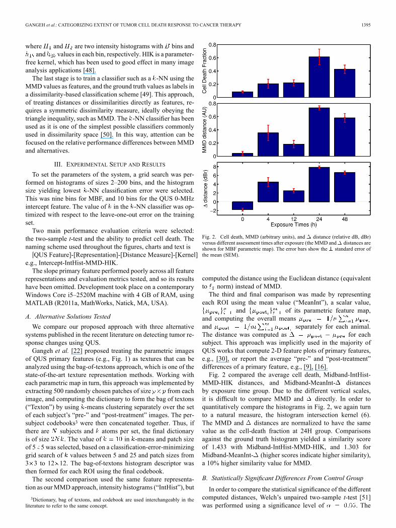
Fig. 1. Representative B-mode Images (first row), the low-magnification microscopic images of ISEL-stained tumors (second row), as well as “pre-treatment”(third row) and “post-treatment” (fourth row) QUS parametric maps of the mid-band fit parameter for each group assessed at a different time after chemotherapyexposure. Rectangles on the B-mode scans indicate where the ROIs are taken from. The color bar represents a scale encompassing approximately 35 dBr and thescale bar represents mm.
features: 1) the intercept of the fit line to the calibrated y-axis,termed the 0-MHz intercept (or intercept in short); 2) the slope ofthe fit line, termed the spectral slope (SS), and 3) the mid-bandfit (MBF), the normalized power (in dBr) at the center frequency. These quantitative parameters can be related to acoustic con-
centration, effective scatterer size, and ultrasound backscatterpower, respectively [35], [36]. The parameters were extractedat each position of the sliding window, thus forming three para-metric maps. Typical “pre-” and “post-treatment” MBF para-metric maps are shown in Fig. 1 over different exposure timeintervals.Previous studies applied different sets of QUS parameters
for tissue characterization [24], [37], [38], [39] and therapyresponse monitoring [9], [18], [22], [30], [40]. In this study,0-MHz intercept, SS, and MBF were extracted. However, sinceSS did not show statistically significant changes between pre-and post-treatment ROIs, it was not reported here.2) Feature Transformations: Several feature transformations
have been introduced that attempt to reduce the region of in-terest (ROI) dimensionality or to represent it in a more dis-criminative fashion. Probability density estimates, used as fea-tures, can be effective data descriptors for a large, multidimen-sional object, such as an image. Due to the very high intrinsicdimensionality and unconstrained input environments of thesedomains, we believe it is inappropriate to assume a distributionon the data. Alternatively, nonparametric density estimates ofthe features can be used to represent the data. No assumptions
are made about an underlying distribution of the data (whetherit be Gaussian, Poisson, etc.). Nonparametric models supportmultimodal data, without the complexities of estimating mix-ture models at the cost of being frequently higher-dimensionalthan a parametric model.Traditionally, the mean of the parametric maps are frequently
used in the literature to represent them [9], [30]. While thismay perform well in some applications, it ignores the richinformation contained in parametric maps. Recently, severalresearchers have proposed treating parametric maps as images,and have applied texture analysis on them. For example, atexton-based approach was applied by Gangeh et al. to dis-tinguish between “pre-” and “post-treatment” images [22].Texton-based approach learns a dictionary of texture atoms(textons) and subsequently represent each ROI by a histogramof these textons [41], [42], [43], [44].In this study, each ROI was represented with the intensity his-
tograms (as a nonparametric density estimator) of the two cor-responding parametric maps. A comparison of each histogram-based parameter with its mean of the parametric map as well aswith the texton-based approach is reported in the results section.
D. Maximum Mean Discrepancy
Maximum mean discrepancy (MMD) is a kernel-basedmodern approach that addresses the problem of comparing thedata samples from two probability distributions. The motivationof using MMD in this study is two fold. 1) It is assumed that the

1394 IEEE TRANSACTIONS ON MEDICAL IMAGING, VOL. 33, NO. 6, JUNE 2014
parametric maps taken from “pre-” and “post-treatment” comefrom two distributions and the distance between these twodistributions can be computed using MMD. If the distance isfar, it indicates that the treatment has been effective and the datasamples are indeed from two different distributions. However,if they are close, it means that the parametric maps from “pre-”and “post-treatment” are not very much different, thus, thetreatment has been ineffective [10]. 2) By using a kernel-basedapproach such as MMD instead of simple measures like thenorm, we benefit from mapping data to a high (possibly
infinite) dimensional feature space where computing samplemeans in this space takes into account high order statistics (upto infinity), hence, better discrimination can be achieved.The concept of MMD [25] is based on Müller’s definition of
an integral probability metric [45]. This metric was designedas a measure to compare the dissimilarity of probability mea-sures1 , and depends on finding a function from amongstthe space of functions that can maximize the distance
(1)
If and , i.e., and have distributions and, respectively, (1) can be written as
(2)
where is the expectation function. If it is assumed that is aunit ball in RKHS , it can be proved that, if and only if [28], a nice property which is very usefulin our application. Hence, from now on, the definition of MMDis restricted on the class of functions as the unit ball in RKHS.Since is an RKHS, according to the Riesz rep-
resentation theorem [46], there is a feature mappingsuch that and
, where isthe inner product operator and is a positive definitekernel function. Based on this, (2) can be written as [26], [28]
(3)
By working with the squared version of the MMD andknowing that , we are able to identify anelegant method of computing it using kernels
(4)
where and .In practice, (4) is computed empirically using finite number of
data samples taken from the distributions and . To this end,if and are and data samplesdrawn independently and identically distributed (i.i.d) from
1A probability measure has unit area.
and , respectively, the empirical can be computedusing
(5)
As can be seen from (4) and (5), the data ismapped from the orig-inal feature space to theRKHSusing a kernel , a positivedefinite functionwhichcanperformnonlinear transformationsonthe data, thereby potentially enhancing the accuracy of linear dis-criminants in this alternate feature space. EmpiricalMMDcanbecomputed efficiently for data samples intime2, and therefore compared to other dissimilaritymeasures fordistributions such as Parzen estimation or divergences, MMD isfarmore suited to real-time image analysis.While MMD was originally proposed in the literature as a
technique for statistical hypothesis test [25], [26], [28], it is pro-posed here as a feature to represent the distance between “pre-”and “post-treatment” data samples of each subject in a dissim-ilarity space. This enables using computed MMD values in asupervised learning approach, e.g., by submitting them as fea-tures to a classifier. Moreover, this provides the possibility forcombining them with other features (if needed) to enhance dis-crimination power of the designed classification system. It isexpected that considering MMD as a dissimilarity measure tobe useful in exploiting intra-group variance information avail-able frommultiple samples/instances taken of each of the “pre-”or “post-treatment” populations. This view on MMD values hasbeen already proved to be useful in scene change point detec-tion in an unsupervised learning paradigm [47].
E. Proposed Computer-Aided-Prognosis System
After the parametric maps have been prepared for each ROI,the 0-MHz intercept and MBF values for each parametric mapare separately gathered into normalized (to unit area) histogramsof intensity values. These uniformly spaced histograms havebins, and constitute a rotation- and scale-invariant nonpara-
metric density estimate of the feature.The MMD distance between “pre-” and “post-treatment”
samples of each subject (animal) is then computed using theempirical formulation given in (5). The first term in (5) com-putes the similarities among all “pre-treatment” ROIs ofeach subject whereas the last term computes the same for all“post-treatment” ROIs of the same subject. The middle term,on the other hand, computes the pairwise similaritiesbetween the ROIs in the “pre-treatment” and the ROIsin the “post-treatment” of the same subject. The histogramintersection kernel (HIK) has been selected as the kernel inthese computations with the formulation
(6)
2An alternative approach has been proposed in [28] with a computational costof , which is useful in case of existing large data samples but it is notthe case in this study.
GANGEH et al.: CATEGORIZING EXTENT OF TUMOR CELL DEATH RESPONSE TO CANCER THERAPY 1395
where and are two intensity histograms with bins andand values in each bin, respectively. HIK is a parameter-
free kernel, which has been used to good effect in many imageanalysis applications [48].The last stage is to train a classifier such as a -NN using the
MMD values as features, and the ground truth values as labels ina dissimilarity-based classification scheme [49]. This approach,of treating distances or dissimilarities directly as features, re-quires a symmetric dissimilarity measure, ideally obeying thetriangle inequality, such as MMD. The -NN classifier has beenused as it is one of the simplest possible classifiers commonlyused in dissimilarity space [50]. In this way, attention can befocused on the relative performance differences between MMDand alternatives.
III. EXPERIMENTAL SETUP AND RESULTS
To set the parameters of the system, a grid search was per-formed on histograms of sizes 2–200 bins, and the histogramsize yielding lowest -NN classification error were selected.This was nine bins for MBF, and 10 bins for the QUS 0-MHzintercept feature. The value of in the -NN classifier was op-timized with respect to the leave-one-out error on the trainingset.Two main performance evaluation criteria were selected:
the two-sample t-test and the ability to predict cell death. Thenaming scheme used throughout the figures, charts and text isQUS Feature]-[Representation]-[Distance Measure]-[Kernel]
e.g., Intercept-IntHist-MMD-HIK.The slope primary feature performed poorly across all feature
representations and evaluation metrics tested, and so its resultshave been omitted. Development took place on a contemporaryWindows Core i5–2520M machine with 4 GB of RAM, usingMATLAB (R2011a, MathWorks, Natick, MA, USA).
A. Alternative Solutions Tested
We compare our proposed approach with three alternativesystems published in the recent literature on detecting tumor re-sponse changes using QUS.Gangeh et al. [22] proposed treating the parametric images
of QUS primary features (e.g., Fig. 1) as textures that can beanalyzed using the bag-of-textons approach, which is one of thestate-of-the-art texture representation methods. Working witheach parametric map in turn, this approach was implemented byextracting 500 randomly chosen patches of size from eachimage, and computing the dictionary to form the bag of textons(“Texton”) by using -means clustering separately over the setof each subject’s “pre-” and “post-treatment” images. The per-subject codebooks3 were then concatenated together. Thus, ifthere are subjects and atoms per set, the final dictionaryis of size . The value of in -means and patch sizeof 5 5 was selected, based on a classification-error-minimizinggrid search of values between 5 and 25 and patch sizes from3 3 to 12 12. The bag-of-textons histogram descriptor wasthen formed for each ROI using the final codebook.The second comparison used the same feature representa-
tion as ourMMD approach, intensity histograms (“IntHist”), but
3Dictionary, bag of textons, and codebook are used interchangeably in theliterature to refer to the same concept.
Fig. 2. Cell death, MMD (arbitrary units), and distance (relative dB, dBr)versus different assessment times after exposure (the MMD and distances areshown for MBF parametric map). The error bars show the standard error ofthe mean (SEM).
computed the distance using the Euclidean distance (equivalentto norm) instead of MMD.The third and final comparison was made by representing
each ROI using the mean value (“MeanInt”), a scalar value,and of its parametric feature map,
and computing the overall meansand separately for each animal.The distance was computed as for eachsubject. This approach was implicitly used in the majority ofQUS works that compute 2-D feature plots of primary features,e.g., [30], or report the average “pre-” and “post-treatment”differences of a primary feature, e.g., [9], [16].Fig. 2 compared the average cell death, Midband-IntHist-
MMD-HIK distances, and Midband-MeanInt- distancesby exposure time group. Due to the different vertical scales,it is difficult to compare MMD and directly. In order toquantitatively compare the histograms in Fig. 2, we again turnto a natural measure, the histogram intersection kernel (6).The MMD and distances are normalized to have the samevalue as the cell-death fraction at 24H group. Comparisonsagainst the ground truth histogram yielded a similarity scoreof 1.433 with Midband-IntHist-MMD-HIK, and 1.303 forMidband-MeanInt- (higher scores indicate higher similarity),a 10% higher similarity value for MMD.
B. Statistically Significant Differences From Control Group
In order to compare the statistical significance of the differentcomputed distances, Welch’s unpaired two-sample t-test [51]was performed using a significance level of . The

1396 IEEE TRANSACTIONS ON MEDICAL IMAGING, VOL. 33, NO. 6, JUNE 2014
TABLE ITEST OF SATISTICAL SGNIFICANCE (UPAIRED, TWO-SAMPLE T-TEST). -VALUES SHOWN FOR EACH EXPOSURE GROUP VERSUS. THE CONTROL GROUP.
DENOTES ; DENOTES ; DENOTES
first group consisted of all of the control subjects’ distances,while the second group contained a treatment group’s distances.Results are reported in Table I for the different feature-distancecombinations, and are roughly arranged in order of ascendingvalues.Almost all of the feature-distance combinations has a(the standard threshold of significance) after 24 h of treat-
ment, and when using the MBF. The results aremost interesting for the (Intercept/Midband)-MeanInt- com-binations which stand almost alone in reporting significant dif-ferences for the 4H and 12H groups (the exception is Midband-Texton-MMD-HIK for the 12H group). As a t-test between con-trol group and noncontrol group ground truth cell-death frac-tions, (Table I) indicates that the 4H and 12H groups do notpossess statistically significant differences, this suggests that themean intensity feature and distance are overly sensitive andare reporting a false positive, in contrast to MMD.As a practical matter, it is unlikely that a clinician would
change treatment protocols after such a short period, when it isknown that the peak response is typically seen 24 h after treat-ment administration (Fig. 2) [30], [40]. In [9], monitoring pho-todynamic therapy effects were investigated in preclinical dataafter intervals as short as 1 h. Although they did not report thevalues per exposure group, they showed that the maximum
changes in MBF occurred 12 h after treatment while the max-imum cell death appeared to happen 24 h after treatment.Statistical power tests were performed in order to evaluate the
reliability of performed statistical tests of significance, consid-ering the use of t-test with relatively small sample size in eachgroup [52], [53]. Obtained results indicated that there were noprinciple objection for using the t-test in this study since the ob-tained statistical power was above 80% for the cases where astatistical significance was reported for the proposed method.
C. Predicting Cell Death Over a Threshold
Next, a supervised binary classifier was trained, using the dis-tance value between “pre-” and “post-treatment” populations asthe sole feature, and cell-death fractions as ground truth. Thetarget is to predict whether or not a subject will have cell deathgreater than a threshold . Ten-fold cross-validation at subjectlevel was performed with the -nearest neighbors ( -NN) clas-sifier to successively test the instances. The -NN is the most
common choice in dissimilarity representation [49], which isable to represent complex, multimodal classification surfaces.Classification was performed using two cell death thresholds,
20% and 40%. Based on the distribution of cell deaths in thepopulation, noticeable gaps around the 20% and 40% levelshave been observed, and therefore it was hypothesized thatthese levels were less susceptible to misclassifications causedby noise in the feature values. The classification accuracy,area under curve (AUC) of the receiver-operator curve (ROC),and Type I (1—Sensitivity) and Type II (1—Specificity) errorrates are summarized in Tables II and III for the 20% and 40%thresholds, respectively. Entries are sorted in order of ascendingtest error. Class priors were set to their observed frequencies,and class-weighted classification error results are reported.Generally, the MBF feature proved more discriminative in
terms of classification error compared to the 0-MHz intercept.MMD with the histogram of MBF values had the lowest errorat both threshold levels.
D. Discussion
The results of this paper demonstrated the viability of the pro-posed computer-aided-prognosis system using QUS methods atclinically relevant conventional frequencies in conjunction withadvanced machine learning techniques in supervised learningparadigm. The proposed system can progressively monitor andcategorize the level of cell death with high accuracy and nonin-vasively on xenograft tumor models in vivo.Overall, the Midband-IntHist-MMD-HIK combination had
the strongest performance amongst the different feature-dis-tance combinations on the evaluated metrics and thresholds.Fig. 3 visually compares the MMD versus the Euclidean dis-tances in dissimilarity space. With MMD, we can observe thatlow cell death subjects are clustered more tightly into a cornercompared to norm, and the inter-class distance is increased.This reduces the likelihood of -NN errors as well as the errorsof classifiers employing linear discriminant functions. Similarscatter plots are obtained if each primary feature, i.e., MBFor intercept, is plotted separately that explains why a -NNclassifier performs better using MMD as dissimilarity measurecompared with norm. Combinations of features in earlyfeature fusion configurations, as well as late classifier-level

GANGEH et al.: CATEGORIZING EXTENT OF TUMOR CELL DEATH RESPONSE TO CANCER THERAPY 1397
TABLE IICLASSIFICATION ERROR PERCENTAGES AND , PEDICTING WHETHER CELL DEATH IS LESS/MORE THAN . THE PARAMETER IN -NN CLASSIFIERWAS OPTIMIZED BY LEAVE-ONE-SUBJECT-OUT ON THE TRAINING SET. TEN-FOLD CROSS-VALIDATION AT SUBJECT LEVEL WAS USED; RESULTS SHOWN ARE
AVERAGED OVER TEN RUNS. AREA UNDER (THE RECEIVER-OPERATOR) CURVE (AUC) AND TYPE I/II ERROR ARE PROVIDED
TABLE IIICLASSIFICATION ERROR PERCENTAGES AND , PREDICTING WHETHER CELL DEATH IS LESS/MORE THAN . THE PARAMETER IN -NN CLASSIFIER
WAS OPTIMIZED BY LEAVE-ONE-SUBJECT-OUT ON THE TRAINING SET. TEN-FOLD CROSS-VALIDATION AT SUBJECT LEVEL WAS USED; RESULTS SHOWN AREAVERAGED OVER TEN RUNS. AREA UNDER (THE RECEIVER-OPERATOR) CURVE (AUC) AND TYPE I/II ERROR ARE PROVIDED
fusion (using Intercept-IntHist-MMD-HIK together with Mid-band-IntHist-MMD-HIK, for example), slightly improvesclassification error over using individual features especiallyat the threshold , which can be explained using thegraphs shown in Fig. 3. As can be seen from the right scatterplots in Fig. 3, especially top right graph, the two classes canbe well separated in the space of two primary features usingMMD as the dissimilarity measure.Obtained results implied that changes in the spectral slope
parametric maps in response to treatment were less promi-nent, compared to the MBF and 0-MHz intercept, and werenot shown to be statistically significant. In addition to smallscattering structures (ensemble of few cells/nuclei undergoingapoptosis), larger scattering structures, such as patches ofresponse and developed gland-like features in MDA humanbreast tumors, could also affect the spectral slope in an oppositemanner. As a working hypothesis, nonsignificant change ofslope suggest that both small and large scattering structurespotentially play a role at these frequencies [10]. Specifically,such different sized scattering structures can possibly cancelout their effects on slope (one increasing, the other decreasingslope), resulting in nonsignificant overall slope alterations.Intensity histograms outperformed the texton-based approach
in the study here. One explanation is to note that the texton-based approach is a dictionary learning approach, which meansthat the signals/data are represented using a few textons froma usually overcomplete set of learned dictionary elements. The
Fig. 3. Scatter plots of the xenograft tumors with various levels of histolog-ical cell death presented over the different distance feature planes of MBF and0-MHZ intercept. distance is on left, MMD on right.
learned dictionary can only be representative when there are suf-ficiently large data samples to train the dictionary. When there

1398 IEEE TRANSACTIONS ON MEDICAL IMAGING, VOL. 33, NO. 6, JUNE 2014
are limited number of training samples, which is the case inthis study, learned dictionary may not be sufficiently represen-tative of the data and using some predefined basis (dictionaryatoms) may lead to a better representation of the underlying dis-tribution and consequently yield better performance in the de-signed system. This was particularly confirmed in another studyon the classification of lung parenchyma in computed tomog-raphy (CT) images where, for example, local binary patterns(LBPs) as predefined operators outperformed texton-based ap-proach in small sample sizes [54]. Based on this explanation,intensity histogram, which does not need any learning step for adictionary to represent the underlying data distributions, is ex-pected to outperform a data-drivenmethod like texton-based ap-proach.Whenmore data samples are available for the estimationof the dictionary, the texton-based approach may surpass otherapproaches in performance.In this paper, the histogram intersection kernel (HIK) was
used for the computation of dissimilarities using MMD. Themain advantage of HIK compared with other kernels is that itis parameter free and thus, no tuning has to be performed. How-ever, other kernels such as the radial basis function (RBF) kernelcan also be used. The RBF kernel has the main advantage ofmapping the data to an infinite dimensional feature space, wherethe computation of the MMD can take into account the statisticsof the data samples up to infinity order [27]. Although the RBFkernel has good analytical properties [55] and is default kernelfor generic data [56], it usually performs poorly on histogramdescriptors [57] used frequently in image analysis and computervision. One critical issue in using the RBF kernel is the propertuning of its parameter that has a great impact on the perfor-mance of the systems designed using MMD as feature. This isespecially a critical issue when there is a limited number of datasamples as the tuning of the RBF kernel may lead to overfittingand hence, degrade the generalization of the designed system.The HIK, can prevent overfitting in this situation. The HIK hasalso been recommended as the kernel of choice in other works[48], [58] especially on histogram descriptors.In this study, using the MMD distance as a feature, statisti-
cally significant differences were detected using the unpairedt-test between treated animals and untreated controls within 24h after treatment administration. Moreover, it was demonstratedthat using the proposed method, cell death can be categorizednoninvasively to less/more than 20% and 40% with high accu-racy, i.e., 84.7% and 88.2%, respectively. At these two thresh-olds, a sensitivity of 85.8% and 80% was achieved, respectivelywhile the specificity was 82% and 91.7%. This demonstrates thecapability of the proposed approach in categorizing cell deathto low, mid, and high levels, which is important in monitoringcancer therapy effects.The results of this study are in line with recent findings in-
dicating the effectiveness of QUS at conventional frequencyin monitoring tumor responses to cancer therapy in preclin-ical models [10], [16] and clinical settings [12]. Also, the tech-nique proposed in this paper can be considered as a comple-mentary approach to other imaging modalities used in moni-toring cancer therapy effects such as positron emission tomog-raphy and magnetic resonance imaging [5], [7], [59] with the
main advantages of being cheaper and alleviating the require-ment for using an external contrast agent. Furthermore, the pro-posed computer-aided-prognosis system, which was tested onpreclinical data, can be easily adopted to clinical settings [12],[60] in a step forward towards personalized medicine.
IV. CONCLUSION
A computer-aided-prognosis systemwas presented for nonin-vasive tumor response assessment using QUS parametric maps,containing several aspects novel to analysis of QUS data: a dis-similarity-based classification scheme employing theMMDdis-tance measure as features, and the use of intensity histograms ofprimary features. Three alternative, commonly used feature rep-resentation and distance schemes were implemented for com-parison purposes. While all showed statistically significant dif-ferences between “pre-” and “post-treatment” groups, signif-icant improvements in classification accuracy were observedusing MMD and intensity histograms, for both the MBF and0-MHz intercept features. The system has a classification ac-curacy of 84.7% and 88.2% when predicting cell death at thethresholds of and , respectively.The techniques utilized in this work can be applied to other
treatments and pathologies, not just for tumor response, but forthe broader problem of pathology detection or treatment re-sponse monitoring using medical imaging. The work here maycontribute to one possible path forward for a fast, noninvasiveand inexpensive computed aided diagnosis system, which canfuse together other metadata and predictors about the patient,such as age, gender, and family history to assist clinicians. TheMMD can be used in an additional context in such a setting, thatis, to identify the additional features and metadata that will bestatistically discriminative between populations.
ACKNOWLEDGMENT
The authors would like to thank A. Al-Mahrouki andW. Tranfor assisting with the experiments.
REFERENCES[1] J. S. Ross, “Cancer biomarkers, companion diagnostics and personal-
ized oncology,” Biomarkers Med., vol. 5, no. 3, pp. 277–279, 2011.[2] E. C. Hayden, “Personalized cancer therapy gets closer,” Nature, vol.
458, no. 7235, pp. 131–132, 2009.[3] K. Sikora, “Personalized cancer therapy,” Personal. Med., vol. 2, no.
1, pp. 5–9, 2005.[4] L. Fass, “Imaging and cancer: A review,”Molecular Oncol., vol. 2, no.
2, pp. 115–152, Aug. 2008.[5] K. Brindle, “New approaches for imaging tumour responses to treat-
ment,” Nat. Rev. Cancer, vol. 8, no. 2, pp. 94–107, Feb. 2008.[6] G. J. Czarnota and M. C. Kolios, “Ultrasound detection of cell death,”
Imag. Med., vol. 2, no. 1, pp. 17–28, 2010.[7] A. Sadeghi-Naini, O. Falou, J. M. Hudson, C. Bailey, P. N. Burns, M.
J. Yaffe, G. J. Stanisz, M. C. Kolios, and G. J. Czarnota, “Imaginginnovations for cancer therapy response monitoring,” Imag. Med., vol.4, no. 3, pp. 311–327, Jun. 2012.
[8] M. C. Kolios and G. J. Czarnota, “Potential use of ultrasound for thedetection of cell changes in cancer treatment,” Future Oncol., vol. 5,no. 10, pp. 1527–1532, 2009.
[9] B. Banihashemi, R. Vlad, B. Debeljevic, A. Giles, M. C. Kolios, andG. J. Czarnota, “Ultrasound imaging of apoptosis in tumor response:Novel preclinical monitoring of photodynamic therapy effects,”Cancer Res., vol. 68, no. 20, pp. 8590–8596, Oct. 2008.

GANGEH et al.: CATEGORIZING EXTENT OF TUMOR CELL DEATH RESPONSE TO CANCER THERAPY 1399
[10] A. Sadeghi-Naini, N. Papanicolau, O. Falou, H. Tadayyon, J. Lee, J.Zubovits, A. Sadeghian, R. Karshafian, A. Al-Mahrouki, A. Giles, M.C. Kolios, and G. J. Czarnota, “Low-frequency quantitative ultrasoundimaging of cell death in vivo,”Med. Phys., vol. 40, no. 8, p. 082901(1-13), 2013.
[11] R. Gerl and D. L. Vaux, “Apoptosis in the development and treatmentof cancer,” Carcinogenesis, vol. 26, no. 2, pp. 263–270, Feb. 2005.
[12] A. Sadeghi-Naini, O. Falou, J. Zubovits, R. Dent, S. Verma, M. E.Trudeau, J. F. Boileau, J. Spayne, S. Iradji, E. Sofroni, J. Lee, S.Lemon-Wong, M. J. Yaffe, M. C. Kolios, and G. J. Czarnota, “Quanti-tative ultrasound evaluation of cell death response in locally advancedbreast cancer patients receiving chemotherapy,” Clin. Cancer Res.,vol. 19, no. 8, pp. 2163–2174, 2013.
[13] G. J. Czarnota, M. C. Kolios, J. Abraham, M. Portnoy, F. P. Ot-tensmeyer, J. W. Hunt, and M. D. Sherar, “Ultrasound imaging ofapoptosis: high-resolution non-invasive monitoring of programmedcell death in vitro, in situ and in vivo,” Br. J. Cancer, vol. 81, no. 3,pp. 520–527, Oct. 1999.
[14] J. W. Hunt, A. E. Worthington, A. Xuan, M. C. Kolios, G. J. Czarnota,andM. D. Sherar, “Amodel based upon pseudo regular spacing of cellscombined with the randomisation of the nuclei can explain the signif-icant changes in high-frequency ultrasound signals during apoptosis,”Ultrasound Med. Biol., vol. 28, no. 2, pp. 217–226, 2002.
[15] V. Rouffiac, C. Bouquet, N. Lassau, P. Opolon, S. Koscielny, P. Per-onneau, M. Perricaudet, and A. Roche, “Validation of a new methodfor quantifying in vivo murine tumor necrosis by sonography,” Invest.Radiol., vol. 39, no. 6, pp. 350–356, 2004.
[16] A. Sadeghi-Naini, O. Falou, H. Tadayyon, A. Al-Mahrouki, W. Tran,N. Papanicolau, M. C. Kolios, and G. J. Czarnota, “Conventional fre-quency ultrasonic biomarkers of cancer treatment response in vivo,”Translat. Oncol., vol. 6, no. 3, pp. 234–243, Jun. 2013.
[17] G. J. Czarnota, M. C. Kolios, H. Vaziri, S. Benchimol, F. P. Ottens-meyer, M. D. Sherar, and J. W. Hunt, “Ultrasonic biomicroscopy ofviable, dead and apoptotic cells,” Ultrasound Med. Biol., vol. 23, no.6, pp. 961–965, 1997.
[18] A. Sadeghi-Naini, O. Falou, and G. J. Czarnota, “Quantitative ultra-sound spectral parametric maps: Early surrogates of cancer treatmentresponse,” in Proc. 2012 Annu. Int. Conf. IEEE Eng. Med. Biol. Soc.,Aug. 2012, pp. 2672–2675.
[19] F. L. Lizzi, M. Greenebaum, E. J. Feleppa, M. Elbaum, and D. J.Coleman, “Theoretical framework for spectrum analysis in ultrasonictissue characterization,” J. Acoust. Soc. Am., vol. 73, no. 4, pp.1366–1373, Apr. 1983.
[20] T. J. Larkin, H. C. Canuto, M. I. Kettunen, T. C. Booth, D.-E. Hu,A. S. Krishnan, S. E. Bohndiek, A. A. Neves, C. McLachlan, M. P.Hobson, and K. M. Brindle, Magnetic Resonance in Medicine “Anal-ysis of image heterogeneity using 2-D Minkowski functionals detectstumor responses to treatment,” Feb. 2013 [Online]. Available: http://onlinelibrary.wiley.com/journal/10.1002/(ISSN)1522–2594
[21] T. Hastie, R. Tibshirani, and J. J. H. Friedman, The Elements of Statis-tical Learning, 1st ed ed. New York: Springer, 2001.
[22] M. J. Gangeh, A. Sadeghi-Naini, M. S. Kamel, and G. Czarnota,“Assessment of cancer therapy effects using texton-based charac-terization of quantitative ultrasound parametric images,” in Proc.Int. Symp. Biomed. Imag.: From Nano to Macro, 2013, pp.1372–1375.
[23] L. Sørensen, S. B. Shaker, and M. d. Bruijne, “Quantitative analysis ofpulmonary emphysema using local binary patterns,” IEEE Trans. Med.Imag., vol. 29, no. 2, pp. 559–569, Feb. 2010.
[24] E. J. Feleppa, J. Mamou, C. R. Porter, and J. Machi, “Quantitativeultrasound in cancer imaging,” Seminars Oncol., vol. 38, no. 1, pp.136–150, Feb. 2011.
[25] A. Gretton, K. Borgwardt, M. Rasch, B. Schölkopf, and A. Smola,“A kernel method for the two-sample-problem,” in Adv. in Neural In-form. Processing Syst. Cambridge, MA: MIT Press, 2006, vol. 19,pp. 513–520.
[26] A. Gretton, K.M. Borgwardt, M. Rasch, B. Schölkopf, and A. J. Smola,A kernel method for the two-sample problem Max Planck Inst. Biol.Cybern., Tech. Rep. 157, 2008.
[27] S. Jegelka, A. Gretton, B. Schölkopf, B. K. Sriperumbudur, and U.V. Luxburg, “Generalized clustering via kernel embeddings,” in Proc.32nd Annu. German Conf. Adv. Artif. Intell., 2009, pp. 144–152.
[28] A. Gretton, K.M. Borgwardt, M. J. Rasch, B. Schölkopf, and A. Smola,“A kernel two-sample test,” J. Mach. Learn. Res., vol. 13, pp. 723–773,2012.
[29] Y. Rathi, J. Malcolm, O. Michailovich, J. Goldstein, L. Seidman,R. W. McCarley, C.-F. Westin, and M. E. Shenton, “Biomarkersfor identifying first-episode schizophrenia patients using diffusionweighted imaging,” in Proc. MICCAI, Jan. 2010, vol. 13, pp.657–665.
[30] R. M. Vlad, S. Brand, A. Giles, M. C. Kolios, and G. J. Czarnota,“Quantitative ultrasound characterization of responses to radiotherapyin cancer mouse models,” Clin. Cancer Res., vol. 15, no. 6, pp.2067–2075, Mar. 2009.
[31] B. Porat, Digital processing of random signals: Theory and methods.Mineola, NY: Dover, 1994.
[32] R. Romijn, J. Thijssen, and G. v. Beuningen, “Estimation of scatterersize from backscattered ultrasound: a simulation study,” IEEE Trans.Ultrason., Ferroelectr. Freq. Control, vol. 36, no. 6, pp. 593–606,Nov., 1989.
[33] F. Dong, E. L. Madsen, M. C. MacDonald, and J. A. Zagzebski, “Non-linearity parameter for tissue-mimicking materials,” Ultrasound Med.Biol., vol. 25, no. 5, pp. 831–838, 1999.
[34] L. X. Yao, J. A. Zagzebski, and E. L. Madsen, “Backscatter coefficient-measurements using a reference phantom to extract depth-dependentinstrumentation factors ,” Ultrason. Imag., vol. 12, no. 1, pp. 58–70,1990.
[35] F. Lizzi, M. Ostromogilsky, E. Feleppa, M. Rorke, and M. Yaremko,“Relationship of ultrasonic spectral parameters to features of tissue mi-crostructure,” IEEE Trans. Ultrason., Ferroelectr. Freq. Control, vol.34, no. 3, pp. 319–329, May 1987.
[36] F. L. Lizzi, M. Astor, T. Liu, C. Deng, D. J. Coleman, andR. H. Silverman, “Ultrasonic spectrum analysis for tissue assaysand therapy evaluation,” Int. J. Imag. Syst. Technol., vol. 8,no. 1, pp. 3–10, 1997.
[37] M. Oelze, W. O’Brien, J. Blue, and J. Zachary, “Differentiation andcharacterization of rat mammary fibroadenomas and 4T1 mouse car-cinomas using quantitative ultrasound imaging,” IEEE Trans. Med.Imag., vol. 23, no. 6, pp. 764–771, Jun. 2004.
[38] E. Feleppa, A. Kalisz, J. Sokil-Melgar, F. Lizzi, T. Liu, A. Rosado,M. Shao, W. Fair, Y. Wang, M. Cookson, V. Reuter, and W. Heston,“Typing of prostate tissue by ultrasonic spectrum analysis,” IEEETrans. Ultrason., Ferroelectr. Freq. Control, vol. 43, no. 4, pp.609–619, Jul. 1996.
[39] M. Yang, T. M. Krueger, J. G. Miller, and M. R. Holland, “Charac-terization of anisotropic myocardial backscatter using spectral slope,intercept and midband fit parameters,” Ultrason. Imag., vol. 29, no. 2,pp. 122–134, 2007.
[40] J. Lee, R. Karshafian, N. Papanicolau, A. Giles, M. C. Kolios, and G.J. Czarnota, “Quantitative ultrasound for the monitoring of novel mi-crobubble and ultrasound radiosensitization,” Ultrasound Med. Biol.,vol. 38, no. 7, pp. 1212–1221, Jul. 2012.
[41] B. Julesz, “Textons, the elements of texture perception, and their inter-actions,” Nature, vol. 290, no. 5802, pp. 91–97, 1981.
[42] M. Varma and A. Zisserman, “A statistical approach to texture classi-fication from single images,” Int. J. Comput. Vis., vol. 62, no. 1-2, pp.61–81, Apr. 2005.
[43] T. Leung and J. Malik, “Representing and recognizing the visual ap-pearance of materials using three-dimensional textons,” Int. J. Comput.Vis., vol. 43, pp. 29–44, Jun. 2001.
[44] M. Varma and A. Zisserman, “A statistical approach to materialclassification using image patch exemplars,” IEEE Trans. PatternAnal. Mach. Intell., vol. 31, no. 11, pp. 2032–2047, Nov. 2009.
[45] A. Müller, “Integral probability metrics and their generating classes offunctions,” Adv. Appl. Probabil., pp. 429–443, 1997.
[46] M. Reid and B. Simon, Methods of Modern Mathematical Physics.San Diego, CA: Academic, 1980, vol. 1, Funct. Anal..
[47] M. Diu, M. J. Gangeh, andM. S. Kamel, “Unsupervised visual change-point detection using maximum mean discrepancy,” in Proc. 10th Int.Conf. Image Anal. Recognit., 2013, pp. 336–345.
[48] S. Lazebnik, C. Schmid, and J. Ponce, “Beyond bags of features:Spatial pyramid matching for recognizing natural scene categories,”in Proc. IEEE Conf. Comput. Vis. Pattern Recognit., 2006, pp.2169–2178.

1400 IEEE TRANSACTIONS ON MEDICAL IMAGING, VOL. 33, NO. 6, JUNE 2014
[49] E. Pekalska and R. P. W. Duin, “Dissimilarity representations allowfor building good classifiers,” Pattern Recognit. Lett., vol. 23, no. 8,pp. 943–956, Jun. 2002.
[50] E. Pekalska, P. Paclik, and R. P. W. Duin, “A generalized kernel ap-proach to Dissimilarity-based classification,” J. Mach. Learn. Res., vol.2, no. 2, pp. 175–211, 2002.
[51] H. Motulsky, Intuitive Biostatistics: A Nonmathematical Guide to Sta-tistical Thinking, 2nd ed. New York: Oxford Univ. Press, 2010.
[52] S. Siegel and N. J. Castellan, Nonparametric Statistics for the Behav-ioral Sciences. New York: McGraw-Hill, 1988.
[53] J. C. F. d. Winter, “Using the student’s t-test with extremely smallsample sizes,” Practical Assessment, Res. Evaluat., vol. 18, no. 10,2013.
[54] L. Sørensen, M. J. Gangeh, S. B. Shaker, and M. d. Bruijne, “Tex-ture classification in pulmonary CT,” in Lung Imaging and ComputerAided Diagnosis, A. El-Baz and J. S. Sure, Eds. Boca Raton, CA:CRC Press, 2007, pp. 343–367.
[55] C. M. Bishop, Pattern Recognition and Machine Learning. NewYork: Springer, 2006.
[56] R. Fan, P. Chen, and C. Lin, “Working set selection using the secondorder information for training SVM,” J. Mach. Learn. Res., vol. 6, pp.1889–1918, 2005.
[57] J. Wu, “A fast dual method for HIK SVM learning,” in Proc. 11th Eur.Conf. Comput. Vis., 2010, pp. 552–565.
[58] J. Zhang, M. Marszalek, S. Lazebnik, and C. Schmid, “Local featuresand kernels for classification of texture and object categories: A com-prehensive study,” Int. J. Comput. Vis., vol. 73, no. 2, pp. 213–238,2007.
[59] T. H. Witney and K. M. Brindle, “Imaging tumour cell metabo-lism using hyperpolarized 13C magnetic resonance spectroscopy,”Biochem. Soc. Translat., vol. 38, no. 5, pp. 1220–1224, 2010.
[60] N. R. Jagannathan, M. Kumar, V. Seenu, O. Coshic, S. N. Dwivedi,P. K. Julka, A. Srivastava, and G. K. Rath, “Evaluation of totalcholine from in-vivo volume localized proton MR spectroscopy andits response to neoadjuvant chemotherapy in locally advanced breastcancer,” Br. J. Cancer, vol. 84, no. 8, pp. 1016–1022, Apr. 2001.
Related Documents