UCE Birmingham Faculty of Health Dip HE Rehabilitation Work (Visual impairment) Cataract morphology By Konrad Pesudovs and David B Elliott Contents Abstract Introduction Cataract morphology Cortical cataracts Nuclear cataracts Posterior subcapsular cataract Clinical cataract classification schema Assessment of vision in the cataract patient Deciding on the need for surgery Acknowledgments Abstract Cataract is the most common condition requiring assessment and referral from optometric practice. This article examines the features of the main morphological types of age-related cataract: nuclear cortical and posterior subcapsular. The steps to take in the assessment and management of cataract patients are discussed including: examination for cataract, visual assessment methods to confirm whether vision loss is present, discussion with the patient to establish if visual disability is present, and establishing criteria for referral. Keywords Cataract, disability, morphology, referral vision. Introduction Cataract is a frequent condition found in patients attending an optometric practice and, given the demographic changes within the population, their numbers will increase in the coming years. This article discusses the optometric assessment and management of age-related cataract. The scope includes the categorisation of cataract morphology, the assessment of vision, the assessment of visual disability, and how to determine when to refer a patient for surgery. Copyright © UCE Birmingham 64029999.doc Visit us at www.sightlossmatters.com Document revised 28 Sep 06

Welcome message from author
This document is posted to help you gain knowledge. Please leave a comment to let me know what you think about it! Share it to your friends and learn new things together.
Transcript

862019 Cataract Morphology
httpslidepdfcomreaderfullcataract-morphology 19
UCE Birmingham Faculty of Health
Dip HE Rehabilitation Work(Visual impairment)
Cataract morphology
By Konrad Pesudovs and David B Elliott
ContentsAbstract
Introduction
Cataract morphology
Cortical cataracts
Nuclear cataracts
Posterior subcapsular cataract
Clinical cataract classification schema
Assessment of vision in the cataract patient
Deciding on the need for surgery
Acknowledgments
Abstract
Cataract is the most common condition requiring assessment and referralfrom optometric practice This article examines the features of the mainmorphological types of age-related cataract nuclear cortical and posterior subcapsular The steps to take in the assessment and management of cataract patients are discussed including examination for cataract visualassessment methods to confirm whether vision loss is present discussionwith the patient to establish if visual disability is present and establishingcriteria for referral
Keywords
Cataract disability morphology referral vision
IntroductionCataract is a frequent condition found in patients attending an optometricpractice and given the demographic changes within the population their numbers will increase in the coming years This article discusses theoptometric assessment and management of age-related cataract Thescope includes the categorisation of cataract morphology theassessment of vision the assessment of visual disability and how to
determine when to refer a patient for surgery
Copyright copy UCE Birmingham
64029999docVisit us at wwwsightlossmatterscom
Document revised 28 Sep 06
862019 Cataract Morphology
httpslidepdfcomreaderfullcataract-morphology 29
UCE Birmingham Faculty of Health
Dip HE Rehabilitation Work(Visual impairment)
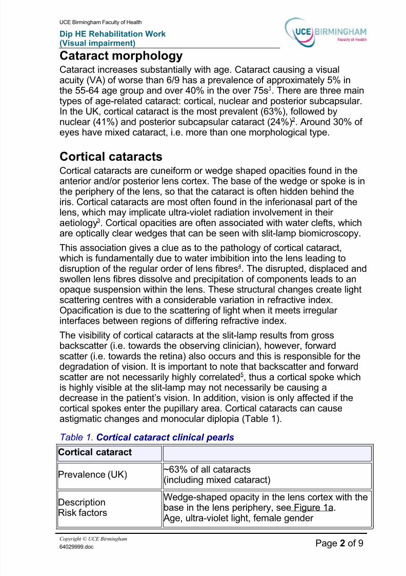
Cataract morphologyCataract increases substantially with age Cataract causing a visualacuity (VA) of worse than 69 has a prevalence of approximately 5 inthe 55-64 age group and over 40 in the over 75s1 There are three main
types of age-related cataract cortical nuclear and posterior subcapsularIn the UK cortical cataract is the most prevalent (63) followed bynuclear (41) and posterior subcapsular cataract (24)2 Around 30 of eyes have mixed cataract ie more than one morphological type
Cortical cataractsCortical cataracts are cuneiform or wedge shaped opacities found in theanterior andor posterior lens cortex The base of the wedge or spoke is inthe periphery of the lens so that the cataract is often hidden behind the
iris Cortical cataracts are most often found in the inferionasal part of thelens which may implicate ultra-violet radiation involvement in their aetiology3 Cortical opacities are often associated with water clefts whichare optically clear wedges that can be seen with slit-lamp biomicroscopy
This association gives a clue as to the pathology of cortical cataractwhich is fundamentally due to water imbibition into the lens leading todisruption of the regular order of lens fibres4 The disrupted displaced andswollen lens fibres dissolve and precipitation of components leads to anopaque suspension within the lens These structural changes create light
scattering centres with a considerable variation in refractive indexOpacification is due to the scattering of light when it meets irregular interfaces between regions of differing refractive index
The visibility of cortical cataracts at the slit-lamp results from grossbackscatter (ie towards the observing clinician) however forwardscatter (ie towards the retina) also occurs and this is responsible for thedegradation of vision It is important to note that backscatter and forwardscatter are not necessarily highly correlated5 thus a cortical spoke whichis highly visible at the slit-lamp may not necessarily be causing adecrease in the patientrsquos vision In addition vision is only affected if thecortical spokes enter the pupillary area Cortical cataracts can causeastigmatic changes and monocular diplopia (Table 1)
Table 1 Cortical cataract clinical pearls
Cortical cataract
Prevalence (UK)~63 of all cataracts(including mixed cataract)
DescriptionRisk factors
Wedge-shaped opacity in the lens cortex with the
base in the lens periphery see Figure 1aAge ultra-violet light female gender
Copyright copy UCE Birmingham
64029999doc Page 2 of 9
862019 Cataract Morphology
httpslidepdfcomreaderfullcataract-morphology 39
UCE Birmingham Faculty of Health
Dip HE Rehabilitation Work(Visual impairment)
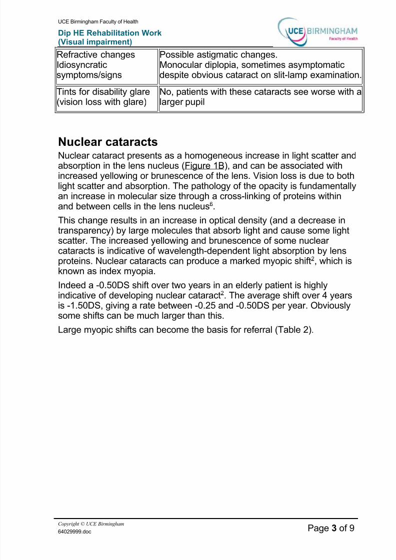
Refractive changesIdiosyncraticsymptomssigns
Possible astigmatic changesMonocular diplopia sometimes asymptomaticdespite obvious cataract on slit-lamp examination
Tints for disability glare(vision loss with glare)
No patients with these cataracts see worse with alarger pupil
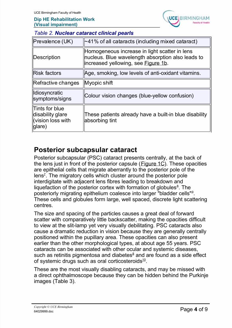
Nuclear cataractsNuclear cataract presents as a homogeneous increase in light scatter andabsorption in the lens nucleus (Figure 1B) and can be associated withincreased yellowing or brunescence of the lens Vision loss is due to bothlight scatter and absorption The pathology of the opacity is fundamentally
an increase in molecular size through a cross-linking of proteins withinand between cells in the lens nucleus6
This change results in an increase in optical density (and a decrease intransparency) by large molecules that absorb light and cause some lightscatter The increased yellowing and brunescence of some nuclear cataracts is indicative of wavelength-dependent light absorption by lensproteins Nuclear cataracts can produce a marked myopic shift2 which isknown as index myopia
Indeed a -050DS shift over two years in an elderly patient is highly
indicative of developing nuclear cataract2 The average shift over 4 yearsis -150DS giving a rate between -025 and -050DS per year Obviouslysome shifts can be much larger than this
Large myopic shifts can become the basis for referral (Table 2)
Copyright copy UCE Birmingham
64029999doc Page 3 of 9
862019 Cataract Morphology
httpslidepdfcomreaderfullcataract-morphology 49
UCE Birmingham Faculty of Health
Dip HE Rehabilitation Work(Visual impairment)
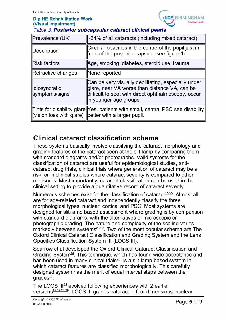
Table 2 Nuclear cataract clinical pearls
Prevalence (UK) ~41 of all cataracts (including mixed cataract)
Description
Homogeneous increase in light scatter in lens
nucleus Blue wavelength absorption also leads toincreased yellowing see Figure 1b
Risk factors Age smoking low levels of anti-oxidant vitamins
Refractive changes Myopic shift
Idiosyncraticsymptomssigns
Colour vision changes (blue-yellow confusion)
Tints for blue
disability glare(vision loss withglare)
These patients already have a built-in blue disabilityabsorbing tint
Posterior subcapsular cataractPosterior subcapsular (PSC) cataract presents centrally at the back of the lens just in front of the posterior capsule (Figure 1C) These opacities
are epithelial cells that migrate aberrantly to the posterior pole of thelens7 The migratory cells which cluster around the posterior poleinterdigitate with adjacent lens fibres leading to breakdown andliquefaction of the posterior cortex with formation of globules8 Theposteriorly migrating epithelium coalesce into larger bladder cells9These cells and globules form large well spaced discrete light scatteringcentres
The size and spacing of the particles causes a great deal of forwardscatter with comparatively little backscatter making the opacities difficultto view at the slit-lamp yet very visually debilitating PSC cataracts also
cause a dramatic reduction in vision because they are generally centrallypositioned within the pupillary area These opacities can also presentearlier than the other morphological types at about age 55 years PSCcataracts can be associated with other ocular and systemic diseasessuch as retinitis pigmentosa and diabetes8 and are found as a side effectof systemic drugs such as oral corticosteroids10
These are the most visually disabling cataracts and may be missed witha direct ophthalmoscope because they can be hidden behind the Purkinjeimages (Table 3)
Copyright copy UCE Birmingham
64029999doc Page 4 of 9
862019 Cataract Morphology
httpslidepdfcomreaderfullcataract-morphology 59
UCE Birmingham Faculty of Health
Dip HE Rehabilitation Work(Visual impairment)
Table 3 Posterior subcapsular cataract clinical pearls
Prevalence (UK) ~24 of all cataracts (including mixed cataract)
DescriptionCircular opacities in the centre of the pupil just in
front of the posterior capsule see figure 1cRisk factors Age smoking diabetes steroid use trauma
Refractive changes None reported
Idiosyncraticsymptomssigns
Can be very visually debilitating especially under glare near VA worse than distance VA can bedifficult to spot with direct ophthalmoscopy occur in younger age groups
Tints for disability glare(vision loss with glare) Yes patients with small central PSC see disabilitybetter with a larger pupil
Clinical cataract classification schemaThese systems basically involve classifying the cataract morphology andgrading features of the cataract seen at the slit-lamp by comparing themwith standard diagrams andor photographs Valid systems for theclassification of cataract are useful for epidemiological studies anti-
cataract drug trials clinical trials where generation of cataract may be arisk or in clinical studies where cataract severity is compared to other measures Most importantly cataract classification can be used in theclinical setting to provide a quantitative record of cataract severity
Numerous schemes exist for the classification of cataract11-25 Almost allare for age-related cataract and independently classify the threemorphological types nuclear cortical and PSC Most systems aredesigned for slit-lamp based assessment where grading is by comparisonwith standard diagrams with the alternatives of microscopic or
photographic grading The nature and complexity of the scaling variesmarkedly between systems2627 Two of the most popular schema are TheOxford Clinical Cataract Classification and Grading System and the LensOpacities Classification System III (LOCS III)
Sparrow et al developed the Oxford Clinical Cataract Classification andGrading System14 This technique which has found wide acceptance andhas been used in many clinical trials28 is a slit-lamp-based system inwhich cataract features are classified morphologically This carefullydesigned system has the merit of equal interval steps between the
grades
14
The LOCS III22 evolved following experiences with 2 earlier versions15172229 LOCS III grades cataract in four dimensions nuclear
Copyright copy UCE Birmingham
64029999doc Page 5 of 9
862019 Cataract Morphology
httpslidepdfcomreaderfullcataract-morphology 69
UCE Birmingham Faculty of Health
Dip HE Rehabilitation Work(Visual impairment)
colour (NC) nuclear opalescence (NO) cortical opacity (C) PSC (P)(Figure 2) Nuclear opalescence and colour are rated on a decimal scalefrom 0 to 7 in steps of 01 and cortical and PSC are rated on a decimalscale from 0 to 6 This is a thoroughly validated system which has beenused extensively for clinical trials29-32
Alternatively cataracts can be classified and approximately gradedwithout these systems The conventional 0 to 4 grading system can beused where 0 is a clear lens + or 1+ represents a mild cataract ++ or 2+a moderate cataract +++ or 3+ a marked cataract and ++++ or 4+ asevere cataract Alternatively PSC and cortical cataract can be graded bythe percentage of the (dilated or otherwise) pupillary area they occupy or by a brief sketch
Assessment of vision in the cataract patientThere are many ways in which vision is affected by cataract includingincreasing myopia and astigmatism monocular diplopia reduced lighttransmission and changes in colour perception33 However visual loss incataract is principally due to increased intraocular light scatter
Light from the object itself is scattered reducing the contrast of its retinalimage In addition wide-angle light scatter from peripheral glare sourcescan produce a veiling luminance on the retina further reducing thecontrast of the retinal image Not surprisingly visual decrement is
greatest in glare or bright light conditions (notes on prescribing tints andcoatings for cataract patients are found in Table 4)
Table 4 Spectacle tints and coatings for use in the cataract patient
Tints for discomfort glare
Tints can be useful to alleviate discomfort
Tints for disability glare
(vision loss withglare)
In general tints provide no benefit as the effect of thereduction in glare light is balanced by the reduction of lightfrom the object of regard In general broad- brimmed hats
are better Some patients with small central PSC cataractmay benefit from a tint due to its effect on pupil size (seeTable 3)
UV-blockingtints
These can alleviate some disability glare fromfluorescence within the lens and perhaps slow cataractprogression particularly of cortical cataract
AR lenscoatings
These may be useful for patients with cataract especiallythose who drive at night or use a VDU
Copyright copy UCE Birmingham
64029999doc Page 6 of 9
862019 Cataract Morphology
httpslidepdfcomreaderfullcataract-morphology 79
UCE Birmingham Faculty of Health
Dip HE Rehabilitation Work(Visual impairment)
It is well established that some patients with cataract who retain goodVA have significant visual problems In these cases contrast sensitivityand glare testing can be used to evaluate the level of disability
Contrast SensitivityContrast sensitivity at low to intermediate spatial frequencies can bereduced in patients with cataract when VA is good3435 In these casescataract surgery can return contrast sensitivity to age-matched normalvalues35
Additionally in cataract patients contrast sensitivity can be a better indicator of various aspects of real world vision than VA such as drivingorientation mobility and face expression recognition35-37 The bestavailable test for use in cataract patients is the Pelli-Robson chart (Figure
3) At the recommended working distance of 1m the letters are equivalentto 6273 Snellen and the chart gives an indication of contrast sensitivityat a spatial frequency of approximately 05 to 2 cdeg (just below the peakof the contrast sensitivity curve) In many cataract patients Pelli-Robsoncontrast sensitivity will be normal (150 log contrast sensitivity or above)
Any patient with a log contrast sensitivity of 135 or below is likely to becomplaining of poor vision no matter how good the VA is Poor contrastsensitivity is more likely with nuclear and PSC cataracts38-42
Glare testingIncreased forward light scatter causes cataract patients to see poorly inbright light or glare conditions Consider the effect of a dirty windscreenon your vision when driving Vision is satisfactory until sunlight or lightcoming from car headlights hits the screen when vision can be reduceddramatically Similarly with some cataract patients VA can be adequatein the relatively low illumination conditions of the examination room butconsiderably reduced when outdoors or when driving at night (Panel 1)
Panel 1 Typical PSC and glare case report 52
A healthy 45-year-old prison guard complained of a gradual decrease invision over the previous year This decrease only occurred in brightsunlight such as when guarding prisoners working outside Before hisvisit his loss of vision had been so great as to allow two convicts toescape His VA was measured to be 66 in both eyes in the examinationroom However VA measured in bright light levels was 6120 Slit-lampexamination with a dilated pupil revealed small PSC cataracts
The case history in (see Panel 1) highlights that a patient attending for anexamination in the UK in the winter with no symptoms and a VA of 69 or
Copyright copy UCE Birmingham
64029999doc Page 7 of 9
862019 Cataract Morphology
httpslidepdfcomreaderfullcataract-morphology 89
UCE Birmingham Faculty of Health
Dip HE Rehabilitation Work(Visual impairment)
better could have profoundly reduced vision in a sunny environment Anysuggestion of small PSC cataracts should immediately indicate glaretesting and pupillary dilation with a careful slit-lamp examination
Glare tests measure the reduction in a patients vision due to a glare
source (glare loss) and indicate the effect on vision of increased lightscatter and a smaller pupil Glare test scores have been shown tocorrelate with VA measured outdoors and to correlate better with glaresymptoms than conventionally measured VA in patients with cataract4344Simple methods of measuring disability glare involve measuring VA under glare conditions such as while directing a bright penlight or ophthalmoscope light into the patients eye45 Due to the inverse squarelaw and light scatter varying as a function of the square of the glare angle(the angle of the glare source to the visual axis) it is very important tostandardise the angle and distance of the penlight to the eye Typical
standards are 10cm and 30 degrees A more standardised version of such tests is the Brightness Acuity Tester
Disability glare scores are usually taken as the level of VA under the glarecondition Alternatively the impact of glare can be recorded as glare lossie the number of lines on the chart lost when the glare source isintroduced Testing can be done using conventional VA low contrast VAor contrast sensitivity charts
Assessment of disability in the cataract patientThe decision of when to refer a patient for cataract surgery should beprimarily dependent on whether the patientrsquos reduced vision interfereswith their desired lifestyle Referral should NOT be based on VAmeasurements alone
For example if a patient is a taxi driver and cataract is reducing his visionso that he cannot see well enough to drive at night or on sunny days heshould be referred for surgery Conversely if a patient is perfectly happywith their vision and has no restrictions on their lifestyle despite having618 or even 624 VA there is no need to refer for surgery although youmay discuss the possibility of future referral with them46 Therefore it isimportant to determine whether the patientrsquos desired lifestyle is affectedby their reduced vision When cataract is identified it is useful to asksome specific questions which relate to cataract-induced visual loss
For example Do you have any problem with glare or in bright sunlightand if pertinent Do you have any problems driving at night or on sunnydays In addition ask about the effect of vision on the patients jobsports or hobbies There are formal instruments (questionnaires) for themeasurement of visual disability caused by cataract such as the VF-14
and the VDA4748 While these questionnaires may not be appropriate for routine use in optometric practice they do contain questions which theoptometrist may find helpful to ask
Copyright copy UCE Birmingham
64029999doc Page 8 of 9
862019 Cataract Morphology
httpslidepdfcomreaderfullcataract-morphology 99
UCE Birmingham Faculty of Health
Dip HE Rehabilitation Work(Visual impairment)
Deciding on the need for surgery
The nexus between vision disability and cataract grading
The referral of patients with cataract for surgery must be based on
whether the patientrsquos reduced vision is interfering with their desiredlifestyle Referral is then justified using clinical vision tests such asdistance and near visual acuity contrast sensitivity disability glare and for monocular pseudophakes anisometropia and stereopsis The presenceof co-morbid eye disease (including amblyopia) needs carefulassessment to determine whether or not the cataract is the major causeof the visual loss An apportionment of the visual loss and the visualdisability to each condition using potential vision tests49-51 and clinicalacumen should be made This last quality is the most difficult to teach butin simple terms it involves an assessment of whether the level of vision
severity of cataract and degree of symptoms seem in balance Finally adiscussion of the risk and benefits of cataract surgery as well as the costsand inconvenience is important and this should follow the line of aninformed consent discussion similar to that which the patient willencounter when they meet the cataract surgeon (Figure 5)
AcknowledgmentsKonrad Pesudovs is supported by an Australian National Health andMedical Research Council (NHMRC) Sir Neil Hamilton Fairley Research
FellowshipKonrad PesudovsBScOptom PhD MCOptomFVCO FAAOBradford University and Flinders University(Australia)andDavid B ElliottPhD MCOptom FAAO
Bradford UniversityCorrespondenceDr Konrad PesudovsDepartment of OptometryUniversity of BradfordBradfordWest Yorkshire BD7 1DP UKE-mail KPesudovsbradfordacuk
You have reached the end of this text
Copyright copy UCE Birmingham
64029999doc Page 9 of 9
862019 Cataract Morphology
httpslidepdfcomreaderfullcataract-morphology 29
UCE Birmingham Faculty of Health
Dip HE Rehabilitation Work(Visual impairment)
Cataract morphologyCataract increases substantially with age Cataract causing a visualacuity (VA) of worse than 69 has a prevalence of approximately 5 inthe 55-64 age group and over 40 in the over 75s1 There are three main
types of age-related cataract cortical nuclear and posterior subcapsularIn the UK cortical cataract is the most prevalent (63) followed bynuclear (41) and posterior subcapsular cataract (24)2 Around 30 of eyes have mixed cataract ie more than one morphological type
Cortical cataractsCortical cataracts are cuneiform or wedge shaped opacities found in theanterior andor posterior lens cortex The base of the wedge or spoke is inthe periphery of the lens so that the cataract is often hidden behind the
iris Cortical cataracts are most often found in the inferionasal part of thelens which may implicate ultra-violet radiation involvement in their aetiology3 Cortical opacities are often associated with water clefts whichare optically clear wedges that can be seen with slit-lamp biomicroscopy
This association gives a clue as to the pathology of cortical cataractwhich is fundamentally due to water imbibition into the lens leading todisruption of the regular order of lens fibres4 The disrupted displaced andswollen lens fibres dissolve and precipitation of components leads to anopaque suspension within the lens These structural changes create light
scattering centres with a considerable variation in refractive indexOpacification is due to the scattering of light when it meets irregular interfaces between regions of differing refractive index
The visibility of cortical cataracts at the slit-lamp results from grossbackscatter (ie towards the observing clinician) however forwardscatter (ie towards the retina) also occurs and this is responsible for thedegradation of vision It is important to note that backscatter and forwardscatter are not necessarily highly correlated5 thus a cortical spoke whichis highly visible at the slit-lamp may not necessarily be causing adecrease in the patientrsquos vision In addition vision is only affected if thecortical spokes enter the pupillary area Cortical cataracts can causeastigmatic changes and monocular diplopia (Table 1)
Table 1 Cortical cataract clinical pearls
Cortical cataract
Prevalence (UK)~63 of all cataracts(including mixed cataract)
DescriptionRisk factors
Wedge-shaped opacity in the lens cortex with the
base in the lens periphery see Figure 1aAge ultra-violet light female gender
Copyright copy UCE Birmingham
64029999doc Page 2 of 9
862019 Cataract Morphology
httpslidepdfcomreaderfullcataract-morphology 39
UCE Birmingham Faculty of Health
Dip HE Rehabilitation Work(Visual impairment)
Refractive changesIdiosyncraticsymptomssigns
Possible astigmatic changesMonocular diplopia sometimes asymptomaticdespite obvious cataract on slit-lamp examination
Tints for disability glare(vision loss with glare)
No patients with these cataracts see worse with alarger pupil
Nuclear cataractsNuclear cataract presents as a homogeneous increase in light scatter andabsorption in the lens nucleus (Figure 1B) and can be associated withincreased yellowing or brunescence of the lens Vision loss is due to bothlight scatter and absorption The pathology of the opacity is fundamentally
an increase in molecular size through a cross-linking of proteins withinand between cells in the lens nucleus6
This change results in an increase in optical density (and a decrease intransparency) by large molecules that absorb light and cause some lightscatter The increased yellowing and brunescence of some nuclear cataracts is indicative of wavelength-dependent light absorption by lensproteins Nuclear cataracts can produce a marked myopic shift2 which isknown as index myopia
Indeed a -050DS shift over two years in an elderly patient is highly
indicative of developing nuclear cataract2 The average shift over 4 yearsis -150DS giving a rate between -025 and -050DS per year Obviouslysome shifts can be much larger than this
Large myopic shifts can become the basis for referral (Table 2)
Copyright copy UCE Birmingham
64029999doc Page 3 of 9
862019 Cataract Morphology
httpslidepdfcomreaderfullcataract-morphology 49
UCE Birmingham Faculty of Health
Dip HE Rehabilitation Work(Visual impairment)
Table 2 Nuclear cataract clinical pearls
Prevalence (UK) ~41 of all cataracts (including mixed cataract)
Description
Homogeneous increase in light scatter in lens
nucleus Blue wavelength absorption also leads toincreased yellowing see Figure 1b
Risk factors Age smoking low levels of anti-oxidant vitamins
Refractive changes Myopic shift
Idiosyncraticsymptomssigns
Colour vision changes (blue-yellow confusion)
Tints for blue
disability glare(vision loss withglare)
These patients already have a built-in blue disabilityabsorbing tint
Posterior subcapsular cataractPosterior subcapsular (PSC) cataract presents centrally at the back of the lens just in front of the posterior capsule (Figure 1C) These opacities
are epithelial cells that migrate aberrantly to the posterior pole of thelens7 The migratory cells which cluster around the posterior poleinterdigitate with adjacent lens fibres leading to breakdown andliquefaction of the posterior cortex with formation of globules8 Theposteriorly migrating epithelium coalesce into larger bladder cells9These cells and globules form large well spaced discrete light scatteringcentres
The size and spacing of the particles causes a great deal of forwardscatter with comparatively little backscatter making the opacities difficultto view at the slit-lamp yet very visually debilitating PSC cataracts also
cause a dramatic reduction in vision because they are generally centrallypositioned within the pupillary area These opacities can also presentearlier than the other morphological types at about age 55 years PSCcataracts can be associated with other ocular and systemic diseasessuch as retinitis pigmentosa and diabetes8 and are found as a side effectof systemic drugs such as oral corticosteroids10
These are the most visually disabling cataracts and may be missed witha direct ophthalmoscope because they can be hidden behind the Purkinjeimages (Table 3)
Copyright copy UCE Birmingham
64029999doc Page 4 of 9
862019 Cataract Morphology
httpslidepdfcomreaderfullcataract-morphology 59
UCE Birmingham Faculty of Health
Dip HE Rehabilitation Work(Visual impairment)
Table 3 Posterior subcapsular cataract clinical pearls
Prevalence (UK) ~24 of all cataracts (including mixed cataract)
DescriptionCircular opacities in the centre of the pupil just in
front of the posterior capsule see figure 1cRisk factors Age smoking diabetes steroid use trauma
Refractive changes None reported
Idiosyncraticsymptomssigns
Can be very visually debilitating especially under glare near VA worse than distance VA can bedifficult to spot with direct ophthalmoscopy occur in younger age groups
Tints for disability glare(vision loss with glare) Yes patients with small central PSC see disabilitybetter with a larger pupil
Clinical cataract classification schemaThese systems basically involve classifying the cataract morphology andgrading features of the cataract seen at the slit-lamp by comparing themwith standard diagrams andor photographs Valid systems for theclassification of cataract are useful for epidemiological studies anti-
cataract drug trials clinical trials where generation of cataract may be arisk or in clinical studies where cataract severity is compared to other measures Most importantly cataract classification can be used in theclinical setting to provide a quantitative record of cataract severity
Numerous schemes exist for the classification of cataract11-25 Almost allare for age-related cataract and independently classify the threemorphological types nuclear cortical and PSC Most systems aredesigned for slit-lamp based assessment where grading is by comparisonwith standard diagrams with the alternatives of microscopic or
photographic grading The nature and complexity of the scaling variesmarkedly between systems2627 Two of the most popular schema are TheOxford Clinical Cataract Classification and Grading System and the LensOpacities Classification System III (LOCS III)
Sparrow et al developed the Oxford Clinical Cataract Classification andGrading System14 This technique which has found wide acceptance andhas been used in many clinical trials28 is a slit-lamp-based system inwhich cataract features are classified morphologically This carefullydesigned system has the merit of equal interval steps between the
grades
14
The LOCS III22 evolved following experiences with 2 earlier versions15172229 LOCS III grades cataract in four dimensions nuclear
Copyright copy UCE Birmingham
64029999doc Page 5 of 9
862019 Cataract Morphology
httpslidepdfcomreaderfullcataract-morphology 69
UCE Birmingham Faculty of Health
Dip HE Rehabilitation Work(Visual impairment)
colour (NC) nuclear opalescence (NO) cortical opacity (C) PSC (P)(Figure 2) Nuclear opalescence and colour are rated on a decimal scalefrom 0 to 7 in steps of 01 and cortical and PSC are rated on a decimalscale from 0 to 6 This is a thoroughly validated system which has beenused extensively for clinical trials29-32
Alternatively cataracts can be classified and approximately gradedwithout these systems The conventional 0 to 4 grading system can beused where 0 is a clear lens + or 1+ represents a mild cataract ++ or 2+a moderate cataract +++ or 3+ a marked cataract and ++++ or 4+ asevere cataract Alternatively PSC and cortical cataract can be graded bythe percentage of the (dilated or otherwise) pupillary area they occupy or by a brief sketch
Assessment of vision in the cataract patientThere are many ways in which vision is affected by cataract includingincreasing myopia and astigmatism monocular diplopia reduced lighttransmission and changes in colour perception33 However visual loss incataract is principally due to increased intraocular light scatter
Light from the object itself is scattered reducing the contrast of its retinalimage In addition wide-angle light scatter from peripheral glare sourcescan produce a veiling luminance on the retina further reducing thecontrast of the retinal image Not surprisingly visual decrement is
greatest in glare or bright light conditions (notes on prescribing tints andcoatings for cataract patients are found in Table 4)
Table 4 Spectacle tints and coatings for use in the cataract patient
Tints for discomfort glare
Tints can be useful to alleviate discomfort
Tints for disability glare
(vision loss withglare)
In general tints provide no benefit as the effect of thereduction in glare light is balanced by the reduction of lightfrom the object of regard In general broad- brimmed hats
are better Some patients with small central PSC cataractmay benefit from a tint due to its effect on pupil size (seeTable 3)
UV-blockingtints
These can alleviate some disability glare fromfluorescence within the lens and perhaps slow cataractprogression particularly of cortical cataract
AR lenscoatings
These may be useful for patients with cataract especiallythose who drive at night or use a VDU
Copyright copy UCE Birmingham
64029999doc Page 6 of 9
862019 Cataract Morphology
httpslidepdfcomreaderfullcataract-morphology 79
UCE Birmingham Faculty of Health
Dip HE Rehabilitation Work(Visual impairment)
It is well established that some patients with cataract who retain goodVA have significant visual problems In these cases contrast sensitivityand glare testing can be used to evaluate the level of disability
Contrast SensitivityContrast sensitivity at low to intermediate spatial frequencies can bereduced in patients with cataract when VA is good3435 In these casescataract surgery can return contrast sensitivity to age-matched normalvalues35
Additionally in cataract patients contrast sensitivity can be a better indicator of various aspects of real world vision than VA such as drivingorientation mobility and face expression recognition35-37 The bestavailable test for use in cataract patients is the Pelli-Robson chart (Figure
3) At the recommended working distance of 1m the letters are equivalentto 6273 Snellen and the chart gives an indication of contrast sensitivityat a spatial frequency of approximately 05 to 2 cdeg (just below the peakof the contrast sensitivity curve) In many cataract patients Pelli-Robsoncontrast sensitivity will be normal (150 log contrast sensitivity or above)
Any patient with a log contrast sensitivity of 135 or below is likely to becomplaining of poor vision no matter how good the VA is Poor contrastsensitivity is more likely with nuclear and PSC cataracts38-42
Glare testingIncreased forward light scatter causes cataract patients to see poorly inbright light or glare conditions Consider the effect of a dirty windscreenon your vision when driving Vision is satisfactory until sunlight or lightcoming from car headlights hits the screen when vision can be reduceddramatically Similarly with some cataract patients VA can be adequatein the relatively low illumination conditions of the examination room butconsiderably reduced when outdoors or when driving at night (Panel 1)
Panel 1 Typical PSC and glare case report 52
A healthy 45-year-old prison guard complained of a gradual decrease invision over the previous year This decrease only occurred in brightsunlight such as when guarding prisoners working outside Before hisvisit his loss of vision had been so great as to allow two convicts toescape His VA was measured to be 66 in both eyes in the examinationroom However VA measured in bright light levels was 6120 Slit-lampexamination with a dilated pupil revealed small PSC cataracts
The case history in (see Panel 1) highlights that a patient attending for anexamination in the UK in the winter with no symptoms and a VA of 69 or
Copyright copy UCE Birmingham
64029999doc Page 7 of 9
862019 Cataract Morphology
httpslidepdfcomreaderfullcataract-morphology 89
UCE Birmingham Faculty of Health
Dip HE Rehabilitation Work(Visual impairment)
better could have profoundly reduced vision in a sunny environment Anysuggestion of small PSC cataracts should immediately indicate glaretesting and pupillary dilation with a careful slit-lamp examination
Glare tests measure the reduction in a patients vision due to a glare
source (glare loss) and indicate the effect on vision of increased lightscatter and a smaller pupil Glare test scores have been shown tocorrelate with VA measured outdoors and to correlate better with glaresymptoms than conventionally measured VA in patients with cataract4344Simple methods of measuring disability glare involve measuring VA under glare conditions such as while directing a bright penlight or ophthalmoscope light into the patients eye45 Due to the inverse squarelaw and light scatter varying as a function of the square of the glare angle(the angle of the glare source to the visual axis) it is very important tostandardise the angle and distance of the penlight to the eye Typical
standards are 10cm and 30 degrees A more standardised version of such tests is the Brightness Acuity Tester
Disability glare scores are usually taken as the level of VA under the glarecondition Alternatively the impact of glare can be recorded as glare lossie the number of lines on the chart lost when the glare source isintroduced Testing can be done using conventional VA low contrast VAor contrast sensitivity charts
Assessment of disability in the cataract patientThe decision of when to refer a patient for cataract surgery should beprimarily dependent on whether the patientrsquos reduced vision interfereswith their desired lifestyle Referral should NOT be based on VAmeasurements alone
For example if a patient is a taxi driver and cataract is reducing his visionso that he cannot see well enough to drive at night or on sunny days heshould be referred for surgery Conversely if a patient is perfectly happywith their vision and has no restrictions on their lifestyle despite having618 or even 624 VA there is no need to refer for surgery although youmay discuss the possibility of future referral with them46 Therefore it isimportant to determine whether the patientrsquos desired lifestyle is affectedby their reduced vision When cataract is identified it is useful to asksome specific questions which relate to cataract-induced visual loss
For example Do you have any problem with glare or in bright sunlightand if pertinent Do you have any problems driving at night or on sunnydays In addition ask about the effect of vision on the patients jobsports or hobbies There are formal instruments (questionnaires) for themeasurement of visual disability caused by cataract such as the VF-14
and the VDA4748 While these questionnaires may not be appropriate for routine use in optometric practice they do contain questions which theoptometrist may find helpful to ask
Copyright copy UCE Birmingham
64029999doc Page 8 of 9
862019 Cataract Morphology
httpslidepdfcomreaderfullcataract-morphology 99
UCE Birmingham Faculty of Health
Dip HE Rehabilitation Work(Visual impairment)
Deciding on the need for surgery
The nexus between vision disability and cataract grading
The referral of patients with cataract for surgery must be based on
whether the patientrsquos reduced vision is interfering with their desiredlifestyle Referral is then justified using clinical vision tests such asdistance and near visual acuity contrast sensitivity disability glare and for monocular pseudophakes anisometropia and stereopsis The presenceof co-morbid eye disease (including amblyopia) needs carefulassessment to determine whether or not the cataract is the major causeof the visual loss An apportionment of the visual loss and the visualdisability to each condition using potential vision tests49-51 and clinicalacumen should be made This last quality is the most difficult to teach butin simple terms it involves an assessment of whether the level of vision
severity of cataract and degree of symptoms seem in balance Finally adiscussion of the risk and benefits of cataract surgery as well as the costsand inconvenience is important and this should follow the line of aninformed consent discussion similar to that which the patient willencounter when they meet the cataract surgeon (Figure 5)
AcknowledgmentsKonrad Pesudovs is supported by an Australian National Health andMedical Research Council (NHMRC) Sir Neil Hamilton Fairley Research
FellowshipKonrad PesudovsBScOptom PhD MCOptomFVCO FAAOBradford University and Flinders University(Australia)andDavid B ElliottPhD MCOptom FAAO
Bradford UniversityCorrespondenceDr Konrad PesudovsDepartment of OptometryUniversity of BradfordBradfordWest Yorkshire BD7 1DP UKE-mail KPesudovsbradfordacuk
You have reached the end of this text
Copyright copy UCE Birmingham
64029999doc Page 9 of 9
862019 Cataract Morphology
httpslidepdfcomreaderfullcataract-morphology 59
UCE Birmingham Faculty of Health
Dip HE Rehabilitation Work(Visual impairment)
Table 3 Posterior subcapsular cataract clinical pearls
Prevalence (UK) ~24 of all cataracts (including mixed cataract)
DescriptionCircular opacities in the centre of the pupil just in
front of the posterior capsule see figure 1cRisk factors Age smoking diabetes steroid use trauma
Refractive changes None reported
Idiosyncraticsymptomssigns
Can be very visually debilitating especially under glare near VA worse than distance VA can bedifficult to spot with direct ophthalmoscopy occur in younger age groups
Tints for disability glare(vision loss with glare) Yes patients with small central PSC see disabilitybetter with a larger pupil
Clinical cataract classification schemaThese systems basically involve classifying the cataract morphology andgrading features of the cataract seen at the slit-lamp by comparing themwith standard diagrams andor photographs Valid systems for theclassification of cataract are useful for epidemiological studies anti-
cataract drug trials clinical trials where generation of cataract may be arisk or in clinical studies where cataract severity is compared to other measures Most importantly cataract classification can be used in theclinical setting to provide a quantitative record of cataract severity
Numerous schemes exist for the classification of cataract11-25 Almost allare for age-related cataract and independently classify the threemorphological types nuclear cortical and PSC Most systems aredesigned for slit-lamp based assessment where grading is by comparisonwith standard diagrams with the alternatives of microscopic or
photographic grading The nature and complexity of the scaling variesmarkedly between systems2627 Two of the most popular schema are TheOxford Clinical Cataract Classification and Grading System and the LensOpacities Classification System III (LOCS III)
Sparrow et al developed the Oxford Clinical Cataract Classification andGrading System14 This technique which has found wide acceptance andhas been used in many clinical trials28 is a slit-lamp-based system inwhich cataract features are classified morphologically This carefullydesigned system has the merit of equal interval steps between the
grades
14
The LOCS III22 evolved following experiences with 2 earlier versions15172229 LOCS III grades cataract in four dimensions nuclear
Copyright copy UCE Birmingham
64029999doc Page 5 of 9
862019 Cataract Morphology
httpslidepdfcomreaderfullcataract-morphology 69
UCE Birmingham Faculty of Health
Dip HE Rehabilitation Work(Visual impairment)
colour (NC) nuclear opalescence (NO) cortical opacity (C) PSC (P)(Figure 2) Nuclear opalescence and colour are rated on a decimal scalefrom 0 to 7 in steps of 01 and cortical and PSC are rated on a decimalscale from 0 to 6 This is a thoroughly validated system which has beenused extensively for clinical trials29-32
Alternatively cataracts can be classified and approximately gradedwithout these systems The conventional 0 to 4 grading system can beused where 0 is a clear lens + or 1+ represents a mild cataract ++ or 2+a moderate cataract +++ or 3+ a marked cataract and ++++ or 4+ asevere cataract Alternatively PSC and cortical cataract can be graded bythe percentage of the (dilated or otherwise) pupillary area they occupy or by a brief sketch
Assessment of vision in the cataract patientThere are many ways in which vision is affected by cataract includingincreasing myopia and astigmatism monocular diplopia reduced lighttransmission and changes in colour perception33 However visual loss incataract is principally due to increased intraocular light scatter
Light from the object itself is scattered reducing the contrast of its retinalimage In addition wide-angle light scatter from peripheral glare sourcescan produce a veiling luminance on the retina further reducing thecontrast of the retinal image Not surprisingly visual decrement is
greatest in glare or bright light conditions (notes on prescribing tints andcoatings for cataract patients are found in Table 4)
Table 4 Spectacle tints and coatings for use in the cataract patient
Tints for discomfort glare
Tints can be useful to alleviate discomfort
Tints for disability glare
(vision loss withglare)
In general tints provide no benefit as the effect of thereduction in glare light is balanced by the reduction of lightfrom the object of regard In general broad- brimmed hats
are better Some patients with small central PSC cataractmay benefit from a tint due to its effect on pupil size (seeTable 3)
UV-blockingtints
These can alleviate some disability glare fromfluorescence within the lens and perhaps slow cataractprogression particularly of cortical cataract
AR lenscoatings
These may be useful for patients with cataract especiallythose who drive at night or use a VDU
Copyright copy UCE Birmingham
64029999doc Page 6 of 9
862019 Cataract Morphology
httpslidepdfcomreaderfullcataract-morphology 79
UCE Birmingham Faculty of Health
Dip HE Rehabilitation Work(Visual impairment)
It is well established that some patients with cataract who retain goodVA have significant visual problems In these cases contrast sensitivityand glare testing can be used to evaluate the level of disability
Contrast SensitivityContrast sensitivity at low to intermediate spatial frequencies can bereduced in patients with cataract when VA is good3435 In these casescataract surgery can return contrast sensitivity to age-matched normalvalues35
Additionally in cataract patients contrast sensitivity can be a better indicator of various aspects of real world vision than VA such as drivingorientation mobility and face expression recognition35-37 The bestavailable test for use in cataract patients is the Pelli-Robson chart (Figure
3) At the recommended working distance of 1m the letters are equivalentto 6273 Snellen and the chart gives an indication of contrast sensitivityat a spatial frequency of approximately 05 to 2 cdeg (just below the peakof the contrast sensitivity curve) In many cataract patients Pelli-Robsoncontrast sensitivity will be normal (150 log contrast sensitivity or above)
Any patient with a log contrast sensitivity of 135 or below is likely to becomplaining of poor vision no matter how good the VA is Poor contrastsensitivity is more likely with nuclear and PSC cataracts38-42
Glare testingIncreased forward light scatter causes cataract patients to see poorly inbright light or glare conditions Consider the effect of a dirty windscreenon your vision when driving Vision is satisfactory until sunlight or lightcoming from car headlights hits the screen when vision can be reduceddramatically Similarly with some cataract patients VA can be adequatein the relatively low illumination conditions of the examination room butconsiderably reduced when outdoors or when driving at night (Panel 1)
Panel 1 Typical PSC and glare case report 52
A healthy 45-year-old prison guard complained of a gradual decrease invision over the previous year This decrease only occurred in brightsunlight such as when guarding prisoners working outside Before hisvisit his loss of vision had been so great as to allow two convicts toescape His VA was measured to be 66 in both eyes in the examinationroom However VA measured in bright light levels was 6120 Slit-lampexamination with a dilated pupil revealed small PSC cataracts
The case history in (see Panel 1) highlights that a patient attending for anexamination in the UK in the winter with no symptoms and a VA of 69 or
Copyright copy UCE Birmingham
64029999doc Page 7 of 9
862019 Cataract Morphology
httpslidepdfcomreaderfullcataract-morphology 89
UCE Birmingham Faculty of Health
Dip HE Rehabilitation Work(Visual impairment)
better could have profoundly reduced vision in a sunny environment Anysuggestion of small PSC cataracts should immediately indicate glaretesting and pupillary dilation with a careful slit-lamp examination
Glare tests measure the reduction in a patients vision due to a glare
source (glare loss) and indicate the effect on vision of increased lightscatter and a smaller pupil Glare test scores have been shown tocorrelate with VA measured outdoors and to correlate better with glaresymptoms than conventionally measured VA in patients with cataract4344Simple methods of measuring disability glare involve measuring VA under glare conditions such as while directing a bright penlight or ophthalmoscope light into the patients eye45 Due to the inverse squarelaw and light scatter varying as a function of the square of the glare angle(the angle of the glare source to the visual axis) it is very important tostandardise the angle and distance of the penlight to the eye Typical
standards are 10cm and 30 degrees A more standardised version of such tests is the Brightness Acuity Tester
Disability glare scores are usually taken as the level of VA under the glarecondition Alternatively the impact of glare can be recorded as glare lossie the number of lines on the chart lost when the glare source isintroduced Testing can be done using conventional VA low contrast VAor contrast sensitivity charts
Assessment of disability in the cataract patientThe decision of when to refer a patient for cataract surgery should beprimarily dependent on whether the patientrsquos reduced vision interfereswith their desired lifestyle Referral should NOT be based on VAmeasurements alone
For example if a patient is a taxi driver and cataract is reducing his visionso that he cannot see well enough to drive at night or on sunny days heshould be referred for surgery Conversely if a patient is perfectly happywith their vision and has no restrictions on their lifestyle despite having618 or even 624 VA there is no need to refer for surgery although youmay discuss the possibility of future referral with them46 Therefore it isimportant to determine whether the patientrsquos desired lifestyle is affectedby their reduced vision When cataract is identified it is useful to asksome specific questions which relate to cataract-induced visual loss
For example Do you have any problem with glare or in bright sunlightand if pertinent Do you have any problems driving at night or on sunnydays In addition ask about the effect of vision on the patients jobsports or hobbies There are formal instruments (questionnaires) for themeasurement of visual disability caused by cataract such as the VF-14
and the VDA4748 While these questionnaires may not be appropriate for routine use in optometric practice they do contain questions which theoptometrist may find helpful to ask
Copyright copy UCE Birmingham
64029999doc Page 8 of 9
862019 Cataract Morphology
httpslidepdfcomreaderfullcataract-morphology 99
UCE Birmingham Faculty of Health
Dip HE Rehabilitation Work(Visual impairment)
Deciding on the need for surgery
The nexus between vision disability and cataract grading
The referral of patients with cataract for surgery must be based on
whether the patientrsquos reduced vision is interfering with their desiredlifestyle Referral is then justified using clinical vision tests such asdistance and near visual acuity contrast sensitivity disability glare and for monocular pseudophakes anisometropia and stereopsis The presenceof co-morbid eye disease (including amblyopia) needs carefulassessment to determine whether or not the cataract is the major causeof the visual loss An apportionment of the visual loss and the visualdisability to each condition using potential vision tests49-51 and clinicalacumen should be made This last quality is the most difficult to teach butin simple terms it involves an assessment of whether the level of vision
severity of cataract and degree of symptoms seem in balance Finally adiscussion of the risk and benefits of cataract surgery as well as the costsand inconvenience is important and this should follow the line of aninformed consent discussion similar to that which the patient willencounter when they meet the cataract surgeon (Figure 5)
AcknowledgmentsKonrad Pesudovs is supported by an Australian National Health andMedical Research Council (NHMRC) Sir Neil Hamilton Fairley Research
FellowshipKonrad PesudovsBScOptom PhD MCOptomFVCO FAAOBradford University and Flinders University(Australia)andDavid B ElliottPhD MCOptom FAAO
Bradford UniversityCorrespondenceDr Konrad PesudovsDepartment of OptometryUniversity of BradfordBradfordWest Yorkshire BD7 1DP UKE-mail KPesudovsbradfordacuk
You have reached the end of this text
Copyright copy UCE Birmingham
64029999doc Page 9 of 9

862019 Cataract Morphology
httpslidepdfcomreaderfullcataract-morphology 79
UCE Birmingham Faculty of Health
Dip HE Rehabilitation Work(Visual impairment)
It is well established that some patients with cataract who retain goodVA have significant visual problems In these cases contrast sensitivityand glare testing can be used to evaluate the level of disability
Contrast SensitivityContrast sensitivity at low to intermediate spatial frequencies can bereduced in patients with cataract when VA is good3435 In these casescataract surgery can return contrast sensitivity to age-matched normalvalues35
Additionally in cataract patients contrast sensitivity can be a better indicator of various aspects of real world vision than VA such as drivingorientation mobility and face expression recognition35-37 The bestavailable test for use in cataract patients is the Pelli-Robson chart (Figure
3) At the recommended working distance of 1m the letters are equivalentto 6273 Snellen and the chart gives an indication of contrast sensitivityat a spatial frequency of approximately 05 to 2 cdeg (just below the peakof the contrast sensitivity curve) In many cataract patients Pelli-Robsoncontrast sensitivity will be normal (150 log contrast sensitivity or above)
Any patient with a log contrast sensitivity of 135 or below is likely to becomplaining of poor vision no matter how good the VA is Poor contrastsensitivity is more likely with nuclear and PSC cataracts38-42
Glare testingIncreased forward light scatter causes cataract patients to see poorly inbright light or glare conditions Consider the effect of a dirty windscreenon your vision when driving Vision is satisfactory until sunlight or lightcoming from car headlights hits the screen when vision can be reduceddramatically Similarly with some cataract patients VA can be adequatein the relatively low illumination conditions of the examination room butconsiderably reduced when outdoors or when driving at night (Panel 1)
Panel 1 Typical PSC and glare case report 52
A healthy 45-year-old prison guard complained of a gradual decrease invision over the previous year This decrease only occurred in brightsunlight such as when guarding prisoners working outside Before hisvisit his loss of vision had been so great as to allow two convicts toescape His VA was measured to be 66 in both eyes in the examinationroom However VA measured in bright light levels was 6120 Slit-lampexamination with a dilated pupil revealed small PSC cataracts
The case history in (see Panel 1) highlights that a patient attending for anexamination in the UK in the winter with no symptoms and a VA of 69 or
Copyright copy UCE Birmingham
64029999doc Page 7 of 9
862019 Cataract Morphology
httpslidepdfcomreaderfullcataract-morphology 89
UCE Birmingham Faculty of Health
Dip HE Rehabilitation Work(Visual impairment)
better could have profoundly reduced vision in a sunny environment Anysuggestion of small PSC cataracts should immediately indicate glaretesting and pupillary dilation with a careful slit-lamp examination
Glare tests measure the reduction in a patients vision due to a glare
source (glare loss) and indicate the effect on vision of increased lightscatter and a smaller pupil Glare test scores have been shown tocorrelate with VA measured outdoors and to correlate better with glaresymptoms than conventionally measured VA in patients with cataract4344Simple methods of measuring disability glare involve measuring VA under glare conditions such as while directing a bright penlight or ophthalmoscope light into the patients eye45 Due to the inverse squarelaw and light scatter varying as a function of the square of the glare angle(the angle of the glare source to the visual axis) it is very important tostandardise the angle and distance of the penlight to the eye Typical
standards are 10cm and 30 degrees A more standardised version of such tests is the Brightness Acuity Tester
Disability glare scores are usually taken as the level of VA under the glarecondition Alternatively the impact of glare can be recorded as glare lossie the number of lines on the chart lost when the glare source isintroduced Testing can be done using conventional VA low contrast VAor contrast sensitivity charts
Assessment of disability in the cataract patientThe decision of when to refer a patient for cataract surgery should beprimarily dependent on whether the patientrsquos reduced vision interfereswith their desired lifestyle Referral should NOT be based on VAmeasurements alone
For example if a patient is a taxi driver and cataract is reducing his visionso that he cannot see well enough to drive at night or on sunny days heshould be referred for surgery Conversely if a patient is perfectly happywith their vision and has no restrictions on their lifestyle despite having618 or even 624 VA there is no need to refer for surgery although youmay discuss the possibility of future referral with them46 Therefore it isimportant to determine whether the patientrsquos desired lifestyle is affectedby their reduced vision When cataract is identified it is useful to asksome specific questions which relate to cataract-induced visual loss
For example Do you have any problem with glare or in bright sunlightand if pertinent Do you have any problems driving at night or on sunnydays In addition ask about the effect of vision on the patients jobsports or hobbies There are formal instruments (questionnaires) for themeasurement of visual disability caused by cataract such as the VF-14
and the VDA4748 While these questionnaires may not be appropriate for routine use in optometric practice they do contain questions which theoptometrist may find helpful to ask
Copyright copy UCE Birmingham
64029999doc Page 8 of 9
862019 Cataract Morphology
httpslidepdfcomreaderfullcataract-morphology 99
UCE Birmingham Faculty of Health
Dip HE Rehabilitation Work(Visual impairment)
Deciding on the need for surgery
The nexus between vision disability and cataract grading
The referral of patients with cataract for surgery must be based on
whether the patientrsquos reduced vision is interfering with their desiredlifestyle Referral is then justified using clinical vision tests such asdistance and near visual acuity contrast sensitivity disability glare and for monocular pseudophakes anisometropia and stereopsis The presenceof co-morbid eye disease (including amblyopia) needs carefulassessment to determine whether or not the cataract is the major causeof the visual loss An apportionment of the visual loss and the visualdisability to each condition using potential vision tests49-51 and clinicalacumen should be made This last quality is the most difficult to teach butin simple terms it involves an assessment of whether the level of vision
severity of cataract and degree of symptoms seem in balance Finally adiscussion of the risk and benefits of cataract surgery as well as the costsand inconvenience is important and this should follow the line of aninformed consent discussion similar to that which the patient willencounter when they meet the cataract surgeon (Figure 5)
AcknowledgmentsKonrad Pesudovs is supported by an Australian National Health andMedical Research Council (NHMRC) Sir Neil Hamilton Fairley Research
FellowshipKonrad PesudovsBScOptom PhD MCOptomFVCO FAAOBradford University and Flinders University(Australia)andDavid B ElliottPhD MCOptom FAAO
Bradford UniversityCorrespondenceDr Konrad PesudovsDepartment of OptometryUniversity of BradfordBradfordWest Yorkshire BD7 1DP UKE-mail KPesudovsbradfordacuk
You have reached the end of this text
Copyright copy UCE Birmingham
64029999doc Page 9 of 9
862019 Cataract Morphology
httpslidepdfcomreaderfullcataract-morphology 89
UCE Birmingham Faculty of Health
Dip HE Rehabilitation Work(Visual impairment)
better could have profoundly reduced vision in a sunny environment Anysuggestion of small PSC cataracts should immediately indicate glaretesting and pupillary dilation with a careful slit-lamp examination
Glare tests measure the reduction in a patients vision due to a glare
source (glare loss) and indicate the effect on vision of increased lightscatter and a smaller pupil Glare test scores have been shown tocorrelate with VA measured outdoors and to correlate better with glaresymptoms than conventionally measured VA in patients with cataract4344Simple methods of measuring disability glare involve measuring VA under glare conditions such as while directing a bright penlight or ophthalmoscope light into the patients eye45 Due to the inverse squarelaw and light scatter varying as a function of the square of the glare angle(the angle of the glare source to the visual axis) it is very important tostandardise the angle and distance of the penlight to the eye Typical
standards are 10cm and 30 degrees A more standardised version of such tests is the Brightness Acuity Tester
Disability glare scores are usually taken as the level of VA under the glarecondition Alternatively the impact of glare can be recorded as glare lossie the number of lines on the chart lost when the glare source isintroduced Testing can be done using conventional VA low contrast VAor contrast sensitivity charts
Assessment of disability in the cataract patientThe decision of when to refer a patient for cataract surgery should beprimarily dependent on whether the patientrsquos reduced vision interfereswith their desired lifestyle Referral should NOT be based on VAmeasurements alone
For example if a patient is a taxi driver and cataract is reducing his visionso that he cannot see well enough to drive at night or on sunny days heshould be referred for surgery Conversely if a patient is perfectly happywith their vision and has no restrictions on their lifestyle despite having618 or even 624 VA there is no need to refer for surgery although youmay discuss the possibility of future referral with them46 Therefore it isimportant to determine whether the patientrsquos desired lifestyle is affectedby their reduced vision When cataract is identified it is useful to asksome specific questions which relate to cataract-induced visual loss
For example Do you have any problem with glare or in bright sunlightand if pertinent Do you have any problems driving at night or on sunnydays In addition ask about the effect of vision on the patients jobsports or hobbies There are formal instruments (questionnaires) for themeasurement of visual disability caused by cataract such as the VF-14
and the VDA4748 While these questionnaires may not be appropriate for routine use in optometric practice they do contain questions which theoptometrist may find helpful to ask
Copyright copy UCE Birmingham
64029999doc Page 8 of 9
862019 Cataract Morphology
httpslidepdfcomreaderfullcataract-morphology 99
UCE Birmingham Faculty of Health
Dip HE Rehabilitation Work(Visual impairment)
Deciding on the need for surgery
The nexus between vision disability and cataract grading
The referral of patients with cataract for surgery must be based on
whether the patientrsquos reduced vision is interfering with their desiredlifestyle Referral is then justified using clinical vision tests such asdistance and near visual acuity contrast sensitivity disability glare and for monocular pseudophakes anisometropia and stereopsis The presenceof co-morbid eye disease (including amblyopia) needs carefulassessment to determine whether or not the cataract is the major causeof the visual loss An apportionment of the visual loss and the visualdisability to each condition using potential vision tests49-51 and clinicalacumen should be made This last quality is the most difficult to teach butin simple terms it involves an assessment of whether the level of vision
severity of cataract and degree of symptoms seem in balance Finally adiscussion of the risk and benefits of cataract surgery as well as the costsand inconvenience is important and this should follow the line of aninformed consent discussion similar to that which the patient willencounter when they meet the cataract surgeon (Figure 5)
AcknowledgmentsKonrad Pesudovs is supported by an Australian National Health andMedical Research Council (NHMRC) Sir Neil Hamilton Fairley Research
FellowshipKonrad PesudovsBScOptom PhD MCOptomFVCO FAAOBradford University and Flinders University(Australia)andDavid B ElliottPhD MCOptom FAAO
Bradford UniversityCorrespondenceDr Konrad PesudovsDepartment of OptometryUniversity of BradfordBradfordWest Yorkshire BD7 1DP UKE-mail KPesudovsbradfordacuk
You have reached the end of this text
Copyright copy UCE Birmingham
64029999doc Page 9 of 9
Related Documents

![Overview of Congenital, Senile and Metabolic Cataractrelated cataract [7] and metabolic cataract [8]. Congenital & Senile Cataract Cataract is a clouding of the eye’s natural lens](https://static.cupdf.com/doc/110x72/5f361b7a353bcc123d74d127/overview-of-congenital-senile-and-metabolic-cataract-related-cataract-7-and-metabolic.jpg)