Catalyst F ALL 2008 VOLUME 3, NUMBER 2 Founded in 1888 as the Marine Biological Laboratory Osamu Shimomura Wins Nobel Prize in Chemistry for Discovery of Green Fluorescent Protein IN THIS ISSUE 2 The MBL’s Vision in Microscopy and Imaging 8 The Message in the Movement 10 Brain Power page 4 Seeing What No One Has Seen

Welcome message from author
This document is posted to help you gain knowledge. Please leave a comment to let me know what you think about it! Share it to your friends and learn new things together.
Transcript

CatalystFall 2008
Volume 3, Number 2
Founded in 1888 as the Marine Biological Laboratory
Osamu Shimomura Wins Nobel Prize in Chemistry for Discovery of Green Fluorescent Protein
I N T H I S I S S U E
2The MBL’s Vision
in Microscopy and Imaging
8The Message
in the Movement
10Brain Power
page 4
Seeing What No One Has Seen

Dear Friends,
These are thrilling times for the MBL. As this issue of MBL Catalyst was going to press, we received news that MBL distinguished scientist Osamu Shimomura had been awarded the Nobel Prize in Chemistry for his discovery of green fluorescent protein (GFP), an indispensable tool in biomedical research for seeing the inner workings of live cells. This well-deserved honor came on the heels of a $15 million award to the MBL from Howard Hughes Medical Institute to renovate Loeb Laboratory, the cornerstone of our educational program. These funds, combined with a $10 million appropriation for infrastructure improvements at the MBL written into the $1 billion Massachusetts Life Sciences bill, recently signed into law by Governor Deval Patrick, and approved by the Massachusetts Life Sciences Center, will enable the modernization of Loeb by the summer of 2010. These outstanding awards and the honor bestowed upon Dr. Shimomura are further testament to the leadership role that the MBL plays in advancing biology and the life sciences.
As this issue of MBL Catalyst shows, the MBL has long been a leader in microscopy and its hand-in-hand counterpart, biological discovery. In all of the MBL courses, students and faculty work with cutting-edge microscopes and cameras, which are generously loaned by commercial vendors from around the globe. As a result, our courses are not only places to learn—they are places to innovate at the leading edge of biological research. Investigators, too, enjoy access to a wealth of imaging expertise and novel, high-end equipment developed at the MBL. It’s not surprising that the MBL is known worldwide as a center for discovering and inventing new technologies in biological imaging.
To build on this strength, this year the MBL established the Cellular Dynamics Program, which unifies our resident research on sophisticated instrumentation and methods for studying cells. We see great opportunities for expanding the imaging section of this program, whether it be in areas of historical strength at the MBL or along horizons not even yet imagined. The MBL abounds with explorations in microscopy and imaging, and we fully expect its future in these fields to be as visionary as its past.
My sincere thanks to Shinya Inoué, distinguished scientist at the MBL and guest science editor for this issue of MBL Catalyst. A pioneer in live-cell imaging for more than half a century, Shinya continues to think hard about research in this area. He is an inspiration to us all, and I appreciate his thoughtful contributions to this issue.
Gary Borisy Director and Chief Executive OfficerMarine Biological Laboratory
Fall 2008 Volume 3, Number 2
MBL Catalyst is published twice yearly by the Office of Communications at the MBL in Woods Hole, Massachusetts. The MBL is an international, independent, nonprofit institution dedicated to discovery and to improving the human condition through creative research in the biological, biomedical, and environmental sciences. Founded in 1888 as the Marine Biological Laboratory, the MBL is the oldest private marine laboratory in the Western Hemisphere.
Director & CEO: Gary G. Borisy Editor-in-Chief: Pamela Clapp Hinkle Guest Science Editor: Shinya Inoué Editor/Writer: Diana Kenney Contributors: Gary Borisy, Joseph Caputo, Gina Hebert, Jennifer Lippincott-SchwartzDesign: Beth Ready Liles
Photography: Inside front cover: T. Kleindinst; O. Shimomura. Table of Contents: R. Oldenbourg; T. Clark (www.tamaraclark.com); S. Inoué; T. Kleindinst. Pp. 2–3: (clockwise from top): R. Fink; T. Kleindinst; S. Tiozzo; J. Sáenz; G. Goshima, N. Stuurman, R. Wollman, and MBL Physiology Course students; J.C. Weber; L. Henson; J. Gray; M. Shribak. Center: M. Shribak. Pp. 4–5: O. Shimomura; T. Kleindinst. Pp. 6–7 (clockwise, left to right): H. Ducklow; A. Moss; U.S. Geological Survey; E. Gladyshev. Pp. 8–9 (clockwise, starting at top left): T. Kleindinst; G. von Dassow; S. Inoué and O. Shimomura; T. Kleindinst; S. Inoué. Pp. 10–11: J. Lichtman; S. Inoué. Pp. 12 - 13 (clockwise, left to right): T. Kleindinst; R. Rikhy and J. Lippincott-Schwartz. p. 14: B. Liles; A. Dolan; D. Patterson (provided by micro*scope; microscope.mbl.edu). p. 15: T. Clark (www.tamaraclark.com); S. Inoué; O. Shimomura; T. Kleindinst. p. 16: J. Lippincott-Schwartz. p. 17: E.B. Wilson. Back page: N. Warren; P. Wilmot.
About the cover: In Brainbow transgenic mice, neurons are fluorescently labeled in up to 160 different colors (J. Lichtman) Inset photo: Osamu Shimomura (T. Kleindinst).
Online extras: For full image descriptions, supplemental materials, and other information related to this issue, visit:
www.MBL.edu/catalyst
Send correspondence to: MBL Communications Office7 MBL Street, Woods Hole, MA 02543508-289-7423, [email protected]
Fr o m t h e Di r e c t o r
CatalystMBL

Catalyst
De pa rt m e n t s
6 News & Notes
The latest findings from our laboratories and field sites.
12 mbl mo m e N t
The Proving GroundsThe latest and best imagingapplications are not only taught, butinvented in the MBL’s Analytical andQuantitative Light Microscopy course.Greenfield “Kip” Sluder reflects on thecourse’s successful mix of academic andcommercial expertise.
14 Gi F t s & Gr a N t s
14 ac c o l a d e s
15 co o l to o l
From Jellyfish to Post-GenomicsSurprisingly, a glowing protein from a jellyfish, GFP, turns out to be the right tool at the right time in biomedical research.
16 sc i e N t i s t ’s ey e Vi e w
Scaling the Super-Resolution Peak Jennifer Lippincott-Schwartz tells of her adventures in breaking the resolution barrier in fluorescence microscopy, thanks to a modified GFP.
17 me m o r a b i l i a
A Cell and its FateIn the early days of photomicrography, Edmund B. Wilson images the descendents of a single cell.
Seeing What No One Has SeenThe living world yields its secrets, again and again, to innovators in microscopy at the MBL. It’s an inspiring view, and it continues to expand.
2Fe at u r e s
A Light in the Darkness
The luminous glow of marine animals leads Osamu Shimomura from war-torn Japan to a Nobel Prize for his discovery of green fluorescent protein (GFP).
4
The Message in the Movement
With his pioneering inventions in light microscopy, Shinya Inoué uncovers the hidden dynamics of the dividing cell.
8
in t h i s i s s u e
Brain Power
In the MBL Neurobiology course, microscopists of every stripe converge on the ultimate challenge of visualizing brain circuitry. Jeff Lichtman and Jean Livet’s Brainbow, based on GFP technology, is one dazzling approach.
10
MBL Catalyst FALL 2008

Photos: clockwise from top: an efflorescence of squid; Microscope Facility manager Louie Kerr and Physiology Course student Paul Miller; innervation of an ascidian oral siphon; ascidian’s cerebral ganglion; filamentous cyanobacteria; Drosophila mitotic spindles; various developmental stages of foraminifer; microtubles and DNS staining in the 2 to 4 cell division stage of sea urchin embryos; Botrylloides metamorphosed; diatom Arachnoidiscus. Center: aster
Seeing What No One Has Seen
“If I have seen farther than others, it is because I was standing on the shoulder of giants.”— Sir Isaac Newton
MBL
The story of microscopy and imaging at the MBL is a multitude of tales. It’s as colorful as the field itself, which is in the midst of a renaissance. Scientists are pushing the boundaries of vision to places once thought impossible, and are amazed to see—and ask questions about—basic processes of life that could never be seen before.
How are neurons arrayed in the brain? How do proteins move and behave in cells?
Such questions now seem answerable, thanks largely to a miniature “flashlight” called green fluorescent protein (GFP). Osamu Shimomura, a gifted chemist and MBL distinguished scientist, first purified GFP from the jellyfish Aequorea in 1961. Decades later, scientists realized they could produce GFP inside the cells of an experimental animal, shine blue light on the GFP, and it would fluoresce green and illuminate the cells in briliant detail. GFP has revolutionized biological imaging, and the scientists behind the breakthrough, Shimomura included, received the Nobel Prize in Chemistry this year. As this issue of MBL Catalyst demonstrates, ingenious ways to use fluorescent proteins to explore the microscopic world are continually being discovered at the MBL.
MBL Catalyst FALL 20082

No less visionary is the work of another MBL distinguished scientist, Shinya Inoué, whose inventions in microscopy pioneered the imaging of live cells and ushered in the modern era of electronic imaging. Inoué has provided keen insight into cell division, the intricate process at the heart of embryonic development, growth, and many illnesses, such as cancer. Today, Inoué inspires an international,
innovative group of researchers at the MBL who are developing a predictive model of the dividing cell.
The MBL has attracted great microscopists since its earliest days, from E.B. Wilson in the 1880s to
Keith Porter in the 1930s to Bob Allen in the 1980s to the innovative scientists working today in the MBL’s Cellular Dynamics Program. Many have seen and found things under the microscope that no one had seen or imagined before. Their accomplishments cannot all be described in this issue of MBL Catalyst; some stories will be told later. In the world of imaging, the MBL is at the center of laudable past and a future of tremendous promise.
“The MBL is the proving ground for
new optics.” —James Galbraith,
National Institutes of Health
3 MBL Catalyst FALL 2008
Osamu Shimomura dims the lights in his home laboratory. Cupped in his hand are silvery brown, dried out sea fireflies that he has carefully shaken out of a jar marked “Cipridina 1944.” He runs his other hand under a water faucet and then begins gently crushing the tiny crustaceans, rubbing them into a grainy paste. Soon a soft blue luminescence lights the hollow of his hand, which deepens in intensity when he presses harder. “Ah, this one is pretty good,” he murmurs.
The dim glow of this marine creature led Shimomura from the darkest days of his life, in post-war Japan, to his receipt this year of one of science’s great honors, the Nobel Prize in Chemistry. Soon after the award was announced, a clearly astonished Shimomura offered these words to young scientists: “Never give up. If you find an interesting subject, study it through to the finish. If you confront difficulties, overcome them. Don’t be discouraged.”
Shimomura’s dedication to pure science, his perseverance even in the most difficult times, has rewarded him far beyond earning the esteem of his peers. The Nobel Prize recognized Shimomura’s discovery of green fluorescent protein (GFP) in the jellyfish Aequorea, which in its application has launched a technical renaissance in the imaging of cells. (See story on Page 15). But when Shimomura found GFP, he was searching for something else entirely: the molecule that gives Aequorea its natural ability to generate light, called bioluminescence. (Fluorescence is not the generation of light, but its conversion from one wavelength to another.) Shimomura did also discover that luminescing protein in Aequorea, and he named it aequorin.
“GFP was a just a by-product in my work,” says Shimomura, who is now 80. “My target was aequorin. I wanted to understand, chemically, how light is emitted from animals.”
Over three decades of heroic effort, Shimomura and his wife, Akemi, collected some 850,000 jellyfish from the waters off Friday Harbor Laboratories in Washington State, and extracted minute amounts of aequorin from each animal’s luminescent rim. He spent countless hours analyzing the photoprotein at Princeton University, where he was a researcher, and later at the MBL. But
Shimomura did achieve his goal. “I am very proud of my work to show how aequorin emits light,” he says. “It took a long, long time and it was very difficult.”
It’s a classic example of how basic research can have unexpected, yet enormously important applications in other fields. “Even if GFP was a by-product in my work, I’m very glad it’s made huge contributions to science,” Shimomura says.
None of this would have happened if Shimomura hadn’t first purified the bioluminescent molecule from Cipridina at Nagoya University in Japan in 1956. This extremely difficult feat had eluded chemists at Princeton for 40 years, and Shimomura accomplished it largely through independent study and experimentation. It was the turning point of his life.
“When I succeeded with that crystallization, I was so happy I couldn’t sleep for three days,” Shimomura says. “I was ten times happier than when I won the Nobel Prize. That success gave me self-confidence. Then, I knew, I can do anything!” He is silent for a while. “Before that time, my life was very dark. Since the atomic bomb, nothing was good. That success gave me some light, somewhere.”
A Light in the Darkness
4 MBL Catalyst FALL 2008

The study of bioluminescence lights the path of
2008 Nobel LaureateOsamu Shimomura’s life
Shimomura had found his way to Nagoya University against all odds in 1955, when Japan was struggling to emerge from the devastating bombings of World War II. Ten years earlier, on August 9, 1945, the 16-year-old Shimomura had been working in a military plane factory about 10 miles outside of Nagasaki. Like many of Japan’s youth, he had been taken out of school to work for the war effort. Shortly before 11 AM, he heard the sound of enemy planes flying over Nagasaki, and he and a friend ran outside and climbed a hill to watch them. A few minutes later, thinking the danger had passed, Shimomura went back into the factory and sat down on his chair.
“At that moment, a big flash came. It blinded me for about 30 seconds. I couldn’t see anything because of the brightness. Then about 40 seconds later was a very strong, not a sound, but a pressure wave. I had pain in my ears, I couldn’t hear for a couple of minutes. We wondered, what has happened? We knew it was an explosion. We didn’t know it was an atomic bomb.”
At 5 PM, Shimomura left the factory to walk home, three miles away. “On the way home, then started the black rain,” he says of the nuclear fallout. “I was soaked. I was wearing a white shirt, and I became completely gray.
When I got home, my grandmother saw me and quickly made a bath and washed everything off.” Shimomura thinks this reduced the amount of radiation damage he received.
In the following days, Shimomura was shocked by the sight of Nagasaki and victims of the bomb. The next years were bleak. “I tried to get into a school, but I couldn’t get in because I had no school record. I had no teachers to ask for a recommendation.” Three years later, “the pharmacy school at Nagasaki University, which had been completely destroyed by the atomic bomb, made a temporary campus within a 10 minute walk from my home. Somehow, I was able to enter that school.”
Shimomura had no interest in pharmacy, he says, but he had “no choice. I had no other place to go.” He graduated in 1951, but stayed on as a supervisor of student experiments. In 1955, one of the professors, Shungo Yasanuga, recognizing Shimomura’s potential, took him to Nagoya University to meet a well-known molecular biologist, Fujio Egami. But Egami wasn’t there. Before leaving, they briefly visited Professor Yoshimasa Hirata, an organic chemist. Hirata might have misunderstood the visit because after chatting for a few minutes, he told Shimomura, “Please come to my lab. You may start at any time.”
“That was puzzling,” Shimomura says. “We had just met.” But he thought the offer might be “a direction given by heaven.” A month later, he accepted.
On Shimomura’s first day, Hirata crushed Cipridina in his palm, added water, and demonstrated its bioluminescence. He asked Shimomura to determine the structure of Cypridina’s light-emitting luciferin molecule, which was known to be extremely unstable. Shimomura’s unlikely success one year later would be the beginning of a new life, as Frank H. Johnson of the Princeton bioluminescence group invited Shimomura to join them. And it was Johnson who gave Shimomura the challenge of isolating the bioluminescent molecule from Aequorea, during which Shimomura also discovered GFP.
Osamu Shimomura exemplifies patient devotion to scientific inquiry, no matter how hard the problem, in a world too often focused on quick results. “Osamu is very steady, very hardworking, very interested in the basic aspects of a problem,” says J. Woodland Hastings of Harvard University, a good friend and collaborator of Shimomura’s who was instrumental in bringing him to the MBL in 1982. In science, Hastings says, “Osamu sets his own directions.”
5 MBL Catalyst FALL 2008

New Ecosystem is Forming as Western Antarctica Rapidly Warms
Dramatic food-web shifts are emerging on the Antarctic Peninsula, where the climate is warming faster than anywhere else on Earth, report MBL Ecosystems Center co-director Hugh Ducklow and his colleagues in American Scientist. Working out of Palmer Station in Western Antarctica, the scientists are observing widespread changes in a polar ecosystem where nearly all species are attuned to the seasonal cycles of sea ice. As annual sea ice cover is decreasing, the algae that live in sea-ice pockets during the winter are rapidly losing habitat. This, in turn, is diminishing the population of Antarctic krill—the preferred food source for penguins, whales, and other top predators in Western Antarctica. Ice-dependent seabirds and seals, such as the Adélie penguin and Weddell seal, are in rapid decline near Palmer Station. They are gradually being replaced by non-ice-dependent species that are migrating into the area. “We are looking at a replacement ecosystem which doesn’t have any analog in the historical or fossil record,” says Ducklow. “A very interesting area of research for us is to try and construct (future) scenarios.” According to the Intergovernmental Panel on Climate Change, this lack of an historical analog is a general problem in predicting the effects of climate warming. “There will be an ecosystem” in Western Antarctica, Ducklow says. “It will just be different.” (American Scientist 96: 302-310, 2008) •
In a paper published in Science, MBL Whitman
investigators Andrew Bass of Cornell University, Edwin
Gilland of Howard University College of Medicine, and
Robert Baker of New York University Medical Center
show that the sophisticated neural circuitry used by
midshipman (a close relative of the toadfish) to vocalize
their mating calls develops in a similar region of the
central nervous system as the circuitry that allows a
human to laugh. The results are evidence that the ability
to make and respond to sound is an ancient part of the
vertebrate success story. The research is an example of
the growing field of evolutionary neurobiology, which
aims to understand the evolution of behavior through
neurobiology. According to Bass, fish are an incredibly
successful group, making up nearly half of the living
species of vertebrates, and vocal communication may
be partly responsible. “The kind of work we’re doing
contributes to answering questions as to why these
animals are so successful,” Bass says. “We’re only
touching the tip of the iceberg here.” (Science 321: 417-
421, 2008) •
When Fish Talk, Scientists Listen
6 MBL Catalyst FALL 2008
ne w s & no t e s

We’d save a fortune on bandages if we had the regenerative abilities of a comb jelly: Surgical scars would be a thing of the past and cuts would heal overnight. How a delicate, small, 500+ million-year-old creature can have this capacity, while humans don’t, is the focus of ongoing investigation at the MBL. Ctenophores, or comb jellies, are one of the most ancient organisms on Earth, which is one of the reasons why Whitman investigator Anthony Moss of Auburn University finds them so intriguing. Moss and his graduate student, Matt Dodson, are investigating how the comb jelly can quickly repair its skin-like epithelium in a few minutes to a few hours—depending on the injury—without scarring. They use high resolution differential interference and fluorescence microscopy in addition to analyses of gene expression and electron microscopy to describe the cellular and molecular bases of wound healing in this jellyfish-like animal. Moss and Dodson hope to determine which molecules involved in ctenophore wound repair could be useful for a better understanding of human wound care. •
Where do you get your genes? If you are an animal, you inherit them from your parents at the moment of conception, and that’s about it. Unless you are a bdelloid rotifer, that is. In a paper published in Science, Irina Arkhipova and Matthew Meselson, scientists at the MBL’s Bay Paul Center and at Harvard University, and Harvard graduate student Eugene Gladyshev describe a startling discovery of numerous chunks of foreign DNA in the genome of the bdelloid rotifer, an asexual, microscopic, freshwater animal that has managed to diversify into more than 360 species over 40 million years of evolution. The results provide evidence for massive horizontal gene transfer—from bacteria, fungi, even from plants—into the bdelloid rotifer genome. While horizontal gene transfer is common in bacterial species, it was unheard of in the animal kingdom on such a massive scale—until this study. “It is quite amazing that bdelloids are able to recruit foreign genes, which were acquired from remarkably diverse sources, to function in the new host,” says Arkhipova. “Bdelloids may have the capacity for tapping into the entire environmental gene pool, which may be of (evolutionarily) adaptive significance during expansion into new ecological niches, and may even contribute to bdelloid speciation,” she says. (Science 320: 1210-1213, 2008) •
Thinking it Through: Scientists Call For Policy to Guide Biofuels Industry Toward Sustainable Practices
As the U.S. and other nations commit to the path of biofuels production, a group of scientists is calling for sustainable practices in an industry that will, as MBL Ecosystems Center co-director Jerry Mellilo says, “reshape the Earth’s landscape in a significant way.” In a paper published in Science, Melillo and 22 co-authors call for science-based policy in the emerging global biofuels industry, which by 2050 could command as much land as is currently farmed for food. While the industry has significant momentum, no environmental performance standards are currently in place. Earlier this year, the 2008 Farm Bill was passed, which provides subsidies for growers of biofuels crops and for refiners who convert those crops to ethanol. Also, the U.S. Legislature approved a mandate in 2007 for the production of 16 billion gallons of cellulosic ethanol per year by 2022. “We have a lot of information that can help policy makers think through the long-term consequences of this kind of mandate,” Melillo says. “We can help society avoid, or at least reduce, some of the negative consequences of the expansion of biofuels programs in the United States and around the world. Science can help all of us use renewable resources, such as biofuels, in a sustainable way.” (Science 322: 49-50, 2008) •
The Power of Self-Healing
No Sex, But Plenty of Gene Transfer
7 MBL Catalyst FALL 2008
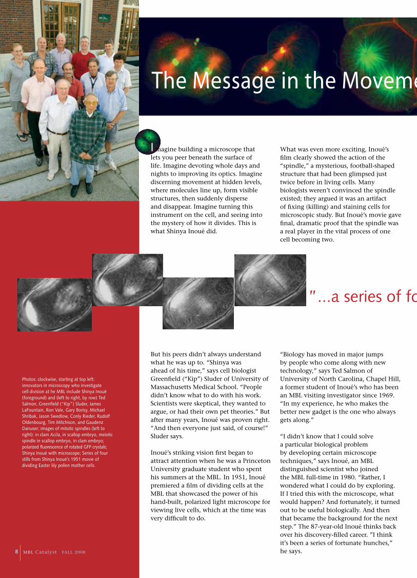
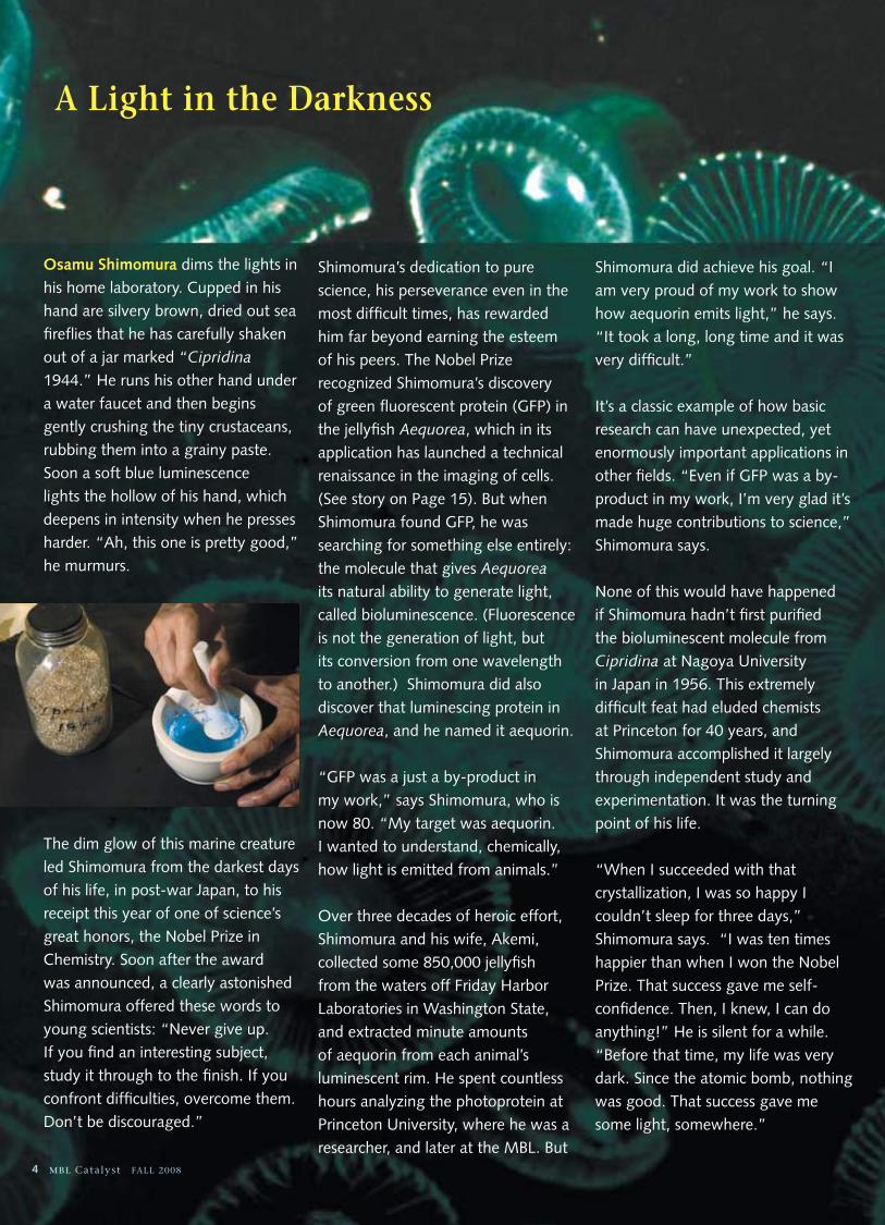
I magine building a microscope that lets you peer beneath the surface of life. Imagine devoting whole days and nights to improving its optics. Imagine discerning movement at hidden levels, where molecules line up, form visible structures, then suddenly disperse and disappear. Imagine turning this instrument on the cell, and seeing into the mystery of how it divides. This is what Shinya Inoué did.
But his peers didn’t always understand what he was up to. “Shinya was ahead of his time,” says cell biologist Greenfield (“Kip”) Sluder of University of Massachusetts Medical School. “People didn’t know what to do with his work. Scientists were skeptical, they wanted to argue, or had their own pet theories.” But after many years, Inoué was proven right. “And then everyone just said, of course!” Sluder says.
Inoué’s striking vision first began to attract attention when he was a Princeton University graduate student who spent his summers at the MBL. In 1951, Inoué premiered a film of dividing cells at the MBL that showcased the power of his hand-built, polarized light microscope for viewing live cells, which at the time was very difficult to do.
What was even more exciting, Inoué’s film clearly showed the action of the “spindle,” a mysterious, football-shaped structure that had been glimpsed just twice before in living cells. Many biologists weren’t convinced the spindle existed; they argued it was an artifact of fixing (killing) and staining cells for microscopic study. But Inoué’s movie gave final, dramatic proof that the spindle was a real player in the vital process of one cell becoming two.
“Biology has moved in major jumps by people who come along with new technology,” says Ted Salmon of University of North Carolina, Chapel Hill, a former student of Inoué’s who has been an MBL visiting investigator since 1969. “In my experience, he who makes the better new gadget is the one who always gets along.”
“I didn’t know that I could solve a particular biological problem by developing certain microscope techniques,” says Inoué, an MBL distinguished scientist who joined the MBL full-time in 1980. “Rather, I wondered what I could do by exploring. If I tried this with the microscope, what would happen? And fortunately, it turned out to be useful biologically. And then that became the background for the next step.” The 87-year-old Inoué thinks back over his discovery-filled career. ”I think it’s been a series of fortunate hunches,” he says.
Photos: clockwise, starting at top left: innovators in microscopy who investigate cell division at he MBL include Shinya Inoué (foreground) and (left to right, by row) Ted Salmon, Greenfield (“Kip”) Sluder, James LaFountain, Ron Vale, Gary Borisy, Michael Shribak, Jason Swedlow, Conly Rieder, Rudolf Oldenbourg, Tim Mitchison, and Gaudenz Danuser; images of mitotic spindles (left to right): in clam Acila, in scallop embryo, meiotic spindle in scallop embryo, in clam embryo; polarized fluorescence of rotated GFP crystals; Shinya Inoué with microscope; Series of four stills from Shinya Inoué’s 1951 movie of dividing Easter lily pollen mother cells.
”...a series of fortunate hunches.”
The Message in the Movement
8 MBL Catalyst FALL 2008

“The modern revolution in microscopy started at the MBL, when Shinya Inoué, Nina Allen, and Bob Allen began using video cameras and computers to image the interior of cells. What they saw was absolutely fantastic, things that no person had seen before.”
— Ron Vale, HHMI/UCSF
Inoué’s hunches—and his gadgets—have indeed been very good over the years. With his polarized light microscope, for example, Inoué clearly saw that the spindle was made of bunches of fibers, which themselves were made of even finer fibrils. But what most excited his eye was seeing that the fibrils weren’t static, but fluctuated dynamically as the cell went through division (called mitosis). By experimenting, Inoué found that the fibrils disappeared when he
cooled the cell or exposed it to certain drugs—and reappeared when he reversed the conditions. Inoué sensed that this submicroscopic movement of the fibrils must be related to an essential job for the spindle: to attach to the chromosomes and accurately separate them during cell division.
So in the late 1960s, Inoué proposed the counterintuitive idea that the spindle fibrils—which by then were known to be microtubules—repeatedly fall apart and reassemble, and that this action creates forces to pull or push chromosomes during cell division. This was far from a mainstream idea—most biologists conceived of the spindle as a stable structure, like a muscle—and it was met with incomprehension and resistance for many years. Still, in the 1970s and 1980s, Inoué and Salmon made significant progress to confirm the energetics of the
”...a series of fortunate hunches.”
The Message in the Movement
concept. But the picture didn’t fall into place until 1984, when Tim Mitchison and Marc Kirschner at the University of California, San Francisco, discovered that spindle microtubules dynamically grow or shrink at their ends, either by adding subunits called tubulin, or by dropping them off. (MBL director and CEO Gary Borisy and his colleagues at the University of Chicago characterized the assembly properties of tubulin in a test tube in the late 1960s and 1970s). This dynamic instability of microtubules
generates forces that move chromosomes in the dividing cell.
“Shinya Inoué and his students really understood the spindle more than anyone else, but almost none of Inoué’s contemporaries
picked up on it,” says Mitchison, who began collaborating with Salmon in the late 1980s.
Meanwhile, the cell division puzzle—and cell biology in general—got a tremendous boost in the early 1980s at the MBL, when Inoué and Robert D. Allen of Dartmouth College realized that connecting a video camera to a light microscope brought huge gains in contrast. Suddenly, fine details of cells were visible that had never been seen before. “Everyone was shocked that the video could do so much better than the eye,” Inoué says. The spindle microtubules, for example, could now be individually imaged for the first time.
“In a light microscope, you can’t see a single microtubule, because it doesn’t scatter enough light to generate enough contrast for your eye to pick it up,” Salmon says. “But with a video camera, you can take that very, very tiny (light) signal and amplify it up, so the microtubule looks like a telephone pole.”
Salmon made beautiful movies of dynamic instability in living cells using this new technology, called VE-DIC microscopy. “We could actually measure how the ends of microtubules grow and shorten in real life,” Salmon says. “We called it video biochemistry.”
And the discovery of VE-DIC microscopy had other profound consequences in the quest to understand the intricacies of cell division. In a story that will have to be told another time, VE-DIC led to the dramatic discovery at the MBL of kinesin, a motor protein. Today, scientists know that chromosome movement involves a complex mix of forces, with both dynamic instability and motor proteins taking part.
Most scientists now use fluorescent protein tags, such as GFP, or fluorescent dyes to visualize molecules in cells. But Inoué and Salmon see an important, continuing role for polarized light microscopy.
“Fluorescence microscopy allows you to discriminate different molecules,” Inoué says. “But if you combined it with polarized light, then you could tell what those specific molecules are doing. You can find out the dynamics of each molecule, how they are moving, changing shape.”
For Inoué, still, the message is in the movement. •
9 MBL Catalyst FALL 2008

brain Power
the MBL neurobiology course takes on the greatest imaging challenge of all
Some things in life are easy to visualize, say, the vacation of your dreams. The brain, with its intricate circuitry, is definitely not one of them.
“It is unimaginably complex. We don’t have anything to compare it to,” says Jeff Lichtman of Harvard University, who has taught imaging in the MBL Neurobiology course for more than 20 years. “You look at any little piece of the brain and you are amazed at how much stuff is in there.”
In even the simplest animal, a maze of cells and wires is packed in the brain, while humans have hundreds of billions of neurons and trillions of synapses, or neural connections. Just getting a picture of neural circuits—let alone understanding how they work—demands all the prowess that modern imaging has to offer. These goals are actively pursued in the Neurobiology course, whose faculty include some of the best imagers in the world.
“What the faculty do,” says course co-director Holly Cline of The Scripps Research Institute, “is bring certain ideas and tools to the course, maybe a certain kind of transgenic animal, and they lay them out as a smorgasbord for the students.” The students learn diverse approaches to the ultimate challenge of visualizing neural circuits, and then they pursue a research project around one of the faculty options. “Many wonderful things, many scientific papers, have come out of the imaging section of this course,” Lichtman says.
10 MBL Catalyst FALL 2008

brain PowerOne of the wonderful aspects of what Jeff Lichtman calls “the green fluorescent protein revolution” is it’s not just green—it’s a rainbow of color. Over the past decade, GFP has been modified to emit light at dozens of different frequencies, primarily by Roger Tsien, who shared this year’s Nobel Prize in Chemistry. Proteins discovered in other sea creatures, such as corals, have broadened the spectral range of fluorescent proteins even further.
For years before Lichtman became co-director of the MBL Neurobiology course in 2000, he had sought a way to label neurons, which are densely arrayed in brain tissue, with a few different colors of fluorescent proteins, so one could tell them apart under the microscope. He had “a long history of failed attempts,” he admits, but his luck would soon change. One of the Neurobiology students that year was Jean Livet from France, who had scant experience with imaging, but strength in molecular neurobiology. The two had perfectly complementary expertise to bring Lichtman’s vision into reality.
“MBL Neurobiology was the best course I ever took,” Livet says. “It allowed me to realize the limits of the molecular approach that had been mine so far, and the extraordinary possibilities offered by combining techniques.”
Experts in several specialties—light microscopy, fluorescence microscopy, electron microscopy, and in vivo imaging—guide the students. “These are different aspects of imaging, and they all bring different pieces of information about neural circuitry to the story,” says Cline.
One approach is to get a static map, or “wiring diagram,” of neural circuits in various animals. Lichtman’s Brainbow (see sidebar) is a leap in this direction, as is the map of the fruit fly brain being pursued by course lecturer Ann-Shyn Chang of National Tsing Hua University. Some faculty are creating 3-D maps of the molecular components of synapses and circuits, including Stephen Smith and Kristina Micheva of Stanford University Medical Center.
And while these mapping efforts, often called “connectomics,” won’t reveal the function of neural circuitry—which neurons are firing, how information is flowing—they are a place to start. As cell biologist Jason Swedlow of University of Dundee puts it, “In many cases in biological imaging, we just want to get a picture of what the darn thing looks like. The premise is, if we can see how this thing is put together, that will be the start of understanding what it is doing.”
Lichtman likens the situation to genomics. “The Human Genome Project got a physical map of all the genes, but of course it didn’t reveal the way genes work, which is highly dynamic. In connectomics, we are still trying to get the map.”
Other researchers are forging ahead without the map and are imaging neural circuits in living animals, called in vivo imaging. In Neurobiology course research this year, one student used two-photon microscopy to observe microglial cells tagged with fluorescent protein inside the brain of a mouse.
“The exciting contribution of in vivo imaging is that it shows that the map of connections is changing over time and with experience,” says Cline, whose own lab pursues in vivo imaging of visual circuitry in the frog brain. “We need to have both aspects: the static wiring diagram and information on the dynamic connectivity in the living animal.”
Still, an open question, and even a conundrum, is how structural and functional imaging studies will eventually converge. “We even staged a debate about this in Neurobiology this year,” says Cline. “What will we be able to learn about the brain simply by determining static connectomics, and what won’t we be able to learn? It was fun, a true exercise in articulating ideas.” •
Two years after the course, Livet joined Lichtman’s lab at Harvard University as a postdoctoral associate. There they developed, with Joshua Sanes, a novel imaging technique they called Brainbow, in which engineered transgenes in the animal express random amounts of red, green, and blue fluorescent protein inside each neuron. Much in the way a TV monitor works, this has the spectacular result of generating more than 100 different colors of neurons. Brainbow’s gorgeous images of neural circuitry landed on the cover of the scientific journal Nature, and the associated paper was one of the most acclaimed in science in 2007.
“It really took Jean’s concerted effort and sophistication as a molecular biologist to come up with strategies to do this in a straightforward way,” Lichtman says. Livet, for his part, had absorbed the imaging expertise that abounds in Neurobiology. “The course proved an unforgettable experience for me,” says Livet, who is now an investigator at the Vision Institute in Paris, France. “I am now continuing this approach of combining molecular and imaging techniques with my own research team.” •
A Dazzling Technique
Photos, left to right: a motor nerve innervating ear muscle in a mouse; GFP nerve ganlion; mouse brainstem
11 MBL Catalyst FALL 2008

The Proving Grounds
...with
Greenfield “Kip” SluderCo-Director, MBL Analytical & Quantitative Light Microscopy course
MBL Does the course teach microscopy in the context of cell biology?
KS No, we don’t teach biology at all. We look at very few cells; often we look at little fluorescent beads. We have a basic program that covers how microscopes work, how contrast is produced, and then we teach some of the fancier techniques. One feature we have been emphasizing—and this is where David Wolf and Jason Swedlow, who is a new AQLM co-director, have been particularly good — is bringing
quantification into the course. We have a student competition, called Spectral Conflict, that involves quantifying something that is seemingly
easy, but is actually quite difficult to do precisely. We find this really activates the students, puts them into intimate contact with the commercial faculty, and gets the commercial faculty excited about using their instruments in a quantitative fashion.
MBL What might you quantify?
KS In one exercise, we mixed together beads that had different amounts of fluorophor in them, and the students had to quantify the fluorescence in the different classes of beads. So first, they had to figure out how to quantify the intensity over a microscopic sphere. What the students didn’t know was that the range of fluorescent intensities was outside the dynamic range of their instrumentation! So they had to deal with that. Another year, the problem was to measure the diameter of spider silk.
MBL What do the students and the faculty get out of the course?
KS AQLM is a mixture between a didactic course and a scientific meeting. Every year we do the meat and potatoes instruction, but we also throw in some whiz-bang stuff, the emerging technologies. That keeps our high-end commercial faculty coming back, because they can learn the latest research on things they may be thinking about producing. The commercial faculty are in many ways the backbone of our course. They do all the laboratory instruction.
Greenfield “Kip” Sluder is professor of cell biology at University of Massachusetts Medical School in Worcester. Sluder studied with Shinya Inoué, Gordon Ellis, and Hidemi Sato at the University of Pennsylvania, earning his doctorate with Sato in 1976. After a postdoctoral fellowship with Dan Mazia at the University of California, Berkeley, he joined the faculty of the Worcester Foundation for Experimental Biology in 1981, remaining there until 1997. Sluder’s research applies microscopic and biophysical methods to investigate the mechanisms that control various aspects of cell division. He is co-author with David Wolf of the textbook Digital Microscopy, now in its third edition.
The MBL hosts two short courses on microscopy each year: Analytical and Quantitative Light Microscopy (AQLM), founded by Shinya Inoué in 1980; and Optical Microscopy and Imaging in the Biomedical Sciences, started by Robert Allen of Dartmouth College in 1978. These courses pioneered a format that is now widely copied: The faculty includes both academic scientists and commercial imaging experts, who bring the “latest and greatest” microscopes and cameras for students to use. Greenfield “Kip” Sluder has co-directed AQLM since 1990 with David Wolf, chief scientific officer and vice president for research and development at Sensor and BioHybrid Technologies. Below, Sluder shares his thoughts on the course.
mBl mo m e n t
12 MBL Catalyst FALL 2008
They get to demonstrate their equipment, which has sales implications, but more importantly, they get to see what biologists are interested in, so they can develop their product further. As for the students, many of whom are university faculty, they get a didactic education; they can test drive the latest equipment; and they are introduced to a big part of the microscopy community. We also get some students from industry. These are people
who could run us around the block any 10 ways on certain issues, but they don’t know the big picture. They get to see the spectrum of light microscopy
all in one place, which is hard to get elsewhere. As for the academic faculty — every year I learn something. It’s always exciting to see the new stuff.
MBL If you compare the course syllabus from 1980 to today’s, it’s obvious the field has grown tremendously in the past 28 years. What caused this burst in innovation in light microscopy?
KS When the course started, electronic imaging was just coming online. Before, it was only used in the surveillance industry, in the military. So a big advance was attaching a video camera to a microscope. And when Bob Allen did this one day in his MBL course, he and Shinya Inoué noted that by controlling black level and gain on the camera, they could get far better contrast on the video screen than you could by eye. The early images weren’t great, in unskilled hands. But Inoué and Allen had very skilled hands and they had the mindset of looking for noise, looking for contrast. They knew what they were after. At the same time, low-light level imaging devices used in the Vietnam War were becoming unclassified. And personal computers
were becoming practical and more powerful. People started writing software for biological imaging, including Shinya Inoué and his son, Ted, who started Universal Imaging Corporation. And there were big gains in fluorescence technology. All of these things were being developed at a very high rate and came together synergistically to advance certain imaging applications.
MBL What role did the AQLM course play in all this innovation?
KS In terms of biological applications for video microscopy, AQLM and Bob and Nina Allen’s course were the proving grounds. Every year the commercial faculty came with their latest and greatest equipment—some would go home delighted, and some would go home mad. And then they would come back the next year with something even better.
The AQLM course provided some real help to the commercial people in terms of application development, particularly for low-light level cameras. And still, today, the course plays a very important role in the development of tools that are practical for the biologist. Now, we have far more applications to choose from than in the early days of the course. All the video cameras have been replaced by digital cameras, of which there are various kinds. Same with confocal microscopes — these come in all sorts of flavors. And in both of these areas, AQLM has been instrumental in defining what instrument you may want to use for what application.
MBL Where do you see light microscopy heading?
KS It’s really limited only by people’s imaginations. Look at green fluorescent protein (GFP), for example. That technology had its origins in the 1900s, when people started looking at jellyfish that glowed at night. That research might have seemed sleepy then, and the NIH wouldn’t even fund that kind of investigation today. But GFP has revolutionized imaging in cell biology. At the early stages, you might have thought, who cares about this weirdo phenomenon in jellyfish? It required someone’s imagination to say, “I care,” and to identify GFP as a protein, and for someone else to realize that you could transfect a cell with a piece of DNA that would encode this protein, and that it would glow inside the cell. These things start off as a thread, and then become a rope. •
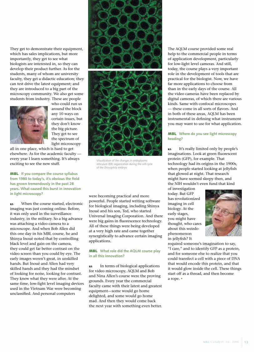
Visualization of the changes in endoplasmic reticulum (ER) organization during the cell cycle of the Drosophila embryo
13 MBL Catalyst FA:: 2008

ac c o l a D e s
MBL Distinguished Scientist Osamu Shimomura was awarded the 2008 Nobel Prize in Chemistry for his discovery of green fluorescent protein. Shimomura shares the prize with Martin Chalfie of Columbia University, New York, and Roger Y. Tsien of University of California, San Diego. Chalfie is a former lecturer in the MBL’s Neural Systems and Behavior course and Tsien is a former lecturer in the MBL Neurobiology course. Tsien was also the Forbes Lecturer in 2005. Shimomura also recently received the Order of Culture, the highest honor given annually to citizens of Japan by the Emperor of Japan.
MBL Corporation Member, Honorary Trustee, and former Chairman of the Board Sheldon J. Segal and the Population Council in New York were awarded the Prix Galien USA 2008 Pro Bono Humanum Award for their science-based, global effort in support of reproductive planning and family health.
The American Academy of Arts and Sciences inducted John Hobbie, distinguished scientist and senior research scholar in the MBL’s Ecosystems Center, into its 2008 class of fellows. Four MBL alumni and 10 course faculty, including Physiology course co-director Timothy Mitchison (Harvard Medical School) and Michael Dickinson (California Institute of Technology), a faculty member in the Neural Systems and Behavior course, were also among this year’s class of fellows.
Gary Ruvkun (Harvard Medical School), co-director of the MBL’s Molecular Biology of Aging course, was one of three recipients of the 2008 Lasker Award for Basic Medical Research. The Lasker Awards are often considered the American version of the Nobel Prize, and many Lasker recipients have gone on to win the Nobel. Ruvkun also received a 2008 Gairdner International Award which recognizes the world’s leading medical research scientists. Since 1957, 70 of 288 Gairdner winners have gone on to win the Nobel Prize.
Pamela Clapp Hinkle was named the MBL’s Director of Development and External Relations. Hinkle, a 26-year MBL veteran, has served the institution in a variety of capacities, most recently as Director of Communications. In her new position, Hinkle is responsible for all aspects of communications, fundraising, and government and external relations in support of the MBL’s mission and strategic direction.
Gi F t s & Gr a n t s
14 MBL Catalyst FALL 2008
The Howard Hughes Medical Institute awarded $15 million and the
Massachusetts Life Sciences Center awarded $10 million to fund top-to-bottom renovations of the MBL’s Loeb Laboratory, home to the laboratory’s intensive, full-immersion graduate
and postdoctoral-level laboratory courses. Preliminary construction is scheduled to begin in March 2009 with an expected completion date of June 2010.
Charles and Patricia Robertson have pledged $2 million to create the Robertson Education Discretionary Fund. An anonymous donor awarded $1.5 million to establish the Women in Ecological Science Quasi-Endowed Fund. Ecosystems Center Senior Scientist Zoe Cardon is the initial recipient of this support.
The Alfred P. Sloan Foundation awarded $1.2 million for “The Rare Biosphere and the Human Habitat.” Josephine Bay Paul Center director Mitch Sogin is the principal investigator.
The National Institutes of Health awarded $920,978 for “BioCurrents Research Center.” Senior scientist Peter Smith is the principal investigator.

Inside every cell are hundreds or thousands of proteins, each with a specific role to play in the life of an organism. Before we can understand how muscles contract, how the immune system defends, or how the nervous system performs, we must first understand how proteins work in cells. Yet proteins are too small to be seen by light microscopy alone. For decades, scientists could only deduce their behavior.
Green fluorescent protein (GFP) from the jellyfish Aequorea has changed all that. Discovered by Osamu Shimomura in 1961, GFP is now the gold-standard tool in microscopy for illuminating protein processes, with far-reaching consequences for research on conditions from cancer to AIDS. This year, Shimomura of the MBL, Martin Chalfie of Columbia University, and Roger Tsien of University of California, San Diego, received the Nobel Prize in Chemistry for the discovery and development of GFP.
When Shimomura purified what he called “green protein” from Aequorea, few scientists took note. It did interest J. Woodland Hastings and James Morin of Harvard University, who studied the green protein at the MBL in the late 1960s and renamed it GFP. Shimomura also characterized the protein further in the 1970s.
Green Fluorescent Protein
Yet GFP remained obscure until the early 1990s, when Chalfie realized how tremendously useful it could be in biological research. For half a century, scientists had known how to chemically synthesize fluorescent molecules and attach them to antibodies, which would specifically bind to proteins in cells. These tiny fluorescent tags allowed microscopists to see the otherwise invisible proteins. But the technique had a significant limitation: it could only be performed on nonliving cells. Chalfie wondered if a simpler genetic manipulation could achieve the same result, but in live cells. Could the GFP gene be inserted into the genome of an experimental animal? Could GFP be fused to interesting proteins in the animal, and would it light them up for study? Chalfie obtained the GFP gene from Douglas Prasher, who had sequenced it in 1992. The exciting answer to these questions, Chalfie found out, was “yes.”
Since then, GFP has been fused to thousands of different proteins, allowing scientists to study their myriad activities in real time. Roger Tsien modified GFP to make it glow brighter, and he also extended its color palette beyond green. Thanks to Aequorea, the future of biomedical research is indeed bright. As Ted Salmon of University of North Carolina says, “We’ve got the human genome sequenced, and genomes generate proteins. The name of the game now is to understand how proteins function in cells.”
From Jellyfish to Post-Genomics
co o l to o l
In the jellyfish Aequorea (left), blue light emitted by the protein aequorin is transferred to a second protein, GFP, which re-emits it as green light.This bioluminescence system is the basis for GFP technology.
15 MBL Catalyst FALL 2008
Scaling the Super-Resolution Peak
Jennifer Lippincott-Schwartz, a faculty
member in the MBL Physiology course, is a
senior investigator at the National Institutes
of Health (NIH), where she is Chief of the
Section on Organelle Biology, Cell Biology and
Metabolism Program, National Institute of
Child Health and Human Development. Her
laboratory uses live-cell imaging approaches,
particularly novel GFP technologies, to analyze
the spatio-temporal behavior and binding
interactions of molecules in cells. In 2003,
Lippincott-Schwartz received the NIH Award of
Merit for her “fundamental contribution to the
understanding of how intracellular organelles
are assembled and inherited, and how proteins
move within cells.” In 2008, Lippincott-
Schwartz was elected to the National Academy
of Sciences. •
By Jennifer Lippincott-Schwartz
Fluorescent proteins purified from marine organisms and used as glowing tags inside cells have
transformed biological imaging, as the 2008 Nobel Prize in Chemistry acknowledges. In my own
laboratory, research to modify and create new uses for green fluorescent protein (GFP) has led us to a
revolutionary new technique in light microscopy. This has been a thrilling adventure, one that shows
the power of interdisciplinary research. A collaboration at the MBL helped lead to exciting extensions
of our new technique, which lets us track single molecules inside cells as never before.
It started one day in 1995 when I attended a seminar by Eric Betzig, a brilliant physicist who was
visiting the National Institutes of Health (NIH). Afterwards, over lunch, Eric pitched an idea to
me that made total sense. Eric had thought of a way to get around a big problem in fluorescence
microscopy: poor resolution at the molecular scale. Typically, under a light microscope, fluorescently
tagged molecules that are less than 200 nanometers apart blur into a fuzzy blob, so the individual
molecules can’t be distinguished. Eric’s idea was to configure a light microscope so that only a small
number of fluorescent molecules in the cell light up at once. After imaging those molecules, different
molecules could be activated and imaged, and so on. After repeating this many times, the image layers
could be combined into one, super-resolution image of the cell. To make this work, Eric needed a
“photoconvertible” fluorescent protein tag, meaning its illumination can be switched on or off at will,
such as the photoactivatable GFP developed in my lab by George Patterson. This GFP only fluoresces
when hit with violet light.
I was extremely excited about Eric’s proposal. This was such a simple idea, and it only needed the
right fluorescent probes and the right microscope setup. We immediately got to work. For probes,
George Patterson and Mike Davidson of Florida State University speedily generated photoconvertible
fluorescent protein tags attached to different cellular proteins, while for the microscopy setup, Eric
tapped his close colleague, Harald Hess. After a few months of hard work in a small lab space at
NIH, the microscope prototype was built, and we had proven the technique works. Called PALM
(photoactivated localization microscopy), it can resolve the most precisely localized molecules at
separations of just a few nanometers. The scientific community’s response to PALM was immediate
and enthusiastic.
We were thrilled, but we didn’t stop there. Initially, we were looking at fixed cells, but we knew
scientists would really get excited if we could apply PALM to living cells. In the summer of 2007, I
was invited to teach a two-week rotation in the MBL Physiology course. I invited Eric to accompany
me, and he brought along Hari Shroff, who had been a Physiology student the year before. It was a
fantastic experience. We worked around the clock, trying many different approaches. By the end of the
rotation, we had gotten live-cell PALM to work! Not only that, we got PALM to work with two different
colors of fluorescent probes, and we demonstrated that PALM could be used to track single molecules
in live cells. It was a spectacular session, and it led to several publications. When I came back to the
Physiology course this year with physicist Suliana Manley from my lab, we taught the students how
to assemble a PALM system from scratch. They used it to look at minute structures, like HIV particles,
that were always thought beyond the reach of light microscopy.
Fluorescence techniques like PALM have dramatically expanded the range of what we can see in cells.
It’s a great time to be in biology! •
sc i e n t i s t ’s ey e Vi e w
16 MBL Catalyst FALL 2008
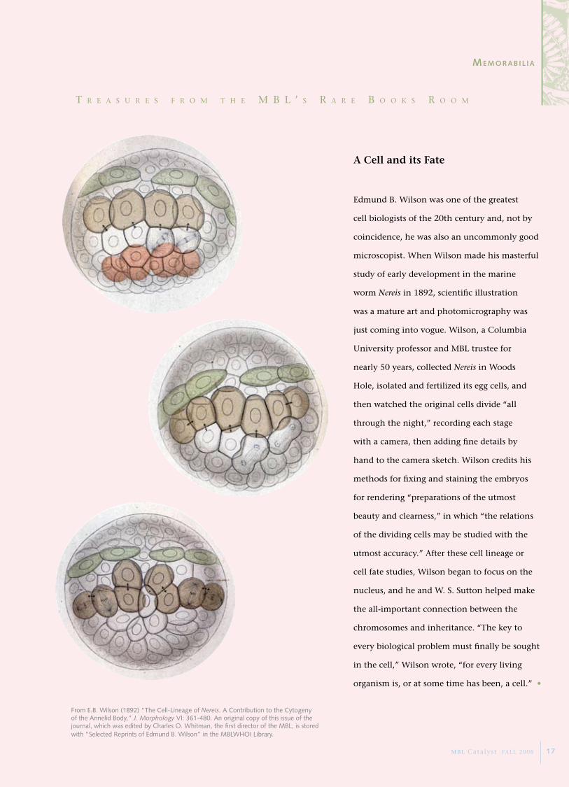
From E.B. Wilson (1892) “The Cell-Lineage of Nereis. A Contribution to the Cytogeny of the Annelid Body,” J. Morphology VI: 361-480. An original copy of this issue of the journal, which was edited by Charles O. Whitman, the first director of the MBL, is stored with “Selected Reprints of Edmund B. Wilson” in the MBLWHOI Library.
T r e a s u r e s f r o m T h e m B L ’ s r a r e B o o k s r o o m
A Cell and its Fate
Edmund B. Wilson was one of the greatest
cell biologists of the 20th century and, not by
coincidence, he was also an uncommonly good
microscopist. When Wilson made his masterful
study of early development in the marine
worm Nereis in 1892, scientific illustration
was a mature art and photomicrography was
just coming into vogue. Wilson, a Columbia
University professor and MBL trustee for
nearly 50 years, collected Nereis in Woods
Hole, isolated and fertilized its egg cells, and
then watched the original cells divide “all
through the night,” recording each stage
with a camera, then adding fine details by
hand to the camera sketch. Wilson credits his
methods for fixing and staining the embryos
for rendering “preparations of the utmost
beauty and clearness,” in which “the relations
of the dividing cells may be studied with the
utmost accuracy.” After these cell lineage or
cell fate studies, Wilson began to focus on the
nucleus, and he and W. S. Sutton helped make
the all-important connection between the
chromosomes and inheritance. “The key to
every biological problem must finally be sought
in the cell,” Wilson wrote, “for every living
organism is, or at some time has been, a cell.” •
me m o r a B i l i a
MBL Catalyst FALL 2008 17

CatalystTranslational Research When Osamu Shimomura discovered a green,
glowing protein in a jellyfish, he didn’t dream it would be applied as a tool that has
revolutionized biomedical research. Yet marine organisms are key to critical advances in
medicine and drug development, which is why they have been intensively studied at the
MBL for more than a century. MBL investigations of terrestrial organisms, too, and of
our changing environment have a direct impact on making the world a better place. In
the next MBL Catalyst, read about applications of MBL research, from the development
of new therapeutics, to the alleviation of poverty, to the preservation of our planet.
in t h e ne x t cata ly s t
MBL7 MBL StreetWoods Hole, MA 02543 USA
NON-PROFIT ORG.
U.S. POSTAGE PAID
PERMIT NO. 55
PLYMOUTH, MA
www.MBL.edu
Related Documents