Cat Dissection Anatomy & Physiology Unit 7 Name ________________________________ Teacher______OLSEN_______ Hr ______ The name of my cat friend is: __________________________________ Thanks for helping me learn more about myself!

Welcome message from author
This document is posted to help you gain knowledge. Please leave a comment to let me know what you think about it! Share it to your friends and learn new things together.
Transcript
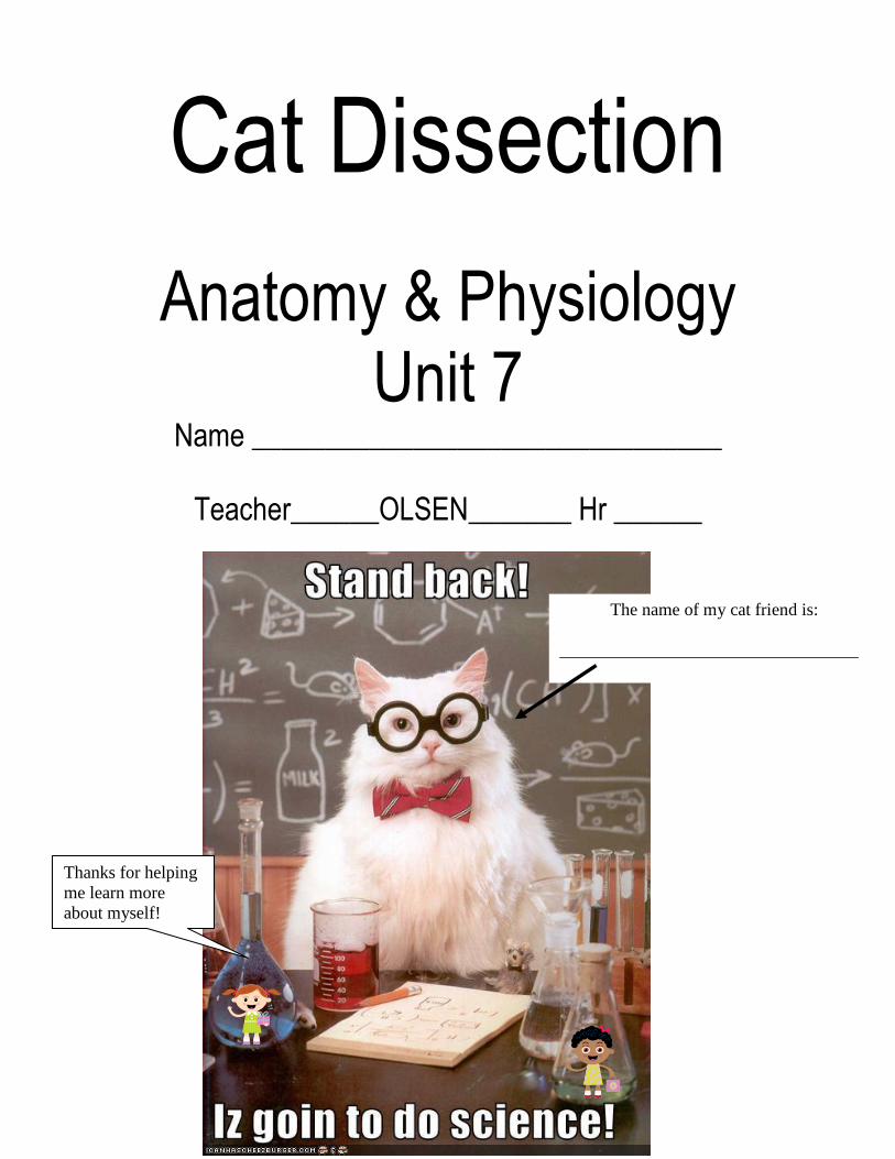
Cat Dissection
Anatomy & Physiology Unit 7
Name ________________________________
Teacher______OLSEN_______ Hr ______
The name of my cat friend is:
__________________________________
Thanks for helping
me learn more
about myself!
25
Introduction Felis catus
The cat, also known as the domestic cat or housecat, to distinguish it from other felines and felids, is a small furry domesticated carnivorous mammal that is valued by humans for its companionship and for its ability to hunt vermin and household pests. Cats have been associated with humans for at least 9,500 years, and are currently the most popular pet in the world. Owing to their close association with humans, cats are now found almost everywhere on Earth. The question of using cats for medical science dissection and learning can and should be raised. The ethical argument against the use of cats would be stronger if cats were bred specifically to be killed for dissection. However, the cats we use are the product of uncontrolled reproduction of pets. The surplus wind up at the animal shelter. At the animal shelter, the majority of cats are "euthenized" in a hypobaric chamber. In this chamber, the air is pumped out until the animal first passes out, and eventually dies of oxygen starvation. In the great majority of cases, the carcasses are then either cremated or buried. It is clear that using these animals which have already been euthenized yields at least one positive outcome of their sad deaths, one of advancing the teaching of medical science. Until the pet population explosion is under control and there is no surplus of euthenized cats, it would seem that a constructive use of a social tragedy is to be encouraged.
Ethical Expectations These rats were, at one time, living animals. Although they have been euthanized they deserve to be treated with respect (both in life and in death). Dissections of specimens can sometimes be exciting, scary, or even a little bit unsettling. However, this organism is an animal, just as you and I are. As such, we expect that all students during this unit will behave in a fashion that displays acknowledgement of the rats’ integrity as a specimen, and as a learning tool. The following behaviors will not be tolerated:
Mutilation (inappropriate damage or destruction) of the cat or any of its parts
Horseplay or harassment of others
Inappropriate use of sexual slang, drawings or references
Misuse of tools If any students or groups are found to be doing any of the above actions, the following consequences could be imposed:
After-school detention
Cleaning of dissection of tools and trays
Written letter of apology to cat for mistreatment.
During Lab Students are expected to use their time efficiently and effectively in lab. There are three essential tasks
that students should be completing for each section of the rat dissection: 1. Procedure:
Follow the specific dissection directions provided for each section.
26
2. Identification and Labeling:
Use the provided and suggested cat dissection manuals to determine what structures need to be identified. Use your Cat Dissection Packet to help you. Remember, you will be responsible for these parts on the lab practical.
3. Questions:
Answer ALL questions for a section before moving onto the next section. These questions are designed to test your ability to locate specific structures and determine the important purposes, functions, or relationships between these structures. Use your Cat Dissection Packet to help you. Remember, you will be responsible for these parts on the lab practical.
Assessment The end-of-unit assessment will be in the form of a lab practical (100pts KS). Specimens will be displayed on dissection trays where students will need to do one or more of the following tasks:
Identify a structure
Describe the function, purpose, or importance of a structure
Relate an already identified structure or function to an important idea or concept that was studied during the unit.
Optional Gloves Gloves may be worn during dissection, but are the sole responsibility of the student(s) using them. Gloves will not be provided or stored for any students during the dissection. Often, students will share the cost of purchasing a pack of gloves for the dissection unit. Students can plan on needed gloves for at least five days of dissection.
Why Do We Dissect?
We have chosen cats for our dissection experience for a variety of reasons. Cats are mammals, just like humans! Therefore, they share many of the same internal and external structures that humans do, and perform many of the same functions. Also, they are larger than other dissection specimins. This dissection will provide you with an opportunity to apply your knowledge and understanding of human anatomy and physiology to another organism with similar characteristics. Some of these traits include:
o Vertebrates (have backbones) o Hair-covered bodies o Mammary glands for nursing young o Young are nourished in mother’s uterus o Breathe with lungs (throughout lifetime) o Diaphragm separating thoracic and digestive cavities o Four-chambered heart o Warm-blooded o Two pairs of limbs

27
1. Write a response to the question, “Why are we dissecting in science class?” ______________________________________________________________________________________
______________________________________________________________________________________
2. Why dissect a cat and not a different thing?
______________________________________________________________________________________
______________________________________________________________________________________
______________________________________________________________________________________
Location Terms
Procedure: 1) Obtain a cat from your teacher and remove it from the original storage bag and throw away the original
storage bag. 2) Place the cat on the dissecting tray so it looks like the diagram below.
Identification and Diagram Labeling: You are responsible for knowing the location of the following terms. Locate each term using your Dissection Guide. Label them on the diagram below and/or describe their location in the notes section of the table.
Terms: Notes:
Anterior
Posterior
Dorsal
Ventral
Questions:
3. What would be an easy for you to remember these 4 terms? Write this idea here: __________________________________________________________________________________________
__________________________________________________________________________________________
http://www.allaboutdrawings.com/images/cat-stretching-drawing.jpg

28
External Structures
Procedure: 1) Lay your cat on its dorsal surface. 2) This next step may require some force. Lay the cat as flat as possible on its dorsal surface so its ventral
surface is clearly visible. If it needs to be tied down, ask your teacher. 3) Be sure to view rats of BOTH sexes during this section.
Identification and Diagram Labeling: You are responsible for knowing the location of the following terms. Locate each term using your Dissection Guide. Label them on the diagram below and/or describe their location in the notes section of the table.
Terms: Notes:
Vibrissae (stout long hairs around the mouth & eyes)
External nares (nostrils)
Philitrum (groove separating the nares; fused in humans)
Pinnae (external ear structures)
Nails and pads (sometimes declawed)
Mammary papillae (nipples present on both M and F)
Anus (just ventral to the tail)
Female Cat:
Terms: Notes:
Urogenital aperture (contains both the urethra and vagina)
Male Cat:
Terms: Notes:
Scrotum (sack containing the testes; sometimes neutered)
Prepuce (slight swelling in which the penis is retracted; contains the urethra)

29
These pictures are with the tail pointing up. Don't ID #9. Female Cat: Male Cat:
Questions: If you do not already know the answers to these questions, reference your Dissection Guides.
4. How can you differentiate between a male and female cat from the external features? __________________ 5. What external clue is there for the approximate number of “babies” that a female cat could produce?
______________________________________________________________________________________
Respiratory System
Procedure: 1) Begin dissection by inserting the dissecting scissors into the urethra and penetrating the body cavity just
below the surface. *Caution: Do not cut too deep; this may damage tissues. 2) Following the diagram of cut lines in your Dissection Guides, proceed to cut open the cat just inside the
body cavity, through the ribcage, toward the head and then down each limb. This will expose internal organs. Don’t forget the neck area as well.
3) Split open the cavity and open the flaps of tissue. Attempt to pin down the tissue if necessary.
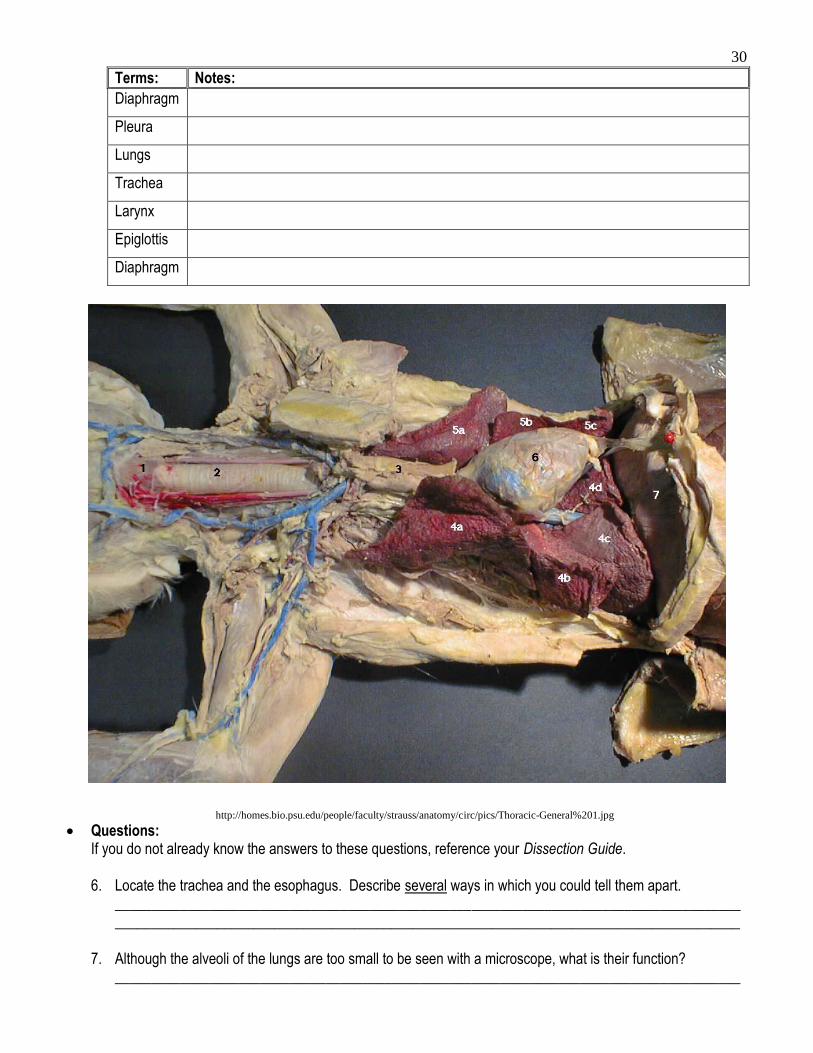
Identification and Diagram Labeling: You are responsible for knowing the location of the following terms. Locate each term using your Dissection Guides. Label them on the diagram below and/or describe their location in the notes section of the table. Recommended Guides: Bohensky p88-92, p72-76; Gilbert p48-51, 28-31
http://fp.dl.kent.edu/hyork/catext.htm
30
Terms: Notes:
Diaphragm
Pleura
Lungs
Trachea
Larynx
Epiglottis
Diaphragm
Questions: If you do not already know the answers to these questions, reference your Dissection Guide.
6. Locate the trachea and the esophagus. Describe several ways in which you could tell them apart.
____________________________________________________________________________________________________________________________________________________________________________
7. Although the alveoli of the lungs are too small to be seen with a microscope, what is their function?
______________________________________________________________________________________
http://homes.bio.psu.edu/people/faculty/strauss/anatomy/circ/pics/Thoracic-General%201.jpg

31
8. Cut a slit at the absolute top of the larynx. Locate the epiglottis (looks like a tongue). What is its function?
______________________________________________________________________________________
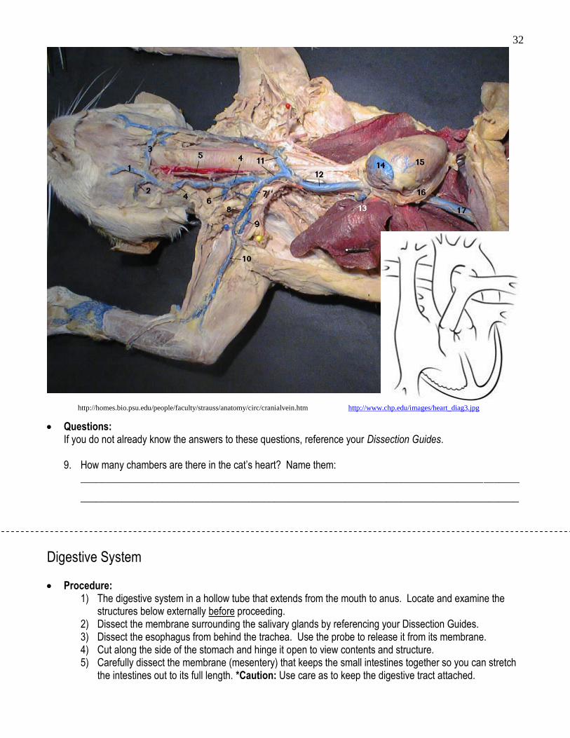
Circulatory System
Procedure: 1) Since the chest cavity has already been opened and the rib cage opened, the heart should be in plain
view. Locate and examine the structures below externally before proceeding. 2) Carefully hold the heart between your fingers and make an incision with a scalpel at the tip of the heart
toward the anterior end. 3) Hinge open to view the chambers of the heart. Make sure to view an injected heart if yours os not.
Identification and Diagram Labeling: You are responsible for knowing the location of the following terms. Label them on the diagram below and/or describe their location in the notes section of the table. Recommended Guides: Bohensky p104-120; Gilbert p48-55
Terms: Notes:
Pericardium
Heart
Right atrium
Left atrium
Right ventricle
Left ventricle
Coronary arteries
Aortic arch
Thorasic aorta
Anterior vena cava
Posterior vena cave
Carotid arteries
Renal arteries & viens
Pulmonary arteries & veins
Spleen *note: This is very difficult to identify and is not located in the pictures below. It is a half moon shaped dark tissue under the liver. It is the same color as the liver.
32
Questions: If you do not already know the answers to these questions, reference your Dissection Guides. 9. How many chambers are there in the cat’s heart? Name them:
______________________________________________________________________________________
______________________________________________________________________________________
Digestive System
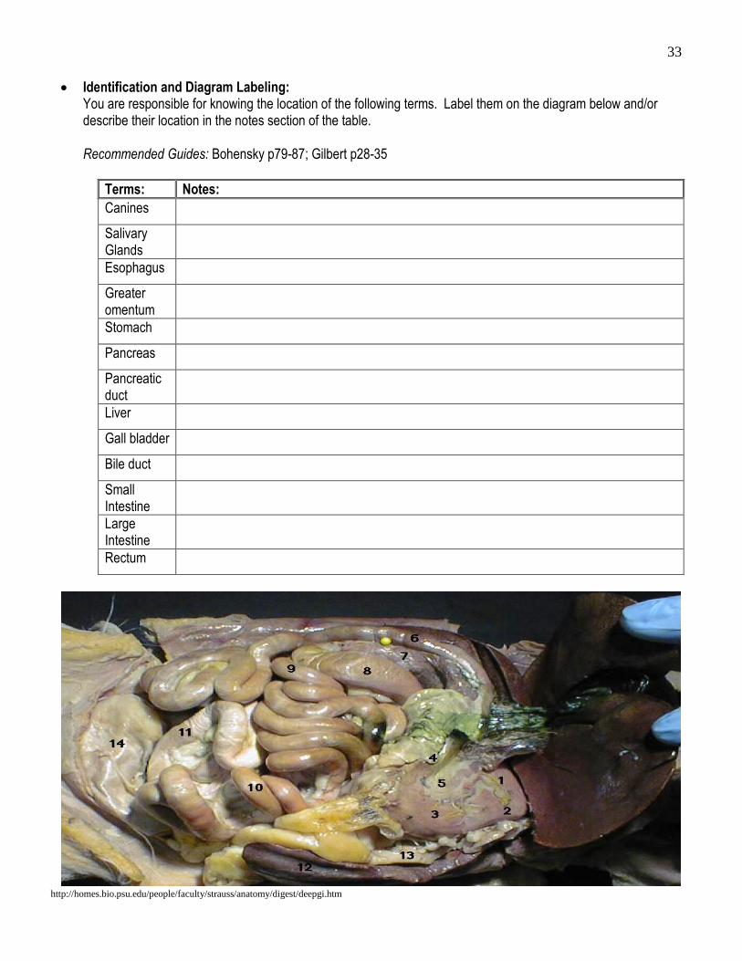
Procedure: 1) The digestive system in a hollow tube that extends from the mouth to anus. Locate and examine the
structures below externally before proceeding. 2) Dissect the membrane surrounding the salivary glands by referencing your Dissection Guides. 3) Dissect the esophagus from behind the trachea. Use the probe to release it from its membrane. 4) Cut along the side of the stomach and hinge it open to view contents and structure. 5) Carefully dissect the membrane (mesentery) that keeps the small intestines together so you can stretch
the intestines out to its full length. *Caution: Use care as to keep the digestive tract attached.
http://homes.bio.psu.edu/people/faculty/strauss/anatomy/circ/cranialvein.htm http://www.chp.edu/images/heart_diag3.jpg
33
Identification and Diagram Labeling: You are responsible for knowing the location of the following terms. Label them on the diagram below and/or describe their location in the notes section of the table. Recommended Guides: Bohensky p79-87; Gilbert p28-35
Terms: Notes:
Canines
Salivary Glands
Esophagus
Greater omentum
Stomach
Pancreas
Pancreatic duct
Liver
Gall bladder
Bile duct
Small Intestine
Large Intestine
Rectum
http://homes.bio.psu.edu/people/faculty/strauss/anatomy/digest/deepgi.htm
34
Questions: If you do not already know the answers to these questions, reference your Dissection Guidse. 10. Locate one or more of the parotid, mandibular, and sublinqual glands. By their location and the fact that they
are part of the digestive system, what must their function be? ____________________________________________________________________________________________________________________________________________________________________________
11. Locate the trachea and the esophagus. Describe several ways in which you could tell them apart.
____________________________________________________________________________________________________________________________________________________________________________
12. Where in the digestive system does mechanical digestion occur? __________________________________
13. Describe the interior of the stomach.
______________________________________________________________________________________
14. What are the general functions of the cardiac and pyloric sphincters of the stomach? ______________________________________________________________________________________
15. If your cat does not have stomach contents see if you can see another groups and describe it here:
______________________________________________________________________________________
16. Besides the glands in the walls of the duodenum, what accessory organ releases numerous digestive enzymes in to the digestive system at that site? ________________________________________________
17. What does the liver secrete to aid in the process of digestion and what is the function of that secretion?
______________________________________________________________________________________
18. What sac stores the liver’s secretion? _______________________________________________________
19. Determine the approximate length of the small intestine, which ends at the caecum. ____________________
20. Why is the small intestine sooooooooo long? __________________________________________________
21. Chemical digestion occurs at three points along the digestive track. Name those three points and what
type(s) of foods are being chemically digested at each one.
______________________________________________________________________________________
______________________________________________________________________________________
______________________________________________________________________________________
22. What is the function(s) of the large intestine (colon)?
_____________________________________________________________________________________

35
Urinary System
Procedure: 1) Place the intestines off to the side outside of the body cavity do you can view the inside dorsal surface of
the cat. 2) Once you have located all the urinary parts, dissect the membrane around the kidney and lift it out of the
cavity without detaching it from the ureter. 3) Slice along the side of the kidney with the scalpel and hinge open to view the inside.
Identification and Diagram Labeling: You are responsible for knowing the location of the following terms. Label them on the diagram below and/or describe their location in the notes section of the table. Recommended Guides: Bohensky p123-133; Gilbert p38-47
Terms: Notes:
Kidneys
Ureters
Urinary Bladder
Renal cortex
Renal medulla
Renal Pelvis
Urethra
*Note: the diagram to the right is also a diagram for the female reproductive system. After doing the reproductive system, come back label these parts here as well.
http://www.tutorvista.com/content/biology/biology-iii/animal-morphology/respiratory-excretory-nervous-reproductive-system-rat.php
http://homes.bio.psu.edu/people/faculty/strauss/anatomy/urogen/dissectkidney.htm
36
Questions: If you do not already know the answers to these questions, reference your Dissection Guides. 23. Locate a kidney. Trace the pathway of the urine through the following structures of the excretory system.
Describe the function of each: A. Kidneys: _____________________________________________________________________
B. Ureters: ______________________________________________________________________
C. Urinary bladder: _______________________________________________________________
D. Urethra: _____________________________________________________________________
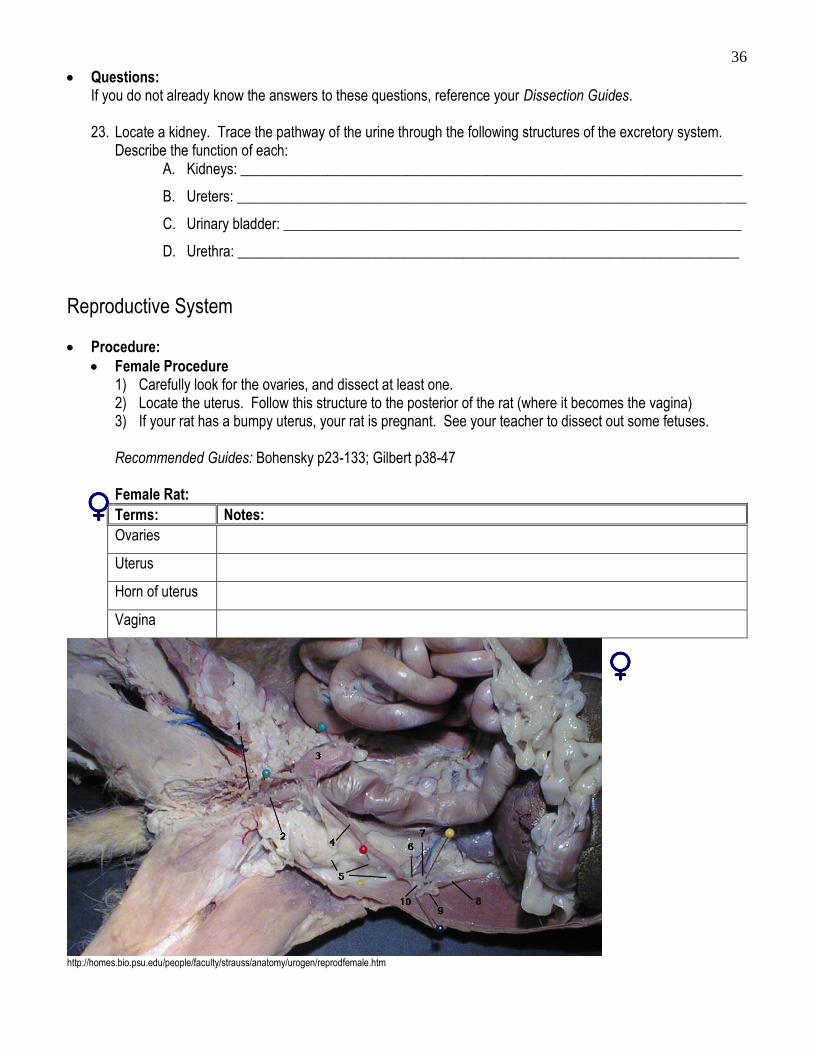
Reproductive System
Procedure:
Female Procedure 1) Carefully look for the ovaries, and dissect at least one. 2) Locate the uterus. Follow this structure to the posterior of the rat (where it becomes the vagina) 3) If your rat has a bumpy uterus, your rat is pregnant. See your teacher to dissect out some fetuses. Recommended Guides: Bohensky p23-133; Gilbert p38-47
Female Rat:
Terms: Notes:
Ovaries
Uterus
Horn of uterus
Vagina
http://homes.bio.psu.edu/people/faculty/strauss/anatomy/urogen/reprodfemale.htm

37
Male Procedure 4) If necessary, cut additional tissue near the pelvis so that the pelvic bones can separate and the legs can
lay out flat. 5) Remember that some of the male parts lie outside of the abdominal cavity! 6) Make an incision along the edge of the scrotum, and hinge open the flap of skin. 7) Use a probe to remove the testes from the scrotum (so that they sit outside of the scrotal sac). 8) Locate the epididymis (directly atop <anterior> to the testes). 9) Look back into the abdominal cavity to find the seminal vesicles.
Recommended Guides: Bohensky p123-133; Gilbert p38-47 Male Rat:
Terms: Notes:
Scrotum
Testes
Epididymis
Seminal vesicles
Penis
http://homes.bio.psu.edu/people/faculty/strauss/anatomy/urogen/reprodmale.htm
Questions: If you do not already know the answers to these questions, reference your Dissection Guides. 24. Male: Cut open one of the scrotal sacs so that you can see the testis and epididymis. What is the
function(s) of each of those two structures? ____________________________________________________________________________________________________________________________________________________________________________

38
25. Female: Locate the uterus and the two ovaries attached to it. Cut open the uterus to check for embryos. Notice its large “V” shape. Why is the cat’s uterus such a different shape than a human’s pear-shaped uterus? (Hint: Think about your answer to #5.) ____________________________________________________________________________________________________________________________________________________________________________
Nervous System
Procedure: This is one of the most difficult areas to dissect in the cat! You are not required to attempt this part of the dissection, BUT you are required to view these parts of the central nervous system on someone’s specimen in class, if not your own.
PATIENCE is required for this part of the dissection. Please see your teacher for assistance before starting!
1) Place the specimen on the dissecting tray so that you are looking down at its dorsal side. 2) Find the base of the neck, where the bottom dorsal part of the cranium meets the backbone. 3) Use a scalpel or dissecting scissors to remove as much of the skin and muscle tissue as possible from
the cranium. 4) Carefully use the tip of the dissecting instrument and bone cutter to slowly chip away small portions of
the bone. Caution: The skull sits directly on top of the brain tissue! It is important to not ‘dig’ at the bone. This may damage the brain tissue.
5) Continue until most of the dorsal and posterior portions of the brain can be seen.
Identification and Diagram Labeling: You are responsible for knowing the location of the following terms. Label them on the diagram below and/or describe their location in the notes section of the table. Recommended Guides: Bohensky p136-156; Gilbert p62-75
Terms: Notes:
Cerebrum
Cerebellum
Medulla
Brain stem
Vegas nerve
http://bio.bd.psu.edu/cat/nervous_system/index.htm
39
Questions: If you do not already know the answers to these questions, reference your Dissection Guides. 26. What is the function of the cerebrum’? __________________________________________________________________________________
__________________________________________________________________________________
27. What is the function of the cerebellum’? __________________________________________________________________________________
__________________________________________________________________________________
28. Where is the medulla oblongata located, and what is its function? __________________________________________________________________________________
__________________________________________________________________________________
Review Websites Google "cat dissection" websites to help you study for your lab practical. Remember, you are
responsible for knowing all of the structures that you identified during the dissection. Any questions that you answered are also fair game!
Related Documents