RESEARCH ARTICLE Open Access A gallotannin-rich fraction from Caesalpinia spinosa (Molina) Kuntze displays cytotoxic activity and raises sensitivity to doxorubicin in a leukemia cell line Diana M Castañeda 1† , Luis Miguel Pombo 2† , Claudia Patricia Urueña 1 , John Fredy Hernandez 1 and Susana Fiorentino 1* Abstract Background: Enhancement of tumor cell sensitivity may help facilitate a reduction in drug dosage using conventional chemotherapies. Consequently, it is worthwhile to search for adjuvants with the potential of increasing chemotherapeutic drug effectiveness and improving patient quality of life. Natural products are a very good source of such adjuvants. Methods: The biological activity of a fraction enriched in hydrolysable polyphenols (P2Et) obtained from Caesalpinia spinosa was evaluated using the hematopoietic cell line K562. This fraction was tested alone or in combination with the conventional chemotherapeutic drugs doxorubicin, vincristine, etoposide, camptothecin and taxol. The parameters evaluated were mitochondrial depolarization, caspase 3 activation, chromatin condensation and clonogenic activity. Results: We found that the P2Et fraction induced mitochondrial depolarization, activated caspase 3, induced chromatin condensation and decreased the clonogenic capacity of the K562 cell line. When the P2Et fraction was used in combination with chemotherapeutic drugs at sub-lethal concentrations, a fourfold reduction in doxorubicin inhibitory concentration 50 (IC 50 ) was seen in the K562 cell line. This finding suggested that P2Et fraction activity is specific for the molecular target of doxorubicin. Conclusions: Our results suggest that a natural fraction extracted from Caesalpinia spinosa in combination with conventional chemotherapy in combination with natural products on leukemia cells may increase therapeutic effectiveness in relation to leukemia. Keywords: Adjuvants, Gallotannins, C.spinosa, Tumor, Leukemia Background Caesalpinia spinosa is a shrub commonly named divi- divi. It is acknowledged to have antimicrobial and anti- oxidant activity, and is traditionally known for its antitumor activity [1]. An ethanol extract from the fruit of C.spinosa has been proven to have antimicrobial activity against gram-positive and gram-negative bac- teria, probably due to the presence of hydrolysable tan- nins in the fruits [2]. Hydrolysable tannins are a group of gallic acid esters associated with polyols (glucose, glu- citol, shikimic acid, quinic acid and quercitol, among others), where the galloyl groups can be further cross- linked by etherification or oxidation to form complex structures. The gallotannins (gallic acid esters) are the simplest hydrolysable tannins, were 1,2,3,4,6-penta-O- galloyl-b-D-glucose (pentagalloyl glucose [PGG]) is the prototype and central compound of the biosynthetic pathway [3]. The presence of PGG, as well as * Correspondence: [email protected] † Contributed equally 1 Grupo de Inmunobiología y Biología Celular, Facultad de Ciencias, Pontificia Universidad Javeriana, Bogotá, Colombia, Carrera 7 N. 43-82 Building 52, Office 608 Full list of author information is available at the end of the article Castañeda et al. BMC Complementary and Alternative Medicine 2012, 12:38 http://www.biomedcentral.com/1472-6882/12/38 © 2012 Castañeda et al; licensee BioMed Central Ltd. This is an Open Access article distributed under the terms of the Creative Commons Attribution License (http://creativecommons.org/licenses/by/2.0), which permits unrestricted use, distribution, and reproduction in any medium, provided the original work is properly cited.

Welcome message from author
This document is posted to help you gain knowledge. Please leave a comment to let me know what you think about it! Share it to your friends and learn new things together.
Transcript
RESEARCH ARTICLE Open Access
A gallotannin-rich fraction from Caesalpiniaspinosa (Molina) Kuntze displays cytotoxic activityand raises sensitivity to doxorubicin in a leukemiacell lineDiana M Castañeda1†, Luis Miguel Pombo2†, Claudia Patricia Urueña1, John Fredy Hernandez1 andSusana Fiorentino1*
Abstract
Background: Enhancement of tumor cell sensitivity may help facilitate a reduction in drug dosage usingconventional chemotherapies. Consequently, it is worthwhile to search for adjuvants with the potential ofincreasing chemotherapeutic drug effectiveness and improving patient quality of life. Natural products are a verygood source of such adjuvants.
Methods: The biological activity of a fraction enriched in hydrolysable polyphenols (P2Et) obtained fromCaesalpinia spinosa was evaluated using the hematopoietic cell line K562. This fraction was tested alone or incombination with the conventional chemotherapeutic drugs doxorubicin, vincristine, etoposide, camptothecin andtaxol. The parameters evaluated were mitochondrial depolarization, caspase 3 activation, chromatin condensationand clonogenic activity.
Results: We found that the P2Et fraction induced mitochondrial depolarization, activated caspase 3, inducedchromatin condensation and decreased the clonogenic capacity of the K562 cell line. When the P2Et fraction wasused in combination with chemotherapeutic drugs at sub-lethal concentrations, a fourfold reduction in doxorubicininhibitory concentration 50 (IC50) was seen in the K562 cell line. This finding suggested that P2Et fraction activity isspecific for the molecular target of doxorubicin.
Conclusions: Our results suggest that a natural fraction extracted from Caesalpinia spinosa in combination withconventional chemotherapy in combination with natural products on leukemia cells may increase therapeuticeffectiveness in relation to leukemia.
Keywords: Adjuvants, Gallotannins, C.spinosa, Tumor, Leukemia
BackgroundCaesalpinia spinosa is a shrub commonly named divi-divi. It is acknowledged to have antimicrobial and anti-oxidant activity, and is traditionally known for itsantitumor activity [1]. An ethanol extract from the fruitof C.spinosa has been proven to have antimicrobial
activity against gram-positive and gram-negative bac-teria, probably due to the presence of hydrolysable tan-nins in the fruits [2]. Hydrolysable tannins are a groupof gallic acid esters associated with polyols (glucose, glu-citol, shikimic acid, quinic acid and quercitol, amongothers), where the galloyl groups can be further cross-linked by etherification or oxidation to form complexstructures. The gallotannins (gallic acid esters) are thesimplest hydrolysable tannins, were 1,2,3,4,6-penta-O-galloyl-b-D-glucose (pentagalloyl glucose [PGG]) is theprototype and central compound of the biosyntheticpathway [3]. The presence of PGG, as well as
* Correspondence: [email protected]† Contributed equally1Grupo de Inmunobiología y Biología Celular, Facultad de Ciencias, PontificiaUniversidad Javeriana, Bogotá, Colombia, Carrera 7 N. 43-82 Building 52,Office 608Full list of author information is available at the end of the article
Castañeda et al. BMC Complementary and Alternative Medicine 2012, 12:38http://www.biomedcentral.com/1472-6882/12/38
© 2012 Castañeda et al; licensee BioMed Central Ltd. This is an Open Access article distributed under the terms of the CreativeCommons Attribution License (http://creativecommons.org/licenses/by/2.0), which permits unrestricted use, distribution, andreproduction in any medium, provided the original work is properly cited.
gallotannins as mono, di or tri-galloylquinic acids, havebeen reported in Caesalpinia species corresponding to40% to 60% of the fruit composition, depending upontheir ecological habitat [4].Gallic acid and its derivatives have proven selective
antitumor activities, such as: reduction in biochemicalmarkers associated with skin cancer [5]; cell deathinduction in several cancer cell lines, including leukemia[6,7], murine myeloma [8] and squamous carcinoma [9].In addition, a beverage containing epigallocatechin gal-late (EGCG) has been reported to promote tumorregression in patients with low-grade lymphomas [10].Galloylquinic derivatives, such as 4,5-di-O-galloylquinicacid, have shown moderate cytotoxicity against mela-noma cells (RPMI-7951) but not in other cell lines [11].Extensive studies have been carried out on PGG andhave demonstrated that this compound has a numberbiological activities related to cancer therapy and pre-vention, such as antiangiogenic, antiproliferative, anti-inflammatory and antioxidant [3]. However, there arelimited studies supporting the use of polyphenols in thetreatment of hematological malignancies, and evenfewer involving hydrolysable polyphenols. In the presentstudy, taking into account that polyphenol antioxidantactivity has been clearly implicated in the control ofthese malignancies [12], and that C.spinosa has a highcontent of polyphenols and is widely distributed in ourcountry, we have evaluated the anti-tumor activity of C.spinosa pod extracts and complex fractions using theerythroleukemia cell line (K652) as a model of hemato-logical malignancy.
MethodsPlant materialC.spinosa pods were collected in Villa de Leyva, Boyacá,Colombia in March 2007 and identified by Luis CarlosJiménez from the Colombian National Herbarium; vou-cher specimen number COL 523714.
Plant extraction and purificationThree kg of fresh pods from C.spinosa were dried underairflow in a solar oven at 35°C and ground down toobtain 1.8 kg of plant material. Subsequently the plantmaterial was extracted with ethanol (96%, 10 L) in arecirculating percolator (twice per day) over a period of10 days. The ethanol crude extract (80 g) was concen-trated under vacuum, trapped on silica gel and excesshumidity removed at 25°C. Afterwards, the ethanolextract was fractionated with the following solvents: pet-roleum ether (1.5 L); chloroform (2 L); ethyl acetate (2L); ethanol (2 L) and water (2 L) (aqueous fraction).From the ethyl acetate fraction we obtained an abundantprecipitate which we named (P2Et). This correspondedto 2.78% of the ethanol extract and a supernatant which
we named (S2Et) corresponded to 1.11%. The P2Et,S2Et and aqueous fractions were selected for biologicaltesting based on their cytotoxic activity. The extractionprotocol was performed three times and the chromato-graphic profiles of the components were verified.The quality control carried out on the P2Et fraction
gave the following results: foreign matter less than 2%;total ash less than 8%; ash that was insoluble in hydro-chloric acid less than 1%; no evidence of heavy metalsand pesticides. These result met the British Herbal Phar-macopoeia quality parameters.
Phytochemical characterizationFraction characterization was determined by means ofstandard phytochemical tests. In the total ethanolextract the presence of alkaloids or nitrogen compoundswere not identified using Dragenddorff, Valser, Reineck-ate and Mayer’s reagents. The Shinoda test (Mg in HCl)was positive suggesting the presence of flavanones, flava-nonols, flavones, flavonols or isoflavones. Hydrogen per-oxide evidenced the presence of naphthoquinones and/or anthraquinones. The presence of steroids wasdemonstrated using Liebermann Burchard reagent. Lowconcentrations of steroidal saponins and/or triterpenoidswere detected using hemolysis and foam tests. In orderto assess the presence of anthraquinone glycosides,Borntrager’s reaction (treatment with ammonia solution)was used. The presence of tannins was verified usingferric chloride solution, gelatin and lead acetate [13].P2Et, S2Et and aqueous fractions exhibited the presenceof leucoanthocyanidins, the absence of quinones and asignificant tannin content, especially in P2Et fraction.
Thin layer chromatography (TLC)Chromatographic analysis was carried out on TLC alu-minum sheets (10 × 5 cm) (Merck) silica gel 60 F 254.Three solvent systems were used: Petroleum ether -ethyl acetate - formic acid (40:60:1); chloroform - ethylacetate - acetic acid (50:50:1); and toluene - acetonitrile- formic acid (70:30:1). After basic hydrolysis, the P2Etand S2Et fractions were dissolved in methanol (1%) anddetected using UV (254 nm), FeCl3 (10%) and vanillin-sulfuric acid (VS)/110°C. Gallic acid was used as a posi-tive control.
HPLC - PDA-MSHPLC analysis was carried out in an Alliance 2795(Waters®, UK) with a PDA detector (996). A Sunfire(Waters) column C18 - 2.1 × 150 mm × 5 μm was used,with a flow rate of 0.25 ml/min and a linear gradientfrom 95% solvent A (H2O + 1%CH3COOH) and 5% sol-vent B (CH3CN) to 60% in solvent A and 40% in solventB, over a period of 25 min. The mass spectrum (MS)analysis was carried out using a LCT (Micromass®, UK)
Castañeda et al. BMC Complementary and Alternative Medicine 2012, 12:38http://www.biomedcentral.com/1472-6882/12/38
Page 2 of 10
mass spectrometer with an ESI source. The percentagerelative abundance was determined using quercetin asan internal standard (0.0625 μg/μl). Runs were per-formed in triplicate.
Tumor cell line and normal cellsThe cell lines used as cancer cells were K562, a humanerythroleukemia and MCF7, a human breast adenocarci-noma, from the American Type Culture Collection(ATCC); and the cell lines used as normal cells werehuman peripheral blood mononuclear cells (PBMC) andhuman fibroblasts obtained from normal healthy donorsafter informed consent was given. This project wasapproved by the ethics committee (founded in 2002) ofthe Science Faculty at a meeting on August 21, 2007.The culture conditions under which the cell lines weremaintained have already been reported [14].
In vitro cytotoxicity assaysThe cytotoxic effects of the fractions and conventionalchemotherapeutic drugs (doxorubicin, etoposide, vincris-tine and taxol) were evaluated using normal and tumorcells by means of trypan blue and the methylthiazol tet-razolium (MTT) assay, as previously reported [14]. TheP2Et fraction was dissolve in ethanol and the corre-sponding vehicle was used as a negative control.
Measurement of mitochondrial membrane potentialThe cells were treated with different concentrations ofthe P2Et fraction or valinomycin (positive control, 0.1μg/ml) for 4, 8 and 12 h for K562 cells, and for 6, 12and 24 h for MCF7 cells. The mitochondrial membranepotential (MMP) was measured using JC-1 dye, as pre-viously described [14].
Annexin V assayPhosphatidylserine (PS) externalization was assessed byflow cytometry using Annexin V-FITC (MolecularProbes, Invitrogen Corp, Carlsbad, CA, USA)/PI (Sigma,Saint Louis, MO, USA). K562 and MCF7 cells (3 × 10⁵)were treated with doxorubicin, ethanol or the P2Et frac-tion for 48 h. After treatment, cells were suspended inAnnexin buffer (Hepes 100 mM, NaCl 140 mM, CaCl22.5 mM) and incubated with Annexin V-FITC for 8 minat room temperature. Then the cells were incubatedwith PI for 2 min at 4°C, acquired on a FACSAria I(Becton Dickinson, New Jersey, USA) and analyzed withFlowJo software (Tree Star Inc., Ashland, USA). Resultsare expressed as the mean ± SE of three independentexperiments.
Caspase 3 assaysCaspase 3 activity was estimated using the caspase 3 col-orimetric assay kit, which detects enzyme activity based
on the cleavage of Asp-Glu-Val-Asp (DEVD)-pNA(R&D Systems Inc., Minneapolis, MN, USA). Briefly,cells (2 × 10⁵ cells/ml) were cultured using differentconcentrations of the P2Et fraction and doxorubicin(positive control) or ethanol (negative control) for 48 h.After the cells were ice lysed for 10 min the enzymeactivity was measure on 96-well flat-bottom microplateswith 50 μl of supernatant. The supernatant was pre-pared by centrifuging at 10,000 × g for 1 min (100-200μg of total protein), and then adding 50 μl of reactionbuffer supplemented with 10 μl of DTT and 5 μl of cas-pase 3 colorimetric substrate DEVD-pNA. Next cellswere incubated for 1 ± 2 h at 37°C and caspase-3 activ-ity was measured at 405 nm on a spectrophotometer(Multiskan Labsystem). The increase in caspase 3 activ-ity was calculated relative to the absorbance value of thenegative control.
DNA fragmentation and cell cycle analysisDAPI (4’,6-diamidino-2-phenylindole, Sigma) stainedcells were monitored under a microscope as previouslydescribed [14]. Slides were mounted using prolong anti-fade kit (Molecular Probes, Eugene, Oregon, USA) andcells were analyzed under a fluorescence microscope(Olympus, Japan). Cell cycle analysis was undertaken aspreviously reported [14].
Clonogenic assaysThe clonogenic assays were performed as previouslydescribed [14]. Briefly, K562 human cells (2.5 × 10⁵
cells/well) were plated (96-well plate) and treated withthe P2Et fraction at 40 and 20 μg/ml, or with 15 and 6μg/ml etoposide, or 0.2% ethanol (in PBS) and incubatedfor 24 h under a humidified environment at 37°C and5% CO2. After treatment cells were re-plated onto 0.5%agar dishes (60 mm, 20,000 cells/dish), incubated for 14days (37°C and 5% CO2) and stained with violet crystal(0.4% in ethanol). Cell colonies with more than 50 cellswere counted. Treatments were performed in triplicate,and results expressed as mean ± SE.
P2Et fraction adjuvant activityP2Et fraction adjuvant activity was assessed using K562and MCF7 cells in combination with the well-knowncancer treatment drugs doxorubicin, vincristine, taxoland camptothecin. Cell viability was evaluated by meansof the MTT assay. K562 and MCF7 cells (5 × 10³ cells/well) were seeded in 96-well plates and treated for 6 hwith sublethal concentrations of the P2Et fraction (1.6μg/ml, 27 fold less than the IC50 value for K562 cellsand 15.5 μg/ml for MCF7 cells); washed and incubatedin fresh medium with each drug for 48 h at 37°C inhumid atmosphere and 5% CO2. Sublethal concentra-tions of the chemotherapeutic drugs had been previously
Castañeda et al. BMC Complementary and Alternative Medicine 2012, 12:38http://www.biomedcentral.com/1472-6882/12/38
Page 3 of 10
determined by MTT assay. Results are expressed as cellviability percentage relative to the control (100 × Treat-ment OD/Negative control OD).
Statistical analysisData is presented as the mean ± SE. The data were ana-lyzed by one- and two-way ANOVA and differencesbetween control and treated groups were determinedusing the Bonferroni and Tukey tests. Differences wereconsidered significant for p < 0.05 and were determinedusing the GraphPad prism 5.0 software.
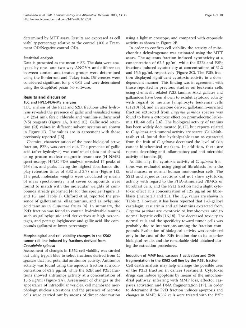
Results and discussionTLC and HPLC-PDA-MS analysesTLC analysis of the P2Et and S2Et fractions after hydro-lysis revealed the presence of gallic acid visualized usingUV (254 nm), ferric chloride and vainillin-sulfuric acid(VS) reagents (Figure 1A, B and 1C). Gallic acid reten-tion (Rf) values in different solvent systems are shownin Figure 1D. The values are in agreement with thosepreviously reported [15].Chemical characterization of the most biological active
fraction, P2Et, was carried out. The presence of gallicacid (after hydrolysis) was confirmed (data not shown)using proton nuclear magnetic resonance (H-NMR)spectroscopy. HPLC-PDA analysis revealed 17 peaks at263 nm, and peaks having the highest absorbance dis-play retention times of 3.32 and 3.78 min (Figure 1E).The peak molecular weights were calculated by meansof mass spectrometry, and seven compounds werefound to match with the molecular weights of com-pounds already published [4] for this species (Figure 1Fand 1G, and Table 1). Clifford et al. reported the pre-sence of gallotannins, ellagitannins, and galloylquinicacid tannins in C.spinosa fruits [4]. In summary, theP2Et fraction was found to contain hydrolysable tanninssuch as galloylquinic acid derivatives at high percen-tages, and pentagalloylglucose and gallic acid-like com-pounds (gallates) at lower percentages.
Morphological and cell viability changes in the K562tumor cell line induced by fractions derived fromCaesalpinia spinosaAssessment of changes in K562 cell viability was carriedout using trypan blue to select fractions derived from C.spinosa that had potential antitumor activity. Antitumoractivity was found using the aqueous fraction at a con-centration of 62.5 μg/ml, while the S2Et and P2Et frac-tions showed antitumor activity at a concentration of15.6 μg/ml (Figure 2A). Assessment of changes in theappearance of intracellular vesicles, cell membrane mor-phology, nuclear alterations and the presence of necroticcells were carried out by means of direct observation
using a light microscope, and compared with etoposideactivity as shown in Figure 2B.In order to confirm cell viability the activity of mito-
chondria dehydrogenase was estimated using the MTTassay. The aqueous fraction induced cytotoxicity at aconcentration of 62.5 μg/ml, while the S2Et and P2Etfractions induced cytotoxicity at concentrations of 31.2and 15.6 μg/ml, respectively (Figure 2C). The P2Et frac-tion displayed significant cytotoxic activity in a dose-dependent manner. This finding was in agreement withthose reported in previous studies on leukemia cellsusing chemically related P2Et tannins. Alkyl gallates andgallamides have been shown to exhibit cytotoxic activitywith regard to murine lymphocyte leukemia cells(L1210) [6], and an acetone derived gallotannin-enrichedfraction extracted from Eugenia jambos species wasfound to have a cytotoxic effect on promyelocytic leuke-mia HL-60 cells [16]. The biological activity of tanninshas been widely documented [6,17], but reports relatedto C. spinosa anti-tumoral activity are scarce. Gali-Muh-tasib et al. found that hydrolysable tannins extractedfrom the fruit of C. spinosa decreased the level of skincancer biochemical markers. In addition, there arereports describing anti-inflammatory and anti-microbialactivity of tannins [5].Additionally, the cytotoxic activity of C. spinosa frac-
tions was evaluated using gingival fibroblasts from theoral mucosa or normal human mononuclear cells. TheS2Et and aqueous fractions did not show cytotoxicactivity with regard to the PHA-stimulated PBMCs orfibroblast cells, and the P2Et fraction had a slight cyto-toxic effect at a concentration of 125 μg/ml on fibro-blasts (Figure 2D and 2E). The IC50 values are shown inTable 2. However, it has been reported that 1-O-galloylcastalagin, casuarinin and gallotannins extracted fromEugenia jambos are cytotoxic to lymphocytes and tonormal hepatic cells [16,18]. The decreased toxicity tonormal cells and the specificity toward tumor cells wasprobably due to interactions among the fraction com-pounds. Evaluation of biological activity was continuedonly in the case of the P2Et fraction due to its superiorbiological results and the remarkable yield obtained dur-ing the extraction procedures.
Induction of MMP loss, caspase 3 activation and DNAfragmentation in the K562 cell line by the P2Et fractionCell death analysis may help envisage the possible usesof the P2Et fraction in cancer treatment. Cytotoxicdrugs can induce apoptosis by means of the mitochon-drial pathway, inferring with MMP loss, effector cas-pases activation and DNA fragmentation [19]. In orderto determine if the P2Et fraction induces apoptosis andchanges in MMP, K562 cells were treated with the P2Et
Castañeda et al. BMC Complementary and Alternative Medicine 2012, 12:38http://www.biomedcentral.com/1472-6882/12/38
Page 4 of 10
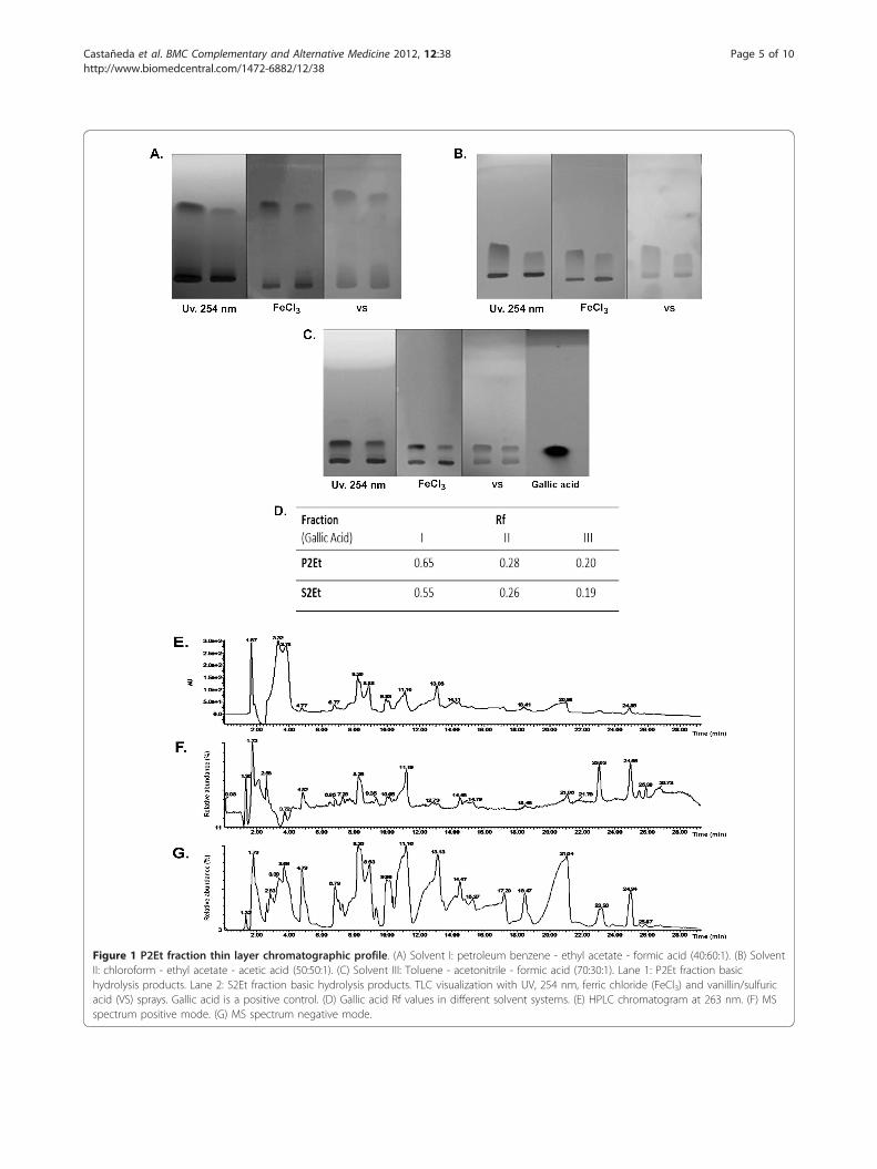
Figure 1 P2Et fraction thin layer chromatographic profile. (A) Solvent I: petroleum benzene - ethyl acetate - formic acid (40:60:1). (B) SolventII: chloroform - ethyl acetate - acetic acid (50:50:1). (C) Solvent III: Toluene - acetonitrile - formic acid (70:30:1). Lane 1: P2Et fraction basichydrolysis products. Lane 2: S2Et fraction basic hydrolysis products. TLC visualization with UV, 254 nm, ferric chloride (FeCl3) and vanillin/sulfuricacid (VS) sprays. Gallic acid is a positive control. (D) Gallic acid Rf values in different solvent systems. (E) HPLC chromatogram at 263 nm. (F) MSspectrum positive mode. (G) MS spectrum negative mode.
Castañeda et al. BMC Complementary and Alternative Medicine 2012, 12:38http://www.biomedcentral.com/1472-6882/12/38
Page 5 of 10
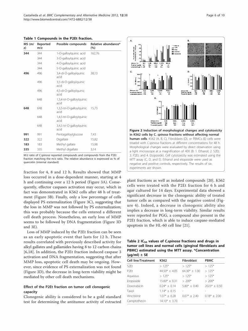
fraction for 4, 8 and 12 h. Results showed that MMPloss occurred in a dose-dependent manner, starting at 4h and continuing over a 12 h period (Figure 3A). Conse-quently, effector caspases activation may occur, which infact was demonstrated in K562 cells after 48 h of treat-ment (Figure 3B). Finally, only a low percentage of cellsdisplayed PS externalization (Figure 3C), suggesting thatthe loss in MMP was not followed by PS externalization;this was probably because the cells entered a differentcell death process. Nonetheless, an early loss of MMPseems to be followed by DNA fragmentation (Figure 3Dand 3E).Loss of MMP induced by the P2Et fraction can be seen
as an early apoptotic event that lasts for 12 h. Theseresults correlated with previously described activity foralkyl gallates and gallamides having 8 to 12 carbon chains[6,18]. In addition, the P2Et fraction induced caspase 3activation and DNA fragmentation, suggesting that afterMMP loss, apoptotic cell death may be ongoing. How-ever, since evidence of PS externalization was not found(Figure 3D), the decrease in long-term viability might bemediated by other cell death mechanisms.
Effect of the P2Et fraction on tumor cell clonogeniccapacityClonogenic ability is considered to be a gold standardtest for determining the antitumor activity of extracted
plant fractions as well as isolated compounds [20]. K562cells were treated with the P2Et fraction for 6 h andagar cultured for 14 days. Experimental data showed asignificant decrease in the clonogenic ability of treatedtumor cells as compared with the negative control (Fig-ure 4). Indeed, a decrease in clonogenic ability alsoimplies a decrease in long-term viability. Similar resultswere reported for PGG, a compound also present in theP2Et fraction, which is able to induce caspase-mediatedapoptosis in the HL-60 cell line [21].
Table 1 Compounds in the P2Et fraction.
MS (m/z)
Reportedm/z
Possible compounds Relative abundance*(%)
344 344 1-O-galloylquinic acid 162,76
344 3-O-galloylquinic acid
344 4-O-galloylquinic acid
344 5-O-galloylquinic acid
496 496 3,4-di-O-galloylquinicacid
38,13
496 3,5-di-O-galloylquinicacid
496 4,5-di-O-galloylquinicacid
648 1,3,4-tri-O-galloylquinicacid
648 648 1,3,5-tri-O-galloylquinicacid
15,73
648 1,4,5-tri-O-galloylquinicacid
648 3,4,5-tri-O-galloylquinicacid
991 991 Pentagalloylglucose 7,43
322 322 Digallate 15,82
183 183 Methyl gallate 15,98
335 335 Methyl digallate 3,14
M/z ratio of C.spinosa reported compounds and compounds from the P2Etfraction matching the m/z ratio. The relative abundance is expressed as % ofquercetin (internal standard)
Figure 2 Induction of morphological changes and cytotoxicityin K562 cells by C. spinosa fractions without affecting normalhuman cells. K562 (A, B, C), Fibroblasts (D), or PBMCs (E) cells weretreated with C.spinosa fractions at different concentrations for 48 h.Morphological changes were evaluated by direct observation usinga light microscope at a magnification of 40X (B: 1. Ethanol; 2. S2Et;3. P2Et; and 4. Etoposide). Cell cytotoxicity was estimated using theMTT assay (C, D, and E). Ethanol and etoposide were used asnegative and positive controls, respectively. The results of sixexperiments are shown.
Table 2 IC50 values of C.spinosa fractions and drugs intumor cell lines and normal cells (gingival fibroblasts andPBMC) estimated using the MTT assay. *Concentration(μg/ml) ± SE
Cell line/Treatment K562 Fibroblast PBMC
S2Et > 125* > 125* > 125*
P2Et 44.50* ± 4.05 64.30* ± 1.00 > 125*
Aqueous > 125* > 125* > 125*
Etoposide 15.60* ± 0.31 > 200* > 200*
Doxorubicin 0.24* ± 0.14 5.80* ± 0.40 2023* ± 0.50
Taxol 1.14* ± 0.15 - -
Vincristine 1.07* ± 0.28 0.07* ± 2.40 0.18* ± 2.00
Camptothecin 14.10* ± 3.70 - -
Castañeda et al. BMC Complementary and Alternative Medicine 2012, 12:38http://www.biomedcentral.com/1472-6882/12/38
Page 6 of 10
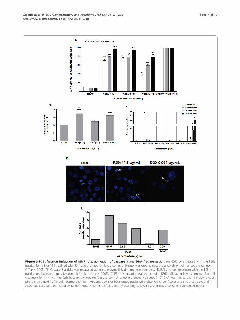
Figure 3 P2Et fraction induction of MMP loss, activation of caspase 3 and DNA fragmentation. (A) K562 cells treated with the P2Etfraction for 4, 8 or 12 h, stained with JC-1 and analyzed by flow cytometry. Ethanol was used as negative and valinomycin as positive controls(*** p < 0.001). (B) Caspase 3 activity was measured using the enzyme-linked immunosorbent assay (ELISA) after cell treatment with the P2Etfraction or doxorubicin (positive control) for 48 h (** p < 0.005). (C) PS externalization was estimated in K562 cells using flow cytometry after celltreatment for 48 h with the P2Et fraction, doxorubicin (positive control) or ethanol (negative control). (D) DNA was stained with 4’,6-diamidino-2-phenylindole (DAPI) after cell treatment for 48 h. Apoptotic cells or fragmented nuclei were observed under fluorescent microscope (40X). (E)Apoptotic cells were estimated by random observation in six fields and by counting cells with strong fluorescence or fragmented nuclei.
Castañeda et al. BMC Complementary and Alternative Medicine 2012, 12:38http://www.biomedcentral.com/1472-6882/12/38
Page 7 of 10
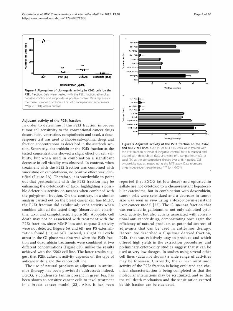
Adjuvant activity of the P2Et fractionIn order to determine if the P2Et fraction improvestumor cell sensitivity to the conventional cancer drugsdoxorubicin, vincristine, camptothecin and taxol, a dose-response test was used to choose sub-optimal drugs andfraction concentrations as described in the Methods sec-tion. Separately, doxorubicin or the P2Et fraction at thetested concentrations showed a slight effect on cell via-bility, but when used in combination a significantdecrease in cell viability was observed. In contrast, whentreatment with the P2Et fraction was combined withvincristine or camptothecin, no positive effect was iden-tified (Figure 5A). Therefore, it is worthwhile to pointout that pretreatment with the P2Et fraction may beenhancing the cytotoxicity of taxol, highlighting a possi-ble deleterious activity on taxanes when combined withthe polyphenol fraction. On the contrary, in a similaranalysis carried out on the breast cancer cell line MCF7,the P2Et fraction did exhibit adjuvant activity whencombine with all the tested drugs (doxorubicin, vincris-tine, taxol and camptothecin, Figure 5B). Apoptotic celldeath may not be associated with treatment with theP2Et fraction, since MMP loss and caspase 3 activitywere not detected (Figure 6A and 6B) nor PS externali-zation found (Figure 6C). Instead, a slight cell cyclearrest in the G1 phase was observed when the P2Et frac-tion and doxorubicin treatments were combined at twodifferent concentrations (Figure 6D), unlike the resultsachieved with the K562 cell line. The latter results sug-gest that P2Et adjuvant activity depends on the type ofanticancer drug and the cancer cell line.The use of natural products as adjuvants in antitu-
mor therapy has been previously addressed; indeed,EGCG, a condensate tannin present in green tea, hasbeen shown to sensitize cancer cells to taxol treatmentin a breast cancer model [22]. Also, it has been
reported that EGCG (at low doses) and epicatechingallate are not cytotoxic to a chemoresistant hepatocel-lular carcinoma, but in combination with doxorubicin,tumor cells were sensitized and a decrease in tumorsize was seen in vivo using a doxorubicin-resistantliver cancer model [23]. The C. spinosa fraction thatwas enriched in gallotannins not only exhibited cyto-toxic activity, but also activity associated with conven-tional anti-cancer drugs, demonstrating once again theefficiency of natural products as potential sources ofadjuvants that can be used in antitumor therapy.Herein, we described a C.spinosa derived fraction,P2Et, that was relatively easy to produce and whichoffered high yields in the extraction procedures; andpreliminary cytotoxicity studies suggest that it can beused at very low dosages. In studies using several othercell lines (data not shown) a wide range of activitiesmay be foreseen. Currently, the in vivo antitumoractivity of the P2Et fraction is being evaluated and che-mical characterization is being completed so that themolecular interactions may be scrutinized; and so thatthe cell death mechanism and the sensitization exertedby this fraction can be elucidated.
Figure 4 Abrogation of clonogenic activity in K562 cells by theP2Et fraction. Cells were treated with the P2Et fraction, ethanol asnegative control and etoposide as positive control. Data representsthe mean number of colonies ± SE of 3 independent experiments.***(p < 0.001) versus control.
Figure 5 Adjuvant activity of the P2Et fraction on the K562and MCF7 cell lines. K562 (A) or MCF7 (B) cells were treated withthe P2Et fraction or ethanol (negative control) for 6 h, washed andtreated with doxorubicin (Dx), vincristine (Vt), camptothecin (Ct) ortaxol (Tx) at the concentrations shown over a 48 h period. Cellcytotoxicity was estimated using the MTT assay. Data representthree independent experiments. *** (p < 0.001).
Castañeda et al. BMC Complementary and Alternative Medicine 2012, 12:38http://www.biomedcentral.com/1472-6882/12/38
Page 8 of 10
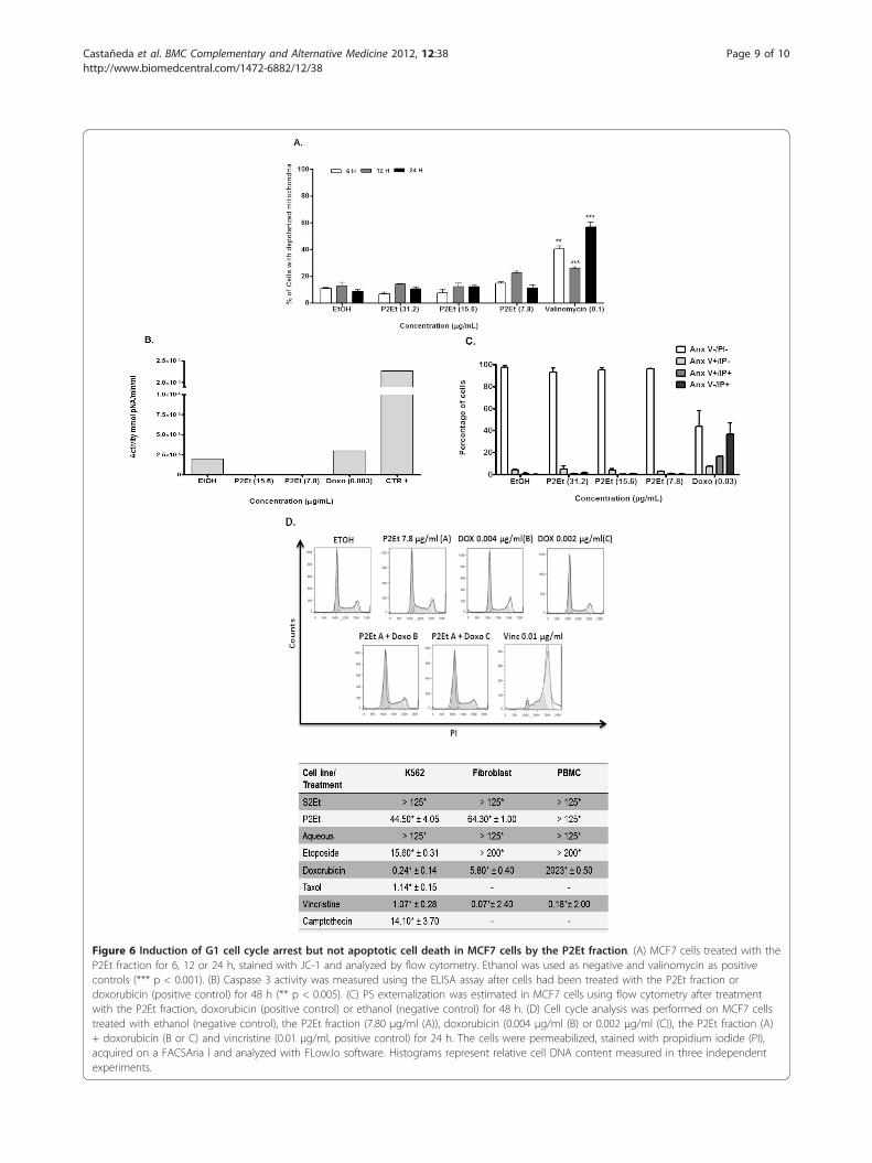
Figure 6 Induction of G1 cell cycle arrest but not apoptotic cell death in MCF7 cells by the P2Et fraction. (A) MCF7 cells treated with theP2Et fraction for 6, 12 or 24 h, stained with JC-1 and analyzed by flow cytometry. Ethanol was used as negative and valinomycin as positivecontrols (*** p < 0.001). (B) Caspase 3 activity was measured using the ELISA assay after cells had been treated with the P2Et fraction ordoxorubicin (positive control) for 48 h (** p < 0.005). (C) PS externalization was estimated in MCF7 cells using flow cytometry after treatmentwith the P2Et fraction, doxorubicin (positive control) or ethanol (negative control) for 48 h. (D) Cell cycle analysis was performed on MCF7 cellstreated with ethanol (negative control), the P2Et fraction (7.80 μg/ml (A)), doxorubicin (0.004 μg/ml (B) or 0.002 μg/ml (C)), the P2Et fraction (A)+ doxorubicin (B or C) and vincristine (0.01 μg/ml, positive control) for 24 h. The cells were permeabilized, stained with propidium iodide (PI),acquired on a FACSAria I and analyzed with FLowJo software. Histograms represent relative cell DNA content measured in three independentexperiments.
Castañeda et al. BMC Complementary and Alternative Medicine 2012, 12:38http://www.biomedcentral.com/1472-6882/12/38
Page 9 of 10
ConclusionsOur results suggest that the therapeutic efficacy of con-ventional chemotherapeutic drugs when combined witha blend of natural polyphenols may be increased in rela-tion to leukemia or breast cancer cells. However, themolecular mechanisms underlying this activity have yetto be explained and are currently under study at ourlaboratory.
AcknowledgementsThe authors thank the Science Faculty of the Pontificia UniversidadJaveriana, Fundación Universitaria Juan N. Corpas for their support and theInstituto Colombiano para el Desarrollo de la Ciencia y la Tecnología“Francisco Jose de Caldas” (COLCIENCIAS) Bogotá, Colombia, for financialsupport.
Author details1Grupo de Inmunobiología y Biología Celular, Facultad de Ciencias, PontificiaUniversidad Javeriana, Bogotá, Colombia, Carrera 7 N. 43-82 Building 52,Office 608. 2Grupo de Farmacología Vegetal, Fundación Universitaria Juan N.Corpas, Bogotá, Colombia, Carrera 111 # 159A61.
Authors’ contributionsThe present work was conceived, directed and coordinated by SF. In vitrocytotoxicity assays, MMP measurements, Annexin V determinations,clonogenic assays, cell cycle analysis and cell line maintenance wasundertaken by DC. Caspase 3 activity analysis and the DAPI DNAfragmentation test was carried out by CU. Plant fraction preparation,characterizations and statistical analysis were conducted by LMP. JHcollaborated in the writing of the manuscript and analysis of the results. Allof the authors have read the manuscript and agree with its contents.
Competing interestsThe authors declare that they have no competing interests.
Received: 27 September 2011 Accepted: 10 April 2012Published: 10 April 2012
References1. Chanwitheesuk A, Teerawutgulrag A, Kilburn J, Rakariyatham N:
Antimicrobial gallic acid from Caesalpinia mimosoides lamk. Food Chem2007, 100:1044-1048.
2. Kloucek P, Polesny Z, Svobodova B, Vlkova E, Kokoska L: Antibacterialscreening of some Peruvian medicinal plants used in Calleria District. JEthnopharmacol 2005, 99(2):309-312.
3. Zhang J, Li L, Kim SH, Hagerman AE, Lu J: Anti-cancer, anti-diabetic andother pharmacologic and biological activities of penta-galloyl-glucose.Pharm Res 2009, 26(9):2066-2080.
4. Clifford MN, Stoupi S, Kuhnert N: Profiling and characterization by LC-MSnof the galloylquinic acids of green tea, tara tannin, and tannic acid. JAgric Food Chem 2007, 55(8):2797-2807.
5. Gali-Muhtasib HU, Yamout SZ, Sidani MM: Tannins protect against skintumor promotion induced by ultraviolet-B radiation in hairless mice.Nutr Canc 2000, 37(1):73-77.
6. Locatelli C, Rosso R, Santos-Silva MC, de Souza CA, Licinio MA, Leal P,Bazzo ML, Yunes RA, Creczynski-Pasa TB: Ester derivatives of gallic acidwith potential toxicity toward L1210 leukemia cells. Bioorg Med Chem2008, 16(7):3791-3799.
7. Inoue M, Suzuki R, Koide T, Sakaguchi N, Ogihara Y, Yabu Y: Antioxidant,gallic acid, induces apoptosis in HL-60RG cells. Biochem Biophys ResCommun 1994, 204(2):898-904.
8. Pellegrina CD, Padovani G, Mainente F, Zoccatelli G, Bissoli G, Mosconi S,Veneri G, Peruffo A, Andrighetto G, Rizzi C, et al: Anti-tumour potential ofa gallic acid-containing phenolic fraction from Oenothera biennis. CancerLett 2005, 226(1):17-25.
9. Chia YC, Rajbanshi R, Calhoun C, Chiu RH: Anti-neoplastic effects of gallicacid, a major component of Toona sinensis leaf extract, on oralsquamous carcinoma cells. Molecules 2010, 15(11):8377-8389.
10. Shanafelt TD, Lee YK, Call TG, Nowakowski GS, Dingli D, Zent CS, Kay NE:Clinical effects of oral green tea extracts in four patients with low gradeB-cell malignancies. Leuk Res 2006, 30(6):707-712.
11. Kashiwada Y, Nonaka G, Nishioka I, Chang JJ, Lee KH: Antitumor agents,129. Tannins and related compounds as selective cytotoxic agents. J NatProd 1992, 55(8):1033-1043.
12. Kelkel M, Jacob C, Dicato M, Diederich M: Potential of the dietaryantioxidants resveratrol and curcumin in prevention and treatment ofhematologic malignancies. Molecules 2010, 15(10):7035-7074.
13. Chaudhari M, Mengi S: Evaluation of phytoconstituents of Terminaliaarjuna for wound healing activity in rats. Phytother Res 2006,20(9):799-805.
14. Uruena C, Cifuentes C, Castaneda D, Arango A, Kaur P, Asea A, Fiorentino S:Petiveria alliacea extracts uses multiple mechanisms to inhibit growth ofhuman and mouse tumoral cells. BMC Compl Alternative Med 2008, 8:60.
15. Sharma OP, Bhat TK, Singh B: Thin-layer chromatography of gallic acid,methyl gallate, pyrogallol, phloroglucinol, catechol, resorcinol,hydroquinone, catechin, epicatechin, cinnamic acid, p-coumaric acid,ferulic acid and tannic acid. J Chrom A 1998, 822:167-171.
16. Yang LL, Lee CY, Yen KY: Induction of apoptosis by hydrolyzable tanninsfrom Eugenia jambos L. on human leukemia cells. Cancer Lett 2000,157(1):65-75.
17. Kim NS, Jeong SI, Hwang BS, Lee YE, Kang SH, Lee HC, Oh CH: Gallic acidinhibits cell viability and induces apoptosis in human monocytic cell lineU937. J Med Food 2011, 14(3):240-246.
18. Serrano A, Palacios C, Roy G, Cespon C, Villar ML, Nocito M, Gonzalez-Porque P: Derivatives of gallic acid induce apoptosis in tumoral cell linesand inhibit lymphocyte proliferation. Arch Biochem Biophys 1998,350(1):49-54.
19. Kroemer G, Galluzzi L, Vandenabeele P, Abrams J, Alnemri ES, Baehrecke EH,Blagosklonny MV, El-Deiry WS, Golstein P, Green DR, et al: Classification ofcell death: recommendations of the Nomenclature Committee on CellDeath 2009. Cell Death Differ 2009, 16(1):3-11.
20. Chen R, Gandhi V, Plunkett W: A sequential blockade strategy for thedesign of combination therapies to overcome oncogene addiction inchronic myelogenous leukemia. Cancer Res 2006, 66(22):10959-10966.
21. Pan MH, Lin JH, Lin-Shiau SY, Lin JK: Induction of apoptosis by penta-O-galloyl-beta-D-glucose through activation of caspase-3 in humanleukemia HL-60 cells. Eur J Pharmacol 1999, 381(2-3):171-183.
22. Luo T, Wang J, Yin Y, Hua H, Jing J, Sun X, Li M, Zhang Y, Jiang Y:(-)-Epigallocatechin gallate sensitizes breast cancer cells to paclitaxel ina murine model of breast carcinoma. Breast Cancer Res 2010, 12(1):R8.
23. Liang G, Tang A, Lin X, Li L, Zhang S, Huang Z, Tang H, Li QQ: Green teacatechins augment the antitumor activity of doxorubicin in an in vivomouse model for chemoresistant liver cancer. Int J Oncol 2010,37(1):111-123.
Pre-publication historyThe pre-publication history for this paper can be accessed here:http://www.biomedcentral.com/1472-6882/12/38/prepub
doi:10.1186/1472-6882-12-38Cite this article as: Castañeda et al.: A gallotannin-rich fraction fromCaesalpinia spinosa (Molina) Kuntze displays cytotoxic activity and raisessensitivity to doxorubicin in a leukemia cell line. BMC Complementaryand Alternative Medicine 2012 12:38.
Castañeda et al. BMC Complementary and Alternative Medicine 2012, 12:38http://www.biomedcentral.com/1472-6882/12/38
Page 10 of 10
Related Documents