of January 12, 2014. This information is current as Nonapoptotic Roles Caspase-8 Serves Both Apoptotic and Lapidot and David Wallach Erika Gustafsson, Parameswaran Ramakrishnan, Tsvee Jurewicz, Ari Waisman, Ori Brenner, Rebecca Haffner, Varfolomeev, Yael Pewzner-Jung, Nir Yogev, Anna Tae-Bong Kang, Tehila Ben-Moshe, Eugene E. http://www.jimmunol.org/content/173/5/2976 2004; 173:2976-2984; ; J Immunol References http://www.jimmunol.org/content/173/5/2976.full#ref-list-1 , 14 of which you can access for free at: cites 37 articles This article Subscriptions http://jimmunol.org/subscriptions is online at: The Journal of Immunology Information about subscribing to Permissions http://www.aai.org/ji/copyright.html Submit copyright permission requests at: Email Alerts http://jimmunol.org/cgi/alerts/etoc Receive free email-alerts when new articles cite this article. Sign up at: Print ISSN: 0022-1767 Online ISSN: 1550-6606. Immunologists All rights reserved. Copyright © 2004 by The American Association of 9650 Rockville Pike, Bethesda, MD 20814-3994. The American Association of Immunologists, Inc., is published twice each month by The Journal of Immunology by guest on January 12, 2014 http://www.jimmunol.org/ Downloaded from by guest on January 12, 2014 http://www.jimmunol.org/ Downloaded from

Welcome message from author
This document is posted to help you gain knowledge. Please leave a comment to let me know what you think about it! Share it to your friends and learn new things together.
Transcript

of January 12, 2014.This information is current as
Nonapoptotic RolesCaspase-8 Serves Both Apoptotic and
Lapidot and David WallachErika Gustafsson, Parameswaran Ramakrishnan, Tsvee Jurewicz, Ari Waisman, Ori Brenner, Rebecca Haffner,Varfolomeev, Yael Pewzner-Jung, Nir Yogev, Anna Tae-Bong Kang, Tehila Ben-Moshe, Eugene E.
http://www.jimmunol.org/content/173/5/29762004; 173:2976-2984; ;J Immunol
Referenceshttp://www.jimmunol.org/content/173/5/2976.full#ref-list-1
, 14 of which you can access for free at: cites 37 articlesThis article
Subscriptionshttp://jimmunol.org/subscriptions
is online at: The Journal of ImmunologyInformation about subscribing to
Permissionshttp://www.aai.org/ji/copyright.htmlSubmit copyright permission requests at:
Email Alertshttp://jimmunol.org/cgi/alerts/etocReceive free email-alerts when new articles cite this article. Sign up at:
Print ISSN: 0022-1767 Online ISSN: 1550-6606. Immunologists All rights reserved.Copyright © 2004 by The American Association of9650 Rockville Pike, Bethesda, MD 20814-3994.The American Association of Immunologists, Inc.,
is published twice each month byThe Journal of Immunology
by guest on January 12, 2014http://w
ww
.jimm
unol.org/D
ownloaded from
by guest on January 12, 2014
http://ww
w.jim
munol.org/
Dow
nloaded from
Caspase-8 Serves Both Apoptotic and Nonapoptotic Roles1
Tae-Bong Kang,2* Tehila Ben-Moshe,2* Eugene E. Varfolomeev,3* Yael Pewzner-Jung,*Nir Yogev,* Anna Jurewicz,§ Ari Waisman,¶ Ori Brenner, † Rebecca Haffner,†
Erika Gustafsson,� Parameswaran Ramakrishnan,* Tsvee Lapidot,‡ and David Wallach4*
Knockout of caspase-8, a cysteine protease that participates in the signaling for cell death by receptors of the TNF/nerve growthfactor family, is lethal to mice in utero. To explore tissue-specific roles of this enzyme, we established its conditional knockout usingthe Cre/loxP recombination system. Consistent with its role in cell death induction, deletion ofcaspase-8 in hepatocytes protectedthem from Fas-induced caspase activation and death. However, application of the conditional knockout approach to investigatethe cause of death ofcaspase-8 knockout embryos revealed that this enzyme also serves cellular functions that are nonapoptotic.Its deletion in endothelial cells resulted in degeneration of the yolk sac vasculature and embryonal death due to circulatory failure.Caspase-8 deletion in bone-marrow cells resulted in arrest of hemopoietic progenitor functioning, and in cells of the myelomono-cytic lineage, its deletion led to arrest of differentiation into macrophages and to cell death. Thus, besides participating in cell deathinduction by receptors of the TNF/nerve growth factor family, caspase-8, apparently independently of these receptors, alsomediates nonapoptotic and perhaps even antiapoptotic activities.The Journal of Immunology, 2004, 173: 2976–2984.
P rogrammed cell death was initially thought to occurthrough induction of death-dedicated cellular proteins.However, it is now known to be mediated mainly by pro-
teins that exist in the cell throughout its life (1). Moreover, someof these proteins display, in addition to their death-oriented func-tion, activities that contribute to the functioning of the living cell.Clarification of the dual nature of these proteins is central to ourunderstanding of the mechanisms of programmed cell death and itsphysiological significance.
The caspase cysteine protease family is known mainly for theparticipation of some of its members in programmed cell death ineukaryotes (2). However, increasing evidence suggests that thedeath-inducing caspases, beside their apoptotic role, also havefunctions that contribute to activities of living cells (see, e.g., Refs.3–9). Studies of caspase-8 have focused on the ability of thiscaspase to interact with receptors of the TNF/nerve growth factor(NGF)5 family and to signal for their cell death-inducing effect
(10–12). However, deletion of caspase-8 in mice resulted in deathin utero, which was associated with cardiac deformations, neuraltube defects, and hemopoietic progenitor deficiency (13, 14). Be-cause no such phenotype results from targeting of any of theknown TNF/NGF family members that use caspase-8 in their sig-naling, these findings suggested that caspase-8 serves other func-tional roles as well. In this study, we report the establishment ofconditional knockout of the caspase-8 gene using the Cre/loxPrecombination system, and its use for exploring these otherfunctions.
Materials and MethodsGeneration of mice carrying conditional caspase-8 allele(Casp8F/�) and their use for ubiquitous or tissue-specificinducible deletion of the caspase-8 gene
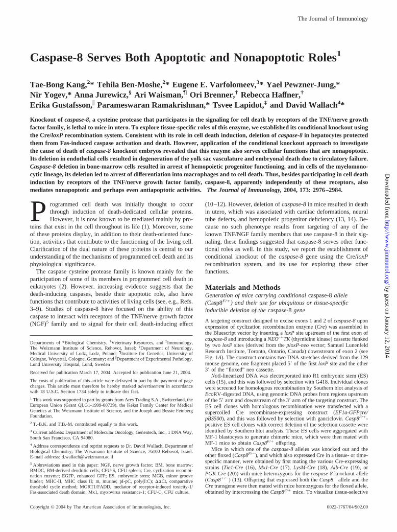
A targeting construct designed to excise exons 1 and 2 of caspase-8 uponexpression of cyclization recombination enzyme (Cre) was assembled inthe Bluescript vector by inserting a loxP site upstream of the first exon ofcaspase-8 and introducing a NEOr�TK (thymidine kinase) cassette flankedby two loxP sites (derived from the ploxP-neo vector; Samuel LunenfeldResearch Institute, Toronto, Ontario, Canada) downstream of exon 2 (seeFig. 1A). The construct contains two DNA stretches derived from the 129mouse genome, one fragment placed 5� of the first loxP site and the other3� of the “floxed” neo cassette.
NotI-linearized DNA was electroporated into R1 embryonic stem (ES)cells (15), and this was followed by selection with G418. Individual cloneswere screened for homologous recombination by Southern blot analysis ofEcoRV-digested DNA, using genomic DNA probes from regions upstreamof the 5� arm and downstream of the 3� arm of the targeting construct. TheES cell clones with homologous recombination were transfected with asupercoiled Cre recombinase-expressing construct (EF1a-GFPcre/pBS500), and this was followed by selection with ganciclovir. Casp8F/�-positive ES cell clones with correct deletion of the selection cassette wereidentified by Southern blot analysis. These ES cells were aggregated withMF-1 blastocysts to generate chimeric mice, which were then mated withMF-1 mice to obtain Casp8F/� offspring.
Mice in which one of the caspase-8 alleles was knocked out and theother floxed (Casp8F/�), and which also expressed Cre in a tissue- or time-specific manner, were obtained by first mating the various Cre-expressingstrains (Tie1-Cre (16), Mx1-Cre (17), LysM-Cre (18), Alb-Cre (19), orPGK-Cre (20)) with mice heterozygous for the caspase-8 knockout allele(Casp8�/�) (13). Offspring that expressed both the Casp8� allele and theCre transgene were then mated with mice homozygous for the floxed allele,obtained by intercrossing the Casp8F/� mice. To visualize tissue-selective
Departments of *Biological Chemistry, †Veterinary Resources, and ‡Immunology,The Weizmann Institute of Science, Rehovot, Israel; §Department of Neurology,Medical University of Lodz, Lodz, Poland; ¶Institute for Genetics, University ofCologne, Weyertal, Cologne, Germany; and �Department of Experimental Pathology,Lund University Hospital, Lund, Sweden
Received for publication March 17, 2004. Accepted for publication June 21, 2004.
The costs of publication of this article were defrayed in part by the payment of pagecharges. This article must therefore be hereby marked advertisement in accordancewith 18 U.S.C. Section 1734 solely to indicate this fact.1 This work was supported in part by grants from Ares Trading S.A., Switzerland, theEuropean Union (Grant QLG1-1999-00739), the Kekst Family Center for MedicalGenetics at The Weizmann Institute of Science, and the Joseph and Bessie FeinbergFoundation.2 T.-B.K. and T.B.-M. contributed equally to this work.3 Current address: Department of Molecular Oncology, Genentech, Inc., 1 DNA Way,South San Francisco, CA 94080.4 Address correspondence and reprint requests to Dr. David Wallach, Department ofBiological Chemistry, The Weizmann Institute of Science, 76100 Rehovot, Israel.E-mail address: [email protected] Abbreviations used in this paper: NGF, nerve growth factor; BM, bone marrow;BMDC, BM-derived dendritic cells; CFU-S, CFU spleen; Cre, cyclization recombi-nation enzyme; EGFP, enhanced GFP; ES, embryonic stem; MGB, minor groovebinder; MHC-II, MHC class II; m, murine; pI-pC, poly(I:C); ��Ct, comparativethreshold cycle method; MORT1/FADD, mediator of receptor-induced toxicity-1/Fas-associated death domain; Mx1, myxovirus resistance-1; CFU-C, CFU culture.
The Journal of Immunology
Copyright © 2004 by The American Association of Immunologists, Inc. 0022-1767/04/$02.00
by guest on January 12, 2014http://w
ww
.jimm
unol.org/D
ownloaded from
expression of functional Cre, some of the offspring of the latter matingwere mated with the Z/EG reporter mouse strain that expresses enhancedGFP (EGFP) upon Cre-mediated excision (21).
To induce deletion of caspase-8 in mice expressing Cre under the con-trol of the myxovirus resistance-1 (Mx1) promoter, mice were injected i.p.with either recombinant type I mouse IFN (106 U/mouse, kindly donatedby Dr. C. Weissmann) (Imperial College School of Medicine at St. Mary’s,London, U.K.) or poly(I:C) (pI-pC) dsRNA (5) (250 �g/mouse; Sigma-Aldrich, St. Louis, MO). Unless otherwise indicated, the mice were in-jected with pI-pC three times at 2-day intervals. Liver cell or bonemarrow (BM) cell function of the Mx1-Cre-expressing mice was as-sessed 2 days after injection of IFN or pI-pC.
In all experiments, the phenotype of the Cre-expressing Casp8F/� micewas compared with that of Cre-expressing Casp8F/� mice from the samelitter. In terms of all the parameters tested in this study, the phenotype ofthe Cre-expressing Casp8F/� mice was indistinguishable from that of wild-type mice, irrespective of the Cre strain used.
Genotyping the mice
Genotyping of the various caspase-8 alleles was done by Southern blot andPCR analyses of tail DNA. Southern blots were analyzed after digestion ofthe DNA with EcoRV, using genomic DNA probes from regions upstreamof the first exon and the 5� arm, and downstream of the 3� arm (betweenexons 5 and 6) of the targeting construct (Fig. 1, A and B). For the PCRanalyses, we used the oligonucleotides 5�-TAGCCTCTTTGGGGTTGTTCTACTG-3� (sense) and 5�-TGGGGCTTCGTTTAGTCTCTACTTC-3�(antisense) for the knockout allele, 5�-TAGCCTCTTTGGGGTTGTTCTACTG-3� (sense) and 5�-CGCGGTCGACTTATCAAGAGGTAGAAGAGCTGTAAC-3�(antisense)forthefloxedallele,and5�-CGTTGATGCCGGTGAACGTG-3� (sense) and 5�-AGCTGGCTGGTGGCAGATGG-3� (antisense)for the Cre transgenes.
Assessing the consequences of Fas triggering in vivo
Mice were injected i.p. with anti-Fas Ab (Jo-2, 15 �g/mouse; BD Bio-sciences, San Diego, CA). Tissues were harvested at the indicated timesafterward. Caspase expression and processing were analyzed by Westernblotting using rat anti-mouse caspase-8 mAb (1G12; kindly donated byDrs. A. Strasser and L. A. O’Reilly, Walter and Eliza Hall Institute, Mel-bourne, Australia) and anti-mouse caspase-3 (H-227; Santa Cruz Biotech-nology, Santa Cruz, CA), after extraction of the tissues in cell-lysis buffer(20 mM Tris-Cl (pH 7.4), 135 mM NaCl, 1 mM EDTA, 1% Triton X-100,and 10% glycerol) containing 1� complete protease inhibitor mixture(Roche Diagnostics, Mannheim, Germany).
Histology and immunostaining
Embryos and the tissues of adult mice were fixed in 10% phosphate-buff-ered formalin pH 7.4, embedded in paraffin, cut into 4-�m sections, andstained with H&E. To detect cells expressing processed caspase-3, paraffinsections of E10.5 embryos were deparaffinized, rehydrated, and incubatedwith anticleaved caspase-3 (Asp175) Ab (no. 9661; Cell Signaling Tech-nology, Beverly, MA), according to the manufacturer’s instructions. Thiswas followed by staining either with Cy3-conjugated goat anti-rabbit Ab(Jackson ImmunoResearch Laboratories, West Grove, PA) or (for immu-nohistochemical detection) with biotinylated peroxidase anti-rabbit sec-ondary Ab (DAKO Envision� System, peroxidase (3-amino-9-ethyl cara-zloe); DakoCytomation, Glostrup, Denmark). Immunofluorescent sectionswere observed with a confocal microscope (LSM 510; Carl Zeiss, Thorn-wood, NY), using an excitation wavelength of 543 nm and differentialinterference contrast optics. The immunostained cells were quantified usingthe ImagePro program (Media Cybernetics, Silver Spring, MD), with atleast 2500 cells examined in each of 10 yolk sacs of Casp8F/�Tie1-Creembryos and 10 yolk sacs of Casp8F/�:Tie1-Cre embryos.
For whole-mount staining with Ab against PECAM-1 (CD31), yolk sacswere dissected in PBS, fixed for 2 h at 4°C in 2% paraformaldehyde,washed overnight at 4°C in PBS, and treated for 1 h at 4°C in PBS con-taining 0.3% H2O2. After three washings with PBS, the embryos and yolksacs were incubated for 1 h at 4°C in blocking solution (Casblock; ZymedLaboratories, San Francisco, CA) containing 0.1% Triton X-100, and werethen incubated overnight at 4°C with rat anti-mouse PECAM-1 Ab (BDBiosciences) in the same blocking solution. The embryos and yolk sacswere then washed five times, each for 1 h at 4°C in PBS, and incubatedovernight at 4°C with biotinylated peroxidase donkey anti-rat Ab (JacksonImmunoResearch Laboratories) in the same blocking solution. After fivemore washings, each for 1 h at 4°C, staining was completed using theVectastain ABC kit (Vector Laboratories, Burlingame, CA) according tothe manufacturer’s instructions. The color was resolved with 3.3�-diami-nobenzidine (Sigma-Aldrich).
Flow cytometric analysis
To exclude dead cells, propidium iodide (Sigma-Aldrich) was included inall staining. For quantification of progenitor cell levels in the BM, single-cell BM suspensions were stained with allophycocyanin-anti-CD117 Ab(c-kit, 2B8; eBioscience, San Diego, CA) and biotin-conjugated Abs forlineage (Lin) markers (CD3, I45-2C11; B220, RA3-6B2; Mac-1, M1/70;Gr-1, RB6-8C5; TER119) (BD Pharmingen, San Diego, CA) and stainedusing PE-conjugated streptavidin (BD Pharmingen). Dying cells in the BMculture were quantified by staining of the nonattached cells with PE-anti-CD11b (M1/70) Ab and FITC-Annexin V. (Note that the cells were gatedfor lack of propidium iodide staining, and thus, their annexin V stainingindicates an early stage of death.) Mature macrophages were stained withAPC-anti-CD11b (M1/70) Ab and PE-anti-F4/80 Ab (MCA 497PE; Sero-tec, Oxford, U.K.). T and B cells in lymphoid organs were analyzed after
FIGURE 1. Generation of conditional caspase-8 knockout mice and as-sessment of Cre-mediated deletion. A, Schematic representation of the tar-geting strategy. The figure shows the structure of the mouse caspase-8 genebefore and after its recombination with the targeting vector and after de-letion of the NEOr�TK cassette, as well as after further deletion of itsfloxed region. The positions of the loxP sites are designated by triangles.The exons, deduced by comparison with the cDNA sequence, are denotedby black boxes. The positions of the 5� and 3� external genomic DNAprobes used for Southern blot analysis of the recombination are underlined.EcoRV restriction sites (RV) are marked by vertical lines. B, Cre-loxP-mediated deletion of the flanked caspase-8 gene. Southern blot analysis(using the 3� probe shown in A) of DNA from the littermate mice obtainedby crossing Casp8F/� mice with the Casp8�/�:PGK-Cre transgene. PCRassessment of occurrence of the Cre transgene is shown at the bottom. The6.8-kb fragment corresponding to the deleted floxed allele (�) could beobserved only in floxed mice that also expressed Cre. Homozygous dele-tion and combination of the deleted and knockout (ko) allele were lethal inutero and could be found only in embryos before midgestation. C, Genomiclocation of the oligonucleotide sequences used to assess the extent ofcaspase-8 deletion by PCR (A–C) and by real-time PCR (D–E). The figureshows the locations of oligonucleotides used to assess levels of the wild-type (WT), targeted (floxed, F), knockout (KO), and deleted floxed (�)caspase-8 alleles. The underlying short bar corresponds to the genomicregion whose level was assessed by real-time PCR.
2977The Journal of Immunology
by guest on January 12, 2014http://w
ww
.jimm
unol.org/D
ownloaded from
the cells were stained with PE-anti-B220 (RA3-6B2; eBioscience), allo-phycocyanin-anti-IgM (II/41; BD Pharmingen), PE-anti-CD4 (RM4-5; BDPharmingen), and allophycocyanin-anti-CD8 (53-6.7; eBioscience) Abs.PE-anti-Ly6G (1A8; BD Pharmingen) and allophycocyanin-anti-CD11bAbs were used for granulocyte analysis, and allophycocyanin-anti-CD11c(N418) and PE-anti-MHC class II (PE-anti-MHC-II) (M5/114.15.2; eBio-science) were used for dendritic cell analysis. Before staining, the cellswere incubated with Fc-block (anti-CD16/32 Ab; BD Pharmingen) toblock nonspecific Ab binding. Stained cells were analyzed on a FACS-Calibur using the CellQuest software (BD Biosciences).
In vitro progenitor assay
Hemopoietic progenitors in mouse embryos were assayed as previouslydescribed (13). To determine the number of clonogenic BM progenitors,single-cell suspensions were harvested from the femoral BM and the nu-cleated cells were counted and diluted to 2 � 105 cells/ml in IMDM con-taining 2% FCS. For each myeloid clonogenic progenitor assay, 2 � 104
cells were mixed with 1 ml of methylcellulose medium containing recombi-nant murine stem cell factor, recombinant murine (m)IL-3, recombinant hu-man IL-6, and erythropoietin (M3434; StemCell Technologies, Vancouver,British Columbia, Canada). The cells were plated and grown in humidifiedchambers at 37°C and 5% CO2. After 10–14 days, erythroid burst-formingunit, CFU granulo-macrophagic, and CFU granulocytic-erythroid-megacaryo-cytic-macrophagic progenitors were scored by microscopic analysis. Colonyidentification was confirmed by staining of sampled colonies with May-Grun-wald-Giemsa stain. CFU B cells were assayed by plating BM suspension (5 �104 cells) in a methylcellulose medium (M3630; StemCell Technologies) sup-plemented with 10 ng/ml IL-7, and scoring the colonies after 7 days of incu-bation. CFU granulocytes were assayed by culturing samples of 1 � 105 BMcells for 7 days in a methylcellulose medium supplemented with 20 ng/mlmrG-CSF (PeproTech, Rocky Hill, NJ) as described (22). Cell patterns in thecolonies were assessed by May-Grunwald-Giemsa staining.
In vivo CFU spleen (CFU-S) assays
For the CFU-S assay, BM from pI-pC-treated Casp8F/�:Mx1-Cre andCasp8F/�Mx1-Cre mice were depleted of mature T cells using anti-mouseCD4 (L3T4) and CD-8 (Ly-2) MicroBeads (Miltenyi Biotec, BergischGladbach, Germany). Single-cell suspensions (1 � 105 cells) were theninjected into the tail veins of irradiated (8.5 Gy, 137C source) 10-wk-oldC57BL/6 female mice. In each experiment, cells from each donor wereinjected into ten recipient mice. The recipients were treated with Ciproxin(Bayer, Leverkusen, Germany) in their drinking water (6.7 mg/l) through-out the experiment. Mice were killed 8 or 13 days after transplantation.Spleens were weighed and fixed in Bouin’s solution. Colonies werecounted under the microscope.
Generation of radiation chimera
BM cells (2 � 106 per recipient) harvested from the femora of Casp8F/�:Mx1-Cre and Casp8F/�Mx1-Cre (Ly5.2) mice were injected into irradiated(8.5 Gy, 137C source) C57BL/6-Ly5.1 recipient mice (Charles River Lab-oratories, Lyon, France) via the tail vein or, conversely, BM cells fromC57BL/6-Ly5.1 mice were injected into the tail veins of similarly irradi-ated Casp8F/�:Mx1-Cre and Casp8F/�Mx1-Cre recipients. After 8–10 wk,the recipient mice were injected with pI-pC to induce deletion ofcaspase-8, and BM cells derived from these mice were assayed for CFU asdescribed above. Donor engraftment in the recipient mice was quantifiedby using FITC-CD45.2 (anti-Ly5.2, clone 104) and PE-CD45.1 (anti-Ly5.1, clone A20) Abs, and was found to be over 95%.
Reconstitution of T and B lymphocytes in RAG-1�/� mice
Casp8F/�:Mx1-Cre and Casp8F/�Mx1-Cre mice were injected with pI-pCto induce deletion of caspase-8, and their femoral BM cells were harvestedand transplanted (1 � 106 cells per recipient mouse) into irradiated (4 Gy,137C source) RAG-1�/� mice (The Jackson Laboratory, Bar Harbor, ME)via the tail vein. The recipient mice were euthanized 7 wk or 6 mo aftertransplantation, and their BM and lymphoid organ cells were harvested andanalyzed.
Preparation of BM-derived and peritoneal macrophages
Primary cultures of BM-derived macrophages were generated from 3- to4-mo-old mice. Femoral BM cells were cultured with DMEM supple-mented with 20% FCS and 30% L929 cell-conditioned medium. Afterovernight culture, nonadherent cells were harvested and suspended in freshmedium. Aliquots of 2.5 � 105 cells were cultured for 7–10 days at 37°Cand 5% CO2 in 35-mm culture wells. Fresh culture medium was added onday 3 and replaced on day 6. Adherent cells were quantified by the MTT
assay (23) in 96-well plates seeded with 5 � 103 cells per well. More than90% of the adherent cells stained positively with anti-CD11b and theF4/80 Ab.
Peritoneal cells were harvested by rinsing the mouse peritoneal cavitywith sterile PBS (10 ml/mouse) containing 2% FCS. The cells were washedonce with PBS and suspended in RPMI 1640 medium supplemented with10% FCS, glutamine, and penicillin/streptomycin. They were then platedon 35-mm Nunc culture dishes (Nunc, Roskilde, Denmark) at a density of3 � 106 cells per dish, and cultured for 2–4 h at 37°C and 5% CO2 to allowmacrophage adherence.
Preparation of BM-derived granulocytes and peritonealneutrophils
Femoral BM cells (see above) were cultured at a density of 5 � 105
cells/ml with RPMI 1640 medium supplemented with 10% FCS and 20ng/ml mrG-CSF (PeproTech). Granulocyte yield on the 7th day of cultur-ing was assessed by trypan blue exclusion and FACS analysis after im-munostaining with anti-Ly6G Ab.
Peritoneal neutrophils were harvested 4 h after injection of 3% thiogly-colate (Difco, Detroit, MI) as described (24), followed by purification ofLy6G� cells by MACS magnetic cell separation system (Miltenyi Biotec).FACS analysis using anti-CD11b and Ly6G Abs confirmed that the purityof the cells was �90%.
Preparation of BM-derived dendritic cells (BMDC)
BMDC were prepared as described (25) by culturing femoral BM cells(2 � 105 cells/ml) with RPMI 1640 medium supplemented with 10% FCS,50 �M 2-ME, and 10 ng/ml mrGM-CSF (PeproTech) for 8 days. Cellswere sorted with the aid of anti-CD11c and anti-MHC-II Abs (for isolationof mature MHC-IIhigh and immature MHC-IIlow BMDC).
PCR and real-time PCR for assessing deletion efficiency
The effectiveness of Cre-mediated deletion of the floxed caspase-8 allele wasroughly estimated by PCR. DNA was extracted from the adherent culturedperitoneal macrophages or BM-derived macrophages, and from liver andspleen samples, as described (26). DNA was subjected to PCR analysis usingthree primers: 5�-TAGCCTCTTTGGGGTTGTTCTACTG-3� (A in Fig. 1C;sense for the wild-type, knockout, floxed, and deleted allele), 5�-CGCGGTCGACTTATCAAGAGGTAGAAGAGCTGTAAC-3� (B in Fig.1C; antisense for the wild-type, knockout, and floxed allele), and 5�-GCGAACACGCCGTGTTTCAAGGGC-3� (C in Fig. 1C; antisense for thedeleted allele).
For more precise quantitative evaluation, the extent of deletion wasassessed by real-time PCR. The assay was conducted in a reaction volumeof 20 �l containing 10 ng DNA, 300 nM oligonucleotide primers, 200 nMoligonucleotide 3�-minor groove binder (MGB) probes, and 10 �l of Taq-Man Universal PCR Master Mix (Applied Biosystems, Foster City, CA).Primers and probes were designed using Primer Express software (AppliedBiosystems). PCR was initiated by incubating the reaction mixture for 2min at 50°C for activation of the AmpErase UNG (Applied Biosystems).This was followed by incubation for 10 min at 95°C, and then by 40 cyclesof 15 s at 95°C and 1 min at 60°C on an ABI Prism 7000 SequenceDetection System (Applied Biosystems).
Levels of caspase-8 were normalized on the basis of quantification ofthe NF-�B-inducing kinase (NIK) gene in the same DNA samples using 5�-AGCCTCCTCTACCGCCAGAA-3� (sense), 5�-GTGCCAGACTCCTCCTTGCT-3� (antisense), and (5�-6-FAM-ACCAGAACCGAGCAAAMGB-3�(probe). The comparative threshold cycle method (��Ct) was used to de-termine the level of caspase-8 (27). The caspase-8 oligonucleotide primersapplied were 5�-GGAAACAAGCTGGTAGCTGACA-3� (D in Fig. 1C;sense), 5�-CCTGGGTCAACACAAGATGCT-3� (E in Fig. 1C; antisense),and 5�-6-FAM-TTAACTTCCTCACTTGATCAT-MGB-3� (Applied Bio-systems). To define the dynamic range and the efficiency of the assay, DNAsamples were varied by the use of successive 2-fold dilutions in the rangeof 0.5–25 ng per reaction. All dilution and negative control samples weretested in triplicate. Analysis of �Ct at the chosen range of template dilu-tions confirmed that the plot of log input vs �Ct has a slope of �0.1,implying that the efficiencies of the real-time PCR for caspase-8 and forNIK were about equal.
Ct were determined by plotting normalized fluorescent signal againstcycle number, and the caspase-8 copy number was calculated by the ex-pression 2���Ct, where ��Ct � (Ctcasp8 � CtNIK)sample DNA � (Ctcasp8 �CtNIK)nondeleted control DNA.
Using this method, the 2���Ct values were expected to be close to 1 insamples without caspase8 deletions and close to 0.5 in samples with 100%caspase-8 deletions. The percentage of caspase-8 deletion in each sample
2978 CASPASE-8-FUNCTION IN VIVO
by guest on January 12, 2014http://w
ww
.jimm
unol.org/D
ownloaded from
was deducted from the 2���Ct values according to the following calcula-tion: percentage of deletion � (1 � 2���Ct) � 200.
ResultsGeneration of caspase-8 conditional knockout mice
Applying the strategy used for the full knockout of caspase-8 (13),we generated mice with conditional caspase-8 allele (Casp8F/�)by using a targeting construct designed to excise exons 1 and 2 thatencode the two N-terminal death-effector domain motifs in theprotein (Fig. 1A). ES cell clones that underwent homologous re-combination with the transfected construct were transfected with aCre-expressing construct to delete the loxP site-flanked selectioncassette, and were then aggregated with blastocysts to generategermline-competent chimeras. Development was normal in micehomozygous for the floxed allele and in mice in which one of thecaspase-8 alleles was floxed and the other knocked out (13); Casp-8f/l�, whereas Casp-8fl/� mice in which the floxed allele was de-leted by their mating with a Cre-deleter strain (20) (Fig. 1B) diedduring embryogenesis, as expected from the embryonic lethal phe-notype of caspase-8 knockout mice.
Caspase-8 plays an apoptotic role in hepatocytes in vivo
Although there is extensive evidence for caspase-8 participation indeath induction in cultured cells, there is no reported direct evi-dence that caspase-8 serves a similar function in vivo. With theobject of using Cre-mediated ablation of caspase-8 to assess the invivo relevance of its in vitro apoptotic function we attempted todelete caspase-8 in hepatocytes, cells whose apoptotic death inresponse to Fas triggering has often been suggested to occur in acaspase-8-dependent manner.
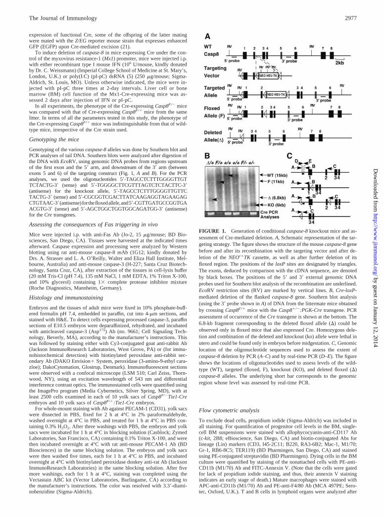
Crossing the Casp8F/� mice with a transgenic mouse line thatexpresses Cre under control of the liver-specific albumin promoter(Alb-Cre; Ref. 19) resulted in effective decrease of caspase-8 ex-pression in the liver, but not in other organs (Fig. 2A). A real-timePCR test designed to allow quantification of caspase-8 deletion(Fig. 1C) confirmed that deletion of the floxed allele occurred to
about the same high extent in the livers of Casp-8fl� and controlCasp-8fl� mice (Fig. 2B). The Casp-8F/� Alb-Cre mice appearednormal. However, whereas injection of anti-Fas Ab (15 �g/mouse)in the control Casp8F/�:Alb-Cre littermates or in wild-type miceresulted in death of all mice within 6 h, the Casp8F/�Alb-Cre mice(n � 20) injected with the Ab were still alive months afterward.Histological and Western blot analysis after anti-Fas injection dis-closed massive apoptosis and hemorrhage, associated with earlyinduction of caspase-3 processing, in the livers (though not in thespleens) of control Casp8F/�:Alb-Cre mice, but not ofCasp8F/�Alb-Cre mice (Fig. 2, C and D).
Expression of the albumin promoter begins before birth, thusdictating deficiency of caspase-8 in the liver of the Casp8F/�Alb-Cre mouse almost throughout its life. To exclude the possibilitythat the resistance to anti-Fas observed in the Casp8F/�Alb-Cremice was secondary to other changes induced in the liver owing tothis prolonged caspase-8 deficiency, we crossed the Casp8F/�
mice with the Mx1-Cre line, which expresses Cre under the controlof the �� IFN-inducible Mx1 promoter. This line responds to in-jection of IFN or of the IFN inducer, pI-pC, by effective inductionof Cre and consequent deletion of floxed genes in various tissues,including the liver (17). As in the case of the Casp8F/� Alb-Cremice, the liver cells of Casp8F/�Mx1-Cre mice in which caspase-8was deleted (by pI-pC injection) a few days before anti-Fas injec-tion were resistant to the cytotoxicity of the anti-Fas Abs (Fig. 2E).
Caspase-8 function in endothelial cells plays a crucial role inembryonic development
Caspase-8 knockout embryos show evidence of circulatory failureat the time of death (13, 14). In an attempt to elucidate the mech-anism underlying this failure, we specifically targeted caspase-8 inendothelial cells by crossing mice in which one of the caspase-8alleles had been knocked out and the other floxed (Casp8F/�) withmice that express the Cre recombinase under control of the endo-thelium-specific Tie1 promoter (Ref. 16; see Fig. 3K for demon-stration of the expression pattern of this Cre transgene). Whereas
FIGURE 2. Deletion of the caspase-8 gene in hepatocytes endows them with resistance to Fas cytotoxicity. A–D, Comparison of Casp8F/�:Alb-Cre andCasp8F/�Alb-Cre mice. A, Western blot analysis of procaspase-8 expression in various tissues. B, Assessment of the levels of the various caspase-8 alleles by PCR(top) and by real-time PCR (numbers at the bottom) in the livers and spleens. C, Evaluation of the extent of caspase-3 processing in the tissues by Western blottingat various times after their injection with anti-Fas Ab. D, Liver histology 6 h after injection of anti-Fas Ab. E, Liver histology of pI-pC-pretreated Casp8F/�:Mx1-Cre and Casp8F/�Mx1-Cre mice 6 h after injection of anti-Fas Ab. Arrows in D and E point to pyknotic nuclei in apoptotic cells.
2979The Journal of Immunology
by guest on January 12, 2014http://w
ww
.jimm
unol.org/D
ownloaded from
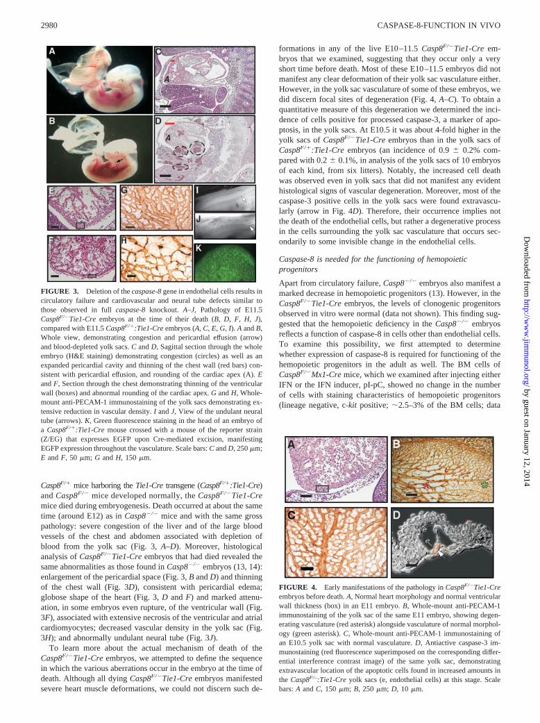
Casp8F/� mice harboring the Tie1-Cre transgene (Casp8F/�:Tie1-Cre)and Casp8F/� mice developed normally, the Casp8F/�Tie1-Cremice died during embryogenesis. Death occurred at about the sametime (around E12) as in Casp8�/� mice and with the same grosspathology: severe congestion of the liver and of the large bloodvessels of the chest and abdomen associated with depletion ofblood from the yolk sac (Fig. 3, A–D). Moreover, histologicalanalysis of Casp8F/�Tie1-Cre embryos that had died revealed thesame abnormalities as those found in Casp8�/� embryos (13, 14):enlargement of the pericardial space (Fig. 3, B and D) and thinningof the chest wall (Fig. 3D), consistent with pericardial edema;globose shape of the heart (Fig. 3, D and F) and marked attenu-ation, in some embryos even rupture, of the ventricular wall (Fig.3F), associated with extensive necrosis of the ventricular and atrialcardiomyocytes; decreased vascular density in the yolk sac (Fig.3H); and abnormally undulant neural tube (Fig. 3J).
To learn more about the actual mechanism of death of theCasp8F/�Tie1-Cre embryos, we attempted to define the sequencein which the various aberrations occur in the embryo at the time ofdeath. Although all dying Casp8F/�Tie1-Cre embryos manifestedsevere heart muscle deformations, we could not discern such de-
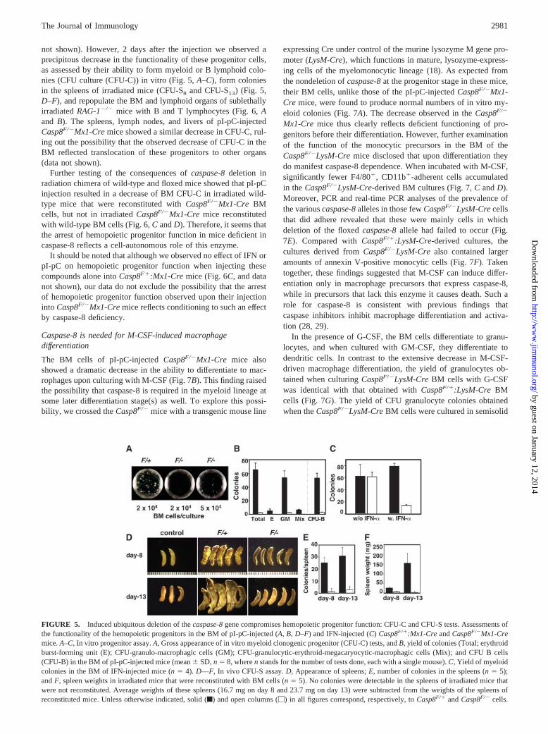
formations in any of the live E10–11.5 Casp8F/�Tie1-Cre em-bryos that we examined, suggesting that they occur only a veryshort time before death. Most of these E10–11.5 embryos did notmanifest any clear deformation of their yolk sac vasculature either.However, in the yolk sac vasculature of some of these embryos, wedid discern focal sites of degeneration (Fig. 4, A–C). To obtain aquantitative measure of this degeneration we determined the inci-dence of cells positive for processed caspase-3, a marker of apo-ptosis, in the yolk sacs. At E10.5 it was about 4-fold higher in theyolk sacs of Casp8F/�Tie1-Cre embryos than in the yolk sacs ofCasp8F/�:Tie1-Cre embryos (an incidence of 0.9 0.2% com-pared with 0.2 0.1%, in analysis of the yolk sacs of 10 embryosof each kind, from six litters). Notably, the increased cell deathwas observed even in yolk sacs that did not manifest any evidenthistological signs of vascular degeneration. Moreover, most of thecaspase-3 positive cells in the yolk sacs were found extravascu-larly (arrow in Fig. 4D). Therefore, their occurrence implies notthe death of the endothelial cells, but rather a degenerative processin the cells surrounding the yolk sac vasculature that occurs sec-ondarily to some invisible change in the endothelial cells.
Caspase-8 is needed for the functioning of hemopoieticprogenitors
Apart from circulatory failure, Casp8�/� embryos also manifest amarked decrease in hemopoietic progenitors (13). However, in theCasp8F/�Tie1-Cre embryos, the levels of clonogenic progenitorsobserved in vitro were normal (data not shown). This finding sug-gested that the hemopoietic deficiency in the Casp8�/� embryosreflects a function of caspase-8 in cells other than endothelial cells.To examine this possibility, we first attempted to determinewhether expression of caspase-8 is required for functioning of thehemopoietic progenitors in the adult as well. The BM cells ofCasp8F/�Mx1-Cre mice, which we examined after injecting eitherIFN or the IFN inducer, pI-pC, showed no change in the numberof cells with staining characteristics of hemopoietic progenitors(lineage negative, c-kit positive; 2.5–3% of the BM cells; data
FIGURE 3. Deletion of the caspase-8 gene in endothelial cells results incirculatory failure and cardiovascular and neural tube defects similar tothose observed in full caspase-8 knockout. A–J, Pathology of E11.5Casp8F/�Tie1-Cre embryos at the time of their death (B, D, F, H, J),compared with E11.5 Casp8F/�:Tie1-Cre embryos (A, C, E, G, I). A and B,Whole view, demonstrating congestion and pericardial effusion (arrow)and blood-depleted yolk sacs. C and D, Sagittal section through the wholeembryo (H&E staining) demonstrating congestion (circles) as well as anexpanded pericardial cavity and thinning of the chest wall (red bars) con-sistent with pericardial effusion, and rounding of the cardiac apex (A). Eand F, Section through the chest demonstrating thinning of the ventricularwall (boxes) and abnormal rounding of the cardiac apex. G and H, Whole-mount anti-PECAM-1 immunostaining of the yolk sacs demonstrating ex-tensive reduction in vascular density. I and J, View of the undulant neuraltube (arrows). K, Green fluorescence staining in the head of an embryo ofa Casp8F/�:Tie1-Cre mouse crossed with a mouse of the reporter strain(Z/EG) that expresses EGFP upon Cre-mediated excision, manifestingEGFP expression throughout the vasculature. Scale bars: C and D, 250 �m;E and F, 50 �m; G and H, 150 �m.
FIGURE 4. Early manifestations of the pathology in Casp8F/�Tie1-Creembryos before death. A, Normal heart morphology and normal ventricularwall thickness (box) in an E11 embryo. B, Whole-mount anti-PECAM-1immunostaining of the yolk sac of the same E11 embryo, showing degen-erating vasculature (red asterisk) alongside vasculature of normal morphol-ogy (green asterisk). C, Whole-mount anti-PECAM-1 immunostaining ofan E10.5 yolk sac with normal vasculature. D, Antiactive caspase-3 im-munostaining (red fluorescence superimposed on the corresponding differ-ential interference contrast image) of the same yolk sac, demonstratingextravascular location of the apoptotic cells found in increased amounts inthe Casp8F/–:Tie1-Cre yolk sacs (e, endothelial cells) at this stage. Scalebars: A and C, 150 �m; B, 250 �m; D, 10 �m.
2980 CASPASE-8-FUNCTION IN VIVO
by guest on January 12, 2014http://w
ww
.jimm
unol.org/D
ownloaded from
not shown). However, 2 days after the injection we observed aprecipitous decrease in the functionality of these progenitor cells,as assessed by their ability to form myeloid or B lymphoid colo-nies (CFU culture (CFU-C)) in vitro (Fig. 5, A–C), form coloniesin the spleens of irradiated mice (CFU-S8 and CFU-S13) (Fig. 5,D–F), and repopulate the BM and lymphoid organs of sublethallyirradiated RAG-1�/� mice with B and T lymphocytes (Fig. 6, Aand B). The spleens, lymph nodes, and livers of pI-pC-injectedCasp8F/�Mx1-Cre mice showed a similar decrease in CFU-C, rul-ing out the possibility that the observed decrease of CFU-C in theBM reflected translocation of these progenitors to other organs(data not shown).
Further testing of the consequences of caspase-8 deletion inradiation chimera of wild-type and floxed mice showed that pI-pCinjection resulted in a decrease of BM CFU-C in irradiated wild-type mice that were reconstituted with Casp8F/�Mx1-Cre BMcells, but not in irradiated Casp8F/�Mx1-Cre mice reconstitutedwith wild-type BM cells (Fig. 6, C and D). Therefore, it seems thatthe arrest of hemopoietic progenitor function in mice deficient incaspase-8 reflects a cell-autonomous role of this enzyme.
It should be noted that although we observed no effect of IFN orpI-pC on hemopoietic progenitor function when injecting thesecompounds alone into Casp8F�:Mx1-Cre mice (Fig. 6C, and datanot shown), our data do not exclude the possibility that the arrestof hemopoietic progenitor function observed upon their injectioninto Casp8F/�Mx1-Cre mice reflects conditioning to such an effectby caspase-8 deficiency.
Caspase-8 is needed for M-CSF-induced macrophagedifferentiation
The BM cells of pI-pC-injected Casp8F/�Mx1-Cre mice alsoshowed a dramatic decrease in the ability to differentiate to mac-rophages upon culturing with M-CSF (Fig. 7B). This finding raisedthe possibility that caspase-8 is required in the myeloid lineage atsome later differentiation stage(s) as well. To explore this possi-bility, we crossed the Casp8F/� mice with a transgenic mouse line
expressing Cre under control of the murine lysozyme M gene pro-moter (LysM-Cre), which functions in mature, lysozyme-express-ing cells of the myelomonocytic lineage (18). As expected fromthe nondeletion of caspase-8 at the progenitor stage in these mice,their BM cells, unlike those of the pI-pC-injected Casp8F/�Mx1-Cre mice, were found to produce normal numbers of in vitro my-eloid colonies (Fig. 7A). The decrease observed in the Casp8F/�
Mx1-Cre mice thus clearly reflects deficient functioning of pro-genitors before their differentiation. However, further examinationof the function of the monocytic precursors in the BM of theCasp8F/�LysM-Cre mice disclosed that upon differentiation theydo manifest caspase-8 dependence. When incubated with M-CSF,significantly fewer F4/80�, CD11b�-adherent cells accumulatedin the Casp8F/�LysM-Cre-derived BM cultures (Fig. 7, C and D).Moreover, PCR and real-time PCR analyses of the prevalence ofthe various caspase-8 alleles in those few Casp8F/�LysM-Cre cellsthat did adhere revealed that these were mainly cells in whichdeletion of the floxed caspase-8 allele had failed to occur (Fig.7E). Compared with Casp8F/�:LysM-Cre-derived cultures, thecultures derived from Casp8F/�LysM-Cre also contained largeramounts of annexin V-positive monocytic cells (Fig. 7F). Takentogether, these findings suggested that M-CSF can induce differ-entiation only in macrophage precursors that express caspase-8,while in precursors that lack this enzyme it causes death. Such arole for caspase-8 is consistent with previous findings thatcaspase inhibitors inhibit macrophage differentiation and activa-tion (28, 29).
In the presence of G-CSF, the BM cells differentiate to granu-locytes, and when cultured with GM-CSF, they differentiate todendritic cells. In contrast to the extensive decrease in M-CSF-driven macrophage differentiation, the yield of granulocytes ob-tained when culturing Casp8F/�LysM-Cre BM cells with G-CSFwas identical with that obtained with Casp8F/�:LysM-Cre BMcells (Fig. 7G). The yield of CFU granulocyte colonies obtainedwhen the Casp8F/�LysM-Cre BM cells were cultured in semisolid
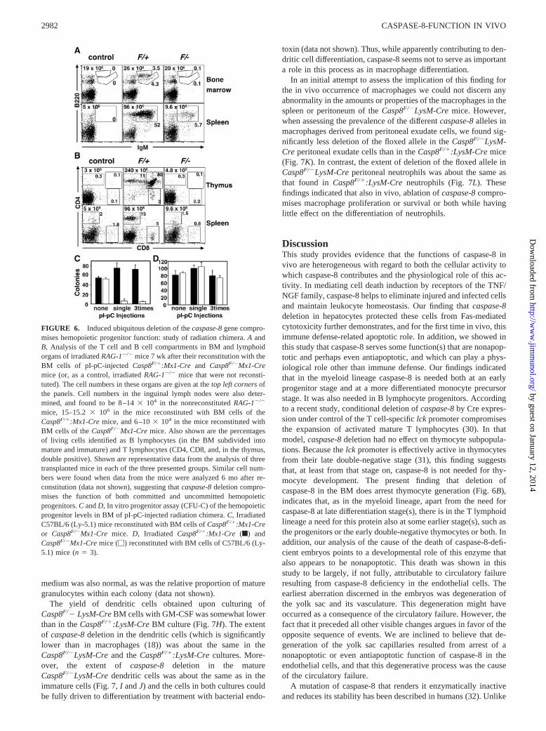
FIGURE 5. Induced ubiquitous deletion of the caspase-8 gene compromises hemopoietic progenitor function: CFU-C and CFU-S tests. Assessments ofthe functionality of the hemopoietic progenitors in the BM of pI-pC-injected (A, B, D–F) and IFN-injected (C) Casp8F/�:Mx1-Cre and Casp8F/�Mx1-Cremice. A–C, In vitro progenitor assay. A, Gross appearance of in vitro myeloid clonogenic progenitor (CFU-C) tests, and B, yield of colonies (Total; erythroidburst-forming unit (E); CFU-granulo-macrophagic cells (GM); CFU-granulocytic-erythroid-megacaryocytic-macrophagic cells (Mix); and CFU B cells(CFU-B) in the BM of pI-pC-injected mice (mean SD, n � 8, where n stands for the number of tests done, each with a single mouse). C, Yield of myeloidcolonies in the BM of IFN-injected mice (n � 4). D—F, In vivo CFU-S assay. D, Appearance of spleens; E, number of colonies in the spleens (n � 5);and F, spleen weights in irradiated mice that were reconstituted with BM cells (n � 5). No colonies were detectable in the spleens of irradiated mice thatwere not reconstituted. Average weights of these spleens (16.7 mg on day 8 and 23.7 mg on day 13) were subtracted from the weights of the spleens ofreconstituted mice. Unless otherwise indicated, solid (f) and open columns (�) in all figures correspond, respectively, to Casp8F/� and Casp8F/� cells.
2981The Journal of Immunology
by guest on January 12, 2014http://w
ww
.jimm
unol.org/D
ownloaded from
medium was also normal, as was the relative proportion of maturegranulocytes within each colony (data not shown).
The yield of dendritic cells obtained upon culturing ofCasp8F/� LysM-Cre BM cells with GM-CSF was somewhat lowerthan in the Casp8F/�:LysM-Cre BM culture (Fig. 7H). The extentof caspase-8 deletion in the dendritic cells (which is significantlylower than in macrophages (18)) was about the same in theCasp8F/�LysM-Cre and the Casp8F/�:LysM-Cre cultures. More-over, the extent of caspase-8 deletion in the matureCasp8F/�LysM-Cre dendritic cells was about the same as in theimmature cells (Fig. 7, I and J) and the cells in both cultures couldbe fully driven to differentiation by treatment with bacterial endo-
toxin (data not shown). Thus, while apparently contributing to den-dritic cell differentiation, caspase-8 seems not to serve as importanta role in this process as in macrophage differentiation.
In an initial attempt to assess the implication of this finding forthe in vivo occurrence of macrophages we could not discern anyabnormality in the amounts or properties of the macrophages in thespleen or peritoneum of the Casp8F/�LysM-Cre mice. However,when assessing the prevalence of the different caspase-8 alleles inmacrophages derived from peritoneal exudate cells, we found sig-nificantly less deletion of the floxed allele in the Casp8F/�LysM-Cre peritoneal exudate cells than in the Casp8F/�:LysM-Cre mice(Fig. 7K). In contrast, the extent of deletion of the floxed allele inCasp8F/�LysM-Cre peritoneal neutrophils was about the same asthat found in Casp8F/�:LysM-Cre neutrophils (Fig. 7L). Thesefindings indicated that also in vivo, ablation of caspase-8 compro-mises macrophage proliferation or survival or both while havinglittle effect on the differentiation of neutrophils.
DiscussionThis study provides evidence that the functions of caspase-8 invivo are heterogeneous with regard to both the cellular activity towhich caspase-8 contributes and the physiological role of this ac-tivity. In mediating cell death induction by receptors of the TNF/NGF family, caspase-8 helps to eliminate injured and infected cellsand maintain leukocyte homeostasis. Our finding that caspase-8deletion in hepatocytes protected these cells from Fas-mediatedcytotoxicity further demonstrates, and for the first time in vivo, thisimmune defense-related apoptotic role. In addition, we showed inthis study that caspase-8 serves some function(s) that are nonapop-totic and perhaps even antiapoptotic, and which can play a phys-iological role other than immune defense. Our findings indicatedthat in the myeloid lineage caspase-8 is needed both at an earlyprogenitor stage and at a more differentiated monocyte precursorstage. It was also needed in B lymphocyte progenitors. Accordingto a recent study, conditional deletion of caspase-8 by Cre expres-sion under control of the T cell-specific lck promoter compromisesthe expansion of activated mature T lymphocytes (30). In thatmodel, caspase-8 deletion had no effect on thymocyte subpopula-tions. Because the lck promoter is effectively active in thymocytesfrom their late double-negative stage (31), this finding suggeststhat, at least from that stage on, caspase-8 is not needed for thy-mocyte development. The present finding that deletion ofcaspase-8 in the BM does arrest thymocyte generation (Fig. 6B),indicates that, as in the myeloid lineage, apart from the need forcaspase-8 at late differentiation stage(s), there is in the T lymphoidlineage a need for this protein also at some earlier stage(s), such asthe progenitors or the early double-negative thymocytes or both. Inaddition, our analysis of the cause of the death of caspase-8-defi-cient embryos points to a developmental role of this enzyme thatalso appears to be nonapoptotic. This death was shown in thisstudy to be largely, if not fully, attributable to circulatory failureresulting from caspase-8 deficiency in the endothelial cells. Theearliest aberration discerned in the embryos was degeneration ofthe yolk sac and its vasculature. This degeneration might haveoccurred as a consequence of the circulatory failure. However, thefact that it preceded all other visible changes argues in favor of theopposite sequence of events. We are inclined to believe that de-generation of the yolk sac capillaries resulted from arrest of anonapoptotic or even antiapoptotic function of caspase-8 in theendothelial cells, and that this degenerative process was the causeof the circulatory failure.
A mutation of caspase-8 that renders it enzymatically inactiveand reduces its stability has been described in humans (32). Unlike
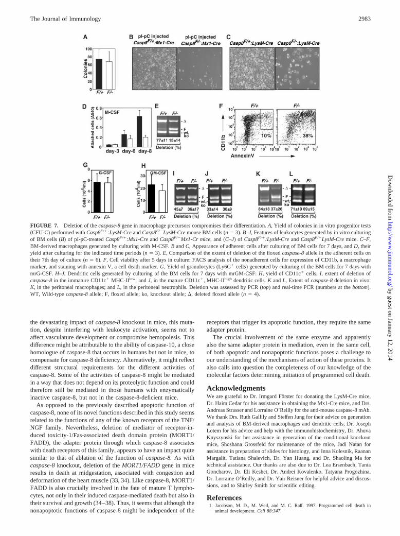
FIGURE 6. Induced ubiquitous deletion of the caspase-8 gene compro-mises hemopoietic progenitor function: study of radiation chimera. A andB, Analysis of the T cell and B cell compartments in BM and lymphoidorgans of irradiated RAG-1�/� mice 7 wk after their reconstitution with theBM cells of pI-pC-injected Casp8F/�:Mx1-Cre and Casp8F/�Mx1-Cremice (or, as a control, irradiated RAG-1�/� mice that were not reconsti-tuted). The cell numbers in these organs are given at the top left corners ofthe panels. Cell numbers in the inguinal lymph nodes were also deter-mined, and found to be 8–14 � 104 in the nonreconstituted RAG-1�/�
mice, 15–15.2 � 106 in the mice reconstituted with BM cells of theCasp8F/�:Mx1-Cre mice, and 6–10 � 104 in the mice reconstituted withBM cells of the Casp8F/�Mx1-Cre mice. Also shown are the percentagesof living cells identified as B lymphocytes (in the BM subdivided intomature and immature) and T lymphocytes (CD4, CD8, and, in the thymus,double positive). Shown are representative data from the analysis of threetransplanted mice in each of the three presented groups. Similar cell num-bers were found when data from the mice were analyzed 6 mo after re-constitution (data not shown), suggesting that caspase-8 deletion compro-mises the function of both committed and uncommitted hemopoieticprogenitors. C and D, In vitro progenitor assay (CFU-C) of the hemopoieticprogenitor levels in BM of pI-pC-injected radiation chimera. C, IrradiatedC57BL/6 (Ly-5.1) mice reconstituted with BM cells of Casp8F/�:Mx1-Creor Casp8F/�Mx1-Cre mice. D, Irradiated Casp8F/�:Mx1-Cre (f) andCasp8F/�Mx1-Cre mice (�) reconstituted with BM cells of C57BL/6 (Ly-5.1) mice (n � 3).
2982 CASPASE-8-FUNCTION IN VIVO
by guest on January 12, 2014http://w
ww
.jimm
unol.org/D
ownloaded from
the devastating impact of caspase-8 knockout in mice, this muta-tion, despite interfering with leukocyte activation, seems not toaffect vasculature development or compromise hemopoiesis. Thisdifference might be attributable to the ability of caspase-10, a closehomologue of caspase-8 that occurs in humans but not in mice, tocompensate for caspase-8 deficiency. Alternatively, it might reflectdifferent structural requirements for the different activities ofcaspase-8. Some of the activities of caspase-8 might be mediatedin a way that does not depend on its proteolytic function and couldtherefore still be mediated in those humans with enzymaticallyinactive caspase-8, but not in the caspase-8-deficient mice.
As opposed to the previously described apoptotic function ofcaspase-8, none of its novel functions described in this study seemsrelated to the functions of any of the known receptors of the TNF/NGF family. Nevertheless, deletion of mediator of receptor-in-duced toxicity-1/Fas-associated death domain protein (MORT1/FADD), the adapter protein through which caspase-8 associateswith death receptors of this family, appears to have an impact quitesimilar to that of ablation of the function of caspase-8. As withcaspase-8 knockout, deletion of the MORT1/FADD gene in miceresults in death at midgestation, associated with congestion anddeformation of the heart muscle (33, 34). Like caspase-8, MORT1/FADD is also crucially involved in the fate of mature T lympho-cytes, not only in their induced caspase-mediated death but also intheir survival and growth (34–38). Thus, it seems that although thenonapoptotic functions of caspase-8 might be independent of the
receptors that trigger its apoptotic function, they require the sameadapter protein.
The crucial involvement of the same enzyme and apparentlyalso the same adapter protein in mediation, even in the same cell,of both apoptotic and nonapoptotic functions poses a challenge toour understanding of the mechanisms of action of these proteins. Italso calls into question the completeness of our knowledge of themolecular factors determining initiation of programmed cell death.
AcknowledgmentsWe are grateful to Dr. Irmgard Forster for donating the LysM-Cre mice,Dr. Haim Cedar for his assistance in obtaining the Mx1-Cre mice, and Drs.Andreas Strasser and Lorraine O’Reilly for the anti-mouse caspase-8 mAb.We thank Drs. Ruth Gallily and Steffen Jung for their advice on generationand analysis of BM-derived macrophages and dendritic cells, Dr. JosephLotem for his advice and help with the immunohistochemistry, Dr. AhuvaKnyszynski for her assistance in generation of the conditional knockoutmice, Shoshana Grossfeld for maintenance of the mice, Jadi Natan forassistance in preparation of slides for histology, and Inna Kolesnik, RaananMargalit, Tatiana Shalevich, Dr. Yan Huang, and Dr. Shaoling Ma fortechnical assistance. Our thanks are also due to Dr. Lea Ersenbach, TaniaGoncharov, Dr. Eli Keshet, Dr. Andrei Kovalenko, Tatyana Progozhina,Dr. Lorraine O’Reilly, and Dr. Yair Reisner for helpful advice and discus-sions, and to Shirley Smith for scientific editing.
References1. Jacobson, M. D., M. Weil, and M. C. Raff. 1997. Programmed cell death in
animal development. Cell 88:347.
FIGURE 7. Deletion of the caspase-8 gene in macrophage precursors compromises their differentiation. A, Yield of colonies in in vitro progenitor tests(CFU-C) performed with Casp8F/�:LysM-Cre and Casp8F/�LysM-Cre mouse BM cells (n � 3). B–J, Features of leukocytes generated by in vitro culturingof BM cells (B) of pI-pC-treated Casp8F/�:Mx1-Cre and Casp8F/�Mx1-Cr mice, and (C–J) of Casp8F/�:LysM-Cre and Casp8F/�LysM-Cre mice. C–F,BM-derived macrophages generated by culturing with M-CSF. B and C, Appearance of adherent cells after culturing of BM cells for 7 days, and D, theiryield after culturing for the indicated time periods (n � 3). E, Comparison of the extent of deletion of the floxed caspase-8 allele in the adherent cells ontheir 7th day of culture (n � 6). F, Cell viability after 5 days in culture: FACS analysis of the nonadherent cells for expression of CD11b, a macrophagemarker, and staining with annexin V, a cell death marker. G, Yield of granulocytes (Ly6G� cells) generated by culturing of the BM cells for 7 days withmrG-CSF. H–J, Dendritic cells generated by culturing of the BM cells for 7 days with mrGM-CSF: H, yield of CD11c� cells; I, extent of deletion ofcaspase-8 in the immature CD11c� MHC-IIlow; and J, in the mature CD11c�, MHC-IIhigh dendritic cells. K and L, Extent of caspase-8 deletion in vivo:K, in the peritoneal macrophages; and L, in the peritoneal neutrophils. Deletion was assessed by PCR (top) and real-time PCR (numbers at the bottom).WT, Wild-type caspase-8 allele; F, floxed allele; ko, knockout allele; �, deleted floxed allele (n � 4).
2983The Journal of Immunology
by guest on January 12, 2014http://w
ww
.jimm
unol.org/D
ownloaded from
2. Shi, Y. 2002. Mechanisms of caspase activation and inhibition during apoptosis.Mol. Cell 9:459.
3. Alam, A., L. Y. Cohen, S. Aouad, and R. P. Sekaly. 1999. Early activation ofcaspases during T lymphocyte stimulation results in selective substrate cleavagein nonapoptotic cells. J. Exp. Med. 190:1879.
4. De Maria, R., A. Zeuner, A. Eramo, C. Domenichelli, D. Bonci, F. Grignani,S. M. Srinivasula, E. S. Alnemri, U. Testa, and C. Peschle. 1999. Negative reg-ulation of erythropoiesis by caspase-mediated cleavage of GATA-1. Nature401:489.
5. Kennedy, N. J., T. Kataoka, J. Tschopp, and R. C. Budd. 1999. Caspase activationis required for T cell proliferation. J. Exp. Med. 190:1891.
6. Faouzi, S., B. E. Burckhardt, J. C. Hanson, C. B. Campe, L. W. Schrum,R. A. Rippe, and J. J. Maher. 2001. Anti-Fas induces hepatic chemokines andpromotes inflammation by an NF-�B-independent, caspase-3-dependent path-way. J. Biol. Chem. 276:49077.
7. Boissonnas, A., O. Bonduelle, B. Lucas, P. Debre, B. Autran, and B. Combadiere.2002. Differential requirement of caspases during naive T cell proliferation. Eur.J. Immunol. 32:3007.
8. Coletti, D., E. Yang, G. Marazzi, and D. Sassoon. 2002. TNF� inhibits skeletalmyogenesis through a PW1-dependent pathway by recruitment of caspase path-ways. EMBO J. 21:631.
9. Franchi, L., I. Condo, B. Tomassini, C. Nicolo, and R. Testi. 2003. A caspaselikeactivity is triggered by LPS and is required for survival of human dendritic cells.Blood 102:2910.
10. Boldin, M. P., T. M. Goncharov, Y. V. Goltsev, and D. Wallach. 1996. Involve-ment of MACH, a novel MORT1/FADD-interacting protease, in Fas/APO-1- andTNF receptor-induced cell death. Cell 85:803.
11. Muzio, M., A. M. Chinnaiyan, F. C. Kischkel, K. O’Rourke, A. Shevchenko,J. Ni, C. Scaffidi, J. D. Bretz, M. Zhang, R. Gentz, et al. 1996. FLICE, a novelFADD-homologous ICE/CED-3-like protease, is recruited to the CD95 (Fas/APO-1) death–inducing signaling complex. Cell 85:817.
12. Wallach, D., E. E. Varfolomeev, N. L. Malinin, Y. V. Goltsev, A. V. Kovalenko,and M. P. Boldin. 1999. Tumor necrosis factor receptor and Fas signaling mech-anisms. Annu. Rev. Immunol. 17:331.
13. Varfolomeev, E. E., M. Schuchmann, V. Luria, N. Chiannilkulchai,J. S. Beckmann, I. L. Mett, D. Rebrikov, V. M. Brodianski, O. C. Kemper,O. Kollet, et al. 1998. Targeted disruption of the mouse Caspase 8 gene ablatescell death induction by the TNF receptors, Fas/Apo1, and DR3 and is lethalprenatally. Immunity 9:267.
14. Sakamaki, K., T. Inoue, M. Asano, K. Sudo, H. Kazama, J. Sakagami, S. Sakata,M. Ozaki, S. Nakamura, S. Toyokuni, et al. 2002. Ex vivo whole-embryo cultureof caspase-8-deficient embryos normalize their aberrant phenotypes in the devel-oping neural tube and heart. Cell Death Differ. 9:1196.
15. Nagy, A., and T. Rossant. 1993. Production of completely ES cell derived tissues.In Gene Targeting: A Practical Approach. A. L. Joyner, ed. Oxford Univ. Press,Oxford, p. 147.
16. Gustafsson, E., C. Brakebusch, K. Hietanen, and R. Fassler. 2001. Tie-1-directedexpression of Cre recombinase in endothelial cells of embryoid bodies and trans-genic mice. J. Cell Sci. 114:671.
17. Kuhn, R., F. Schwenk, M. Aguet, and K. Rajewsky. 1995. Inducible gene tar-geting in mice. Science 269:1427.
18. Clausen, B. E., C. Burkhardt, W. Reith, R. Renkawitz, and I. Forster. 1999.Conditional gene targeting in macrophages and granulocytes using LysMcremice. Transgenic Res. 8:265.
19. Kellendonk, C., C. Opherk, K. Anlag, G. Schutz, and F. Tronche. 2000. Hepa-tocyte-specific expression of Cre recombinase. Genesis 26:151.
20. Lallemand, Y., V. Luria, R. Haffner-Krausz, and P. Lonai. 1998. Maternallyexpressed PGK-Cre transgene as a tool for early and uniform activation of the Cresite-specific recombinase. Transgenic Res. 7:105.
21. Novak, A., C. Guo, W. Yang, A. Nagy, and C. G. Lobe. 2000. Z/EG, a doublereporter mouse line that expresses enhanced green fluorescent protein upon Cre-mediated excision. Genesis 28:147.
22. Mitsui, T., S. Watanabe, Y. Taniguchi, S. Hanada, Y. Ebihara, T. Sato, T. Heike,M. Mitsuyama, T. Nakahata, and K. Tsuji. 2003. Impaired neutrophil maturationin truncated murine G-CSF receptor-transgenic mice. Blood 101:2990.
23. Mosmann, T. 1983. Rapid colorimetric assay for cellular growth and survival:application to proliferation and cytotoxicity assays. J. Immunol. Methods 65:55.
24. Ajuebor, M. N., A. M. Das, L. Virag, R. J. Flower, C. Szabo, and M. Perretti.1999. Role of resident peritoneal macrophages and mast cells in chemokine pro-duction and neutrophil migration in acute inflammation: evidence for an inhibi-tory loop involving endogenous IL-10. J. Immunol. 162:1685.
25. Fainaru, O., E. Woolf, J. Lotem, M. Yarmus, O. Brenner, D. Goldenberg,V. Negreanu, Y. Bernstein, D. Levanon, S. Jung, and Y. Groner. 2004. Runx3regulates mouse TGF-�-mediated dendritic cell function and its absence resultsin airway inflammation. EMBO J. 23:969.
26. Laird, P. W., A. Zijderveld, K. Linders, M. A. Rudnicki, R. Jaenisch, andA. Berns. 1991. Simplified mammalian DNA isolation procedure. Nucleic AcidsRes. 19:4293.
27. Livak, K. J., and T. D. Schmittgen. 2001. Analysis of relative gene expressiondata using real-time quantitative PCR and the 2���CT method. Methods 25:402.
28. Kim, S. O., K. Ono, and J. Han. 2001. Apoptosis by pan-caspase inhibitors inlipopolysaccharide-activated macrophages. Am. J. Physiol. 281:L1095.
29. Sordet, O., C. Rebe, S. Plenchette, Y. Zermati, O. Hermine, W. Vainchenker,C. Garrido, E. Solary, and L. Dubrez-Daloz. 2002. Specific involvement ofcaspases in the differentiation of monocytes into macrophages. Blood 100:4446.
30. Salmena, L., B. Lemmers, A. Hakem, E. Matysiak-Zablocki, K. Murakami,P. Y. Au, D. M. Berry, L. Tamblyn, A. Shehabeldin, E. Migon, et al. 2003.Essential role for caspase 8 in T-cell homeostasis and T-cell-mediated immunity.Genes Dev. 17:883.
31. Buckland, J., D. J. Pennington, L. Bruno, and M. J. Owen. 2000. Co-ordinationof the expression of the protein tyrosine kinase p56lck with the pre-T cell receptorduring thymocyte development. Eur. J. Immunol. 30:8.
32. Chun, H. J., L. Zheng, M. Ahmad, J. Wang, C. K. Speirs, R. M. Siegel, J. K. Dale,J. Puck, J. Davis, C. G. Hall, et al. 2002. Pleiotropic defects in lymphocyteactivation caused by caspase-8 mutations lead to human immunodeficiency. Na-ture 419:395.
33. Yeh, W. C., J. L. Pompa, M. E. McCurrach, H. B. Shu, A. J. Elia, A. Shahinian,M. Ng, A. Wakeham, W. Khoo, K. Mitchell, et al. 1998. FADD: essential forembryo development and signaling from some, but not all, inducers of apoptosis.Science 279:1954.
34. Zhang, J., D. Cado, A. Chen, N. H. Kabra, and A. Winoto. 1998. Fas-mediatedapoptosis and activation-induced T-cell proliferation are defective in mice lack-ing FADD/Mort1. Nature 392:296.
35. Walsh, C. M., B. G. Wen, A. M. Chinnaiyan, K. O’Rourke, V. M. Dixit, andS. M. Hedrick. 1998. A role for FADD in T cell activation and development.Immunity 8:439.
36. Newton, K., A. W. Harris, M. L. Bath, K. G. Smith, and A. Strasser. 1998. Adominant interfering mutant of FADD/MORT1 enhances deletion of autoreactivethymocytes and inhibits proliferation of mature T lymphocytes. EMBO J. 17:706.
37. Zornig, M., A. O. Hueber, and G. Evan. 1998. p53-dependent impairment ofT-cell proliferation in FADD dominant-negative transgenic mice. Curr. Biol.8:467.
38. Kabra, N. H., C. Kang, L. C. Hsing, J. Zhang, and A. Winoto. 2001. T cell-specific FADD-deficient mice: FADD is required for early T cell development.Proc. Natl. Acad. Sci. USA 98:6307.
2984 CASPASE-8-FUNCTION IN VIVO
by guest on January 12, 2014http://w
ww
.jimm
unol.org/D
ownloaded from
Related Documents


