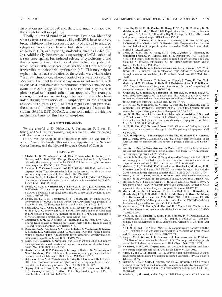
10.1128/MCB.20.18.6731-6740.2000. 2000, 20(18):6731. DOI: Mol. Cell. Biol. Gordon C. Shore Mai Nguyen, David G. Breckenridge, Axel Ducret and from Mitochondria c Fragmentation and Release of Cytochrome Fas-Mediated Apoptotic Membrane Caspase-Resistant BAP31 Inhibits http://mcb.asm.org/content/20/18/6731 Updated information and services can be found at: These include: REFERENCES http://mcb.asm.org/content/20/18/6731#ref-list-1 at: This article cites 48 articles, 27 of which can be accessed free CONTENT ALERTS more» articles cite this article), Receive: RSS Feeds, eTOCs, free email alerts (when new http://journals.asm.org/site/misc/reprints.xhtml Information about commercial reprint orders: http://journals.asm.org/site/subscriptions/ To subscribe to to another ASM Journal go to: on July 26, 2014 by guest http://mcb.asm.org/ Downloaded from on July 26, 2014 by guest http://mcb.asm.org/ Downloaded from

Welcome message from author
This document is posted to help you gain knowledge. Please leave a comment to let me know what you think about it! Share it to your friends and learn new things together.
Transcript
10.1128/MCB.20.18.6731-6740.2000.
2000, 20(18):6731. DOI:Mol. Cell. Biol. Gordon C. ShoreMai Nguyen, David G. Breckenridge, Axel Ducret and from Mitochondriac
Fragmentation and Release of Cytochrome Fas-Mediated Apoptotic Membrane Caspase-Resistant BAP31 Inhibits
http://mcb.asm.org/content/20/18/6731Updated information and services can be found at:
These include:
REFERENCEShttp://mcb.asm.org/content/20/18/6731#ref-list-1at:
This article cites 48 articles, 27 of which can be accessed free
CONTENT ALERTS more»articles cite this article),
Receive: RSS Feeds, eTOCs, free email alerts (when new
http://journals.asm.org/site/misc/reprints.xhtmlInformation about commercial reprint orders: http://journals.asm.org/site/subscriptions/To subscribe to to another ASM Journal go to:
on July 26, 2014 by guesthttp://m
cb.asm.org/
Dow
nloaded from
on July 26, 2014 by guesthttp://m
cb.asm.org/
Dow
nloaded from
MOLECULAR AND CELLULAR BIOLOGY,0270-7306/00/$04.0010
Sept. 2000, p. 6731–6740 Vol. 20, No. 18
Copyright © 2000, American Society for Microbiology. All Rights Reserved.
Caspase-Resistant BAP31 Inhibits Fas-Mediated ApoptoticMembrane Fragmentation and Release of Cytochrome c
from MitochondriaMAI NGUYEN, DAVID G. BRECKENRIDGE, AXEL DUCRET, AND GORDON C. SHORE*
Department of Biochemistry, McGill University, Montreal, Quebec, Canada H3G 1Y6, andMerck-Frosst Center for Therapeutic Research, Kirkland, Quebec, Canada H9H 3L1
Received 3 May 2000/Returned for modification 5 June 2000/Accepted 12 June 2000
BAP31 is a 28-kDa integral membrane protein of the endoplasmic reticulum whose cytosolic domaincontains two identical caspase recognition sites (AAVD.G) that are preferentially cleaved by initiator caspases,including caspase 8. Cleavage of BAP31 during apoptosis generates a p20 fragment that remains integrated inthe membrane and, when expressed ectopically, is a potent inducer of cell death. To examine the consequencesof maintaining the structural integrity of BAP31 during apoptosis, the caspase recognition aspartate residueswere mutated to alanine residues, and Fas-mediated activation of caspase 8 and cell death were examined inhuman KB epithelial cells stably expressing the caspase-resistant mutant crBAP31. crBAP31 only modestlyslowed the time course for activation of caspases, as assayed by the processing of procaspases 8 and 3 and themeasurement of total DEVDase activity. As a result, cleavage of the caspase targets poly(ADP-ribosyl) poly-merase and endogenous BAP31, as well as the redistribution of phosphatidylserine and fragmentation of DNA,was observed. In contrast, cytoplasmic membrane blebbing and fragmentation and apoptotic redistribution ofactin were strongly inhibited, cell morphology was retained near normal, and the irreversible loss of cell growthpotential following removal of the Fas stimulus was delayed. Of note, crBAP31-expressing cells also resistedFas-mediated release of cytochrome c from mitochondria, and the mitochondrial electrochemical potential wasonly partly reduced. These results argue that BAP31 cleavage is important for manifesting cytoplasmicapoptotic events associated with membrane fragmentation and reveal an unexpected cross talk betweenmitochondria and the endoplasmic reticulum during Fas-mediated apoptosis in vivo.
Programmed cell death is characterized by a series of mor-phological and structural changes culminating in the coordi-nated packaging of cellular contents into apoptotic bodies,which are ultimately eliminated via phagocytosis by neighbor-ing cells. Early events in this process typically include cellrounding, loss of phospholipid asymmetry in the cell mem-brane, extensive cytoplasmic membrane blebbing and fragmen-tation, nuclear pyknosis, and internucleosomal DNA cleavage(26). Although much remains to be learned about the mecha-nisms underlying these events, programmed cell death isachieved in most cell death pathways as a consequence of theproteolytic cleavage of a diverse array of structural and regu-latory proteins by the executors of apoptosis, the caspase fam-ily of cysteine proteases (33, 46, 48). Several of the caspasetargets are known to have critical roles in at least some of theapoptotic processes. These targets include the DFF40/CADinhibitor, DFF45/ICAD, which plays a role in the fragmenta-tion of DNA (23, 36), and the actin-associated capping proteingelsolin, which plays a role in cytoplasmic membrane blebbing(17). In addition, the activation of several kinases by caspasecleavage, including PAK2 (20, 34) and the Ste20-related ki-nases MST1 (14, 19) and SLK (35), contributes to the loss offocal adhesions and the retraction and disassembly of actinstress fibers, events that are associated with elaborate changesto the actomyosin network and membrane remodeling (26).
In contrast to many death-stimulating pathways, the proxi-mal molecular events following activation of Fas (also called
CD95) with either Fas ligand or agonistic anti-Fas antibody arewell understood. Receptor stimulation results in the assemblyof a death-inducing signaling complex that includes theadapter protein FADD and procaspase 8 (4, 5, 7, 28, 40). Theresulting activation of this initiator caspase (25) ultimatelyleads to the processing of effector procaspases, includingcaspase 3, and apoptosis. A cytosolic target of caspase 8, pro-apoptotic BID, is cleaved, and the truncated product (tBID)translocates to mitochondria, where it stimulates the release ofintermembrane proteins, including cytochrome c (15, 21, 24).BID appears to be a critical effector of this pathway, at least incertain contexts, because death receptor-induced redistribu-tion of cytochrome c was not observed in Bid2/2 mouse thy-mocytes and hepatocytes (47). Released cytochrome c in turncontributes to the activation of initiator caspase 9 (22), whichin many death pathways is important for subsequent processingof downstream effector procaspases (22, 39). In contrast, Fasstimulation of certain cell types activates high levels of caspase8 that are sufficient to process effector procaspases directly,whereas other cell types, at least in culture, activate low levelsof procaspase 8 and likely depend on mitochondrial events foramplification of the caspase cascade (37, 38).
Here we show that human epithelial cells expressing acaspase-resistant mutant of BAP31 (crBAP31), a preferredsubstrate for initiator caspases 8 and 1 (30), are resistant to anumber of cytoplasmic changes that typically occur during Fas-mediated apoptosis. BAP31 is a 28-kDa integral membraneprotein that is ubiquitously expressed (1, 27; unpublished data)and highly enriched in the endoplasmic reticulum (ER) (3, 30),where it forms a homo-oligomer (31). The protein containsthree predicted transmembrane segments within its amino ter-minus that confer a topology in the ER membrane in which the
* Corresponding author. Mailing address: Department of Biochem-istry, McIntyre Medical Sciences Building, McGill University, 3655Drummond St., Montreal, Quebec, Canada H3G 1Y6. Phone: (514)398-7282. Fax: (514) 398-7384. E-mail: [email protected].
6731
on July 26, 2014 by guesthttp://m
cb.asm.org/
Dow
nloaded from
short hydrophilic amino terminus and an approximately 37-amino-acid loop connecting TM2 and TM3 face the ER lumenand a 14-kDa domain, terminating in a canonical KKXX ERretention signal, faces the cytosol (30). The cytosolic tail ispredicted to contain a weak death effector and overlappingcoiled-coil (DECC) domain flanked on either side by identicalcaspase recognition sites. These sites are cleaved in response todiverse death stimuli in vivo (13, 31) and are preferably cleavedby caspases 8 and 1 and only weakly cleaved by effector caspase3 in vitro (30). The resulting membrane-integrated BAP31fragment, called p20, is a potent inducer of apoptosis whenexpressed ectopically, suggesting that BAP31 cleavage maycontribute in some manner to the death process. Moreover, theinfluence of BAP31 may also extend to a role in regulatingcaspases, which would be consistent with the observation thatBAP31 can associate with procaspase 8, BCL-2, and CED-4 incotransfected cells (30, 31). To investigate the contribution ofBAP31 cleavage to apoptosis, therefore, we have created a cellline that expresses a caspase-resistant mutant form of the pro-tein and have examined its influence on the cell death pathwayinitiated by independent stimulation of caspase 8 activity by theFas signaling complex.
MATERIALS AND METHODS
Plasmids and transfectants. cDNA encoding human BAP31 with the Flagpeptide epitope sequence inserted between the codons for amino acids 242 and243 was incorporated into the pcDNA3.1 expression vector, as previously docu-mented (30). Site-directed mutagenesis was performed to convert the codons foraspartate residues at positions 164 and 238 to codons for alanine residues, andthe authenticity of the crBAP31-Flag mutant expression vector was confirmed byDNA sequence analysis. pcDNA3.1 vectors expressing CrmA (gift from V. Dix-it), BAP31-Flag, and crBAP31-Flag were stably expressed in human KB epithe-lial cells, as previously described (30).
Antibodies and immunoblots. The following antibodies were employed: mouseanti-Flag (Sigma), chicken anti-human BAP31 (30), rabbit antibodies against thecatalytic subunits of human caspase 8 (gift from D. Nicholson) and caspase 3(gift from R. Sekaly), mouse anti-poly (ADP-ribosyl) polymerase (anti-PARP)(Biomol), mouse monoclonal antibodies against cytochrome c (2G8.B6 and7H8.2C12) (gift from R. Jemmerson), and rabbit anti-g-actin (gift from P.Braun). Cell extracts containing equivalent amounts of protein were resolved bysodium dodecyl sulfate-polyacrylamide gel electrophoresis (SDS-PAGE), andthe proteins were transferred to nitrocellulose, incubated with a primary anti-body, and visualized with a secondary antibody coupled to enhanced chemilu-minescence.
Immunofluorescence. Cells were fixed in 4% paraformaldehyde and incubatedeither with mouse monoclonal antibody 2G8.B6 (anti-cytochrome c) followed bygoat anti-mouse immunoglobulin G (IgG) conjugated to Texas red or withanti-g-actin followed by goat anti-rabbit IgG coupled to Alexa 488 (MolecularProbes). Cells were visualized by fluorescence confocal microscopy.
Electron microscopy. Cells were treated with anti-Fas antibody for varioustimes. After two washes in phosphate-buffered saline (PBS), cell pellets werefixed in 2.5% glutaraldehyde–0.1 M sodium cacodylate for 2 h, washed, treatedfor 1 h with 0.1% osmium tetroxide, and finally dehydrated in acetone. Thepellets were infiltrated with Epon-acetone and embedded in Epon. Thin sections(approximately 100 nm thick) stained with 4% uranyl acetate–lead citrate andthick sections (approximately 0.5 mm thick) stained with toluidine blue wereexamined by electron and conventional light microscopy, respectively.
Apoptosis assays. Human KB epithelial cells were maintained in a-minimalessential medium supplemented with 10% fetal bovine serum and treated at ap-proximately 80% confluency with mouse activating anti-Fas antibody (Upstate Bio-technology) and 10 mg of cycloheximide (CHX) per ml. At the times indicatedbelow, cells were collected and analyzed. Cell viability and the structural integrity ofthe plasma membrane were measured by the ability of cells to exclude trypan blue,as determined microscopically. For assessing phosphatidylserine redistribution andmitochondrial transmembrane potential, cells were incubated for 15 min at 37°C inPBS containing 2% fetal bovine serum and a 40 mM concentration of the mitochon-drial-potential-sensitive dye DiOC6 (Molecular Probes) or 0.1 mM fluorescein-con-jugated human annexin V (R & D). Following two washes in the same medium,fluorescence was measured by flow cytometry. Caspase activity in whole-cell extractswas obtained by treating the cells with a solution containing 50 mM HEPES (pH7.4), 1% Triton X-100, 5 mM EDTA, and 2 mM dithiothreitol and incubating themwith 50 mM acetyl-Asp-Glu-Val-Asp-7-amino-4-methylcoumarin (AcDEVD-amc)for 30 min at 37°C. Fluorescence in the linear range of DEVDase activity wasmeasured with a plate reader (Tecan).
Cytochrome c. Cells (4 3 106) were washed in PBS and suspended in 0.1 ml of200 mM mannitol–70 mM sucrose–1 mM EDTA–10 mM HEPES (pH 7.5). After
one cycle of freeze-thaw, the cells were homogenized with 35 strokes in amotorized Teflon-glass homogenizer operating at 2,000 rpm and then centri-fuged at 800 3 g for 10 min to remove nuclei and cell debris. The supernatant wascentrifuged at 100,000 3 g for 10 min, and the membrane pellet was resuspendedin homogenization buffer to the same volume as that of the 100,000 3 g super-natant. Equivalent volumes of the pellets and supernatants were subjected toSDS-PAGE and immunoblotting with mouse monoclonal antibody 7H8.2C12(anti-cytochrome c).
RESULTS
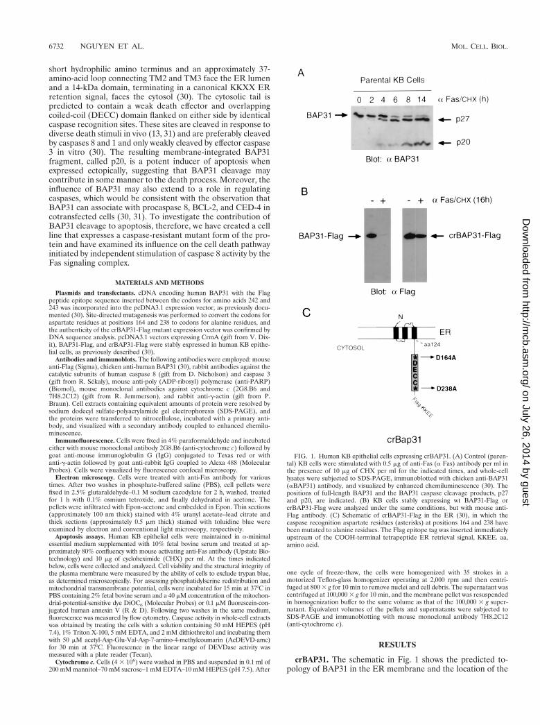
crBAP31. The schematic in Fig. 1 shows the predicted to-pology of BAP31 in the ER membrane and the location of the
FIG. 1. Human KB epithelial cells expressing crBAP31. (A) Control (paren-tal) KB cells were stimulated with 0.5 mg of anti-Fas (a Fas) antibody per ml inthe presence of 10 mg of CHX per ml for the indicated times, and whole-celllysates were subjected to SDS-PAGE, immunoblotted with chicken anti-BAP31(aBAP31) antibody, and visualized by enhanced chemiluminescence (30). Thepositions of full-length BAP31 and the BAP31 caspase cleavage products, p27and p20, are indicated. (B) KB cells stably expressing wt BAP31-Flag orcrBAP31-Flag were analyzed under the same conditions, but with mouse anti-Flag antibody. (C) Schematic of crBAP31-Flag in the ER (30), in which thecaspase recognition aspartate residues (asterisks) at positions 164 and 238 havebeen mutated to alanine residues. The Flag epitope tag was inserted immediatelyupstream of the COOH-terminal tetrapeptide ER retrieval signal, KKEE. aa,amino acid.
6732 NGUYEN ET AL. MOL. CELL. BIOL.
on July 26, 2014 by guesthttp://m
cb.asm.org/
Dow
nloaded from
caspase recognition sites (AAVD.G) flanking the DECC do-main in the cytosolic tail (30). Treatment of human KB epi-thelial cells with agonistic mouse antibody against Fas (0.5mg/ml), in the presence of 10 mg of CHX per ml to enhancesensitivity to Fas activation (38), resulted in cleavage of the246-amino-acid full-length BAP31, generating two products ofapproximately 27 and 20 kDa (Fig. 1A). These correspondedto cleavages at aspartates 238 and 164, respectively (30).crBAP31 was produced by mutating these residues to alanineresidues, and both the construct encoding crBAP31 and thatencoding wild-type (wt) BAP31 were further manipulated toinclude a Flag epitope inserted immediately upstream of theER retention signal, KKEE, located at the extreme carboxyterminus of the protein. KB cell lines stably expressing wtBAP31 or crBAP31 were then examined by immunoblottingwith anti-Flag antibody. crBAP31-Flag, but not BAP31-Flag,was found to remain structurally intact in the face of sustainedstimulation with anti-Fas (Fig. 1B). In both cases, the proteinswere expressed at approximately three times the level of en-dogenous BAP31, as assessed using a BAP31 polyclonal anti-body (data not shown).
crBAP31 inhibits Fas-mediated apoptotic membrane bleb-bing and fragmentation. Control (parental) KB epithelial cellsand KB cells stably expressing crBAP31-Flag or the cowpox
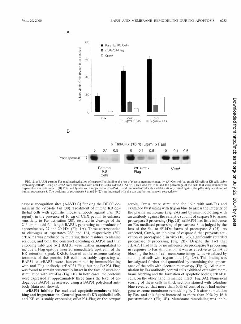
serpin, CrmA, were stimulated for 16 h with anti-Fas andexamined by staining with trypan blue to assess the integrity ofthe plasma membrane (Fig. 2A) and by immunoblotting withan antibody against the catalytic subunit of caspase 8 to assessprocaspase 8 processing (Fig. 2B). crBAP31 had little influenceon Fas-mediated processing of procaspase 8, as judged by theloss of the 54- to 55-kDa forms of procaspase 8 (25). Asexpected, CrmA, an inhibitor of caspase 8 that prevents acti-vation of procaspase 8 in vivo (10, 28), significantly retardedprocaspase 8 processing (Fig. 2B). Despite the fact thatcrBAP31 had little or no influence on procaspase 8 processingin response to Fas stimulation, it was as effective as CrmA atblocking the loss of cell membrane integrity, as visualized bystaining of cells with trypan blue (Fig. 2A). This finding wasinvestigated further and quantified by examining the appear-ance of the cells with electron microscopy (Fig. 3). After stim-ulation by Fas antibody, control cells exhibited extensive mem-brane blebbing and the formation of apoptotic bodies; crBAP31cells, on the other hand, remained intact (Fig. 3A). Numericalscoring of these cells in thick sections stained with toluidineblue revealed that more than 60% of control cells had under-gone extreme membrane remodeling by 7 h after stimulationby Fas, and this figure increased to more than 90% by 16 hpoststimulation (Fig. 3B). Membrane remodeling was inhib-
FIG. 2. crBAP31 permits Fas-mediated activation of caspase 8 but inhibits the loss of plasma membrane integrity. (A) Control (parental) KB cells or KB cells stablyexpressing crBAP31-Flag or CrmA were stimulated with anti-Fas–CHX (aFas/CHX) or CHX alone for 16 h, and the percentage of the cells that were stained withtrypan blue was determined. (B) Total cell lysates were subjected to SDS-PAGE and immunoblotted with a rabbit antibody raised against the p18 catalytic subunit ofhuman procaspase 8. The positions of procaspase 8 a and b (25) are indicated with the top and bottom arrows, respectively.
VOL. 20, 2000 BAP31 AND MEMBRANE REMODELING DURING APOPTOSIS 6733
on July 26, 2014 by guesthttp://m
cb.asm.org/
Dow
nloaded from
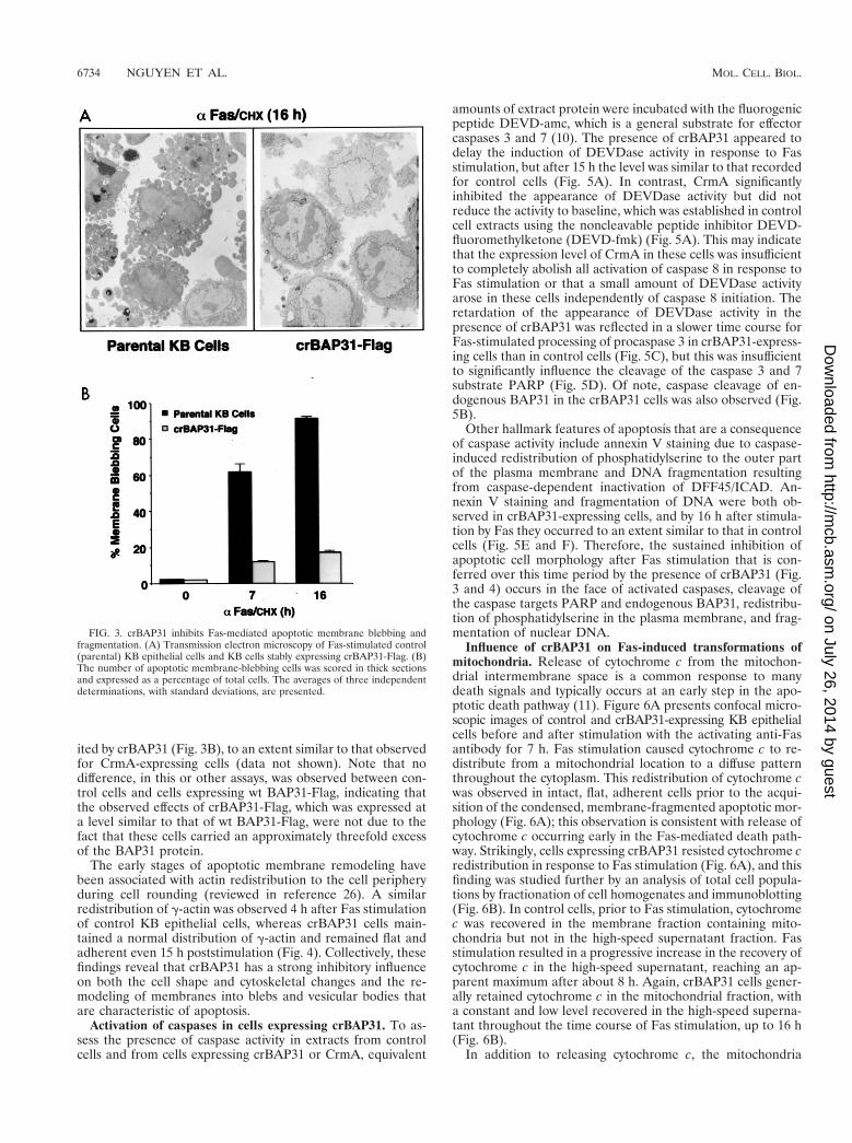
ited by crBAP31 (Fig. 3B), to an extent similar to that observedfor CrmA-expressing cells (data not shown). Note that nodifference, in this or other assays, was observed between con-trol cells and cells expressing wt BAP31-Flag, indicating thatthe observed effects of crBAP31-Flag, which was expressed ata level similar to that of wt BAP31-Flag, were not due to thefact that these cells carried an approximately threefold excessof the BAP31 protein.
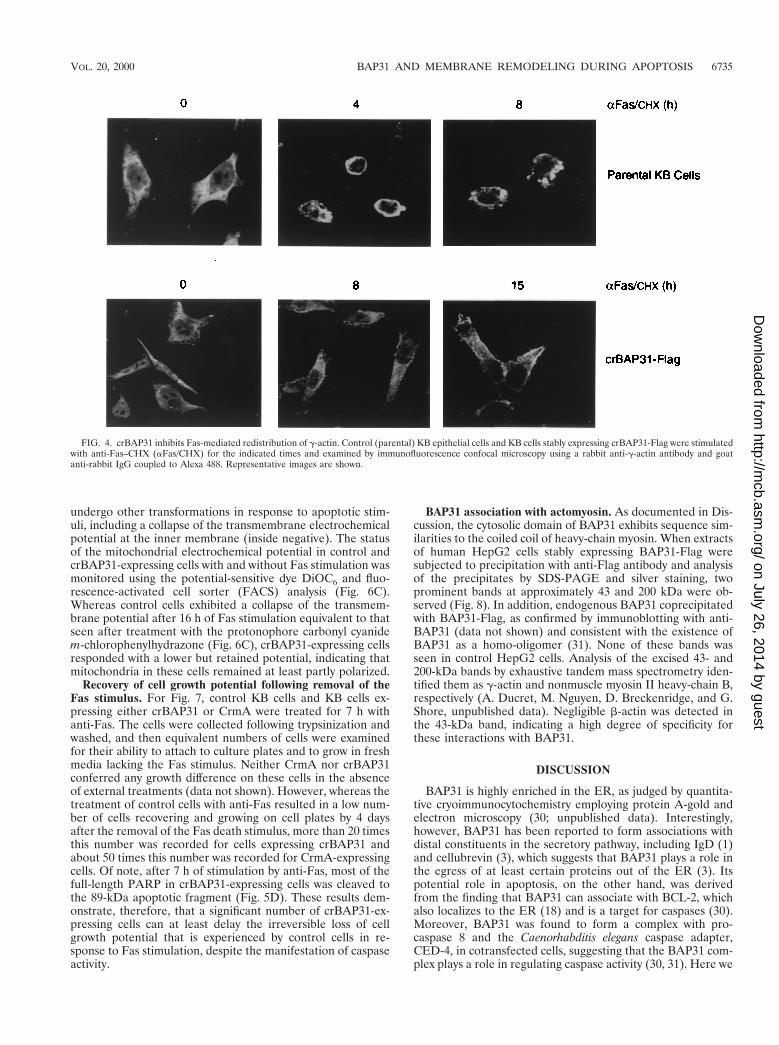
The early stages of apoptotic membrane remodeling havebeen associated with actin redistribution to the cell peripheryduring cell rounding (reviewed in reference 26). A similarredistribution of g-actin was observed 4 h after Fas stimulationof control KB epithelial cells, whereas crBAP31 cells main-tained a normal distribution of g-actin and remained flat andadherent even 15 h poststimulation (Fig. 4). Collectively, thesefindings reveal that crBAP31 has a strong inhibitory influenceon both the cell shape and cytoskeletal changes and the re-modeling of membranes into blebs and vesicular bodies thatare characteristic of apoptosis.
Activation of caspases in cells expressing crBAP31. To as-sess the presence of caspase activity in extracts from controlcells and from cells expressing crBAP31 or CrmA, equivalent
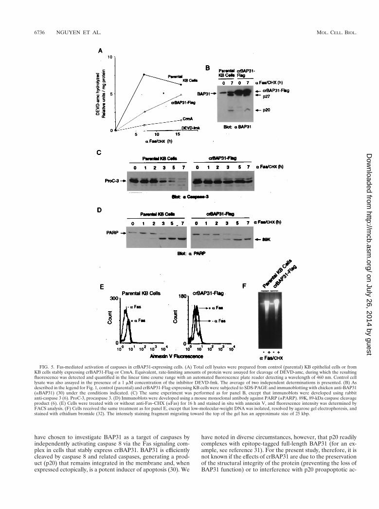
amounts of extract protein were incubated with the fluorogenicpeptide DEVD-amc, which is a general substrate for effectorcaspases 3 and 7 (10). The presence of crBAP31 appeared todelay the induction of DEVDase activity in response to Fasstimulation, but after 15 h the level was similar to that recordedfor control cells (Fig. 5A). In contrast, CrmA significantlyinhibited the appearance of DEVDase activity but did notreduce the activity to baseline, which was established in controlcell extracts using the noncleavable peptide inhibitor DEVD-fluoromethylketone (DEVD-fmk) (Fig. 5A). This may indicatethat the expression level of CrmA in these cells was insufficientto completely abolish all activation of caspase 8 in response toFas stimulation or that a small amount of DEVDase activityarose in these cells independently of caspase 8 initiation. Theretardation of the appearance of DEVDase activity in thepresence of crBAP31 was reflected in a slower time course forFas-stimulated processing of procaspase 3 in crBAP31-express-ing cells than in control cells (Fig. 5C), but this was insufficientto significantly influence the cleavage of the caspase 3 and 7substrate PARP (Fig. 5D). Of note, caspase cleavage of en-dogenous BAP31 in the crBAP31 cells was also observed (Fig.5B).
Other hallmark features of apoptosis that are a consequenceof caspase activity include annexin V staining due to caspase-induced redistribution of phosphatidylserine to the outer partof the plasma membrane and DNA fragmentation resultingfrom caspase-dependent inactivation of DFF45/ICAD. An-nexin V staining and fragmentation of DNA were both ob-served in crBAP31-expressing cells, and by 16 h after stimula-tion by Fas they occurred to an extent similar to that in controlcells (Fig. 5E and F). Therefore, the sustained inhibition ofapoptotic cell morphology after Fas stimulation that is con-ferred over this time period by the presence of crBAP31 (Fig.3 and 4) occurs in the face of activated caspases, cleavage ofthe caspase targets PARP and endogenous BAP31, redistribu-tion of phosphatidylserine in the plasma membrane, and frag-mentation of nuclear DNA.
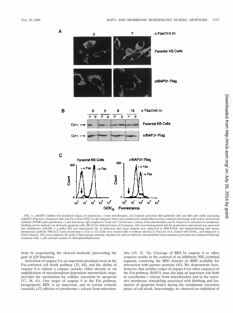
Influence of crBAP31 on Fas-induced transformations ofmitochondria. Release of cytochrome c from the mitochon-drial intermembrane space is a common response to manydeath signals and typically occurs at an early step in the apo-ptotic death pathway (11). Figure 6A presents confocal micro-scopic images of control and crBAP31-expressing KB epithelialcells before and after stimulation with the activating anti-Fasantibody for 7 h. Fas stimulation caused cytochrome c to re-distribute from a mitochondrial location to a diffuse patternthroughout the cytoplasm. This redistribution of cytochrome cwas observed in intact, flat, adherent cells prior to the acqui-sition of the condensed, membrane-fragmented apoptotic mor-phology (Fig. 6A); this observation is consistent with release ofcytochrome c occurring early in the Fas-mediated death path-way. Strikingly, cells expressing crBAP31 resisted cytochrome credistribution in response to Fas stimulation (Fig. 6A), and thisfinding was studied further by an analysis of total cell popula-tions by fractionation of cell homogenates and immunoblotting(Fig. 6B). In control cells, prior to Fas stimulation, cytochromec was recovered in the membrane fraction containing mito-chondria but not in the high-speed supernatant fraction. Fasstimulation resulted in a progressive increase in the recovery ofcytochrome c in the high-speed supernatant, reaching an ap-parent maximum after about 8 h. Again, crBAP31 cells gener-ally retained cytochrome c in the mitochondrial fraction, witha constant and low level recovered in the high-speed superna-tant throughout the time course of Fas stimulation, up to 16 h(Fig. 6B).
In addition to releasing cytochrome c, the mitochondria
FIG. 3. crBAP31 inhibits Fas-mediated apoptotic membrane blebbing andfragmentation. (A) Transmission electron microscopy of Fas-stimulated control(parental) KB epithelial cells and KB cells stably expressing crBAP31-Flag. (B)The number of apoptotic membrane-blebbing cells was scored in thick sectionsand expressed as a percentage of total cells. The averages of three independentdeterminations, with standard deviations, are presented.
6734 NGUYEN ET AL. MOL. CELL. BIOL.
on July 26, 2014 by guesthttp://m
cb.asm.org/
Dow
nloaded from
undergo other transformations in response to apoptotic stim-uli, including a collapse of the transmembrane electrochemicalpotential at the inner membrane (inside negative). The statusof the mitochondrial electrochemical potential in control andcrBAP31-expressing cells with and without Fas stimulation wasmonitored using the potential-sensitive dye DiOC6 and fluo-rescence-activated cell sorter (FACS) analysis (Fig. 6C).Whereas control cells exhibited a collapse of the transmem-brane potential after 16 h of Fas stimulation equivalent to thatseen after treatment with the protonophore carbonyl cyanidem-chlorophenylhydrazone (Fig. 6C), crBAP31-expressing cellsresponded with a lower but retained potential, indicating thatmitochondria in these cells remained at least partly polarized.
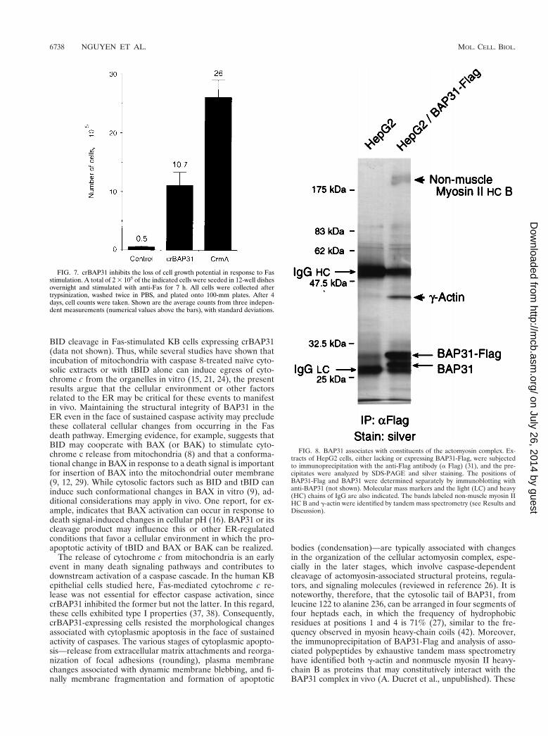
Recovery of cell growth potential following removal of theFas stimulus. For Fig. 7, control KB cells and KB cells ex-pressing either crBAP31 or CrmA were treated for 7 h withanti-Fas. The cells were collected following trypsinization andwashed, and then equivalent numbers of cells were examinedfor their ability to attach to culture plates and to grow in freshmedia lacking the Fas stimulus. Neither CrmA nor crBAP31conferred any growth difference on these cells in the absenceof external treatments (data not shown). However, whereas thetreatment of control cells with anti-Fas resulted in a low num-ber of cells recovering and growing on cell plates by 4 daysafter the removal of the Fas death stimulus, more than 20 timesthis number was recorded for cells expressing crBAP31 andabout 50 times this number was recorded for CrmA-expressingcells. Of note, after 7 h of stimulation by anti-Fas, most of thefull-length PARP in crBAP31-expressing cells was cleaved tothe 89-kDa apoptotic fragment (Fig. 5D). These results dem-onstrate, therefore, that a significant number of crBAP31-ex-pressing cells can at least delay the irreversible loss of cellgrowth potential that is experienced by control cells in re-sponse to Fas stimulation, despite the manifestation of caspaseactivity.
BAP31 association with actomyosin. As documented in Dis-cussion, the cytosolic domain of BAP31 exhibits sequence sim-ilarities to the coiled coil of heavy-chain myosin. When extractsof human HepG2 cells stably expressing BAP31-Flag weresubjected to precipitation with anti-Flag antibody and analysisof the precipitates by SDS-PAGE and silver staining, twoprominent bands at approximately 43 and 200 kDa were ob-served (Fig. 8). In addition, endogenous BAP31 coprecipitatedwith BAP31-Flag, as confirmed by immunoblotting with anti-BAP31 (data not shown) and consistent with the existence ofBAP31 as a homo-oligomer (31). None of these bands wasseen in control HepG2 cells. Analysis of the excised 43- and200-kDa bands by exhaustive tandem mass spectrometry iden-tified them as g-actin and nonmuscle myosin II heavy-chain B,respectively (A. Ducret, M. Nguyen, D. Breckenridge, and G.Shore, unpublished data). Negligible b-actin was detected inthe 43-kDa band, indicating a high degree of specificity forthese interactions with BAP31.
DISCUSSION
BAP31 is highly enriched in the ER, as judged by quantita-tive cryoimmunocytochemistry employing protein A-gold andelectron microscopy (30; unpublished data). Interestingly,however, BAP31 has been reported to form associations withdistal constituents in the secretory pathway, including IgD (1)and cellubrevin (3), which suggests that BAP31 plays a role inthe egress of at least certain proteins out of the ER (3). Itspotential role in apoptosis, on the other hand, was derivedfrom the finding that BAP31 can associate with BCL-2, whichalso localizes to the ER (18) and is a target for caspases (30).Moreover, BAP31 was found to form a complex with pro-caspase 8 and the Caenorhabditis elegans caspase adapter,CED-4, in cotransfected cells, suggesting that the BAP31 com-plex plays a role in regulating caspase activity (30, 31). Here we
FIG. 4. crBAP31 inhibits Fas-mediated redistribution of g-actin. Control (parental) KB epithelial cells and KB cells stably expressing crBAP31-Flag were stimulatedwith anti-Fas–CHX (aFas/CHX) for the indicated times and examined by immunofluorescence confocal microscopy using a rabbit anti-g-actin antibody and goatanti-rabbit IgG coupled to Alexa 488. Representative images are shown.
VOL. 20, 2000 BAP31 AND MEMBRANE REMODELING DURING APOPTOSIS 6735
on July 26, 2014 by guesthttp://m
cb.asm.org/
Dow
nloaded from
have chosen to investigate BAP31 as a target of caspases byindependently activating caspase 8 via the Fas signaling com-plex in cells that stably express crBAP31. BAP31 is efficientlycleaved by caspase 8 and related caspases, generating a prod-uct (p20) that remains integrated in the membrane and, whenexpressed ectopically, is a potent inducer of apoptosis (30). We
have noted in diverse circumstances, however, that p20 readilycomplexes with epitope-tagged full-length BAP31 (for an ex-ample, see reference 31). For the present study, therefore, it isnot known if the effects of crBAP31 are due to the preservationof the structural integrity of the protein (preventing the loss ofBAP31 function) or to interference with p20 proapoptotic ac-
FIG. 5. Fas-mediated activation of caspases in crBAP31-expressing cells. (A) Total cell lysates were prepared from control (parental) KB epithelial cells or fromKB cells stably expressing crBAP31-Flag or CrmA. Equivalent, rate-limiting amounts of protein were assayed for cleavage of DEVD-amc, during which the resultingfluorescence was detected and quantified in the linear time course range with an automated fluorescence plate reader detecting a wavelength of 460 nm. Control celllysate was also assayed in the presence of a 1 mM concentration of the inhibitor DEVD-fmk. The average of two independent determinations is presented. (B) Asdescribed in the legend for Fig. 1, control (parental) and crBAP31-Flag-expressing KB cells were subjected to SDS-PAGE and immunoblotting with chicken anti-BAP31(aBAP31) (30) under the conditions indicated. (C) The same experiment was performed as for panel B, except that immunoblots were developed using rabbitanti-caspase 3 (6). ProC-3, procaspase 3. (D) Immunoblots were developed using a mouse monoclonal antibody against PARP (aPARP). 89K, 89-kDa caspase cleavageproduct (6). (E) Cells were treated with or without anti-Fas–CHX (aFas) for 16 h and stained in situ with annexin V, and fluorescence intensity was determined byFACS analysis. (F) Cells received the same treatment as for panel E, except that low-molecular-weight DNA was isolated, resolved by agarose gel electrophoresis, andstained with ethidium bromide (32). The intensely staining fragment migrating toward the top of the gel has an approximate size of 25 kbp.
6736 NGUYEN ET AL. MOL. CELL. BIOL.
on July 26, 2014 by guesthttp://m
cb.asm.org/
Dow
nloaded from
tivity by sequestering the cleaved molecule (preventing thegain of p20 function).
Activation of caspase 8 is an important proximal event in theFas-activated cell death pathway (25, 44), and the ability ofcaspase 8 to initiate a caspase cascade, either directly or viaamplification of mitochondrion-dependent intermediate steps,provides the mechanism for cellular execution by apoptosis(37, 38, 41). One target of caspase 8 in the Fas pathway,proapoptotic BID, is an important, and in certain contextsessential, (47) effector of cytochrome c release from mitochon-
dria (15, 21, 24). Cleavage of BID by caspase 8 or othercaspases results in the removal of an inhibitory NH2-terminalsegment, rendering the BH3 domain of tBID available forinteraction with partner proteins (45). We demonstrate here,however, that another target of caspase 8 (or other caspases) inthe Fas pathway, BAP31, may also play an important role bothin cytochrome c release from mitochondria and in the exten-sive membrane remodeling associated with blebbing and for-mation of apoptotic bodies during the cytoplasmic executionphase of cell death. Interestingly, we observed no inhibition of
FIG. 6. crBAP31 inhibits Fas-mediated release of cytochrome c from mitochondria. (A) Control (parental) KB epithelial cells and KB cells stably expressingcrBAP31-Flag were stimulated with anti-Fas (aFas/CHX) for the indicated times and examined by immunofluorescence confocal microscopy with mouse monoclonalantibody 2G8.B6 (anti-cytochrome c) and anti-mouse IgG coupled to Texas red. Cytochrome c release from mitochondria can be observed in cells prior to membraneblebbing (arrow indicates an obviously apoptotic cell). (B) At the indicated times of treatment, cells were homogenized and the postnuclear supernatant was separatedinto membranes (100,000 3 g pellet [P]) and supernatant (S), as indicated, and equal aliquots were subjected to SDS-PAGE and immunoblotting with mousemonoclonal antibody 7H8.2C12 (anti-cytochrome c) (Cyt c). (C) Cells were treated with or without anti-Fas (a Fas) for 16 h, stained with DiOC6, and subjected toFACS analysis. The arrow indicates the peak of fluorescence intensity obtained for cells in which the mitochondrial electrochemical potential was collapsed followingtreatment with 1 mM carbonyl cyanide m-chlorophenylhydrazone.
VOL. 20, 2000 BAP31 AND MEMBRANE REMODELING DURING APOPTOSIS 6737
on July 26, 2014 by guesthttp://m
cb.asm.org/
Dow
nloaded from
BID cleavage in Fas-stimulated KB cells expressing crBAP31(data not shown). Thus, while several studies have shown thatincubation of mitochondria with caspase 8-treated naıve cyto-solic extracts or with tBID alone can induce egress of cyto-chrome c from the organelles in vitro (15, 21, 24), the presentresults argue that the cellular environment or other factorsrelated to the ER may be critical for these events to manifestin vivo. Maintaining the structural integrity of BAP31 in theER even in the face of sustained caspase activity may precludethese collateral cellular changes from occurring in the Fasdeath pathway. Emerging evidence, for example, suggests thatBID may cooperate with BAX (or BAK) to stimulate cyto-chrome c release from mitochondria (8) and that a conforma-tional change in BAX in response to a death signal is importantfor insertion of BAX into the mitochondrial outer membrane(9, 12, 29). While cytosolic factors such as BID and tBID caninduce such conformational changes in BAX in vitro (9), ad-ditional considerations may apply in vivo. One report, for ex-ample, indicates that BAX activation can occur in response todeath signal-induced changes in cellular pH (16). BAP31 or itscleavage product may influence this or other ER-regulatedconditions that favor a cellular environment in which the pro-apoptotic activity of tBID and BAX or BAK can be realized.
The release of cytochrome c from mitochondria is an earlyevent in many death signaling pathways and contributes todownstream activation of a caspase cascade. In the human KBepithelial cells studied here, Fas-mediated cytochrome c re-lease was not essential for effector caspase activation, sincecrBAP31 inhibited the former but not the latter. In this regard,these cells exhibited type I properties (37, 38). Consequently,crBAP31-expressing cells resisted the morphological changesassociated with cytoplasmic apoptosis in the face of sustainedactivity of caspases. The various stages of cytoplasmic apopto-sis—release from extracellular matrix attachments and reorga-nization of focal adhesions (rounding), plasma membranechanges associated with dynamic membrane blebbing, and fi-nally membrane fragmentation and formation of apoptotic
bodies (condensation)—are typically associated with changesin the organization of the cellular actomyosin complex, espe-cially in the later stages, which involve caspase-dependentcleavage of actomyosin-associated structural proteins, regula-tors, and signaling molecules (reviewed in reference 26). It isnoteworthy, therefore, that the cytosolic tail of BAP31, fromleucine 122 to alanine 236, can be arranged in four segments offour heptads each, in which the frequency of hydrophobicresidues at positions 1 and 4 is 71% (27), similar to the fre-quency observed in myosin heavy-chain coils (42). Moreover,the immunoprecipitation of BAP31-Flag and analysis of asso-ciated polypeptides by exhaustive tandem mass spectrometryhave identified both g-actin and nonmuscle myosin II heavy-chain B as proteins that may constitutively interact with theBAP31 complex in vivo (A. Ducret et al., unpublished). These
FIG. 7. crBAP31 inhibits the loss of cell growth potential in response to Fasstimulation. A total of 2 3 105 of the indicated cells were seeded in 12-well dishesovernight and stimulated with anti-Fas for 7 h. All cells were collected aftertrypsinization, washed twice in PBS, and plated onto 100-mm plates. After 4days, cell counts were taken. Shown are the average counts from three indepen-dent measurements (numerical values above the bars), with standard deviations.
FIG. 8. BAP31 associates with constituents of the actomyosin complex. Ex-tracts of HepG2 cells, either lacking or expressing BAP31-Flag, were subjectedto immunoprecipitation with the anti-Flag antibody (a Flag) (31), and the pre-cipitates were analyzed by SDS-PAGE and silver staining. The positions ofBAP31-Flag and BAP31 were determined separately by immunoblotting withanti-BAP31 (not shown). Molecular mass markers and the light (LC) and heavy(HC) chains of IgG are also indicated. The bands labeled non-muscle myosin IIHC B and g-actin were identified by tandem mass spectrometry (see Results andDiscussion).
6738 NGUYEN ET AL. MOL. CELL. BIOL.
on July 26, 2014 by guesthttp://m
cb.asm.org/
Dow
nloaded from
associations are lost for p20 and, therefore, might contribute tothe apoptotic cell morphology.
Finally, a limited number of proteins have been identifiedwhose caspase-resistant mutants, like crBAP31, have relativelybroad inhibitory influences on the ability of the cell to undergocytoplasmic apoptosis. These include structural proteins, suchas gelsolin (17), and signaling molecules, such as PAK2 (20,34). Additionally, however, cells expressing crBAP31 exhibiteda resistance against Fas-induced release of cytochrome c andthe collapse of the mitochondrial electrochemical potential,which presumably preserves or delays the cell from acquiringirreversible mitochondrial dysfunction (43). This may help toexplain why at least a fraction of these cells were viable after7 h of Fas stimulation, whereas control cells were not (Fig. 7).Moreover, the identification of caspase-resistant mutants, suchas crBAP31, that have death-inhibiting influences may be rel-evant to recent suggestions that caspases can play roles inphysiological cell stimuli other than apoptosis. For example,cleavage of certain caspase targets but not of others has beennoted during stimulation of T lymphocytes in the apparentabsence of apoptosis (2). Collateral regulation that preservesthe structural integrity of certain key caspase substrates, in-cluding BAP31, DFF45/ICAD, and gelsolin, might provide themechanistic basis for this lack of apoptosis.
ACKNOWLEDGMENTS
We are grateful to D. Nicholson, R. Jemmersen, P. Braun, R.Sekaly, and V. Dixit for providing reagents and to J. Mui for helpingwith electron microscopy.
D.G.B. was the recipient of a studentship from the Medical Re-search Council of Canada. This work was supported by the NationalCancer Institute and the Medical Research Council of Canada.
REFERENCES
1. Adachi, T., W. W. Schamel, K. M. Kim, T. Watanabe, B. Becker, P. J.Nielsen, and M. Reth. 1996. The specificity of association of the IgD mole-cule with the accessory proteins BAP31/BAP29 lies in the IgD transmem-brane sequence. EMBO J. 15:1534–1541.
2. Alam, A., L. Y. Cohen, S. Aouad, and R.-P. Sekaly. 1999. Early activation ofcaspases during T lymphocyte stimulation results in selective substrate cleav-age in non-apoptotic cells. J. Exp. Med. 190:1879–1890.
3. Annaert, W. G., B. Becker, U. Kistner, M. Reth, and R. Jahn. 1997. Exportof cellubrevin from the endoplasmic reticulum is controlled by BAP31.J. Cell Biol. 139:1397–1410.
4. Boldin, M. P., E. E. Varfolomeev, Z. Pancer, I. L. Mett, J. H. Camonis, andD. Wallach. 1995. A novel protein that interacts with the death domain ofFas/APO-1 contains a sequence motif related to the death domain. J. Biol.Chem. 270:7795–7798.
5. Boldin, M. P., T. M. Goncharov, Y. V. Goltsev, and D. Wallach. 1996.Involvement of MACH, a novel MORT1/FADD-interacting protease, inFas/APO-1- and TNF receptor-induced cell death. Cell 85:803–815.
6. Boulakia, C. A., G. Chen, F. W. H. Ng, J. G. Teodoro, P. E. Branton, D. W.Nicholson, G. G. Poirier, and G. C. Shore. 1996. Bcl-2 and adenovirus E1B19 kDa protein prevent E1A-induced processing of CPP32 and cleavage ofpoly(ADP-ribose) polymerase. Oncogene 12:529–535.
7. Chinnaiyan, A. M., K. O’Rourke, M. Tewari, and V. M. Dixit. 1995. FADD,a novel death domain-containing protein, interacts with the death domain ofFas and initiates apoptosis. Cell 81:505–512.
8. Desagher, S., A. Osen-Sand, A. Nichols, R. Eskes, S. Montessuit, S. Lauper,K. Mandrell, B. Antonsson, and J.-C. Martinou. 1999. Bid-induced confor-mational change of Bax is responsible for mitochondrial cytochrome c re-lease during apoptosis. J. Cell Biol. 144:891–901.
9. Eskes, R., S. Desagher, B. Antonsson, and J.-C. Martinou. 2000. Bid inducesthe oligomerization and insertion of Bax into the outer mitochondrial mem-brane. Mol. Cell. Biol. 20:929–935.
10. Garcia-Calvo, M., E. P. Peterson, B. Leiting, R. Ruel, D. W. Nicholson, andN. A. Thornberry. 1998. Inhibition of human caspases by peptide-based andmacromolecular inhibitors. J. Biol. Chem. 273:32608–32613.
11. Goldstein, J. C., N. J. Waterhouse, P. Juin, G. I. Evan, and D. R. Green.2000. The coordinate release of cytochrome c during apoptosis is rapid,complete, and kinetically invariant. Nat. Cell Biol. 2:156–167.
12. Goping, I. S., A. Gross, J. N. Lavoie, M. Nguyen, R. Jemmerson, K. Roth,S. J. Korsmeyer, and G. C. Shore. 1998. Regulated targeting of Bax tomitochondria. J. Cell Biol. 143:207–215.
13. Granville, D. J., C. M. Carthy, H. Jiang, F. W. Ng, G. C. Shore, B. M.McManus, and D. W. C. Hunt. 1998. Rapid cytochrome c release, activationof caspases 3, 6, 7 and 8, followed by Bap31 cleavage in HeLa cells treatedwith photodynamic therapy. FEBS Lett. 437:5–10.
14. Graves, J. D., Y. Gotoh, K. E. Draves, D. Ambrose, D. K. Han, M. Wright,J. Chernoff, E. A. Clark, and E. G. Krebbs. 1998. Caspase-mediated activa-tion and induction of apoptosis by the mammalian Ste20-like kinase Mst1.EMBO J. 17:2224–2234.
15. Gross, A., X.-M. Yin, K. Wang, M. C. Wei, J. Jockel, C. Milliman, H.Erdjument-Bromage, P. Tempst, and S. J. Korsmeyer. 1999. Caspase-cleaved Bid targets mitochondria and is required for cytochrome c release,while Bcl-XL prevents this release but not tumor necrosis factor-R1/Fasdeath. J. Biol. Chem. 274:1156–1163.
16. Kahled, A. R., K. Kim, R. Hofmeisterand, and S. K. Durum. 1999. With-drawal of IL-7 induces Bax translocation from cytosol to mitochondriathrough a rise in intracellular pH. Proc. Natl. Acad. Sci. USA 96:14476–14481.
17. Kothakota, S., T. Azuma, C. Reihart, A. Klippel, J. Tang, K. Chu, T. J.McGarry, M. W. Kirschner, K. Koth, D. J. Kwiatkowski, and L. T. Williams.1997. Caspase-3-generated fragment of gelsolin: effector of morphologicalchange in apoptosis. Science 278:294–298.
18. Krajewski, S., S. Tanaka, S. Takayama, M. Schibler, W. Fenton, and J. C.Reed. 1993. Investigation of the subcellular distribution of the Bcl-2 onco-protein: residence in the nuclear envelope, endoplasmic reticulum, and outermitochondrial membranes. Cancer Res. 53:4701–4714.
19. Lee, K. K., M. Murakawa, E. Nishida, S. Tsubuki, K. Sakamaki, and S.Yonehara. 1998. Proteolytic activation of MST/Krs, STE20-related proteinkinase, by caspase during apoptosis. Oncogene 16:3029–3037.
20. Lee, N., H. MacDonald, C. Reihard, R. Halenbeck, A. Roulston, T. Shi, andL. T. Williams. 1997. Activation of hPAK65 by caspase cleavage inducessome of the morphological and biochemical changes of apoptosis. Proc. Natl.Acad. Sci. USA 94:13642–13647.
21. Li, H., H. Zhu, C.-J. Xu, and J. Yuan. 1998. Cleavage of Bid by caspase-8mediates the mitochondrial damage in the Fas pathway of apoptosis. Cell94:491–501.
22. Li, P., D. Nijhawan, I. Budihardjo, S. Srinivasula, M. Ahmad, E. S. Alnemri,and X. Wang. 1997. Cytochrome c and dATP-dependent formation of anApaf-1/caspase-9 complex initiates apoptotic protease cascade. Cell 91:479–489.
23. Liu, X., H. Zou, C. Slaughter, and X. Wang. 1997. DFF, a heterodimericprotein that functions downstream of caspase-3 to trigger DNA fragmenta-tion during apoptosis. Cell 89:175–184.
24. Luo, X., I. Budihardjo, H. Zou, C. Slaughter, and X. Wang. 1998. Bid, a Bcl-2interacting protein, mediates cytochrome c release from mitochondria inresponse to activation of cell death receptors. Cell 94:481–490.
25. Medema, J. P., C. Scaffidi, F. C. Kischkel, A. Shevchenko, M. Mann, P. H.Krammer, and M. E. Peter. 1997. FLICE is activated by association with theCD95 death inducing signaling complex (DISC). EMBO J. 16:2794–2804.
26. Mills, J. C., N. L. Stone, and R. N. Pittman. 1999. Extranuclear apoptosis:the role of the cytoplasm in the execution phase. J. Cell Biol. 146:703–707.
27. Mosser, J., C. O. Sarde, S. Vicaire, J. R. Yates, and J. L. Mandel. 1994. Anew human gene (DXS1357E) with ubiquitous expression, located at Xq28adjacent to the adrenoleukodystrophy gene. Genomics 22:469–471.
28. Muzio, M., A. M. Chinnaiyan, F. C. Kischkel, F. C. O’Rourke, A.Shevchenko, J. Ni, C. Scaffidi, J. D. Bretz, M. Zhang, R. Gentz, M. Mann,P. H. Krammer, M. E. Peter, and V. M. Dixit. 1996. FLICE, a novel FADD-homologous ICE/Ced-3-like protease, is recruited to the CD95 (Fas/APO-1)death-inducing signaling complex. Cell 85:817–827.
29. Nechestan, A., C. L. Smith, Y.-T. Hsu, and R. J. Youle. 1999. Conformationof the Bax C-terminus regulates subcellular location and cell death. EMBOJ. 18:2330–2341.
30. Ng, F. W. H., M. Nguyen, T. Kwan, P. E. Branton, D. W. Nicholson, J. A.Cromlish, and G. C. Shore. 1997. p28 Bap31, a Bcl-2/Bcl-XL- and pro-caspase-8-associated protein in the endoplasmic reticulum. J. Cell Biol. 139:327–338.
31. Ng, F. W. H., and G. C. Shore. 1998. Bcl-XL cooperatively associates with theBap31 complex in the endoplasmic reticulum, dependent on procaspase-8and Ced-4 adaptor. J. Biol. Chem. 139:327–338.
32. Nguyen, M., P. Walton, P. E. Branton, S. J. Korsmeyer, and G. C. Shore.1994. Role of membrane anchor domain of Bcl-2 in suppression of apoptosiscaused by E1B-defective adenovirus. J. Biol. Chem. 269:16521–16524.
33. Nicholson, D. W. 1999. Caspase structure, proteolytic substrates, and func-tion during apoptotic cell death. Cell Death Differ. 6:1028–1042.
34. Rudel, T., and G. M. Bokoch. 1997. Membrane and morphological changesin apoptotic cells regulated by caspase-mediated activation of PAK2. Science276:1571–1574.
35. Sabourin, L. A., P. Seale, J. Wagner, and M. A. Rudnicki. 2000. Caspase 3cleavage of the Ste20-related kinase SLK releases and activates an apoptosis-inducing kinase domain and an actin-disassembling region. Mol. Cell. Biol.20:684–696.
36. Sakahira, H., M. Enari, and S. Nagata. 1998. Cleavage of CAD inhibitor in
VOL. 20, 2000 BAP31 AND MEMBRANE REMODELING DURING APOPTOSIS 6739
on July 26, 2014 by guesthttp://m
cb.asm.org/
Dow
nloaded from

CAD activation and DNA degradation during apoptosis. Nature 391:96–99.37. Scaffidi, C., S. Fulda, A. Srinivasan, C. Friesen, F. Li, K. J. Tomaselli, K.-M.
Debatin, P. H. Krammer, and M. E. Peter. 1998. Two CD95 (APO-1/Fas)signaling pathways. EMBO J. 17:1675–1687.
38. Scaffidi, C., I. Schmitz, J. Zha, S. J. Korsmeyer, P. H. Krammer, and M. E.Peter. 1999. Differential modulation of apoptosis sensitivity in CD95 type Iand type II cells. J. Biol. Chem. 274:22532–22538.
39. Slee, E. A., M. T. Harte, R. M. Kluck, B. B. Wolf, C. A. Casiano, D. D.Newmeyer, H.-G. Wang, J. C. Reed, D. W. Nicholson, E. S. Alnemri, D. R.Green, and S. Martin. 1999. Ordering the cytochrome c-initiated caspasecascade: hierarchical activation of caspases -2, -3, -6, -7, -8, and -10 in acaspase-9-dependent manner. J. Cell Biol. 144:281–292.
40. Srinivasula, S. M., M. Ahmad, T. Fernandes-Alnemri, G. Litwack, and E. S.Alnemri. 1996. Molecular ordering of the Fas-apoptotic pathway: the Fas/APO-1 protease Mch5 is a CrmA-inhibitable protease that activates multipleCed-3/ICE-like cysteine proteases. Proc. Natl. Acad. Sci. USA 93:14486–14491.
41. Stennicke, H. R., J. M. Jurgensmeier, H. Shin, Q. Deveraux, B. B. Wolf, X.Yang, Q. Zhou, H. M. Ellerby, D. Bredesen, D. R. Green, J. C. Reed, C. J.Froelich, and G. S. Salvesen. 1998. Pro-caspase-3 is a major physiologicaltarget of caspase-8. J. Biol. Chem. 273:27084–27090.
42. Strehler, E. E., M. A. Strehler-Page, J. C. Perriard, M. Periasamy, and B.Nadal-Ginard. 1986. Complete nucleotide and encoded amino acid se-quence of a mammalian myosin heavy chain gene. J. Mol. Biol. 190:291–317.
43. Vander Heiden, V. G., and C. B. Thompson. 1999. Bcl-2 proteins: regulatorsof apoptosis or of mitochondrial homeostasis. Nat. Cell Biol. 1:E209–E216.
44. Varfolomeev, E. E., M. Schuchman, V. Luria, N. Chiannilkulchai, J. S.Beckmann, I. L. Mett, D. Rebrikov, V. M. Brodianski, O. C. Kemper, O.Kollet, T. Lapidot, D. Soffer, T. Sobe, K. B. Abraham, T. Goncharov, H.Holtmann, P. Lonia, and D. Wallach. 1998. Targeted disruption of themouse caspase-8 gene ablates cell death induction by the TNF receptors,Fas/APO-1, and DR3 and is lethal prenatally. Immunity 9:267–276.
45. Wang, K., X.-M. Yin, D. T. Chao, C. L. Milliman, and S. J. Korsmeyer. 1996.BID: a novel BH3 domain-only death agonist. Genes Dev. 10:2859–2869.
46. Wolf, B. B., and D. R. Green. 1999. Suicidal tendencies: apoptotic cell deathby caspase family proteases. J. Biol. Chem. 274:20049–20052.
47. Yin, X.-M., K. Wang, A. Gross, Y. Zhao, S. Zinlle, B. Klocke, K. A. Roth, andS. J. Korsmeyer. 1999. Bid-deficient mice are resistant to Fas-induced hep-atocellular apoptosis. Nature 400:886–891.
48. Zheng, T. S., S. Hunot, K. Kuida, and R. A. Flavell. 1999. Caspase knock-outs: matters of life and death. Cell Death Differ. 6:1043–1053.
6740 NGUYEN ET AL. MOL. CELL. BIOL.
on July 26, 2014 by guesthttp://m
cb.asm.org/
Dow
nloaded from
Related Documents