Cases in Gastroenterology and Liver disease 25-2-2015 Revision Course Parveen Kumar Professor of Medicine and Education Barts and the London

Welcome message from author
This document is posted to help you gain knowledge. Please leave a comment to let me know what you think about it! Share it to your friends and learn new things together.
Transcript

Cases inGastroenterology and Liver disease
25-2-2015 Revision Course
Parveen KumarProfessor of Medicine and Education
Barts and the London

Introduction
• Discuss a few cases
• Emphasise common and difficult problems
• Cannot cover everything you need to know

Acknowledgements/Conflicts of interest All taken from:
Kumar and Clark’s Clinical Medicine 8th edition 2012
• Essentials of Clinical Medicine - Ballinger• Pass Finals - Smith, Carty and Langmead
• Kumar and Clark’s Medical Management and Therapeutics• Kumar and Clark’s Clinical cases
Thanks to Dr Andrew Smith and Dr William Dooley

Diseases
• GORD
PUD
• Coeliac disease
• Inflammatory Bowel disease
Crohn’s
Ulcerative Colitis
• Irritable Bowel syndrome
• Diverticular disease
• Carcinoma
Introduction

Case 1
• A 47 year old man attends A&E with worsening abdominal and chest pain. It’s a sharp, burning pain and is worse after eating. This evening, he vomited and noticed some fresh red blood.
• PH: Nil
• DH: 75mg Aspirin daily bought OTC
• SH: Works as an accountant, married with 2 children. Drinks 1 bottle of wine a week. Ex-smoker (10 pack year history)
• What more do you want to know?

What should you think of?• Chest and abdo pain
Pneumonia/abdo /?systemic• Burning GORD• Vomiting blood Is he hypovolaemic?
Resus• Where is he bleeding from?
Oesophagus/stomach
NB ALARM symptoms?Dysphagia Weight loss
GI bleeding VomitingAbdominal mass

Case 1 Continued
• Obs: P89, BP 127/85, RR 16 T36.8 Sats 98% in air
• Examination: CVS/Resp/Neuro NADAbdo: Epigastric tenderness, no peritonism. BS present.
PR normal, no melaena
xxxxxxxxxxxx
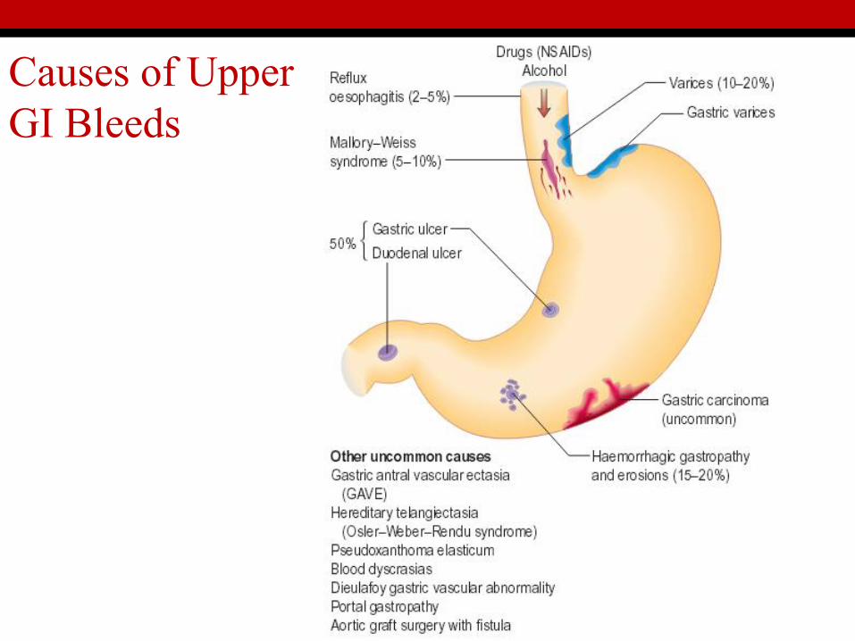
Causes of Upper GI Bleeds

Our patient found to have a GU
Helicobacter Pylori positive on
CLO test on biopsy
• Class I carcinogen• Risk of Gastric
carcinoma 3-6x• Almost all PUs• 60% of un-investigated
dyspepsia have NUD

H. pylori
• Example eradication regimens are:• Omeprazole 20 mg,
clarithromycin 500 mg and amoxicillin 1 g all twice daily, or;
• Omeprazole 20 mg, metronidazole 400 mg and clarithromycin 500 mg – all twice daily.
• These should be given for 7 or 14 days.

H. pylori
• Example eradication regimens are:• Omeprazole 20 mg,
clarithromycin 500 mg and amoxicillin 1 g all twice daily, or;
• Omeprazole 20 mg, metronidazole 400 mg and clarithromycin 500 mg – all twice daily.
• These should be given for 7 or 14 days.

H. pyloriExample eradication
regimens are:• Omeprazole 20 mg,
clarithromycin 500 mg and amoxicillin 1 g all twice daily, or;
• Omeprazole 20 mg, metronidazole 400 mg and clarithromycin 500 mg – all twice daily.
• Quadruple therapy• These should be given
for (7 or )14 days.

If GORD had been a problem …think of…..
Barrett’s Oesophagus
• Long standing reflux• Pre-malignant-
adenocarcinoma• Middle-aged men• Histology-intestinal
metaplasiacarcinoma• Management

Same case – different scenario….
• A 47 year old man attends A&E with worsening abdominal and chest pain. It’s a sharp, burning pain and is worse after eating. This evening,
he vomited and noticed some fresh red blood.
He vomited large quantities of fresh blood
OE HR 120 bpm , BP 102/60, sweaty


Upper GI bleeding cont
Endoscopy Treatments:• Ulcers
• Adrenaline injection
• Sclerosant injection
• Heat coagulation
(DO dual therapy)
New powder spray for bleeding
• Varices• Sclerosent Injection
• Banding
Surgery is needed for uncontrolled bleeding

Oesophageal varices - liver disease

Rockall assessment score


Case 2• A 75 year old lady presents to your GP practice with
difficulty swallowing for 2 months.
• PH: GORD, Hypertension• DH: Ramipril 5mg od, Gaviscon PRN• SH: Retired widow. 5 cigarettes a day for 50 years.
No alcohol.
• O/E Cachexic, nil else.
• What more do you want to know?

Case 2• A 75 year old lady presents to your GP practice with difficulty
swallowing for about 2 months.
• PMHx: GORD, Hypertension• DHx: Ramipril 5mg od, Gaviscon PRN• SHx: Retired widow. 5 cigarettes a day for 50 years.
No alcohol.
• O/E Cachexic, nil else.
• Elderly, can’t swallow, short history, lost weight++• Social circumstances, ?Lives alone, ?family. Who is with her

Causes of Dysphagia
?
??


• OGD with biopsy
• + Staging scans
Oesophageal carcinoma

Tis Carcinoma in situ Nx Nodes cannot be assessed
T1 Invading lamina propria N0 No node spread
T2 Invading muscularis propria N1 Regional Node Metastases
T3 Invading adventia M0 No distant Spread
T4 Invading adjacent structures M1 Distant Metastasis
>70% present at Stage III +


Case 3
• A 28 year old man presents to A&E complaining of 3 weeks of loose stools, associated with abdominal pain. He opens his bowels 8-12 times a day. On occasion, there is some fresh red blood mixed in with the stools.
• PH: Appendicetomy aged 8• Medic: Nil. Allergies: Penicillin • SH: Non-smoker. Drinks 3-4 pints a week.
He works as a holiday rep.
• What further questions would you ask?

Case 3
• A 28 year old man presents to A&E complaining of 3 weeks of loose stools, associated with abdominal pain. He is opening his bowels 8-12 times a day. On occasion, there is some fresh red blood mixed in with the stools.
• PMHx: Appendicetomy aged 8• DHx: Nil. Penicillin Allergy.• SHx: Non-smoker. Drinks 3-4 pints a week.
He works as a holiday rep.

Case 3 - interpretation
• Young • Man• 3 weeks ( most G’enteritis self limiting 48hr)• Abdo pain….? Helpful• Blood in stools …• 8-12 x/day ( what about night ?)• Holiday repDiff Diag?Bloody D ….infective ( travel), IBDImmunosuppressed?

Some Causes of Diarrhoea
• Infective▫ Bacterial
▫ Viral
▫ Protozoal
• Inflammatory Bowel Disease▫ Crohn’s
▫ Ulcerative Colitis
• Alcohol excess
• Irritable Bowel Syndrome
• Hyperthyroidism
• Malabsorptive States
• Diverticular Disease
• Constipation (with overflow)
• Drugs
• Ischaemic
• Radiation Colitis
• Malignancy
• Bacterial Overgrowth
• Fictitious
http://www.continence.org.au/data/images/bristol_stool_chart.gif

For stool gazers !

Causes of Travellers’ Diarrhoea


Gastroenteritis Management Algorithm

Gastroenteritis Management Algorithm
Case 3 Continued• The patient’s stool and blood cultures are negative.
• His pain and diarrhoea persist. He looks ill.• Hb 98g/L MCV 78fl CRP 86
What further investigations would you consider?
• Immediate AXR• IV infusion• Unprepared sigmoid/colonoscopy + biopsy

http
://w
ww
.cro
hnsf
orum
.com
/sho
wth
read
.php
?t=
4727


Manage
Management of Acute Colitis ( contd)

Differences between Crohn’s and UC

Summary of treatments for
Crohn’s and UC
Get off steroids asap , if you can

Extra-intestinal manifestations of IBD

Complications of IBD


Case 4
• A 56 year old man comes to his GP with a 6 week history of constipation. He occasionally notices some red blood in the stools.
• What further questions would you ask him?NB Older, recent change in bowel habit, blood PR?weight loss?change in medication

Causes of Constipation

Causes of Rectal Bleeding

Case 4 contdOn Examination:
• He looks thinner than when you last saw him.• CVS and Resp examinations normal.• Abdo – Bowel sounds present. No organomegaly.• PR – A mass is felt in the posterior aspect of the rectum.
There is some blood on the finger on removal.

Risk Factors and Distribution (%) of Colorectal Ca.

InvestigationsThe purpose of investigation is to confirm the diagnosis and stage the
tumour.
• Colonoscopy with biopsy is gold standard.• CT colonography and barium enema can be used.
• Blood tests• FBC may show anaemia. LFTs may be abnormal in metastases.• Carcinoembryonic antigen (CEA) are often raised
• Radiology• CT scan of the chest, abdomen and pelvis is the initial staging
investigation to look for local spread and metastatic disease. • MRI and endoanal ultrasound are used to locally stage rectal cancer.
• (Do NOT do Faecal occult blood tests…… Only used in population screening studies but are not of value diagnostically)

• Treatment is primarily surgical (avoiding stomas if possible)
• Adjuvent chemotherapy increases survival in Stage II and III tumours
• Radiotherpy can be used in low rectal tumours.
• Chemoradiotherapy may be used in palliation
Treatment and Staging


Case 5
• A 34 year old woman attends your GP practice complaining of feeling generally unwell for a couple of weeks. She complains of nausea and has had a temperature during this time.
• On examination, you notice she is jaundiced and has 4 cm smooth hepatomegaly.
• Anything else you would like to know?• What would you like to do next?

Downloaded from: StudentConsult (on 25 October 2005 12:22 PM)
© 2005 Elsevier
Causes of Jaundice
Gilberts


Causes of Hepatomegaly Causes of Splenomegaly
Infective
Viral HepatitisEBV
MalariaLeishmaniasis
InfectiveEBV
MalariaLeishmaniasis
MalignantHepatocellular Ca.
LeukaemiaLymphoma
Secondary Ca.
MalignantLeukaemiaLymphoma
Metabolic/Infiltration/
Inflammatory
FattyAmyloid
HaemochromatosisStorage Diseases
Sarcoid
Metabolic/Infiltration/
Inflammatory
AmyloidSarcoidStorageDiseases
Haemolytic Anaemia Haemoglobinopathies
SLE
CardiovascularRight Heart Failure
Budd-ChiariCardiovascular Portal Hypertension
OtherReidel’s Lobe
Low Diaphragm

Case 5 ContinuedFBCs and U+Es: Normal
Liver Biochemistry: AST 1134, ALT 1456, ALP 145 GGT 188 Bil 34
Liver function: Alb 36 INR 1.1
Autoantibody screen: ASMA, ANCA and ANA negative
HBsAg +HBeAg +Anti-HBs –Anti-Hbe –Anti-HBc IgM +Anti-HBc IgG +

Summary of the Viral Hepatitis Viruses

Summary of the Viral Hepatitis Viruses

Summary of the Viral Hepatitis Viruses

Hepatitis B

Hepatitis B virusSerology and Course

Treatment of HBV InfectionAcute – Mainly symptomatic. The majority (>90%) will recover and clear
the virus.
Chronic Infection – May be inactive or show chronic hepatitis.• Criteria for treatment is based on:
• Presence of HBeAg,• HBV DNA level (>20000)• serum ALT (> x2 normal)• Liver histology (biopsy is not indicated if the above features are present)
Treatment options:• Pegylated α-interferon given subcutaneously, once weekly.
Response rate of 25-45% at 1 year. • Entecavir – Nucleoside analogue 1-5mg oral x1/day
Response rate of 67-90% at one year. • Tenofovir – Reverse Transcriptase Inhibitor –300mg oral x1/day
Response of 76-93 at one year.

Hepatitis C


Case 6• A 63 year old man is brought into hospital by his family
who are concerned with his drinking. He has been drinking more and more since his wife passed away 2 years ago; he is currently having a large bottle of whisky every 1-2 days.
• Family say he seems to be more confused today and has recently developed a number of unexplained bruises.
• Are you concerned? What would you like to do?

Case 6 ? Differential diagnosis
• Alcohol• Confusion • Subdural?• Encephalopathy?• Other cerebral event?• Wernicke- Korsakoff ?
Bruising - coagulopathy? due to falls? Low platelets

http://news.bbc.co.uk/1/hi/in_depth/629/629/7202183.stm
ALCOHOL

Physical Effects of Excessive Alcohol Use
MCV

Case 6 cont:
On ExaminationHis Mini-mental score is 5/10.He is jaundiced with a number of bruises. He has a course flapping tremor.Cranial nerves normal.HS 1+2+nil. Chest is clear.
Abdomen is mildly distended with shifting dullness. No organomegaly is felt.
PR – empty rectum
What would you do now?

Case 6 cont:
On ExaminationHis Mini-mental score is 5/10.He is jaundiced with a number of bruises. He has a course flapping tremor.Cranial nerves normal.HS 1+2+nil. Chest is clear.
Abdomen is mildly distended with shifting dullness. No organomegaly is felt.
PR – empty rectum
So he is :YellowEncephalopathicAscitesie Complications of Chronic liver disease

Signs of ChronicLiver Disease and Causes of Ascites

Case 6 contd:
FBCs: Hb 108 MCV 102 WCC 8.2 Plt 156U+Es: Na 130 K 4.6 Urea 8.9 Creat 265LB: AST 1467 ALT 677 ALP 137 GGT 237
Bil 38 LFTs: Alb 24 INR 1.8 PT 22 secondsα-fetaprotein – normal
Viral and Autoimmune Screen NegativeSeptic Screen Clear


Grading of Hepatic Encephalopathy

Causes of Fulminant Liver Failure
Fulminant Liver FailureDefined as severe hepatic failure in which hepatic
encepthalopathy is present within 2 weeks

Causes of Cirrhosis

Child-Pugh Score for Cirrhosis

Indicators of Poor Prognosis
Average 5 year survival is 50%
Complications
Cirrhosis Outcomes

Liver Failure Management• Liaise with specialist liver centre. Manage in high-intensity ward.
• Treat the cause• Nutritional Support (high carb, low to high protein, vitamins)• Watch for sepsis• Strict fluid status, daily weights and observations.• Ovoid sedatives and drugs metabolised in liver
• Treat Symptoms:• Ascites: Na and fluid restriction. Spironolactone (then furosemide can
help. Paracentesis and replacement of albumin may be needed.• Bleeding – Vitamin K• Laxatives• Cerebral oedema – mannitol, hyperventilate
• Always think of liver transplantation early

The End
Any Questions?
Bibliography
Kumar and Clark’s Clinical Medicine 8th ed
Essentials of Clinical Medicine - Ballinger
Pass Finals – Smith, Carty and Langmead
Kumar and Clark’s Medical Management and therapeutics
Kumar and Clark’s Clinical cases
Related Documents


