Case Report Relapsing Polychondritis Beata Sosada, Katarzyna Loza, and Ewelina Bialo-Wojcicka Department of Dermatology, Miedzyleski Specialist Hospital in Warsaw, ul. Bursztynowa 2, 04-479 Warsaw, Poland Correspondence should be addressed to Beata Sosada; [email protected] Received 30 June 2014; Accepted 13 September 2014; Published 30 September 2014 Academic Editor: Alexander A. Navarini Copyright © 2014 Beata Sosada et al. is is an open access article distributed under the Creative Commons Attribution License, which permits unrestricted use, distribution, and reproduction in any medium, provided the original work is properly cited. Relapsing polychondritis (RP) is a rare systemic disease characterized by recurrent, widespread chondritis of the auricular, nasal, and tracheal cartilages. Additional clinical features include audiovestibular dysfunction, ocular inflammation, vasculitis, myocarditis, and nonerosive arthritis. Although the cause remains unknown, the etiology is suspected to be autoimmune. We describe a case of a 31-year-old woman with a four-month history of bilateral auricular and nasal chondritis. Infectious and neoplastic diseases were excluded by imaging and laboratory examinations. RP was diagnosed based on three McAdam’s criteria. e patient was medicated with oral prednisolone and methotrexate with positive clinical response. In this case clinical history and detailed physical examination were fundamental in concluding the correct diagnosis and administrating the appropriate medication. 1. Introduction Relapsing polychondritis (RP) is a rare inflammatory disease primarily affecting the cartilaginous structures of the ear, nose, joints, tracheobronchial tree, and cardiovascular sys- tem. Cardiovascular and respiratory complications of RP are associated with high morbidity and mortality. e first case of RP was described in 1923 by Jaksch-Wartenhorst [1]. e term “relapsing polychondritis” was first used by Pearson et al. in 1960 in their review of 12 cases [2]. RP was usually observed in the fourth and fiſth decade of life with no sex predilection [3–5]. e McAdam’s criteria were the initial diagnostic criteria of RP [3] and required meeting three out of six of the follow- ing: bilateral auricular chondritis, nonerosive seronegative inflammatory arthritis, nasal chondritis, ocular inflamma- tion, respiratory tract chondritis, and audiovestibular dam- age. Modified criteria have been proposed by Damiani and Levine [4] which include meeting one McAdam’s criterion plus histopathological confirmation or two McAdam’s criteria plus response to corticosteroids or dapsone. Currently, the diagnosis of RP relies mostly on the criteria established by Michet et al. [5] which require the presence of a proven inflammation in at least two of three of the auricular, nasal, or laryngotracheal cartilages or the proven inflammation in one of these cartilages plus two other signs, including ocular inflammation, vestibular dysfunction, seronegative inflammatory arthritis, or hearing loss (Table 1). e exact cause of RP is still unknown but the disease is mostly seen as an immune-mediated disease, as there is a well-documented overlap of RP with other rheumatic and autoimmune diseases [3, 6]. Although a large number of cases have been reported recently and the knowledge on the clinical spectrum, pathogenesis, and management in RP has grown considerably, only limited microscopic data is available in the literature [4, 7]. e histologic features of the chondritis include loss of basophilic staining of the cartilage matrix followed by cartilage destruction with replacement by fibrous tissue and cellular infiltration with plasma cells and lymphocytes. A rare disease RP is described occurring extremely rarely in young women. 2. Case Report A 31-year-old Caucasian woman was consulted in our depart- ment for recurrent swellings of both pinnae which had been present for approximately 4 months. About two weeks before coming to hospital she suffered from pain and tenderness of both auricles, the nose as well as the leſt elbow. Her personal and family history was unremarkable. She was a smoker (10 pack-years). During physical examination both Hindawi Publishing Corporation Case Reports in Dermatological Medicine Volume 2014, Article ID 791951, 4 pages http://dx.doi.org/10.1155/2014/791951

Welcome message from author
This document is posted to help you gain knowledge. Please leave a comment to let me know what you think about it! Share it to your friends and learn new things together.
Transcript
Case ReportRelapsing Polychondritis
Beata Sosada, Katarzyna Loza, and Ewelina Bialo-Wojcicka
Department of Dermatology, Miedzyleski Specialist Hospital in Warsaw, ul. Bursztynowa 2, 04-479 Warsaw, Poland
Correspondence should be addressed to Beata Sosada; [email protected]
Received 30 June 2014; Accepted 13 September 2014; Published 30 September 2014
Academic Editor: Alexander A. Navarini
Copyright © 2014 Beata Sosada et al. This is an open access article distributed under the Creative Commons Attribution License,which permits unrestricted use, distribution, and reproduction in any medium, provided the original work is properly cited.
Relapsing polychondritis (RP) is a rare systemic disease characterized by recurrent, widespread chondritis of the auricular,nasal, and tracheal cartilages. Additional clinical features include audiovestibular dysfunction, ocular inflammation, vasculitis,myocarditis, and nonerosive arthritis. Although the cause remains unknown, the etiology is suspected to be autoimmune. Wedescribe a case of a 31-year-old woman with a four-month history of bilateral auricular and nasal chondritis. Infectious andneoplastic diseases were excluded by imaging and laboratory examinations. RP was diagnosed based on three McAdam’s criteria.The patient was medicated with oral prednisolone and methotrexate with positive clinical response. In this case clinical historyand detailed physical examination were fundamental in concluding the correct diagnosis and administrating the appropriatemedication.
1. Introduction
Relapsing polychondritis (RP) is a rare inflammatory diseaseprimarily affecting the cartilaginous structures of the ear,nose, joints, tracheobronchial tree, and cardiovascular sys-tem. Cardiovascular and respiratory complications of RP areassociatedwith highmorbidity andmortality.The first case ofRPwas described in 1923 by Jaksch-Wartenhorst [1].The term“relapsing polychondritis” was first used by Pearson et al. in1960 in their review of 12 cases [2]. RP was usually observedin the fourth and fifth decade of life with no sex predilection[3–5].
The McAdam’s criteria were the initial diagnostic criteriaof RP [3] and required meeting three out of six of the follow-ing: bilateral auricular chondritis, nonerosive seronegativeinflammatory arthritis, nasal chondritis, ocular inflamma-tion, respiratory tract chondritis, and audiovestibular dam-age. Modified criteria have been proposed by Damiani andLevine [4] which include meeting one McAdam’s criterionplus histopathological confirmation or twoMcAdam’s criteriaplus response to corticosteroids or dapsone. Currently, thediagnosis of RP relies mostly on the criteria established byMichet et al. [5] which require the presence of a proveninflammation in at least two of three of the auricular, nasal,or laryngotracheal cartilages or the proven inflammationin one of these cartilages plus two other signs, including
ocular inflammation, vestibular dysfunction, seronegativeinflammatory arthritis, or hearing loss (Table 1).
The exact cause of RP is still unknown but the diseaseis mostly seen as an immune-mediated disease, as there isa well-documented overlap of RP with other rheumatic andautoimmune diseases [3, 6]. Although a large number ofcases have been reported recently and the knowledge onthe clinical spectrum, pathogenesis, and management in RPhas grown considerably, only limited microscopic data isavailable in the literature [4, 7]. The histologic features of thechondritis include loss of basophilic staining of the cartilagematrix followed by cartilage destruction with replacementby fibrous tissue and cellular infiltration with plasma cellsand lymphocytes. A rare disease RP is described occurringextremely rarely in young women.
2. Case Report
A 31-year-old Caucasian womanwas consulted in our depart-ment for recurrent swellings of both pinnae which had beenpresent for approximately 4 months. About two weeks beforecoming to hospital she suffered from pain and tendernessof both auricles, the nose as well as the left elbow. Herpersonal and family history was unremarkable. She was asmoker (10 pack-years). During physical examination both
Hindawi Publishing CorporationCase Reports in Dermatological MedicineVolume 2014, Article ID 791951, 4 pageshttp://dx.doi.org/10.1155/2014/791951
2 Case Reports in Dermatological Medicine
(a) (b) (c)
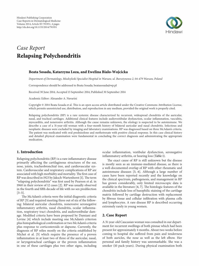
Figure 1: (a, b) Cauliflower ears. Swelling and erythema of the cartilaginous part of the ear, sparing the lobule which lacks cartilage. (c) TheRaynaud’s phenomenon.
Table 1: Diagnostic criteria for RP.
McAdam et al. [3]
(1) Recurrent chondritis of bothauricles(2) Nonerosive inflammatorypolyarthritis(3) Chondritis of nasal cartilages(4) Inflammation of ocular structures(5) Chondritis of respiratory tract(6) Cochlear and/or vestibular damage(requirement—three out of six criteria)
Damiani and Levine [4]
(1) Three out of six McAdam et al.’s [3]criteria(2) One out of six McAdam et al.’s [3]criteria and a positive histologicconfirmation(3) Two out of six McAdam et al.’scriteria and response to corticosteroidor dapsone(requirement—any of these)
Michet et al. [5]
(1) Proven inflammation in two out ofthree cartilages: auricular, nasal, andlaryngotracheal(2) Proven inflammation in one of theabove and meeting two other signsfrom ocular inflammation, hearingloss, vestibular dysfunction, orseronegative inflammatory arthritis(requirement—any of these)
pinnae lost their firmness, became soft and floppy, andhad a cauliflower-like appearance (Figures 1(a) and 1(b)). Inaddition Raynaud’s phenomenonwas found (Figure 1(c)) andevidenced by nailfold capillaroscopy.
Routine blood investigations revealed normocytic nor-mochromic anemia, elevated erythrocyte sedimentation rate.The rheumatoid factor was within normal limits. Other
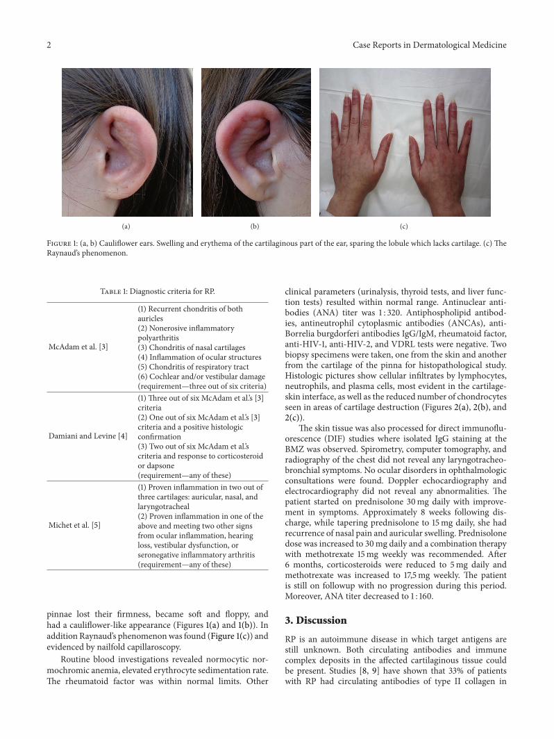
clinical parameters (urinalysis, thyroid tests, and liver func-tion tests) resulted within normal range. Antinuclear anti-bodies (ANA) titer was 1 : 320. Antiphospholipid antibod-ies, antineutrophil cytoplasmic antibodies (ANCAs), anti-Borrelia burgdorferi antibodies IgG/IgM, rheumatoid factor,anti-HIV-1, anti-HIV-2, and VDRL tests were negative. Twobiopsy specimens were taken, one from the skin and anotherfrom the cartilage of the pinna for histopathological study.Histologic pictures show cellular infiltrates by lymphocytes,neutrophils, and plasma cells, most evident in the cartilage-skin interface, as well as the reduced number of chondrocytesseen in areas of cartilage destruction (Figures 2(a), 2(b), and2(c)).
The skin tissue was also processed for direct immunoflu-orescence (DIF) studies where isolated IgG staining at theBMZ was observed. Spirometry, computer tomography, andradiography of the chest did not reveal any laryngotracheo-bronchial symptoms. No ocular disorders in ophthalmologicconsultations were found. Doppler echocardiography andelectrocardiography did not reveal any abnormalities. Thepatient started on prednisolone 30mg daily with improve-ment in symptoms. Approximately 8 weeks following dis-charge, while tapering prednisolone to 15mg daily, she hadrecurrence of nasal pain and auricular swelling. Prednisolonedose was increased to 30mg daily and a combination therapywith methotrexate 15mg weekly was recommended. After6 months, corticosteroids were reduced to 5mg daily andmethotrexate was increased to 17,5mg weekly. The patientis still on followup with no progression during this period.Moreover, ANA titer decreased to 1 : 160.
3. Discussion
RP is an autoimmune disease in which target antigens arestill unknown. Both circulating antibodies and immunecomplex deposits in the affected cartilaginous tissue couldbe present. Studies [8, 9] have shown that 33% of patientswith RP had circulating antibodies of type II collagen in

Case Reports in Dermatological Medicine 3
(a) (b) (c)
Figure 2: (a)The dermis contains a mild focal lymphohistiocytic infiltrate. H&E, ×100. (b) Degenerative and inflammatory changes affectingthe marginal chondrocytes with loss of basophilia and poor alcian blue staining of the cartilaginous tissue. H&E, ×40. (c) The inflammatorycells infiltrate, including lymphocytes, plasma cells, and histiocytes, infiltrate the degenerative cartilage. H&E, ×100.
the active phase of the disease and their titres also corre-sponded to the disease activity. Autoimmunity to collagentype II has also been described in systemic lupus erythe-matosus (SLE) and rheumatoid arthritis. Other studies [10, 11]showed that the antibodies are generated against not onlynative and denatured collagen type II but also collagen typesIX and XI, which form the major extracellular scaffold inthe cartilage. Matrilin-1 is a cartilage-specific protein and ishighly expressed in tracheal and nasal but not in normal adultarticular cartilage [12]. Saxne and Heinegard in their studies[12, 13] revealed that an increased serum level of matrilin-1could be found in patients with RP in the active phase,suggesting that the release of matrilin-1 resulted from thedestruction of the involved cartilage. However, neither anti-collagen type II nor anti-matrilin-1 antibodies are sensitiveand specific enough and consequently cannot be used fordiagnostic purposes. The diagnosis of RP is largely based onthe clinical features and the role of laboratory and imaginginvestigations is purely supportive to rule out other relatedor associated systemic diseases. Clinical, histopathological,and DIF features together or in combination are helpful inthe final diagnosis. The treatment of RP is symptomatic andshould be tailored to each individual patient based on diseaseactivity and severity.
Glucocorticoid therapy is fundamental in the treat-ment of RP and is used chronically in most patients. Lesssevere symptoms are generally treated with nonsteroid anti-inflammatory drugs. Dapsone may also be used as an ini-tial therapy but results in many adverse reactions. Severesymptoms of disease, including ocular or laryngotrachealinvolvement, systemic vasculitis, and severe polychondritisrequire systemic corticosteroids. In patients intolerant to,rarely unresponsive to, steroid therapy or in whom a steroidsparing therapy is required, immunosuppressants play a role.Immunosuppressive agents like methotrexate, azathioprine,and cyclosporine may be given to patients with severerespiratory or vascular involvement and to thosewith steroid-resistant or steroid-dependent disease. Trentham and Le [14]observed that methotrexate in dose of 17,5mg/week wasthe most effective nonsteroid drug in causing symptomaticbenefit and reducing the steroid requirement. Intravenouscyclophosphamide and plasmapheresis could be used in
patientswith organ-threatening and life-threatening diseases,including glomerulonephritis or acute airways obstruction.The autoimmune theory of pathogenesis of RP makesimmunomodulatory agents (biologics) an important treat-ment alternative to other medical therapies. However, datafrom clinical trials is scarce; there are many case reportsof satisfactory response to biologic therapy in RP. Standardmanagement cannot be established due to its rarity.
Conflict of Interests
The authors declare that there is no conflict of interestsregarding the publication.
Acknowledgment
The authors would like to thank Dr. Kazimierz Kalbarczyk,who unfortunately passed away this year. He was a greatdermatopathologist in Department of Dermatology at theMiedzyleski Specialist Hospital in Warsaw and gave oppor-tunity to all young dermatologists to discover the fascinatingworld of dermatopathology.
References
[1] R. Jaksch-Wartenhorst, “Polychondropathia,”Wiener Archiv furInnere Medizin, vol. 6, pp. 93–100, 1923.
[2] C. M. Pearson, H. M. Kline, and V. D. Newcomer, “Relapsingpolychondritis,”The New England Journal of Medicine, vol. 263,pp. 51–58, 1960.
[3] L. P. McAdam, M. A. O’Hanlan, and R. C. M. Pearson, “Relaps-ing polychondritis: prospective study of 23 patients and a reviewof the literature,”Medicine, vol. 55, no. 3, pp. 193–215, 1976.
[4] J. M. Damiani and H. L. Levine, “Relapsing polychondritis.Report of ten cases,” The Laryngoscope, vol. 89, no. 6, pp. 929–946, 1979.
[5] C. J. Michet Jr., C. H. McKenna, H. S. Luthra, and W.M. O’Fallon, “Relapsing polychondritis: survival and predic-tive role of early disease manifestations,” Annals of InternalMedicine, vol. 104, no. 1, pp. 74–78, 1986.
4 Case Reports in Dermatological Medicine
[6] J.-C. Piette, R. El-Rassi, and Z. Amoura, “Antinuclear antibodiesin relapsing polychondritis,” Annals of the Rheumatic Diseases,vol. 58, no. 10, pp. 656–657, 1999.
[7] S. Frisenda, C. Perricone, and G. Valesini, “Cartilage as a targetof autoimmunity: a thin layer,” Autoimmunity Reviews, vol. 12,no. 5, pp. 591–598, 2013.
[8] J. M. Foidart, S. Abe, G. R. Martin et al., “Antibodies to type IIcollagen in relapsing polychondritis,”The New England Journalof Medicine, vol. 299, no. 22, pp. 1203–1207, 1978.
[9] L. Giroux, F. Paquin, M. J. Guerard Desjardins, and A. Lefaivre,“Relapsing polychondritis: an autoimmune disease,” Seminarsin Arthritis and Rheumatism, vol. 13, no. 2, pp. 182–187, 1983.
[10] C. L. Yang, J. Brinckmann, H. F. Rui et al., “Autoantibodiesto cartilage collagens in relapsing polychondritis,” Archives ofDermatological Research, vol. 285, no. 5, pp. 245–249, 1993.
[11] S. Alsalameh, J. Mollenhauer, F. Scheuplein et al., “Preferentialcellular and humoral immune reactivities to native and dena-tured collagen types IX and XI in a patient with fatal relapsingpolychondritis,” Journal of Rheumatology, vol. 20, no. 8, pp.1419–1424, 1993.
[12] T. Saxne and D. Heinegard, “Involvement of nonarticular car-tilage, as demonstrated by release of a cartilage-specific protein,in rheumatoid arthritis,” Arthritis and Rheumatism, vol. 32, no.9, pp. 1080–1086, 1989.
[13] T. Saxne and D. Heinegard, “Serum concentrations of two car-tilage matrix proteins reflecting different aspects of cartilageturnover in relapsing polychondritis,” Arthritis and Rheuma-tism, vol. 38, no. 2, pp. 294–296, 1995.
[14] D. E. TrenthamandC.H. Le, “Relapsing polychondritis,”Annalsof Internal Medicine, vol. 129, no. 2, pp. 114–122, 1998.

Submit your manuscripts athttp://www.hindawi.com
Stem CellsInternational
Hindawi Publishing Corporationhttp://www.hindawi.com Volume 2014
Hindawi Publishing Corporationhttp://www.hindawi.com Volume 2014
MEDIATORSINFLAMMATION
of
Hindawi Publishing Corporationhttp://www.hindawi.com Volume 2014
Behavioural Neurology
EndocrinologyInternational Journal of
Hindawi Publishing Corporationhttp://www.hindawi.com Volume 2014
Hindawi Publishing Corporationhttp://www.hindawi.com Volume 2014
Disease Markers
Hindawi Publishing Corporationhttp://www.hindawi.com Volume 2014
BioMed Research International
OncologyJournal of
Hindawi Publishing Corporationhttp://www.hindawi.com Volume 2014
Hindawi Publishing Corporationhttp://www.hindawi.com Volume 2014
Oxidative Medicine and Cellular Longevity
Hindawi Publishing Corporationhttp://www.hindawi.com Volume 2014
PPAR Research
The Scientific World JournalHindawi Publishing Corporation http://www.hindawi.com Volume 2014
Immunology ResearchHindawi Publishing Corporationhttp://www.hindawi.com Volume 2014
Journal of
ObesityJournal of
Hindawi Publishing Corporationhttp://www.hindawi.com Volume 2014
Hindawi Publishing Corporationhttp://www.hindawi.com Volume 2014
Computational and Mathematical Methods in Medicine
OphthalmologyJournal of
Hindawi Publishing Corporationhttp://www.hindawi.com Volume 2014
Diabetes ResearchJournal of
Hindawi Publishing Corporationhttp://www.hindawi.com Volume 2014
Hindawi Publishing Corporationhttp://www.hindawi.com Volume 2014
Research and TreatmentAIDS
Hindawi Publishing Corporationhttp://www.hindawi.com Volume 2014
Gastroenterology Research and Practice
Hindawi Publishing Corporationhttp://www.hindawi.com Volume 2014
Parkinson’s Disease
Evidence-Based Complementary and Alternative Medicine
Volume 2014Hindawi Publishing Corporationhttp://www.hindawi.com
Related Documents