IIUM Journal of Orofacial and Health Sciences (2020) 1(2): 91-105 91 CASE REPORT Orthodontic treatment of an adult patient with aggressive periodontitis – A case report Mohd Zambri Mohamed Makhbul 1* , Izrawatie Mardiana Shapeen 2 , Wan Nurazreena Wan Hassan 3 1 Orthodontic Unit, Klinik Pergigian Cahaya Suria (Sementara), Level 1, Bangunan UTC Pudu Sentral, Jalan Pudu, 50100 Kuala Lumpur, Malaysia. 2 Periodontic Unit, Klinik Pergigian Cahaya Suria (Sementara), Level 1, Bangunan UTC Pudu Sentral, Jalan Pudu, 50100 Kuala Lumpur, Malaysia. 3 Department of Paediatric Dentistry and Orthodontics & Clinical Craniofacial Dentistry Research Group, Faculty of Dentistry, University of Malaya, Kuala Lumpur, Malaysia. ___________________________________________________________________________ Abstract A 26-year-old man with an aggressive periodontitis sought for orthodontic treatment to improve the appearance of his smile. He presented with generalised anterior spacing, missing lower left central incisor and deep traumatic bite. He was treated successfully with a combination of orthodontic and periodontal treatment. After 18 months of orthodontic treatment and follow up by the periodontist, his alignment of teeth was improved, a stable occlusion was achieved, and occlusal trauma was prevented. As a result, the patient’s smile appearance and self- confidence were improved. Orthodontic tooth movement is not only to correct the alignment of his teeth but also to improve the bone level especially at the anterior region. This case report shows the successful treatment outcome in aggressive periodontitis patient which requires good collaboration between the orthodontist and the periodontist. Keywords: aggressive periodontitis, bone level, orthodontist and periodontist, occlusal trauma, traumatic bite ___________________________________________________________________________ *Corresponding Author Email address: [email protected] Tel: +603-207215512 Introduction Awareness for orthodontic treatment among periodontic patients is increasing due to pathologic dental migration, which compromise the facial aesthetics (Brunsvold, 2005; Feng et al., 2005). Periodontitis is the inflammation of the supporting tissues of the teeth, caused by specific microorganisms, which leads to progressive destruction of the periodontal ligament and alveolar bone with either pocket formation, recession, or both (Gyawali & Bhattarai, 2017). Interdisciplinary approach by the orthodontist and periodontist is required in the orthodontic management of cases with compromised periodontium (Vinod et al., 2012). Aggressive forms of periodontal disease have been defined based on the following primary features (Lang et al., 1999): non‐ contributory medical history, rapid attachment loss and bone destruction, and familial aggregation of cases. The general secondary features of aggressive periodontitis is: amounts of microbial deposits inconsistent with the severity of

Welcome message from author
This document is posted to help you gain knowledge. Please leave a comment to let me know what you think about it! Share it to your friends and learn new things together.
Transcript
IIUM Journal of Orofacial and Health Sciences (2020) 1(2): 91-105
91
CASE REPORT
Orthodontic treatment of an adult patient with aggressive periodontitis – A case report Mohd Zambri Mohamed Makhbul1*, Izrawatie Mardiana Shapeen2, Wan Nurazreena Wan Hassan3
1 Orthodontic Unit, Klinik Pergigian Cahaya Suria (Sementara), Level 1, Bangunan UTC Pudu Sentral, Jalan Pudu, 50100 Kuala Lumpur, Malaysia. 2 Periodontic Unit, Klinik Pergigian Cahaya Suria (Sementara), Level 1, Bangunan UTC Pudu Sentral, Jalan Pudu, 50100 Kuala Lumpur, Malaysia. 3 Department of Paediatric Dentistry and Orthodontics & Clinical Craniofacial Dentistry Research Group, Faculty of Dentistry, University of Malaya, Kuala Lumpur, Malaysia.
___________________________________________________________________________
Abstract
A 26-year-old man with an aggressive periodontitis sought for orthodontic treatment to improve
the appearance of his smile. He presented with generalised anterior spacing, missing lower
left central incisor and deep traumatic bite. He was treated successfully with a combination of
orthodontic and periodontal treatment. After 18 months of orthodontic treatment and follow up
by the periodontist, his alignment of teeth was improved, a stable occlusion was achieved,
and occlusal trauma was prevented. As a result, the patient’s smile appearance and self-
confidence were improved. Orthodontic tooth movement is not only to correct the alignment
of his teeth but also to improve the bone level especially at the anterior region. This case report
shows the successful treatment outcome in aggressive periodontitis patient which requires
good collaboration between the orthodontist and the periodontist.
Keywords: aggressive periodontitis, bone level, orthodontist and periodontist, occlusal trauma, traumatic bite
___________________________________________________________________________ *Corresponding Author Email address: [email protected] Tel: +603-207215512
Introduction Awareness for orthodontic treatment among
periodontic patients is increasing due to
pathologic dental migration, which
compromise the facial aesthetics
(Brunsvold, 2005; Feng et al., 2005).
Periodontitis is the inflammation of the
supporting tissues of the teeth, caused by
specific microorganisms, which leads to
progressive destruction of the periodontal
ligament and alveolar bone with either
pocket formation, recession, or both
(Gyawali & Bhattarai, 2017). Interdisciplinary
approach by the orthodontist and
periodontist is required in the orthodontic
management of cases with compromised
periodontium (Vinod et al., 2012).
Aggressive forms of periodontal disease
have been defined based on the following
primary features (Lang et al., 1999): non‐
contributory medical history, rapid
attachment loss and bone destruction, and
familial aggregation of cases. The general
secondary features of aggressive
periodontitis is: amounts of microbial
deposits inconsistent with the severity of
IIUM Journal of Orofacial and Health Sciences (2020) 1(2): 91-105
92
periodontal tissue destruction. The other
secondary features were laboratory features
such as elevated proportions of
Aggregatibacter actinomycetemcomitans
(previously named Actinobacillus
actinomycetemcomitans) and, in some
populations, Porphyromonas gingivalis;
phagocyte abnormalities; hyper‐responsive
macrophage phenotype, including elevated
production of prostaglandin and interleukin
in response to bacterial endotoxins (Lang et
al., 1999).
Aggressive periodontitis affects adolescents,
and the percentage of adolescents is highest
among orthodontic patients (Bagga, 2010).
Prevalence of aggressive periodontitis
varies widely among various races and
ethnicities from 0.1% to 15% (Albandar et al.,
2007). Besides, genetics, age, and
environment may also influence it. Females
are found to be more affected than male
(Hormand & Frandsen, 1979).
Patients with an aggressive periodontitis
may lose the interproximal attachment and
this is the main factor for the pathological
migration of teeth (Martinez-Canut et al.,
1997). Changes the position such as
proclination, rotation, spacing and extrusion
the anterior teeth may compromise
aesthetics (Towfighi et al., 1997).
Orthodontic treatment can facilitate
improvement by light intrusive orthodontic
forces to correct the pathological extrusion
and migration of teeth (Garat et al., 2005;
Panwar et al., 2010). Nonetheless,
orthodontic treatment should be started only
after the clinician is convinced that the
patient is well motivated and can follow the
oral hygiene instructions well (Gyawali &
Bhattarai, 2017).
During the treatment, it is important to
reinforce good oral hygiene. Clinicians
should inform the patients the consequences
of poor oral hygiene on the teeth and
supporting structures. Orthodontic patients
with aggressive periodontitis also require a
separate periodontal appointment with a
periodontist once every 3 months (Levin et
al., 2012). Good communication and
understanding between the orthodontist and
periodontist are essential to achieve
successful results and avoid unwanted
complications. This case described the
management of aggressive periodontitis
through the interdisciplinary approach for
improving the aesthetics and fulfilled the
patient expectation. The correction of
extruded upper central incisors with
controlled intrusion led to a decrease in the
clinical crown length, better access for oral
hygiene procedures, better gingival form,
and a more suitable distribution of occlusal
forces (Rabie et al., 1998).
Diagnosis and etiology
A 26 year old male presented in October
2015 at the Orthodontic – Periodontic -
Restorative Joint Specialist Clinic in Klinik
Pergigian Cahaya Suria, Kuala Lumpur. His
chief complaint was “spacing and forward
position of the upper teeth”. He was very
keen to improve his esthetics, which affected
his self-confidence. Patient’s medical history
was non-contributory. He was a light smoker.
He had mentioned during history taking
interview that his mother’s siblings had lost
their teeth at early age and wearing
dentures. Pre-treatment records indicated
that the patient had full mouth plaque score
of 51% and bleeding score of 38%. The
presence of plaque was noted to be at
supragingival areas and thin in thickness.
These scores had improved following
completion of initial periodontal therapy to
23% and 36%, respectively, prior to referral
to orthodontist.
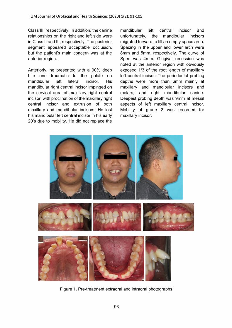
He presented with a symmetrical face and
Class I skeletal profile (Figure 1). Intraorally,
he had Class II division 1 malocclusion with
an overjet of 10mm. The molar relationship
on the right and left side were in Class I and
IIUM Journal of Orofacial and Health Sciences (2020) 1(2): 91-105
93
Class III, respectively. In addition, the canine
relationships on the right and left side were
in Class II and III, respectively. The posterior
segment appeared acceptable occlusion,
but the patient’s main concern was at the
anterior region.
Anteriorly, he presented with a 90% deep
bite and traumatic to the palate on
mandibular left lateral incisor. His
mandibular right central incisor impinged on
the cervical area of maxillary right central
incisor, with proclination of the maxillary right
central incisor and extrusion of both
maxillary and mandibular incisors. He lost
his mandibular left central incisor in his early
20’s due to mobility. He did not replace the
mandibular left central incisor and
unfortunately, the mandibular incisors
migrated forward to fill an empty space area.
Spacing in the upper and lower arch were
8mm and 5mm, respectively. The curve of
Spee was 4mm. Gingival recession was
noted at the anterior region with obviously
exposed 1/3 of the root length of maxillary
left central incisor. The periodontal probing
depths were more than 6mm mainly at
maxillary and mandibular incisors and
molars; and right mandibular canine.
Deepest probing depth was 9mm at mesial
aspects of left maxillary central incisor.
Mobility of grade 2 was recorded for
maxillary incisor.
Figure 1. Pre-treatment extraoral and intraoral photographs
IIUM Journal of Orofacial and Health Sciences (2020) 1(2): 91-105
94
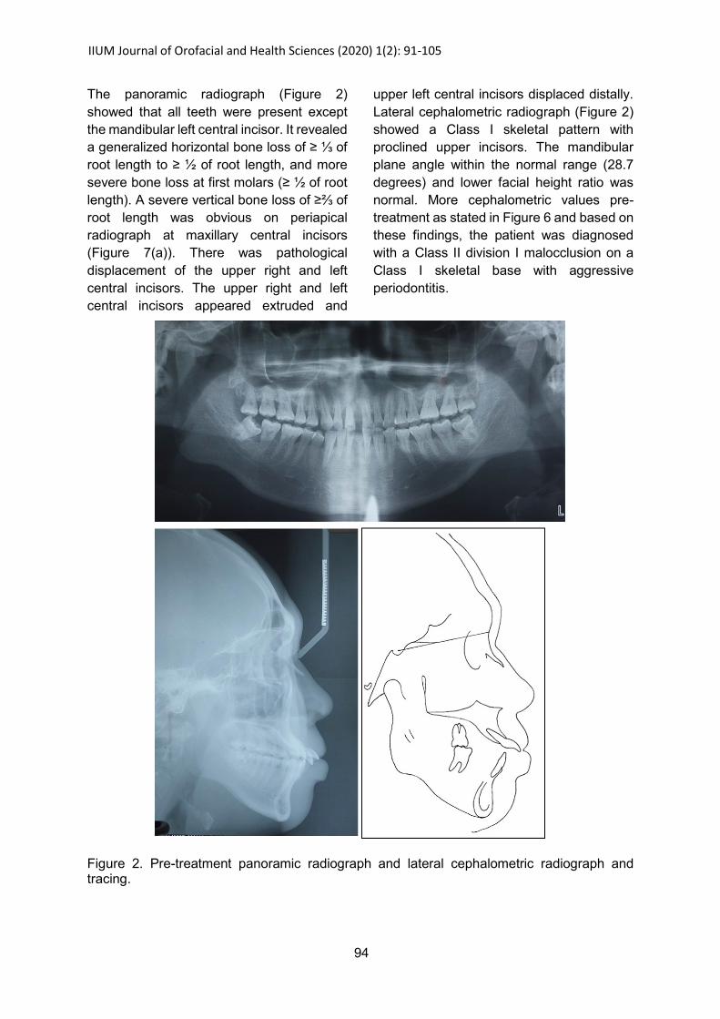
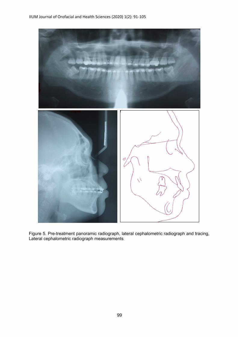
The panoramic radiograph (Figure 2)
showed that all teeth were present except
the mandibular left central incisor. It revealed
a generalized horizontal bone loss of ≥ ⅓ of
root length to ≥ ½ of root length, and more
severe bone loss at first molars (≥ ½ of root
length). A severe vertical bone loss of ≥⅔ of
root length was obvious on periapical
radiograph at maxillary central incisors
(Figure 7(a)). There was pathological
displacement of the upper right and left
central incisors. The upper right and left
central incisors appeared extruded and
upper left central incisors displaced distally.
Lateral cephalometric radiograph (Figure 2)
showed a Class I skeletal pattern with
proclined upper incisors. The mandibular
plane angle within the normal range (28.7
degrees) and lower facial height ratio was
normal. More cephalometric values pre-
treatment as stated in Figure 6 and based on
these findings, the patient was diagnosed
with a Class II division I malocclusion on a
Class I skeletal base with aggressive
periodontitis.
Figure 2. Pre-treatment panoramic radiograph and lateral cephalometric radiograph and tracing.
IIUM Journal of Orofacial and Health Sciences (2020) 1(2): 91-105
95
Treatment options
The treatment options that were given to the
patient in the interdisciplinary Orthodontic-
Periodontic-Restorative Joint Clinic are as
follows:
1. Comprehensive periodontal treatment
until maintenance phase, followed by
orthodontic treatment and later with
Prosthodontic treatment. The patient
needs to start with 2-3 years orthodontic
treatment with regular follow up (every 6-
8 weeks) with the orthodontist. In
addition to that, patient have to be
committed to the periodontal health care
maintenance and attends regular review
(every 3 months) with the periodontist
throughout the active orthodontic
treatment. After the course of the
orthodontic treatment, the missing tooth
was planned for replacement with a
prosthesis, followed by night time (life
time) upper and lower retainers. The
patient was warned of potential loss of
vitality of the upper central incisors and
thus, needed close monitoring.
2. Endodontic treatment for upper right and
left central incisors followed by crown
placement. This treatment option saved
cost and time for the patient. However,
the traumatic overbite would not be
corrected and it may cause continued
trauma on the palatal mucosa.
3. Extraction of the upper central incisors
and fabrication of prosthesis with either
fixed prosthesis or partial removable
prosthesis on upper central incisors. This
treatment option may cause the patient
to lose two sound teeth, which would be
replaced with prostheses. The overbite
would be not be corrected and the lower
incisors may occlude on the fixed
prosthesis or acrylic and may cause
mobility of the lower incisors.
4. Orthodontic extrusion of upper centrals
incisors to create bone for further
prosthesis treatment. This treatment
option require orthodontic treatment and
the overbite would be not corrected.
After the discussions and considering the
risks and complications, the patient decided
to proceed with combined orthodontic,
periodontic and prosthodontic treatment
which was the first option given to him. The
patient agreed with the explained treatment
and signed the written consent. The patient
was referred to Periodontist prior to
Orthodontic treatment.
The orthodontic treatment objectives were
built on the complete treatment objectives for
periodontal health (Xie et al., 2014).
The complete periodontal treatment
objectives for this patient consisted of:
i) The medical problem was non-
contributory and the patient was
instructed to stop the smoking habit
(Azouni & Tarakji, 2014).
ii) Motivation and customized oral hygiene
instructions were given in order for him to
maintain good oral hygiene. The initial
periodontal therapy was directed
towards elimination or suppression of the
infecting microorganisms and providing
an environment conducive to long‐term
maintenance, of which include full mouth
scaling and root planing (subgingival
debridement). The subgingival
debridement with combined systematic
antibiotics as an adjunctive (Guerrero et
al., 2005). Reassessment was made for
all the periodontal parameters and
ensured stable. The full-mouth plaque
index was targeted to be within 25%, the
full-mouth percentage of positive
bleeding on probing sites less than 30%,
and no residual pockets deeper than 5
mm (Xie et al., 2014).
iii) Patient was referred to the orthodontist
and restorative specialist to achieve
stable occlusion and restore aesthetics
(Azouni & Tarakji, 2014).
IIUM Journal of Orofacial and Health Sciences (2020) 1(2): 91-105
96
iv) Once orthodontic treatment was finished,
the maintenance phase began. The
patient was required to be reviewed
every 3 to 6 months to prevent
reinfection and recurrence (Xie et al.,
2014).
After the initial phase of periodontal therapy,
the patient was referred to Orthodontist. The
Orthodontic treatment objectives were to:
i) Secure the optimum oral hygiene before
starting orthodontic treatment.
ii) Ensure the vertical control in the
reduction of overbite.
iii) Eliminate dental crowding, intrusion of
upper centrals, level and align the teeth.
iv) Retract upper incisors to close the
spaces and maintain the space for lower
left central incisor.
v) Obtain ideal overbite and overjet.
vi) Achieve a mutually protective functional
occlusion.
vii) Retain the corrected results and referred
to prosthodontist for the replacement of
the missing teeth.
Treatment progress
Patient was instructed to do his full medical
examination to exclude the systemic
diseases. Once patient notified his medical
condition was clear, the patient was seen by
Periodontist for the oral hygiene instructions
and increased his motivation to maintain
good oral hygiene. The scaling and
subgingival debridement with combined
systemic antibiotics regime as an adjunctive.
This treatment approach has been
thoroughly validated in randomized
controlled clinical trials (Aimetti et al., 2012;
Guerrero et al., 2005; Mestnik et al., 2010;
Mestnik et al., 2012): achievement of
adequate supragingival plaque control
(<25% of tooth sites with detectable plaque);
rigorous subgingival instrumentation with a
combination of hand and ultrasonic
instruments completed within 2 days; and an
adjunctive systemic antibiotic regime.
After three months the patient was referred
to orthodontist when the periodontal status
satisfied the referral criteria, which were
proper infection control, full-mouth plaque
index within 25%, the full-mouth percentage
of positive bleeding on probing sites less
than 30%, and no residual pockets deeper
than 5 mm (Xie et al., 2014). Patient was
able to maintain satisfactory periodontal
parameters throughout active orthodontic
treatment as required.
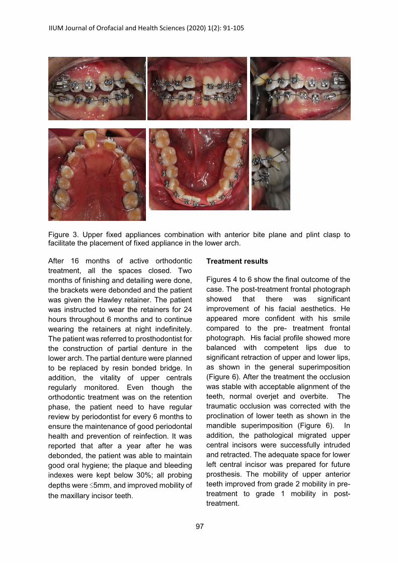
Orthodontic treatment started in December
2015 and finished in June 2017. It took 18
months to achieve the stable and good
occlusion. In order raise the bite for the
placement of upper and lower fixed
appliances, the upper removable appliance
with an anterior bite plane and plint clasp
was constructed. After two weeks, pre-
adjusted edgewise brackets (0.022x0.028-
in, MBT prescription) were bonded to all the
teeth except the upper second molars
(Figure 3).
Upper and lower 0.012-in nickel titanium
archwires were placed and treatment
progressed up to 0.019x0.025-in stainless
steel archwires. Initial alignment followed by
levelling in the upper and lower arches was
achieved in 6 months. Upper and lower
0.019x0.025-in stainless steel archwires
were maintained for 2 months in order to fully
express the torque. The anterior bite plane
was removed and en-masse retraction of the
upper arch was done with elastic chains
(150g) to close the remaining spaces. In the
lower arch, same mechanics was used as an
upper in order to retract lower arch. The
space for lower left central incisor was
maintained with dead coil spring.
IIUM Journal of Orofacial and Health Sciences (2020) 1(2): 91-105
97
Figure 3. Upper fixed appliances combination with anterior bite plane and plint clasp to facilitate the placement of fixed appliance in the lower arch.
After 16 months of active orthodontic
treatment, all the spaces closed. Two
months of finishing and detailing were done,
the brackets were debonded and the patient
was given the Hawley retainer. The patient
was instructed to wear the retainers for 24
hours throughout 6 months and to continue
wearing the retainers at night indefinitely.
The patient was referred to prosthodontist for
the construction of partial denture in the
lower arch. The partial denture were planned
to be replaced by resin bonded bridge. In
addition, the vitality of upper centrals
regularly monitored. Even though the
orthodontic treatment was on the retention
phase, the patient need to have regular
review by periodontist for every 6 months to
ensure the maintenance of good periodontal
health and prevention of reinfection. It was
reported that after a year after he was
debonded, the patient was able to maintain
good oral hygiene; the plaque and bleeding
indexes were kept below 30%; all probing
depths were ≤5mm, and improved mobility of
the maxillary incisor teeth.
Treatment results
Figures 4 to 6 show the final outcome of the
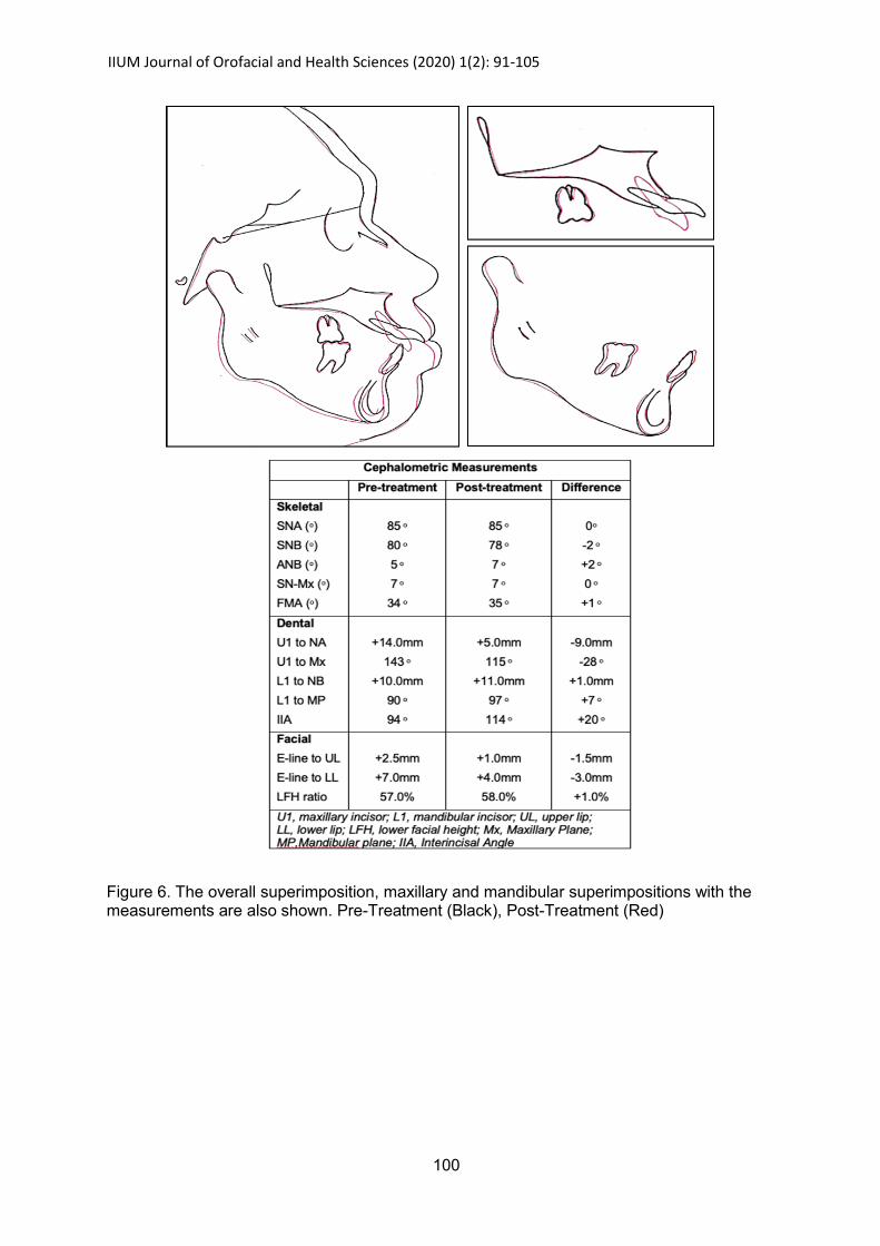
case. The post-treatment frontal photograph
showed that there was significant
improvement of his facial aesthetics. He
appeared more confident with his smile
compared to the pre- treatment frontal
photograph. His facial profile showed more
balanced with competent lips due to
significant retraction of upper and lower lips,
as shown in the general superimposition
(Figure 6). After the treatment the occlusion
was stable with acceptable alignment of the
teeth, normal overjet and overbite. The
traumatic occlusion was corrected with the
proclination of lower teeth as shown in the
mandible superimposition (Figure 6). In
addition, the pathological migrated upper
central incisors were successfully intruded
and retracted. The adequate space for lower
left central incisor was prepared for future
prosthesis. The mobility of upper anterior
teeth improved from grade 2 mobility in pre-
treatment to grade 1 mobility in post-
treatment.
IIUM Journal of Orofacial and Health Sciences (2020) 1(2): 91-105
98
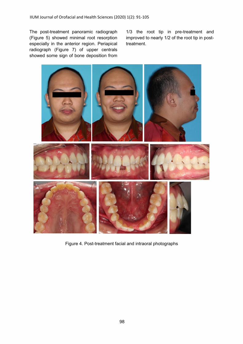
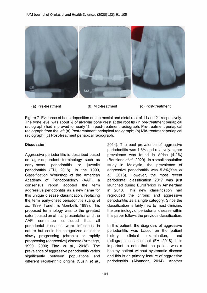
The post-treatment panoramic radiograph
(Figure 5) showed minimal root resorption
especially in the anterior region. Periapical
radiograph (Figure 7) of upper centrals
showed some sign of bone deposition from
1/3 the root tip in pre-treatment and
improved to nearly 1/2 of the root tip in post-
treatment.
Figure 4. Post-treatment facial and intraoral photographs
IIUM Journal of Orofacial and Health Sciences (2020) 1(2): 91-105
99
Figure 5. Pre-treatment panoramic radiograph, lateral cephalometric radiograph and tracing, Lateral cephalometric radiograph measurements
IIUM Journal of Orofacial and Health Sciences (2020) 1(2): 91-105
100
Figure 6. The overall superimposition, maxillary and mandibular superimpositions with the measurements are also shown. Pre-Treatment (Black), Post-Treatment (Red)
IIUM Journal of Orofacial and Health Sciences (2020) 1(2): 91-105
101
(a) Pre-treatment (b) Mid-treatment (c) Post-treatment
Figure 7. Evidence of bone deposition on the mesial and distal root of 11 and 21 respectively. The bone level was about ⅓ of alveolar bone crest at the root tip (in pre-treatment periapical radiograph) had improved to nearly ½ in post-treatment radiograph. Pre-treatment periapical radiograph from the left (a) Post-treatment periapical radiograph; (b) Mid-treatment periapical radiograph; (c) Post-treatment periapical radiograph.
Discussion
Aggressive periodontitis is described based
on age‑dependent terminology such as
early‑onset periodontitis or juvenile
periodontitis (FH, 2018). In the 1999,
Classification Workshop of the American
Academy of Periodontology (AAP), a
consensus report adopted the term
aggressive periodontitis as a new name for
this unique disease classification, replacing
the term early-onset periodontitis (Lang et
al., 1999; Tonetti & Mombelli, 1999). This
proposed terminology was to the greatest
extent based on clinical presentation and the
AAP committee concluded that all
periodontal diseases were infectious in
nature but could be categorized as either
slowly progressing (chronic) or rapidly
progressing (aggressive) disease (Armitage,
1999, 2000; Fine et al., 2018). The
prevalence of aggressive periodontitis varies
significantly between populations and
different racial/ethnic origins (Susin et al.,
2014). The pool prevalence of aggressive
periodontitis was 1.6% and relatively higher
prevalence was found in Africa (4.2%)
(Bouziane et al., 2020). In a small population
study in Malaysia, the prevalence of
aggressive periodontitis was 5.3%(Yee et
al., 2016). However, the most recent
periodontal classification 2017 was just
launched during EuroPerio9 in Amsterdam
in 2018. This new classification had
regrouped the chronic and aggressive
periodontitis as a single category. Since the
classification is fairly new to most clinician,
the terminology of periodontal disease within
this paper follows the previous classification.
In this patient, the diagnosis of aggressive
periodontitis was based on the patient
history, clinical examination, and
radiographic assessment (FH, 2018). It is
important to note that the patient was a
healthy patient without systematic disease
and this is an primary feature of aggressive
periodontitis (Albandar, 2014). Another
IIUM Journal of Orofacial and Health Sciences (2020) 1(2): 91-105
102
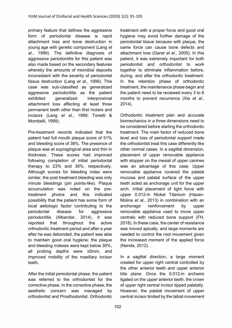
primary feature that defines the aggressive
form of periodontal disease is rapid
attachment loss and bone destruction in
young age with genetic component (Lang et
al., 1999). The definitive diagnosis of
aggressive periodontitis for this patient was
also made based on the secondary features
whereby the amounts of microbial deposits
inconsistent with the severity of periodontal
tissue destruction (Lang et al., 1999). This
case was sub-classified as generalized
aggressive periodontitis as the patient
exhibited generalized interproximal
attachment loss affecting at least three
permanent teeth other than first molars and
incisors (Lang et al., 1999; Tonetti &
Mombelli, 1999).
Pre-treatment records indicated that the
patient had full mouth plaque score of 51%
and bleeding score of 38%. The presence of
plaque was at supragingival area and thin in
thickness. These scores had improved
following completion of initial periodontal
therapy to 23% and 36%, respectively.
Although scores for bleeding index were
similar, the post treatment bleeding was only
minute bleedings (pin points-like). Plaque
accumulation was noted on the pre-
treatment photos and this indicated
possibility that the patient has some form of
local aetiologic factor contributing to the
periodontal disease for aggressive
periodontitis (Albandar, 2014). It was
reported that throughout the active
orthodontic treatment period and after a year
after he was debonded, the patient was able
to maintain good oral hygiene; the plaque
and bleeding indexes were kept below 30%;
all probing depths were ≤5mm, and
improved mobility of the maxillary incisor
teeth.
After the initial periodontal phase, the patient
was referred to the orthodontist for the
corrective phase. In the corrective phase, the
aesthetic concern was managed by
orthodontist and Prosthodontist. Orthodontic
treatment with a proper force and good oral
hygiene may avoid further damage of the
periodontal tissue because with plaque, the
same force can cause bone defects and
attachment loss (Garat et al., 2005). In this
patient, it was extremely important for both
periodontist and orthodontist to work
together to eliminate inflammation before,
during, and after the orthodontic treatment.
In the retention phase of orthodontic
treatment, the maintenance phase begin and
the patient need to be reviewed every 3 to 6
months to prevent recurrence (Xie et al.,
2014).
Orthodontic treatment plan and accurate
biomechanics in a three dimensions need to
be considered before starting the orthodontic
treatment. The main factor of reduced bone
level and loss of periodontal support made
the orthodontist treat this case differently like
other normal cases. In a sagittal dimension,
placement of upper removable appliance
with stopper on the mesial of upper canines
was an advantage of this case. Upper
removable appliance covered the palatal
mucosa and palatal surface of the upper
teeth acted as anchorage unit for the upper
arch. Initial placement of light force with
upper 0.012-in Nickel Titanium (Hazan-
Molina et al., 2013) in combination with an
anchorage reinforcement by upper
removable appliance used to move upper
centrals with reduced bone support (FH,
2018). In these case, the center of resistance
was moved apically, and large moments are
needed to control the root movement given
the increased moment of the applied force
(Nanda, 2012).
In a sagittal direction, a large moment
created for upper right central controlled by
the other anterior teeth and upper anterior
bite plane. Once the 0.012-in archwire
ligated on the upper anterior teeth, the crown
of upper right central incisor tipped palatally.
However, the palatal movement of upper
central incisor limited by the labial movement
IIUM Journal of Orofacial and Health Sciences (2020) 1(2): 91-105
103
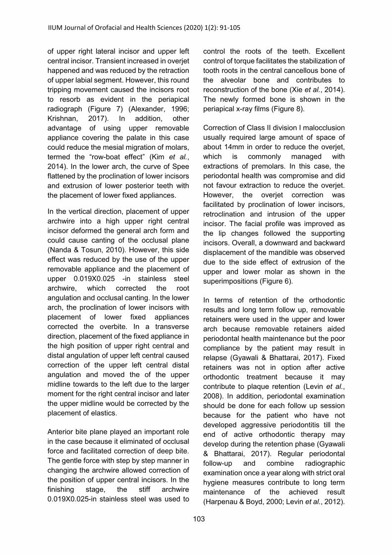
of upper right lateral incisor and upper left
central incisor. Transient increased in overjet
happened and was reduced by the retraction
of upper labial segment. However, this round
tripping movement caused the incisors root
to resorb as evident in the periapical
radiograph (Figure 7) (Alexander, 1996;
Krishnan, 2017). In addition, other
advantage of using upper removable
appliance covering the palate in this case
could reduce the mesial migration of molars,
termed the “row-boat effect” (Kim et al.,
2014). In the lower arch, the curve of Spee
flattened by the proclination of lower incisors
and extrusion of lower posterior teeth with
the placement of lower fixed appliances.
In the vertical direction, placement of upper
archwire into a high upper right central
incisor deformed the general arch form and
could cause canting of the occlusal plane
(Nanda & Tosun, 2010). However, this side
effect was reduced by the use of the upper
removable appliance and the placement of
upper 0.019X0.025 -in stainless steel
archwire, which corrected the root
angulation and occlusal canting. In the lower
arch, the proclination of lower incisors with
placement of lower fixed appliances
corrected the overbite. In a transverse
direction, placement of the fixed appliance in
the high position of upper right central and
distal angulation of upper left central caused
correction of the upper left central distal
angulation and moved the of the upper
midline towards to the left due to the larger
moment for the right central incisor and later
the upper midline would be corrected by the
placement of elastics.
Anterior bite plane played an important role
in the case because it eliminated of occlusal
force and facilitated correction of deep bite.
The gentle force with step by step manner in
changing the archwire allowed correction of
the position of upper central incisors. In the
finishing stage, the stiff archwire
0.019X0.025-in stainless steel was used to
control the roots of the teeth. Excellent
control of torque facilitates the stabilization of
tooth roots in the central cancellous bone of
the alveolar bone and contributes to
reconstruction of the bone (Xie et al., 2014).
The newly formed bone is shown in the
periapical x-ray films (Figure 8).
Correction of Class II division I malocclusion
usually required large amount of space of
about 14mm in order to reduce the overjet,
which is commonly managed with
extractions of premolars. In this case, the
periodontal health was compromise and did
not favour extraction to reduce the overjet.
However, the overjet correction was
facilitated by proclination of lower incisors,
retroclination and intrusion of the upper
incisor. The facial profile was improved as
the lip changes followed the supporting
incisors. Overall, a downward and backward
displacement of the mandible was observed
due to the side effect of extrusion of the
upper and lower molar as shown in the
superimpositions (Figure 6).
In terms of retention of the orthodontic
results and long term follow up, removable
retainers were used in the upper and lower
arch because removable retainers aided
periodontal health maintenance but the poor
compliance by the patient may result in
relapse (Gyawali & Bhattarai, 2017). Fixed
retainers was not in option after active
orthodontic treatment because it may
contribute to plaque retention (Levin et al.,
2008). In addition, periodontal examination
should be done for each follow up session
because for the patient who have not
developed aggressive periodontitis till the
end of active orthodontic therapy may
develop during the retention phase (Gyawali
& Bhattarai, 2017). Regular periodontal
follow-up and combine radiographic
examination once a year along with strict oral
hygiene measures contribute to long term
maintenance of the achieved result
(Harpenau & Boyd, 2000; Levin et al., 2012).
IIUM Journal of Orofacial and Health Sciences (2020) 1(2): 91-105
104
Conclusion
Management of patients with an aggressive
periodontitis is a challenge for orthodontist
and periodontist. The importance of having
good oral hygiene before, during and after
orthodontic treatment significantly improve
the function, esthetic and periodontal health
of the patient. Therefore, a good
collaboration between orthodontist and
periodontist are important to ensure the
successful treatment outcome in patient with
aggressive periodontitis.
Acknowledgement
The authors would like to thank all staff at
Orthodontic Unit, Klinik Pergigian Cahaya
Suria (Sementara), Kuala Lumpur for
assistance and contribution to this case
report. Special gratitude to the Director
General of Health Malaysia and the Principle
Director of the Oral Health Division, Ministry
of Health Malaysia for permission to have
this case report published.
Declaration of patient consent
The authors certify that they have obtained
all appropriate patient consent forms. In the
form the patient(s) has/have given
his/her/their consent for his/her/their images
and other clinical information to be reported
in the journal. The patients understand that
their names and initials will not be published,
and due efforts will be made to conceal their
identity, but anonymity cannot be
guaranteed.
References
Aimetti, M., Romano, F., Guzzi, N., & Carnevale, G.
(2012). Full‐mouth disinfection and systemic
antimicrobial therapy in generalized aggressive periodontitis: a randomized, placebo‐controlled trial. Journal of Clinical Periodontology, 39(3), 284-294.
Albandar, J. M. (2014). Aggressive periodontitis: case definition and diagnostic criteria. Periodontology 2000, 65, 13–26.
Albandar, J. M., Brown, L. J., & Loe, H. (1997). Clinical features of early-onset periodontitis. The Journal of the American Dental Association, 128(10), 1393-1399.
Alexander, S. A. (1996). Levels of root resorption associated with continuous arch and sectional arch mechanics. American Journal of Orthodontics and Dentofacial Orthopedics, 110(3), 321-324.
Armitage, G. C. (1999). Development of a classification system for periodontal diseases and conditions. Annals of Periodontology, 4(1), 1-6.
Armitage, G. C. (2000). Development of a classification system for periodontal diseases and conditions. Northwest Dentistry, 79(6), 31-35.
Azouni, K. G., & Tarakji, B. (2014). The trimeric model: a new model of periodontal treatment planning. Journal of Clinical and Diagnostic Research, 8(7), ZE17-20.
Bagga, D. K. (2010). Adult orthodontics versus adolescent orthodontics: an overview. Journal of Oral Health and Community Dentistry 4(2), 42-47.
Bouziane, A., Hamdoun, R., Abouqal, R., & Ennibi, O. (2020). Global prevalence of aggressive periodontitis: A systematic review and meta-analysis. Journal of Clinical Periodontology, 47(4), 406-428.
Brunsvold, M. A. (2005). Pathologic tooth migration. Journal of Periodontology, 76(6), 859-866.
Feng, X., Oba, T., Oba, Y., & Moriyama, K. (2005). An interdisciplinary approach for improved functional and esthetic results in a periodontally compromised adult patient. The Angle Orthodontist, 75(6), 1061-1070.
FH, A. (2018). Orthodontic treatment of an adolescent patient with localized aggressive periodontitis. Journal of International Oral Health, 10, 47‐57.
Fine, D. H., Patil, A. G., & Loos, B. G. (2018). Classification and diagnosis of aggressive periodontitis. Journal of Periodontology, 89 Suppl 1, S103-S119.
Garat, J. A., Gordillo, M. E., & Ubios, A. M. (2005). Bone response to different strength orthodontic forces in animals with periodontitis. Journal of Periodontal Research, 40(6), 441-445.
Guerrero, A., Griffiths, G. S., Nibali, L., Suvan, J., Moles, D. R., Laurell, L., & Tonetti, M. S. (2005). Adjunctive benefits of systemic amoxicillin and metronidazole in non-surgical treatment of generalized aggressive periodontitis: a randomized placebo-controlled clinical trial. Journal of Clinical Periodontology, 32(10), 1096-1107.
Gyawali, R., & Bhattarai, B. (2017). Orthodontic management in aggressive periodontitis. International Scholarly Research Notices, 2017, 8098154.
Harpenau, L. A., & Boyd, R. L. (2000). Long-term follow-up of successful orthodontic-periodontal treatment of localized aggressive periodontitis: a case report. Clinical Orthodontics and Research, 3(4), 220-229.
IIUM Journal of Orofacial and Health Sciences (2020) 1(2): 91-105
105
Hazan-Molina, H., Levin, L., Einy, S., & Aizenbud, D. (2013). Aggressive periodontitis diagnosed during or before orthodontic treatment. Acta Odontologica Scandinavica, 71(5), 1023-1031.
Hormand, J., & Frandsen, A. (1979). Juvenile periodontitis. Localization of bone loss in relation to age, sex, and teeth. Journal of Clinical Periodontology, 6(6), 407-416. Kim,
S.-J., Kim, J.-W., Choi, T.-H., & Lee, K.-J. (2014). Combined use of miniscrews and continuous arch for intrusive root movement of incisors in Class II division 2 with gummy smile. The Angle Orthodontist, 84, 910–918.
Krishnan, V. (2017). Root resorption with orthodontic mechanics: pertinent areas revisited. Australian Dental Journal, 62 Suppl 1, 71-77.
Lang, N., Bartold, P. M., Cullinan, M., Jeffcoat, M., Mombelli, A., Murakami, S. et al. (1999). Consensus report: aggressive periodontitis. Annals of Periodontology, 4(1), 53.
Levin, L., Einy, S., Zigdon, H., Aizenbud, D., & Machtei, E. E. (2012). Guidelines for periodontal care and follow-up during orthodontic treatment in adolescents and young adults. Journal of Applied Oral Science, 20(4), 399-403.
Levin, L., Samorodnitzky-Naveh, G. R., & Machtei, E. E. (2008). The association of orthodontic treatment and fixed retainers with gingival health. Journal of Periodontology, 79(11),
2087-2092. Martinez-Canut, P., Carrasquer, A., Magan, R., &
Lorca, A. (1997). A study on factors associated with pathologic tooth migration. Journal of Clinical Periodontology, 24(7), 492-497.
Meng, H., Xu, L., Li, Q., Han, J., & Zhao, Y. (2007). Determinants of host susceptibility in aggressive periodontitis. Periodontol 2000, 43, 133-159.
Mestnik, M. J., Feres, M., Figueiredo, L. C., Duarte, P. M., Lira, E. A. G., & Faveri, M. (2010). Short‐term benefits of the adjunctive use of metronidazole plus amoxicillin in the microbial profile and in the clinical parameters of subjects with generalized aggressive periodontitis. Journal of Clinical Periodontology, 37(4), 353-365.
Mestnik, M. J., Feres, M., Figueiredo, L. C., Soares, G., Teles, R. P., Fermiano, D., et al. (2012). The effects of adjunctive metronidazole plus amoxicillin in the treatment of generalized aggressive periodontitis: a 1-year double-blinded, placebo-controlled, randomized clinical trial. Journal of Clinical Periodontology, 39(10), 955-961.
Nanda, R. (2012). Esthetics and Biomechanics in Orthodontics‐E‐Book: St. Louis : Elsevier
Health Sciences. Nanda, R. S., & Tosun, Y. S. (2010). Biomechanics In
Orthodontics, Principles And Practice:
Quintessence Publishing Co Inc. Panwar, M., Jayan, B., Mandlik, V. B., & Jha, A. K.
(2010). Combined Periodontal and Orthodontic Treatment of Pathologic Migration of Anterior Teeth. Medical Journal Armed Forces India, 66(1), 67-69.
Rabie, A. B., Deng, Y. M., & Jin, L. J. (1998). Adjunctive orthodontic treatment of periodontally involved teeth: case reports. Quintessence International, 29(1), 13-19.
Susin, C., Haas, A. N., & Albandar, J. M. (2014). Epidemiology and demographics of aggressive periodontitis. Periodontol 2000, 65(1), 27-45.
Tonetti, M. S., & Mombelli, A. (1999). Early-onset periodontitis. Annals of Periodontology, 4(1), 39-53.
Towfighi, P. P., Brunsvold, M. A., Storey, A. T., Arnold, R. M., Willman, D. E., & McMahan, C. A. (1997). Pathologic migration of anterior teeth in patients with moderate to severe periodontitis. Journal of Periodontology, 68(10), 967-972.
Vinod, K., Reddy, Y. G., Reddy, V. P., Nandan, H., & Sharma, M. (2012). Orthodontic-periodontics interdisciplinary approach. Journal of Indian Society of Periodontology, 16(1), 11-15.
Xie, Y., Zhao, Q., Tan, Z., & Yang, S. (2014). Orthodontic treatment in a periodontal patient with pathologic migration of anterior teeth. American Journal of Orthodontics and Dentofacial Orthopedics, 145(5), 685-693.
Yee, L. F., Subramaniam, U., Raman, R., & Loo, C. S. (2016). Prevalence of Aggressive Periodontitis in Newly Referred Patients in a Periodontal Specialist Government Clinic. Malaysian Dental Journal, 39(1), 1-25.
Related Documents