Case Report Oral Lesion as Unusual First Manifestation of Multiple Myeloma: Case Reports and Review of the Literature A. Romano, 1,2,3 M. S. Marescalco, 1,3,4 Chiara Liardo, 4 L. Villari, 5 C. Vetro, 1,3 C. Conticello, 1 F. Di Raimondo, 1 and S. Ferlito 4 1 Department of Clinical and Molecular Biomedicine, Section of Haematology, University of Catania, Via Citelli 6, 95124 Catania, Italy 2 Ospedale Ferrarotto, Divisione di Ematologia, Via Citelli 6, 95124 Catania, Italy 3 Scuola Superiore di Catania, Via San Nullo 5 I, 95125 Catania, Italy 4 1st Section of Dentistry, Department of Surgery, University of Catania, Via Citelli 6, 95124 Catania, Italy 5 Division of Pathology, AOU “Policlinico-Vittorio Emanuele”, Via Plebiscito 628, 95100 Catania, Italy Correspondence should be addressed to A. Romano; [email protected] Received 1 August 2014; Revised 11 October 2014; Accepted 13 October 2014; Published 25 November 2014 Academic Editor: Akimichi Ohsaka Copyright © 2014 A. Romano et al. is is an open access article distributed under the Creative Commons Attribution License, which permits unrestricted use, distribution, and reproduction in any medium, provided the original work is properly cited. Extramedullary plasmacytoma (EMP) and solitary bone plasmacytoma (SBP) represent a disease continuum through a multistage process of cell differentiation, survival, proliferation, and dissemination, strictly related to multiple myeloma (MM), the second most common hematological malignancy. Herein, we report two cases of recurrent oral plasmacytoma progressed to MM, in which the first clinical sign of a more widespread disease was limited to the mouth. Based on our experience, we recommend a strict workup for the differential diagnosis between EMP, SBP, and MM for patients with oral plasmacytoma, including radiological exam of the skeleton, magnetic resonance imaging (MRI) of the bone, and positive emission tomography (FDG-PET). MRI and possibly PET can all be used to more sensitively detect EM plasmacytoma sites. 1. Introduction Multiple myeloma (MM) is a multifocal plasma cell pro- liferation in the bone marrow, in which neoplastic cells replace normal marrow cells, produce excess immunoglob- ulin, and infiltrate the bone. In a few cases, neoplastic plasma cells can be disposed in a localized mass within the bone, in a condition known as solitary bone plasmacytoma [1] (SBP) or outside thus involving soſt tissues, in a condition defined as extramedullary plasmacytoma [2] (SEP). Clinical manifestations of SEP are generally not specific and may include swelling, pain, numbness, bleeding, mobile teeth, and xerostomia [3–5]. MM is defined by ≥10% of plasma cell infiltration in the bone marrow, ≥30 g/L of monoclonal protein and presence of hypercalcemia, renal insufficiency, anemia, and bone lytic lesions (identified by the acronym of CRAB symptoms) [6, 7]. Up to 90% MM patients complain of symptoms due to bone disease, defined as multiple destructive lytic lesions of the skeleton, including severe demineralization and osteoporosis through pathological fractures [8]. Involved areas include skeleton segments at high content of bone marrow such as skull, spine, sternum, vertebrae, pelvis, and hip. Jawbone is MM site in 30% cases [9, 10]. Oral lesions are seldom the first sign of disease [11–14]. Solitary bone plasmacytoma (SBP) is an area of lytic bone destruction in an otherwise asymptomatic patient, occurring generally in axial skeleton (70% in spine, seldom in the mouth [15, 16], including the temporomandibular joint [17, 18]), in absence of plasma cell infiltration of the bone marrow, without serum/urine M-protein. Patients complain of bone pain, and in critical cases, SBP may cause cord/root compression. High dosage radiotherapy (>5000 Gy) is oſten curative. However, lesions larger than 5 cm, immunoparesis, and persistence of paraprotein aſter radiotherapy are adverse prognostic factors for progression to MM [1]. Hindawi Publishing Corporation Case Reports in Hematology Volume 2014, Article ID 529452, 7 pages http://dx.doi.org/10.1155/2014/529452

Welcome message from author
This document is posted to help you gain knowledge. Please leave a comment to let me know what you think about it! Share it to your friends and learn new things together.
Transcript
Case ReportOral Lesion as Unusual First Manifestation of MultipleMyeloma: Case Reports and Review of the Literature
A. Romano,1,2,3 M. S. Marescalco,1,3,4 Chiara Liardo,4 L. Villari,5
C. Vetro,1,3 C. Conticello,1 F. Di Raimondo,1 and S. Ferlito4
1 Department of Clinical and Molecular Biomedicine, Section of Haematology, University of Catania, Via Citelli 6,95124 Catania, Italy
2 Ospedale Ferrarotto, Divisione di Ematologia, Via Citelli 6, 95124 Catania, Italy3 Scuola Superiore di Catania, Via San Nullo 5 I, 95125 Catania, Italy4 1st Section of Dentistry, Department of Surgery, University of Catania, Via Citelli 6, 95124 Catania, Italy5 Division of Pathology, AOU “Policlinico-Vittorio Emanuele”, Via Plebiscito 628, 95100 Catania, Italy
Correspondence should be addressed to A. Romano; [email protected]
Received 1 August 2014; Revised 11 October 2014; Accepted 13 October 2014; Published 25 November 2014
Academic Editor: Akimichi Ohsaka
Copyright © 2014 A. Romano et al. This is an open access article distributed under the Creative Commons Attribution License,which permits unrestricted use, distribution, and reproduction in any medium, provided the original work is properly cited.
Extramedullary plasmacytoma (EMP) and solitary bone plasmacytoma (SBP) represent a disease continuum through a multistageprocess of cell differentiation, survival, proliferation, anddissemination, strictly related tomultiplemyeloma (MM), the secondmostcommon hematological malignancy. Herein, we report two cases of recurrent oral plasmacytoma progressed to MM, in which thefirst clinical sign of a more widespread disease was limited to the mouth. Based on our experience, we recommend a strict workupfor the differential diagnosis between EMP, SBP, and MM for patients with oral plasmacytoma, including radiological exam of theskeleton, magnetic resonance imaging (MRI) of the bone, and positive emission tomography (FDG-PET). MRI and possibly PETcan all be used to more sensitively detect EM plasmacytoma sites.
1. Introduction
Multiple myeloma (MM) is a multifocal plasma cell pro-liferation in the bone marrow, in which neoplastic cellsreplace normal marrow cells, produce excess immunoglob-ulin, and infiltrate the bone. In a few cases, neoplastic plasmacells can be disposed in a localized mass within the bone,in a condition known as solitary bone plasmacytoma [1](SBP) or outside thus involving soft tissues, in a conditiondefined as extramedullary plasmacytoma [2] (SEP). Clinicalmanifestations of SEP are generally not specific and mayinclude swelling, pain, numbness, bleeding,mobile teeth, andxerostomia [3–5].
MM is defined by ≥10% of plasma cell infiltration in thebone marrow, ≥30 g/L of monoclonal protein and presenceof hypercalcemia, renal insufficiency, anemia, and bone lyticlesions (identified by the acronymof CRAB symptoms) [6, 7].Up to 90% MM patients complain of symptoms due to bone
disease, defined as multiple destructive lytic lesions of theskeleton, including severe demineralization and osteoporosisthrough pathological fractures [8]. Involved areas includeskeleton segments at high content of bone marrow such asskull, spine, sternum, vertebrae, pelvis, and hip. Jawbone isMM site in 30% cases [9, 10]. Oral lesions are seldom the firstsign of disease [11–14].
Solitary bone plasmacytoma (SBP) is an area of lyticbone destruction in an otherwise asymptomatic patient,occurring generally in axial skeleton (70% in spine, seldomin the mouth [15, 16], including the temporomandibular joint[17, 18]), in absence of plasma cell infiltration of the bonemarrow, without serum/urine M-protein. Patients complainof bone pain, and in critical cases, SBP may cause cord/rootcompression. High dosage radiotherapy (>5000Gy) is oftencurative. However, lesions larger than 5 cm, immunoparesis,and persistence of paraprotein after radiotherapy are adverseprognostic factors for progression to MM [1].
Hindawi Publishing CorporationCase Reports in HematologyVolume 2014, Article ID 529452, 7 pageshttp://dx.doi.org/10.1155/2014/529452
2 Case Reports in Hematology
Extramedullary plasmacytoma (SEP) occurs rarely (<1%of cases) and preferentially at level of head andneck [3], upperairways including nose-pharynx, tonsils, and paranasal sinus[4] and rarely at the mouth [5]. Like in SBP the M-proteinis low or absent; plasma cell infiltration in the bone marrowis less than 5%, in absence of end-organ damage and diffuseosteolytic lesions [2].
Here we report two cases of oral plasmacytoma, in whichthe first clinical sign of amorewidespread disease was limitedto the mouth.
2. Case A
A 72-year-old Caucasian male presented to dental divisioncomplaining of pain and difficulty in chewing due to anovergrowth of soft tissue located in the alveolar ridge of theleftmandible. He reported the extraction of the residual rootsof 3.7 which occurred two weeks before. After two days, thealveolarmucosa grew up over the postextraction site. No painor alterations of gingival mucosa were reported before theextraction of residual roots.
Extraoral examination revealed normal facial morphol-ogy, in absence of lymph node swelling.The orthopantomog-raphy did not show any sign of bone involvement. At intraoralexamination, a softmass was present (1.5 cm) with ill-definedcontours being pink in colour and slightly ulcerated on topfor the action of antagonist dental elements.
After the patient provided written informed consent,the soft tissue was biopsied. The histological examinationrevealed a granulation tissue dissociated by lymphoplasma-cytic infiltrate. Seven days after the biopsy the soft massrecurred. Three days later, an excisional biopsy was per-formed (Figure 1). Histological analysis showed the presenceof a poorly differentiated plasma cell neoplasm. Microscop-ically the tissue appeared heavily infiltrated by neoplasticproliferation with widespread growth, consisting of ele-ments of plasmablastic/plasmacytic morphology and someof them frankly pleomorphic and anaplastic (Figure 2),compatible with the diagnosis of extramedullary plas-mablastic/anaplastic plasmacytomaCD138+, CD38+, EMA+,vimentin+, CD79a weak, CD20−, and CD3−.The patient wastherefore sent to the department of haematology for inquiriesregarding the stage of disease and for subsequent treatment.
According to medical history, the patient was diabetic,asthmatic smoker. A comprehensive metabolic panel andcomplete blood count (CBC) revealed a total protein of8.2 g/dL, due to a concomitant low amount of iron, withoutimmunoparesis, in absence of a detectable serum or urineparaprotein by immunofixation. The bone marrow biopsywas negative for clonal plasma cells.
The patient underwent bone imaging: the skeletal surveywas negative for osteolytic lesions; the fluorodeoxyglucose-positron emission tomography (FDG-PET) scan was weaklypositive at level of mandible and the second cervical vertebra(SUVmax = 2.5), otherwise normal. This finding was con-firmed atmagnetic resonance imaging (MRI), otherwise nor-mal; thus we considered these findings without an oncologicmeaning.
Table 1: Clinical variables at diagnosis of patients A and B.
Case A Case B Normal rangeHemoglobin, g/L 10.5 10.6 12.1–15.1Serum lactate dehydrogenase, U/L 377 256 256–450Serum beta-2 microglobulin, mg/L 2.8 2.2 1.2–2.4Serum albumin (g/dL) 4.0 4.1 3.5–5.0Serum AST (U/L) 14 15 5–42Serum ALT (U/L) 42 31 5–42Serum calcium (mmol/L) 2.22 2.40 2.15–2.55Serum creatinine (micromol/L) 108 89 60–125Blood urea nitrogen (mmol/L) 5.2 4.8 2.5–8.0ISS stage I I —Durie and Salmon Stage 1 1 —
In absence of CRAB criteria, SEP was diagnosed andthe patient received radiotherapy at level of mandible andvertebra. The treatment was complicated by swelling, weightloss, and worsening asthenia.
Threemonths later, FDG-PET scanwas repeated showingpersistence of positivity, but in absence of symptoms.
Further three months later, clinical conditions worsened;magnetic resonance imaging (MRI) showed nodular lesionswith intense enhancement in vertebrae, in the pelvic bone,and in the sternum. FDG-PET scan showed diffusely positivesignal in the left jaw, sternum, and ankle, confirming thesystemic nature of the disease. The bone marrow biopsyshowed an infiltration of 30% monoclonal plasma cells.
In absence of circulating monoclonal component inserum and urine, with evidence of immunoparesis, nonsecre-toryMMwas diagnosed, stage II in accord with InternationalStaging System [24]. Laboratory findings are reported inTable 1.
Two months later, at eight months from the first occur-rence of oral plasmacytoma, the patient started systemicchemotherapy with bortezomib 1.3 g/mq at days 1, 4, 8, and11, thalidomide 100mg for 21 days, and dexamethasone 40mgweekly without benefit. The patient died of pneumonia twomonths later.
3. Case B
A 63-year-old Caucasian man, affected by monoclonal gam-mopathy of undetermined significance (MGUS) since fiveyears, presented with an evolving swelling in the maxilla, atlevel of left posterior region with involvement of palate andgingiva. At the intraoral examination, the mass consistencywas hard elastic and normal in color and covered with intactoral mucosa (Figure 1(c)).
The maxillofacial computerized tomography (CT) scanconfirmed the presence of a solid neoplasia, with maximumdimensions 25 × 45mm, causing a structural rearrangementof the bone. An osteolytic involvement of the upper alve-olar process and the maxillary sinus was evident, whichappeared partially obliterated in its basal portion. The neo-plasm was extended to the homolateral nasal cavity withhyperdense swelling at the nose-maxillary region adjoining.
Case Reports in Hematology 3
(a) (b)
(c) (d)
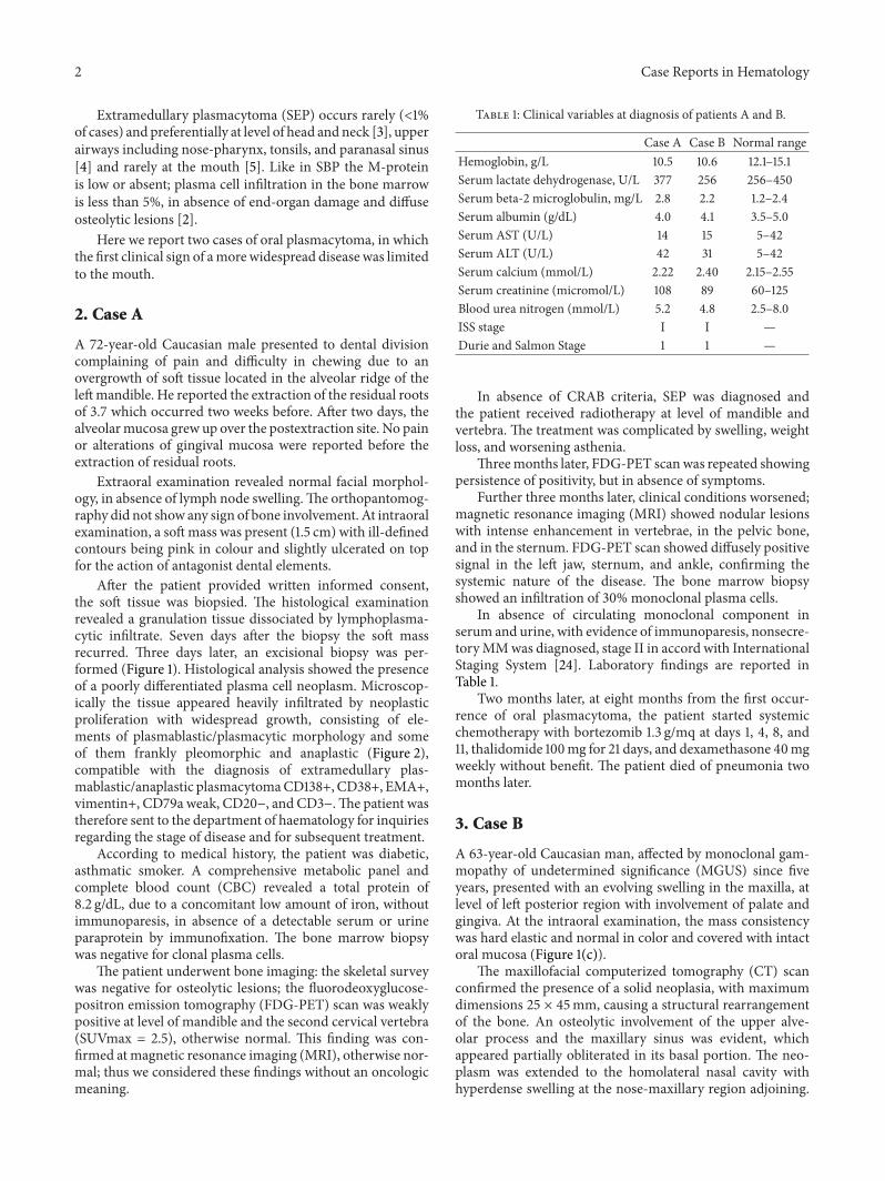
Figure 1: Extramedullary plasmacytoma, macroscopic features. (a) The soft mass neoplasm appears pink in colour and has floatingconsistency with a diameter of about 1.5 cm, occupying the area of dental elements 37 and 38. It has ill-defined contours and is slightlyulcerated on the top for the effects of antagonist dental elements, causing algia and difficulty in chewing for the patient. No sign of boneinvolvement at the orthopantomography (Case A). (b) The patient presented a relapse of the neoplasm seven days after the biopsy, with theenlargement of the lesion to the entire edentulous left side of the jaw, until the first premolar. The mucosa covering the lesion was of irregularcolour and the traumatism due to the antagonist dental elements was accentuated (Case A). (c) The mass presented as a swelling in the leftposterior region of the maxilla, with maximum dimensions 25 × 45mm, involving also palate and gingiva. The mass consistency was hardelastic and normal in colour and covered with intact oral mucosa (Case B). (d) The maxillofacial CT scan showed an important osteolyticinvolvement of the upper alveolar process and the partial obliteration of the basal portion of the left maxillary sinus.The lesion extended alsoto the homolateral nasal cavity. Concomitant enlargement of lateral cervical and submandibular lymph nodes, with the biggest having 13mmdiameter (Case B).
Later enlarged cervical and submandibular lymphnodeswereevident; the largest had 13mm diameter.
The patient reported that the presence of soft tissue in thesame site occurred around six months before and was alreadybiopsied, compatible with diagnosis of mucosal plasmacy-toma.The histological revision in our division confirmed thelow-grade nature of that lesion (weak plasma cell infiltration,
presence of small necrosis foci, low proliferation index, 3-4%,immunophenotype: CD138+, CD38+, EMA+, vimentin+,CD79a weak, CD20−, CD3−, CD45RO−, SMA−, myosin−,and S-100−, Figure 2). Hematological restaging confirmedpersistency of MGUS, with immunofixation positive for IgA,stable M-protein (2200mg/dL), no end-organ damage signs,including bone evaluation with X-ray scan and MRI, and
4 Case Reports in Hematology
(a) (b)
(c) (d)
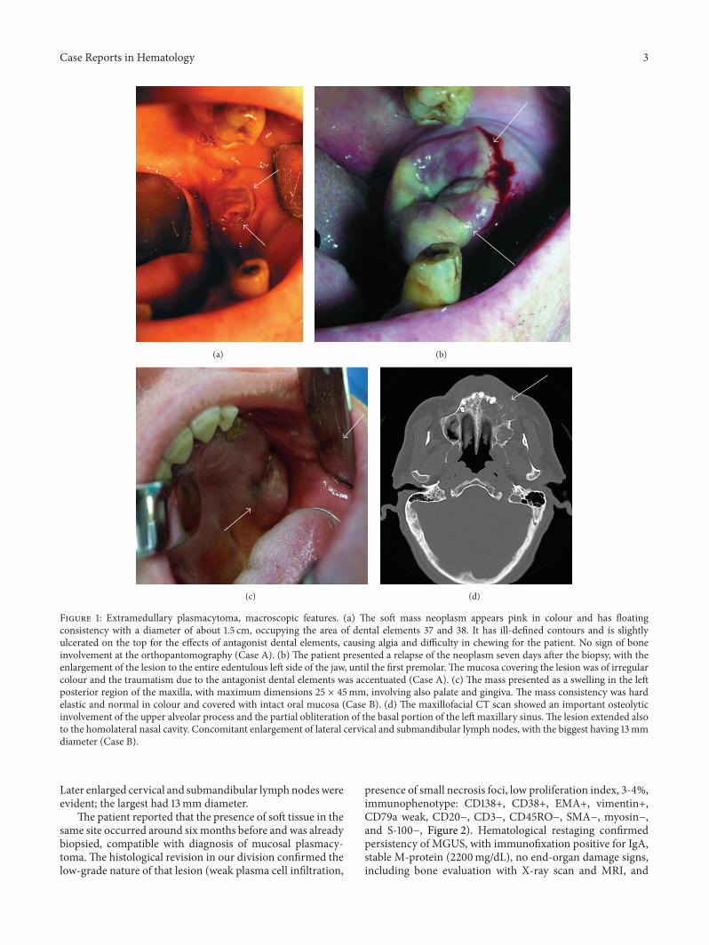
Figure 2: Extramedullary plasmacytoma, microscopic features. (a) The submucosa was heavily infiltrated by atypical plasma cells(hematoxylin eosine stain × 20, Case A). (b) MUM-1 + atypical plasma cells (nuclear staining pattern) (×40). (c) Some of the neoplasticplasma cells (pleomorphic or showing atypical mitosis stain × 63, Case B). (d) CD138 + atypical plasma cells (membranous staining pattern,×40, Case B).
6% plasma cell infiltration in the bone marrow. Since theabsence of organ damage (see biochemistry Table 1) andCRAB symptoms, the patient remained in follow-up withoutstarting therapy.
Because of the recurrence of oral lesion two months later,new incisional biopsy was given. Histological examinationof the 1.5 cm soft tissue fragment showed a massive compo-nent of well-differentiated plasma cells, secreting monotypicimmunoglobulins with lambda light chain restriction.
Diagnosis of MGUS progression to extramedullarymyeloma stage II was stated. The patient started systemicchemotherapy with lenalidomide 25mg and dexamethasone40mg weekly, achieving partial remission. He is currentlyunder maintenance therapy with 5mg lenalidomide daily, ingood clinical conditions.
Whereas thalidomide and lenalidomide might be ofhelp in high-risk and EM-myeloma, most often, protea-some inhibitors, such as bortezomib, have more often beendescribed to induce response and seem the better option touse.
4. Discussion
Herein, we report two cases of MM patients with unusualextramedullary sites (mouth) in which the systemic disease
was anticipated by localized mouse disease. Both patientsreceived first localized treatment (only radiotherapy) andstrict follow-up, thus to shift to systemic chemotherapy later.
Maxillofacial lesions are very rare as first myeloma mani-festation (<0.5%) and are more frequent in advanced diseasewith extensive skeletal involvement, including the skull [14].A Japanese series reported oral manifestations of MM inalmost 50% of initially diagnosed mouth plasmocytoma[18].
At X-ray scans, osteolytic lesions in maxilla appear welldefined, without any sign of bone reaction [10],more frequentin the jaw than in the maxilla [14, 25].
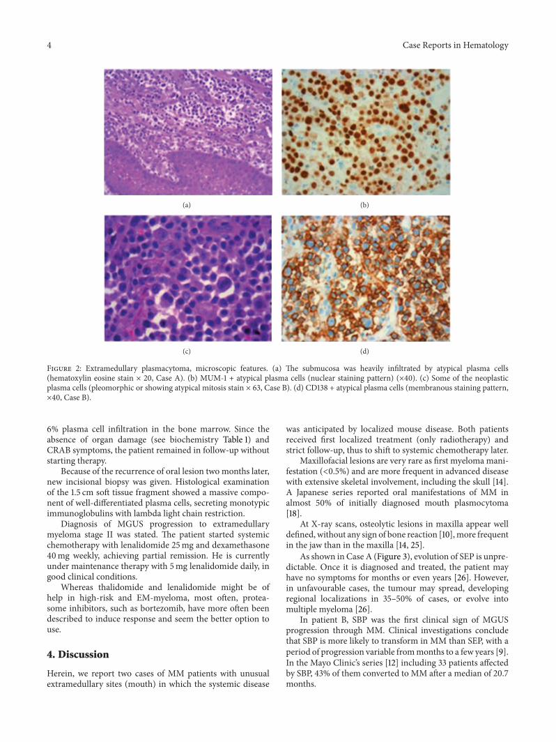
As shown in Case A (Figure 3), evolution of SEP is unpre-dictable. Once it is diagnosed and treated, the patient mayhave no symptoms for months or even years [26]. However,in unfavourable cases, the tumour may spread, developingregional localizations in 35–50% of cases, or evolve intomultiple myeloma [26].
In patient B, SBP was the first clinical sign of MGUSprogression through MM. Clinical investigations concludethat SBP is more likely to transform in MM than SEP, with aperiod of progression variable frommonths to a few years [9].In the Mayo Clinic’s series [12] including 33 patients affectedby SBP, 43% of them converted to MM after a median of 20.7months.
Case Reports in Hematology 5
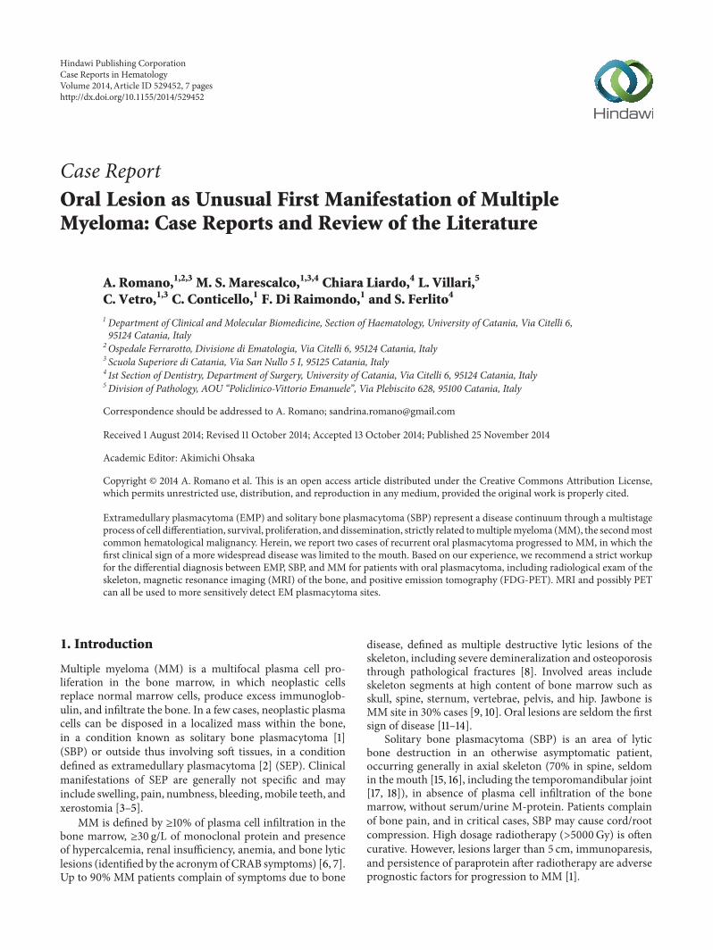
The patient underwent a biopsy in anotherhospital for a soft tissue lesion in the leftposterior region of the maxilla. Histologicaldiagnosis was compatible with mucosalplasmacytoma
Six months later he came to our dentaldepartment for the first time, referred by thedepartment of haematology, because the lesionhad recurred in the same site. He reported afive-year clinical history of monoclonalgammopathy of undetermined significance(MGUS)
Case B
CT scan and incisional biopsy were performed at ourdental department, with histological diagnosisof MGUS progression to extramedullarymyeloma stage IIPatient started systemic chemotherapy with
partial remission and he is currently undermaintenance therapy, in good clinicalconditions
Histological revision of the biopsy was performed atthe other hospital and haematological restaging without starting therapy
First visit of the patient with a soft tissue massin the left mandible at our dental division
First biopsy
Ten days later a second biopsy (excisional) wasperformed due to the recurrence of the lesion.Histological diagnosis was compatible withextramedullary plasmablastic/anaplasticplasmacytoma
Radiotherapy at level of mandible andvertebra
of clinical conditions, MRI, FDG-PET,and bone marrow biopsy were performed,leading to a diagnosis of nonsecretory MM,stage II
After two months of systemic chemotherapythe patient died of pneumonia
At eight months from the first occurrence oforal plasmacytoma, the patient startedsystemic chemotherapy without benefit
Three months later, FDG-PET showedpersistence of positivity, without symptoms
Referral to our department of haematologyfor staging and treatment of the disease. Aftera comprehensive metabolic panel and completeblood count, bone marrow biopsy,and bone marrow imaging (FDG-PET and MRI),SEP was diagnosed
Case A
Further three months later, due to the worsening
Figure 3
As shown in this report, localized PCN can be the firstsign of systemic disease, including nonsecretory MM.
Thus, whoever is involved in dental care should searchfor MM signs in each patient with mouth plasmacytomas,
including bone scan and if clinically indicated MRI and/orFDG-PET.The integrated use ofMRI and FDG-PET can helpclinicians to exclude other extramedullary localizations [27,28]. Patients affected by extramedullary MM, both primary

6 Case Reports in Hematology
Table 2
Number of patients Regimen description Response rate Notes Reference
27554 w EMP Bortezomib based + ASCT Not reported
Retrospective seriesEMP is an independent factor
for PFS and OS only intransplant-ineligible patients
Lee et al. [19]
1 (1) VTD + ASCT(2) Bortezomib + tanespimycin Not reported Case report Kumar et al. [20]
97 (plasmacytomaevolved to MM)
Radiotherapy versus novelagents/chemotherapy 91.8%
Retrospective seriesImmunoparesis was the only
negative predictor ofprogression to MM
Katodritou et al. [21]
36(EMP with invasion ofthe spinal cord)
Bortezomib based 13/36 (includingcomplete remission)
Zhang and Zhong[22]
50(intracranial MM) Bortezomib based (𝑛 = 29) 18/29 (CR + VGPR) Retrospective series Gozzetti et al. [23]
and progressing from SBP/SEP, have still a poor outcomedespite the success of novel agents, due to acquiredmolecularlesion, including TP53 mutations [29, 30].
Preliminary observations in extramedullary MM haveshown no efficacy of thalidomide [31] and controversialresults for bortezomib [32, 33], more often associated withrelapse with extramedullary spreading. Encouraging reportsarise from lenalidomide based regimens, as our Case Bconfirms.
Whereas IMIDs (thalidomide, lenalidomide) might beof help in high-risk and EM-myeloma, most often, protea-some inhibitors, such as bortezomib, have more often beendescribed to induce response and seem better to use.
The incidence of EMPs is 7% to 18% at MM diagnosisand up to 20% at relapse. The current notion that EMPs aremore frequent after treatment with novel agents remains tobe proven, especially considering that different patterns ofdisease recurrence can emerge as patients live longer in theera of novel drugs. Possible mechanisms of extramedullaryspread include decreased adhesion molecule expression anddownregulation of chemokine receptors. High-dose ther-apy with autologous stem-cell transplantation (ASCT) canovercome the negative prognostic impact of extramedullarydisease in younger selected patients. EMPs do not typicallyrespond to thalidomide alone, but, in contrast, responsesto bortezomib have been reported. A potential first-linetreatment option seems to be a bortezomib-containing reg-imen followed by ASCT, whenever possible [34]. Table 2summarizes recent reports about outcome of EM-myelomatreated with bortezomib, where overall responses can achieveup to 50% [19–23].
5. Conclusions
SBP and SEP can evolve to MM. MM signs should be eval-uated in each patient with mouth plasmacytomas, includingbone scan and if clinically indicated MRI and/or FDG-PET.When SBP and SEP progress to MM, novel agents includinglenalidomide and bortezomib are safe and efficacious indisease management.
Conflict of Interests
F. Di Raimondo has received honoraria from Janssen-Cilagand Celgene. All others declare no conflict of interests.
Acknowledgment
Authors would like to commemorate Dr. E. Salomone, whocontributed to this paper and passed away unexpectedly.
References
[1] R. H. Liebross, C. S. Ha, J. D. Cox, D. Weber, K. Delasalle,and R. Alexanian, “Solitary bone plasmacytoma: outcomeand prognostic factors following radiotherapy,” InternationalJournal of Radiation Oncology, Biology, Physics, vol. 41, no. 5, pp.1063–1067, 1998.
[2] C. Alexiou, R. J. Kau, H. Dietzfelbinger et al., “Extramedullaryplasmacytoma: tumor occurrence and therapeutic concepts,”Cancer, vol. 85, no. 11, pp. 2305–2314, 1999.
[3] F. Tesei, U. Caliceti, G. Sorrenti et al., “Extramedullary plasmo-cytoma (EMP) of the head and neck: a series of 22 cases,” ActaOtorhinolaryngologica Italica, vol. 15, no. 6, pp. 437–442, 1995.
[4] S. Majumdar, U. Raghavan, and N. S. Jones, “Solitary plas-macytoma and extramedullary plasmacytoma of the paranasalsinuses and soft palate,” Journal of Laryngology and Otology, vol.116, no. 11, pp. 962–965, 2002.
[5] J. Seoane, A. De La Cruz, M. Pomareda, P. I. Varela-Centelles,and L. Pias, “Primary extramedullary plasmacytoma of thepalate,” Otolaryngology—Head and Neck Surgery, vol. 120, no.4, p. 530, 1999.
[6] M. Dimopoulos, R. Kyle, J.-P. Fermand et al., “Consensusrecommendations for standard investigative workup: reportof the International Myeloma Workshop Consensus Panel 3,”Blood, vol. 117, no. 18, pp. 4701–4705, 2011.
[7] N. W. C. J. van de Donk, H. M. Lokhorst, K. C. Anderson, andP. G. Richardson, “How I treat plasma cell leukemia,” Blood, vol.120, no. 12, pp. 2376–2389, 2012.
[8] E. Terpos, L. A. Moulopoulos, and M. A. Dimopoulos, “Advan-ces in imaging and the management of myeloma bone disease,”Journal of Clinical Oncology, vol. 29, no. 14, pp. 1907–1915, 2011.
Case Reports in Hematology 7
[9] B. Rodrıguez-Caballero, S. Sanchez-Santolino, B. Garcıa-Mon-tesinos-Perea, M.-F. Garcia-Reija, J. Gomez-Roman, and R.Saiz-Bustillo, “Mandibular solitary plasmocytoma of the jaw: acase report,”Medicina Oral, Patologıa Oral y Cirugıa Bucal, vol.16, no. 5, pp. e647–e650, 2011.
[10] J. Seoane, J. M. Aguirre Urizar, G. Esparza Gomez, M. SuarezCunqueiro, J. Campos Trapero, and M. Pomareda, “The spec-trum of plasma cell neoplasia in oral pathology,”Medicina Oral,vol. 8, no. 4, pp. 269–280, 2003.
[11] J. J. Pisano, R. Coupland, S.-Y. Chen, and A. S. Miller, “Plas-macytoma of the oral cavity and jaws: a clinicopathologic studyof 13 cases,” Oral Surgery, Oral Medicine, Oral Pathology, OralRadiology, and Endodontics, vol. 83, no. 2, pp. 265–271, 1997.
[12] M. E. Lae, E. F. Vencio, C. Y. Inwards, K. K. Unni, and A. G.Nascimento, “Myeloma of the jaw bones: a clinicopathologicstudy of 33 cases,”Head&Neck, vol. 25, no. 5, pp. 373–381, 2003.
[13] S. Ferreira, C. Baldaia, N. Fatela et al., “Severe intrahepaticcholestasis, erythrocytosis and hypoglycemia: unusual present-ing features of systemic AL amyloidosis,” Scandinavian Journalof Gastroenterology, vol. 43, no. 3, pp. 375–379, 2008.
[14] L. S. S. Pinto, E. B. Campagnoli, J. E. Leon, M. A. Lopes, andJ. Jorge, “Maxillary lesion presenting as a first sign of multiplemyeloma: case report,”Medicina Oral, Patologia Oral y CirugiaBucal, vol. 12, no. 5, pp. E344–E347, 2007.
[15] S. Razaq, H. P. Kessler, and W. Phillips, “Oral and maxillofacialpathology case of the month. Solitary plasmacytoma of bone,”Texas Dental Journal, vol. 125, no. 2, pp. 182–190, 2008.
[16] S. Anil, “Solitary plasmacytoma of the maxilla—a case reportand review of the literature,”General Dentistry, vol. 55, no. 1, pp.39–43, 2007.
[17] D. Lesmes and Z. Laster, “Plasmacytoma in the temporo-mandibular joint: a case report,” British Journal of Oral andMaxillofacial Surgery, vol. 46, no. 4, pp. 322–324, 2008.
[18] E.M. Canger, P. Celenk, A. Alkan, andO.Gunhan, “Mandibularinvolvement of solitary plasmocytoma: a case report,”MedicinaOral, Patologia Oral y Cirugia Bucal, vol. 12, no. 1, pp. E7–E9,2007.
[19] S. E. Lee, J. H. Kim, Y. W. Jeon et al., “Impact of extramedullaryplasmacytomas on outcomes according to treatment approachin newly diagnosed symptomatic multiple myeloma,” Annals ofHematology, 2014.
[20] A. K. L. Kumar, C. Dakhil, M. T. Satyan, and N. Haideri, “Extra-medullary progression of multiple myeloma despite concomi-tant medullary response to multiple combination therapies andautologous transplant: a case report,” Journal of Medical CaseReports, vol. 8, article 299, 2014.
[21] E. Katodritou, E. Terpos, A. S. Symeonidis et al., “Clinicalfeatures, outcome, and prognostic factors for survival andevolution to multiple myeloma of solitary plasmacytomas: areport of the Greek myeloma study group in 97 patients,”American Journal of Hematology, vol. 89, no. 8, pp. 803–808,2014.
[22] J. Zhang and Y. Zhong, “Clinical analysis of 36 multiplemyeloma patients with extramedullary plasmacytoma invasionof the spinal canal,” Hematological Oncology, 2014.
[23] A. Gozzetti, A. Cerase, F. Lotti et al., “Extramedullary intracra-nial localization of multiple myeloma and treatment with novelagents: a retrospective survey of 50 patients,” Cancer, vol. 118,no. 6, pp. 1574–1584, 2012.
[24] P. R. Greipp, J. S. Miguel, B. G. M. Dune et al., “Internationalstaging system formultiple myeloma,” Journal of Clinical Oncol-ogy, vol. 23, no. 15, pp. 3412–3420, 2005.
[25] G. Lambertenghi-Deliliers, E. Bruno, A. Cortelezzi, L. Fuma-galli, and A. Morosini, “Incidence of jaw lesions in 193 patientswith multiple myeloma,” Oral Surgery Oral Medicine and OralPathology, vol. 65, no. 5, pp. 533–537, 1988.
[26] C. M. Garcia, J. A. Dominguez, P. T. Roman, J. Pando-Pinto,and A. B. Huelva, “Primary extramedullary plasmocytoma ofthe tongue base,” Acta Otorrinolaringologica Espanola, vol. 50,no. 8, pp. 657–659, 1999.
[27] C. P. Shortt, T. G. Gleeson, K. A. Breen et al., “Whole-bodyMRIversus PET in assessment of multiple myeloma disease activity,”The American Journal of Roentgenology, vol. 192, no. 4, pp. 980–986, 2009.
[28] S. D’Sa, N. Abildgaard, J. Tighe, P. Shaw, and M. Hall-Craggs,“Guidelines for the use of imaging in the management ofmyeloma,” British Journal of Haematology, vol. 137, no. 1, pp. 49–63, 2007.
[29] L. Billecke, E. M. M. Penas, A. M. May et al., “Cytogeneticsof extramedullary manifestations in multiple myeloma,” BritishJournal of Haematology, vol. 161, no. 1, pp. 87–94, 2013.
[30] L. Billecke, E.M.M. Penas, A.M.May et al., “Similar incidencesof TP53 deletions in extramedullary organ infiltrations, softtissue and osteolyses of patients with multiple myeloma,”Anticancer Research, vol. 32, no. 5, pp. 2031–2034, 2012.
[31] L. Rosinol, M. T. Cibeira, J. Blade et al., “Extramedullary multi-ple myeloma escapes the effect of thalidomide,”Haematologica,vol. 89, no. 7, pp. 832–836, 2004.
[32] R. Laura, M. T. Cibeira, C. Uriburu et al., “Bortezomib: aneffective agent in extramedullary disease in multiple myeloma,”European Journal of Haematology, vol. 76, no. 5, pp. 405–408,2006.
[33] A. Romano, C. Conticello, and F. Di Raimondo, “Bortezomibfor the treatment of previously untreated multiple myeloma,”Immunotherapy, vol. 5, no. 4, pp. 327–352, 2013.
[34] J. Blade, C. Fernandez de Larrea, L. Rosinol, M. T. Cibeira, R.Jimenez, and R. Powles, “Soft-tissue plasmacytomas in multiplemyeloma: incidence, mechanisms of extramedullary spread,and treatment approach,” Journal of Clinical Oncology, vol. 29,no. 28, pp. 3805–3812, 2011.

Submit your manuscripts athttp://www.hindawi.com
Stem CellsInternational
Hindawi Publishing Corporationhttp://www.hindawi.com Volume 2014
Hindawi Publishing Corporationhttp://www.hindawi.com Volume 2014
MEDIATORSINFLAMMATION
of
Hindawi Publishing Corporationhttp://www.hindawi.com Volume 2014
Behavioural Neurology
EndocrinologyInternational Journal of
Hindawi Publishing Corporationhttp://www.hindawi.com Volume 2014
Hindawi Publishing Corporationhttp://www.hindawi.com Volume 2014
Disease Markers
Hindawi Publishing Corporationhttp://www.hindawi.com Volume 2014
BioMed Research International
OncologyJournal of
Hindawi Publishing Corporationhttp://www.hindawi.com Volume 2014
Hindawi Publishing Corporationhttp://www.hindawi.com Volume 2014
Oxidative Medicine and Cellular Longevity
Hindawi Publishing Corporationhttp://www.hindawi.com Volume 2014
PPAR Research
The Scientific World JournalHindawi Publishing Corporation http://www.hindawi.com Volume 2014
Immunology ResearchHindawi Publishing Corporationhttp://www.hindawi.com Volume 2014
Journal of
ObesityJournal of
Hindawi Publishing Corporationhttp://www.hindawi.com Volume 2014
Hindawi Publishing Corporationhttp://www.hindawi.com Volume 2014
Computational and Mathematical Methods in Medicine
OphthalmologyJournal of
Hindawi Publishing Corporationhttp://www.hindawi.com Volume 2014
Diabetes ResearchJournal of
Hindawi Publishing Corporationhttp://www.hindawi.com Volume 2014
Hindawi Publishing Corporationhttp://www.hindawi.com Volume 2014
Research and TreatmentAIDS
Hindawi Publishing Corporationhttp://www.hindawi.com Volume 2014
Gastroenterology Research and Practice
Hindawi Publishing Corporationhttp://www.hindawi.com Volume 2014
Parkinson’s Disease
Evidence-Based Complementary and Alternative Medicine
Volume 2014Hindawi Publishing Corporationhttp://www.hindawi.com
Related Documents