CASE REPORT Open Access Paediatric T-cell lymphoma of the appendix: a case report Yoshifumi Matsushita 1 and Morishige Takeshita 2* Abstract A 7-year-old boy with no history of malnutrition or diarrhoea complained of acute abdominal pain, was diagnosed with acute appendicitis, and underwent appendectomy. Histologically, a diffuse infiltrate of large atypical lymphoid cells was found in the entire appendiceal wall. Immunohistochemical examination revealed that the tumour cells expressed T-cell receptor (TCR)-βF1, CD3, CD4, CD25, cytotoxic-related protein TIA1 and granzyme-B, but were negative for CD8, Foxp3, CD20, CD30 and CD56. Polymerase chain reaction (PCR) revealed clonal bands of TCR-γ gene products in the tumour tissue. No anti-cytomegalovirus antibody-positive cells were detected. In situ hybridization revealed no nuclear signals of Epstein-Barr virus (EBV)-encoded RNA. Helicobacter pylori infection was detected in tumour tissue by anti-East Asian cytotoxin-associated gene (Cag) A antibody and PCR using its specific primers. The patient received chemotherapy and has remained in remission for 2 years. To the best of our knowledge, only two cases of appendiceal T-cell non-Hodgkin lymphoma (NHL) have been reported, both in elderly patients. We believe that this is the first reported case of childhood CD4- and TIA1-positive cytotoxic T (Th1)-cell NHL in the appendix or gastrointestinal tract. Helicobacter pylori infection might be an initiator of atypical cytotoxic T-cell proliferation. Virtual Slides: The virtual slide(s) for this article can be found here: http://www.diagnosticpathology.diagnomx.eu/ vs/1302380563830412. Keywords: T-cell lymphoma, Appendix, Child Background The specification of non-Hodgkin lymphoma (NHL) in the “WHO Classification 2008” is based on recent pro- gress in immunohistochemical and genetic analysis and clinical findings, including prognostic data [1]. Among extranodal NHLs, the alimentary tract is the most fre- quently affected site [1,2]. In the small intestine, diffuse large B-cell NHL (DLBCL) is the most common subtype of lymphoma, followed by mucosa-associated lymphoid tissue (MALT) lymphoma in the elderly, while Burkitt’ s lymphoma is the predominant childhood NHL. Appen- diceal involvement is extremely rare, constituting less than 1% of all small- and large-intestinal NHLs [2,3]. About 50 cases of appendiceal NHL have been previ- ously reported, but immunohistochemical and genetic examinations have been performed in only a limited number of recent cases, including two T/natural killer (NK)-cell NHL cases [2-5]. Among intestinal T/NK-cell NHLs, enteropathy-associated T-cell lymphoma (EATL) has been frequently reported in the elderly [1,6-8]. Type I EATL is a CD4- and CD8-negative and CD30-positive large-cell lymphoma associated with coeliac disease, and is seen in northern Europe and the United States. Type II EATL is a CD56-positive and CD8-positive or -nega- tive medium-sized lymphoma that is less strongly asso- ciated with coeliac disease. There are several reports of nasal-type NK-cell lymphomas with Epstein-Barr virus (EBV) infection in intestine and colon, which are usually encountered in the elderly and rarely in children [7,9]. The present case is a CD3-, CD4- and TIA1-positive and CD30-negative (Th1) large-cell lymphoma without EBV infection. CD4-positive T/NK-cell lymphoma has occa- sionally been reported in the stomach, but is rarely seen in the intestine [10,11]. The intestinal bacterial florae, Helicobacter pylori and Campylobacter jejuni, may be initiators of abnormal lymphocytic proliferation in the * Correspondence: [email protected] 2 Department of Pathology, Faculty of Medicine, Fukuoka University, Nanakuma 7-45-1, Jonan-ku, Fukuoka 814-0180, Japan Full list of author information is available at the end of the article © 2013 Matsushita and Takeshita; licensee BioMed Central Ltd. This is an Open Access article distributed under the terms of the Creative Commons Attribution License (http://creativecommons.org/licenses/by/2.0), which permits unrestricted use, distribution, and reproduction in any medium, provided the original work is properly cited. Matsushita and Takeshita Diagnostic Pathology 2013, 8:2 http://www.diagnosticpathology.org/content/8/1/2

Welcome message from author
This document is posted to help you gain knowledge. Please leave a comment to let me know what you think about it! Share it to your friends and learn new things together.
Transcript

Matsushita and Takeshita Diagnostic Pathology 2013, 8:2http://www.diagnosticpathology.org/content/8/1/2
CASE REPORT Open Access
Paediatric T-cell lymphoma of the appendix: acase reportYoshifumi Matsushita1 and Morishige Takeshita2*
Abstract
A 7-year-old boy with no history of malnutrition or diarrhoea complained of acute abdominal pain, was diagnosedwith acute appendicitis, and underwent appendectomy. Histologically, a diffuse infiltrate of large atypical lymphoidcells was found in the entire appendiceal wall. Immunohistochemical examination revealed that the tumour cellsexpressed T-cell receptor (TCR)-βF1, CD3, CD4, CD25, cytotoxic-related protein TIA1 and granzyme-B, but werenegative for CD8, Foxp3, CD20, CD30 and CD56. Polymerase chain reaction (PCR) revealed clonal bands of TCR-γgene products in the tumour tissue. No anti-cytomegalovirus antibody-positive cells were detected. In situhybridization revealed no nuclear signals of Epstein-Barr virus (EBV)-encoded RNA. Helicobacter pylori infection wasdetected in tumour tissue by anti-East Asian cytotoxin-associated gene (Cag) A antibody and PCR using its specificprimers. The patient received chemotherapy and has remained in remission for 2 years. To the best of ourknowledge, only two cases of appendiceal T-cell non-Hodgkin lymphoma (NHL) have been reported, both inelderly patients. We believe that this is the first reported case of childhood CD4- and TIA1-positive cytotoxic T(Th1)-cell NHL in the appendix or gastrointestinal tract. Helicobacter pylori infection might be an initiator of atypicalcytotoxic T-cell proliferation.Virtual Slides: The virtual slide(s) for this article can be found here: http://www.diagnosticpathology.diagnomx.eu/vs/1302380563830412.
Keywords: T-cell lymphoma, Appendix, Child
BackgroundThe specification of non-Hodgkin lymphoma (NHL) inthe “WHO Classification 2008” is based on recent pro-gress in immunohistochemical and genetic analysis andclinical findings, including prognostic data [1]. Amongextranodal NHLs, the alimentary tract is the most fre-quently affected site [1,2]. In the small intestine, diffuselarge B-cell NHL (DLBCL) is the most common subtypeof lymphoma, followed by mucosa-associated lymphoidtissue (MALT) lymphoma in the elderly, while Burkitt’slymphoma is the predominant childhood NHL. Appen-diceal involvement is extremely rare, constituting lessthan 1% of all small- and large-intestinal NHLs [2,3].About 50 cases of appendiceal NHL have been previ-ously reported, but immunohistochemical and geneticexaminations have been performed in only a limited
* Correspondence: [email protected] of Pathology, Faculty of Medicine, Fukuoka University,Nanakuma 7-45-1, Jonan-ku, Fukuoka 814-0180, JapanFull list of author information is available at the end of the article
© 2013 Matsushita and Takeshita; licensee Biothe Creative Commons Attribution License (htdistribution, and reproduction in any medium
number of recent cases, including two T/natural killer(NK)-cell NHL cases [2-5]. Among intestinal T/NK-cellNHLs, enteropathy-associated T-cell lymphoma (EATL)has been frequently reported in the elderly [1,6-8]. TypeI EATL is a CD4- and CD8-negative and CD30-positivelarge-cell lymphoma associated with coeliac disease, andis seen in northern Europe and the United States. TypeII EATL is a CD56-positive and CD8-positive or -nega-tive medium-sized lymphoma that is less strongly asso-ciated with coeliac disease. There are several reports ofnasal-type NK-cell lymphomas with Epstein-Barr virus(EBV) infection in intestine and colon, which are usuallyencountered in the elderly and rarely in children [7,9].The present case is a CD3-, CD4- and TIA1-positive andCD30-negative (Th1) large-cell lymphoma without EBVinfection. CD4-positive T/NK-cell lymphoma has occa-sionally been reported in the stomach, but is rarely seenin the intestine [10,11]. The intestinal bacterial florae,Helicobacter pylori and Campylobacter jejuni, may beinitiators of abnormal lymphocytic proliferation in the
Med Central Ltd. This is an Open Access article distributed under the terms oftp://creativecommons.org/licenses/by/2.0), which permits unrestricted use,, provided the original work is properly cited.
Matsushita and Takeshita Diagnostic Pathology 2013, 8:2 Page 2 of 6http://www.diagnosticpathology.org/content/8/1/2
stomach and intestine [12,13]. Here, we present what is,to the best of our knowledge, the first report of a child-hood case of appendiceal CD4-positive T-cell NHL anddiscuss the influence of H. pylori infection.
Case presentationClinical historyA 7-year-old boy was referred to our hospital with com-plaints of abdominal discomfort and high fever. Four daysbefore admission, the patient complained of uneasinessand sneezing and was afebrile. On the following day, thepatient complained of increasing abdominal pain and ap-petite loss. One day before admission, the symptoms wor-sened and the patient’s temperature rose to 39.1°C. Therewas no history of recurrent diarrhoea, malnutrition or fail-ure to thrive. On admission, the white blood cell countremained within normal limits, but C-reactive protein waselevated to a concentration of 3.6 mg/dL. An abdominalultrasound revealed a mildly swollen appendix. Inaddition, a few mildly swollen lymph nodes, up to 10 mmin diameter, were seen in the mesentery. A diagnosis ofacute appendicitis was made and appendectomy was per-formed on the second day of hospitalisation.
Material and methodsImmunohistochemistryThe antibodies used in this study were as follows: TCR-βF1 (Endogen, Rockford, IL, USA); CD3, CD5, CD7, CD8,CD25, CD56, CD57, and terminal deoxynucleotidyl trans-ferase (TdT) (Novocastra, Newcastle, UK); CD4 (MBL,Nagoya, Japan); Foxp3 (e-Bioscience, San Diego, CA,USA); TIA-1 (Immunotech, Marseille, France); GranzymeB (Chemicon, Temecula, CA, USA); CD20 (Nichirei,Tokyo, Japan); and CD79a, CD30, CD15, anaplastic lymph-oma kinase (ALK), myeloperoxidase, epithelial membraneantigen (EMA), AE1/AE3 and anti-cytomegalovirus(CMV) antibody (Dako Cytomation, Glostrup, Denmark).Anti-East Asian H. pylori CagA antibody was kindly pro-vided by Dr. T. Uchida, Department of MolecularMedicine, Oita University, Japan [14].
In situ hybridisation for detection of Epstein-Barr virus(EBV)-encoded RNAsTissue sections were digested with proteinase K andincubated in a solution of 50% formamide containingdigoxigenin/biotin-labelled EBV-encoded RNA (EBER)oligonucleotide probes (Dako Cytomation). A peroxidase-conjugated anti-FITC antibody was applied to the sectionsto detect the hybridized probes.
Polymerase chain reaction (PCR) for T-cell receptor (TCR)-γ and H. pylori-associated genesFor evaluations of genes associated with TCR-γ and H.pylori, DNA was extracted from paraffin-embedded
tumour sections. TCR-γ gene analysis was performedaccording to the BIOMED II PCR method [15].The East Asian-type CagA genes were detected using pri-
mer sets CAGJF/CAGTR and CAGTF/CAGJR, whichyielded 222- and 293- to 299-bp products, respectively [16].
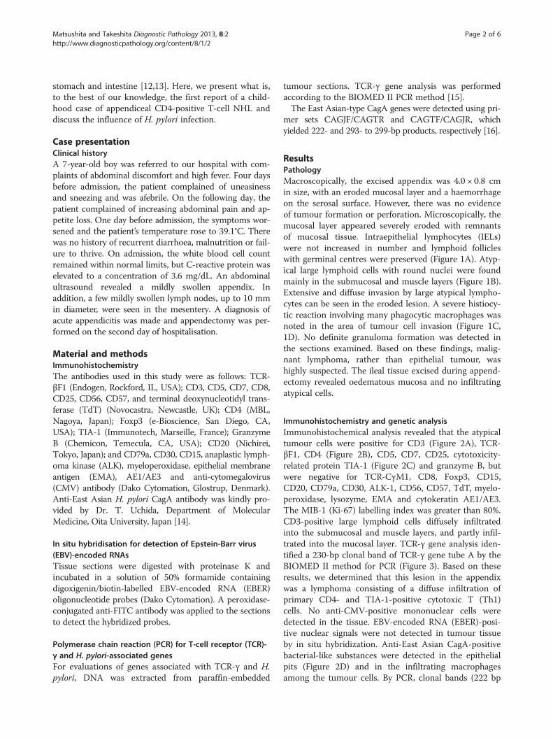
ResultsPathologyMacroscopically, the excised appendix was 4.0 × 0.8 cmin size, with an eroded mucosal layer and a haemorrhageon the serosal surface. However, there was no evidenceof tumour formation or perforation. Microscopically, themucosal layer appeared severely eroded with remnantsof mucosal tissue. Intraepithelial lymphocytes (IELs)were not increased in number and lymphoid follicleswith germinal centres were preserved (Figure 1A). Atyp-ical large lymphoid cells with round nuclei were foundmainly in the submucosal and muscle layers (Figure 1B).Extensive and diffuse invasion by large atypical lympho-cytes can be seen in the eroded lesion. A severe histiocy-tic reaction involving many phagocytic macrophages wasnoted in the area of tumour cell invasion (Figure 1C,1D). No definite granuloma formation was detected inthe sections examined. Based on these findings, malig-nant lymphoma, rather than epithelial tumour, washighly suspected. The ileal tissue excised during append-ectomy revealed oedematous mucosa and no infiltratingatypical cells.
Immunohistochemistry and genetic analysisImmunohistochemical analysis revealed that the atypicaltumour cells were positive for CD3 (Figure 2A), TCR-βF1, CD4 (Figure 2B), CD5, CD7, CD25, cytotoxicity-related protein TIA-1 (Figure 2C) and granzyme B, butwere negative for TCR-CγM1, CD8, Foxp3, CD15,CD20, CD79a, CD30, ALK-1, CD56, CD57, TdT, myelo-peroxidase, lysozyme, EMA and cytokeratin AE1/AE3.The MIB-1 (Ki-67) labelling index was greater than 80%.CD3-positive large lymphoid cells diffusely infiltratedinto the submucosal and muscle layers, and partly infil-trated into the mucosal layer. TCR-γ gene analysis iden-tified a 230-bp clonal band of TCR-γ gene tube A by theBIOMED II method for PCR (Figure 3). Based on theseresults, we determined that this lesion in the appendixwas a lymphoma consisting of a diffuse infiltration ofprimary CD4- and TIA-1-positive cytotoxic T (Th1)cells. No anti-CMV-positive mononuclear cells weredetected in the tissue. EBV-encoded RNA (EBER)-posi-tive nuclear signals were not detected in tumour tissueby in situ hybridization. Anti-East Asian CagA-positivebacterial-like substances were detected in the epithelialpits (Figure 2D) and in the infiltrating macrophagesamong the tumour cells. By PCR, clonal bands (222 bp
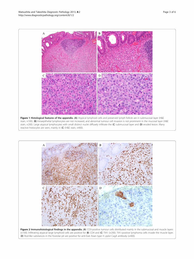
Figure 1 Histological features of the appendix. (A) Atypical lymphoid cells and preserved lymph follicle are in submucosal layer (H&Estain, ×100). (B) Intraepithelial lymphocytes are not increased, and abnormal tumour cell invasion is not prominent in the mucosal layer (H&Estain, ×200). Large atypical lymphocytes with small distinct nuclei diffusely infiltrate the (C) submucosal layer and (D) eroded lesion. Manyreactive histiocytes are seen, mainly in (C) (H&E stain, ×400).
Figure 2 Immunohistological findings in the appendix. (A) CD3-positive tumour cells distributed mainly in the submucosal and muscle layers(×100). Infiltrating atypical large lymphoid cells are positive for (B) CD4 and (C) TIA1 (×200). TIA1-positive lymphoma cells invade the muscle layer.(D) Rod-like substances in the foveolar pit are positive for anti-East Asian type H. pylori CagA antibody (×400).
Matsushita and Takeshita Diagnostic Pathology 2013, 8:2 Page 3 of 6http://www.diagnosticpathology.org/content/8/1/2
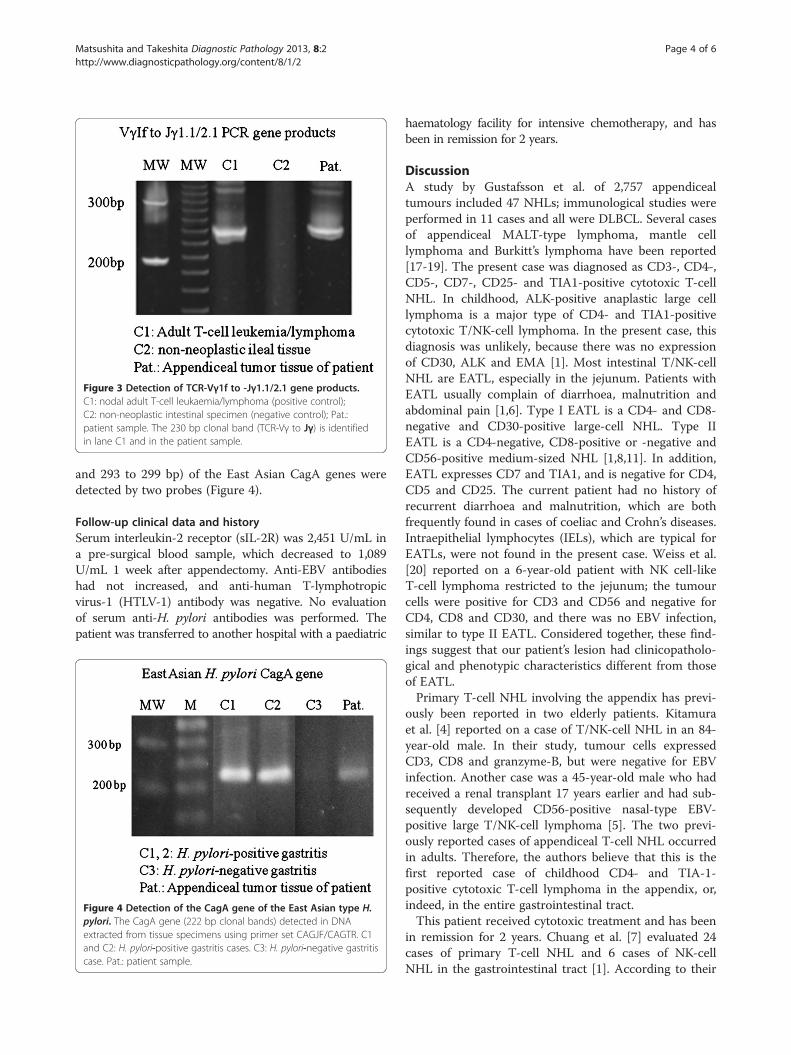
Figure 3 Detection of TCR-Vγ1f to -Jγ1.1/2.1 gene products.C1: nodal adult T-cell leukaemia/lymphoma (positive control);C2: non-neoplastic intestinal specimen (negative control); Pat.:patient sample. The 230 bp clonal band (TCR-Vγ to Jγ) is identifiedin lane C1 and in the patient sample.
Matsushita and Takeshita Diagnostic Pathology 2013, 8:2 Page 4 of 6http://www.diagnosticpathology.org/content/8/1/2
and 293 to 299 bp) of the East Asian CagA genes weredetected by two probes (Figure 4).
Follow-up clinical data and historySerum interleukin-2 receptor (sIL-2R) was 2,451 U/mL ina pre-surgical blood sample, which decreased to 1,089U/mL 1 week after appendectomy. Anti-EBV antibodieshad not increased, and anti-human T-lymphotropicvirus-1 (HTLV-1) antibody was negative. No evaluationof serum anti-H. pylori antibodies was performed. Thepatient was transferred to another hospital with a paediatric
Figure 4 Detection of the CagA gene of the East Asian type H.pylori. The CagA gene (222 bp clonal bands) detected in DNAextracted from tissue specimens using primer set CAGJF/CAGTR. C1and C2: H. pylori-positive gastritis cases. C3: H. pylori-negative gastritiscase. Pat.: patient sample.
haematology facility for intensive chemotherapy, and hasbeen in remission for 2 years.
DiscussionA study by Gustafsson et al. of 2,757 appendicealtumours included 47 NHLs; immunological studies wereperformed in 11 cases and all were DLBCL. Several casesof appendiceal MALT-type lymphoma, mantle celllymphoma and Burkitt’s lymphoma have been reported[17-19]. The present case was diagnosed as CD3-, CD4-,CD5-, CD7-, CD25- and TIA1-positive cytotoxic T-cellNHL. In childhood, ALK-positive anaplastic large celllymphoma is a major type of CD4- and TIA1-positivecytotoxic T/NK-cell lymphoma. In the present case, thisdiagnosis was unlikely, because there was no expressionof CD30, ALK and EMA [1]. Most intestinal T/NK-cellNHL are EATL, especially in the jejunum. Patients withEATL usually complain of diarrhoea, malnutrition andabdominal pain [1,6]. Type I EATL is a CD4- and CD8-negative and CD30-positive large-cell NHL. Type IIEATL is a CD4-negative, CD8-positive or -negative andCD56-positive medium-sized NHL [1,8,11]. In addition,EATL expresses CD7 and TIA1, and is negative for CD4,CD5 and CD25. The current patient had no history ofrecurrent diarrhoea and malnutrition, which are bothfrequently found in cases of coeliac and Crohn’s diseases.Intraepithelial lymphocytes (IELs), which are typical forEATLs, were not found in the present case. Weiss et al.[20] reported on a 6-year-old patient with NK cell-likeT-cell lymphoma restricted to the jejunum; the tumourcells were positive for CD3 and CD56 and negative forCD4, CD8 and CD30, and there was no EBV infection,similar to type II EATL. Considered together, these find-ings suggest that our patient’s lesion had clinicopatholo-gical and phenotypic characteristics different from thoseof EATL.Primary T-cell NHL involving the appendix has previ-
ously been reported in two elderly patients. Kitamuraet al. [4] reported on a case of T/NK-cell NHL in an 84-year-old male. In their study, tumour cells expressedCD3, CD8 and granzyme-B, but were negative for EBVinfection. Another case was a 45-year-old male who hadreceived a renal transplant 17 years earlier and had sub-sequently developed CD56-positive nasal-type EBV-positive large T/NK-cell lymphoma [5]. The two previ-ously reported cases of appendiceal T-cell NHL occurredin adults. Therefore, the authors believe that this is thefirst reported case of childhood CD4- and TIA-1-positive cytotoxic T-cell lymphoma in the appendix, or,indeed, in the entire gastrointestinal tract.This patient received cytotoxic treatment and has been
in remission for 2 years. Chuang et al. [7] evaluated 24cases of primary T-cell NHL and 6 cases of NK-cellNHL in the gastrointestinal tract [1]. According to their

Matsushita and Takeshita Diagnostic Pathology 2013, 8:2 Page 5 of 6http://www.diagnosticpathology.org/content/8/1/2
report, using univariate and multivariate COX propor-tional hazard regression analysis, NK-cell lineage wasassociated with poor prognosis. EBV infection plays animportant role in the progression of various NHLs [21].We speculate that the early clinical stage and EBV-freestatus of the current patient predicted better prognosis.However, this is a single case and the follow-up periodwas limited. Identification of additional cases of intes-tinal T/NK-cell NHL and long-term follow-up is neces-sary in order to fully understand the clinical features ofappendiceal T/NK-cell NHL.In Japan, gastric carcinoma and MALT-type lymphoma
have higher incidences compared with those occurringin other regions of the world [22]. It was strongly sug-gested that the East Asian CagA gene and protein havea great influence on the tumourigenesis of these twodisorders [14,15]. Kiriya et al. [23] demonstrated that theT-cell reaction against the captured, round-shaped H.pylori seen in dendritic cells of Peyer’s patches in thesmall intestine plays a critical role in H. pylori gastritis.CD4-positive T cells, including Th1 and regulatory Tcells, are distributed in the gastric mucosa in H. pyloriinfection [24], and cases of primary CD4-, CD5-, CD25-and TIA1-positive cytotoxic T-cell lymphoma have beenreported in the stomach [10]. Among CD4-positive Tcells, neoplastic cells of the present case had phenotypicfindings regarding TIA1 expression similar to those ofTh1 effector cells [25]. Helicobacter pylori infectionmight play a role in abnormal proliferation of CD4-positive cytotoxic T (Th1) cells. However, althoughKüpeli et al. [26] in Turkey reported that 3 of 15 cases(20%) of childhood systemic NHL had serological H. pyl-ori infection and that 2 cases were T-cell type ALCL,they suggested that H. pylori infection was not an agentresponsible for lymphomagenesis.
ConclusionWe present a rare paediatric case of appendiceal CD3-,CD4- and TIA1-positive cytotoxic T (Th1)-cell lymph-oma. Further studies are necessary to examine the rela-tionships between H. pylori infection, including theAsian variety, and NHL.
Competing interestsThe authors declare that they have no conflicts of interest.
Authors’ contributionsYM carried out initial pathological diagnosis of this case. YM and MTparticipated in the sequence alignment and drafted the manuscript. Bothauthors read and approved the final manuscript.
AcknowledgementsThe authors thank Dr. Hiroaki Kumazawa, Department of Surgery, and Dr.Hiroshi Kobayashi, Department of Paediatrics, Chidoribashi Hospital, forpatient care. Written informed consent was obtained from his family forpublication and any accompanying images.
Author details1Laboratory of Pathology, Chidoribashi Hospital, 5-18-1 Chiyo, Hakata-ku,Fukuoka 812-8633, Japan. 2Department of Pathology, Faculty of Medicine,Fukuoka University, Nanakuma 7-45-1, Jonan-ku, Fukuoka 814-0180, Japan.
Received: 20 October 2012 Accepted: 13 December 2012Published: 9 January 2013
References1. Swerdlow SH, Campo E, Harris NL, Jaffe ES, Pileri SA, Stein H, Thiele J,
Vardiman JW: WHO classification of tumours of haematopoietic and lymphoidtissues. Lyon: International Agency for Research on Cancer; 2008.
2. Gustafsson BI, Siddique L, Chan A, Dong M, Dorozdov I, Kidd M, Modlin IM:Uncommon cancers of the small intestine, appendix and colon: Ananalysis of SEER 1973–2004, and current diagnosis and therapy. Int JOncol 2008, 33:1121–1131.
3. O’Donnell ME, Badger SA, Beattie GC, Carson J, Garstin WIH: Malignantneoplasms of the appendix. Int J Colorectal Dis 2007, 22:1239–1248.
4. Kitamura Y, Ohta T, Terada T: Primary T-cell non-Hodgkin's malignantlymphoma of the appendix. Pathol Int 2000, 50:313–317.
5. Ratuapli SK, Murarka S, Miller KA, Ferraro JC, Zafar H: Epstein-Barr virus-positive large T-cell lymphoma presenting an acute appendicitis17 years after cadaveric renal transplant: a case report. J Med CaseReports 2011, 5:5.
6. Dalabie J, Holte H, Vose JM, Ullrich F, Jaffe ES, Savage KJ, Connors JM,Rimsza L, Harris NL, Müller-Hermelink K, et al: Enteropathy-associated T-celllymphoma: clinical and histological findings from the internationalperipheral T-cell lymphoma project. Blood 2011, 118:148–155.
7. Chuang S-S, Chang S-T, Chuang W-Y, Huang W-T, Hsieh P-P, Tsou M-H, LiaoY-L, Lin S-H, Hsieh Y-H, Lu C-L, et al: NK-cell lineage predicts poor survivalin primary intestinal NK-cell and T-cell lymphoma. Am J Surg Pathol 2009,33:1230–1240.
8. Takeshita M, Nakamura S, Kikuma K, Nakayama Y, Nimura S, Yao T, Urabe S,Ogawara S, Yonemasu H, Matsushita Y, et al: Pathological andimmunohistological findings and genetic aberrations of intestinalenteropathy-associated T-cell lymphoma in Japan. Histopathol 2011,58:395–407.
9. Sun J, Lu Z, Yang D, Chen J: Primary intestinal T-cell and NK-celllymphomas: a clinicopathological and molecular study from Chinafocused on type II enteropathy-associated T-cell lymphoma and primaryNK-cell lymphoma. Mod Pathol 2011, 24:983–992.
10. Kawamoto K, Nakamura S, Iwashita A, Watanabe J, Oshiro Y, Nakayama Y,Nimura S, Kimura N, Aoyagi K, Yao T, et al: Clinicopathologicalcharacteristics of primary gastric T-cell lymphoma. Histopathol 2009,55:641–651.
11. Tse E, Gill H, Loong F, Kim SJ, Ng S-B, Tang T, Ko Y-H, Chng W-J, Lim S-T,Kim WS, Kwong Y-L: Type II enteropathy-associated T-cell lymphoma: Amulticenter analysis from the Asia lymphoma study group. Am J Hematol2012, 87:663–668.
12. Memeo L, Jhang J, Hisshoosh H, Green PH, Rotterdam H, Bhagat G:Duodenal intraepithelial lymphocytosis with normal villous architecture:common occurrence in H. pylori gastritis. Mod Pathol 2005, 18:1134–1144.
13. Lecuit M, Abachin E, Martin A, Poyart C, Pochart P, Suarez F, Bengoufa D,Feuillard J, Lavergne A, Gordon JI, et al: Immunoproliferative smallintestinal disease associated with Campylobacter jejuni. New Eng J Med2004, 350:239–248.
14. Uchida T, Kanada R, Tsukamoto Y, Hijiya N, Matsuura K, Yano S, Yokoyama S,Kishida T, Kodama M, Murakami K: Immunohistochemical diagnosis of theCagA-gene genotype of Helicobacter pylori with anti-East Asian CagA-specific antibody. Cancer Sci 2007, 98:521–528.
15. van Dongen JJ, Langerak AW, Brüggemann M, Evans PA, Hummel M,Lavender FL, Delabesse E, Davi F, Schuuring E, García-Sanz R, et al: Designand standardization of PCR primers and protocols for detection of clonalimmunoglobulin and T-cell receptor gene recombinations in suspectlymphoproliferations: report of the BIOMED-2 concerted action. BMH4-CT98-3936. Leukemia 2003, 17:2257–2317.
16. Yamaoka Y, Osato M, Sepulveda AR, Gutierrez O, Figura N, Kim JG, KodamaT, Kashima K, Graham DY: Molecular epidemiology of Helicobacter pylori:separation of H. pylori from East Asian and non-Asian countries.Epidemiol Infect 2000, 124:91–96.

Matsushita and Takeshita Diagnostic Pathology 2013, 8:2 Page 6 of 6http://www.diagnosticpathology.org/content/8/1/2
17. Marte A, Sabatino MD, Cautiero P, Accardo M, Romano M, Parmeggiani P:Unexpected finding of laparoscopic appendectomy: appendix MALTlymphoma in children. Pediatr Surg Int 2008, 24:471–473.
18. Rahimi K, Gologan A, Haliotis T, Lamoureux E, Chetty R: Gastrointestinalstromal tumor with autonomic nerve differentiation and coexistentmantle cell lymphoma involving the appendix. Int J Clin Exp Pathol 2009,2:608–613.
19. Khanna M, Buddhavarapu SR: Primary Burkitt’s lymphoma of the appendixpresenting as acute appendicitis: A case report. Gastrointestinal Radiol2008, 2:9–14.
20. Weiss RI, Lazarus KH, Macon WR, Gulley ML, Kjeldsberg CR: Natural killer-like T-cell lymphoma in the small intestine of a child without evidenceof enteropathy. Am J Surg Pathol 1997, 21:964–969.
21. Langer R, Geissinger E, Ruediger T, von Schilling C, Ott G, Mandl-Weber S,Quintanilla-Martinez L, Fend F: Peripheral T-cell lymphoma withprogression to a clonally related, Epstein Barr virus+, cytotoxic,aggressive T-cell lymphoma. Am J Surg Pathol 2010, 34:1382–1387.
22. Hatakeyama M: Helicobacter pylori and gastric carcinogenesis.J Gastroenterol 2009, 44:239–248.
23. Kiriya K, Watanabe N, Nishio A, Okazaki K, Kido M, Saga K, Tanaka J,Akamatsu T, Ohashi S, Asada M, et al: Essential role of Peyer’s patches inthe development of Helicobacter-induced gastritis. Int Immunol 2007,19:435–446.
24. Riedel S, Kraft M, Kucharzik T, Pauels HG, Tiemann M, Steinbüchel A,Domschke W, Lügering N: CD4+ Th1-cells predominate in low-grade B-cell lymphoma of gastric mucosa-associated lymphoid tissue (MALTtype). Scand J Gastroenterol 2001, 11:1198–1203.
25. Zaunders JJ, Dyer WB, Munier ML, Ip S, Liu J, Amyes E, Rawlinson W, DeRose R, Kent SJ, Sullivan JS, et al: CD127+, CCR5+, CD38+++ CD4+ Th1effector cells are an early component of the primary immune responseto vaccinia virus and precede development of inerleukin2+ memoryCD4+ T cells. J Virol 2006, 80:1051–1061.
26. Küpeli S, Varan A, Demir H, Aydin B, Yüce A, Büyükpamukçu M: Associationof Helicobacter pylori and childhood lymphoma. Pediatr Hematol Oncol2007, 29:301–304.
doi:10.1186/1746-1596-8-2Cite this article as: Matsushita and Takeshita: Paediatric T-cell lymphomaof the appendix: a case report. Diagnostic Pathology 2013 8:2.
Submit your next manuscript to BioMed Centraland take full advantage of:
• Convenient online submission
• Thorough peer review
• No space constraints or color figure charges
• Immediate publication on acceptance
• Inclusion in PubMed, CAS, Scopus and Google Scholar
• Research which is freely available for redistribution
Submit your manuscript at www.biomedcentral.com/submit
Related Documents