Case Report Interstitial Keratitis, Vertigo, and Vasculitis: Typical Cogan’s Syndrome Ahad Azami, 1 Nasrollah Maleki, 1 Mohammadreza Kalantar Hormozi, 2 and Zahra Tavosi 3 1 Department of Internal Medicine, Imam Khomeini Hospital, Ardabil University of Medical Sciences, Ardabil, Iran 2 Department of Endocrine and Metabolic Diseases, e Persian Gulf Tropical Medicine Research Center, Bushehr University of Medical Sciences, Bushehr 7514763448, Iran 3 Department of Internal Medicine, Shohadaye Khalije Fars Hospital, Bushehr University of Medical Sciences, Bushehr, Iran Correspondence should be addressed to Nasrollah Maleki; [email protected] Received 8 January 2014; Accepted 30 January 2014; Published 4 March 2014 Academic Editor: Jagdish Butany Copyright © 2014 Ahad Azami et al. is is an open access article distributed under the Creative Commons Attribution License, which permits unrestricted use, distribution, and reproduction in any medium, provided the original work is properly cited. Cogan’s syndrome (CS) is a chronic inflammatory disorder of unknown etiology that most commonly affects young adults. Clinical hallmarks are bilateral interstitial keratitis and vestibuloauditory dysfunction. Association between CS and systemic vasculitis as well as aortitis also exists. e diagnosis of CS is based upon presence of characteristic inflammatory eye disease and vestibuloauditory dysfunction. We describe classic Cogan’s syndrome in a 47-year-old female from Ardabil. e patient was admitted with headache, vertigo, nausea, vomiting, right leg claudication, musculoskeletal pains, bilateral hearing loss, and blindness for the past two months. Ophthalmologic examination revealed that visual acuity was 0.1 bilaterally. Conjunctival hyperemia, bilateral cataract, and interstitial keratitis were detected with a slit lamp examination. Pure tone audiogram (PTA) and auditory brain stem response (ABR) showed bilateral sensorineural hearing loss. e other differential diagnosis of CS was studied and ruled out. Pulse i.v. methylprednisolone and cyclophosphamide were given and were followed by oral prednisolone and cyclophosphamide. Clinical follow-up showed partial improvement. 1. Introduction Cogan’s syndrome (CS) is a rare chronic inflammatory dis- order characterized by nonsyphilitic interstitial keratitis and vestibuloauditory dysfunction [1]. Associations between CS and systemic vasculitis as well as aortitis also exist [2–4]. e peak incidence of CS occurs in the third decade of life. In the two largest series to date, the median age of onset was 22 years (range 5 to 63 years) [2, 3]. CS may also occur in children and the elderly [5–7]. ere is no known gender or racial predominance. Fewer than five percent of patients initially present with systemic manifestations. In these cases, the diag- nosis of CS can only be established aſter the development of eye or inner ear disease [8]. e predominant ocular feature of CS is interstitial keratitis (IK), which typically causes eye redness, pain, photophobia, and blurred vision. On examina- tion of patients with IK, an irregular, granular corneal infil- tration is observed, affecting particularly the posterior part of the cornea, near the limbus. Although IK is the classic eye finding, it is not essential for the diagnosis. Ocular inflam- mation may involve other parts of the eye and lead to iridocyclitis, conjunctivitis, episcleritis, anterior or posterior scleritis, or retinal vasculitis [3, 8–10]. e inner ear manifes- tations of CS are M´ eni` ere’s-like attacks consisting of vertigo, ataxia, nausea, vomiting, tinnitus, and hearing loss [11]. Most patients do not develop features of more widespread systemic vasculitis, with the exception of aortitis and aneurysm or aor- tic insufficiency, occurring in about 12% of patients [8]. A def- inite diagnosis of CS is based upon characteristic involvement of both the eye and inner ear, supported by the histologic abnormalities and exclusion of other conditions. We report a case of a typical Cogan’s syndrome, the workup of the diagnosis, and treatment results. 2. Presentation Case A 47-year-old female was hospitalized due to headache, ver- tigo, nausea, vomiting, right leg claudication, musculoskeletal Hindawi Publishing Corporation Case Reports in Medicine Volume 2014, Article ID 830831, 4 pages http://dx.doi.org/10.1155/2014/830831

Welcome message from author
This document is posted to help you gain knowledge. Please leave a comment to let me know what you think about it! Share it to your friends and learn new things together.
Transcript
Case ReportInterstitial Keratitis, Vertigo, and Vasculitis:Typical Cogan’s Syndrome
Ahad Azami,1 Nasrollah Maleki,1 Mohammadreza Kalantar Hormozi,2 and Zahra Tavosi3
1 Department of Internal Medicine, Imam Khomeini Hospital, Ardabil University of Medical Sciences, Ardabil, Iran2Department of Endocrine and Metabolic Diseases, The Persian Gulf Tropical Medicine Research Center,Bushehr University of Medical Sciences, Bushehr 7514763448, Iran
3Department of Internal Medicine, Shohadaye Khalije Fars Hospital, Bushehr University of Medical Sciences, Bushehr, Iran
Correspondence should be addressed to Nasrollah Maleki; [email protected]
Received 8 January 2014; Accepted 30 January 2014; Published 4 March 2014
Academic Editor: Jagdish Butany
Copyright © 2014 Ahad Azami et al. This is an open access article distributed under the Creative Commons Attribution License,which permits unrestricted use, distribution, and reproduction in any medium, provided the original work is properly cited.
Cogan’s syndrome (CS) is a chronic inflammatory disorder of unknown etiology that most commonly affects young adults.Clinical hallmarks are bilateral interstitial keratitis and vestibuloauditory dysfunction. Association between CS and systemicvasculitis as well as aortitis also exists. The diagnosis of CS is based upon presence of characteristic inflammatory eye diseaseand vestibuloauditory dysfunction. We describe classic Cogan’s syndrome in a 47-year-old female from Ardabil. The patientwas admitted with headache, vertigo, nausea, vomiting, right leg claudication, musculoskeletal pains, bilateral hearing loss, andblindness for the past two months. Ophthalmologic examination revealed that visual acuity was 0.1 bilaterally. Conjunctivalhyperemia, bilateral cataract, and interstitial keratitis were detected with a slit lamp examination. Pure tone audiogram (PTA)and auditory brain stem response (ABR) showed bilateral sensorineural hearing loss. The other differential diagnosis of CS wasstudied and ruled out. Pulse i.v. methylprednisolone and cyclophosphamide were given and were followed by oral prednisoloneand cyclophosphamide. Clinical follow-up showed partial improvement.
1. Introduction
Cogan’s syndrome (CS) is a rare chronic inflammatory dis-order characterized by nonsyphilitic interstitial keratitis andvestibuloauditory dysfunction [1]. Associations between CSand systemic vasculitis as well as aortitis also exist [2–4]. Thepeak incidence of CS occurs in the third decade of life. In thetwo largest series to date, themedian age of onset was 22 years(range 5 to 63 years) [2, 3]. CS may also occur in childrenand the elderly [5–7]. There is no known gender or racialpredominance. Fewer than five percent of patients initiallypresent with systemicmanifestations. In these cases, the diag-nosis of CS can only be established after the development ofeye or inner ear disease [8]. The predominant ocular featureof CS is interstitial keratitis (IK), which typically causes eyeredness, pain, photophobia, and blurred vision. On examina-tion of patients with IK, an irregular, granular corneal infil-tration is observed, affecting particularly the posterior partof the cornea, near the limbus. Although IK is the classic eye
finding, it is not essential for the diagnosis. Ocular inflam-mation may involve other parts of the eye and lead toiridocyclitis, conjunctivitis, episcleritis, anterior or posteriorscleritis, or retinal vasculitis [3, 8–10]. The inner ear manifes-tations of CS are Meniere’s-like attacks consisting of vertigo,ataxia, nausea, vomiting, tinnitus, and hearing loss [11]. Mostpatients do not develop features of more widespread systemicvasculitis, with the exception of aortitis and aneurysm or aor-tic insufficiency, occurring in about 12% of patients [8]. A def-inite diagnosis of CS is based upon characteristic involvementof both the eye and inner ear, supported by the histologicabnormalities and exclusion of other conditions.
We report a case of a typical Cogan’s syndrome, theworkup of the diagnosis, and treatment results.
2. Presentation Case
A 47-year-old female was hospitalized due to headache, ver-tigo, nausea, vomiting, right leg claudication,musculoskeletal
Hindawi Publishing CorporationCase Reports in MedicineVolume 2014, Article ID 830831, 4 pageshttp://dx.doi.org/10.1155/2014/830831
2 Case Reports in Medicine
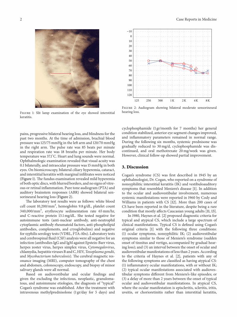
Figure 1: Slit lamp examination of the eye showed interstitialkeratitis.
pains, progressive bilateral hearing loss, and blindness for thepast two months. At the time of admission, brachial bloodpressure was 125/75mmHg in the left arm and 120/70mmHgin the right arm. The pulse rate was 85 beats per minuteand respiration rate was 18 breaths per minute. Her bodytemperature was 37.1∘C. Heart and lung sounds were normal.Ophthalmologic examination revealed that visual acuity was0.1 bilaterally, and intraocular pressure was 15mmHg in botheyes. On biomicroscopy, bilateral ciliary hyperemia, cataract,and interstitial keratitis withmarginal infiltrates were noticed(Figure 1). The fundus examination revealed mild hyperemiaof both optic discs, with blurred borders, andno signs of vitre-ous or retinal inflammation. Pure tone audiogram (PTA) andauditory brainstem responses (ABR) showed bilateral sen-sorineural hearing loss (Figure 2).
The laboratory test results were as follows: white bloodcell count 10,200/mm3, hemoglobin 9.8 g/dL, platelet count340,000/mm3, erythrocyte sedimentation rate 65mm/hr,and C-reactive protein 13.1mg/dL. She tested negative forautoimmune tests (anti-nuclear antibody, anti-neutrophilcytoplasmic antibody rheumatoid factors, anti-phospholipidantibodies, complements, and cryoglobulins) and negativefor syphilis serology tests (VDRL, FTA-Abs). Laboratory testsand cerebrospinal fluid (CSF) analysis were all negative for aninfection (antibodies IgG and IgM against Epstein-Barr virus,herpes zoster virus, herpes simplex virus, Cytomegalovirus,chlamydia, hepatitis viruses B andC,HIV,Toxoplasma gondii,andMycobacterium tuberculosis). The cerebral magnetic res-onance imaging (MRI), computer tomography of the chestand abdomen, colonoscopy, endoscopy, and biopsy of minorsalivary glands were all normal.
Based on audiovestibular and ocular findings andgiven the excluding the infectious, neoplastic, granuloma-tous, and autoimmune etiologies, the diagnosis of “typical”Cogan’s syndrome was established. After the treatment withintravenous methylprednisolone (1 gr/day for 5 days) and
−10
0
10
20
30
40
50
60
70
80
90
100
110
120
125 250 500 1 K 2 K 4 K 8 K
Figure 2: Audiogram showing bilateral moderate sensorineuralhearing loss.
cyclophosphamide (1 gr/month for 7 months) her generalcondition stabilized, anterior eye segment changes improved,and inflammatory parameters remained in normal range.During the following six months, systemic prednisone wasgradually reduced to 30mg/d, cyclophosphamide was dis-continued, and oral methotrexate 20mg/week was given.However, clinical follow-up showed partial improvement.
3. Discussion
Cogan’s syndrome (CS) was first described in 1945 by anophthalmologist, Dr. Cogan, who reported on a syndrome ofnonsyphilitic interstitial keratitis (IK) and vestibuloauditorysymptoms that resembled Meniere’s disease [1]. In additionto the ocular and audiovestibular involvement, numeroussystemic manifestations were reported in 1960 by Cody andWilliams in patients with CS [12]. More than 200 cases ofCS have been reported in the literature, despite being a rarecondition that mostly affects Caucasian young adults [11, 13].
In 1980, Haynes et al. [2] proposed diagnostic criteria fortypical and atypical CS, which include a large spectrum ofclinical manifestations. Typical CS is defined using Cogan’soriginal criteria [1] with the following three conditions:(1) ocular symptoms, nonsyphilitic IK; (2) audiovestibularsymptoms similar to those of Meniere’s syndrome (suddenonset of tinnitus and vertigo, accompanied by gradual hear-ing loss); and (3) an interval between the onset of ocular andaudiovestibularmanifestations of less than 2 years. Accordingto the criteria of Haynes et al. [2], patients with any ofthe following symptoms are classified as having atypical CS:(1) inflammatory ocular manifestations, with or without IK;(2) typical ocular manifestations associated with audioves-tibular symptoms different from Meniere’s-like episodes; or(3) a delay of more than 2 years between the onset of typicalocular and audiovestibular manifestations. In atypical CS,where the ocular manifestation is episcleritis, scleritis, iritis,uveitis, or chorioretinitis rather than interstitial keratitis,
Case Reports in Medicine 3
there is a worse prognosis and a higher frequency of aorticand other systemic manifestations [11].
Recurrent episodes of inner ear disease frequently resultin profound hearing loss. In a retrospective series of 60patients from one center, hearing loss was typically sudden,bilateral, fluctuating, and progressive, resulting in completehearing loss in 73 of 120 ears [8]. Hearing loss in both earswas noted at some point in all patients. Two smaller studiesboth found bilateral deafness in approximately two-thirds ofpatients [2, 3]. Typically, audiometry testing demonstrates asensorineural hearing loss, preferentially involving the lowand high range frequencies; poor speech discrimination isalso observed. In one preliminary study, at least 30 percent ofpatients had a pure tone audiometry threshold of greater thanor equal to 60 dB, a threshold value indicative of moderatelysevere hearing loss [4]. Hearing loss is often bilateral fromonset but in some patients it may be unilateral initially,becoming bilateral later. In the review by Vollertsen et al. [3],which included 78 patients with typical Cogan’s syndrome,bilateral deafness affected 43.5% of patients and occurred amean of 3 months after the onset of the initial symptoms.
When present, the systemic vasculitis associated with CSis a large- or medium- to small-sized vessel vasculitis, or anaortitis. The pattern of vessel involvement may be overlap-ping. Aortitis, which may develop within weeks to years ofdisease onset, has been described in approximately 10 percentof patients [2, 3, 11]. It may cause proximal aorta dilation, aor-tic valvular regurgitation, ostial coronary artery disease, andthoracoabdominal aortic aneurysms [11, 14–18]. A coronaryarteritis has also been described [3, 19, 20]. The large vesselvasculitis associated with CS may also resemble Takayasu’sarteritis, causing an occlusion of the aortic arch vessels withresultant upper and/or lower limb claudication, or renalartery stenosis [14, 19, 21, 22]. A small- or medium-sizedvessel arteritis has been described in some cases [23].
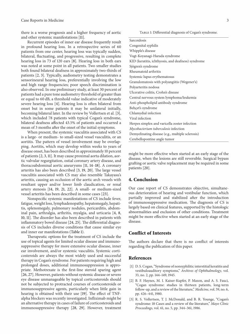
Nonspecific systemic manifestations of CS include fever,fatigue, weight loss, lymphadenopathy, hepatomegaly, hepati-tis, splenomegaly, pulmonary nodules, pericarditis, abdom-inal pain, arthralgia, arthritis, myalgia, and urticaria [4, 8,10, 11]. The disorder has also been described in patients withinflammatory bowel disease [24, 25].The differential diagno-sis of CS includes diverse conditions that cause similar eyeand inner ear manifestations (Table 1).
Therapeutic options for the treatment of CS include theuse of topical agents for limited ocular disease and immuno-suppressive therapy for more extensive ocular disease, innerear involvement, and/or systemic vasculitis. Systemic corti-costeroids are always the most widely used and successfultherapy in Cogan’s syndrome. For patients requiring high andprolonged doses, additional immunosuppression is appro-priate. Methotrexate is the first-line steroid sparing agent[26, 27]. However, patients without systemic disease or severeeye disease unmanageable by topical corticosteroids shouldnot be subjected to protracted courses of corticosteroids orimmunosuppressive agents, particularly when little gain inhearing is obtained with their use [19]. The effect of TNF-alpha blockers was recently investigated. Infliximab might bean alternative therapy in cases of failure of corticosteroids andimmunosuppressive therapy [28, 29]. However, treatment
Table 1: Differential diagnosis of Cogan’s syndrome.
SarcoidosisCongenital syphilisWhipple’s diseaseVogt-Koyanagi-Harada syndromeKID (keratitis, ichthyosis, and deafness) syndromeSjogren’s syndromeRheumatoid arthritisSystemic lupus erythematosusGranulomatosis with polyangiitis (Wegener’s)Polyarteritis nodosaUlcerative colitis, Crohn’s diseaseCentral nervous system lymphoma/leukemiaAnti-phospholipid antibody syndromeBehcet’s syndromeChlamydial infectionViral infectionHerpes simplex and varicella zoster infectionMycobacterium tuberculosis infectionDemyelinating disease (e.g., multiple sclerosis)Cerebellopontine angle tumor
might be more effective when started at an early stage of thedisease, when the lesions are still reversible. Surgical bypassgrafting or aortic valve replacement may be required in somepatients [20].
4. Conclusion
Our case report of CS demonstrates objective, simultane-ous deterioration of hearing and vestibular function, whichpartially improved and stabilized after the introductionof immunosuppressive medication. The diagnosis of CS islargely based on clinical features, supported by the histologicabnormalities and exclusion of other conditions. Treatmentmight be more effective when started at an early stage of thedisease.
Conflict of Interests
The authors declare that there is no conflict of interestsregarding the publication of this paper.
References
[1] D. S. Cogan, “Syndromeof nonsyphilitic interstitial keratitis andvestibuloauditory symptoms,” Archives of Ophthalmology, vol.33, no. 2, pp. 144–149, 1945.
[2] B. F. Haynes, M. I. Kaiser-Kupfer, P. Mason, and A. S. Fauci,“Cogan syndrome: studies in thirteen patients, long-termfollow-up, and a review of the literature,”Medicine, vol. 59, no. 6,pp. 426–441, 1980.
[3] R. S. Vollertsen, T. J. McDonald, and B. R. Younge, “Cogan’ssyndrome: 18 Cases and a review of the literature,”Mayo ClinicProceedings, vol. 61, no. 5, pp. 344–361, 1986.
4 Case Reports in Medicine
[4] E. W. St. Clair and R. M. McCallum, “Cogan’s syndrome,”Current Opinion in Rheumatology, vol. 11, no. 1, pp. 47–52, 1999.
[5] S. Podder and R. C. Shepherd, “Cogan’s syndrome: a rare sys-temic vasculitis,” Archives of Disease in Childhood, vol. 71, no. 2,pp. 163–164, 1994.
[6] H. Fidler and N. S. Jones, “Late onset Cogan’s syndrome,” Jour-nal of Laryngology and Otology, vol. 103, no. 5, pp. 512–514, 1989.
[7] O. Kasapcopur,M.Ashraf, S. Caliskan et al., “Cogan’s syndrome:a rare vasculitis in childhood,” Journal of Rheumatology, vol. 27,no. 7, pp. 1824–1825, 2000.
[8] M. B. Gluth, K. H. Baratz, E. L. Matteson, and C. L. W. Driscoll,“Cogan syndrome: a retrospective review of 60 patientsthroughout a half century,”Mayo Clinic Proceedings, vol. 81, no.4, pp. 483–488, 2006.
[9] P. Shah, R. A. Luqmani, P. I. Murray, W. P. Honan, P. G. J.Corridan, and P. Emery, “Posterior scleritis—an unusual mani-festation of Cogan’s syndrome,”The British Journal of Rheuma-tology, vol. 33, no. 8, pp. 774–775, 1994.
[10] M. Mazlumzadeh and E. L. Matteson, “Cogan’s syndrome:an audiovestibular, ocular, and systemic autoimmune disease,”Rheumatic Disease Clinics of North America, vol. 33, no. 4, pp.855–874, 2007.
[11] A. Grasland, J. Pouchot, E. Hachulla et al., “Typical and atypicalCogan’s syndrome: 32 cases and review of the literature,”Rheumatology, vol. 43, no. 8, pp. 1007–1015, 2004.
[12] D. T. Cody and H. L. Williams, “Cogan’s syndrome,”The Laryn-goscope, vol. 70, pp. 447–478, 1960.
[13] J. Cundiff, S. Kansal, A. Kumar, D. A. Goldstein, and H. H.Tessler, “Cogan’s syndrome: a cause of progressive hearingdeafness,” The American Journal of Otolaryngology, vol. 27, no.1, pp. 68–70, 2006.
[14] A. D. Cochrane and J. Tatoulis, “Cogan’s syndromewith aortitis,aortic regurgitation, and aortic arch vessel stenoses,” Annals ofThoracic Surgery, vol. 52, no. 5, pp. 1166–1167, 1991.
[15] J. Z. Livingston, A. S. Casale, G. M. Hutchins, and E. P. Shapiro,“Coronary involvement in Cogan’s syndrome,” The AmericanHeart Journal, vol. 123, no. 2, pp. 528–530, 1992.
[16] M. Hammer, T. Witte, A. Mugge et al., “Complicated Cogan’ssyndrome with aortic insufficiency and coronary stenosis,”Journal of Rheumatology, vol. 21, no. 3, pp. 552–555, 1994.
[17] J. Cremer, J. Laas, and B. Heublein, “A case of coronary stenosisdeveloping after successful aortic valve repair in Cogan’s syn-drome,”Thoracic and Cardiovascular Surgeon, vol. 41, no. 4, pp.255–257, 1993.
[18] J. F. Tseng, R. P. Cambria, H. T. Aretz, and D. C. Brewster,“Thoracoabdominal aortic aneurysm in Cogan’s syndrome,”Journal of Vascular Surgery, vol. 30, no. 3, pp. 565–568, 1999.
[19] N. B. Allen, C. C. Cox, J. Kisslo, M. R. Jacobs, R. M. McCallum,and B. F. Haynes, “Use of immunosuppressive agents in thetreatment of severe ocular and vascular manifestations ofCogan’s syndrome,” The American Journal of Medicine, vol. 88,no. 3, pp. 296–301, 1990.
[20] R. M. McCallum and B. F. Haynes, “Cogan’s syndrome,” inOcular Infection & Immunity, J. S. Pepose, G. N. Holland, andK. R. Wilhelmus, Eds., p. 446, Mosby, St. Louis, Mo, USA, 1stedition, 1996.
[21] K. Raza, D. Karokis, and G. D. Kitas, “Cogan’s syndrome withTakayasu’s arteritis,” The British Journal of Rheumatology, vol.37, no. 4, pp. 369–372, 1998.
[22] J. P. Vella, J. O’Callaghan, D. Hickey, and J. J. Walshe,“Renal artery stenosis complicating Cogan’s syndrome,”ClinicalNephrology, vol. 47, no. 6, pp. 407–408, 1997.
[23] R. S. Vollertsen, “Vasculitis and Cogan’s syndrome,” RheumaticDisease Clinics of North America, vol. 16, no. 2, pp. 433–439,1990.
[24] A. Jacob, J. G. Ledingham, A. I. G. Kerr, andM. J. Ford, “Ulcera-tive colitis and giant cell arteritis associated with sensorineuraldeafness,” Journal of Laryngology and Otology, vol. 104, no. 11,pp. 889–890, 1990.
[25] H. G.Thomas, “Case report: clinical and radiological features ofCogan’s syndrome—non-syphilitic interstitial keratitis, audio-vestibular symptoms and systemic manifestations,” ClinicalRadiology, vol. 45, no. 6, pp. 418–421, 1992.
[26] J. Pouchot, P. Vinceneux, D. Bouccara, O. Sterkers, B. Richard-son, and B. Bodelet, “Methotrexate as a steroid-sparing agent inCogan’s syndrome: comment on the concise communication byRichardson,” Arthritis and Rheumatism, vol. 38, no. 9, pp. 1348–1349, 1995.
[27] L. Riente, E. Taglione, and S. Berrettini, “Efficacy of methotrex-ate in Cogan’s syndrome,” Journal of Rheumatology, vol. 23, no.10, pp. 1830–1831, 1996.
[28] Z. Touma, R. Nawwar, U. Hadi, M. Hourani, and T. Arayssi,“The use of TNF-𝛼 blockers in Cogan’s syndrome,” Rheumatol-ogy International, vol. 27, no. 10, pp. 995–996, 2007.
[29] M. Fricker, A. Baumann, F. Wermelinger, P. M. Villiger, and A.Helbling, “A novel therapeutic option in Cogan diseases? TNF-𝛼 blockers,” Rheumatology International, vol. 27, no. 5, pp. 493–495, 2007.

Submit your manuscripts athttp://www.hindawi.com
Stem CellsInternational
Hindawi Publishing Corporationhttp://www.hindawi.com Volume 2014
Hindawi Publishing Corporationhttp://www.hindawi.com Volume 2014
MEDIATORSINFLAMMATION
of
Hindawi Publishing Corporationhttp://www.hindawi.com Volume 2014
Behavioural Neurology
EndocrinologyInternational Journal of
Hindawi Publishing Corporationhttp://www.hindawi.com Volume 2014
Hindawi Publishing Corporationhttp://www.hindawi.com Volume 2014
Disease Markers
Hindawi Publishing Corporationhttp://www.hindawi.com Volume 2014
BioMed Research International
OncologyJournal of
Hindawi Publishing Corporationhttp://www.hindawi.com Volume 2014
Hindawi Publishing Corporationhttp://www.hindawi.com Volume 2014
Oxidative Medicine and Cellular Longevity
Hindawi Publishing Corporationhttp://www.hindawi.com Volume 2014
PPAR Research
The Scientific World JournalHindawi Publishing Corporation http://www.hindawi.com Volume 2014
Immunology ResearchHindawi Publishing Corporationhttp://www.hindawi.com Volume 2014
Journal of
ObesityJournal of
Hindawi Publishing Corporationhttp://www.hindawi.com Volume 2014
Hindawi Publishing Corporationhttp://www.hindawi.com Volume 2014
Computational and Mathematical Methods in Medicine
OphthalmologyJournal of
Hindawi Publishing Corporationhttp://www.hindawi.com Volume 2014
Diabetes ResearchJournal of
Hindawi Publishing Corporationhttp://www.hindawi.com Volume 2014
Hindawi Publishing Corporationhttp://www.hindawi.com Volume 2014
Research and TreatmentAIDS
Hindawi Publishing Corporationhttp://www.hindawi.com Volume 2014
Gastroenterology Research and Practice
Hindawi Publishing Corporationhttp://www.hindawi.com Volume 2014
Parkinson’s Disease
Evidence-Based Complementary and Alternative Medicine
Volume 2014Hindawi Publishing Corporationhttp://www.hindawi.com
Related Documents