Hindawi Publishing Corporation Case Reports in Surgery Volume 2012, Article ID 829213, 3 pages doi:10.1155/2012/829213 Case Report Angiosarcoma of the Right Atrium Presenting as Syncope and Hemorrhagic Pericardial Tamponade V. G. Sams, 1 A. Tsapenko, 2 J. N. Kravitz, 2 and T. E. Gaines 1 1 Department of Surgery, University of Tennessee Medical Center, 1924 Alcoa Highway, Box U11, Knoxville, TN 37920, USA 2 Department of Pulmonary Medicine, University of Tennessee Medical Center, 1924 Alcoa Highway, Knoxville, TN 37920, USA Correspondence should be addressed to V. G. Sams, [email protected] Received 21 July 2012; Accepted 30 September 2012 Academic Editors: A. Cho, C. Foroulis, and E. Xenos Copyright © 2012 V. G. Sams et al. This is an open access article distributed under the Creative Commons Attribution License, which permits unrestricted use, distribution, and reproduction in any medium, provided the original work is properly cited. Angiosarcoma of the heart is a rare malignancy that can present in many ways. It is an important diagnosis to consider in patients presenting with otherwise unexplained tamponade-type symptoms. Here we present a case of a young male who presented with hemorrhagic tamponade and underwent resection of a large angiosarcoma of the right atrium. In this case, we describe the rare presentation of angiosarcoma with its diagnostic approaches, hospital course, clinical management, and discussion. 1. Introduction Primary cardiac angiosarcoma is rare and aggressive with metastatic spread approaching ninety percent at the time of diagnosis [1]. It is the most common malignant tumor of the heart and is characterized by rapid growth, local invasion, and distant metastasis [2]. Multiple case reports have been published in the literature with presentations including pericarditis and pleuritis [3], chest pain and shortness of breath [4], and cough and hemoptysis [2]. Prognosis of patients with these primary tumors is very poor [5] with a mean survival of six months and poor chemotherapy response [6]. 2. Case Report This is a 27-year-old white previously healthy male with no past medical history and no risk factors for cardiovascular conditions who presented to the medicine critical care unit via interfacility transfer after an episode of chest pain with syncope while exercising and was hypotensive. He stated he had been experiencing some chest discomfort for several days with exercise intolerance and cold sweats. 2.1. Findings. Upon arrival, the patient was alert and in no acute distress with a heart rate of 112 and irregular and blood pressure of 112/77. His oxygen saturation was 94% on two liters of oxygen via nasal cannula. He had an obvious palpable pulsus paradoxus. All of his laboratory values were within normal limits with the exception of a mildly elevated creatinine. 2.2. Diagnosis and Management. Upon initial presentation, the patient received a therapeutic dose of enoxaparin for a presumptive diagnosis of pulmonary embolism. Follow- ing the drug’s administration, further diagnostic workup included a chest X-ray which demonstrated cardiomegaly (Figure 1(a)), an echocardiogram which demonstrated a pericardial effusion with tamponade and a computed tomog- raphy scan also demonstrating the pericardial effusion (Figure 1(b)). Cardiothoracic surgery was consulted for pericardial drainage and possible biopsy. He was scheduled for a pericardial window the next morning since he had been anti- coagulated on arrival and was hemodynamically stable after IV fluid administration. Intraoperatively, the patient was found to have a bloody pericardial effusion. The pericardial window did not allow adequate exposure to determine the source of the persistent bleeding. At this point we proceeded with a median sternotomy. Exposure of the heart revealed a large right atrial lobulated, bleeding mass. The pulmonary artery and aorta also had plaque-like lesions.

Welcome message from author
This document is posted to help you gain knowledge. Please leave a comment to let me know what you think about it! Share it to your friends and learn new things together.
Transcript
Hindawi Publishing CorporationCase Reports in SurgeryVolume 2012, Article ID 829213, 3 pagesdoi:10.1155/2012/829213
Case Report
Angiosarcoma of the Right Atrium Presenting as Syncope andHemorrhagic Pericardial Tamponade
V. G. Sams,1 A. Tsapenko,2 J. N. Kravitz,2 and T. E. Gaines1
1 Department of Surgery, University of Tennessee Medical Center, 1924 Alcoa Highway, Box U11, Knoxville, TN 37920, USA2 Department of Pulmonary Medicine, University of Tennessee Medical Center, 1924 Alcoa Highway, Knoxville, TN 37920, USA
Correspondence should be addressed to V. G. Sams, [email protected]
Received 21 July 2012; Accepted 30 September 2012
Academic Editors: A. Cho, C. Foroulis, and E. Xenos
Copyright © 2012 V. G. Sams et al. This is an open access article distributed under the Creative Commons Attribution License,which permits unrestricted use, distribution, and reproduction in any medium, provided the original work is properly cited.
Angiosarcoma of the heart is a rare malignancy that can present in many ways. It is an important diagnosis to consider in patientspresenting with otherwise unexplained tamponade-type symptoms. Here we present a case of a young male who presented withhemorrhagic tamponade and underwent resection of a large angiosarcoma of the right atrium. In this case, we describe the rarepresentation of angiosarcoma with its diagnostic approaches, hospital course, clinical management, and discussion.
1. Introduction
Primary cardiac angiosarcoma is rare and aggressive withmetastatic spread approaching ninety percent at the time ofdiagnosis [1]. It is the most common malignant tumor of theheart and is characterized by rapid growth, local invasion,and distant metastasis [2]. Multiple case reports have beenpublished in the literature with presentations includingpericarditis and pleuritis [3], chest pain and shortness ofbreath [4], and cough and hemoptysis [2]. Prognosis ofpatients with these primary tumors is very poor [5] witha mean survival of six months and poor chemotherapyresponse [6].
2. Case Report
This is a 27-year-old white previously healthy male with nopast medical history and no risk factors for cardiovascularconditions who presented to the medicine critical care unitvia interfacility transfer after an episode of chest pain withsyncope while exercising and was hypotensive. He stated hehad been experiencing some chest discomfort for several dayswith exercise intolerance and cold sweats.
2.1. Findings. Upon arrival, the patient was alert and in noacute distress with a heart rate of 112 and irregular and
blood pressure of 112/77. His oxygen saturation was 94% ontwo liters of oxygen via nasal cannula. He had an obviouspalpable pulsus paradoxus. All of his laboratory values werewithin normal limits with the exception of a mildly elevatedcreatinine.
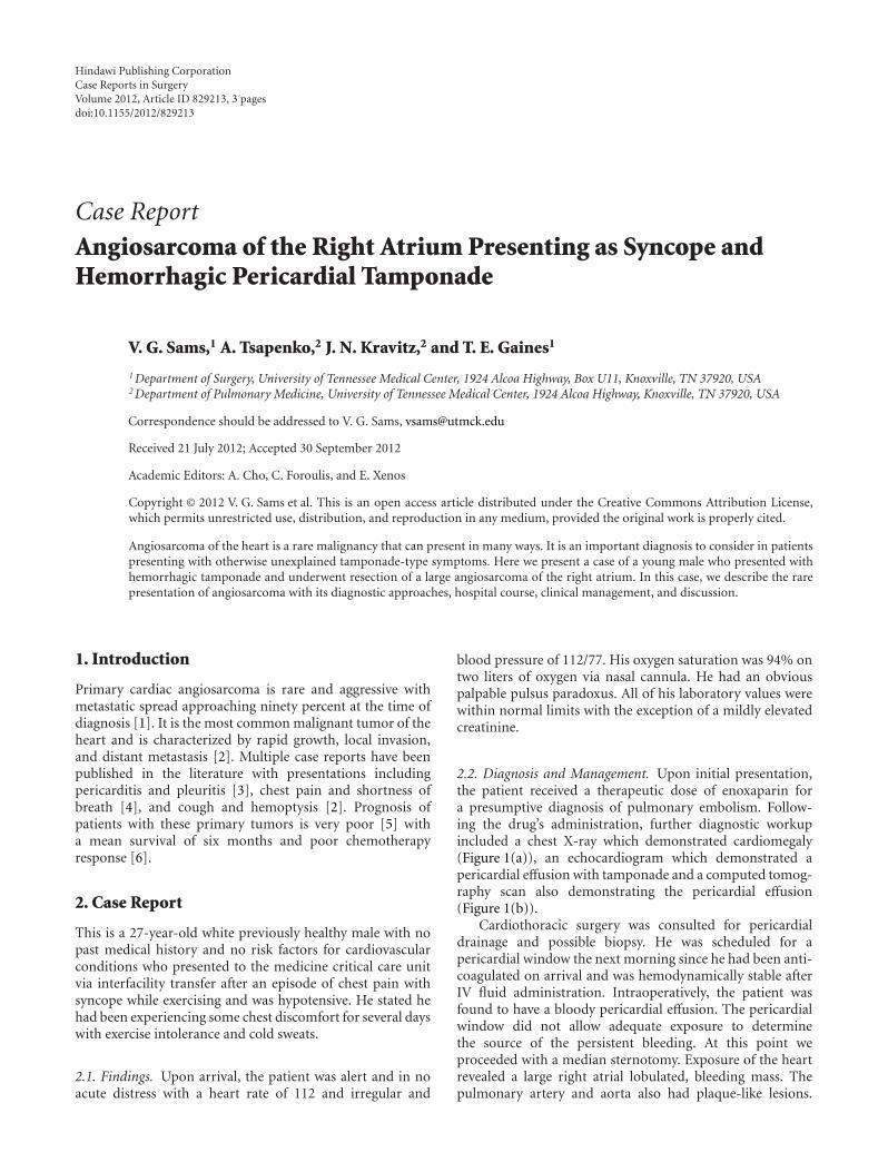
2.2. Diagnosis and Management. Upon initial presentation,the patient received a therapeutic dose of enoxaparin fora presumptive diagnosis of pulmonary embolism. Follow-ing the drug’s administration, further diagnostic workupincluded a chest X-ray which demonstrated cardiomegaly(Figure 1(a)), an echocardiogram which demonstrated apericardial effusion with tamponade and a computed tomog-raphy scan also demonstrating the pericardial effusion(Figure 1(b)).
Cardiothoracic surgery was consulted for pericardialdrainage and possible biopsy. He was scheduled for apericardial window the next morning since he had been anti-coagulated on arrival and was hemodynamically stable afterIV fluid administration. Intraoperatively, the patient wasfound to have a bloody pericardial effusion. The pericardialwindow did not allow adequate exposure to determinethe source of the persistent bleeding. At this point weproceeded with a median sternotomy. Exposure of the heartrevealed a large right atrial lobulated, bleeding mass. Thepulmonary artery and aorta also had plaque-like lesions.

2 Case Reports in Surgery
(a) (b)
Figure 1: Radiographic imaging; chest X-ray (a) and CT scan (b).
(a) (b) (c)
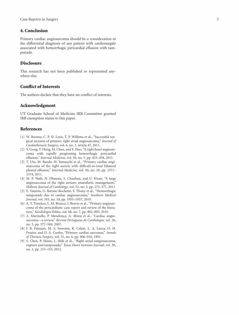
Figure 2: Intraoperative photos of identification (a), excision (b), and reconstruction (c) of the right atrial angiosarcoma.
Intraoperative frozen pathologic analysis suggested sometype of high-grade angiosarcoma. We removed these lesionsas well as performed an extensive node dissection to includepretracheal and right paratracheal lymph nodes. A decisionwas made to excise all gross disease, which involved the entirelateral wall of the right atrium, to best control the bleedingand prevent a recurrent effusion. The patient was placedon cardiopulmonary bypass. The mass was then resected tothe right atrial and right ventricle junction next to the rightcoronary artery and including the sinoatrial node. We thenused a bovine pericardial graft to reconstruct the atriumand placed two temporary right ventricular pacing wires(Figure 2).
The patient was extubated the following morning andmanaged with intravenous pain medication as well aspulmonary toilet. He did well and both mediastinal drainsand right pleural drain were removed. He never requiredventricular pacing and his final pathology was poorlydifferentiated angiosarcoma involving right atrial resectionmargin, virtually all of the lymph nodes, and the plaque-like lesions on the aorta and pulmonary artery. He wasdischarged to home with family with outpatient followupwith medical oncology.
3. Discussion
Due to the rarity and aggressiveness of cardiac angiosarcoma,no curative treatment strategy has been developed andopportunity for meaningful intervention is slim. In this case,the goal of resection was to remove all gross disease in anattempt to eliminate the source of hemorrhage and tampon-ade. The other dilemma in evaluating these patients is thevariability in cardiac angiosarcoma presentations. As seenin our patient, the presumption of pulmonary embolismwith the administration of therapeutic enoxaparin couldhave worsened his bleeding and increased the degree oftamponade. Another differential diagnosis in a young,previously asymptomatic patient with cardiac tamponadeis aortic dissection in which case enoxaparin would alsobe contraindicated. The disease is also difficult to diagnosewith imaging when a complex bloody effusion is present.Surgery is the treatment of choice but often not curative.Patients often have metastatic disease at the time of surgery[7]. Aggressive and complete surgical resection offers the bestpalliation and prolongation of life [8]. Palliative resection inconjunction with chemotherapy and radiation can increasethe length of survival and quality of life [9].
Case Reports in Surgery 3
4. Conclusion
Primary cardiac angiosarcoma should be a consideration inthe differential diagnosis of any patient with cardiomegalyassociated with hemorrhagic pericardial effusion with tam-ponade.
Disclosure
This research has not been published or represented any-where else.
Conflict of Interests
The authors declare that they have no conflict of interests.
Acknowledgment
UT Graduate School of Medicine IRB Committee grantedIRB exemption status to this paper.
References
[1] W. Bouma, C. P. H. Lexis, T. P. Willems et al., “Successful sur-gical excision of primary right atrial angiosarcoma,” Journal ofCardiothoracic Surgery, vol. 6, no. 1, article 47, 2011.
[2] Y. Gong, T. Hong, M. Chen, and Y. Huo, “A right heart angiosar-coma with rapidly progressing hemorrhagic pericardialeffusion,” Internal Medicine, vol. 50, no. 5, pp. 455–458, 2011.
[3] T. Uto, M. Bando, H. Yamauchi et al., “Primary cardiac angi-osarcoma of the right auricle with difficult-to-treat bilateralpleural effusion,” Internal Medicine, vol. 50, no. 20, pp. 2371–2374, 2011.
[4] M. P. Nath, N. Dhawan, S. Chauhan, and U. Kiran, “A largeangiosarcoma of the right atrium: anaesthetic management,”Hellenic Journal of Cardiology, vol. 52, no. 3, pp. 273–277, 2011.
[5] E. Vautrin, G. Barone-Rochette, F. Thony et al., “Hemorrhagictamponade due to cardiac angiosarcoma,” Southern MedicalJournal, vol. 103, no. 10, pp. 1055–1057, 2010.
[6] A. T. Timoteo, L. M. Branco, I. Bravio et al., “Primary angiosar-coma of the pericardium: case report and review of the litera-ture,” Kardiologia Polska, vol. 68, no. 7, pp. 802–805, 2010.
[7] A. Murinello, P. Mendonca, A. Abreu et al., “Cardiac angio-sarcoma—a review,” Revista Portuguesa de Cardiologia, vol. 26,no. 5, pp. 577–584, 2007.
[8] J. B. Putnam, M. S. Sweeney, R. Colon, L. A. Lanza, O. H.Frazier, and D. A. Cooley, “Primary cardiac sarcomas,” Annalsof Thoracic Surgery, vol. 51, no. 6, pp. 906–910, 1991.
[9] S. Chen, P. Hsien, L. Shih et al., “Right atrial sangiosarcoma,rupture and tamponade,” Texas Heart Institute Journal, vol. 30,no. 1, pp. 153–155, 2012.

Submit your manuscripts athttp://www.hindawi.com
Stem CellsInternational
Hindawi Publishing Corporationhttp://www.hindawi.com Volume 2014
Hindawi Publishing Corporationhttp://www.hindawi.com Volume 2014
MEDIATORSINFLAMMATION
of
Hindawi Publishing Corporationhttp://www.hindawi.com Volume 2014
Behavioural Neurology
EndocrinologyInternational Journal of
Hindawi Publishing Corporationhttp://www.hindawi.com Volume 2014
Hindawi Publishing Corporationhttp://www.hindawi.com Volume 2014
Disease Markers
Hindawi Publishing Corporationhttp://www.hindawi.com Volume 2014
BioMed Research International
OncologyJournal of
Hindawi Publishing Corporationhttp://www.hindawi.com Volume 2014
Hindawi Publishing Corporationhttp://www.hindawi.com Volume 2014
Oxidative Medicine and Cellular Longevity
Hindawi Publishing Corporationhttp://www.hindawi.com Volume 2014
PPAR Research
The Scientific World JournalHindawi Publishing Corporation http://www.hindawi.com Volume 2014
Immunology ResearchHindawi Publishing Corporationhttp://www.hindawi.com Volume 2014
Journal of
ObesityJournal of
Hindawi Publishing Corporationhttp://www.hindawi.com Volume 2014
Hindawi Publishing Corporationhttp://www.hindawi.com Volume 2014
Computational and Mathematical Methods in Medicine
OphthalmologyJournal of
Hindawi Publishing Corporationhttp://www.hindawi.com Volume 2014
Diabetes ResearchJournal of
Hindawi Publishing Corporationhttp://www.hindawi.com Volume 2014
Hindawi Publishing Corporationhttp://www.hindawi.com Volume 2014
Research and TreatmentAIDS
Hindawi Publishing Corporationhttp://www.hindawi.com Volume 2014
Gastroenterology Research and Practice
Hindawi Publishing Corporationhttp://www.hindawi.com Volume 2014
Parkinson’s Disease
Evidence-Based Complementary and Alternative Medicine
Volume 2014Hindawi Publishing Corporationhttp://www.hindawi.com
Related Documents