Hindawi Publishing Corporation Case Reports in Radiology Volume 2013, Article ID 235209, 3 pages http://dx.doi.org/10.1155/2013/235209 Case Report Blunt Facial Trauma Causing Isolated Optic Nerve Hematoma R. Parab, C. I. Fung, and Gerrit Van Der Merwe Department of Radiology and Diagnostic Imaging, University of Alberta, and Royal Alexandra Hospital, Edmonton, AB, Canada T5H 3V9 Correspondence should be addressed to Gerrit Van Der Merwe; [email protected] Received 25 October 2012; Accepted 1 January 2013 Academic Editors: E. Kapsalaki, A. V. Khaw, and D. Tsetis Copyright © 2013 R. Parab et al. is is an open access article distributed under the Creative Commons Attribution License, which permits unrestricted use, distribution, and reproduction in any medium, provided the original work is properly cited. Traumatic optic neuropathy is an uncommon, yet serious, result of facial trauma. e authors present a novel case of a 59-year- old gentleman who presented with an isolated blunt traumatic leſt optic nerve hematoma causing vision loss. ere were no other injuries or fractures to report. is case highlights the importance of early recognition of this rare injury and reviews the current literature and management of traumatic optic neuropathy. 1. Introduction A recent prospective Canadian epidemiological study showed that 0.4% of all trauma patients had traumatic optic neuropa- thy [1]. Out of the patients who suffered from a traumatic optic neuropathy, two thirds had significant associated head injuries [1]. Isolated traumatic optic nerve hematoma without evidence of any other traumatic injuries or fractures is a very unusual presentation of traumatic optic neuropathy. Traumatic optic neuropathy is the result of direct or indirect forces causing injury to the optic nerve. Direct trauma refers to trauma precisely to the optic nerve causing transection, avulsion, orbital hemorrhage, orbital sheath hemorrhage, or orbital emphysema [2, 3] whereas indirect injury is caused by the convergence of traumatic forces focused elsewhere, secondarily involving the optic nerve [3]. e injury to the optic nerve may be primary, referring to ischemia caused by shearing and contusion necrosis of the optic nerve at the time of injury [2], or secondary, reflecting posttraumatic injury of the optic nerve due to a self-propagating compartment syndrome caused by edema or hemorrhage in the confined space of the intracanalicular optic nerve sheath [2, 3]. e final common pathway is that optic nerve axonal injury results in retinal ganglion cell death [2, 4] which peaks within two to three weeks [2, 5] and results in permanent vision loss. 2. Case Report A 59-year-old male presented to a tertiary care center in Edmonton, AB, Canada, with monocular leſt-sided vision loss and leſt retrobulbar pain. An hour and a half prior, he had been struck by a piece of heavy equipment at work resulting in a blunt traumatic injury to his leſt eye. No other symptoms suggestive of head injury were described. Orbital examination revealed leſt proptosis and swelling. e leſt pupil was fixed and dilated, measuring 5 mm in diameter. e patient was unable to visualize light with the affected eye, which was also nonreactive. Normal bilateral extraocular muscle movements were maintained. Unenhanced CT orbits revealed moderate edema and fat stranding surrounding the leſt optic nerve complex. e distal optic nerve adjacent to the globe measured 7 mm and demonstrated an irregular contour. Additionally, a small pre- septal hematoma tracked along the lateral aspect of the globe measuring 4 mm in thickness and extending approximately 1 cm posteriorly along the lateral orbital wall. e leſt globe was intact. e right optic nerve was normal in caliber, measuring 4mm in diameter. No other injuries were seen (Figure 1). Within the clinical context, partial transections of the optic nerve complex or a nerve sheath hematoma were the likely diagnoses. Given the possible diagnosis of a nerve

Welcome message from author
This document is posted to help you gain knowledge. Please leave a comment to let me know what you think about it! Share it to your friends and learn new things together.
Transcript

Hindawi Publishing CorporationCase Reports in RadiologyVolume 2013, Article ID 235209, 3 pageshttp://dx.doi.org/10.1155/2013/235209
Case ReportBlunt Facial Trauma Causing Isolated Optic Nerve Hematoma
R. Parab, C. I. Fung, and Gerrit Van Der Merwe
Department of Radiology and Diagnostic Imaging, University of Alberta, and Royal Alexandra Hospital, Edmonton,AB, Canada T5H 3V9
Correspondence should be addressed to Gerrit Van Der Merwe; [email protected]
Received 25 October 2012; Accepted 1 January 2013
Academic Editors: E. Kapsalaki, A. V. Khaw, and D. Tsetis
Copyright © 2013 R. Parab et al.This is an open access article distributed under the Creative Commons Attribution License, whichpermits unrestricted use, distribution, and reproduction in any medium, provided the original work is properly cited.
Traumatic optic neuropathy is an uncommon, yet serious, result of facial trauma. The authors present a novel case of a 59-year-old gentleman who presented with an isolated blunt traumatic left optic nerve hematoma causing vision loss. There were no otherinjuries or fractures to report. This case highlights the importance of early recognition of this rare injury and reviews the currentliterature and management of traumatic optic neuropathy.
1. Introduction
Arecent prospectiveCanadian epidemiological study showedthat 0.4% of all trauma patients had traumatic optic neuropa-thy [1]. Out of the patients who suffered from a traumaticoptic neuropathy, two thirds had significant associated headinjuries [1]. Isolated traumatic optic nerve hematomawithoutevidence of any other traumatic injuries or fractures is a veryunusual presentation of traumatic optic neuropathy.
Traumatic optic neuropathy is the result of direct orindirect forces causing injury to the optic nerve. Directtrauma refers to trauma precisely to the optic nerve causingtransection, avulsion, orbital hemorrhage, orbital sheathhemorrhage, or orbital emphysema [2, 3] whereas indirectinjury is caused by the convergence of traumatic forcesfocused elsewhere, secondarily involving the optic nerve [3].The injury to the optic nerve may be primary, referringto ischemia caused by shearing and contusion necrosis ofthe optic nerve at the time of injury [2], or secondary,reflecting posttraumatic injury of the optic nerve due to aself-propagating compartment syndrome caused by edemaor hemorrhage in the confined space of the intracanalicularoptic nerve sheath [2, 3]. The final common pathway is thatoptic nerve axonal injury results in retinal ganglion cell death[2, 4] which peaks within two to three weeks [2, 5] and resultsin permanent vision loss.
2. Case Report
A 59-year-old male presented to a tertiary care center inEdmonton, AB, Canada, with monocular left-sided visionloss and left retrobulbar pain. An hour and a half prior,he had been struck by a piece of heavy equipment at workresulting in a blunt traumatic injury to his left eye. No othersymptoms suggestive of head injury were described. Orbitalexamination revealed left proptosis and swelling. The leftpupil was fixed and dilated, measuring 5mm in diameter.The patient was unable to visualize light with the affectedeye, which was also nonreactive. Normal bilateral extraocularmuscle movements were maintained.
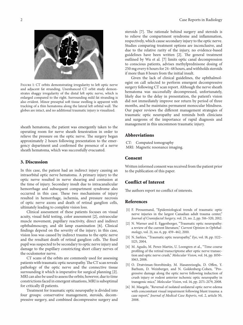
Unenhanced CT orbits revealed moderate edema andfat stranding surrounding the left optic nerve complex. Thedistal optic nerve adjacent to the globe measured 7mm anddemonstrated an irregular contour. Additionally, a small pre-septal hematoma tracked along the lateral aspect of the globemeasuring 4mm in thickness and extending approximately1 cm posteriorly along the lateral orbital wall. The left globewas intact. The right optic nerve was normal in caliber,measuring 4mm in diameter. No other injuries were seen(Figure 1).
Within the clinical context, partial transections of theoptic nerve complex or a nerve sheath hematoma were thelikely diagnoses. Given the possible diagnosis of a nerve
2 Case Reports in Radiology
Figure 1: CT orbits demonstrating irregularity to left optic nerveand adjacent fat stranding. Unenhanced CT orbit study demon-strates shaggy irregularity of the distal left optic nerve, which isenlarged compared to the right. Surrounding mild fat stranding isalso evident. Minor preseptal soft tissue swelling is apparent withtracking of a thin hematoma along the lateral left orbital wall. Theglobes are intact, and no additional traumatic injury is visualized.
sheath hematoma, the patient was emergently taken to theoperating room for nerve sheath fenestration in order torelieve the pressure on the optic nerve. The surgery beganapproximately 2 hours following presentation to the emer-gency department and confirmed the presence of a nervesheath hematoma, which was successfully evacuated.
3. Discussion
In this case, the patient had an indirect injury causing anintraorbital optic nerve hematoma. A primary injury to theoptic nerve resulted in nerve shearing and contusion atthe time of injury. Secondary insult due to intracanalicularhemorrhage and subsequent compartment syndrome alsooccurred in this case. These two mechanisms of injuryresulted in hemorrhage, ischemia, and pressure necrosisof optic nerve axons and death of retinal ganglion cells,ultimately leading to complete vision loss.
Clinical assessment of these patients focuses on visualacuity, visual field testing, color assessment [2], extraocularmuscle movement, pupillary reactivity, direct and indirectophthalmoscopy, and slit lamp examination [6]. Clinicalfindings depend on the severity of the injury; in this case,vision loss was caused by indirect trauma to the optic nerveand the resultant death of retinal ganglion cells. The fixedpupil was suspected to be secondary to optic nerve injury anddamage to the pupillary constricting short ciliary nerves ofthe oculomotor nerve.
CT scans of the orbits are commonly used for assessingpatientswith traumatic optic neuropathy.TheCT scan revealspathology of the optic nerve and the connective tissuesurrounding it which is imperative for surgical planning [2].MRI can also be used to assess the orbits; however, due to timeconstrictions faced in emergent situations,MRI is suboptimalfor critically ill patients.
Treatment for traumatic optic neuropathy is divided intofour groups: conservative management, steroids, decom-pressive surgery, and combined decompressive surgery and
steroids [7]. The rationale behind surgery and steroids isto relieve the compartment syndrome and inflammation,respectively, which cause secondary injury to the optic nerve.Studies comparing treatment options are inconclusive, anddue to the relative rarity of the injury, no evidence-basedguidelines have been written [2]. The general treatmentoutlined by Wu et al. [7] limits optic canal decompressionto conscious patients, advises methylprednisone dosing of250mg every 6 hours for 24–48 hours, andwithholds steroidsif more than 8 hours from the initial insult.
Given the lack of clinical guidelines, the ophthalmol-ogist on call selected to perform emergent decompressivesurgery following CT scan report. Although the nerve sheathhematoma was successfully decompressed, unfortunately,likely due to the delay in presentation, the patient’s visiondid not immediately improve nor return by period of threemonths, and he maintains permanent monocular blindness.Our paper reviews the different management strategies oftraumatic optic neuropathy and reminds both cliniciansand surgeons of the importance of rapid diagnosis andmanagement in this uncommon traumatic injury.
Abbreviations
CT: Computed tomographyMRI: Magnetic resonance imaging.
Consent
Written informed consent was received from the patient priorto the publication of this paper.
Conflict of Interest
The authors report no conflict of interests.
References
[1] F. Pirouzmand, “Epidemiological trends of traumatic opticnerve injuries in the largest Canadian adult trauma center,”Journal of Craniofacial Surgery, vol. 23, no. 2, pp. 516–520, 2012.
[2] N. Warner and E. Eggenberger, “Traumatic optic neuropathy:a review of the current literature,” Current Opinion in Ophthal-mology, vol. 21, no. 6, pp. 459–462, 2010.
[3] N. Sarkies, “Traumatic optic neuropathy,” Eye, vol. 18, pp. 1122–1125, 2004.
[4] M. Agudo, M. Perez-Martin, U. Loongren et al., “Time courseprofiling of the retinal transcriptome after optic nerve transec-tion and optic nerve crush,”Molecular Vision, vol. 14, pp. 1050–1063, 2008.
[5] O. Dratviman-Storobinsky, M. Hasanreisoglu, D. Offen, Y.Barhum, D. Weinberger, and N. Goldenberg-Cohen, “Pro-gressive damage along the optic nerve following induction ofcrush injury or rodent anterior ischemic optic neuropathy intransgenic mice,”Molecular Vision, vol. 14, pp. 2171–2179, 2008.
[6] M. Maegele, “Reversal of isolated unilateral optic nerve edemawith concomitant visual impairment following blunt trauma: acase report,” Journal of Medical Case Reports, vol. 2, article 50,2008.

Case Reports in Radiology 3
[7] N. Wu, Z. Q. Yin, and Y. Wang, “Traumatic optic neuropathytherapy: an update of clinical experimental studies,”The Journalof International Medical Research, vol. 36, no. 5, pp. 883–889,2008.

Submit your manuscripts athttp://www.hindawi.com
Stem CellsInternational
Hindawi Publishing Corporationhttp://www.hindawi.com Volume 2014
Hindawi Publishing Corporationhttp://www.hindawi.com Volume 2014
MEDIATORSINFLAMMATION
of
Hindawi Publishing Corporationhttp://www.hindawi.com Volume 2014
Behavioural Neurology
EndocrinologyInternational Journal of
Hindawi Publishing Corporationhttp://www.hindawi.com Volume 2014
Hindawi Publishing Corporationhttp://www.hindawi.com Volume 2014
Disease Markers
Hindawi Publishing Corporationhttp://www.hindawi.com Volume 2014
BioMed Research International
OncologyJournal of
Hindawi Publishing Corporationhttp://www.hindawi.com Volume 2014
Hindawi Publishing Corporationhttp://www.hindawi.com Volume 2014
Oxidative Medicine and Cellular Longevity
Hindawi Publishing Corporationhttp://www.hindawi.com Volume 2014
PPAR Research
The Scientific World JournalHindawi Publishing Corporation http://www.hindawi.com Volume 2014
Immunology ResearchHindawi Publishing Corporationhttp://www.hindawi.com Volume 2014
Journal of
ObesityJournal of
Hindawi Publishing Corporationhttp://www.hindawi.com Volume 2014
Hindawi Publishing Corporationhttp://www.hindawi.com Volume 2014
Computational and Mathematical Methods in Medicine
OphthalmologyJournal of
Hindawi Publishing Corporationhttp://www.hindawi.com Volume 2014
Diabetes ResearchJournal of
Hindawi Publishing Corporationhttp://www.hindawi.com Volume 2014
Hindawi Publishing Corporationhttp://www.hindawi.com Volume 2014
Research and TreatmentAIDS
Hindawi Publishing Corporationhttp://www.hindawi.com Volume 2014
Gastroenterology Research and Practice
Hindawi Publishing Corporationhttp://www.hindawi.com Volume 2014
Parkinson’s Disease
Evidence-Based Complementary and Alternative Medicine
Volume 2014Hindawi Publishing Corporationhttp://www.hindawi.com
Related Documents