Case Report Bilateral, Simultaneous, Acute Angle Closure Glaucoma in Pseudophakia Induced by Chlorthalidone Indra Durai, 1 Mrunali Mohan Dhavalikar, 2 Chandran Prem Anand, 2 Venkatraman Ganesh, 2 and Ramaswami Krishnadas 1 1 Aravind Eye Care System, Madurai, India 2 Aravind Eye Care System, Coimbatore, India Correspondence should be addressed to Ramaswami Krishnadas; [email protected] Received 31 January 2016; Accepted 10 April 2016 Academic Editor: Stephen G. Schwartz Copyright © 2016 Indra Durai et al. is is an open access article distributed under the Creative Commons Attribution License, which permits unrestricted use, distribution, and reproduction in any medium, provided the original work is properly cited. Purpose. To report two persons with acute, bilateral, and simultaneous angle closure glaucoma in pseudophakia secondary to uveal effusions induced by administration of chlorthalidone. Methods. Case reports. Results. Bilateral shallow anterior chambers and high intraocular pressure with decline in visual acuity were reported in two patients within days of intake of chlorthalidone for systemic hypertension. Gonioscopy confirmed appositional angle closure while choroidal detachment and ciliochoroidal detachment were revealed on ultrasonographic studies. Discontinuing chlorthalidone and institution of aqueous suppressants to reduce IOP and cycloplegics reversed angle closure and glaucoma. Conclusions. Reports of angle closure glaucoma in pseudophakic eyes induced by idiosyncratic reaction to chlorthalidone confirms that osmotic changes in the crystalline lens has no role in the pathogenesis of drug induced glaucoma and reaffirms that glaucoma is secondary to ciliochoroidal detachment and ciliary body rotation and edema. 1. Introduction Pseudophakic angle closure glaucoma is a rare occurrence. It has been reported to frequently occur as a consequence of pupil block with iris bombe from various causes, aqueous misdirection, choroidal detachment and effusions, supra- choroidal hemorrhage, and capsular bag distension syn- drome with anterior shiſt of the iris-lens diaphragm. Bilateral, simultaneous, acute angle closure glaucoma in pseudophakic eyes is a still rarer entity and has been reported as an idiosyn- cratic reaction following administration of sulphonamide containing drugs, acetazolamide and hydrochlorothiazide [1, 2]. We report a series of two patients, both pseudophakic in either eye, who had simultaneous acute angle closure glaucoma following intake of chlorthalidone for management of systemic hypertension. 2. Case Description 2.1. Report of Case 1. A 71-year-old male presented with complaints of sudden blurring of vision in both eyes associated with pain and coloured haloes since morning. He had had bilateral, uneventful clear corneal phacoemulsifica- tion with posterior chamber intraocular lens implantation five years earlier with no other antecedent ocular disease. Medical history was significant for systemic hypertension for which he was undergoing treatment for the past 15 years. He was prescribed Klorzid tablets (chlorthalidone 12.5 mg, German Remedies Limited, Zydus Cadila) for systemic hypertension and AB Phylline tablets for bronchitis for the week preceding his eye consultation. Examination revealed uncorrected acuity of 6/60 (20/200) OU. Vision was correctable to 20/20 OU with -3.75 D Sphere OD and -2.50 D Sphere OS, respectively, with near vision N6 with +2.50 addition in both eyes. Anterior segment examination revealed corneal epithelial edema, shallow anterior chambers, sluggishly reacting pupils, and posterior chamber intraocular lenses in both eyes (Figures 1(a) and 1(b)). Evaluation of fundus revealed normal optic discs and posterior pole. Intraocular pressure (IOP) was 41 mmHg OD and 36 mmHg OS, respectively, by Goldmann applanation tonometry. Gonioscopy performed with a Hindawi Publishing Corporation Case Reports in Ophthalmological Medicine Volume 2016, Article ID 3713818, 5 pages http://dx.doi.org/10.1155/2016/3713818

Welcome message from author
This document is posted to help you gain knowledge. Please leave a comment to let me know what you think about it! Share it to your friends and learn new things together.
Transcript
Case ReportBilateral, Simultaneous, Acute Angle ClosureGlaucoma in Pseudophakia Induced by Chlorthalidone
Indra Durai,1 Mrunali Mohan Dhavalikar,2 Chandran Prem Anand,2
Venkatraman Ganesh,2 and Ramaswami Krishnadas1
1Aravind Eye Care System, Madurai, India2Aravind Eye Care System, Coimbatore, India
Correspondence should be addressed to Ramaswami Krishnadas; [email protected]
Received 31 January 2016; Accepted 10 April 2016
Academic Editor: Stephen G. Schwartz
Copyright © 2016 Indra Durai et al. This is an open access article distributed under the Creative Commons Attribution License,which permits unrestricted use, distribution, and reproduction in any medium, provided the original work is properly cited.
Purpose. To report two persons with acute, bilateral, and simultaneous angle closure glaucoma in pseudophakia secondary to uvealeffusions induced by administration of chlorthalidone.Methods. Case reports. Results. Bilateral shallow anterior chambers and highintraocular pressure with decline in visual acuity were reported in two patients within days of intake of chlorthalidone for systemichypertension. Gonioscopy confirmed appositional angle closure while choroidal detachment and ciliochoroidal detachment wererevealed on ultrasonographic studies. Discontinuing chlorthalidone and institution of aqueous suppressants to reduce IOP andcycloplegics reversed angle closure and glaucoma. Conclusions. Reports of angle closure glaucoma in pseudophakic eyes inducedby idiosyncratic reaction to chlorthalidone confirms that osmotic changes in the crystalline lens has no role in the pathogenesisof drug induced glaucoma and reaffirms that glaucoma is secondary to ciliochoroidal detachment and ciliary body rotation andedema.
1. Introduction
Pseudophakic angle closure glaucoma is a rare occurrence.It has been reported to frequently occur as a consequenceof pupil block with iris bombe from various causes, aqueousmisdirection, choroidal detachment and effusions, supra-choroidal hemorrhage, and capsular bag distension syn-dromewith anterior shift of the iris-lens diaphragm. Bilateral,simultaneous, acute angle closure glaucoma in pseudophakiceyes is a still rarer entity and has been reported as an idiosyn-cratic reaction following administration of sulphonamidecontaining drugs, acetazolamide and hydrochlorothiazide [1,2]. We report a series of two patients, both pseudophakicin either eye, who had simultaneous acute angle closureglaucoma following intake of chlorthalidone formanagementof systemic hypertension.
2. Case Description
2.1. Report of Case 1. A 71-year-old male presented withcomplaints of sudden blurring of vision in both eyes
associated with pain and coloured haloes since morning. Hehad had bilateral, uneventful clear corneal phacoemulsifica-tion with posterior chamber intraocular lens implantationfive years earlier with no other antecedent ocular disease.Medical history was significant for systemic hypertension forwhich he was undergoing treatment for the past 15 years.He was prescribed Klorzid tablets (chlorthalidone 12.5mg,German Remedies Limited, Zydus Cadila) for systemichypertension and AB Phylline tablets for bronchitis for theweek preceding his eye consultation.
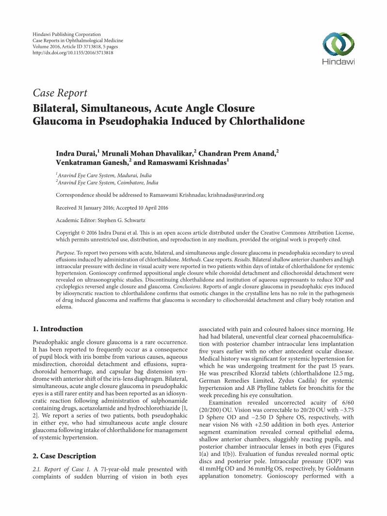
Examination revealed uncorrected acuity of 6/60(20/200)OU. Vision was correctable to 20/20OU with −3.75D Sphere OD and −2.50 D Sphere OS, respectively, withnear vision N6 with +2.50 addition in both eyes. Anteriorsegment examination revealed corneal epithelial edema,shallow anterior chambers, sluggishly reacting pupils, andposterior chamber intraocular lenses in both eyes (Figures1(a) and 1(b)). Evaluation of fundus revealed normal opticdiscs and posterior pole. Intraocular pressure (IOP) was41mmHgOD and 36mmHgOS, respectively, by Goldmannapplanation tonometry. Gonioscopy performed with a
Hindawi Publishing CorporationCase Reports in Ophthalmological MedicineVolume 2016, Article ID 3713818, 5 pageshttp://dx.doi.org/10.1155/2016/3713818
2 Case Reports in Ophthalmological Medicine
(a) (b)
Figure 1: Slit lamp photograph of the left eye of the patient in Case Report 1 with shallow anterior chambers.
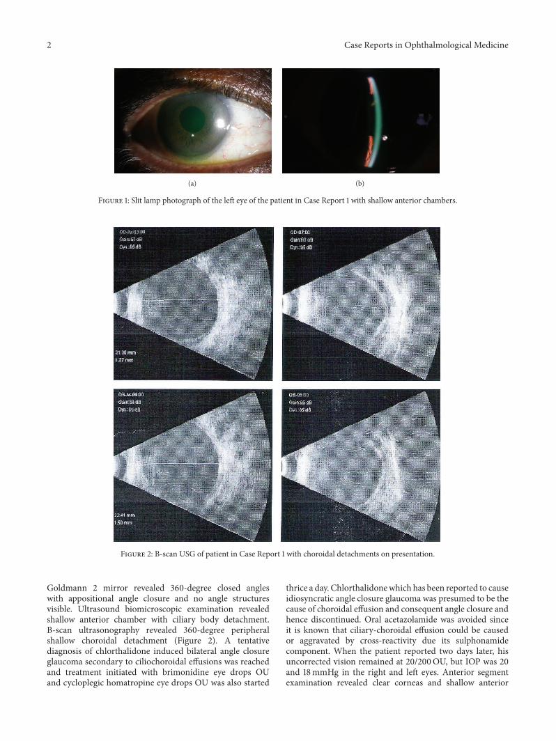
Figure 2: B-scan USG of patient in Case Report 1 with choroidal detachments on presentation.
Goldmann 2 mirror revealed 360-degree closed angleswith appositional angle closure and no angle structuresvisible. Ultrasound biomicroscopic examination revealedshallow anterior chamber with ciliary body detachment.B-scan ultrasonography revealed 360-degree peripheralshallow choroidal detachment (Figure 2). A tentativediagnosis of chlorthalidone induced bilateral angle closureglaucoma secondary to ciliochoroidal effusions was reachedand treatment initiated with brimonidine eye drops OUand cycloplegic homatropine eye drops OU was also started
thrice a day. Chlorthalidonewhich has been reported to causeidiosyncratic angle closure glaucoma was presumed to be thecause of choroidal effusion and consequent angle closure andhence discontinued. Oral acetazolamide was avoided sinceit is known that ciliary-choroidal effusion could be causedor aggravated by cross-reactivity due its sulphonamidecomponent. When the patient reported two days later, hisuncorrected vision remained at 20/200OU, but IOP was 20and 18mmHg in the right and left eyes. Anterior segmentexamination revealed clear corneas and shallow anterior
Case Reports in Ophthalmological Medicine 3
chambers (OD > OS). Fundus examination was normal inboth eyes. Gonioscopy revealed closed, angle configurationOU. UBM and B-scan ultrasonography were characteristicof ciliary body and choroidal detachments, as observedearlier. Treatment with topical aqueous suppressants andcycloplegics was continued. A week later, the patientsuncorrected visual acuity was 20/30OD and 20/20OS.Vision improved to 20/20OU with −1.00 × 90D cylinderOD and −0.50 × 90OS, respectively. Anterior segmentexamination revealed clear corneas and deep anteriorchambers OU. Fundus examination was normal in botheyes. Intraocular pressure (IOP) was 8mmHg OD and11mmHg OS. Gonioscopy revealed wide open angles OU.UBM and B-scan ultrasonography revealed 360-degreeresolving choroidal detachments. The topical medicationswere then discontinued.The patient was advised of periodicalevaluation to exclude any residual glaucoma.
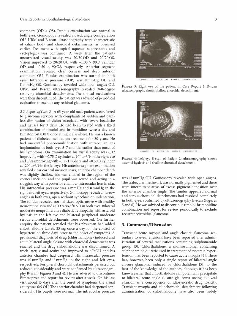
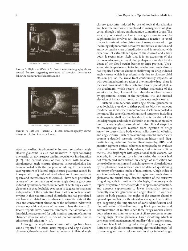
2.2. Report of Case 2. A65-year-oldmale patient was referredto glaucoma services with complaints of sudden and pain-less diminution of vision associated with severe headacheand nausea for 3 days. He had been treated with a fixedcombination of timolol and brimonidine twice a day andBimatoprost 0.01% once at night elsewhere. He was a knownpatient of diabetes mellitus on treatment for 30 years. Hehad uneventful phacoemulsification with intraocular lensimplantation in both eyes 5–7 months earlier than onset ofhis symptoms. On examination his visual acuity was 6/12improving with −0.75D cylinder at 90∘ to 6/9 in the right eyeand 6/24 improvingwith−1.25D sphere and−0.50D cylinderat 120∘ to 6/9 in the left eye. His anterior segment examinationrevealed clear corneal incision scars, anterior chamber depthwas slightly shallow, iris was chaffed in the region of thecorneal incision, and the pupil was round and acting in asluggish way with posterior chamber intraocular lens in situ.His intraocular pressure was 4mmHg and 8mmHg in theright and left eyes, respectively. Gonioscopy revealed narrowangles in both eyes, open without synechiae on indentation.The fundus revealed normal sized optic nerve with healthyneuroretinal rim and aCD ratio of 0.5 : 1 in both eyes. Bilateralmoderate nonproliferative diabetic retinopathy with asteroidhyalosis in the left eye and bilateral peripheral moderateserous choroidal detachments were observed. On furtherenquiry the patient revealed that his physician had addedchlorthalidone tablets 25mg once a day for the control ofhypertension three days prior to the onset of symptoms. Aprovisional diagnosis of drug (chlorthalidone) induced andacute bilateral angle closure with choroidal detachment wasreached and the drug chlorthalidone was discontinued. Aweek later, visual acuity had improved to 6/9OU and hisanterior chamber had deepened. His intraocular pressurewas 10mmHg and 8mmHg in the right and left eyes,respectively. Peripheral choroidal detachments persisted butreduced considerably and were confirmed by ultrasonogra-phy B-scan (Figures 3 and 4). He was advised to discontinueBimatoprost and report for review after a week. On his lastvisit about 15 days after the onset of symptoms the visualacuity was 6/9OU.The anterior chamber had deepened con-siderably. His pupils were normal. The intraocular pressure
Figure 3: Right eye of the patient in Case Report 2: B-scanultrasonography shows shallow choroidal detachment.
Figure 4: Left eye B-scan of Patient 2: ultrasonography showsasteroid hyalosis and shallow choroidal detachment.
was 13mmHg OU. Gonioscopy revealed wide open angles.The trabecular meshwork was normally pigmented and therewere intermittent areas of excess pigment deposition overthe anterior chamber angle. The fundus appeared normaland serous choroidal detachments had resolved completelyin both eyes, confirmed by ultrasonography B-scan (Figures5 and 6). He was advised to discontinue timolol-brimonidinecombination and report for review periodically to excluderecurrence/residual glaucoma.
3. Comments/Discussion
Transient acute myopia and angle closure glaucoma sec-ondary to uveal effusions have been reported after admin-istration of several medications containing sulphonamidegroup [3]. Chlorthalidone, a monsosulfomyl containingsulphonamide diuretic used in treatment of systemic hyper-tension, has been reported to cause acute myopia [4]. Therehas, however, been only a single report of bilateral angleclosure glaucoma induced by chlorthalidone [5], to thebest of the knowledge of the authors, although it has beenknown earlier that chlorthalidone can potentially precipitateto bilateral acute angle closure glaucoma owing to uvealeffusion as a consequence of idiosyncratic drug toxicity.Transient myopia and ciliochoroidal detachment followingadministration of chlorthalidone have also been widely
4 Case Reports in Ophthalmological Medicine
Figure 5: Right eye (Patient 2) B-scan ultrasosonography showsnormal features suggesting resolution of choroidal detachmentfollowing withdrawal of chlorthalidone.
Figure 6: Left eye (Patient 2) B-scan ultrasonography showsresolution of choroidal detachment.
reported earlier. Sulphonamide induced secondary angleclosure glaucoma is also not unknown in eyes followinguneventful cataract surgery and intraocular lens implantation[1, 2]. The current series of two persons with bilateral,simultaneous angle closure glaucoma in pseudophakia hasbeen reported with the purpose of adding to the alreadyvast repertoire of bilateral angle closure glaucoma caused byidiosyncratic drug induced uveal effusions. Accommodativespasm and increase in lens thickness [3] have been postulatedas one of the mechanisms of acute angle closure glaucomainduced by sulphonamides, but reports of acute angle closureglaucoma in pseudophakic eyes seem to suggest mechanismsindependent of the crystalline lens. Earlier reports of acutemyopia in those administered chlorthalidone had postulatedmechanisms related to disturbance in osmotic state of thelens and concomitant alteration of the refractive index withultrasonographic evidence of increase in lens thickness [6].Recent reports have, however, reinforced that alterations inlens thickness accounted for onlyminimal amount of anteriorchamber decrease which is instead, predominantly, due tociliochoroidal effusion [7–10].
Although the sulphonamide group of drugs have beenwidely reported to cause acute myopia and angle closureglaucoma, there have so far been no reports of bilateral angle
closure glaucoma induced by use of topical dorzolamideand brinzolamide widely employed in management of glau-coma, though both are sulphonamide containing drugs. Thewidely hypothesized mechanism of angle closure induced bysulphonamides involves an idiosyncratic reaction in uvealtissues to systemic administration of many classes of drugsincluding sulphonamide derivative antibiotics, diuretics, andantihypertensive class of medications and is associated withexpansion of extracellular space of the choroid and ciliarybody. It seems most likely that it is an expansion of theextravascular compartment, due perhaps to a sudden break-down of the blood-ocular barrier to large proteins. Ultra-sound studies performed in topiramate induced angle closurehad reported anterior chamber shallowing in drug inducedangle closure which is predominantly due to ciliochoroidaleffusion [7]. As the uveal tract continuously expands, aswith continued administration of the causative drug, there isforward movement of the crystalline lens or pseudophakia-iris diaphragm, which results in further shallowing of theanterior chamber, closure of the trabecular outflow pathwayby appositional closure of the peripheral iris, and markedelevation of intraocular pressure from acute angle closure.
Bilateral, simultaneous, acute angle closure glaucoma inpseudophakic eyes due to either pupillary block or aqueousmisdirection is extremely uncommon and seldom reported inliterature.The constellation of signs and symptoms includingacute myopia, shallow chamber due to anterior shift of iris-lens diaphragm, and sudden elevation in intraocular pressuredue to acute angle closure should raise clinical suspicionof idiosyncrasy related reaction from ingestion of drugsknown to cause ciliary body edema, ciliochoroidal effusion,and angle closure. Such clinical findings should immediatelyprompt a detailed systemic medication history and diag-nostic testing including B-scan ultrasonography, UBM, andanterior segment optical coherence tomography to evaluateuveal effusions, ciliary body edema, and anterior shift inthe iris-lens diaphragm with appositional angle closure. Forexample, in the second case in our series, the patient hadnot volunteered information on change of medication forcontrol of hypertension and switching over to chlorthalidoneby his physician was revealed only on detailed questioningon history of systemic intake of medications. A high index ofsuspicion and early recognition of drug induced angle closureglaucoma are crucial since discontinuation of the causativedrug along with institution of cycloplegia, if necessary withtopical or systemic corticosteroids to suppress inflammation,and aqueous suppressants to lower intraocular pressurepromptly reverses glaucoma and angle closure. In our caseseries, in both patients, the angles of the anterior chamberopened up completely without evidence of synechiae in eithereye, suggesting the importance of early identification anddiscontinuation of the offending drug. It is important to avoidadministration of miotics since these drugs worsen ciliarybody edema and anterior rotation of ciliary processes accen-tuating angle closure glaucoma. Laser iridotomy, which iscornerstone of management of pupillary block angle closure,is seldom beneficial in drug induced angle closure glaucoma.Refractory angle closure necessitating choroidal drainage [11]to reverse glaucoma is seldom seen in drug induced angle
Case Reports in Ophthalmological Medicine 5
closure glaucoma with conservative therapy reversing angleclosure and glaucoma in most individuals.
Competing Interests
The authors declare that they have no competing interests.
References
[1] S. Senthil, C. Garudadri, H. B. L. Rao, and R. Mahesh-wari, “Bilateral simultaneous acute angle closure caused bysulphonamide derivatives: a case series,” Indian Journal ofOphthalmology, vol. 58, no. 3, pp. 248–252, 2010.
[2] G. C. Lee, C. P. Tam, H. V. Danesh-Meyer, J. S. Myers,and L. J. Katz, “Bilateral angle closure glaucoma induced bysulphonamide-derived medications,” Clinical & ExperimentalOphthalmology, vol. 35, no. 1, pp. 55–58, 2007.
[3] V. A. Panday and D. J. Rhee, “Review of sulfonamide-inducedacute myopia and acute bilateral angle closure glaucoma,”ComprehensiveOphthalmologyUpdate, vol. 8, no. 5, pp. 271–276,2007.
[4] G. Mahesh, A. Giridhar, S. J. Saikumar, and S. Fegde, “Drug-induced acute myopia following chlorthalidone treatment,”Indian Journal of Ophthalmology, vol. 55, no. 5, pp. 386–388,2007.
[5] J. R. Singer, Z.D. Pearce, S. J.Westhouse, andK. J. Siebert, “Uvealeffusion as a mechanism of bilateral angle-closure glaucomainduced by chlorthalidone,” Journal of Glaucoma, vol. 24, no.1, pp. 84–86, 2015.
[6] O. Pallin and R. Ericsson, “Ultrasound studies in a case ofhygroton-induced myopia,” Acta Ophthalmologica, vol. 43, no.5, pp. 692–696, 1965.
[7] J. E. Craig, T. J. Ong, D. L. Louis, and J. M. Wells, “Mechanismof topiramate-induced acute-onset myopia and angle closureglaucoma,” American Journal of Ophthalmology, vol. 137, no. 1,pp. 193–195, 2004.
[8] H. A. Sen, H. S. O’Halloran, W. Lee, and L. Ky, “Topiramate-inducedacute myopia and retinal striae,” Archives of Ophthal-mology, vol. 119, no. 5, pp. 775–777, 2001.
[9] J. T. Banta, K. Hoffman, D. L. Budenz, E. Ceballos, and D.S. Greenfield, “Presumed topiramate-induced bilateral acuteangle-closure glaucoma,” American Journal of Ophthalmology,vol. 132, no. 1, pp. 112–114, 2001.
[10] F. A. Medeiros, X. Y. Zhang, A. S. Bernd, and R. N. Weinreb,“Angle-closure glaucoma associated with ciliary body detach-ment in patients using topiramate,” Archives of Ophthalmology,vol. 121, no. 2, pp. 282–285, 2003.
[11] R. Parikh, S. Parikh, S.Das, andR.Thomas, “Choroidal drainagein the management of acute angle closure after Topiramatetoxicity,” Journal of Glaucoma, vol. 16, no. 8, pp. 691–693, 2007.

Submit your manuscripts athttp://www.hindawi.com
Stem CellsInternational
Hindawi Publishing Corporationhttp://www.hindawi.com Volume 2014
Hindawi Publishing Corporationhttp://www.hindawi.com Volume 2014
MEDIATORSINFLAMMATION
of
Hindawi Publishing Corporationhttp://www.hindawi.com Volume 2014
Behavioural Neurology
EndocrinologyInternational Journal of
Hindawi Publishing Corporationhttp://www.hindawi.com Volume 2014
Hindawi Publishing Corporationhttp://www.hindawi.com Volume 2014
Disease Markers
Hindawi Publishing Corporationhttp://www.hindawi.com Volume 2014
BioMed Research International
OncologyJournal of
Hindawi Publishing Corporationhttp://www.hindawi.com Volume 2014
Hindawi Publishing Corporationhttp://www.hindawi.com Volume 2014
Oxidative Medicine and Cellular Longevity
Hindawi Publishing Corporationhttp://www.hindawi.com Volume 2014
PPAR Research
The Scientific World JournalHindawi Publishing Corporation http://www.hindawi.com Volume 2014
Immunology ResearchHindawi Publishing Corporationhttp://www.hindawi.com Volume 2014
Journal of
ObesityJournal of
Hindawi Publishing Corporationhttp://www.hindawi.com Volume 2014
Hindawi Publishing Corporationhttp://www.hindawi.com Volume 2014
Computational and Mathematical Methods in Medicine
OphthalmologyJournal of
Hindawi Publishing Corporationhttp://www.hindawi.com Volume 2014
Diabetes ResearchJournal of
Hindawi Publishing Corporationhttp://www.hindawi.com Volume 2014
Hindawi Publishing Corporationhttp://www.hindawi.com Volume 2014
Research and TreatmentAIDS
Hindawi Publishing Corporationhttp://www.hindawi.com Volume 2014
Gastroenterology Research and Practice
Hindawi Publishing Corporationhttp://www.hindawi.com Volume 2014
Parkinson’s Disease
Evidence-Based Complementary and Alternative Medicine
Volume 2014Hindawi Publishing Corporationhttp://www.hindawi.com
Related Documents


