PEER REVIEWED Case Report - An Investigation of Dairy Cow Teat Lesions and Clinical Mastitis John H. Kirk, DVM, MPVM1; William M. Sischo, DVM, PhD2 Veterinary Medicine Extension departm ent of Population Health and Reproduction, School of Veterinary Medicine, University of California Davis, Veterinary Medicine Teaching and Research Center, Tulare, CA 93274 Abstract A California dairyman milking 1,969 cows reported excessive cases of clinical mastitis, and questioned whether teat-end lesions were associated with the prob - lem. Using a system of teat-end lesion classification, the entire milking herd was observed for lesions during January 2002. Overall, 9.9% of the herd had some type of lesion, including warts and severe hyperkeratosis; 67% of lesions were severe teat-end hyperkeratosis. Cows with teat lesions were three times more at risk of clinical mastitis compared to cows without teat lesions. Because of physical limitations of the milking equip- ment and the owner’s reluctance to modify the milking routine, no management changes were made. Resume Un producteur de lait Califomien avec un troupeau de 1969 vaches a rapporte une frequence excessive de mammite clinique et se demandait si les lesions a l’extremite des trayons etaient associees au probleme. Avec l’aide d’un systeme de classification des lesions a l’extremite des trayons, tout le troupeau fut observe pour des lesions en janvier 2002. En tout, 9.9% des vaches du troupeau montraient des lesions de quelque type que ce soit incluant des verrues et des cas severes d’hyperkeratose. Pres de 67% des lesions impliquaient l’hyperkeratose severe a l’extremite des trayons. Les vaches avec des lesions aux trayons avaient trois fois plus de chance que les vaches sans lesions d’avoir de la mammite clinique. En raison des restrictions physiques reliees a l’equipement laitier et du manque d’enthousiasme du proprietaire a changer sa routine de traite, aucun changement au niveau de la gestion n’a ete fait. Introduction Pathogens and non-infectious mechanisms are re - ported to cause injury and lesions on the teat ends of dairy cattle. These are often classified into milking machine effects, environmental effects and infectious agents. The resulting injuries and lesions may affect individual cows or the entire herd, and in some cases may be associated with increased cases of mastitis. Pap- illomatosis, or warts, are common in cattle and may cause problems with milking and mastitis. Teat-end hy - perkeratosis, when severe, is also associated with in- creased prevelance of mastitis. This report describes a field investigation of teat- end lesions in dairy cows, both warts and hyperkerato- sis, and their association with increased clinical mastitis. History The owner of a large (1969 milking cows) Califor- nia dairy reported a continuing problem with warts on cows’teats. Moreover, he suspected that cows with warts had a higher rate of clinical mastitis than cows without warts. He asked for advice on how to reduce the wart and clinical mastitis problems. During the previous 5 months, cows milked in the north side of the milking parlor (84% lactation >1) averaged 79 (+/-31) cases of clinical mastitis per month, while cows milked in the south side (71% in first lactation) averaged 40 (+/-14) cases per month. The average daily milk production was 80 (+/-0.8) and 70 (+/-1.7) lb (36.4 and 31.8 kg) for cows milked in the north and south sides, respectively. Dur- ing this time period, the bulk tank somatic cell count (SCC) ranged from 350,000 to 400,000 cells/ml. Forty percent of cows were in their first lactation. They were housed in a typical western, dry lot dairy. 30 THE BOVINE PRACTITIONER— VOL. 37, NO. 1 © Copyright American Association of Bovine Practitioners; open access distribution.

Welcome message from author
This document is posted to help you gain knowledge. Please leave a comment to let me know what you think about it! Share it to your friends and learn new things together.
Transcript

PEER REVIEW ED
Case Report - An Investigation of Dairy Cow Teat Lesions and Clinical MastitisJohn H. Kirk, D V M , M P V M 1; William M. Sischo, D V M , P h D 2 V e te r in a r y M ed ic in e E x ten sio nd e p a r t m e n t o f P o p u la tio n H e a lth a n d R ep ro d u c tio n , S ch oo l o f V eterin ary M ed ic in e , U n iv e rs ity o f C a lifo rn ia D a v is , V eterin ary M ed ic in e T each in g a n d R esea rch Center, T u lare, C A 9 3 2 7 4
Abstract
A California dairyman milking 1,969 cows reported excessive cases of clinical m astitis, and questioned whether teat-end lesions were associated with the problem. Using a system of teat-end lesion classification, the entire milking herd was observed for lesions during January 2002. Overall, 9.9% of the herd had some type of lesion, including warts and severe hyperkeratosis; 67% of lesions were severe teat-end hyperkeratosis. Cows with teat lesions were three times more at risk of clinical mastitis compared to cows without teat lesions. Because of physical limitations of the milking equipment and the owner’s reluctance to modify the milking routine, no management changes were made.
Resume
Un producteur de lait Califomien avec un troupeau de 1969 vaches a rapporte une frequence excessive de mammite clinique et se dem andait si les lesions a l’extremite des trayons etaient associees au probleme. Avec l’aide d’un systeme de classification des lesions a l’extremite des trayons, tout le troupeau fut observe pour des lesions en janvier 2002. En tout, 9.9% des vaches du troupeau montraient des lesions de quelque type que ce soit in c lu an t des v erru es et des cas severes d’hyperkeratose. Pres de 67% des lesions impliquaient l’hyperkeratose severe a l’extremite des trayons. Les vaches avec des lesions aux trayons avaient trois fois plus de chance que les vaches sans lesions d’avoir de la mammite clinique. En raison des restrictions physiques re liees a l ’equ ipem ent la i t ie r et du m anque d’enthousiasme du proprietaire a changer sa routine de traite, aucun changement au niveau de la gestion n’a ete fait.
Introduction
Pathogens and non-infectious mechanisms are reported to cause injury and lesions on the teat ends of dairy cattle. These are often classified into milking machine effects, environmental effects and infectious agents. The resulting injuries and lesions may affect individual cows or the entire herd, and in some cases may be associated with increased cases of mastitis. Papillomatosis, or warts, are common in cattle and may cause problems with milking and mastitis. Teat-end hyperkeratosis, when severe, is also associated with increased prevelance of mastitis.
This report describes a field investigation of teat- end lesions in dairy cows, both warts and hyperkeratosis, and their association with increased clinical mastitis.
History
The owner of a large (1969 milking cows) California dairy reported a continuing problem with warts on cows’teats. Moreover, he suspected that cows with warts had a higher rate of clinical mastitis than cows without warts. He asked for advice on how to reduce the wart and clinical mastitis problems. During the previous 5 months, cows milked in the north side of the milking parlor (84% lactation >1) averaged 79 (+/-31) cases of clinical mastitis per month, while cows milked in the south side (71% in first lactation) averaged 40 (+/-14) cases per month. The average daily milk production was 80 (+/-0.8) and 70 (+/-1.7) lb (36.4 and 31.8 kg) for cows milked in the north and south sides, respectively. During this time period, the bulk tank somatic cell count (SCC) ranged from 350,000 to 400,000 cells/ml.
Forty percent of cows were in their first lactation. They were housed in a typical western, dry lot dairy.
30 THE BOVINE PRACTITIONER— VOL. 37, NO. 1
© Copyright American Association of Bovine Practitioners; open access distribution.
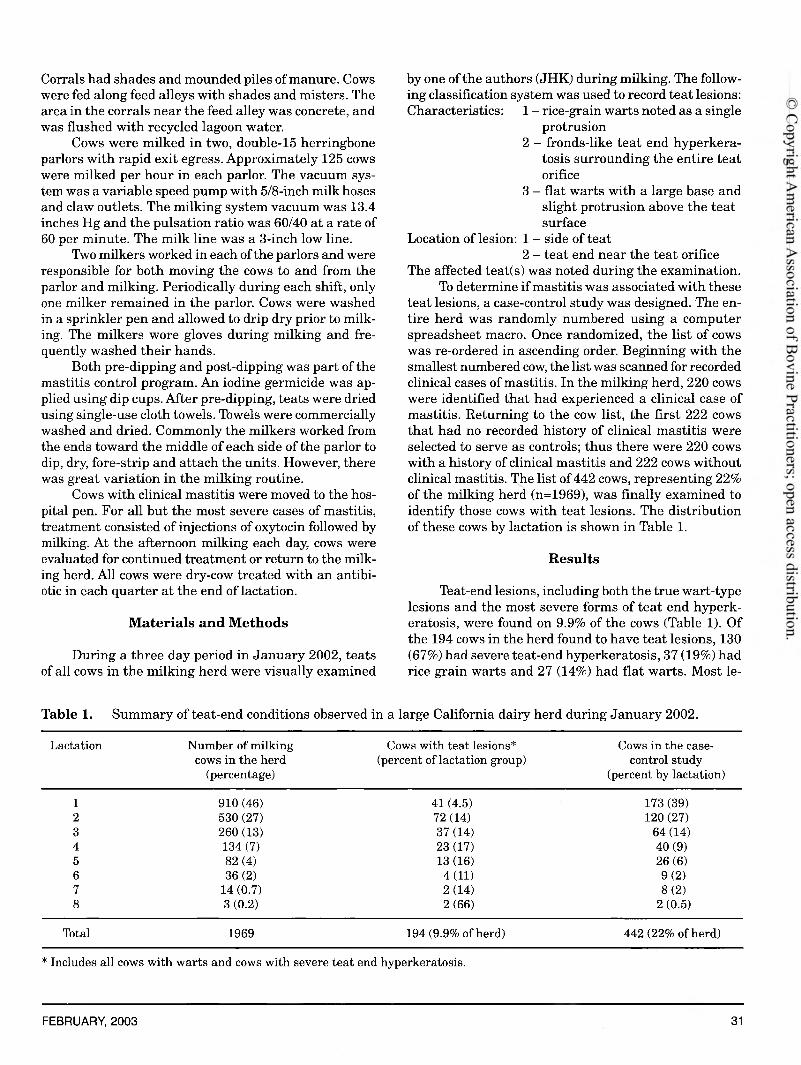
Corrals had shades and mounded piles of manure. Cows were fed along feed alleys with shades and misters. The area in the corrals near the feed alley was concrete, and was flushed with recycled lagoon water.
Cows were milked in two, double-15 herringbone parlors with rapid exit egress. Approximately 125 cows were milked per hour in each parlor. The vacuum system was a variable speed pump with 5/8-inch milk hoses and claw outlets. The milking system vacuum was 13.4 inches Hg and the pulsation ratio was 60/40 at a rate of 60 per minute. The milk line was a 3-inch low line.
Two milkers worked in each of the parlors and were responsible for both moving the cows to and from the parlor and milking. Periodically during each shift, only one milker remained in the parlor. Cows were washed in a sprinkler pen and allowed to drip dry prior to milking. The milkers wore gloves during milking and frequently washed their hands.
Both pre-dipping and post-dipping was part of the mastitis control program. An iodine germicide was applied using dip cups. After pre-dipping, teats were dried using single-use cloth towels. Towels were commercially washed and dried. Commonly the milkers worked from the ends toward the middle of each side of the parlor to dip, dry, fore-strip and attach the units. However, there was great variation in the milking routine.
Cows with clinical mastitis were moved to the hospital pen. For all but the most severe cases of mastitis, treatment consisted of injections of oxytocin followed by milking. At the afternoon milking each day, cows were evaluated for continued treatment or return to the milking herd. All cows were dry-cow treated with an antibiotic in each quarter at the end of lactation.
Materials and Methods
During a three day period in January 2002, teats of all cows in the milking herd were visually examined
by one of the authors (JHK) during milking. The following classification system was used to record teat lesions: Characteristics: 1 - rice-grain warts noted as a single
protrusion2 - fronds-like teat end hyperkera
tosis surrounding the entire teat orifice
3 - flat warts with a large base andslight protrusion above the teat surface
Location of lesion: 1 - side of teat2 - teat end near the teat orifice
The affected teat(s) was noted during the examination.To determine if mastitis was associated with these
teat lesions, a case-control study was designed. The entire herd was randomly numbered using a computer spreadsheet macro. Once randomized, the list of cows was re-ordered in ascending order. Beginning with the smallest numbered cow, the list was scanned for recorded clinical cases of mastitis. In the milking herd, 220 cows were identified that had experienced a clinical case of mastitis. Returning to the cow list, the first 222 cows that had no recorded history of clinical mastitis were selected to serve as controls; thus there were 220 cows with a history of clinical mastitis and 222 cows without clinical mastitis. The list of442 cows, representing 22% of the milking herd (n=1969), was finally examined to identify those cows with teat lesions. The distribution of these cows by lactation is shown in Table 1.
Results
Teat-end lesions, including both the true wart-type lesions and the most severe forms of teat end hyperkeratosis, were found on 9.9% of the cows (Table 1). Of the 194 cows in the herd found to have teat lesions, 130 (67%) had severe teat-end hyperkeratosis, 37 (19%) had rice grain warts and 27 (14%) had flat warts. Most le-
Table 1. Summary of teat-end conditions observed in a large California dairy herd during January 2002.Lactation Number of milking
cows in the herd (percentage)
Cows with teat lesions* (percent of lactation group)
Cows in the case- control study
(percent by lactation)1 910 (46) 41 (4.5) 173(39)2 530 (27) 72 (14) 120 (27)3 260 (13) 37 (14) 64 (14)4 134 (7) 23 (17) 40 (9)5 82 (4) 13 (16) 26 (6)6 36 (2) 4(11) 9(2)7 14 (0.7) 2(14) 8(2)8 3 (0.2) 2(66) 2 (0.5)
Total 1969 194 (9.9% of herd) 442 (22% of herd)* Includes all cows with warts and cows with severe teat end hyperkeratosis.
FEBRUARY, 2003 31
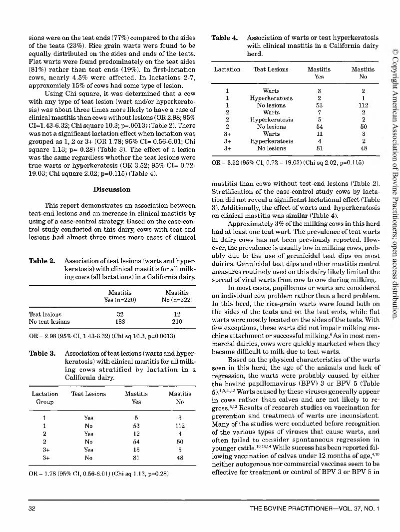
sions were on the teat ends (77%) compared to the sides of the teats (23%). Rice grain warts were found to be equally distributed on the sides and ends of the teats. Flat warts were found predominately on the teat sides (81%) rather than teat ends (19%). In first-lactation cows, nearly 4.5% were affected. In lactations 2-7, approxomiely 15% of cows had some type of lesion.
Using Chi square, it was determined that a cow with any type of teat lesion (wart and/or hyperkeratosis) was about three times more likely to have a case of clinical mastitis than cows without lesions (OR 2.98; 95% CI=1.43-6.32; Chi square 10.3; p=.0013) (Table 2). There was not a significant lactation effect when lactation was grouped as 1, 2 or 3+ (OR 1.78; 95% CI= 0.56-6.01; Chi square 1.13; p= 0.28) (Table 3). The effect of a lesion was the same regardless whether the teat lesions were true warts or hyperkeratosis (OR 3.52; 95% CI= 0.72- 19.03; Chi square 2.02; p=0.115) (Table 4).
D iscussionThis report demonstrates an association between
teat-end lesions and an increase in clinical mastitis by using of a case-control strategy. Based on the case-control study conducted on this dairy, cows with teat-end lesions had almost three times more cases of clinical
Table 2. Association of teat lesions (warts and hyperkeratosis) with clinical mastitis for all milking cows (all lactations) in a California dairy.
Mastitis MastitisYes(n=220) No (n=222)
Teat lesions 32 12No teat lesions 188 210OR - 2.98 (95% Cl, 1.43-6.32) (Chi sq 10.3, p=0.0013)
Table 3. Association of teat lesions (warts and hyperkeratosis) with clinical mastitis for all milking cows s tra tif ie d by lac ta tio n in a California dairy.
LactationGroup
Teat Lesions MastitisYes
MastitisNo
1 Yes 5 31 No 53 1122 Yes 12 42 No 54 503+ Yes 15 53+ No 81 48
OR - 1.78 (95% Cl, 0.56-6.01) (Chi sq 1.13, p=0.28)
Table 4. Association of warts or teat hyperkeratosis with clinical mastitis in a California dairy herd.
Lactation Teat Lesions MastitisYes
MastitisNo
1 Warts 3 21 Hyperkeratosis 2 11 No lesions 53 1122 Warts 7 22 Hyperkeratosis 5 22 No lesions 54 50
3+ Warts 11 33+ Hyperkeratosis 4 23+ No lesions 81 48
OR - 3.52 (95% Cl, 0.72 - 19.03) (Chi sq 2.02, p=0.115)
mastitis than cows without test-end lesions (Table 2). Stratification of the case-control study cows by lactation did not reveal a significant lactational effect (Table 3). Additionally, the effect of warts and hyperkeratosis on clinical mastitis was similar (Table 4).
Approximately 3% of the milking cows in this herd had at least one teat wart. The prevalence of teat warts in dairy cows has not been previously reported. However, the prevalence is usually low in milking cows, probably due to the use of germicidal tea t dips on most dairies. Germicidal teat dips and other mastitis control measures routinely used on this dairy likely limited the spread of viral warts from cow to cow during milking.
In most cases, papillomas or warts are considered an individual cow problem rather than a herd problem. In this herd, the rice-grain warts were found both on the sides of the teats and on the teat ends, while flat warts were mostly located on the sides of the teats. With few exceptions, these warts did not impair milking machine attachment or successful milking.6 As in most commercial dairies, cows were quickly marketed when they became difficult to milk due to teat warts.
Based on the physical characteristics of the warts seen in this herd, the age of the animals and lack of regression, the warts were probably caused by either the bovine papillomavirus (BPV) 3 or BPV 5 (Table 5 ) 1 ,3 ,1 1 ,1 3 Warts caused by these viruses generally appear in cows rather than calves and are not likely to regress.5,13 Results of research studies on vaccination for prevention and treatm ent of warts are inconsistent. Many of the studies were conducted before recognition of the various types of viruses that cause warts, and often failed to consider spontaneous regression in younger cattle.10,13,14 While success has been reported following vaccination of calves under 12 months of age,4,10 neither autogenous nor commercial vaccines seem to be effective for treatm ent or control of BPV 3 or BPV 5 in
32 THE BOVINE PRACTITIONER— VOL. 37, NO. 1

Table 5. Description of bovine papillomavirus (BPV) warts commonly found on cattle.1’3’5111314Type Description Location Age RegressionBPV 1 Typical wart, filamentous,
frond likeTeats, penis Less than 2 years Spontaneous 1-12 months
BPV 2 Multiple, gray, firm, small, often raised on a stalk
Head, neck, dewlap Less than 2 years Spontaneous 1-12 monthsBPV 3 Low, flat, circular, no stalk,
frond like projectionsNon-specific All ages Permanent
BPV 4 Papillomas Gastrointestinal tract, bladderBPV 5 Rice-grain shape, long,
smooth, whiteTeats All ages Permanent
adult cattle.213-14 For either autogenous or commercial vaccines to be successful, they should contain the specific virus that is causing warts in the herd as there is little cross-protection between virus types.10,13
Due to the nature of the warts (BPV 3 or BPV 5) and the mastitis control practices already in place on this dairy, no opportunity was seen to prevent warts on the teats. For individual cases, surgical intervention for the most severe cases was possible. In this herd, however, cows were sent to market rather than electing surgery.
The majority of teat-end lesions in this herd were hyperkeratosis surrounding the teat orifice. Nearly 7% of the herd was affected with severe hyperkeratosis, which was below the reported action level where more than 10% of the cows have very rough (VR) lesions.7 These non- viral lesions found on teats have often been described as rings or fronds with modifiers like smooth, rough or flowered.2’79 Some of the lesions have been reported to be milking machine-related.6 9 The more severe the lesion, the greater the association with mastitis.2’7’9
Several scoring systems have been used to classify bovine teat lesions. An adaptation of scoring systems of
ten used for routine field evaluations is shown in Table 6. Note that in two cases the authors have stipulated the condition or level of conditions that might be associated with an increased risk of mastitis. Other more definitive systems have been proposed for research studies.8
Many risk factors have been associated with the appearance of rough teat ends with fronds or flowering. All of these risk factors were considered in an effort to reduce the problem on this dairy. These risk factors can be grouped as cow, milking machine or milking technique factors.6 Cow factors are related to teat shape, length, teat position; milking speed; production level; stage of lactation; parity; and genetics.7 9 Slower milking cows with long, pointed teats and higher production levels7,9 are at greater risk for developing teat lesions. Obviously these cow factors can only be described and not altered to influence the prevalence of teat end lesions. Environment has also been reported to play a role in teat lesions,6 however, in this dairy the lesions were not this type (burns, chaps, bites).
Milking machine factors, on the other hand, can be manipulated in hope of reducing the number of se-
Table 6. Bovine teat-end condition classification systems currently in use for field evaluations.2,7,9Teat condition Blowery and Neijenhuis, Mein, Mein, Neijenhuis,
Edmondson3,2 Britt et a l b-9 Morgan et a l c’7Perfect 0 N - none N - no ringOrifice appears open,
not circular1 A - slight
Moderate hyperkeratosis, few rough fronds
2 B - moderate S - smooth/ slightly rough ring
Very rough, keratin protruding around sphincter
3 C - thick R - rough with isolated fronds
Advanced protrusion; sphincter appears to be turned inside out
4 D - extreme VR - very rough fronds, “flowered”
aOnly lesions with scores 3 or 4 are likely to contribute to increased mastitis. bAlso add a category for smooth or rough."Mastitis problems more likely when 20% of cows have R or VR, or more than 10% of cows have VR lesions.
FEBRUARY, 2003 33

vere lesions associated with mastitis.2,9 Machine factors associated with teat-end lesions are total time per day milking at less than 2.2 lb (1 kg) milk/minute, whether at the beginning or end of milking, the threshold for automatic takeoff removal, liner type and liner use. Increased prevalence of teat-end lesions are associated with slow or over-milking, liners with stiff mouth pieces or liners mounted under high tension, and liners that are used too long.2,7,9,12 Milking technique factors that influence the number of teat-end lesions are pre-milking preparation and machine-on time.7,9 There is an obvious overlap and combination effects of these factors.
Based on previous experiences in this herd and observations during teat-end evaluations, both milking machine and milking technique factors could have contributed to the severe teat-end hyperkeratosis in this herd. The milking machine factors were total machine- on time and time at low milk flow with high vacuum. The lack of consistency in the pre-milking routine was the primary concern with milking technique.
Unfortunately, the veterinarian providing milking machine maintenance indicated it was not possible to adjust the takeoffs to come off sooner or at a low rate of milk flow. During the past few years several adjustments had been made to reduce machine-on time. The veterinary consultant agreed that the pre-milking preparation routine was not consistent and could be improved. More consistent preparation could lead to better letdown, quicker milk out and less machine- on time at low milk flow and high vacuum. This subject had been broached with the dairyman on several occasions by the consulting veterinarian, but the dairyman was unwilling to change the habits of his longtime milkers. As often happens, the case history ends without resolution of the problem.
Conclusions
The association between excessive cases of clinical mastitis and teat-end lesions was investigated. Using a system of teat-end lesion classification, the entire milking herd was observed for lesions. Overall, 9.9% of the herd had some type of wart or severe hyperkeratosis lesion. About 67% of the lesions were severe teat- end hyperkeratosis. Cows with teat-end lesions were more likely to have clinical mastitis than cows without lesions. Because of physical limitations of the milking
equipment and unwillingness of the owner to modify the milking routine, no changes were made at the time of the investigation to reduce teat-end lesions. Later contact with the owner revealed his awareness of the problem. New circuit boards that could reduce the time for detachment at the end of milking had been purchased. However, six months after the study, the new equipment had not been installed and the number of clinical mastitis cases ranged from 60 to 90 per month.
References
1. Bloch N, Sutton RH, Spradbrow PB: Bovine cutaneous Papillomas associated with bovine papillomavirus type 5 .Arch Viro 138: 373- 377, 1994.2. Blowey R, Edmundson P: Teat Conditions in M astitis Control In Dairy Herds: An Illustrated and Practical Guide. Ipswich, UK, Farming Press, 1995, pp 176-179.3. Campos MS: Bovine Papillomavirus and cancer. Vet Journal 154:175-188, 1997.4. Chandrachud LM, Grindlay GJ, McGarvie GM, et al\ Vaccination of cattle with the N-Terminus of L2 is necessary and sufficient for preventing infections by Bovine Papillomavirus-4. Virology 211: 2204- 2208, 1995.5. Desrochres A, St-Jean G, Kennedy GA: Congential cutaneous papillomatosis in a one-year-old Holstein. Can Vet J 35: 646-647, 1994.6. Hillerton JE, Morgan WF, Farnsworth R, et al\ Evaluation of bovine teat conditions in commercial dairy herds: 2. Infectious factors and infections. Proc 2nd International Symposium on M astitis and Milk Quality, Vancouver, BC, Canada, 2001, pp 352-356.7. Mein GA, Neijenhuis G, Morgan WF, et ah. Evaluation of bovine teat condition in commercial dairy herds: 1. Non-infectious factors. Proc 2nd International Sym posium on M astitis and Milk Quality. Vancouver, BC, Canada, 2001, pp 347-351.8. Neijenhuis F: Teat end callosity classification system. Proc 4th International Dairy Housing Conference, American Society of Ag Engineers, St. Louis, MO, 1998, pp 117-123.9. Neijenhuis G, Mein GA, Britt JS, et ah. Evaluation of bovine teat condition in commercial dairy herds: 4. Relationship between teat- end callosity or hyperkeratosis and mastitis. Proc 2nd International Symposium on M astitis and Milk Quality, Vancouver, BC, Canada, 2001, pp 362-366.10. N ich o lls PK, S ta n ley MA: The im m unology of anim al papillomaviruses. Vet Immun and Immunopath 73: 101-127, 2000.11. Onions D: Papillomaviruses: Progress for human and veterinary medicine. Vet Journal 154: 171-172, 1997.12. Reinemann DJ, Davis MA, Costa D, et ah. Effects of milking vacuum on milking performance and teat condition. Proc 2nd International Sym posium on M astitis and Milk Quality, Vancouver, BC, Canada, 2001, pp 367-371.13. Scott DW: Papillomatosis, in Scott DW (ed): Large Anim al Dermatology, ed 1. Philadelphia, PA, WB Saunders Co, 1988, pp 420-428.14. Smith BP: Viral diseases. Papillomatosis, in Smith BP (ed): Large Anim al Internal Medicine, ed 2. St. Louis MO, Mosby, 1996, pp 1417- 1418.
34 THE BOVINE PRACTITIONER— VOL 37, NO. 1

Call BIOCOR Animal Health at 1-800-HH1-7H80 or contact your BIOCOR Animal Health distributor to learn m ore a b o u t S u rro u n d in a c t iv a te d c a ttle
vaccines and Herd-Vac™ modified live virus cattle vaccines. Be sure to ask for our latest technical bulletin on th e p re v a le n c e a n d c o n tro l o f BVD.
©2002 Biocor Anim al Health, A CSL CompanySurround™ is a trademark of Biocor Animal Health
w w w .b io c o r a h . c o m
Only Surround™ offers you the expanded protection your program needs in the fight against diseases caused by BVD la and BVD lb. And we do it with antigens from proven strains of BVD la and lb sub-genotypes.The prevalence of BVD la and BVD lb in North America accounts for approximately two-thirds of the tested samples containing BVD virus. (See sidebar.)Safe, proven and highly immunogenic — Surround " inactivated cattle vaccines are ideal for beef and dairy cattle in all stages of production.Keep your herd at work with dual protection against BVD.Surround™ cattle vaccines — your best protection against BVD la and lb.Surround: The only vaccine with BVDlb.
New data on prevalence
of B V D l a and l bIt’s been evident for years that BVD presents itself in two genotypes - BVD1 and BVD2. But more recent studies show that the BVD1 genotype is actually comprised of two genetically distinct sub-genotypes - BVDla and BVDlb (1) (2).
These and other findings are causing many to rethink their vaccination strategies against BVD as a more complete picture is emerging:
♦ In North America, the prevalence of BVDla and BVDlb sub-genotypes accounts for approximately two-thirds of the samples containing the BVD virus.
♦ Inactivated BVD vaccines are specific to genotype and sub-genotype. And their cross protective properties are weak or non-existent (2).
For the latest research and information on the prevalence and control of the BVD virus in North America, see us online at www.biocorah.com.
(1) Fulton RW, SalikiJT, et al. 2000. Bovine viral diarrhea
virus cytopathic and noncylopalhic biotypes and
type 1 and 2 genotypes in diagnostic laboratories
accessions: clinical and necropsy samples from cattle.
J.Vet Diag. Invest. 12:23-38
(2) Bolin SR and RidpathJF, 1998. Prevalence of bovine
viral diarrhea virus genotypes and antibodies against
those viral genotypes in fetal bovine serum.
J. Vet Diag. invest. 10:135-139
BIOCORAnimal HealthA CSL Company
Related Documents