Case Report Submitted by: Jordan Emerson Faculty reviewer: Dr. Sandra Oldham, M.D. Date accepted: 29 August 2007 Radiological Category: Principal Modality (1): Principal Modality (2): Neuroradiology None CT

Case Report
Dec 30, 2015
Radiological Category:. Neuroradiology. Principal Modality (1): Principal Modality (2):. CT. None. Case Report. Submitted by:. Jordan Emerson. Faculty reviewer:. Dr. Sandra Oldham, M.D. Date accepted:. 29 August 2007. Case History. - PowerPoint PPT Presentation
Welcome message from author
This document is posted to help you gain knowledge. Please leave a comment to let me know what you think about it! Share it to your friends and learn new things together.
Transcript
Case Report
Submitted by: Jordan Emerson
Faculty reviewer: Dr. Sandra Oldham, M.D.
Date accepted: 29 August 2007
Radiological Category: Principal Modality (1):
Principal Modality (2):
Neuroradiology
None
CT
Case History
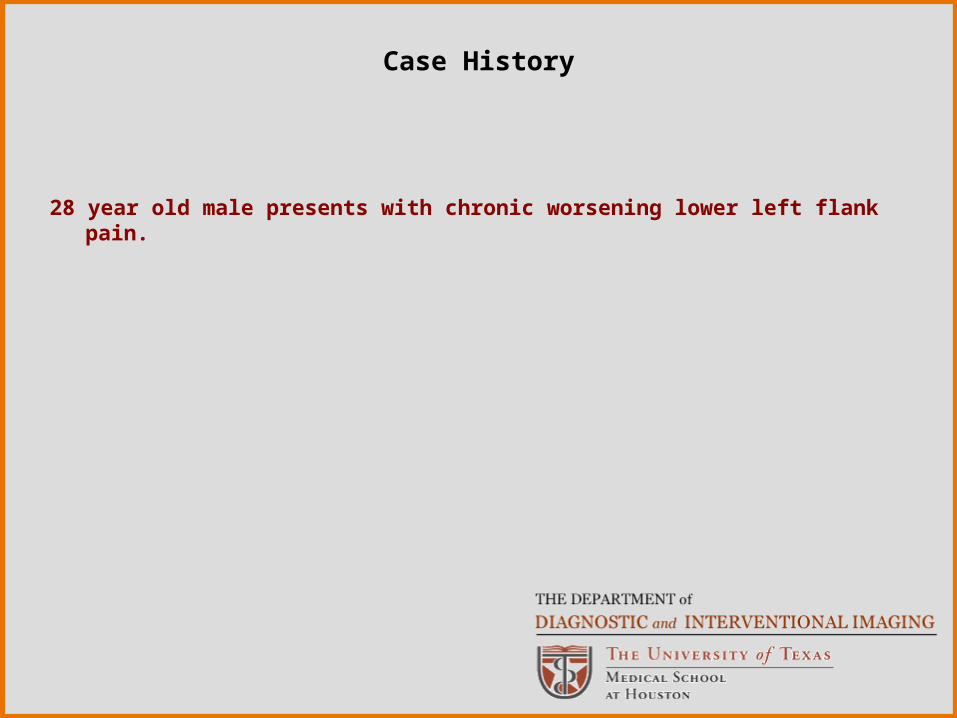
28 year old male presents with chronic worsening lower left flank pain.

Radiological Presentations
Radiological Presentations

Radiological Presentations

Radiological Presentations
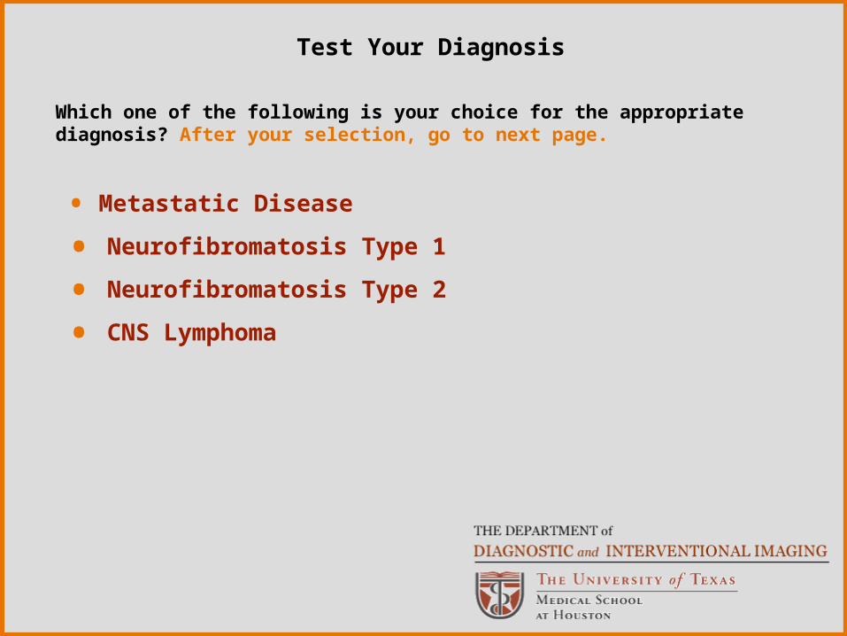
• Metastatic Disease
• Neurofibromatosis Type 1
• Neurofibromatosis Type 2
• CNS Lymphoma
Which one of the following is your choice for the appropriate diagnosis? After your selection, go to next page.
Test Your Diagnosis

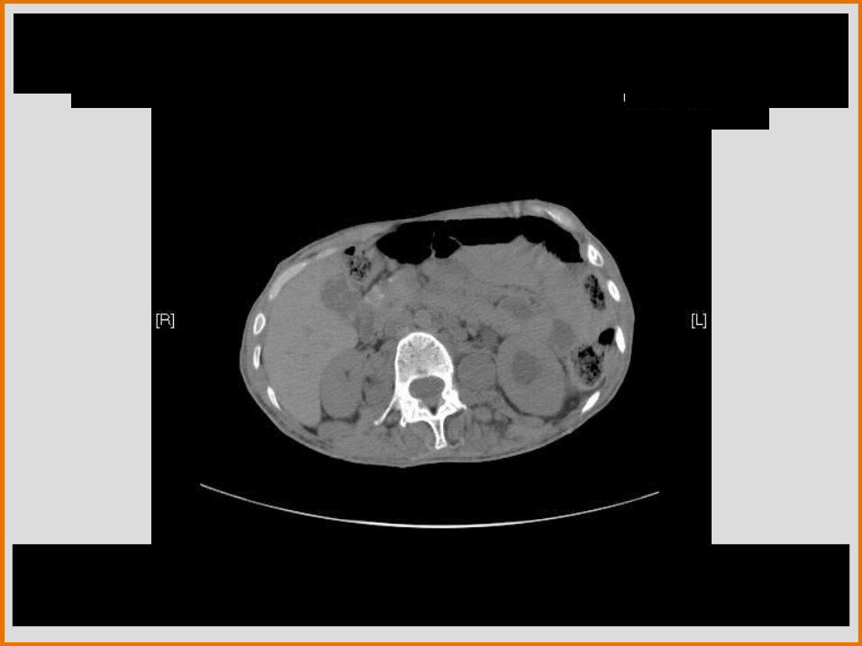
There are numerous smooth, well-defined soft tissue densities without enhancement or calcifications located cutaneously, subcutaneously, and around the spinal column. Also noted is a mass protruding from the neural foramen causing widening in the lumbar region which is consistent with a plexiform neurofibroma. A very large heterogeneous mass of soft tissue density is noted in the left gluteal region. There is marked hydronephrosis on the left side with an increase in the diameter of the ureter. There are no stones within the kidneys or ureter on non-contrast CT.
•Neurofibromatosis type 1
•Neurofibromatosis type 2
• Metastatic disease
Differentials:
Findings and Differentials

Metastatic disease must be considered in a patient with multiple soft tissue masses. Without further work-up for a primary malignancy, metastasis cannot be completely ruled out. Evidence of peripheral nerve involvement (cutaneous and subcutaneous) does go against metastatic disease.
Neurofibromatosis type 2 is known as central neurofibromatosis and occurs at a rate of 1/50K. The gene is located on chromosome 22 and is inherited in an autosomal dominant manner. The common features of NF-2 include bilateral acoustic neuromas, multiple meningiomas, and multiple schwannomas. Skin lesions occur in 30% of these patients. Diagnostic criteria include:
*Bilateral eighth nerve tumors (detected by MRI)
*Family member with NF-2 or a unilateral eighth nerve lesion and 2 of the following
*Juvenile subcapsular lens opacities, glioma, meningioma, or schwannoma
Discussion
With cutaneous, subcutaneous, and plexiform neurofibromas seen on the CT scan, Neurofibromatosis type 1 (von Recklinghausen’s) becomes the most likely diagnosis.
This disease occurs at a prevalence of 1/3000. The gene is located on chromosome 17 and is inherited in an autosomal dominant manner, though half the cases are due to sporadic mutation. The diagnosis is made clinically and the patient must have at least two of the following criteria:
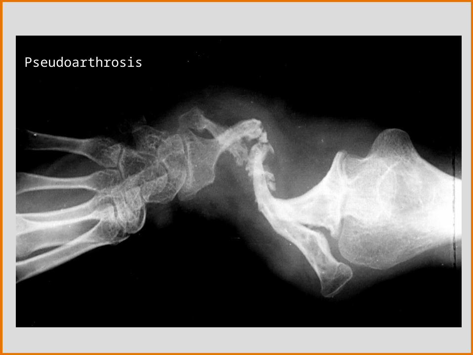
• Six or more café-au-lait macules >5mm in prepubertal and >15mm in postpubertal• Two or more neurofibromas OR one plexiform neurofibroma• Freckling of the axillary or inguinal regions• Optic glioma• Two or more lisch nodules• Distinctive bony lesions (pseudoarthrosis, sphenoid dysplasia or long bone thinning)• First degree relative with NF-1
Discussion

Things to look for in imaging NF-1:
•Neurofibroma = Tumor consisting of schwann cells, fibroblasts, and mast cells. There are 4 types:
1. Cutaneous
2. Subcutaneous
3. Nodular Plexiform – tumor involving a nerve root or major nerve.
4. Diffuse Plexiform – usually a congenital tumor involving fascicles.
** Plexiform are cause of major morbidity due to the angiogenic and invasive properties of these tumors. Also are have risk of
malignant transformation into neurofibrosarcoma.• Optic glioma , Meningioma and Astrocytoma• UBOs – Unidentified Bright Objects• Pheochromocytoma • Meningocele • Renal artery stenosis• GI neurofibromas• Sphenoid bone dysplasia, Long bone thinning, pseudoarthrosis, scoliosis
Discussion
Imaging Modalities for NF-1:
1. Plain film
•Used to evaluate any skull abnormalities (including optic glioma), scoliosis, long bone deformities, and pseudoarthrosis.•GI neurofibromas can be located on barium study
2. CT•Neurofibromas appear as soft tissue density masses•Optic gliomas can be seen as widening of the optic canals•GI neurofibromas seen as ring enhancing with contrast•Sphenoid dysplasia is seen as infiltration and decalcification of the bone•Can be used to evaluate hydrocephalus, hydronephrosis, etc. caused by the tumors
3. MRI•On T2 the neurofibromas present with increased signal at the periphery with decreased attenuation in the center of the tumor = “Target Sign”
Discussion

Imaging Modalities for NF-1 cont.
•UBO = increased signal on T2 in the Basal Ganglia, Thalamus, and sub-cortical white matter•Optic gliomas = isointense enlargement of optic nerves on T1 and T2•Meningiomas = hypointense mass on T1 (can use gadolinium to increase signal)•Pheochromocytoma = hyperintense on T2•Meningoceles = MRI is the study of choice since the pathological anatomy can be identified. Will enhance on T2.
Discussion
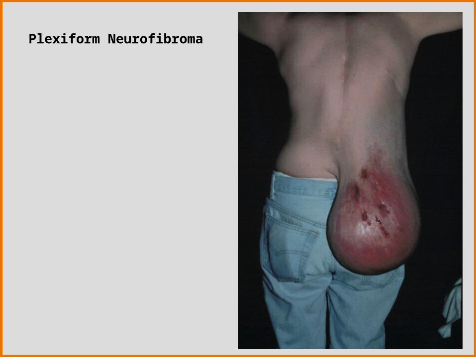
Plexiform Neurofibroma

Findings and DifferentialsLeft Optic Glioma: T1 weighted image
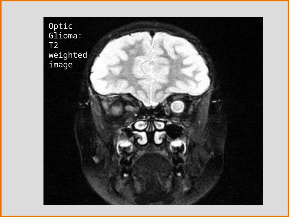
Optic Glioma: T2 weighted image
Findings:
Differentials:
Pseudoarthrosis

Findings:
Differentials:
Findings and DifferentialsScoliosis with an associated Meningocele
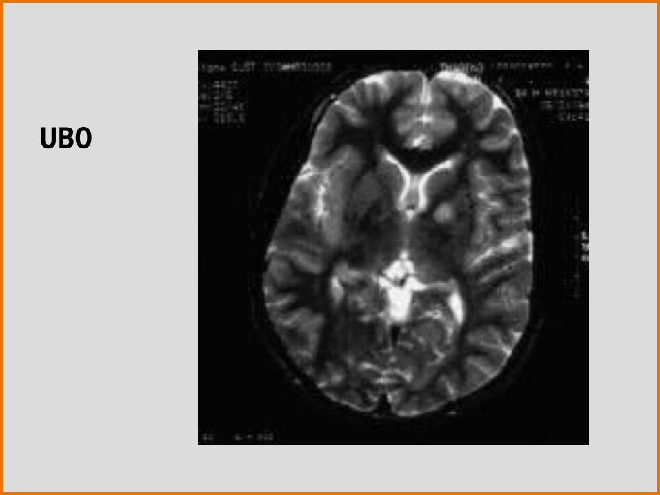
UBO

Neurofibromatosis type 1 (von Recklinghausen’s disease)
Diagnosis

S Aoki, AJ Barkovich, K Nishimura, BO Kjos, T Machida, P Cogen, M Edwards, and D Norman. Neurofibromatosis types 1 and 2: cranial MR findings. Radiology 1989; 172: 527.
James D. Eastwood, David J. Fiorella, James F. MacFall, David M. Delong, James M. Provenzale, and Robert S. Greenwood. Increased Brain Apparent Diffusion Coefficient in Children with Neurofibromatosis Type 1. Radiology 2001; 219: 354.
Dachman AH: CT in neurofibromatosis type I. AJR Am J Roentgenol 2000 Dec; 175(6 )
Stevenson, DA, Zhou, H, Ashrafi, S, et al. Double inactivation of NF1 in tibial pseudarthrosis. Am J Hum Genet 2006; 79:143.
Lewis, RA, Gerson, PL, Axelson, KA, et al. von Recklinghausen neurofibromatosis: incidence of optic gliomata. Ophthalmology 1984; 91:929.
Ducatman, BS, Scheithauer, BW, Piepgras, DG, et al. Malignant peripheral nerve sheath tumors. A clinicopathologic study of 120 cases. Cancer 1986; 57:2006.
Southcott, R, Lucraft, HH. Association of soft tissue sarcomas other than neurofibrosarcoma with neurofibromatosis. Clin Oncol (R Coll Radiol) 2002; 14:431.
www.utdol.comwww.emedicine.com
References
Related Documents