Alireza Sadeghi MD Lutheran Medical Center University Hospital of Brooklyn Downstate Medical Center March 10 th 2006 Case Presentation

Welcome message from author
This document is posted to help you gain knowledge. Please leave a comment to let me know what you think about it! Share it to your friends and learn new things together.
Transcript

Alireza Sadeghi MDLutheran Medical CenterUniversity Hospital of BrooklynDownstate Medical CenterMarch 10th 2006
Case Presentation

• xx years old Caucasian Male• History of Stage III Colon Cancer in xxxx year
– S/P Resection and Chemotherapy• Presented to Thoracic Service at LMC for
elective VATS & Pulmonary Resection for a 3.1 cm lesion in the Right Lower Lobe on ___ date.
• Denied SOB, Weight Loss, Cough, Hemoptysis• No other complaints
Case Presentation

Case Presentation
• Past Medical History:– DM, BPH, HTN
• Past Surgical History:– Colon Cancer (date): S/P
resection and chemotherapy
– Pulmonary Metastasectomy (date) for metastatic colon CA: Left upper lobectomy
– Bladder Cancer (date): TURBT
• Allergies: – PCN
• Medications:– Proscar– Glipizide– Cozaar– Multivitamins
• Social History:– ☻ 51 Pk yr Tobacco– ☻ EtOH

Case Presentation
• Vital Signs– Temp: 98.1 F– BP: 108/61– HR: 70– RR: 18– O2 Sat: 100 % RA
• Physical Exam– No cervical adenopathy– No head/neck/arm
edema– Chest:
• CTA B/L• RRR, S1 & S2, no murmurs
– Abd: S/ND/NT/+BS– Exterm: 2+ distal pulses– Neuro: Intact

Case Presentation• Preoperative Labs:
– WBC: 7.5– Hgb/HCT: 15.3/46.3– Plts: 170– Chem: 144/5.0/108/28/21/1.1/115– INR: 0.9– CEA level:
• 2.1-2.5 ng/ml since initial surgery in (date)
– ABG: 7.45/37/85/25.6/+1.8/99%
Case Presentation
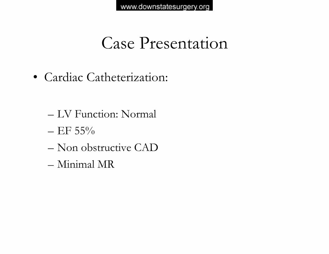
• Cardiac Catheterization:
– LV Function: Normal – EF 55%– Non obstructive CAD– Minimal MR

Case Presentation
6998 L/min142 L/minMVV481.81 L/sec3.79 L/secFEF 25-75%
% PredictedActualPredictedParameters
6616.224.6DLCO (ml/min/mmHg)
866.22 L7.27 LTLC
NA77%78 %FEV1/FVC722.78 L3.86 LFEV1 753.63 L4.87 LFVC
• Pulmonary Function Tests:

Case Presentation
• Imaging:– Surveillance CT Scan (date)
• Lobulated mass in the RLL; posteriomedial location• 3.1 x 2.5 x 2.8 cm• No Axillary LN involvement• Previous Surveillance CT Scans did not reveal any lesions
– 10/28/2004– 05/12/2004– 12/03/2003
– PET Scan (date)• Single Hyper metabolic focus in the Right Lower Lobe

Case Presentation
• Operative Course:– Bronchoscopy: No intraluminal lesions– Double lumen ET intubation– SCD Boots– Patient positioned in R Thoracotomy position– Right VATS performed
• Patient tolerates LLL ventilation: O2 Sat 97-99%• Unable to safely remove the lesion due to its location• Procedure abandoned• Right Muscle Sparing Thoracotomy performed

Case Presentation
• Operative Course:– Exploratory Right Thoracotomy:
• Lesion not amenable to wedge resection due to central location
• Decision made to perform a complete right lower lobectomy
• EBL: 900 cc• 2 chest tubes inserted and the chest was closed.• Patient tolerated procedure well.

Case Presentation
• Pathology Report:
– Right Lower Lobe• Metastatic Moderately Differentiated Adenocarcinoma• 3.1 cm x 2.9 cm x 2.8 cm• Solitary Lesion• Histologically compatible with Colonic Origin

Case Presentation
• Postoperative Course:– POD #1: SICU
• Supportive Management: – 2U PRBC & Pressor Support– HCT 28→33
• Extubated successfully• SCD boots• No evidence postoperative MI• Chest Tubes: 1200 cc/24 hr• Post Extubation ABG on 3 L NC:
– 7.42/34.9/153/22/-1.9/99%


Case Presentation
• Postoperative Course:– POD #2: SICU
• Supportive Care/SCD Boots & HSC– Chest tubes: 300 cc/24 hrs– Apical CT removed
• Diet Advanced
– POD #3: Floor• OOB/Incentive Spirometry/SCD Boots & HSC
– Chest tube: 130 cc/24 hrs– CXR: mild atelectasis

Case Presentation
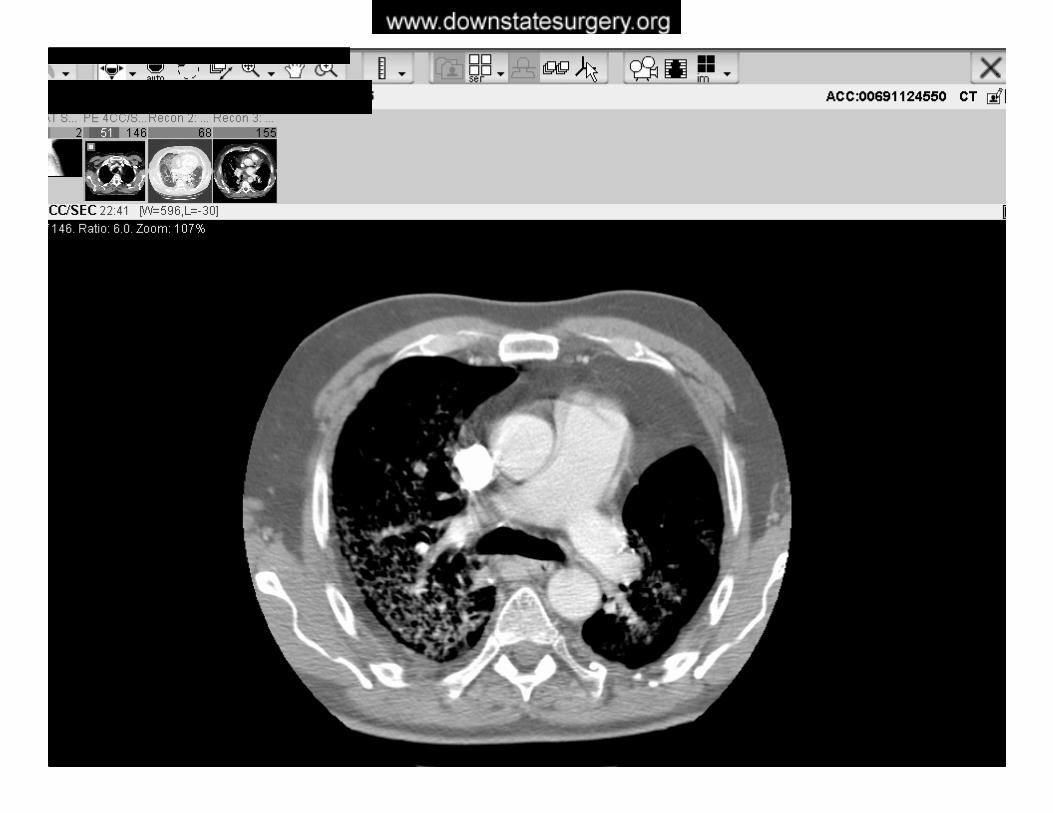
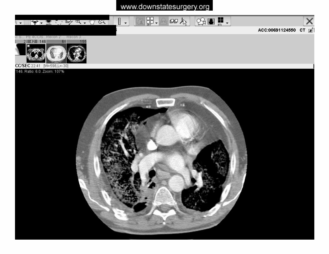
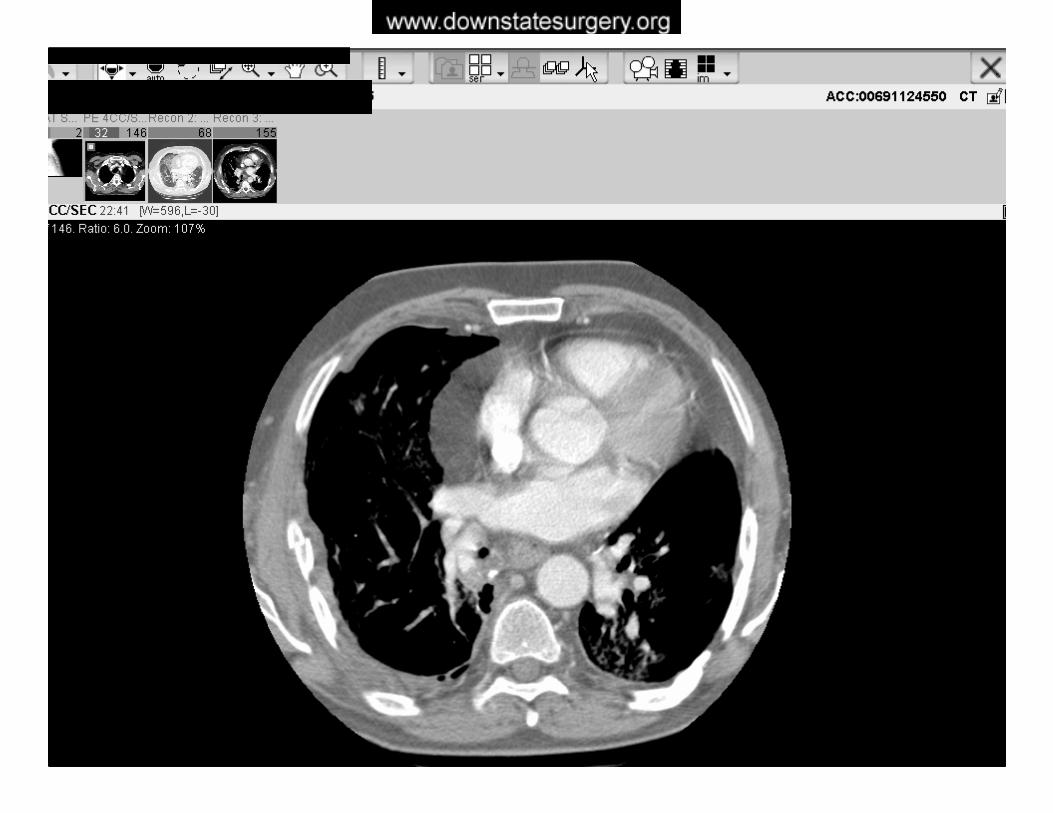
• Postoperative Course:– POD #4: Floor
• Acute SOB with desaturation to 90%• Afebrile/VSS/No evidence of MI• ABG: 7.46/36/225/25/+1.9/100% on NRB mask• A-a Gradient: 371• CTA Chest:
– Large hypo dense filling defect within R Middle Lobe PA– B/L Pneumonitis
• Lower Extremity Duplex: No evidence of DVT• Heparin Anticoagulation Started/Broad Spectrum Abx• Transferred to ICU


Case Presentation
• Postoperative Course:– POD #5: ICU
• Brief initial improvement• IV Anticoagulation therapeutic/Warfarin started• Respiratory Distress continues• Medical Oncology team assumes care of patient• Oxygen Saturation 90-93% on NRB Mask• Patient maintained on BIPAP ventilation• INR therapeutic 2.9• Transferred to MICU

Case Presentation
• Postoperative Course:– POD #5: MICU
• Continued Respiratory Failure• Patient deteriorates• BIPAP• ABG: 7.45/41/63/28/+3.3/93%
– POD #6: MICU• Fulminant Respiratory Failure• BIPAP• ABG: 7.47/38/28• Cardiac Arrest/Unsuccessful ACLS → Pt Expired.
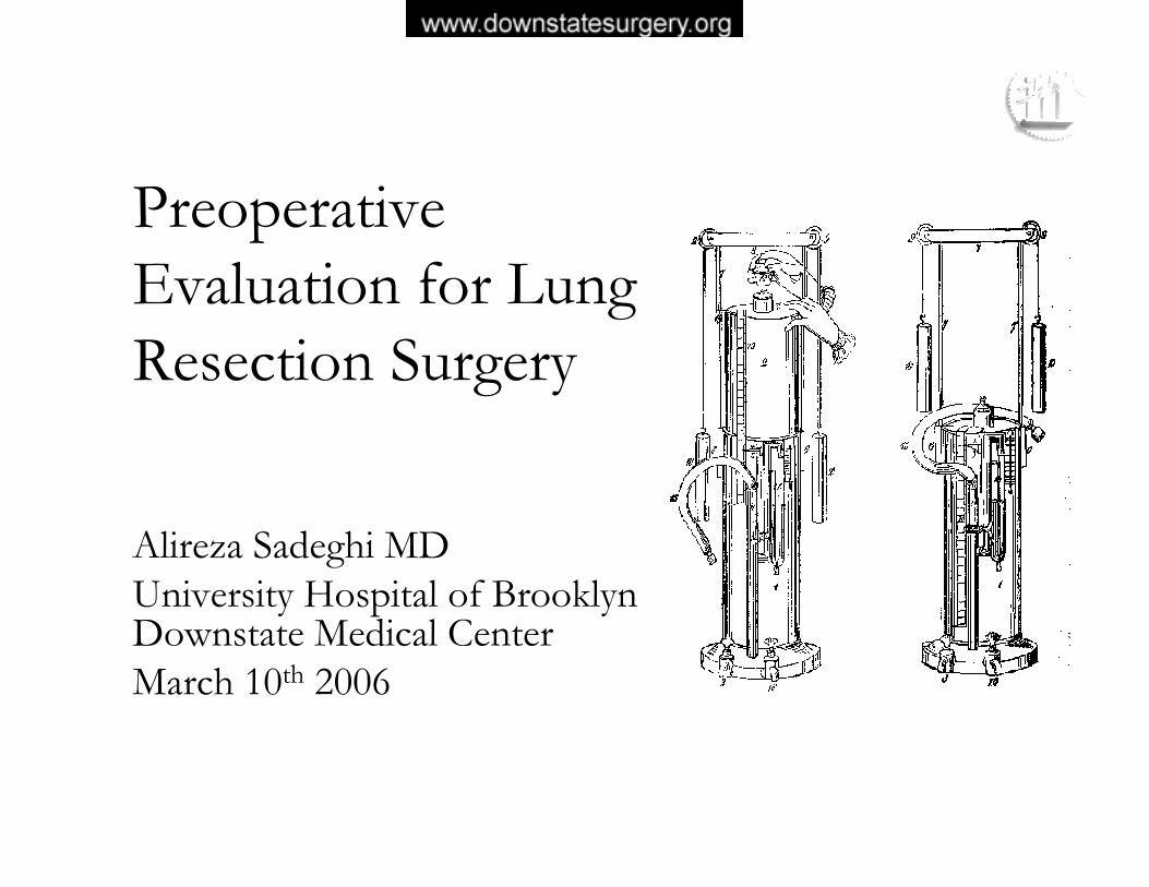
Preoperative Evaluation for Lung Resection Surgery
Alireza Sadeghi MDUniversity Hospital of Brooklyn Downstate Medical CenterMarch 10th 2006

Spirometry
• Recording of the volume of air inhaled & exhaled, plotted against time, during a series of Ventilatory maneuvers.
• Results depict a Normal vs. Abnormal pattern of Ventilatory reserve– Obstructive– Restrictive – Mixed Disorders
Debapriya D et al. Preoperative Evaluation of Patients Undergoing Lung Resection Surgery. CHEST 2003; 123:2096–2103

Mazzone PJ et al. Interpreting pulmonary function tests. Cleveland Clinic Journal Medicine 2003; 70(10):866-81.

Terminology
Mazzone PJ et al. Interpreting pulmonary function tests. Cleveland Clinic Journal Medicine 2003; 70(10):866-81.

Terminology
• Forced vital capacity (FVC):– Total volume of air that can be
exhaled forcefully from TLC– The majority of FVC can be
exhaled in <3 seconds under normal circumstances
– Measured in liters (L)

Terminology
• Forced expiratory volume in 1 second: (FEV1)– Volume of air forcefully
expired from full inflation (TLC) in the first second
– Measured in liters (L)– Normally more than 75-80%
of FVC is exhaled in the first second

Terminology
• Forced expiratory flow 25-75% (FEF25-75)– Mean forced expiratory flow
during middle half of FVC – Measured in L/sec– May reflect effort independent
expiration and the status of the small airways
– Highly variable

FVC• Interpretation of % predicted:
– 80-120% Normal– 70-79% Mild reduction– 50%-69% Moderate reduction– <50% Severe reduction
FEV1• Interpretation of % predicted:
– >75% Normal– 60%-75% Mild obstruction– 50-59% Moderate obstruction– <49% Severe obstruction
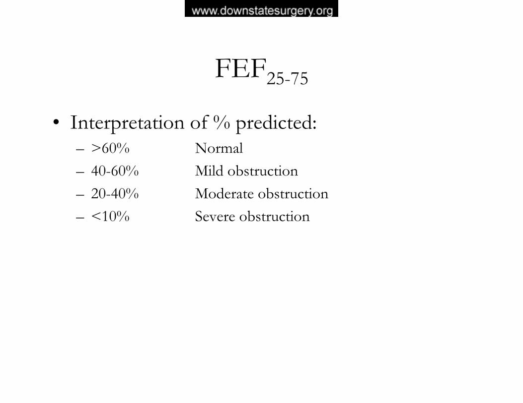
FEF25-75
• Interpretation of % predicted:– >60% Normal– 40-60% Mild obstruction– 20-40% Moderate obstruction– <10% Severe obstruction

Introduction
• Statistics from Center for Disease Control & Prevention – About 30,000 lung resections are performed annually in the
United States
• Commonly performed surgeries for lung cancer include – Pneumonectomy– Lobectomy– Wedge Resection– Segmentectomy
Jeng-Shing Wang. Pulmonary function tests in preoperative pulmonary evaluation. Resp Med 2004; 98:598-605

Introduction
• Indications for Pulmonary Resection:– Neoplastic Disease
• Primary• Metastatic
– Bullous Lung Disease: LVRS– Diagnosis & Management of inflammatory
conditions• Granulomas• Pulmonary infiltrates• Resection of segments destroyed by bronchiectasis
Debapriya D et al. Preoperative Evaluation of Patients Undergoing Lung Resection Surgery. CHEST 2003; 123:2096–2103

Introduction
• Recent Studies: Mortality Rates– Pneumonectomy: 6.8% 1 & 5.7% 2– Bilobectomy: 4.4 % 2– Lobectomy: 3.9% 1– Lesser Resection: 1.4% 2
• Risk of morbidity & mortality – mandatory to assess as accurately as possible which
patients with anatomically resectable disease are suitable candidates for resectional surgery
1. Kadri MA et al Survival and prognosis following resection of primary non-small cell bronchogenic carcinoma. Eur J Cardiovasc Surg 1991; 5:132–6.
2. Damhuis et al. Resection rates and postoperative mortality in 7899 patients with lung cancer. Eur Respir J 1996; 9:7–10.

Purpose
• The Purpose of Preoperative Physiologic Assessment:– Using the least invasive test possible
• Identify the High Risk Patient– Complications– Long term Disability
• Adequately Counsel the patient on treatment options and risks to make an informed decision
• Identify possible steps to reduce risks of peri-operative complications & long-term pulmonary disability
Beckles MA et al. Lung Cancer Guidelines: The physiologic evaluation of patients with lung cancer being considered for resectional surgery. CHEST 2003; 123:105S-114S

Complications
• Postoperative Cardiopulmonary Complications– Acute Hypercapnia– Mechanical Ventilation > 48hrs– Arrhythmias– Pneumonia– Pulmonary Emboli– Myocardial Infarction– Lobar Atelectasis requiring Bronchoscopy

Preoperative Evaluation
• Who should be evaluated? – The general answer
• All patients undergoing lung resection surgery, irrespective of age or extent of the lesion.
• History– Smoking & COPD
• Preoperative Medical Optimization
Debapriya D et al. Preoperative Evaluation of Patients Undergoing Lung Resection Surgery. CHEST 2003; 123:2096–2103

Pulmonary-Specific Evaluation
• The aims of pulmonary-specific evaluation– Assessment of the patient’s physiologic pulmonary
function– Determining the patient candidacy for surgery and
the extent of resection that can be tolerated. • There is no single measure that is a “gold
standard” in accurately predicting complications.• However, certain criteria, when applied have
been shown to be predictive of outcome.
Debapriya D et al. Preoperative Evaluation of Patients Undergoing Lung Resection Surgery. CHEST 2003; 123:2096–2103

The Algorithm
Jeng-Shing Wang. Pulmonary function tests in preoperative pulmonary evaluation. Resp Med 2004; 98:598-605

Risk Factors for Postoperative Cardiopulmonary Complications
• Non Pulmonary Factors– Site of Surgery– Duration of Surgery– Laparoscopic approach– Nutrition– Age– Obesity
• Pulmonary Factors– COPD – Smoking– Obesity– Productive Cough– Wheezing – FEV1/FVC ratio– PaCO2
– ASA Classification
Jeng-Shing Wang. Pulmonary function tests in preoperative pulmonary evaluation. Resp Med 2004; 98:598-605

Pulmonary-Specific Evaluation
• Pulmonary Functions Tests
– Used to evaluate risk for postoperative complications since 1950s
– 1955 first published case of preoperative PFTs• Patients undergoing lung resection for pulmonary TB• MVV <50% & FVC <70%
– 40% Mortality following Thoracotomy
Jeng-Shing Wang. Pulmonary function tests in preoperative pulmonary evaluation. Resp Med 2004; 98:598-605

Pulmonary-Specific Evaluation
• Pulmonary Function Tests Include:– Spirometry – Lung Volumes– Diffusion Capacity– Oximetry – Arterial Blood Gas Analysis– Radionuclide Lung Scanning– Cardiopulmonary Exercise Testing
Jeng-Shing Wang. Pulmonary function tests in preoperative pulmonary evaluation. Resp Med 2004; 98:598-605

Stages of Pulmonary-Specific Evaluation
• Stage I Assessment (Preop lung function)– Spirometry– Arterial Blood Gas Analysis– DLCO
• Stage II Assessment (Postop lung function)– Quantitative Ventilation-Perfusion Scan– Quantitative CT Scan
• Stage III Assessment– Exercise Testing: Oxygen Uptake (VO2 Max)
Debapriya D et al. Preoperative Evaluation of Patients Undergoing Lung Resection Surgery. CHEST 2003; 123:2096–2103

• Spirometry:– Simple, inexpensive, standardized & readily available
• FVC → volume• FEV1 → airflow
– Factor regarded as being the best for predicting complications of lung resection in the initial assessment
• FEF25–75% → airflow• Maximal voluntary ventilation (MVV)→ Muscle Strength
– Maximal inhalation & exhalation over 12 sec: Air Flow & MS– Dependent on patient effort
– Predicted values of pulmonary function depend on age, height, gender and race
Stage I
Debapriya D et al. Preoperative Evaluation of Patients Undergoing Lung Resection Surgery. CHEST 2003; 123:2096–2103

Stage I
• Forced Expiratory Volume One Second: FEV1– Correlates well with the degree of respiratory
impairment in patients with COPD– Indirect measure of pulmonary reserve– In studies evaluating preoperative spirometric values
a reduced FEV1 (<60% of predicted)• Strongest predictor of postoperative complications
– ACCP & BTS Guidelines:• FEV1 > 2 L tolerate pneumonectomy• FEV1 > 1-1.5 L tolerate lobectomy
Mazzone PJ et al. Lung Cancer: Preoperative pulmonary evaluation of the lung resection candidate. Am J Med 2005; 118:578-583

Stage I
• Retrospective Studies in 1970s– British Thoracic Society (BTS) Guidelines compiled
on data from >2000 patients in 3 large series– FEV1 studied at the main factor– Mortality Rate < 5%
• FEV1 > 1.5 L for Lobectomy• FEV1 > 2 L or FEV1 > 80% predicted for
Pneumonectomy
Beckles MA et al. Lung Cancer Guidelines: The physiologic evaluation of patients with lung cancer being considered for resectional surgery. CHEST 2003; 123:105S-114S

Stage I
• Diffusing capacity of the lung for carbon monoxide (DLCO)– Reflects alveolar membrane integrity & pulmonary
capillary blood flow in the patient’s lungs.– In the past (Ferguson et al) 237 patients
• Was the most important predictor of mortality & was the sole predictor of postoperative pulmonary complications.
– In recent studies• Equally significant predictor of postoperative
complications as FEV1Debapriya D et al. Preoperative Evaluation of Patients Undergoing Lung Resection Surgery. CHEST 2003;
123:2096–2103Ferguson MK et al. Diffusing capacity predicts morbidity and mortality after pulmonary resection. J Thorac
Cardiovasc Surg 1988; 96:894

Stage I
• Factors That Enhance CO Diffusion:– Increase in Lung Capillary
Blood Volume– Recruitment & distention
of Pulmonary Capillaries– Supine Position
• Factors That Decrease CO Diffusion:– Age– Standing Position– Decrease in Lung
Capillary Blood Volume– Alveolar disease– Loss of Lung Disease
Beckles MA et al. Lung Cancer Guidelines: The physiologic evaluation of patients with lung cancer being considered for resectional surgery. CHEST 2003; 123:105S-114S

Stage I
• DLCO as a useful marker of operative risk– Ferguson et al in 237 patients
• Relation between preoperative DLCO and M&M
– Preoperative DLCO as percentage predicted had higher correlation with postoperative mortality than FEV1
• DLCO < 60% predicted associated with ↑ mortality.
– DLCO & FEV1 should be viewed as complementary physiologic tests
Beckles MA et al. Lung Cancer Guidelines: The physiologic evaluation of patients with lung cancer being considered for resectional surgery. CHEST 2003; 123:105S-114S

Stage I
• Arterial Blood Gas Analysis (ABG)– Not extensively studied as predictor of postoperative
complication (PCO2>50 mmHg & PO2<60 mmHg)– Hypercapnia (PCO2 >50 mm Hg) in arterial blood
has been a traditional contraindication to lung resection as it indicates chronic respiratory failure.
– In recent studies• Patients with a PCO2 of 45 mm Hg did well
postoperatively• Was not predictive of postoperative complications
Debapriya D et al. Preoperative Evaluation of Patients Undergoing Lung Resection Surgery. CHEST 2003; 123:2096–2103

Stage I
• Arterial Blood Gas Analysis (ABG)– Evidence: – Kearney et al (1994)
• No difference in postoperative complications• Preoperative PCO2 < 45 mmHg vs. PCO2 > 45 mmHg
– 17% vs. 13%• Hypercapnia is now NOT a contraindication to surgery
– Low PPO FEV1
– Poor exercise tolerance• Hypoxemia (SaO2 < 90%) was associated with ↑ risk of
postoperative complications

Original Studies
• Study complications in 500 patients undergoing lung resection and correlated them with preoperative spirometric indexes & type of surgery performed. – Recommendation for Pneumonectomy
• MVV → 55%; FEV1→ 2 L; FEF25–75% →1.6 L/min.
– Recommendation for Lobectomy • MVV → 40%; FEV1 → 1 L; FEF25–75% → 0.6 L/s.
– Recommendation for Segmentectomy or Wedge Resection• MVV → 40%; FEV1 → 0.6 L; FEF25–75% → 0.6 L/sMiller JI, Grossman GD, Hatcher CR. Pulmonary function test criteria for operability and pulmonary
resection. Surg Gynecol Obstet 1981; 153:893–895

Recent Studies
• Summary:– following criteria are predictive of increased
postoperative complications and mortality: • Pneumonectomy:
– FEV1 → <2L or 60% of predicted & MVV → 55% of predicted– DLCO → <50% of predicted & FEF25–75% → 1.6L/s.
• Lobectomy:– FEV1 → <1 L & MVV → <40% of predicted– FEF25–75% → 0.6 L/s, DLCO → <50% of predicted.
• Wedge resection/Segmentectomy: – FEV1 → <0.6 L & DLCO → <50% of predicted.
Stephan F, Boucheseiche S, Hollande J, et al. Pulmonary complication following lung resection: a comprehensive analysis of incidence and possible risk factors. Chest 2000; 118:1263–1270

Stage II
• Quantitative Ventilation-Perfusion Scan:– Measures predicted Postoperative lung function– Quantitative measurement of the contribution of
each lung to pulmonary ventilation & perfusion– Readily available with negligible risk to the patient– Highly accurate in the prediction of postoperative
pulmonary function following resection• Spirometry & Lung Scan
Jeng-Shing Wang. Pulmonary function tests in preoperative pulmonary evaluation. Resp Med 2004; 98:598-605

Stage II
• Inhaled 133Xe or IV 99Tc-Labeled Macroaggregates of Albumin– % of radioactivity contributed by each lung
correlates with the contribution of the function of that lung
– Normally: 19 Segments (10 R & 9 L)• Right Lung (3/2/5): 55 % & Left Lung(3/2/4): 45%
– Calculation:• Predicted Postoperative FEV1 of the remaining lung
Debapriya D et al. Preoperative Evaluation of Patients Undergoing Lung Resection Surgery. CHEST 2003; 123:2096–2103

Stage II
• Using 133Xe Inhalation:– Predicted Postoperative (PPO) FEV1 of < 1 L is
indicative of physiologic inoperability.Kristersson S et al. Prediction of pulmonary function loss due to pneumonectomy using 133Xe radioisometry.
Chest 1972; 62:696–698
• Using 99Tc Macroaggregate of Albumin Perfusion:– Predicted Postoperative (PPO) FEV1 of < 0.8 L is
indicative of surgical inoperability.Olsen GN, Block AJ, Tobias JA. Prediction of postpneumonectomy pulmonary function using quantitative
macroaggregate lung scanning. Chest 1974; 66:13–16

Stage II
• Predictors of Morbidity & Mortality after lung resection– Evidence:
• PPO FEV1 < 40% of predicted → 50% ↑ Mortality• PPO FEV1 > 40% of predicted → 50% ↓ Mortality• PPO DLCO < 40% of predicted → ↑ Mortality
– Best predictor of postoperative respiratory failure• Recommendation:
– If PPO FEV1 & DLCO > 40% (or PPO FEV1 > 1 L) may undergo lung resection including pneumonectomy.
– Otherwise → Exercise Testing
Markos J et al. Preoperative assessment as a predictor of mortality and morbidity after lung resection. Am Rev Respir Dis 1989; 139:902–916

Stage II
• Other Tests:– Bronchospirometry, Lateral position testing & Total
Unilateral pulmonary artery occlusion– Invasive tests & Require specialized equipment with
a high level of technical expertise– These test are no longer performed in the
preoperative evaluation of patients awaiting lung resection
Debapriya D et al. Preoperative Evaluation of Patients Undergoing Lung Resection Surgery. CHEST 2003; 123:2096–2103

Stage III
• Cardio-Pulmonary Exercise Testing (CPET)– Stresses the entire cardiopulmonary & oxygen delivery system
• Provides a good estimate of cardiopulmonary reserve• Pulmonary/cardiac function & peripheral oxygen utilization• Used before the advent of PFTs & sophisticated exercise testing
– Two major types of tests• Fixed exercise challenge
– Sustained level of work (i.e. walking a fixed distance or a flight of stairs)• Incremental exercise challenge
– Work rate is sequentially increased to a desired end point• Submaximal vs. Maximal oxygen consumption
– (VO2 Max)
Debapriya D et al. Preoperative Evaluation of Patients Undergoing Lung Resection Surgery. CHEST 2003; 123:2096–2103

Stage III
• Maximal Oxygen Consumption:– With increasing muscular
work VO2 rises to a point where there is a plateau of the VO2 work rate slope.
• VO2 max is a measure of aerobic capacity of the peripheral tissue– i.e. Oxygen Consumption
Mazzone PJ et al. Lung Cancer: Preoperative pulmonary evaluation of the lung resection candidate. Am J Med 2005; 118:578-583

Stage III
• Fixed Challenge Exercise Testing– Van Nostrand 1968
• Test of endurance in the preoperative evaluation• 50% postoperative mortality rate in patients who failed to
climb one flight of stairs with minimal dyspnea• 11% postoperative mortality rate in patients who
successfully climbed two flight of stairs with minimal dyspnea
Van Nostrand D, Kjelsberg MD, Humphrey EW. Pre-resectional evaluation of risk from pneumonectomy. Surg Gynecol Obstet 1968; 127:306–312
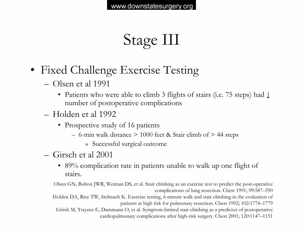
Stage III
• Fixed Challenge Exercise Testing– Olsen et al 1991
• Patients who were able to climb 3 flights of stairs (i.e. 75 steps) had ↓number of postoperative complications
– Holden et al 1992• Prospective study of 16 patients
– 6-min walk distance > 1000 feet & Stair climb of > 44 steps» Successful surgical outcome
– Girsch et al 2001• 89% complication rate in patients unable to walk up one flight of
stairs.Olsen GN, Bolton JWR, Weiman DS, et al. Stair climbing as an exercise test to predict the post-operative
complications of lung resection. Chest 1991; 99:587–590Holden DA, Rice TW, Stelmach K. Exercise testing, 6-minute walk and stair climbing in the evaluation of
patients at high risk for pulmonary resection. Chest 1992; 102:1774–1779Girish M, Trayner E, Dammann O, et al. Symptom-limited stair climbing as a predictor of postoperative
cardiopulmonary complications after high-risk surgery. Chest 2001; 120:1147–1151

Stage III
• Incremental Exercise Testing– Exercise to maximal exertion
• Measurement VO2 max in patients for lung resection• VO2 Max > 1 L/min → No mortality• VO2 Max < 1 L/min → 100% mortality
– Incidence of Postoperative complications• VO2 Max < 15 mL/kg/min → 100% complication rate• VO2 Max 15-20 mL/kg/min → 66% complication rate• VO2 Max > 20 mL/kg/min → 10% complication rate
Eugene J, Brown SE, Light RW, et al. Maximum oxygen consumption: a physiologic guide to pulmonary resection. Surg Forum 1982; 33:260–262
Smith TP, Kinasewitz GT, Tucker WY, et al. Exercise capacity as a predictor of post-thoracotomy morbidity. Am Rev Respir Dis 1984; 129:730–734

Changes in Lung Volume
• Pneumonectomy:– FEV1 reduced by 34 -
36%– FVC reduced by 36 - 40%– VO2 max reduced by 20 -
28%
• Lobectomy:– FEV1 reduced by 9 -
17%– FVC reduced by 7 - 11%– VO2 max reduced by 0 -
13%
Mazzone PJ et al. Lung Cancer: Preoperative pulmonary evaluation of the lung resection candidate. Am J Med 2005; 118:578-583
Decline in Lung Function Varies with the extent of resection

Lung Volume Reduction Surgery
• Selected patients with severe emphysema– Surgery may lead to improvement in lung function
• Lung Nodules:– In individuals who do not meet standard criteria
Mazzone PJ et al. Lung Cancer: Preoperative pulmonary evaluation of the lung resection candidate. Am J Med 2005; 118:578-583

Summary
• Reasons for Resection of Lung Cancer– Poor prognosis without
resection– Low Operative Mortality
Rate• Improve Surgical
Technique• Improved Anesthesia &
Postoperative Care– Modest decline in lung
functionMazzone PJ et al. Lung Cancer: Preoperative pulmonary evaluation of the lung resection candidate. Am J
Med 2005; 118:578-583

Summary
– American College of Chest Physicians in 2003.
• Guidelines for Preoperative Evaluation:– British Thoracic Society & Society of Cardiothoracic
Surgeons of Great Britain and Ireland Working Party in 2001.
British Thoracic Society and Society of Cardiothoracic Surgeons of Great Britain and Ireland Working Party. Guidelines on the selection of patients with lung cancer for surgery. Thorax 2001; 56:89-108
Beckles MA et al. Lung Cancer Guidelines: The physiologic evaluation of patients with lung cancer being considered for resectional surgery. CHEST 2003; 123:105S-114S
Guidelines

Guidelines
British Thoracic Society and Society of Cardiothoracic Surgeons of Great Britain and Ireland Working Party. Guidelines on the selection of patients with lung cancer for surgery. Thorax 2001; 56:89-108
Beckles MA et al. Lung Cancer Guidelines: The physiologic evaluation of patients with lung cancer being considered for resectional surgery. CHEST 2003; 123:105S-114S

The Algorithm
Jeng-Shing Wang. Pulmonary function tests in preoperative pulmonary evaluation. Resp Med 2004; 98:598-605

The End
Related Documents