Case Presentation Case Presentation Ass.prof. Hala AbdulHameed MBBCH , MSc ,MD,FCCP Pulmonolary and Critical Care. Alminya University

Case Presentation
Jan 13, 2016
Case Presentation. Ass.prof. Hala AbdulHameed MBBCH , MSc ,MD,FCCP Pulmonolary and Critical Care. Alminya University. Presenting complaint. A Sudanese male aged 52 years old non smoker he worked as a shepherd C/O - PowerPoint PPT Presentation
Welcome message from author
This document is posted to help you gain knowledge. Please leave a comment to let me know what you think about it! Share it to your friends and learn new things together.
Transcript
Case PresentationCase Presentation
Ass.prof. Hala AbdulHameed MBBCH , MSc ,MD,FCCP
Pulmonolary and Critical Care.Alminya University
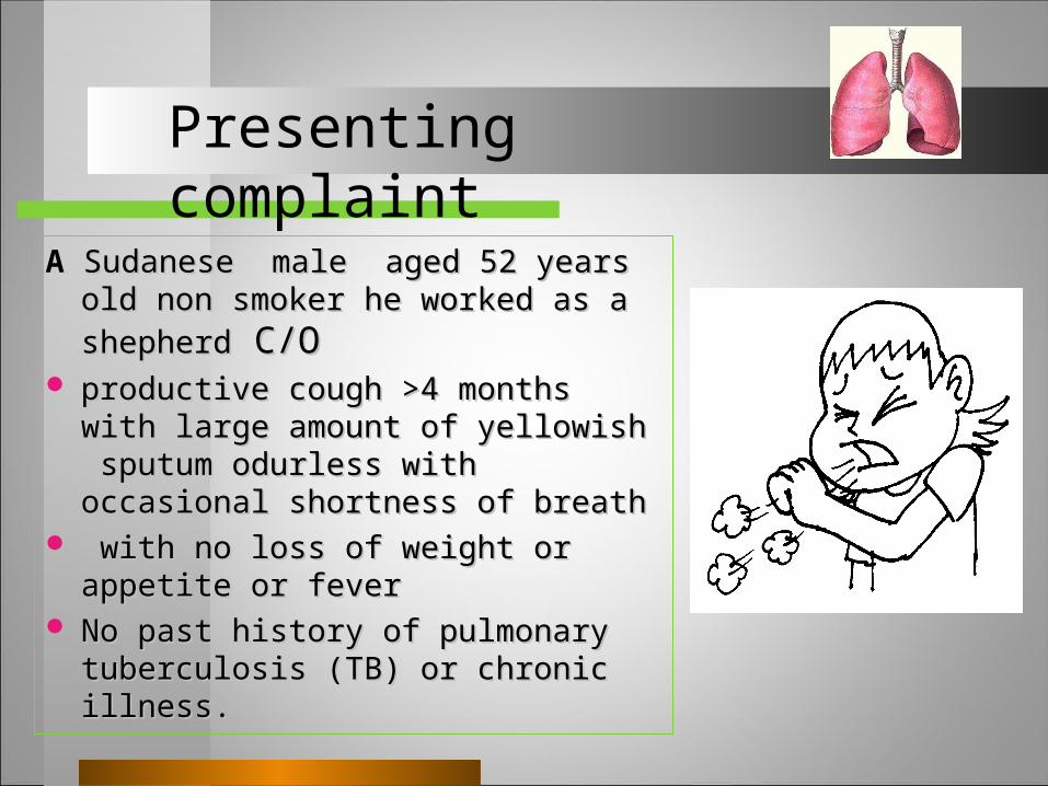
A Sudanese male aged 52 years old non Sudanese male aged 52 years old non
smoker smoker he worked as a shepherdhe worked as a shepherd C/O C/O productive cough >4 months with large productive cough >4 months with large
amount of yellowish sputum odurless amount of yellowish sputum odurless with occasional shortness of breathwith occasional shortness of breath
with no loss of weight or appetite or with no loss of weight or appetite or feverfever
No past history of pulmonary No past history of pulmonary tuberculosis (TB) or chronic illness. tuberculosis (TB) or chronic illness.
Presenting complaint
On examination, the patient was clinically stable
. He was not dyspnoeic
no pyrexia
no clubbing of the fingers.
Physical Examination
no hepatosplenomegally. Over the chest , an impaired percussion
note was detected over the right infrascapular area, but the breath sounds were normal with diminished intensity and there were bilateral scattered mid- crackles
workup followed toward differential diagnosis.
Physical Examination Cont.,
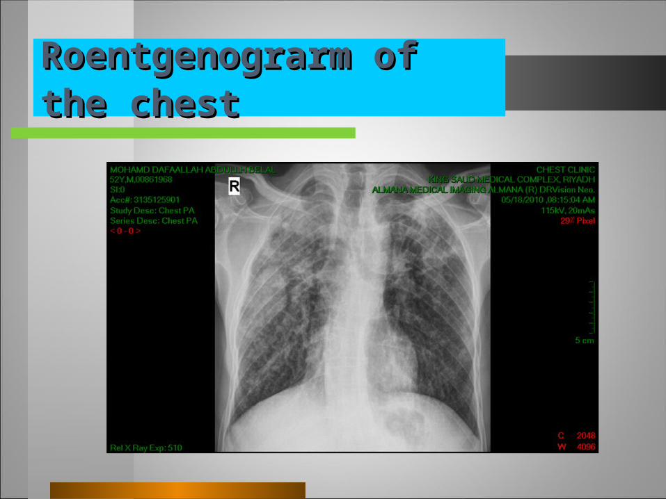
Roentgenograrm of the chestRoentgenograrm of the chest
Laboratory findingsLaboratory findings
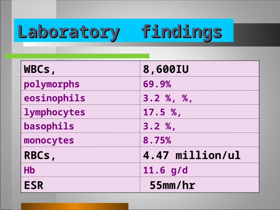
WBCs, 8,600IUpolymorphs 69.9%
eosinophils 3.2 %, %,
lymphocytes 17.5 %,
basophils 3.2 %,
monocytes 8.75%
RBCs, 4.47 million/ulHb 11.6 g/d
ESR 55mm/hr
Laboratory findings Cont.,Laboratory findings Cont.,
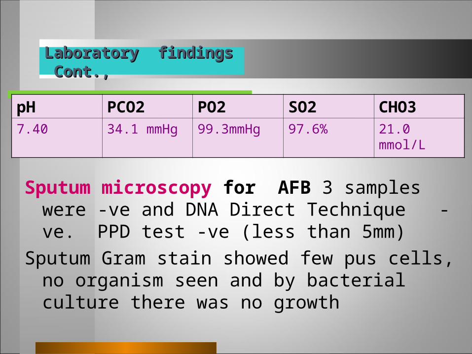
pH PCO2 PO2 SO2 CHO37.40 34.1 mmHg 99.3mmHg 97.6% 21.0 mmol/L
Sputum microscopy for AFB 3 samples were -ve and DNA Direct Technique -ve. PPD test -ve (less than 5mm)
Sputum Gram stain showed few pus cells, no organism seen and by bacterial culture there was no growth
Urine analysis---- at this time showed no red blood cells, and no parasites were identified on urine or stool microscopy also ,urine cytology showed no evidence of malignancy.
Biochemical indices of hepatic and renal function were normal.
Ca 2.23,Na 139, K 4.96,mg 0.89
Laboratory findings Cont.,Laboratory findings Cont.,
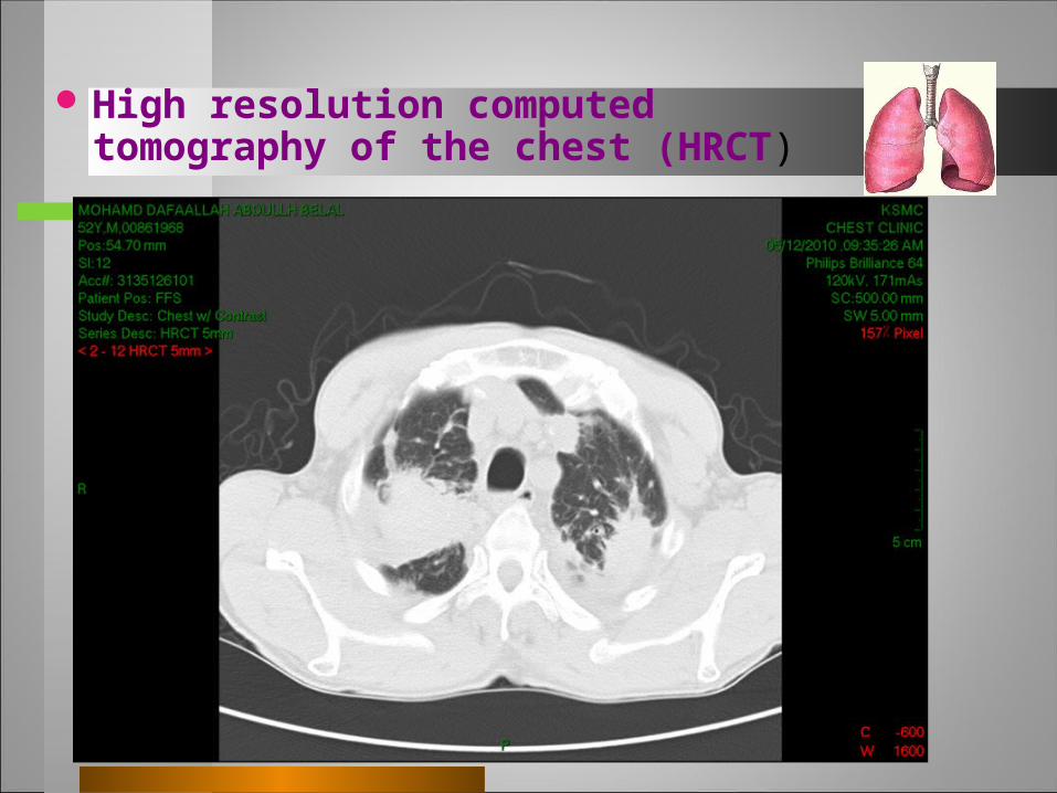
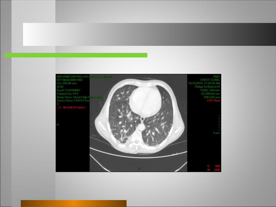
High resolution computed tomography of the chest (HRCT)
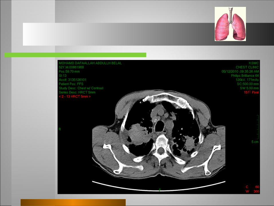
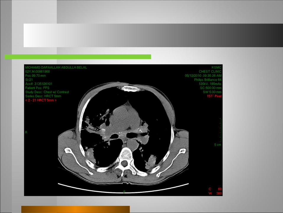
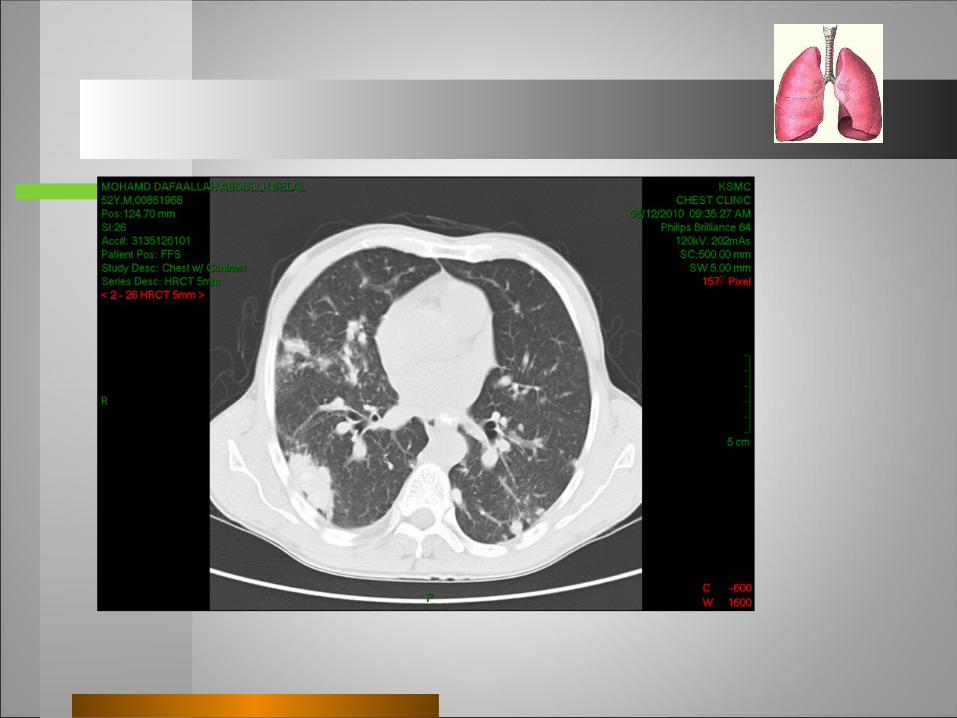
Conclusion:– Bilat. ill defined soft tissue masses in RUL
LUL,RML
- prominent calcified anterior mediastinal and hilar nodes
--- nodular pattern seen throughout both lungs, evenly distributed
CT Abdomen with contrast : Normal except for 2 small stones in the mid pole of the right kidney with no hydronephrosis
Differential Diagnoses
Tuberculous Mycobacterial Infections lung neoplasmsSarcoidosis, , silicosis,atypical pneumonia
Fiberoptic bronchoscopyFiberoptic bronchoscopy
Fiberoptic bronchoscopy revealed no changes in the bronchial tree.
Both the research of the TB bacillus , fungi and other bacteria in bronchial lavage were -ve.
Cytology was also negative for malignant cells. Post bronchoscopic sputum for TB was -ve It was decided, then, thoracotomy and biopsy.
ThoracotomyThoracotomy
Right mini thoracotomy revealed multiple nodules and masses involving most of the right lung, parietal pleura is not involved and wedge biopsy of lung was taken from the right upper lobe.
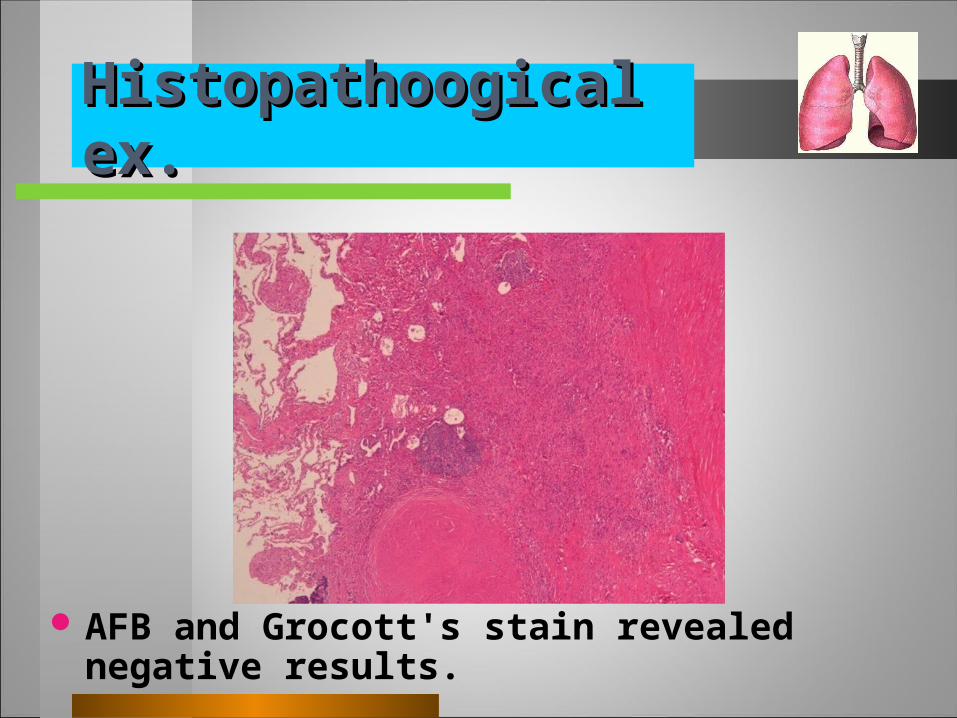
Histopathoogical ex.Histopathoogical ex.
AFB and Grocott's stain revealed negative results.
Start Anti tuberculous or not?Start Anti tuberculous or not?
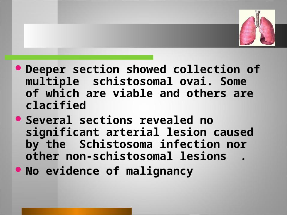
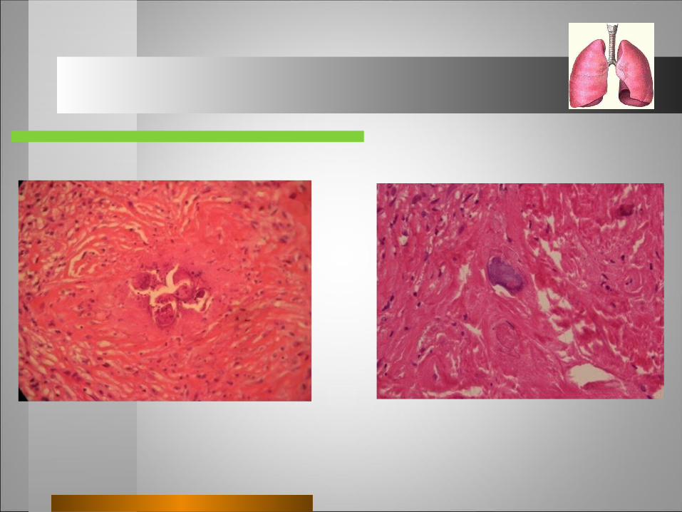
Deeper section showed collection of multiple schistosomal ovai. Some of which are viable and others are clacified
Several sections revealed no significant arterial lesion caused by the Schistosoma infection nor other non-schistosomal lesions .
No evidence of malignancy
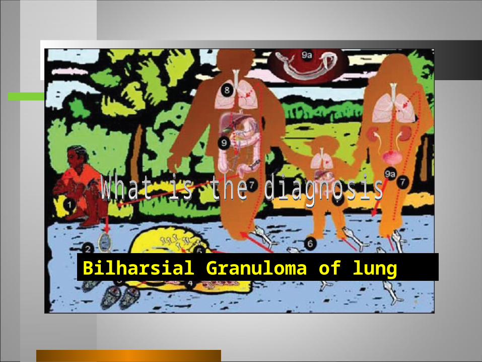
Bilharsial Granuloma of lung
A diagnosis of schistosomiasis should prompt initiation of treatment, even if the patient is asymptomatic, since adult worms can live for years .
The patient was treated with praziquantel (40 mg/kg) as a single dose without complications.
Is patient needs TTT or not ?
One month after treatment, a subjective improvement as regards better general condition, decrease amount of sputum change to whitish a chest CT scan showed a no changes on previous findings i.e. no more lesions appear
F/U
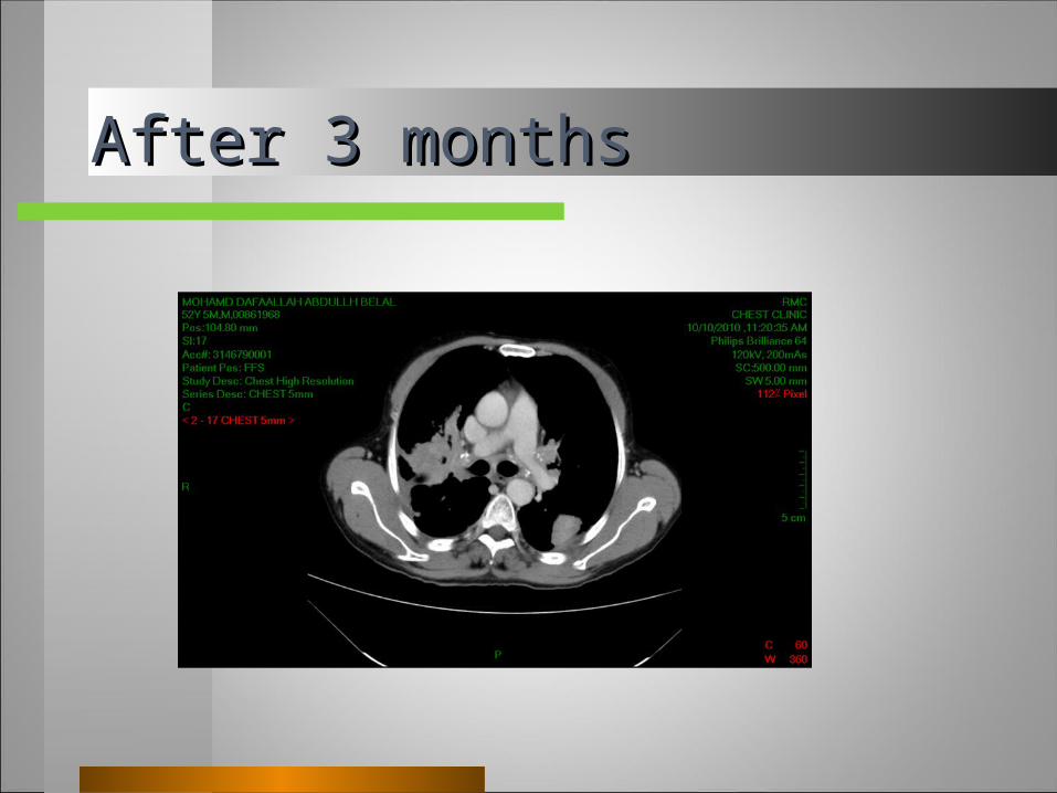
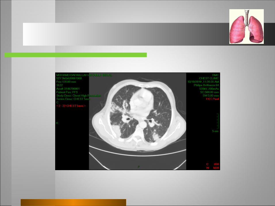
After 3 monthsAfter 3 months
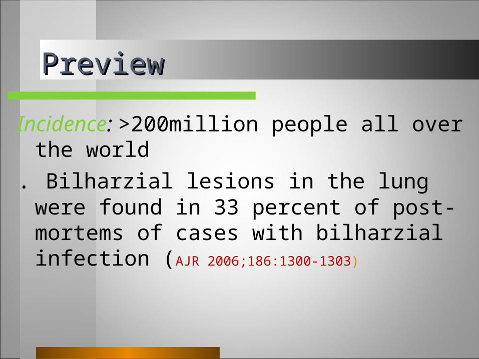
PreviewPreview
Incidence: >200million people all over the world
. Bilharzial lesions in the lung were found in 33 percent of post-mortems of cases with bilharzial infection (AJR 2006;186:1300-1303)
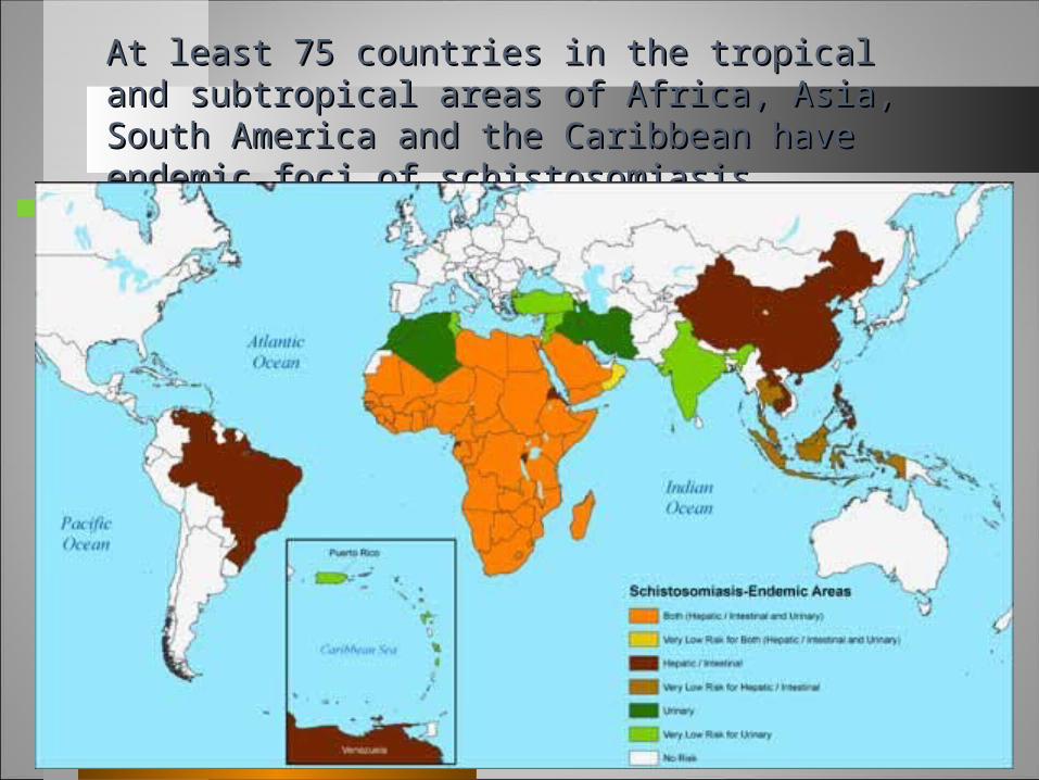
At least 75 countries in the tropical and subtropical areas At least 75 countries in the tropical and subtropical areas of Africa, Asia, South America and the Caribbean have of Africa, Asia, South America and the Caribbean have endemic foci of schistosomiasisendemic foci of schistosomiasis
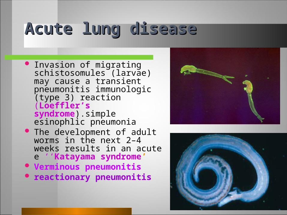
Acute lung diseaseAcute lung disease
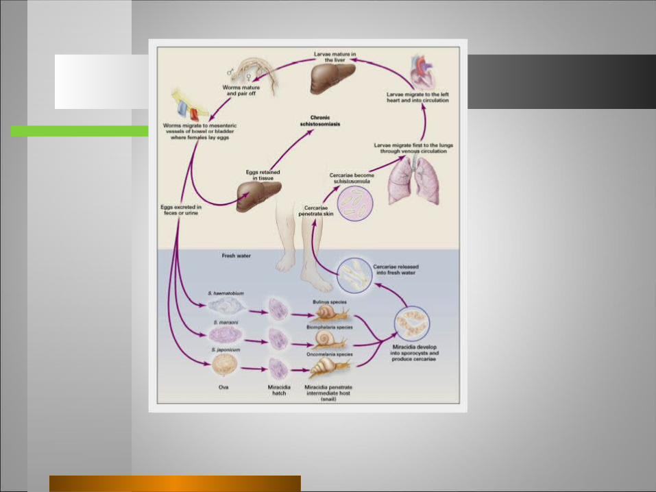
Invasion of migrating schistosomules (larvae) may cause a transient pneumonitis immunologic (type 3) reaction (Loeffler’s syndrome).simple esinophlic pneumonia
The development of adult worms in the next 2–4 weeks results in an acute e ‘‘Katayama syndrome’
Verminous pneumonitis reactionary pneumonitis
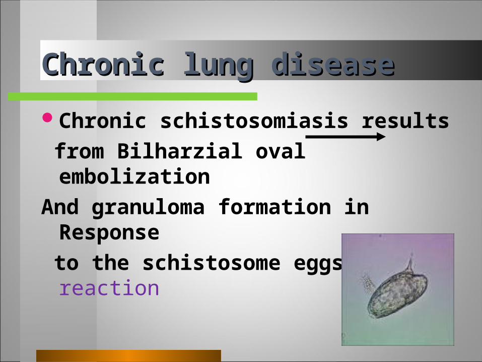
Chronic lung diseaseChronic lung disease
Chronic schistosomiasis results
from Bilharzial oval embolization
And granuloma formation in Response
to the schistosome eggs (type 4) reaction
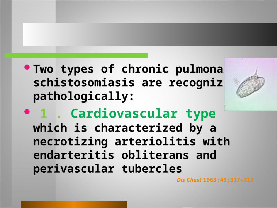
Two types of chronic pulmonary schistosomiasis are recognizable pathologically:
1 . Cardiovascular type which is characterized by a necrotizing arteriolitis with endarteritis obliterans and perivascular tubercles
Dis Chest 1963;43;317-319
Chronic lung diseaseChronic lung disease
Parenchymatous or bronchopulmonary type.
Its pathologic incidence is more common than the former type and it is less serious clinically as in the present case
Rev Bras Ter Intensiva 2009;21(4):461-464
Radiographic appearancesRadiographic appearances
are of interstitial infiltrates, typically nodular or micronodular, and there may be frank fibrosis.
Later CT findings include cardiomegaly and pulmonary arterial enlargement.
Rarely large mass lesions pseudo tumor
DiagnosisDiagnosis
as pulmonary affection occurs several years after infection so eggs may be not found in stool or urine . under this circumstances demonstrating characteristic pathologic changes and ova in tissues or +ve serology settle the diagnosis.
Demonstration of bilharzial ova in the sputum was also reported.
RadioGraphicsJanuary-February 2005 Volume 25 ● Number 1
to stress having bacteriologic proof before accepting the clinical diagnosis of tuberculosis.
It is proposed that, in endemic areas, pulmonary schistosomiasis is considered a differential diagnosis for complex structures, as pulmonary masses even with absense of sure diagnostic criteria, and pulmonary hypertension. pseudotumoral schistosomiasis
Pulmonary bilharziasis may be arrested at any stage, and the patient may live his normal span of life.
diagnosis of schistosomiasis should prompt initiation of treatment, even if the patient is asymptomatic, since adult worms can live for years .
Home MassageHome MassageHome MassageHome Massage

Related Documents