HAL Id: hal-03189260 https://hal.archives-ouvertes.fr/hal-03189260 Submitted on 6 Apr 2021 HAL is a multi-disciplinary open access archive for the deposit and dissemination of sci- entific research documents, whether they are pub- lished or not. The documents may come from teaching and research institutions in France or abroad, or from public or private research centers. L’archive ouverte pluridisciplinaire HAL, est destinée au dépôt et à la diffusion de documents scientifiques de niveau recherche, publiés ou non, émanant des établissements d’enseignement et de recherche français ou étrangers, des laboratoires publics ou privés. CAS and PSI increase coronal alignment accuracy and reduce outliers when compared to traditional technique of medial open wedge high tibial osteotomy: a meta-analysis S. Cerciello, M. Ollivier, K. Corona, B. Kaocoglu, R. Seil To cite this version: S. Cerciello, M. Ollivier, K. Corona, B. Kaocoglu, R. Seil. CAS and PSI increase coronal alignment accuracy and reduce outliers when compared to traditional technique of medial open wedge high tibial osteotomy: a meta-analysis. Knee Surgery, Sports Traumatology, Arthroscopy, Springer Verlag, In press, 10.1007/s00167-020-06253-5. hal-03189260

Welcome message from author
This document is posted to help you gain knowledge. Please leave a comment to let me know what you think about it! Share it to your friends and learn new things together.
Transcript

HAL Id: hal-03189260https://hal.archives-ouvertes.fr/hal-03189260
Submitted on 6 Apr 2021
HAL is a multi-disciplinary open accessarchive for the deposit and dissemination of sci-entific research documents, whether they are pub-lished or not. The documents may come fromteaching and research institutions in France orabroad, or from public or private research centers.
L’archive ouverte pluridisciplinaire HAL, estdestinée au dépôt et à la diffusion de documentsscientifiques de niveau recherche, publiés ou non,émanant des établissements d’enseignement et derecherche français ou étrangers, des laboratoirespublics ou privés.
CAS and PSI increase coronal alignment accuracy andreduce outliers when compared to traditional technique
of medial open wedge high tibial osteotomy: ameta-analysis
S. Cerciello, M. Ollivier, K. Corona, B. Kaocoglu, R. Seil
To cite this version:S. Cerciello, M. Ollivier, K. Corona, B. Kaocoglu, R. Seil. CAS and PSI increase coronal alignmentaccuracy and reduce outliers when compared to traditional technique of medial open wedge high tibialosteotomy: a meta-analysis. Knee Surgery, Sports Traumatology, Arthroscopy, Springer Verlag, Inpress, �10.1007/s00167-020-06253-5�. �hal-03189260�

CAS and PSI increase coronal alignment accuracy and reduce outliers when compared to traditional technique of medial open wedge high tibial osteotomy: a meta‑analysis
S. Cerciello1,2 · M. Ollivier3 · K. Corona4 · B. Kaocoglu5 · R. Seil6,7,8
AbstractPurpose Medial open-wedge high tibial osteotomy (MOWHTO) is an accepted option in the treatment of medial compart-ment osteoarthritis of the knee in young and active patients. Functional results are closely correlated to the correction of the mechanical axis of the lower limb. Although several angular and geometrical methods and values have been proposed in the past, the ideal target is still debated. In addition, it is important to have a deep correlation between the planned correction and the achieved correction after surgery. The aim of the present systematic review was to identify the ideal coronal correction after MOWHTO and the most accurate method to achieve it.Methods A systematic review of the literature was completed on July 3rd 2020 in the Pubmed, Medline, Cochrane Reviews, and Google Scholar databases using the Medical Subject Headings (MeSH) terms: “high tibial osteotomy” AND “accuracy” OR “planning”.Results 28 studies were included; 18 were focused on computer-assisted surgery (CAS) and 10 on patient-specific instru-mentation (PSI). There were 598 patients in the CAS group and 501 in the control group; the rate of outliers was 16% and 38.2% respectively (P = 0.04), while there was no significant difference between the two groups (SMD = − 0.10; 95% CI 1.31 to 1.12; P = n.s.) in terms of coronal accuracy. Likewise, there were 318 patients in the PSI group and 40 in the control group; the rate of outliers was 15% and 40% respectively (P = 0.98), while there was no significant difference between the two groups (SMD = 0.01; 95% CI 0.58 to 0.59; P = 0.98).Conclusions A statistically significant reduced outlier rate and a non-significant increased accuracy emerged with the use of CAS when compared to the traditional surgical technique, whereas the results of PSI were still inconclusive. In addition, it emerged clearly that no consensus still exists on the ideal correction target to be achieved after surgery.Level of evidence III.
Keywords High tibial osteotomy · Medial open wedge · Accuracy · Coronal alignment · Computer assisted surgery · Patient specific instrumentation

Introduction
Medial open-wedge high tibial osteotomy (MOWHTO) is a well-recognized option in the treatment of medial compart-ment osteoarthritis of the knee, medial compartment over-load or spontaneous osteonecrosis of the medial femoral condyle [1, 2]. It was also been proposed in association with ligament reconstruction or meniscal transplantation in varus knees with encouraging outcomes [24]. The MOWHTO aims to correct an extra-articular deformity, shifting the weight bearing line from the medial compartment to a more lateral position, thus unloading cartilage and subchondral bone.
The clinical outcomes are often satisfactory, even at long-term follow-up, with 10 and 15-year survival rates of 92% and 71%, respectively [37]. However, the results depend on the accurate correction of the lower limb alignment accord-ing to the preoperative plan [16]. Historically, it was sug-gested to achieve 8°–10° of postoperative valgus in the anatomical axis (AA) or 3°–5° of valgus in the mechanical axis (MA) [20]. Persistent varus axis can lead to recurrence of deformity whereas overcorrection can accelerate lateral compartment OA [20]. An angular correction of 3°–5° usu-ally corresponds to a mechanical axis that crosses the tibial plateau at around 65–70% of its width (with 0% being the medial and 100% the lateral border). This area has been ini-tially described by Fujisawa et al. [9]. More recently authors advocated for a postoperative mechanical axis (MA) of the lower limb of approximately of 2°–7° valgus which is the range usually associated with favorable clinical outcomes for cartilage repair [17, 18]. Although the theoretical principles seem clear and easy, some fundamental aspects of high tibial medial wedge osteotomy, like the ideal postoperative target, the precision of planning and intraoperative accuracy have not yet clearly addressed.
The present systematic review and meta-analysis has therefore several aims. First of all, to compare the accuracy of new tencologies such as computer assisted surgery (CAS) and patient specific instrumentation (PSI) in achieving the postoperative correction target when compared to the tradi-onal MOWHTO. Second, to compare the rate of postopera-tive outliers among the different surgical options. Finally to identify the ideal anatomic and angular target after MOW-HTO. It was hypotesized that CAS and PSI were more accu-rate and yield less outliers than the traditional technique.
Methods
This systematic review was carried out in accordance with the recommendations proposed by the Preferred Report-ing Items for Systematic Reviews and Meta-analyses (PRISMA) statement [29].
Search strategy
Multiple database, including MEDLINE, EMBASE and the Cochrane Library were searched on the 3rd of July 2020 for studies reporting the accuracy of different meth-ods of planning (conventional, navigation and patient specific guides) in high tibial osteotomy. Medical Sub-ject Headings (MeSH) terms used for the search included “medial open wedge high tibial osteotomy” AND/OR “accuracy” AND/OR “planning”. After the initial elec-tronic search which yielded 110 results, relevant articles and their bibliographies were searched manually. Articles identified were assessed individually for inclusion (Fig. 1).
Study selection
The following exclusion criteria were applied to the arti-cles: (1) non-English literature; (2) biomechanical studies; (3) articles in which standard-procedure HTO, with theindication osteoarthritis and/or genu varum, was not themain objective; (4) case reports (less than 5 knees in thestudy), replies to other pubblications; (5) studies in whichdata on the accuracy of HTO, or the rate of outliers wasunavailable. The remaining articles were included in thissystematic review.
Data extraction
Each study was evaluated for the following variables: type of study, population size, gender and age, correction tar-get in terms of Fujisawa area or angular correction, dura-tion of the surgical procedure, follow-up (FU), accuracy of the achieved correction (in terms of Fujisawa area or angular correction), complications and outliers. All these information was searched and reported for three groups: patients treated with MOWHTO and traditional radio-graphic planning, patients treated with computer assisted MOWHTO and patients operated with the assistance of 3D CT analysis and patient specific instrumentation (PSI). All the articles were evaluated, and data extracted by two authors independently. Any differences in the sensible data between the two authors lead to a new analysis and a dou-ble check of the article. If any data was missing in one of the two authors evaluation, this lead to a new analysis and a double check of the article.
Methodological quality assessment
As no randomized controlled trials were found, the Meth-odological Index for Nonrandomized Studies (MINORS) checklist [39], a tool specifically created to evaluate the

quality of nonrandomized surgical studies, was used to evaluate the quality of the included studies. The checklist includes 12 items, with the last 4 specific to comparative studies. Scoring was as follows: 0, not reported; 1, reported but poorly done and/or inadequate; and 2, reported, well done and adequate. The highest overall score was 16 for non-comparative studies and 24 for comparative studies. Each study was scored by two authors (KC and SC) who reached consensus if disagreement occurred with the intervention of the third author (MO).
Outcomes assessment
The target of coronal alignment after MOWHTO was reported in terms of angular correction referred to the hip-knee angle (HKA), femoro-tibial angle (FTA) or mechanical
medial proximal tibial angle (mMPTA) or in terms of the rate of the lateral tibial plateau intersected by the lower limb mechanical axis. The difference between the preoperative target and the achieved correction (accuracy) was reported. In comparative studies the difference between navigated osteotomies and traditional technique and between 3D CT scan-based PSI and traditional technique was measured. The rate of outliers in terms of postoperative correction (if avail-able), the duration of the surgical procedure (if available), the rate of complications (if available) were reported for the different groups.
Statistical analysis
Statistical analysis was performed with Review Manager (Version 5.3, The Cochrane Collaboration). Risk ratio (RR)
Fig. 1 PRISMA flow chart of the papers selection process

and odds ratio (OR) were used as summary statistics to perform statistical analysis of dichotomous variables, and the mean difference (MD) was used to analyse continuos variables. They were reported with 95% confidence intervals (95% CI), and P value of 0.05 was used as the level of sta-tistical significance. Statistical heterogeneity between trials was evaluated by the Chi square and I-square (I2) test, with significance set at P < 0.10. An I2 above 40% was considered to be significant. The random effect model was used in the presence of significant heterogeneity, while in the absence of significant heterogeneity, the fixed effect was preferred. A sensitivity analysis was conducted by excluding one study in each round and evaluating the influence of any single study on the primary meta-analysis estimate.
Results
Search results
The electronic search yielded 110 studies. After 13 duplica-tions were removed, 97 studies remained; of these, 65 were excluded after review of the abstracts and full-text articles leaving 32 eligible studies. An additional 10 articles were then excluded mainly based on the different ways to analyze the ouctcomes (uniquely on standard X-rays, or using the cable method alone or with a theoretical computer-method, or with picture archiving method). When manually checking the reference list of the included articles, 6 additional studies were found. Finally, 28 articles met the inclusion criteria: 18 were focusing on CAS compared to traditional technique and 10 on PSI compared to traditional technique. Figure 2 shows the flow chart of the study selection process.
Methodological quality assessment
The studies in the CAS/traditional technique subgroup had a mean MINORS score of 15.9 (range 10–18) while in the PSI/traditional subgroup the mean MINORS score was 12.6 (range 10–18), which confirmed the good methodological quality of the available literature (Tables 1, 2).
Patients and study characteristics
Tables 3 and 4 show the characteristics of the studies and the demographic characteristics of the cohorts. When focus-ing on CAS/traditional technique 18 studies were available. There were 598 patients in the study (CAS) group and 501 in the control group. The groups were comparable in terms of age and male/female ratio. The targeted correction was extremely variable in the different studies (Table 1). When focusing on PSI/traditional technique 10 studies were avail-able. There were 318 patients in the study (PSI) group and 40 in the control group. The groups were comparable in terms of age, the rate of female patients was higher in the control group (75%). The targeted correction was extremely variable in the different studies (Table 2).
Outcomes assessment: coronal correction and outliers
In the CAS/traditional technique the pooled results of aggre-gate analysis for comparison of navigated and conventional HTO regarding accuracy of alignment, including subgroup analysis by the method of post-operative alignment: angular correction (mean ± SD) and area (%) described by Fujisawa,
Fig. 2 Comparison of coronal correction in the computer assisted surgery (CAS)/traditional technique studies

the analysis showed that there was no significant difference between the two groups (SMD = − 0.10; 95% CI − 1.31 to 1.12; P = n.s.) (Fig. 2). The rate of outliers was 15.9% in the CAS group and 38.2% in the traditional group. The pooled results showed that the number of outliers was lower in navi-gated HTO than that in conventional HTO (OR = 0.40; 95% CI 0.17 to 0.96; P = 0.04) (Fig. 3).
In the PSI/traditional tecnique studies the rate of outli-ers was 15% in the PSI group and 40% in the traditional group (Table 2). The pooled results showed that there was no significant difference between the two groups (SMD = 0.01; 95% CI − 0.58 to 0.59; P = 0.98) (Fig. 4).
Outcomes assessment: duration of surgery and complications
In the CAS/traditional subgroup the average duration of surgery was much longer in the CAS subgorup (97.4 min), while the rate of complications was lower (7.7%) (Table 5). The pooled results showed that the risk of complications was lower in navigated HTO than that in conventional HTO (OR = 0.38; 95% CI 0.10 to 1.45; P = 0.16) (Fig. 5).
In the PSI/traditional subgroup the average duration of surgery was shorter in the PSI subgroup (60.8 min), while the rate of complications was higher (7.7%) (Table 6)
Table 1 Coronal correction of the computer-assisted surgery (CAS)/traditional technique studies
SG Study group, FTA femoro-tibial angle, FU follow-up
Authors SG 1st target 2nd target FU months Conclusions Minors
Saragaglia 2005 [36] 28 182–186 3 Better navigation 14Maurer 2006 [27] 44 182–185 Better navigation 16Hankemeier 2006
[12]10 80% 0 Better navigation 18
Kim 2009 [19] 47 183–185 62 12 Better navigation 16Lutzner 2010 [25] 10 80% 0 Better navigation 18Iorio 2011 [14] 14 182–186 12.6 No control 12Gebhard 2011 [10] 51 182–183 1.5 No control 10Lee 2012 [23] 39 182–188 62% 6.5 No control 12Akamatsu 2012 [2] 31 190 167–173 12 Better navigation 16Reising 2013 [34] 40 62% No difference less outliers 16Iorio 2013 [13] 14 182–186 39 Better navigation 18Lee 2014 [22] 40 62% 2 Worse navigation 18Ribeiro 2014 [35] 18 62% 62% 12 No difference 16Akamatsu 2016 [1] 31 (FTA) 165–175 62% 24 Better navigation 18Na 2016 [31] 40 62% 3 Better navigation 16Schröter 2016 [38] 56 182–183 1.5 No difference 18Stanley 2016 [40] 52 58% No difference 16Chang 2017 [3] 33 62.5% 12 Better navigation 18
Table 2 Coronal correction of the Patient Specific Instrumentation (PSI)/traditional technique studies
SG Study group, FU Follow-Up, HKA Hip-Knee Angle, mMPTA mechanical medial proximal tibial angle
Authors SG 1st target 2nd target FU monhs Conclusions Minors
Pérez-Mañanes 2016 [33] 8 62% 183–185 Less errors 18Munier 2017 [30] 10 182.4° 12 No control 12Kim 2018 [17] 20 57.2–67.5% 183°–186° Higher accuracy 16Yang 2018 [41] 10 62.5% No control 10Chernchujit 2019 [5] 19 185°–187° No control 12Donnez 2018 [6] ΔmMPTA 88 20 No control 18Chaouche 2019 [4] 100 ΔHKA 24 No control 12Jacquet 2019 [15] 71 ΔHKA 57.2–67.5% 12 No control 12Kuriyama 2019 [21] 47 62.5% No control 12Fucentese 2020 [8] 23 62.5% Planned HKA 9.7° No control 10

Tabl
e 3
Dem
ogra
phic
cha
ract
erist
ics o
f the
com
pute
r ass
isted
surg
ery
(CA
S)/tr
aditi
onal
tech
niqu
e stu
dies
RCS
retro
spec
tive
com
para
tive
study
, PRC
S pr
ospe
ctiv
e co
mpa
rativ
e stu
dy, P
RCOS
pros
pect
ive
coho
rt stu
dy, P
RMS
pros
pect
ive
mul
ticen
ter s
tudy
, Pts
pat
ient
s, N
num
ber, M
mal
es, O
T, o
pera
-tiv
e tim
e, FTA
fem
oro-
tibia
l ang
le
Aut
hors
Type
of s
tudy
Stud
y gr
oup
Con
trol g
roup
Targ
et
Pts (N
)A
ge (m
ean)
Gen
der (
M) %
OT
min
Pts (N
)A
ge (m
ean)
Gen
der (
M) %
OT
(min
)
Sara
gagl
ia 2
005
[36]
RCS
2844
2855
182°
–186
°M
aure
r 200
6 [2
7]RC
S44
6025
2360
182°
–185
°H
anke
mei
er 2
006
[12]
Cad
aver
1053
7082
1053
7059
80%
Kim
200
9 [1
9]RC
S47
5491
.574
.843
53.5
88.4
73.8
183°
–185
°Lu
tzne
r 201
0 [2
5]C
adav
er10
980
%Io
rio 2
011
[14]
PRM
S14
56.5
53.9
78.9
182°
–186
°G
ebha
rd 2
011
[10]
PRCO
S51
5178
105
182°
–183
°Le
e 20
12 [2
3]PR
COS
3953
30.8
182°
–188
°A
kam
atsu
201
2 [2
]RC
S31
6219
.228
5750
190°
Reis
ing
2013
[34]
RCS
4043
.632
141
4043
.632
141
62%
Iorio
201
3 [1
3]PR
CS
1456
.553
813
54.8
63.6
182°
–186
°Le
e 20
14 [2
2]PR
CS
4051
.922
.540
54.4
21.1
62%
Rib
eiro
201
4 [3
5]RC
S18
46.6
94.4
2048
.460
62%
Aka
mat
su 2
016
[1]
PRC
S31
63.6
25.8
3166
.325
.816
5°–1
75° (
FTA
)N
a 20
16 [3
1]RC
S40
55.4
21.2
41.3
2050
4539
.262
%Sc
hröt
er 2
016
[38]
PRC
S56
4583
.984
5747
63.2
6618
2°–1
83°
Stan
ley
2016
[40]
RCS
5249
.278
6547
.686
.958
%C
hang
201
7 [3
]RC
S33
43.5
45.5
172
7443
.375
.711
562
.5%
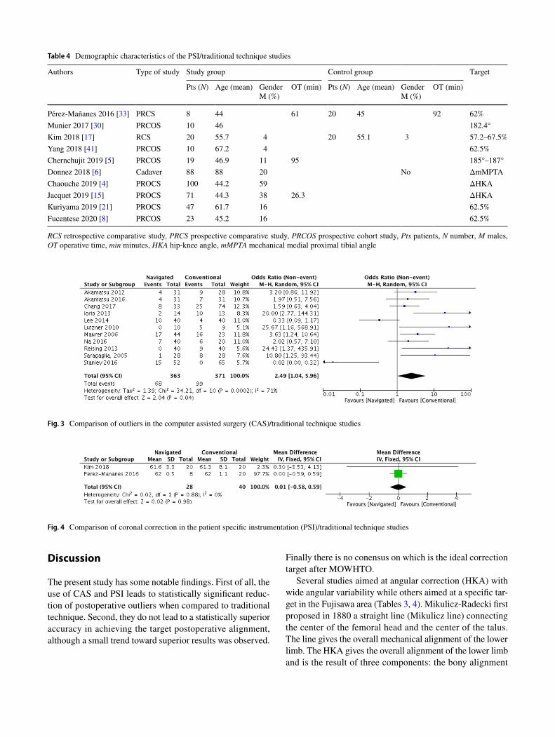
Discussion
The present study has some notable findings. First of all, the use of CAS and PSI leads to statistically significant reduc-tion of postoperative outliers when compared to traditional technique. Second, they do not lead to a statistically superior accuracy in achieving the target postoperative alignment, although a small trend toward superior results was observed.
Finally there is no conensus on which is the ideal correction target after MOWHTO.
Several studies aimed at angular correction (HKA) with wide angular variability while others aimed at a specific tar-get in the Fujisawa area (Tables 3, 4). Mikulicz-Radecki first proposed in 1880 a straight line (Mikulicz line) connecting the center of the femoral head and the center of the talus. The line gives the overall mechanical alignment of the lower limb. The HKA gives the overall alignment of the lower limb and is the result of three components: the bony alignment
Table 4 Demographic characteristics of the PSI/traditional technique studies
RCS retrospective comparative study, PRCS prospective comparative study, PRCOS prospective cohort study, Pts patients, N number, M males, OT operative time, min minutes, HKA hip-knee angle, mMPTA mechanical medial proximal tibial angle
Authors Type of study Study group Control group Target
Pts (N) Age (mean) GenderM (%)
OT (min) Pts (N) Age (mean) GenderM (%)
OT (min)
Pérez-Mañanes 2016 [33] PRCS 8 44 61 20 45 92 62%Munier 2017 [30] PRCOS 10 46 182.4°Kim 2018 [17] RCS 20 55.7 4 20 55.1 3 57.2–67.5%Yang 2018 [41] PRCOS 10 67.2 4 62.5%Chernchujit 2019 [5] PRCOS 19 46.9 11 95 185°–187°Donnez 2018 [6] Cadaver 88 88 20 No ΔmMPTAChaouche 2019 [4] PROCS 100 44.2 59 ΔHKAJacquet 2019 [15] PROCS 71 44.3 38 26.3 ΔHKAKuriyama 2019 [21] PROCS 47 61.7 16 62.5%Fucentese 2020 [8] PRCOS 23 45.2 16 62.5%
Fig. 3 Comparison of outliers in the computer assisted surgery (CAS)/traditional technique studies
Fig. 4 Comparison of coronal correction in the patient specific instrumentation (PSI)/traditional technique studies

Tabl
e 5
Com
pute
r-ass
isted
surg
ery
(CA
S)/tr
aditi
onal
tech
niqu
e
SG st
udy
grou
p, CG
con
trol g
roup
, COMPL
com
plic
atio
ns, O
UTL
out
liers
, FTA
fem
oro-
tibia
l ang
le
Aut
hors
1st t
arge
t1s
t SG
1st C
G2n
d ta
rget
2nd
SG2n
d C
GCO
MPL
SG
%CO
MPL
CG
%O
UTL
SG
%O
UTL
CG
%
Sara
gagl
ia 2
005
[36]
182–
186
183.
418
44
29M
aure
r 200
6 [2
7]18
2–18
538
70H
anke
mei
er 2
006
[12]
80%
72.1
%79
.7%
00
Kim
200
9 [1
9]18
3–18
518
3.9
182.
762
%62
.3%
58.7
%3
2Lu
tzne
r 201
0 [2
5]80
%80
%79
.4%
055
.5Io
rio 2
011
[14]
182–
186
183.
30
Geb
hard
201
1 [1
0]18
2–18
318
15Le
e 20
12 [2
3]18
2–18
818
2.9
62%
59.9
%15
.4A
kam
atsu
201
2 [2
]19
019
0.1
189.
716
7–17
316
9.3
170.
919
.227
.213
32Re
isin
g 20
13 [3
4]62
%58
.6%
58.6
%0
00
22.5
Iorio
201
3 [1
3]18
2–18
618
0.7
180.
80
18.2
1477
Lee
2014
[22]
62%
56.8
%63
.5%
2510
Rib
eiro
201
4 [3
5]18
3–18
618
3.06
183.
3562
%A
kam
atsu
201
6 [1
](F
TA) 1
65–1
7516
8.5
168.
162
%68
.6%
73.5
%29
19.4
12.9
22.6
Na
2016
[31]
62%
64.5
%62
.9%
1830
Schr
öter
201
6 [3
8]18
2–18
318
1.8
181.
77.
15.
3St
anle
y 20
16 [4
0]58
%57
%49
%36
.928
.8C
hang
201
7 [3
]62
.5%
64.3
%57
.3%
0.43
3424
.433
.8To
tal
7.7%
15.9
%16
%38
.2%

of the femur and the tibia, the wear of the articular cartilage and the slackness of the capsule/ligaments complex around the knee. However it does not give any information on where the deformity is located (bone, cartilage or ligaments).
The first step of a traditional MOWHTO is to establish the desired postoperative alignment and therefore to plan the desired correction. The planning should be performed on full leg standing weight bearing X-ray [22]. During planning, it is easier to consider the Mikulicz line in relation to the % of the tibial plateau rather than the HKA. The physiological Mikulicz line crosses the tibial plateau in the geometrical middle, but with a mechanical axis deviation (MAD) of some 4–8 mm to the medial side [32]. As a results, an inter-section at 45% TPW has to be considered normal while its medial shift is associated with a varus deformity. The most common way to plan the desired correction is to follow the Miniaci and Jakob method [28]. It is based on the necessity to shift the Mikulicz line in a more lateral position to correct the varus deformity. Fujisawa et al. were the first to advocate a postoperative Mikulicz line passing at around 65–70% of the tibial width [9]. This principle roughly corresponds to a postoperative HKA of 183°–185°. Although Dugdale and Noyes hypothesized that different intersection points could provide equivalent outcomes [7], the principles of the cor-rection proposed by Jakob and Miniaci remain accepted. In any case, a shift of 65% is set as the upper boundary, but for some surgeons, when osteoarthritis is severe, this upper boundary might be extended up to 75% [26]. On the contrary the general trend is to reduce the shift toward 55–60% adapt-ing the correction to the individual deformity. This wide variability reflects the different target values found in the present review.
A second aspect emerges from the present review and meta-analysis: the addition of technology do not clearly increases the accuracy of MOWHTO, although a trend toward superior accuracy was observed. The accuracy of bony correction is important; traditional technique has sev-eral drawbacks when compared to CAS and PSI. First of all, errors with the traditional technique can result from inap-propriate planning. When transferring angular correction
into millimeters (of opening or closing wedges) there can be mismatches. This may result from poorly performed X-ray (not real dimensions, malrotations of the limb, poorprojections, wrong assessment of the deformity, absenceof correction of intraarticular deformity) or poor planningitself (wrong drawings, lines, HKA correction). PSI involvespatient specific cutting blocks which are manufactured onpatient-specific 3D-CT scan films and therefore this kindof errors are significantly reduced. In addition there can beintroperative errors associated with direct visual inspectionmethods such as the cable method or grid boards [31]. Theycan also be the consequence of high intra-observer variationsand the low reproducibility of intra-operative assessmenttools, including fluoroscopy-based methods [23, 26]. Boththe CAS and PSI are advantageous in reducing the rate andamount of these errors since they guarantee objective intra-operative references and data. Several previous studies haveshown that navigation assisted HTO is superior in termsof reducing outliers from the predicted postoperative limbalignment [10, 11, 23, 34]. In a similar way the use of patientspecific instrumentation (PSI) can be a reliable option toimprove the accuracy of HTOs [15]. In the present reviewseveral studies compared the accuracy of coronal alignmentbetween the traditional technique and the CAS. Although atrend toward increased accuracy was observed in the CASgroup, the pooled results including subgroup of angularcorrection (mean ± SD) and area (%) described by Fujisawashowed that there was no statistically significant differencebetween the two groups. On the contrary a significant reduc-tion of outliers was observed when CAS was used, althoughsome authors still reported no differences in their series [22,34, 35, 38, 40]. Less studies analyzed the accuracy of PSIand only 2 compared this technique to traditional surgery[17, 33]. The pooled results including subgroup of angularcorrection (mean ± SD) and area (%) described by Fujisawashowed that there was no statistically significant differencebetween the two groups.
The present study has some notable limitations. Firstly, although several databases have been accessed with differ-ent combination of appropriate keywords, it is possible that
Fig. 5 Comparison of complication rate in the computer assisted surgery (CAS)/traditional technique studies

some articles may not have been included in our search. Second, the level of evidence of the included studies was not high because the majority were observational comparative studies rather than randomized controlled. Third, almost all of the available articles in the PSI subgroup are case series with no control group; this could reduce the scientific rel-evance of the reported data. Fourtly, the analysis of postop-erative alignment should be performed at the same time in the different studies.
Although the reported limitations the present study yields some relevant informations for daily practice First of all, CAS and PSI ensure a statistically significant reduction in the rate of postoperative outliers. Second, they do not lead to a statistically superior accuracy in achieving the target post-operative alignment, although a small trend toward superior results was observed. Finally, a lack of consensus on which is the ideal postoperative alignement target clearly emerges from the present review.
Conclusions
New technologies such as CAS and PSI yield reduced rate of outliers after MOWHTO when compared to the traditional surgical technique. Coversely they do not lead to statistically significant improvement in the accuracy of postoperative alignment, At the same time, it is clear that no consensus exists on the ideal correction target to achieve after surgery, in terms of angular corrections (HKA) or target zone on the tibial plateau (Fujisawa area and Mikulicz line).
Compliance with ethical standards
Conflict of interest The authors declare that they have no conflicts of interest.
Funding No funding has been received for this study.
Ethical approval Ethical approval was not required for this study.
References
1. Akamatsu Y, Kobayashi H, Kusayama Y, Kumagai K, Saito T(2016) Comparative study of opening-wedge high tibial osteotomy with and without a combined computed tomography-based andimage-free navigation system. Arthroscopy 32(10):2072–2081
2. Akamatsu Y, Mitsugi N, Mochida Y, Taki N, Kobayashi H,Takeuchi R, Saito T (2012) Navigated opening wedge hightibial osteotomy improves intraoperative correction angle com-pared with conventional method. Knee Surg Sports TraumatolArthrosc 20(3):586–593
Tabl
e 6
Pat
ient
spec
ific
instr
umen
tatio
n (P
SI)/t
radi
tiona
l tec
hniq
ue
SG st
udy
grou
p, CG
con
trol g
roup
, COMPL
com
plic
atio
ns, O
UTL
out
liers
, FTA
fem
oro-
tibia
l ang
le, H
KA
hip-
knee
ang
le, m
MPT
A m
echa
nica
l med
ial p
roxi
mal
tibi
al a
ngle
Aut
hor
1st t
arge
t1s
t SG
1st C
G2n
d ta
rget
2nd
SG2n
d C
GCO
MPL
SG
%CO
MPL
CG
%O
UTL
SG
%O
UTL
CG
%
Pére
z-M
añan
es 2
016
[33]
62%
± 0
.5°
± 1
.1°
183–
185
187
187
Mun
ier 2
017
[30]
182.
4°18
2.3°
0K
im 2
018
[17]
57.2
–67.
5%61
.6%
61.3
%18
3°–1
86°
Δ ±
2.3°
Δ ±
6.2
00
2040
Yang
201
8 [4
1]62
.5%
60.2
%4.
9C
hern
chuj
it 20
19 [5
]18
5°–1
87°
Δ ±
5.1
Don
nez
2018
[6]
Δm
MPT
AΔ
mM
PTA
± 0.
2°C
haou
che
2019
[4]
ΔH
KA
ΔH
KA
±1°
10Ja
cque
t 201
9 [1
5]Δ
HK
AΔ
HK
A±
1°57
.2–6
7.5%
Kur
iyam
a 20
19 [2
1]62
.5%
61.5
%24
Fuce
ntes
e 20
20 [8
]62
.5%
Plan
ned
HK
A 9
.7°
8.9°
1326
.1

3. Chang J, Scallon G, Beckert M, Zavala J, Bollier M, Wolf B,Albright J (2017) Comparing the accuracy of high tibial osteot-omies between computer navigation and conventional methods.Comput Assist Surg 22(1):1–8
4. Chaouche S, Jacquet C, Fabre-Aubrespy M, Sharma A, Argen-son JN, Parratte S, Ollivier M (2019) Patient-specific cut-ting guides for open-wedge high tibial osteotomy: safety andaccuracy analysis of a hundred patients continuous cohort. IntOrthop 43(12):2757–2765
5. Chernchujit B, Tharakulphan S, Prasetia R, Chantarapanich N,Jirawison C, Sitthiseripratip K (2019) Preoperative planning ofmedial opening wedge high tibial osteotomy using 3D computer-aided design weight-bearing simulated guidance: technique andpreliminary result. J Orthop Surg 27(1):2309499019831455
6. Donnez M, Ollivier M, Munier M, Berton P, Podgorski JP,Chabrand P, Parratte S (2018) Are three-dimensional patient-specific cutting guides for open wedge high tibial osteotomyaccurate? An in vitro study. J Orthop Surg Res 13(1):171
7. Dugdale TW, Noyes FR, Styer D (1992) Preoperative plan-ning for high tibial osteotomy. The effect of lateral tibiofemo-ral separation and tibiofemoral length. Clin Orthop Relat Res274:248–264
8. Fucentese SF, Meier P, Jud L, Köchli GL, Aichmair A, Vlacho-poulos L, Fürnstahl P (2020) Accuracy of 3D-planned patientspecific instrumentation in high tibial open wedge valgisationosteotomy. J Exp Orthop 7(1):7
9. Fujisawa Y, Masuhara K, Shiomi S (1979) The effect of high tibial osteotomy on osteoarthritis of the knee An arthroscopic study of54 knee joints. Orthop Clin N Am 10(3):585–608
10. Gebhard F, Krettek C, Hufner T, Grutzner PA, Stockle U, Imhoff AB, Lorenz S, Ljungqvist J, Keppler P (2011) Reliability of com-puter-assisted surgery as an intraoperative ruler in navigated high tibial osteotomy. Arch Orthop Trauma Surg 131(3):297–302
11. Han SB, Kim HJ, Lee DH (2017) Effect of computer navigationon accuracy and reliability of limb alignment correction following open-wedge high tibial osteotomy: a meta-analysis. Biomed ResInt 2017:3803457
12. Hankemeier S, Hufner T, Wang G, Kendoff D, Zeichen J, ZhengG, Krettek C (2006) Navigated open-wedge high tibial osteotomy: advantages and disadvantages compared to the conventional tech-nique in a cadaver study. Knee Surg Sports Traumatol Arthrosc14(10):917–921
13. Iorio R, Pagnotelli M, Vadalà A, Giannetti S, Di Sette P, Papan-drea P, Conteduca F, Ferretti A (2013) Open-wedge high tibialosteotomy:comparison between manual and computer-assistedtechniques. Knee Surg Sports Traumatol Arthrosc 21(1):113–119
14. Iorio R, Vadalà A, Giannetti S, Pagnotelli M, Di Sette P, Conted-uca F, Ferretti A (2010) Computer-assisted high tibial osteotomy:preliminary results. Orthopedics 33(10):82–86
15. Jacquet C, Sharma A, Fabre M, Ehlinger M, Argenson JN, Par-ratte S, Ollivier M (2019) Patient-specific high-tibial osteotomy’s cutting-guides decrease operating time and the number of fluoro-scopic images taken after a brief learning curve. Knee Surg Sports Traumatol Arthrosc. https ://doi.org/10.1007/s0016 7-019-05637 -6
16. Jung WH, Takeuchi R, Chun CW, Lee JS, Jeong JH (2015)Comparison of results of medial opening-wedge high tibialosteotomy with and without subchondral drilling. Arthroscopy31(4):673–679
17. Kim HJ, Park J, Shin JY, Park IH, Park KH, Kyung HS (2018)More accurate correction can be obtained using a three-dimen-sional printed model in open-wedge high tibial osteotomy. KneeSurg Sports Traumatol Arthrosc 26(11):3452–3458
18. Kim KI, Seo MC, Song SJ, Bae DK, Kim DH, Lee SH (2017)Change of chondral lesions and predictive factors after medialopen-wedge high tibial osteotomy with a locked plate system. AmJ Sports Med 45(7):1615–1621
19. Kim SJ, Koh YG, Chun YM, Kim YC, Park YS, Sung CH (2009) Medial opening wedge high-tibial osteotomy using a kinematicnavigation system versus a conventional method: a 1 year ret-rospective, comparative study. Knee Surg Sports TraumatolArthrosc 17(2):128–134
20. Koshino T, Morii T, Wada J, Saito H, Ozawa N, Noyori K (1989) High tibial osteotomy with fixation by a blade plate for medialcompartment osteoarthritis of the knee. Orthop Clin N Am20(2):227–243
21. Kuriyama S, Morimoto N, Shimoto T, Takemoto M, Nakamura S,Nishitani K, Ito H, Matsuda S, Higaki H (2019) Clinical efficacyof preoperative 3D planning for reducing surgical errors duringopen-wedge high tibial osteotomy. J Orthop Res 37(4):898–907
22. Lee DH, Han SB, Oh KJ, Lee JS, Kwon JH, Kim JI, Patnaik S,Shetty GM, Nha KW (2014) The weight-bearing scanogram tech-nique provides better coronal limb alignment than the navigation technique in open high tibial osteotomy. Knee 21(2):451–455
23. Lee DH, Nha KW, Park SJ, Han SB (2012) Preoperative and post-operative comparisons of navigation and radiologic limb align-ment measurements after high tibial osteotomy. Arthroscopy28:1842–1850
24. Li Y, Zhang H, Zhang J, Li X, Song G, Feng H (2015) Clinicaloutcome of simultaneous high tibial osteotomy and anterior cruci-ate ligament reconstruction for medial compartment osteoarthritisin young patients with anterior cruciate ligament-deficient knees: a systematic review. Arthroscopy 31(3):507–519
25. Lutzner J, Gross AF, Gunther KP, Kirschner S (2010) Precisionof navigated and conventional open-wedge high tibial osteotomyin a cadaver study. Eur J Med Res 30 15(3):117–120
26. Marti CB, Gautier E, Watchtl SW, Jakob RP (2004) Accuracy offrontal and sagittal plane correction in open-wedge high tibialosteotomy. Arthroscopy 20:366–372
27. Maurer F, Wassmer G (2006) High tibial osteotomy: does naviga-tion improve results? Orthopedics 29(10):S130–S132
28. Miniaci A, Ballmer FT, Ballmer PM, Jakob RP (1989) Proximaltibial osteotomy. A new fixation device. Clin Orthop Relat Res246:250–259
29. Moher D, Liberati A, Tetzlaff Altman DG (2014) Preferred report-ing items for systematic reviews and meta-analyses. Ann InternMed 151:264–269
30. Munier M, Donnez M, Ollivier M, Flecher X, Chabrand P,Argenson JN, Parratte S (2017) Can three-dimensional patient-specific cutting guides be used to achieve optimal correction forhigh tibial osteotomy? Pilot study. Orthop Traumatol Surg Res103(2):245–250
31. Na YG, Eom SH, Kim SJ, Chang MJ, Kim TK (2016) The useof navigation in medial opening wedge high tibial osteotomy canimprove tibial slope maintenance and reduce radiation exposure.Int Orthop 40(3):499–507
32. Paley D, Tetsworth K (1992) Mechanical axis deviation of thelower limbs. Preoperative planning of uniapical angular deformi-ties of the tibia or femur. Clin Orthop Relat Res 280:48–64
33. Pérez-Mañanes R, Burrò JA, Manaute JR, Rodriguez FC, Martin JV (2016) 3D Surgical printing cutting guides for open-wedgehigh tibial osteotomy: do it yourself. J Knee Surg 29(8):690–695
34. Reising K, Strohm PC, Hauschild O, Schmal H, Khattab M, Sud-kamp NP, Niemeyer P (2013) Computer-assisted navigation forthe intraoperative assessment of lower limb alignment in high tib-ial osteotomy can avoid outliers compared with the conventionaltechnique. Knee Surg Sports Traumatol Arthrosc 21(1):181–188
35. Ribeiro Ch, Severino NR, de Barros Moraes, Fucs PM (2014)Opening wedge high tibial osteotomy: navigation system com-pared to the conventional technique in a controlled clinical study. Int Orthop 38(8):1627–1631
36. Saragaglia D, Roberts J (2005) Navigated osteotomies around the knee in 170 patients with osteoarthritis secondary to genu varum.Orthopedics 28(10):s1269–s1274
37. Schallberger A, Jacobi M, Wahl P, Maestretti G, Jakob RP (2011)High tibial valgus osteotomy in unicompartmental medial osteo-arthritis of the knee: a retrospective follow-up study over 13–21years. Knee Surg Sports Traumatol Arthrosc 19(1):122–127
38. Schröter S, Ihle C, Elson DW, Döbele S, Stöckle U, Ateschrang A (2016) Surgical accuracy in high tibial osteotomy: coronal equiva-lence of computer navigation and gap measurement. Knee SurgSports Traumatol Arthrosc 24(11):3410–3417
39. Slim K, Nini E, Forestier D, Kwiatkowski F, Panis Y, Chip-poni J (2003) Methodological index for non-randomized stud-ies (MINORS): development and validation of a new instrument. ANZ J Surg 73(9):712–716
40. Stanley JC, Robinson KG, Devitt BM, Richmond AK, WebsterKE, Whitehead TS, Feller JA (2016) Computer assisted align-ment of opening wedge high tibial osteotomy provides limitedimprovement of radiographic outcomes compared to fluoroscopic alignment. Knee 23(2):289–294
41. Yang JC, Chen CF, Luo CA, Chang MC, Lee OK, Huang Y, LinSC (2018) Clinical experience using a 3D-printed patient-spe-cific instrument for medial opening wedge high tibial osteotomy.Biomed Res Int. https ://doi.org/10.1155/2018/92465 29
Related Documents