1 23 Clinical Oral Investigations ISSN 1432-6981 Clin Oral Invest DOI 10.1007/s00784-015-1470-z Caries removal in deciduous teeth using an Er:YAG laser: a randomized split-mouth clinical trial Rodrigo Alexandre Valério, Maria Cristina Borsatto, Monica Campos Serra, Silvana Aparecida Fernandes Polizeli, Mariana Alencar Nemezio, et al.

Welcome message from author
This document is posted to help you gain knowledge. Please leave a comment to let me know what you think about it! Share it to your friends and learn new things together.
Transcript

1 23
Clinical Oral Investigations ISSN 1432-6981 Clin Oral InvestDOI 10.1007/s00784-015-1470-z
Caries removal in deciduous teeth using anEr:YAG laser: a randomized split-mouthclinical trial
Rodrigo Alexandre Valério, MariaCristina Borsatto, Monica CamposSerra, Silvana Aparecida FernandesPolizeli, Mariana Alencar Nemezio, et al.

1 23
Your article is protected by copyright and
all rights are held exclusively by Springer-
Verlag Berlin Heidelberg. This e-offprint is
for personal use only and shall not be self-
archived in electronic repositories. If you wish
to self-archive your article, please use the
accepted manuscript version for posting on
your own website. You may further deposit
the accepted manuscript version in any
repository, provided it is only made publicly
available 12 months after official publication
or later and provided acknowledgement is
given to the original source of publication
and a link is inserted to the published article
on Springer's website. The link must be
accompanied by the following text: "The final
publication is available at link.springer.com”.

ORIGINAL ARTICLE
Caries removal in deciduous teeth using an Er:YAG laser:a randomized split-mouth clinical trial
Rodrigo Alexandre Valério1 & Maria Cristina Borsatto1 &
Monica Campos Serra2 & Silvana Aparecida Fernandes Polizeli1 &
Mariana Alencar Nemezio1 & Rodrigo Galo3 & Carolina Patrícia Aires4 &
Ana Carolina dos Santos4 & Silmara Aparecida Milori Corona2
Received: 21 November 2014 /Accepted: 3 April 2015# Springer-Verlag Berlin Heidelberg 2015
AbstractObjectives The aim of the present clinical randomized split-mouth study was to evaluate the effectiveness and efficiencyof an Er:YAG laser for caries removal in primary molars,microbiological dentin analysis, and clinical restorations after1 year in 29 children.Materials and methods The children’s teeth were random-ized into two groups: (I) an Er:YAG laser group and (II) abur preparation group. The efficiency of the treatments(the time necessary for the removal of carious tissue)was evaluated based on the time spent on caries removalin the deciduous molars. The effectiveness (caries remov-al capacity) of the caries removal was determined bymeans of a blind test in which the examiner performed atactile and visual examination of the dentin. Microbiolog-ical analysis was performed by counting the Streptococcusmutans and Lactobacillus sp in the remaining dentin.
Clinical analysis of restorations was performed using theUSPHS method in combination with photographs of re-stored teeth, 7 days after the restorative procedure andagain after 1 year. All cavities were restored with theAdper Single Bond 2/Filtek Z350 system. The obtaineddata were analyzed with a significance level of 5 %.Results The Er:YAG laser was less effective and had the sameefficacy as bur preparation during caries removal at the pulpalwall of deciduous molars. In the surrounding walls, bur prep-aration was the more effective method. Regardless of themethod employed, the affected dentin in the pulpal wall hadsimilar amounts of S. mutans and Lactobacillus sp. The res-torations were clinically accepted by the USPHS method overa 1-year period.Conclusion It can be concluded that caries removal with anEr:YAG laser has no influence on the clinical behavior ofrestorations.
* Silmara Aparecida Milori [email protected]
Rodrigo Alexandre Valé[email protected]
Maria Cristina [email protected]
Monica Campos [email protected]
Silvana Aparecida Fernandes [email protected]
Mariana Alencar [email protected]
Rodrigo [email protected]
Carolina Patrícia [email protected]
Ana Carolina dos [email protected]
1 Clinical Pediatric Dentistry Department, Ribeirão Preto School ofDentistry/ São Paulo University, Café Avenue, Monte Alegre,Ribeirão Preto, SP 14040-904, Brazil
2 Restorative Dentistry Department, Ribeirão Preto School ofDentistry/ São Paulo University, Café Avenue, Monte Alegre,Ribeirão Preto, SP 14040-904, Brazil
3 Department of Dental Materials and Prosthodontics, AraraquaraDental School, State Sao Paulo University, Humaitá, 1680 Street,Centro, Araraquara, SP 14801-903, Brazil
4 School of Pharmaceutical Sciences of Ribeirão Preto, São PauloUniversity, Café Avenue, s/n, Monte Alegre, RibeirãoPreto, SP 14040-904, Brazil
Clin Oral InvestDOI 10.1007/s00784-015-1470-z
Author's personal copy

Clinical relevance Irradiation with an Er:YAG laser is appro-priate for caries removal in primary teeth.
Keywords Lasers . Dental caries . Deciduous tooth .
Effectiveness . Efficiency .Microbiological
Introduction
Partial caries removal [1–3] involves the removal of infecteddentin, which is a softened, necrotic, and moist tissue thatcarries a large amount of bacteria [1, 4–6]. The affected dentinon the pulpal and axial walls is dry and less disordered, with asmall number of bacteria. Affected dentin is resistant to re-moval and is capable of remineralization [1, 6, 7] through thetubular sclerosis process and the deposition of tertiary dentin,thus reducing the permeability of the remaining dentin [5].
This procedure has been successfully performed in primary[2] and permanent teeth [3,5], with the advantage of removinga minimum of the remaining sound tooth structure, avoidingpulpal exposure, preserving the vitality of this tissue, andpreventing the progression of lesions [1].
The Er:YAG laser can be used for caries removal, when itswavelength (2.94 μm) coincides with the peak of water ab-sorption and hydroxyl radicals of hydroxyapatite [8]. Thispromotes the effective ablation of the carious tissue [9] viamicroexplosions from the evaporation of the water containedin the mineralized tissue [10]. Furthermore, the Er:YAG laserprovides a conservative treatment [11] in caries removal be-cause of the high absorption in the humid caries tissue. Thisallows for conservative caries excavation without extendingthe preparation into sound tooth structure [12]. Furthermore, itdoes not generate the noise, pressure, or vibration of conven-tional rotary devices [8, 11, 13], making dental treatmentmuch less traumatic, especially for children.
Patients have been shown to have a reduced perception ofpain during laser treatment [13] and thus require less localinfiltrative anesthesia [8, 14]. Laser treatment is thus oftenpreferred over bur preparation by patients [15–17].
The Er:YAG laser, with its bactericidal effect [18], can beemployed in caries removal. Its thermal effects promote thecollapse of bacterial cell structures followed by physicalmicroexplosion [19], reducing the amount of bacteria via theablation and vaporization of intertubular and peritubular den-tin, exposing dentinal tubules, without affecting the surround-ing tissues [20].
To date, there are no studies that evaluate the effectivenessof the Er:YAG laser for caries removal in deciduous teeth andfor the longevity of the restorations performed in thesecavities.
The aim of the present clinical randomized study (split-mouth) was to evaluate the efficiency [13, 21] of the Er:YAGlaser according to the time needed for caries removal in
deciduous molars. The effectiveness [13, 21] of the Er:YAGlaser in caries removal was evaluated by means of a blind testin which the examiner performed a tactile and visual exami-nation of the dentin in deciduous molars. Microbiologicalanalysis was completed by counting Lactobacillus sp andStreptococcus mutans in the remaining dentin. Clinical andphotographic restorations were evaluated based on modifiedUSPHS criteria along with photographs of the restored decid-uous molars after 7 days (baseline) and 1 year after the com-pletion of the restorations.
The null hypotheses to be tested were (1) that caries remov-al using the Er:YAG laser is of similar effectiveness and effi-ciency as bur preparation, (2) that the remaining dentin has thesame number of microorganisms, and (3) that the clinical lon-gevity of the restorations after 1 year is similar between thetwo methods of caries removal.
Materials and methods
Experimental design
The study factor method was employed for caries removalusing (I) the Er:YAG laser (250 mJ/4 Hz) and (II) bur prepa-ration (low speed turbine-control). The experimental samplesfor the randomized split-mouth clinical study consisted of 42children (n=42) and 84 counterpart primary molars with ac-tive carious lesions and cavitation reaching the dentin, locatedat the occlusal surface (class I). Twenty-nine children wereevaluated 1 year after the restorative procedure. The experi-mental design used a randomized complete block, and theresponse variables used to test the efficiency of the cariesremoval were evaluated by means of the time needed for theprocedure, the effectiveness of the partial caries as assessed byvisual and tactile information, microbiological analysis bycounting Lactobacillus sp and S. mutans and clinical (modi-fied USPHS method) and photographic analyses of therestorations.
Ethical aspects
The present study was approved by the Committee of Ethicsin Research at the Ribeirão Preto School of Dentistry – USP(2010.1.159.58.3). The children’s parents or guardians wereinformed about the purpose of the study and signed the Termsof Consent agreeing to participate in the research.
The sample size calculation was based on the amount ofchildren examined over a 9 month period at the Pediatric Den-tal Clinic (2100 children/9 months, all of whom met the in-clusion criteria for this research). The confidence level wasestimated to 95 % with an error of 5 %, representing 1.0 %of the population of Ribeirão Preto, São Paulo, Brazil. Thisanalysis included 33 children. The calculation of n real
Clin Oral Invest
Author's personal copy

samples included 29 children. Thus, after the sample calcula-tion, the sample was established in n=29 children for thepresent research.
Two thousand and one hundred children of both gendersand between the ages of 6 and 10 were examined. Of these, 42(22 boys and 20 girls) met the inclusion criteria and wereaccepted to take part in the study.
The CONSORT guide [22] for randomized clinical trialswas followed for the study design. Figure 1 represents theCONSORT diagram, which discriminates in detail the recruit-ment form, allocation, monitoring, and analysis of the re-search subjects. During the monitoring period, 11 childrenwere lost to follow-up, leaving a total of n=29 participants(13 girls and 16 boys) for the final analysis.
Clinical examinations were performed under adequate illu-mination, followed by standardized radiographic examinationwith bitewing radiographs, using positioned (Jon, São Paulo,SP, Brazil) radiographic film #2 (Kodak, New York, NY,USA), with an exposure of 50 kV, 10 mA, and 0.6 s (Spectro70X, Dabi Atlante, Ribeirão Preto, SP, Brazil). The radiographprocessing was performed automatically (A/T2000 XR, AirTechniques, Melville, New York, NY, USA).
The inclusion criterion for the children included the pres-ence of at least two active carious lesions into the dentin thatwere located on the occlusal surfaces (class I) of contralateraldeciduous molars, with vital pulps and no sealants, amalgam,glass ionomer cement, or composite resin restorations. Theselected teeth all had positive responses to the thermal pulptest performed with Endofrost (Roeko, Langenau, Germany).
Children were excluded if they clinically presented toothpain, spontaneous sensitivity, fistulas, swelling, or mobilitynot compatible with the root rizolisis stage or if they radio-graphically presented with furcal or periapical radiolucencies,increased periodontal space or internal/external dentalreabsorption.
Caries removal
Subject randomization was conducted using a computerspreadsheet. With the aid of a random number generator avail-able at http://randomnumbergenerator.intemodino.com/pt/,the selected children had their names numbered to ordertheir treatment. Teeth were randomly assigned to theexperimental group (Er:YAG laser) or the control group (burpreparation) by coin toss. The different methods of cariesremoval were performed in separate sessions.
The operative field was isolated with a rubber dam(Madeitex, São José dos Campos, SP, Brazil) using clamps#207, #209, or #26 (Duflex, SSWhite, Juiz de Fora, MG,Brazil) depending on the dental morphology of each decidu-ous molar. After absolute isolation was achieved, the cariesremoval was performed with the Er:YAG laser on the
deciduous molar of one hemiarch and with bur preparation(using a low-speed turbine) of the contralateral deciduousmolar.
In the experimental group, the access to the carious lesion(removal of the cavosurface enamel) and the caries removalwas completed with the Er:YAG laser (Fidelis Er III, Fotona,Ljubljana, Slovenia) in the MSP mode, using a pen (R02), atthe noncontact mode with focal distance of 7 mm, a pulseenergy of 250 mJ, a pulse frequency of 4 Hz [14], an outputbeam diameter of 0.9 mm, an energy density of 39 J/cm2, andunder water spray (6 mL/min). Both the patient and the oper-ator wore protective glasses during the laser treatment. Treat-ment took place in a room specifically prepared for this type oftreatment, in accordance with the general guidelines for safelaser application.
In the control group, the caries removal was performedusing spherical carbide drills #½, #1, and #2 (KG Sorensen,Barueri, SP, Brazil), which were compatible with the cavitysize, mounted in low-speed turbines (Dabi Atlante, RibeirãoPreto, SP, Brazil).When necessary, access to the carious lesion(removal of the cavosurface enamel) was performed usingspherical diamond burs #1012 and #1014 (KG Sorensen,Barueri, SP, Brazil), which were also compatible with thecavity and which were mounted in high-speed turbines (DabiAtlante, Ribeirão Preto, SP, Brazil).
Caries removal was initiated in the superficial layer of in-fected dentin from the surrounding walls of deciduous molarsusing either the Er:YAG laser or bur preparation. The affecteddentin, which is hardened, dry, resistant to curettage, and sus-ceptible to remineralization, was left in the pulpal wall [1, 3,5]. It was checked with a probe and evaluated based on clin-ical criteria of consistency and texture [4]. Only the incom-plete removal of the carious tissue from the surrounding wallswas verified according to the clinical hardness criteria [6].Curettes #11, #11½, and #12 (Duflex, SSWhite, Juiz de Fora,MG, Brazil), were used to supplement the total removal,whenever necessary for both groups.
Efficiency evaluation
The treatment efficiency of the control and experimentalgroups was evaluated according to the time required for cariesremoval in the deciduous molars. The infected dentin wascompletely removed from the surrounding walls, and the af-fected dentin present in the pulpal wall was preserved [6]. Aprecision digital timer (CD-2800, InstruthermMeasure Instru-ments Ltda, São Paulo, SP, Brazil) was triggered from the firstpulse emitted by the laser or from the first contact of the drillwith the dental surface. The timer was paused when the oper-ator signaled that caries removal was finished. All teeth wereprepared and restored by the same operator, with the presenceof an auxiliary.
Clin Oral Invest
Author's personal copy

Effectiveness evaluation
To evaluate the effectiveness of the procedures, the exami-ner was trained. To determine the effectiveness of thecaries removal, one examiner, who was blinded to whichmethod was employed, performed a tactile and visual exa-mination. During the tactile and visual examination, ablunt instrument with an active tip was used to evaluatethe caries removal from the surrounding walls accordingto the hardness clinical criteria [6] and at the pulpal wallfollowing the clinical criteria for consistency and texture[4]. The examiner scored the tissue as either A (infecteddentin) or B (affected dentin).
Microbiological evaluation
Immediately after caries removal by the Er:YAG laser or burpreparation, the remaining dentin was collected with sterilecurettes #11, #11½, and #12 (Duflex, SSWhite, Juiz de Fora,MG, Brazil) for microbiological analyses. Thus, at least 5 mgof the remaining dentin was weighed to ±0.01 mg (AnalyticalPlus AP 250D, Ohaus Corp., Florham Park, NJ, USA) insterile microcentrifuge tubes, suspended in 1 mL of 0.9 %NaCl solution and sonicated at an amplitude of 20 % for15 s using a sonic dismembrator (CL-334 Digital Fischer Sci-entific Sonicator, Park Lane, Pittsburgh, USA). From this sus-pension, an aliquot of 50 μL of the sonicated suspension was
Fig. 1 CONSORT schematicexplaining the recruitment,allocation, accompaniment, andanalysis of the research subjects
Clin Oral Invest
Author's personal copy
diluted in 0.9 % NaCl, and serial decimal dilutions were inoc-ulated in duplicate using the drop-counting technique in thefollowing culture media: mitis salivarius agar plus 0.2 unitsbacitracin per milliliter and 15 % sucrose (MSB) for themutans streptococci group [23] and Rogosa SL agar for thelactobacillus group. The plates were incubated in 10% CO2 at37 °C for 48 h. The colony-forming units (CFUs) were count-ed, and the results are expressed as CFU/mg remaining dentin.
Restorative treatment
Depending on the depth of the carious lesion, an indirect pulpcap was performed. For deep cavities, calcium hydroxide ce-ment (Dycal, Dentsply Caulk, Milford, DE, USA ) was used,followed by glass ionomer cement (Ketac Molar; 3 M ESPESeefeld, Schleswig-Holstein, Germany). In medium cavities,only glass ionomer cement (Ketac Molar; 3 M ESPE, Seefeld,Schleswig-Holstein, Germany) was used.
The cavity was conditioned with 37 % phosphoric acid gelfor 15 s for enamel and 7 s for dentin [24] and washed withwater for 1 min. The Adper Single Bond 2 adhesive system(3 M ESPE, Saint Paul, MN, USA) was applied in two layerswith a disposable applicator (KGBrush, KG Sorensen, Cotia,SP, Brazil) and light cured (Ultralux (750 mW/cm2), DabiAtlante, Ribeirão Preto, SP, Brazil) following the manufac-turer’s instructions.
To the restoration, the composite resin Filtek Z350 (3MESPE, Saint Paul, MN, USA) was applied in small incrementsand light cured for 20 s, returning the anatomical shape to theteeth.
After the restoration was complete, the isolation was re-moved and occlusal adjustment was performed with carbonpaper (AccuFilm, Parkell, Farmingdale, NY, USA) and
diamond finishing burs (KG Sorensen, Cotia, SP, Brazil).The children returned after 7 days for the final polishing ofthe restorations with abrasive tips (Enhance, Dentsply Caulk,Milford, DE, USA) mounted in a low-speed turbine (DabiAtlante, Ribeirão Preto, SP, Brazil).
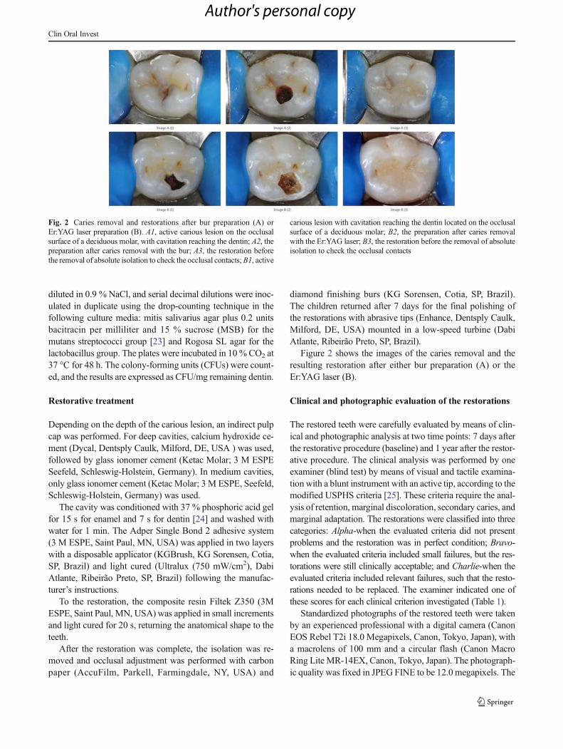
Figure 2 shows the images of the caries removal and theresulting restoration after either bur preparation (A) or theEr:YAG laser (B).
Clinical and photographic evaluation of the restorations
The restored teeth were carefully evaluated by means of clin-ical and photographic analysis at two time points: 7 days afterthe restorative procedure (baseline) and 1 year after the restor-ative procedure. The clinical analysis was performed by oneexaminer (blind test) by means of visual and tactile examina-tion with a blunt instrument with an active tip, according to themodified USPHS criteria [25]. These criteria require the anal-ysis of retention, marginal discoloration, secondary caries, andmarginal adaptation. The restorations were classified into threecategories: Alpha-when the evaluated criteria did not presentproblems and the restoration was in perfect condition; Bravo-when the evaluated criteria included small failures, but the res-torations were still clinically acceptable; and Charlie-when theevaluated criteria included relevant failures, such that the resto-rations needed to be replaced. The examiner indicated one ofthese scores for each clinical criterion investigated (Table 1).
Standardized photographs of the restored teeth were takenby an experienced professional with a digital camera (CanonEOS Rebel T2i 18.0 Megapixels, Canon, Tokyo, Japan), witha macrolens of 100 mm and a circular flash (Canon MacroRing Lite MR-14EX, Canon, Tokyo, Japan). The photograph-ic quality was fixed in JPEG FINE to be 12.0 megapixels. The
Fig. 2 Caries removal and restorations after bur preparation (A) orEr:YAG laser preparation (B). A1, active carious lesion on the occlusalsurface of a deciduous molar, with cavitation reaching the dentin; A2, thepreparation after caries removal with the bur; A3, the restoration beforethe removal of absolute isolation to check the occlusal contacts; B1, active
carious lesion with cavitation reaching the dentin located on the occlusalsurface of a deciduous molar; B2, the preparation after caries removalwith the Er:YAG laser; B3, the restoration before the removal of absoluteisolation to check the occlusal contacts
Clin Oral Invest
Author's personal copy
camera was handled in the manual mode ISO 200, F-22, witha speed of 100 and RGB color space. Each photo was evalu-ated for acceptability and quality, and if it was not acceptable,the photograph was retaken. The same examiner who per-formed the clinical analyses verified the photographs fromthe restored teeth in a flat panel display and could comparethe data obtained from the clinical analyses with the photo-graphs to reach an accurate result.
Statistical analysis
The experimental data on the efficiency and effectiveness werestatistically evaluated by a t test (parametric), with a signifi-cance level of 5%. TheMann-Whitney test, with a significancelevel of 5 %, was employed for the microbiological analysis ofthe remaining dentin after caries removal. The Kruskal-Wallistest (p≤0.05) was used on the values from the evaluation after7 days and after 1 year to assess the following parameters forthe laser - and bur - prepared specimens: retention, marginaldiscoloration, secondary caries, and marginal adaptation. Sta-tistical analysis was performed using SPSS software for Win-dows, version 12.0 (SPSS Inc., Chicago, IL, USA).
Results
The results showed that the efficiency (in seconds) of theEr:YAG laser for caries removal in deciduous molars wasstatistically lower (p=0.019) than that for bur preparation, asshown in Table 2.
The results showed that the effectiveness of the caries re-moval, as measured from the pulpal wall of deciduous molars,was similar between the two groups (p=0.05).
For caries removal in the surrounding walls, the resultsshowed that the bur preparation method was more effective(p=0.0001). Necrotic tissue was observed in 11 samples afterEr:YAG laser irradiation.
The counts of mutans streptococci and lactobacilli in theremaining dentin collected after preparation did not differ(p<0.05) between the two treatments (Table 3).
The clinical and photographic analysis of the restorationswere performed at two time points: 7 days after the restorativeprocedure (baseline) and 1 year after the treatment; the resultsdemonstrated that there were no statistically significant differ-ences between the restorations placed after caries removalwith the Er:YAG laser or the bur, as evaluated according toUSPHS criteria. These criteria included retention, marginaldiscoloration, secondary caries, and marginal adaptation (p≤0.05) (Chart 1).
Discussion
The null hypothesis was rejected for the response variableefficiency. The results from this study demonstrated that theefficiency of the Er:YAG laser for caries removal in deciduousteeth is lower than conventional bur preparation.
These results may be due to the decreased tactile sensitivitywith the Er:YAG laser, making the preparation more difficult,especially for posterior teeth, as treatment must be interruptedin order for the operator to continually verify the presence ofremaining infected dentin with curettes. For conventional burpreparation, the operator knows when to stop treatment basedon haptic feedback.
A previous clinical study [14] described a longer workingtime when the Er:YAG laser was employed, leading to mini-mal discomfort [8] compared to the bur preparation in decid-uous teeth. However, the technique for the total removal of
Table 1 Modified USPHScriteria employed during theevaluation of restorations
Category Score Criteria
Retention Alpha Without loss of restorative material
Charlie With loss of restorative material
Marginal discoloration Alpha Without marginal discoloration
Bravo Slight marginal discoloration, without axial penetration
Charlie Marginal discoloration with axial penetration
Secondary caries Alpha No recurrence of caries
Charlie With recurrence of caries
Marginal adaptation Alpha Perfectly adapted, without visible edges
Bravo Visible edge, but clinically acceptable
Charlie Marginal leakage, clinical failure
Table 2 Efficiency of caries removal after Er:YAG laser/bur prepara-tion (in seconds)
Methods Number Mean Std. error mean
Er:YAG laser 29 110.24 9.83
Bur preparation 29 54.96 5.64
t Test, p=0.019
Clin Oral Invest
Author's personal copy

carious lesions and the cavities were not standardized. Similarworking times were observed for the Er:YAG laser and burpreparation by DenBesten et al. [11] for a sample set contain-ing both deciduous and permanent teeth. Other studies, usingonly permanent teeth, found similar results in in vitro [21] andin vivo samples [15, 16]. However, it is difficult to comparethe results from the present study with those that exist in theliterature because no previous study mentions as the cariesremoval was performed.
The null hypothesis was rejected for the response variableeffectiveness. The Er:YAG laser does not curve, so it wasdifficult to remove all of the carious tissues from the surround-ing walls. In the present study, we sought to maintain thecavosuperficial angle to perform minimally invasive dentistry[13] and preserve unsupported enamel. Thus, 11 samples stillhad necrotic tissue in the surrounding walls after the use of theEr:YAG laser, as determined using sharp curettes. The com-plete removal of carious dentin from the surrounding walls[2–4, 6] is essential to achieve a perfect hermetic seal betweenthe restorative material and the dental substrate, aiming toreduce the supply of exogenous nutrients in the cavities [26].Regarding effectiveness, it is difficult to compare the resultsbetween the published studies, as the effectiveness of lasersdepends on complex interactions between the wavelength,pulse duration, frequency and energy and on the hardness ofthe dental substrate [16]. These procedures are also operatordependent, so there is no way to ensure that only infectedtissue is removed, as the dentin hardness can be related to itsdepth [3]. The results presented in our study demonstrate thatthe Er:YAG laser has the same capacity to remove infecteddentin from the pulpal wall as drills mounted in low-speed
turbines. No statistically significant differences in the com-plete excavation of carious lesion were found in a study byBohari et al. [13] in which they used 245 round bur in a high-speed air turbine hand piece compared to the Er:YAG laser indeciduous teeth. In permanent teeth, Dommisch et al. [15]verified the total removal of carious tissue by visual and tactileexamination and found no statistically significant differencesbetween the use of the Er:YAG laser and a conventional bur.In the study of Schwass et al. [27], the Er:YAG laser irradia-tion resulted in complete removal of demineralized tissue andsome sound enamel was removed to provide access to depthsof the lesion. A comparison of our results with those describedin the literature is difficult because no prior studies have de-scribed how the carious dentin was completely removed fromthe surrounding walls and how caries removal wasstandardized.
The null hypothesis was accepted for the response vari-ables of clinical and photographic restoration evaluation.The results from this study showed no statistically significantdifferences between the restorations placed following burpreparation and those placed following Er:YAG laser prepa-ration in deciduous molars, as assessed by the USPHSmethodproposed by Cvar and Ryge [25] and careful photographicanalysis of restorations after a 1- year period.
This positive result may be due to the ability of the Er:YAGlaser to increase the resistance of enamel to acid deminerali-zation and to reduce acid dissolution, thus preventing the oc-currence of secondary caries [28]. The laser also producesphysical changes, such as melting and recrystallization withpores, which creates a coarse surface that provides amicromechanical bond for adhesives [29]. The increase in
Table 3 The amount ofmicroorganisms remaining in thecavities after caries removal withEr:YAG laser/bur preparation(mean±SD; n=13)
Method Mutans streptococci(CFU/mg remaining dentine×103)
Lactobacilli(CFU/mg remaining dentine×103)
Er:YAG laser 3.6±10.8ª 2.5±7.6a
Bur preparation 2.1±3.3ª 4.0±7.4a
Within columns, distinct letters indicate significant differences among treatment/groups (p<0.05)
Chart 1 Clinical and photographic analysis of the restorations using modified USPHS criteria
Methods Retention Marginal discoloration Secondary caries Marginal adaptation
A B C A B C A B C A B C
Bur preparation
Baseline 29 (100 %) 0 (0 %) 0 (0 %) 29 (100 %) 0 (0 %) 0 (0 %) 29 (100 %) 0 (0 %) 0 (0 %) 29 (100 %) 0 (0 %) 0 (0 %)
1 year 29 (100 %) 0 (0 %) 0 (0 %) 29 (100 %) 0 (0 %) 0 (0 %) 29 (100 %) 0 (0 %) 0 (0 %) 28 (96.5 %) 1 (3.5 %) 0 (0 %)
Er:YAG laser
Baseline 29 (100 %) 0 (0 %) 0 (0 %) 29 (100 %) 0 (0 %) 0 (0 %) 29 (100 %) 0 (0 %) 0 (0 %) 29 (100 %) 0 (0 %) 0 (0 %)
1 year 29 (100 %) 0 (0 %) 0 (0 %) 28 (96.5 %) 1 (3.5 %) 0 (0 %) 29 (100 %) 0 (0 %) 0 (0 %) 27 (93 %) 1 (3.5 %) 1 (3.5 %)
Clin Oral Invest
Author's personal copy

the bond strength in both caries-affected and sound dentin inprimary molars following Er:YAG laser preparation wasverified by Koyuturk et al. [30], and no significant diffe-rence was identified between the laser and bur-preparedcavities using self-etch primers in primary canine teeth[31]. Furthermore, the Er:YAG laser’s bond strength wasfound to be similar to that following bur preparation [32]. Inthe present study, no restorative material was lost in follow-up, as clinically evaluated by the USPHS criteria and pho-tographic analysis.
This study verified that the difficulty in the complete cariesremoval from the surrounding walls of cavities prepared withEr:YAG lasers can be overcome with curettes. This findingsuggests that curettes must always be used after the removal ofcarious lesions with Er:YAG lasers to allow for an enhancedsealing of the adhesive interface.
Clinical and photographic analyses are useful and neces-sary tools for the evaluation of the quality of restorations.Photographic analysis shows an increase in the restorationdefects compared to those clinically and visually observedaccording to the USPHS method [33]. The results from thisstudy are in agreement with Yazici et al. [34] who comparedocclusal restorations using the USPHS method after the re-moval of carious lesions using either a conventional bur oran Er,Cr:YSGG laser.
The null hypothesis was accepted for the response variablemicrobiological evaluation. Although the methods employedfor caries removal could not completely eliminate the viablemicroorganisms, the amount of remaining bacteria was clini-cally irrelevant [15]. The Er:YAG laser reduced the number ofS. mutans and Lactobacillus sp to a similar level as the burpreparation. These results are likely due to the Er:YAG laser’sablation ability on moist surfaces such as infected dentin [27],which contains a greater number of microorganisms than theremaining affected dentin [7]. However, Bönecker et al. [35]observed no differences between the dentin’s consistency andthe level of bacterial colonization. Other changes observed inthe dentin during caries removal, such as changes in the colorand hardness, may not interfere with the level of bacterialviability [5, 36, 37].
The increased temperature during the ablation of the dentincan modify the cell structure [19] of gram-positive bacteria,such as S. mutans and Lactobacillus sp [38], which have re-sistant cell structures that hamper their elimination [39]. Dueto the thermal effects, the Er:YAG laser reduces bacterial via-bility [18] but does not damage the remaining dental structure[20,40].
Here, the presence of S. mutans and Lactobacillus sp wasanalyzed, as they are associated dental caries [37]. Microbio-logical evaluation of carious dentin during caries removal isnot feasible during routine clinical practice, and clinical pa-rameters like hardness, moisture, and color [37] are typicallyused to distinguish infected and affected dentin.
Conclusion
It can be concluded from this study that bur preparation (usingdrills with low-speed rotations) is more efficient for cariesremoval in primary teeth. Both the Er:YAG laser and the burpreparation methods were effective for caries removal fromthe pulpal wall; however, for the surrounding walls, the burpreparation was found to be more effective. The amount ofS. mutans and Lactobacillus sp found on the affected dentinin the pulpal walls was similar after caries removal by bothmethods. The restorations placed after the caries removalusing either the bur preparation or the Er:YAG laser wereclinically acceptable according to USPHS criteria and photo-graphic assessment after a 1- year period.
Acknowledgments This study was supported by FAPESP(2011/00272-5). The author thanks CAPES for the PhD support. Thefunders had no role in the study design, data collection and analysis,decision to publish, or preparation of the manuscript.
Conflict of interest The authors declare that they have no conflicts ofinterest.
References
1. Thompson V, Craig RG, Curro FA, Green WS, Ship JA (2008)Treatment of deep carious lesions by complete excavation or partialremoval: a critical review. J Am Dent Assoc 139:705–712
2. Lula EC, Monteiro-Neto V, Alves CM, Ribeiro CC (2009)Microbiological analysis after complete or partial removal of cari-ous dentin in primary teeth: a randomized clinical trial. Caries Res43:354–358
3. Maltz M, Garcia R, Jardim JJ, de Paula LM, Yamaguti PM, MouraMS, Garcia F, Nascimento C, Oliveira A, Mestrinho HD (2012)Randomized trial of partial vs. stepwise caries removal: 3-year fol-low-up. J Dent Res 91:1026–1031
4. Massara MI, Alves JB, Brandao PR (2002) A traumatic restorativetreatment: clinical, ultrastructural and chemical analysis. Caries Res36:430–436
5. Kidd EAM (2004) How ‘clean’must a cavity be before restoration?Caries Res 38:305–313
6. Maltz M, Oliveira EF, Fontanella V, Carminatti G (2007) Deepcaries lesions after incomplete dentine caries removal: 40-monthfollow-up study. Caries Res 41:493–496
7. Maltz M, Jardim JJ, Mestrinho HD, Yamaguti PM, Podestá K,Moura MS, de Paula LM (2013) Partial removal of carious dentine:a multicenter randomized controlled trial and 18-month follow-upresults. Caries Res 47:103–119
8. Keller U, Hibst R (1997) Effects of Er:YAG Laser in caries treat-ment: a clinical pilot study. Lasers Surg Med 20:32–38
9. Aoki A, Ishikawa I, Yamada T, Otsuki M, Watanabe H, Tagami J,Ando Y, Yamamoto H (1998) Comparison between Er:YAG laserand conventional technique for root caries treatment in vitro. J DentRes 77:1404–1414
10. Hibst R, Keller U (1989) Experimental studies of the application ofthe Er:YAG laser on dental hard substances: I. Measurement of theablation rate. Lasers Surg Med 9:338–344
11. DenBesten PK, White JM, Pelino JEP, Furnish G, Silveira A,Parkins FM (2001) The safety and effectiveness of an Er:YAG laser
Clin Oral Invest
Author's personal copy

for caries removal and cavity preparation in children. Med LaserAppl 16:215–222
12. Baraba A, Perhavec T, Chieffi N, Ferrari M, Anić I, Miletić I (2012)Ablative potential of four different pulses of Er:YAG lasers andlow-speed hand piece. Photomed Laser Surg 30:301–307
13. Bohari MR, Chunawalla YK, Ahmed BM (2012) Clinical evalua-tion of caries removal in primary teeth using conventional,chemomechanical and laser technique: an in vivo study. JContemp Dent Pract 13:40–47
14. Krause F, Braun A, Lotz G, Kneist S, Jepsen S, Eberhard J (2008)Evaluation of selective caries removal in deciduous teeth by a fluo-rescence feedback-controlled Er: YAG laser in vivo. Clin OralInvest 12:209–215
15. Dommisch H, Peus K, Kneist S, Krause F, Braun A, Hedderich J,Jepsen S, Eberhard J (2008) Fluorescence-controlled Er:YAG laserfor caries removal in permanent teeth: a randomized clinical trial.Eur J Oral Sci 116:170–176
16. Mosskull Hjertton P, Bågesund M (2013) Er:YAG laser or high-speed bur for cavity preparation in adolescents. Acta Odontol Scand71:610–615
17. Sarmadi R, Hedman E, Gabre P (2014) Laser in caries treatment –patient’s experiences and opinions. Int J Dent Hyg 12:67–73
18. SchoopU, KlugerW,Moritz A, Nedjelik N, GeorgopoulosA, SperrW (2004) Bactericidal effect of different laser systems in the deeplayers of dentin. Lasers Surg Med 35:111–116
19. Ando Y, Aoki A,Watanabe H, Ishikawa I (1996) Bactericidal effectof erbium YAG laser on periodontopathic bacteria. Lasers SurgMed 19:190–200
20. Dos Santos Antonio MP, Moura-Netto C, Camargo SE,Davidowicz H, Marques MM, Maranhão de Moura AA (2012)Bactericidal effects of two parameters of Er:YAG laser intracanalirradiation: ex-vivo study. Lasers Med Sci 27:1165–1168
21. Messias DCF, de Souza-Gabriel AE, Palma-Dibb RG, RodriguesAL, Serra MC (2006) Efficiency and effectiveness of Er:YAG laseron carious tissue removal. J Oral Laser Appl 6:181–188
22. Schulz KF, Altman DG, Moher D (2010) CONSORT Group -Statement: updated guidelines for reporting parallel grouprandomised trials. Ann Int Med 152:726–732
23. Gold OG, Jordan HV, Hout JV (1973) A selective medium forStreptococcus mutans. Arch Oral Biol 18:1357–1364
24. Torres CP, Chinelatti MA, Gomes-Silva JM, Borsatto MC, Palma-Dibb RG (2007) Tensile bond strength to primary dentin after dif-ferent etching times. J Dent Child 74:113–117
25. Cvar JF, Ryge G (2005) Reprint of criteria for the clinical evaluationof dental restorative materials. Clin Oral Investig 9:215–232
26. Paddick JS, Brailsford SR, Kidd EA, Beighton D (2005)Phenotypic and genotypic selection of microbiota surviving underdental restorations. Appl Environ Microbiol 71:2467–2472
27. Schwass DR, Leichter JW, Purton DG, Swain MV (2013)Evaluating the efficiency of caries removal using an Er:YAG laser
driven by fluorescence feedback control. Arch Oral Biol 58:603–610
28. Apel C, Birker L, Meister J, Weiss C, Gutknecht N (2004) Thecaries-preventive potential of subablative Er:YAG and Er:YSGGlaser radiation in an intraoral model: a pilot study. PhotomedLaser Surg 22:312–317
29. Wen X, Liu L, Nie X, Zhang L, Deng M, Chen Y (2010) Effect ofpulse Nd:YAG laser on bond strength and microleakage of resin tohuman dentine. Photomed Laser Surg 28:741–746
30. Koyuturk AE, Ozmen B, Cortcu M, Tokay U, Tosun G, Erhan SM(2014) Effects of Er:YAG laser on bond strength of self-etchingadhesives to caries-affected dentin. Microsc Res Tech 77:282–288
31. Bahrololoomi Z, Heydari E (2014) Assessment of ToothPreparation via Er:YAG Laser and Bur on Microleakage ofDentin Adhesives. J Dent (Tehran) 11:172–178
32. Yamada Y, Hossain M, Nakamura Y, Murakami Y, Matsumoto K(2002) Microleakage of composite resin restoration in cavities pre-pared by Er:YAG laser irradiation in primary teeth. Eur J PaediatrDent 3:39–45
33. Moncada G, Silva F, Angel P, Oliveira OB Jr, FresnoMC, CisternasP, Fernandez E, Estay J, Martin J (2014) Evaluation of dental res-torations: a comparative study between clinical and digital photo-graphic assessments. Oper Dent 39:45–56
34. Yazici R, Baseren M, Gorucu J (2010) Clinical comparison of bur-and laser-prepared minimally invasive occlusal resin composite res-torations: two-year follow-up. Oper Dent 35:500–507
35. Bönecker M, Toi C, Cleaton-Jones P (2003) Mutans streptococciand lactobacilli in carious dentine before and after AtraumaticRestorative Treatment. J Dent 31:423–428
36. Banerjee A, Kidd EA, Watson TF (2000) In vitro evaluation of fivealternative methods of carious dentine excavation. Caries Res 34:144–150
37. Kidd EA, Ricketts DN, Pitts NB (1993) Occlusal caries diagnosis: achanging challenge for clinicians and epidemiologists. J Dent 21:323–331
38. Albuquerque R Jr, Head TW, Mian H, Rodrigo A, Müller K,Sanches K, Ito IY (2004) Reduction of salivary S aureus andmutans group streptococci by a preprocedural chlorhexidine rinseand maximal inhibitory dilutions of chlorhexidine andcetylpyridinium. Quintessence Int 35:635–640
39. Moritz A, Jakolitsch S, Goharkhay K, Schoop U, Kluger W,Mallinger R, Sperr W, Georgopoulos A (2000) Morphologicchanges correlating to different sensitivities of Escherichia coliand enterococcus faecalis to Nd:YAG laser irradiation through den-tin. Lasers Surg Med 26:250–261
40. Kita T, Ishii K, Yoshikawa K, Yasuo K, Yamamoto K, Awazu K(2014) In vitro study on selective removal of bovine demineralizeddentin using nanosecond pulsed laser at wavelengths around5.8 μm for realizing less invasive treatment of dental caries.Lasers Med Sci 7 [Epub ahead of print].
Clin Oral Invest
Author's personal copy
Related Documents


![ÿ j¯ dû3ñÎ+ÇzU¸ç '´nKyEÅ!ö'8 /® øð ¹Ì ß · [chemo-mechanical debridement] D LhHlJ 1900 Kerr [Kerr Broach] 100 ákJ CD Er:YAG Er:YAG (2.94 um) Er:YAG o Z Er:YAG HV:](https://static.cupdf.com/doc/110x72/5b7512f87f8b9a884c8cea26/y-j-du3niczuc-nkyeaoe8-od-i-ss-chemo-mechanical-debridement.jpg)