Cardiovascular System Northwest Rankin High School Human A&P

Cardiovascular System Northwest Rankin High School Human A&P.
Dec 13, 2015
Welcome message from author
This document is posted to help you gain knowledge. Please leave a comment to let me know what you think about it! Share it to your friends and learn new things together.
Transcript

Cardiovascular SystemNorthwest Rankin High School
Human A&P

Introduction
2
The cardiovascular system consists of the heart and vessels (arteries, capillaries and veins.)
A functional cardiovascular system is vital for supplying oxygen and nutrients to tissues and removing wastes from them.
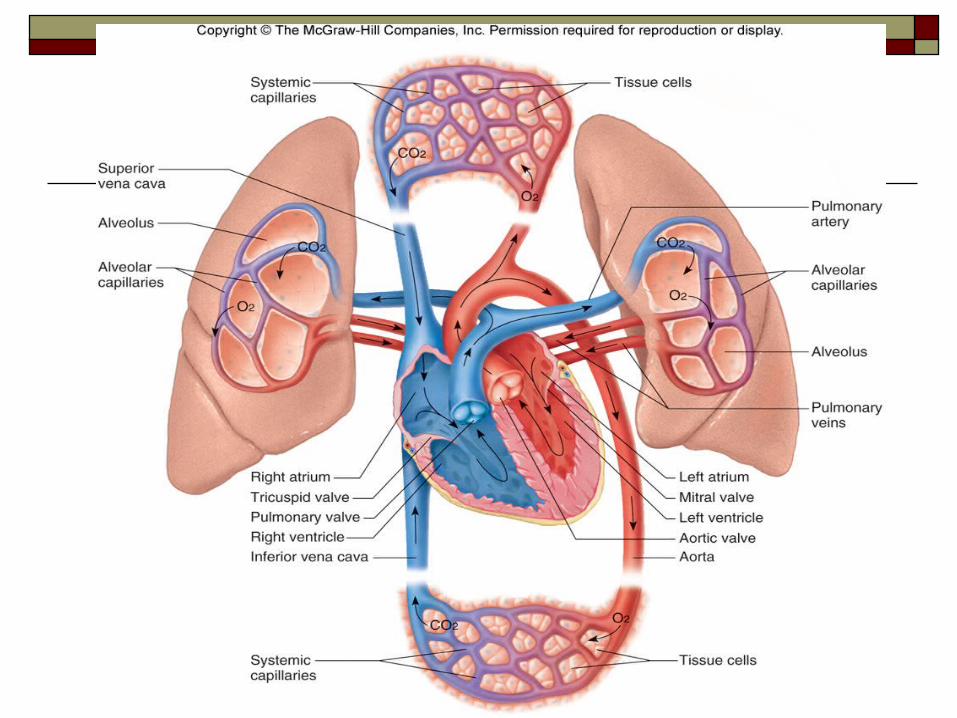
Deoxygenated blood is carried by the pulmonary circuit to the lungs, while the systemic circuit sends oxygenated blood to all body cells.
CopyrightThe McGraw-Hill Companies, Inc. Permission required for reproduction or display.

3

Size of Heart The heart is about the
size of a person’s fist. It is hollow, cone
shaped and weighs less than one pound.
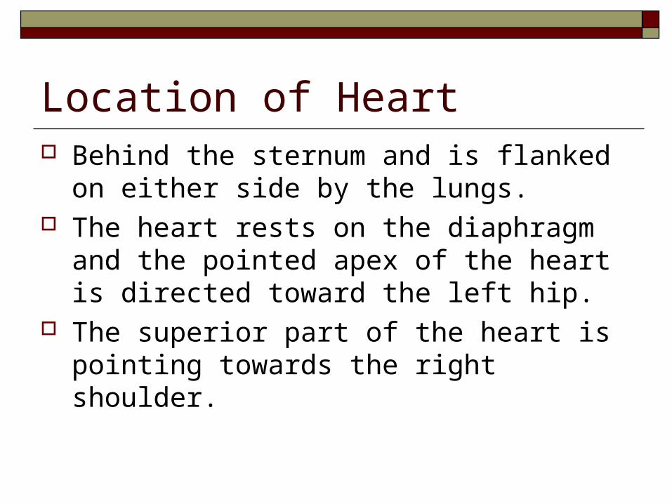
Location of Heart Behind the sternum and is flanked on either
side by the lungs. The heart rests on the diaphragm and the
pointed apex of the heart is directed toward the left hip.
The superior part of the heart is pointing towards the right shoulder.

Walls of the Heart The heart is enclosed by a double sac called
pericardium. The visceral pericardium, or epicardium tightly
hugs the heart. The parietal pericardium is the outside layer that
anchors the heart to the surrounding structures (diaphragm and sternum).
Between the pericardial layers is a fluid to reduce friction.

7

Layer of the Heart Wall Epicardium – outside layer
Made of connective and epithelial tissue Houses blood and lymph capillaries as well as the
coronary arteries
Myocardium – middle layer thick layers of cardiac muscle
Endocardium –the inside layer Very smooth Made of connective and epithelial tissue Contains Purkinje fibers

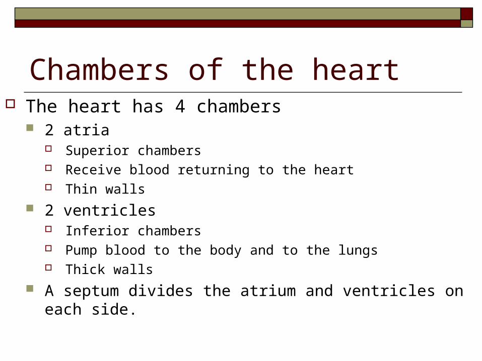
Chambers of the heart The heart has 4 chambers
2 atria Superior chambers Receive blood returning to the heart Thin walls
2 ventricles Inferior chambers Pump blood to the body and to the lungs Thick walls
A septum divides the atrium and ventricles on each side.

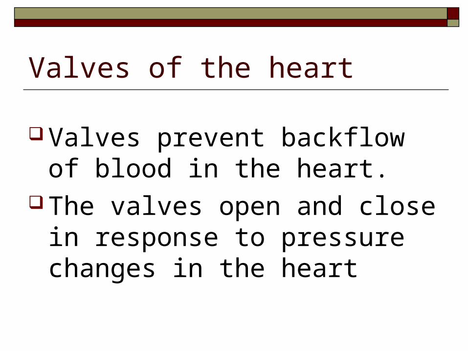
Valves of the heart
Valves prevent backflow of blood in the heart.
The valves open and close in response to pressure changes in the heart


Atrioventricular (AV) Valves Each side of the heart has an AV valve to ensure
one way flow of blood There are two AV valves:
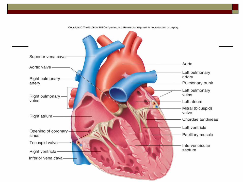
Biscuspid (mitral) valve – LEFT AV valve with 2 cusps, between the left atria and ventricle
Tricuspid valve – RIGHT AV valve with three cusps located between the right atria and ventricle
Each AV valve cusps attach to chordae tendinae which are attached to papillary muscles.

AV Valves The AV valves open during heart relaxing and
close when the heart contracts to prevent blood from returning to the atria.
When the heart is relaxed and blood is passively filling its chambers, the AV- valve flaps hang limply into the ventricles. As the ventricles contract, the pressure begins to rise and forces the valves closed. This prevents backflow into the atria when the ventricles are contracting.

Semilunar Valves Each side of the heart also has a semilunar
valve to further ensure one way flow of blood There are also two semilunar valves
Pulmonary – in the pulmonary artery leaving the heart to the lungs.
Aortic – in the aortic arch leaving the heart to the body.

Semilunar Valves The semilunar valves open during heart contraction
and close to prevent backflow into the ventricles when relaxing.
When the ventricles are contracting and forcing blood out of the heart, the cusps are forced open and flattened against the wall of the arteries by the tremendous force of rushing blood. Then when the ventricles relax the blood begins to flow backward toward the heart, and the cusps fill with blood, closing the valves.

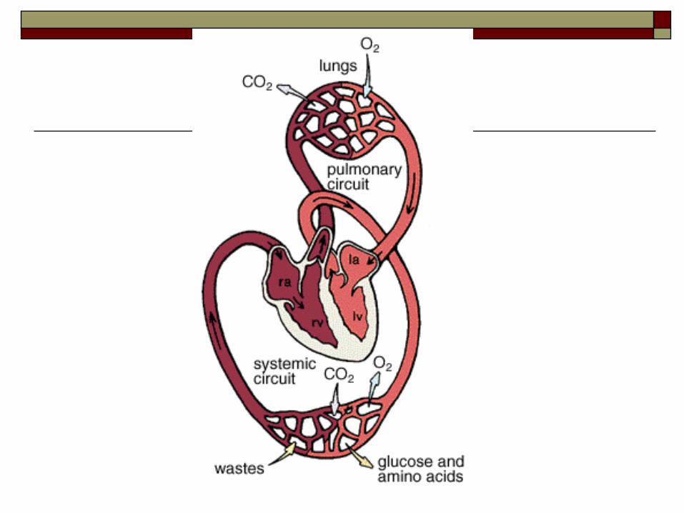
Heart functions as a double pump
Pulmonary circulation – right side of heart to the lungs and back to the left side of the heart. It’s only function is to carry blood to the lungs for gas exchange and then to return it to the heart.
Systemic circulation – left side of the heart through the body tissues and back to the right side of the heart. It supplies oxygen and nutrient rich blood to all body organs.
Path of Blood through the Heart
22
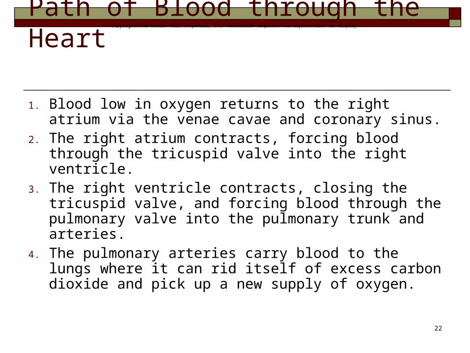
1. Blood low in oxygen returns to the right atrium via the venae cavae and coronary sinus.
2. The right atrium contracts, forcing blood through the tricuspid valve into the right ventricle.
3. The right ventricle contracts, closing the tricuspid valve, and forcing blood through the pulmonary valve into the pulmonary trunk and arteries.
4. The pulmonary arteries carry blood to the lungs where it can rid itself of excess carbon dioxide and pick up a new supply of oxygen.
CopyrightThe McGraw-Hill Companies, Inc. Permission required for reproduction or display.

24
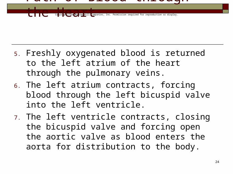
5. Freshly oxygenated blood is returned to the left atrium of the heart through the pulmonary veins.
6. The left atrium contracts, forcing blood through the left bicuspid valve into the left ventricle.
7. The left ventricle contracts, closing the bicuspid valve and forcing open the aortic valve as blood enters the aorta for distribution to the body.
CopyrightThe McGraw-Hill Companies, Inc. Permission required for reproduction or display.Path of Blood through the Heart

Blood Supply to the Heart
26
The first branches off of the aorta, which carry freshly oxygenated blood, are the right and left coronary arteries that feed the heart muscle itself.
Branches of the coronary arteries feed many capillaries of the myocardium.
Cardiac veins drain blood from the heart muscle and carry it to the coronary sinus, which empties into the right atrium.
CopyrightThe McGraw-Hill Companies, Inc. Permission required for reproduction or display.
Heart Actions The cardiac cycle consists of the atria beating in
unison (atrial systole) followed by the contraction of both ventricles, (ventricular systole) then the entire heart relaxes for a brief moment (diastole). It includes events of one complete heartbeat, during
which both atria and ventricles contract and then relax. (0.8 seconds) or 75 times a minute
Common terms: Systole-Ventricle contraction; Diastole- Ventricle relaxation
27
CopyrightThe McGraw-Hill Companies, Inc. Permission required for reproduction or display.
Cardiac Cycle
During the cardiac cycle, pressure within the heart chambers rises and falls with the contraction and relaxation of atria and ventricles.
When the atria fill, pressure in the atria is greater than that of the ventricles, which forces the A-V valves open.
Pressure inside atria rises further as they contract, forcing the remaining blood into the ventricles
28
CopyrightThe McGraw-Hill Companies, Inc. Permission required for reproduction or display.

Cardiac Cycle
When ventricles contract, pressure inside them increases sharply, causing A-V valves to close and the aortic and pulmonary valves to open.
As the ventricles contract, papillary muscles contract, pulling on chordae tendinae and preventing the backflow of blood through the A-V valves.
29
CopyrightThe McGraw-Hill Companies, Inc. Permission required for reproduction or display.

30

Heart Sounds
Heart sounds are due to vibrations in heart tissues as blood rapidly changes velocity within the heart.
Heart sounds can be described as a "lubb-dupp" sound. The first sound (lubb) occurs as ventricles contract
and A-V valves are closing. The second sound (dupp) occurs as ventricles relax
and aortic and pulmonary valves are closing31
CopyrightThe McGraw-Hill Companies, Inc. Permission required for reproduction or display.
http://www.smm.org/heart/lessons/lesson4.htm

Controlling systems of the heart Without a controlling system, the heart would
be an inefficient pump There are two controlling systems
Intrinsic Conduction System Autonomic nervous system

Intrinsic Conduction System Specialized cardiac muscle tissue conducts impulses
throughout the myocardium and comprises the intrinsic conduction system. It is not found anywhere else in the body It causes heart muscle depolarization in only one direction
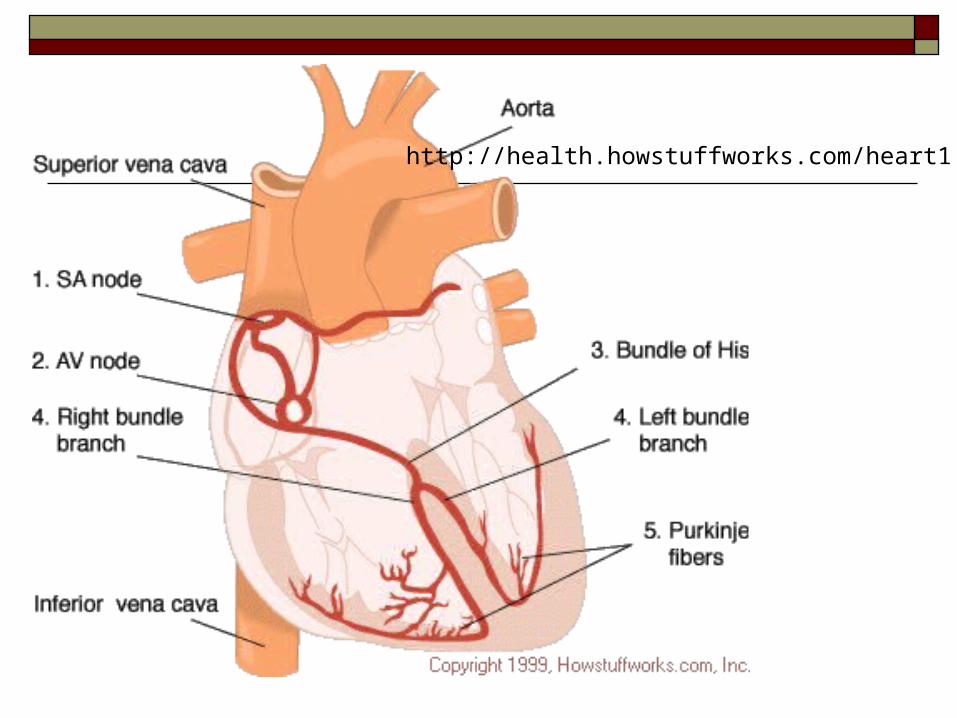
from the atria to the ventricles The Intrinsic Conduction System consists of an SA node
(pacemaker), an AV node , the AV bundle (bundle of His) and bundle branches, and the Purkinje fibers

SA Node A self-exciting mass of specialized cardiac
muscle called the sinoatrial node (S-A node or pacemaker), located on the posterior right atrium, generates the impulses for the heartbeat.
34
CopyrightThe McGraw-Hill Companies, Inc. Permission required for reproduction or display.

AV Node Impulses spread next to next grouping of
cells, it contracts, and impulses then travel to the junctional fibers leading to the atrioventricular node (A-V node) located in the septum.
Junctional fibers are small, allowing the atria to contract before the impulse spreads rapidly over the ventricles.
35
CopyrightThe McGraw-Hill Companies, Inc. Permission required for reproduction or display.

Bundle of His and Purkinje fibers From the AV Node signals spread to the AV
bundle (Bundle of His) And then the branches of the A-V bundle give
rise to Purkinje fibers leading to papillary muscles
These fibers stimulate contraction of the papillary muscles at the same time the ventricles contract.
37
CopyrightThe McGraw-Hill Companies, Inc. Permission required for reproduction or display.

http://health.howstuffworks.com/heart1.htm
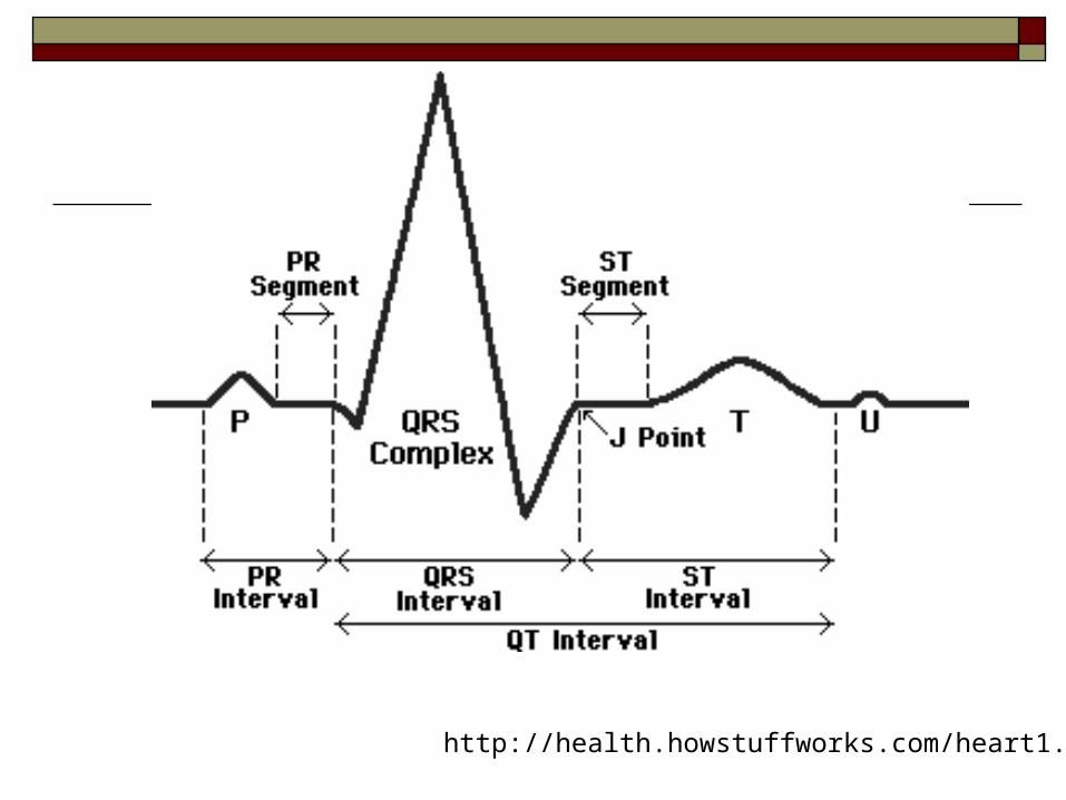
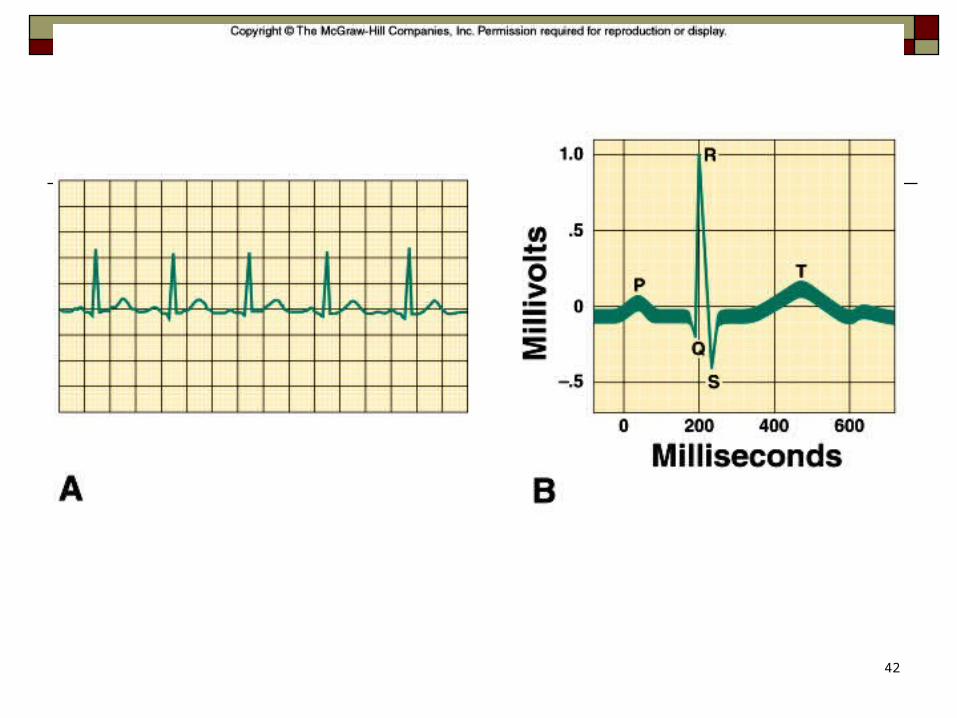
Electrocardiography: ECG All the activity of the heart produces electrical
waves we can measure. The measurement is typically represented as a graph called an electrocardiogram (ECG)
The typical ECG has three recognizable waves P wave: small and signals the depolarization of the atria
immediately before they contract QRS complex: results from the depolarization of the
ventricles, complicated shape; precedes the contraction of the ventricles
T wave: results from currents flowing during the repolarization of the ventricles
http://health.howstuffworks.com/heart1.htm
42
Regulation of the Cardiac Cycle
The amount of blood pumped at any one time must adjust to the current needs of the body
The SA node is innervated by branches of the sympathetic and parasympathetic divisions, so basically the autonomic system of the CNS controls heart rate. Sympathetic impulses increase the speed of heart rate. Heart rate is decreased by parasympathetic impulses
43
CopyrightThe McGraw-Hill Companies, Inc. Permission required for reproduction or display.

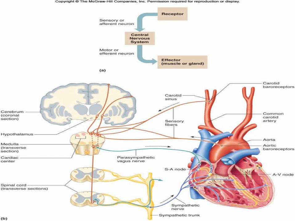
Cardiac control center The cardiac control center of the medulla oblongata
maintains a balance between the sympathetic and parasympathetic divisions of the nervous system in response to messages from baroreceptors which detect changes in blood pressure.
Impulses from the cerebrum or hypothalamus may also influence heart rate, as do body temperature and the concentrations of certain ions.
44
CopyrightThe McGraw-Hill Companies, Inc. Permission required for reproduction or display.
45
Blood Vessels
46
The blood vessels (arteries, arterioles, capillaries, venules, and veins) form a closed tube that carries blood away from the heart, to the cells, and back again.
Large arteries leave heart (aorta) THEN to smaller arteries THEN to arterioles THEN to capillary beds THEN to venules THEN to veins THEN to larger veins (inferior and superior vena cavae) that dump blood back into heart.
CopyrightThe McGraw-Hill Companies, Inc. Permission required for reproduction or display.

Anatomy of Blood Vessels Arteries and veins have 3 layers
Tunica intima (endothelium) – forms slick surface to decrease friction of blood flow (epithelial tissue)
Tunica media – thick in arteries and relatively thin in veins (smooth muscle tissue)
Tunica externa – the outermost tunic basically supports and protects the vessels (connective tissue)
Capillaries have only one layer: tunica intima

Anatomy of Arteries Arteries are strong, elastic vessels adapted for
carrying high-pressure blood. All the major arteries of the systemic circulation are
branches of the aorta, which leaves the left ventricle Arteries are capable of vasoconstriction as directed
by the sympathetic impulses; when impulses are inhibited, vasodilation results.
Arteries become smaller as they divide and give rise to arterioles.

Anatomy of Capillaries
50
Capillaries are the smallest vessels, consisting only of a layer of endothelium through which substances are exchanged with tissue cells.
Capillary permeability varies from one tissue to the next, generally with more permeability in the liver, intestines, and certain glands, and less in muscle and considerably less in the brain (blood-brain barrier).
Precapillary sphincters can regulate the amount of blood entering a capillary bed and are controlled by oxygen concentration in the area. If blood is needed elsewhere in the body, the capillary beds
in less important areas are shut down
CopyrightThe McGraw-Hill Companies, Inc. Permission required for reproduction or display.

51

Exchanges in the Capillaries
52
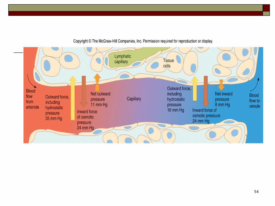
Blood entering capillaries contains high concentrations of oxygen and nutrients that diffuse out of the capillary wall and into the tissues.
Plasma proteins remain in the blood due to their large size. Hydrostatic pressure drives the passage of fluids and very
small molecules out of the capillary at this point. At the venule end, osmosis, due to the osmotic pressure of
the blood, causes much of the tissue fluid to return to the bloodstream.
Lymphatic vessels collect excess tissue fluid and return it to circulation.
CopyrightThe McGraw-Hill Companies, Inc. Permission required for reproduction or display.

53
54

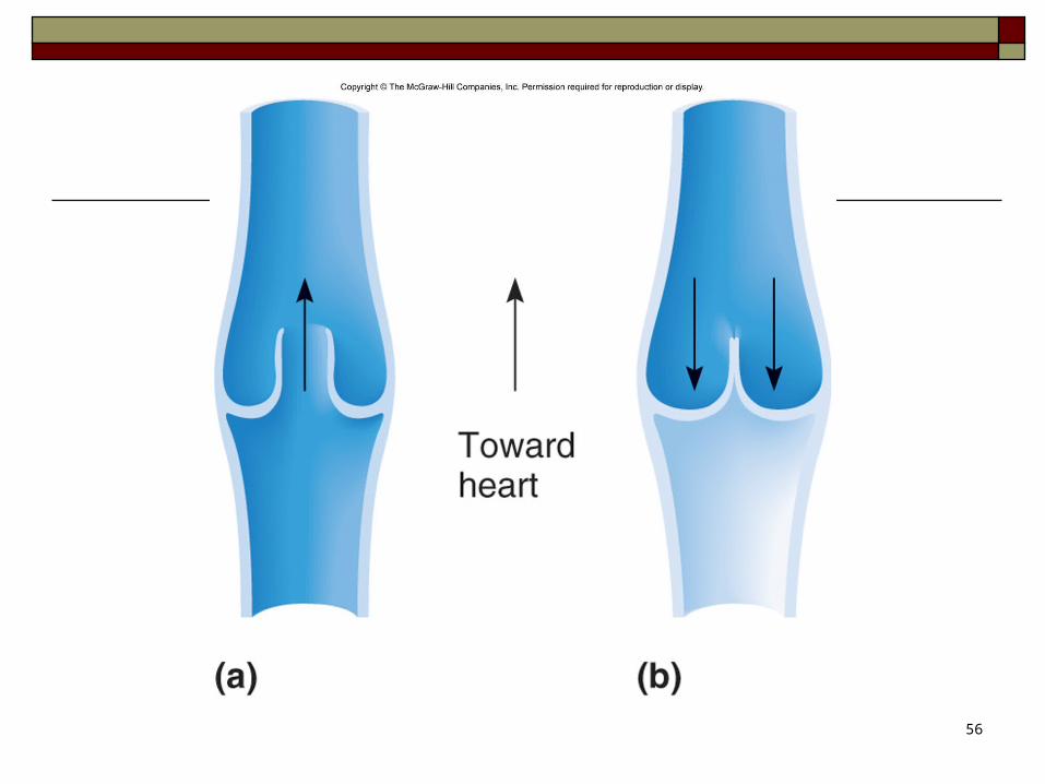
Anatomy of Veins Veins have the same three layers as arteries but the walls are
thinner, their lumens are larger, and they are equipped with valves. These modifications reflect low-pressure nature of veins. The valves prevent the backflow of blood.
The major veins of the systemic circulation ultimately converge on one of the venae cavae. All veins above the diaphragm drain into the superior vena cava, and those below the diaphragm drain into the inferior vena cava. Both venae cavae enter the right atrium of the heart
Varicose veins are caused by incompetent valves in the veins. It is common in the obese and people who stand for long hours
56

Venous Blood Flow
Blood flow through the venous system is only partially the result of heart action and instead also depends on skeletal muscle contraction, breathing movements, and vasoconstriction of veins.
Contractions of skeletal muscle squeeze blood back up veins one valve at a time.
Differences in thoracic and abdominal pressures draw blood back up the veins.
57
CopyrightThe McGraw-Hill Companies, Inc. Permission required for reproduction or display.

Blood Pressure
59
Blood pressure is the force of blood against the inner walls of blood vessels anywhere in the cardiovascular system, although the term "blood pressure" usually refers to arterial pressure.
CopyrightThe McGraw-Hill Companies, Inc. Permission required for reproduction or display.

Arterial Blood Pressure
Arterial blood pressure rises and falls following a pattern established by the cardiac cycle.
During ventricular contraction, arterial pressure is at its highest (systolic pressure).
When ventricles are relaxing, arterial pressure is at its lowest (diastolic pressure).
The surge of blood that occurs with ventricular contraction can be felt at certain points in the body as a pulse
60
CopyrightThe McGraw-Hill Companies, Inc. Permission required for reproduction or display.

61
Arterial pressure depends on 1. heart action2. blood volume3. resistance to flow4. blood viscosity
CopyrightThe McGraw-Hill Companies, Inc. Permission required for reproduction or display.
Factors that Affect Arterial Pressure
62
1. Heart Action Dependent upon stroke volume and heart rate (together
called cardiac output) Stroke Volume: The amount of blood pumped by the left
ventricle of the heart in one contraction Heart Rate: The number of heartbeats per unit of time,
usually per minute. The heart rate is based on the number of contractions of the ventricles.
Cardiac Output: The volume of blood pumped per minute by each ventricle of the heart. Cardiac output is equal to the stroke times the heart rate.
It is used as a measure of the overall health of the heart If cardiac output increases, so does blood pressure.
CopyrightThe McGraw-Hill Companies, Inc. Permission required for reproduction or display.
Factors that Affect Arterial Pressure

63
2. Blood Volume Blood pressure is normally directly proportional
to the volume of blood within the cardiovascular system.
Blood volume varies with age, body size, and gender.
CopyrightThe McGraw-Hill Companies, Inc. Permission required for reproduction or display.
Factors that Affect Arterial Pressure

64
3. Peripheral Resistance Friction between blood and the walls of blood
vessels is a force called peripheral resistance. As peripheral resistance increases, such as during
sympathetic constriction of blood vessels, blood pressure increases.
CopyrightThe McGraw-Hill Companies, Inc. Permission required for reproduction or display.
Factors that Influence Arterial Pressure

65
4. Blood Viscosity The greater the viscosity (ease of flow) of blood,
the greater its resistance to flowing, and the greater the blood pressure.
CopyrightThe McGraw-Hill Companies, Inc. Permission required for reproduction or display.
Factors that Influence Arterial Pressure

66
CopyrightThe McGraw-Hill Companies, Inc. Permission required for reproduction or display.

Control of Blood Pressure
67
Blood pressure is determined by cardiac output and peripheral resistance. The body maintains normal blood pressure by adjusting cardiac output and peripheral resistance. Cardiac output depends on stroke volume and heart
rate, and a number of factors can affect these actions.

Control of Blood Pressure
68
The volume of blood that enters the right atrium is normally equal to the volume leaving the left ventricle. If arterial pressure increases, the cardiac center of
the medulla oblongata sends parasympathetic impulses to decrease heart rate.
If arterial pressure decreases, the medulla oblongata sends sympathetic impulses to increase heart rate to adjust blood pressure.

69
Control of Blood Pressure The vasomotor center of the medulla oblongata can
adjust the sympathetic impulses to smooth muscles in arteriole walls, adjusting blood pressure.
Certain chemicals, such as carbon dioxide, oxygen, and hydrogen ions, can also affect peripheral resistance.
Other factors, such as emotional upset, exercise, and a rise in temperature can result in increased cardiac output and increased blood pressure.
70
CopyrightThe McGraw-Hill Companies, Inc. Permission required for reproduction or display.

71
Related Documents