Cardiovascular Pathophysiology 3 Roman Benacka, MD, PhD Department of Pathophysioloy Medical Faculty, Safarik University, Košice Illustations herein might bve adapted from various printed or electrornic media and serve merely for demonstrational and educational purposes

Welcome message from author
This document is posted to help you gain knowledge. Please leave a comment to let me know what you think about it! Share it to your friends and learn new things together.
Transcript
Cardiovascular
Pathophysiology 3
Roman Benacka, MD, PhD
Department of Pathophysioloy
Medical Faculty, Safarik
University, Košice
Illustations herein might bve adapted from various printed or electrornic media
and serve merely for demonstrational and educational purposes

Physiological review
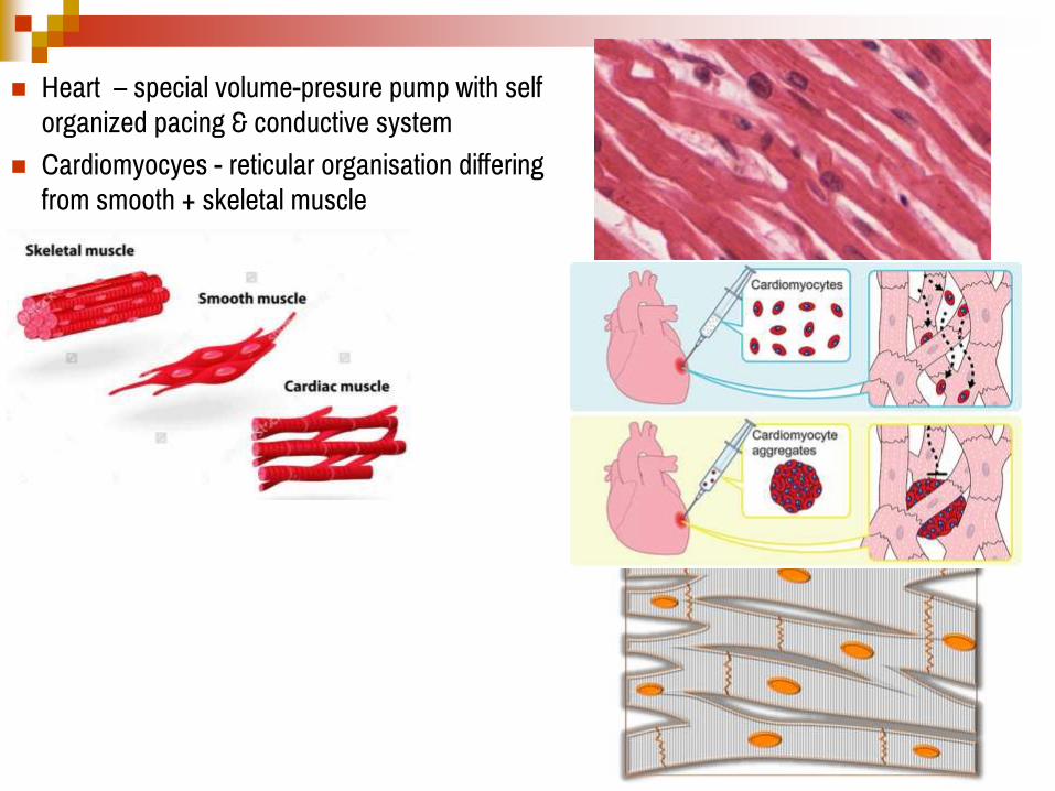
Heart – special volume-presure pump with self
organized pacing & conductive system
Cardiomyocyes - reticular organisation differing
from smooth + skeletal muscle

Cardiomyocytes can conduct electric currents from the cell to cell; intercalated discs elecrtical synapses
Conductive system = not nerves but preformed muscle cells; specific anatomy to organize (direction, speed) heart exitability = basic rhythm
Atria and ventricles are electrically relatively isolated; AV gateway control (+ abnormal bypasses)
Vegetative nerves + hormones modulate chromotropy, dromotropy, batmotropy

Srivastava, D. & Ivey, K. N. Potential of stem-cell-based therapies for heart disease. Nature 441, 1097-1099


Principal ion channels
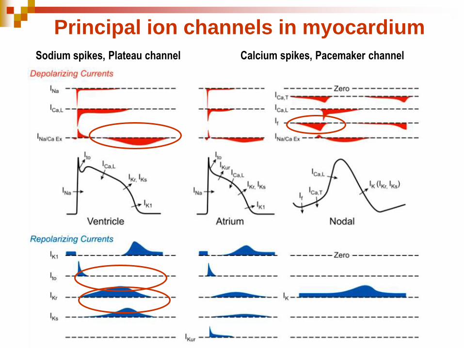
Principal ion channels in myocardium
Calcium spikes, Pacemaker channel Sodium spikes, Plateau channel

Principles of ECG
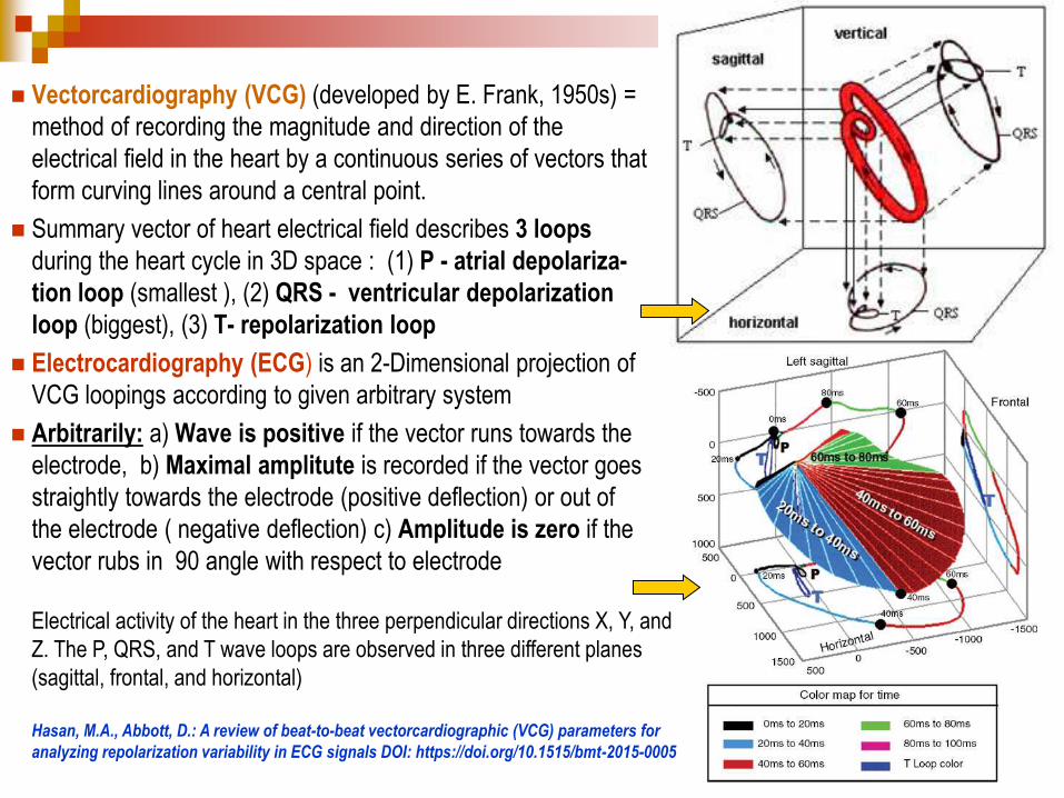
Vectorcardiography (VCG) (developed by E. Frank, 1950s) =
method of recording the magnitude and direction of the
electrical field in the heart by a continuous series of vectors that
form curving lines around a central point.
Summary vector of heart electrical field describes 3 loops
during the heart cycle in 3D space : (1) P - atrial depolariza-
tion loop (smallest ), (2) QRS - ventricular depolarization
loop (biggest), (3) T- repolarization loop
Electrocardiography (ECG) is an 2-Dimensional projection of
VCG loopings according to given arbitrary system
Arbitrarily: a) Wave is positive if the vector runs towards the
electrode, b) Maximal amplitute is recorded if the vector goes
straightly towards the electrode (positive deflection) or out of
the electrode ( negative deflection) c) Amplitude is zero if the
vector rubs in 90 angle with respect to electrode
Electrical activity of the heart in the three perpendicular directions X, Y, and
Z. The P, QRS, and T wave loops are observed in three different planes
(sagittal, frontal, and horizontal)
Hasan, M.A., Abbott, D.: A review of beat-to-beat vectorcardiographic (VCG) parameters for
analyzing repolarization variability in ECG signals DOI: https://doi.org/10.1515/bmt-2015-0005

Generation of ECG waves

Electrode placement

Standard 12- lead ECG recording
I Lateral left ventricle
II Inferior left ventricle
III Inferior left ventricle
aVR Right heart
aVL Left heart
aVF Left heart V6 Lateral ventricle
V5 Lateral ventricle
V4 Anterior
V1 Septal
V2 Antero-septal
V3 Antero-septal
Limb leads Precordial leads
25 mm/s 10 mm/1mV

1. Rhythm regularity and normality, 2. Heart rate,, 3. Electric heart axis, trasmient zone, 4.
Normality of wave composition, intervals, segments analysis – P wave, PQ interval , QRS
complex, ST segment, T wave, QT interval
Normal 12- lead ECG What to look for ?

Evauation of ECG
1. Rhythm
SA rhythm (physiological): P wave is present before QRS, PQ interval is constant;
depoôarisation proceeeds from rostral to caudal parts of the heart
Supraventricular rhythm: P wave is not normal, PQ shorter (atrial rhythms) or P wave and
PQ are absent before QRS; morphology of QRS may be normal; depolarisation may
proceeed both rostrally and caudally ( e.g. AV nodal rhythm) thus atrail depolarisation may
be hidden
Ventricular rhythms: distinct from normal rhythms
2. Axis – left- oriented or right oriented
3. Waves
4. Intervals and segments

Electrocardiography – Frequency calculation
1. Counting large boxes. If a sweep
speed is 0,2 s/large box (LB) = 300
LB/min; HR = 300/number of LB in
between RR intervals
HR = 300/5 = 60 b/min 3. Counting large & small boxes.
2. Counting R waves in 6 sec strip x 10.
HR = 7 x 10 = 70 b/min
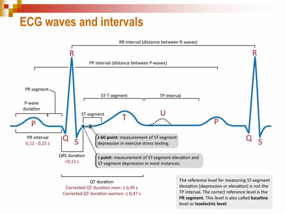
ECG waves and intervals
Arrhythmias

Cardiac arrhythmias (dysrhythmias)
Definition: Cardiac dysrhythmias = group of disorders of cardiac electrical rhythm
autopacing and distribution in which the heartbeat may show irregularities or ECG
abnomalities with no change in normal frequency, or too fast or too slow.
Epidemiology: affect millions of people, occur at any age incl. children; more common
among older people; Sudden cardiac death is the cause 1/2 of deaths due to
cardiovascular disease or about 15% of all deaths globally. About 80% of sudden
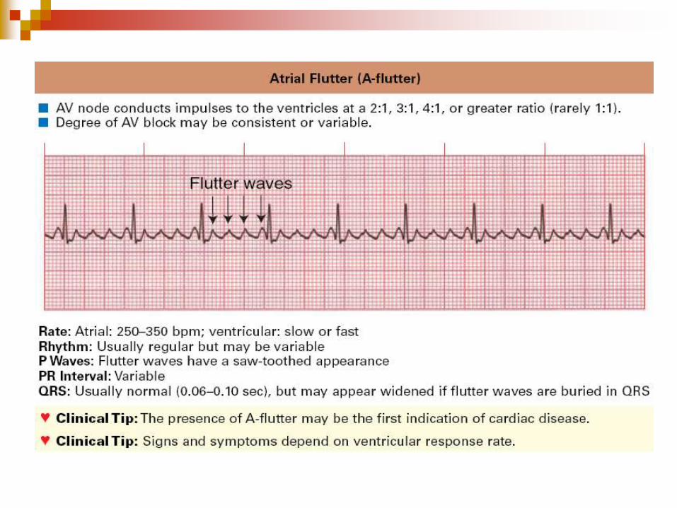
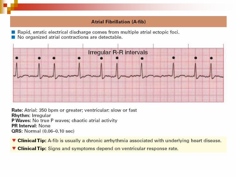
cardiac death ← ventricular arrhythmias. (atrial fibrillation and atrial flutter = 112,000
deaths (2013)
Clinical manifestations:
Many types of arrhythmia have no symptoms, are not serious
Typical symptoms include - palpitations, feeling a pause between heartbeats,
lightheadedness, shortness of breath, chest pain
Sudden serious complications – heart failure, cardiac arrest.

Cardiac arrhythmias (cont.)
Etiology: Specific cardiac and non-specific chanellopathies;
Congenital/acquired defects in electrical conduction system of the heart ( e-g. abnormalities of resting ECG, pre excitation (short PR interval)
Structural cardiac diseases - mitral valve dis., LV aneurysm, congenital heart diseases
Ischemic heart disease = mother of many arrhythmias (conductive system & myocard);, (angina, recent muocardial infarction)
Internal milieu disturbances = ↓ or ↑ K+ hyper-/hypokalemia; ↓ or ↑ Ca2+ hypo/hypercalcemia; ↓ Mg2+, acidosis/alkalosis; hypoxia, hypercarbemia ↓ PaO2, ↑ PaCO
Miscellaneous: Febrile illness, Emotional stress, Smoking, Fatigue
Hormonal dysbalance (thyroid hormones = hyper-/hypothyroidism, growith hormone, estrogenes, testosterone)
Vegetative dystonia - sympathetic hyperreractors (tachycardic arrhythmias; vagal hyperresponsivenes (bradycardia) Phaeochromocytoma
Drugs Anti-arrhythmics, Para/ sympathomimetics (β2 agonists, cocaine), antidepressants, caffeine), Alcohol

Classification:
According to origin: a) Nomotopic (sinus rhythm) = generated in sinoatrial node ; b)
Ectopic = relesed from locations from elsewhere
According to ectopic location: a) Supraventricular arrhythmias (incl. Atrial
arrhythmias + Nodal arrhythmias= atrioventricular node area) b) Ventricular dysrrhythmia
(generated in conductive system (Hiss bunkdle, Tawara bundles + myocardium of ventricles)
According to stability of pacing : a) Rhythms (= paroxysms/ or longer periods (minutes)
with out of normal rhyhmicity, ECG wave composition, etc.) b) Extrabeats (captured beats,
short periods, several or individual QRST complexes)
Extra beats include premature atrial, premature ventricular contractions and premature
junctional contractions.
According to regularity: a) regular (equal RR intervals), (e.g. sinus bradycardia,
tacgycadia) irregular (non-equal RR int.), e.g. sinus arrhythmia, extrasystoles
According to contraction frequency: a) normocardic rhythms = 60 - 100 b/ min in
adults; b) tachycardic rhythms >100 b/ min (hypoxia, ischemia to the heart !!) c)
bradycardic rhythms <60 b/ min in adults
Cardiac arrhythmias (dysrhythmias)

Mechanisms:
Abnomal / hidden /reviced pacemakers
(excuted by patholoical condtirions)
Abnormal automaticity (efects of hormones,
nervous drive)
Triggered activity – EAD, DAD (tetanic
activity, refractery phases)
Reentry circuits – small or long loop
reentries,
Channelopthies – specific disorders
Cardiac arrhythmias

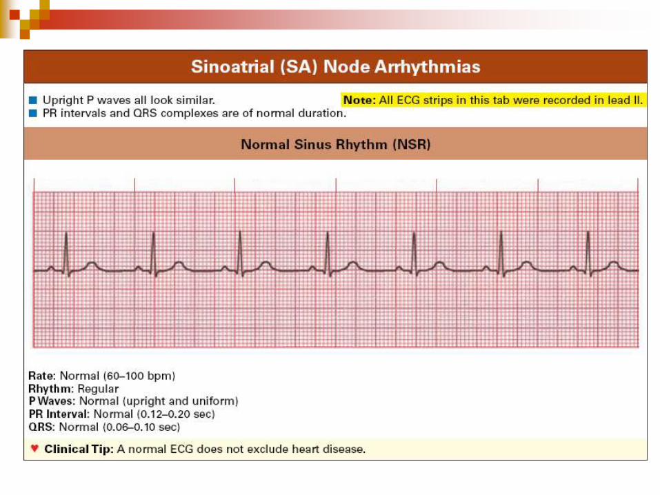
Sinus Arrhythmias
P wave has normal
morphology
QRS and T wave are of
normal morphology


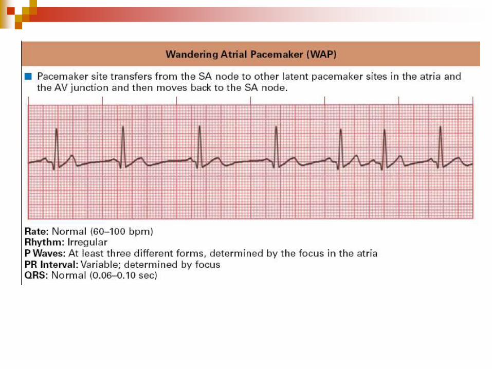
Atrial Arrhythmias
P wave is different from that
generated in SA node
QRS and T wave are of
normal morphology


Junctional
Arrhythmias
The atria and SA node loss their
pacemaking functions
A junctional escape rhythm
begins

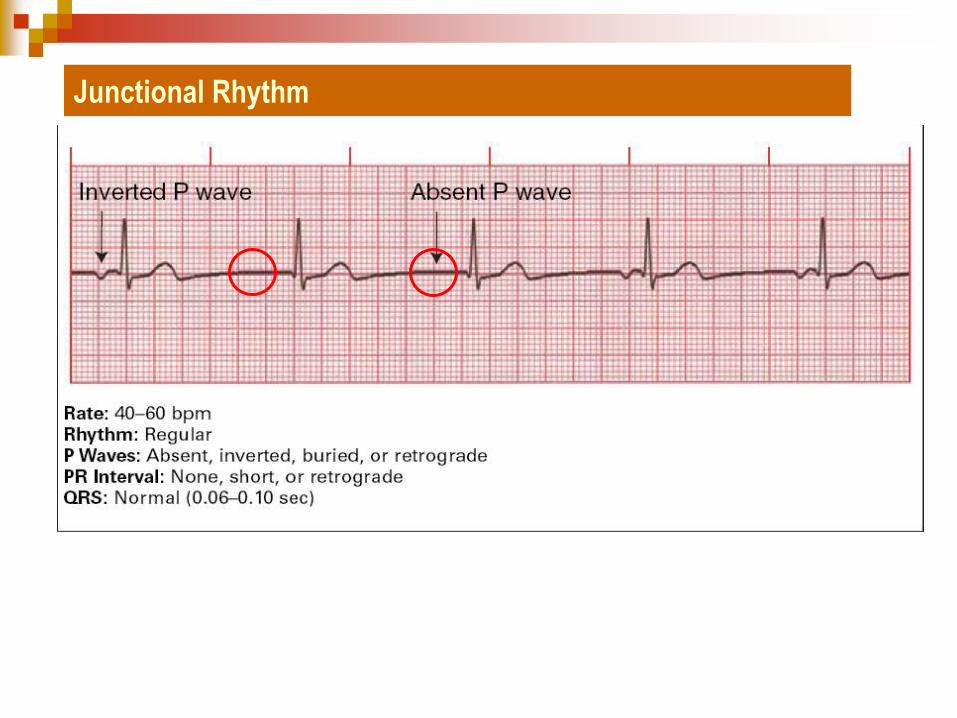
Junctional Rhythm

Accelerated Junctional Rhythm
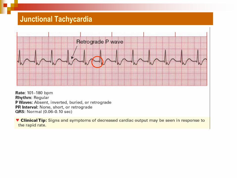
Junctional Tachycardia

Junctional Escape Beat
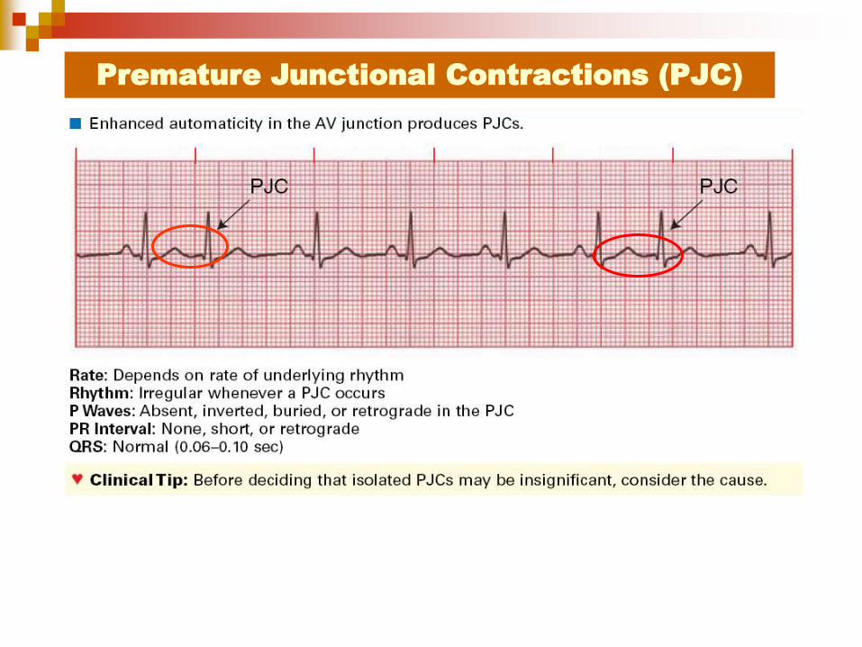
Premature Junctional Contractions (PJC)

Atroventricular blockade

Ventricular
Arrhythmias
The atria and SA node loss their
pacemaking functions
Ventricular loci drive the rhythm

Benacka
Textové pole
Idioventricular rhythm


Sources
http://www.medicalestudy.com/
Cardiac
channelopathies
The atria and SA node loss their
pacemaking functions
Ventricular loci drive the rhythm
Benacka
Písací stroj
ADVANCED

Cardiac channelopathies
Def.: Disorders caused by spontaneous or hereditary mutations of genes coding subunites of ionic channels or transportrers involved in creation of cardiac electrical excitation or conduction or electro-mechanical coupling in cardiomyocytes
Channels are multimeric proteins, where each subunite is encoded various genes in different locuses..
Inherited forms of cardiac channelopathies
Acquired forms of cardiac channeloapthies
Long QT syndrome) (LQTS)
Short QT Syndrome) (SQTS)
Brugada brothers syndrome (BrS)
Catecholaminergic polymorhous
ventricular tachyarrhythmia (CPVT)
Arrythmogenic rightsided ventrikular
cardiomyopathy (ARVC)
Familial forms of atrial fibrilation
Inherited forms of cardiac
channelopathies
Atrial fibrillation
Heart failure
Sick sinus syndrme
Cardiac hypertrophy
myocardial infarction
Acquired forms of cardiac
channeloapthies

Inherited cardiac channelopathies Cardiac channelopathies

Acquired cardiac channelopathies
Cardiac channelopathies

Long QT interval syndrome (LQTS)
Def: Group of cardiac channelopathies typical by
prolongation of repolarisation in cardiomyocytes
due to gene defects in mostly potassium,
sodium, or calcium channels
QT interval (start of Q till end of T) shows
interpersonal and intrapersonal variability -=>
corrected QT (QTc) 0.35 to 0.46 sec. ; 95%
percentil = 0.38 to 0.44 s
Etio: 14 subtypes of disease exist (LQT1, LQT2,
& LQT3 ~ 80-90% of known cases)
KCNQ1, KCNH2, & SCN5A cardaic K+ a Na+
channels; loss-of-function mutations
polymorfisms (postmedical LQTS)
Occ: common 1/2000 - 1/3000 of cardaic
patients
Clinical manifestations:
palpitations, fainting, sudden death due to
ventricular fibrilation
risk of episodes of torsades de pointes
(polymorfic ventricular tachyarrythmias)
induction by hypokalemia, heart attack and
heart ischemia, hypothermia, subarachnoidal
bleeding etc.
Cardiac channelopathies

Short QT interval syndrome (LQTS)
Cardiac channelopathies

Brugada sy. (BRGDA)
Def.: Group of hereditary arrhythmias leading to
sudden unexpected death (ventricular fibrillation)
(Pedro and Joseph Brugada, 1992)
One of reasons of unexplained cardiac death
(sudden unexplained death syndrome, SUDS);
most coomon reason of death in young man without
previous cardiac diasease in Thailand and Laos
Etiology:
(a) Na+ channel in cardiomyocytes (SCN5A) 20%
cases ; 160 types of mutations
(b) Ca2+ channels L-type (CACNA1C and CACNB2
leading to elevation of ST ans shortenieng of QT
(<360 ms).
Type Gene Locus
BS1 SCN5A 3p22.2
BS2 GPD1L 3p22.3
BS3 CACNA1C 12p13.33
BS4 CACNB2 10p12.33-
p12.31
BS5 SCN1B 19q13.12
BS6 KCNE3 11q13.4
BS7 SCN10A 3p22.2
BS8 HEY2 6q22.31
Cardiac channelopathies

Cardiac channelopathies http://upload.wikimedia.org/wikipedia/c
ommons/0/05/Brugada_EKG_Schema.
jpg Dg. typical spontanous changes in ECG or induced by
antiarythmic drugs blocking Na+ channels in 3 various
ECG patterns:
Type 1: ST elevation >2 mm (0.2 mV), J-point elevation;
decrease of ST segment with negativeT-wave.
Type 2: > 2 mm elevation of J-point and > elevation 1 mm
ST with posiktive bipjasic T-wave
Type 3: similar to type 1 or type 2 with elevation of J point
< 2 mm + ST elevation < 1 mm.

Examples of allelic heterogenity of various arrhythmias
Cardiac channelopathies

Alfa- subunite of cardiac Na+ channel
(SCN5A) Ch3p22.2
• Atrial fibrillation, familial, 13
• Brugada syndrome 5
• Cardiac conduction defect,
nonspecific
• Epilepsy, generalized, with febrile
seizures plus, type 1
• Long QT syndrome-3
• Cardiomyopathy, dilated, 1E
• Atrial fibrillation, familial,
• Brugada syndrome 1
• Heart block, nonprogressive
• Heart block, progressive, type IA
• Sick sinus syndrome 1
• Ventricular fibrillation, familial, 1
• Sudden infant death syndrome,
susceptibility
Cardiac channelopathies
Alpha subunite of cardiac
Na+ channel SCN1B 19q13.12
Related Documents


