Cardiovascular abnormality in heat stroke Chiaki Watanabe, MD, Ph.D Department of Cardiology, Takeda General Hospital Kyoto Japan

Welcome message from author
This document is posted to help you gain knowledge. Please leave a comment to let me know what you think about it! Share it to your friends and learn new things together.
Transcript

Cardiovascular abnormality in heat stroke Chiaki Watanabe, MD, Ph.D
Department of Cardiology, Takeda General Hospital
Kyoto Japan

Kyoto Prefecture
Takeda General Hospital

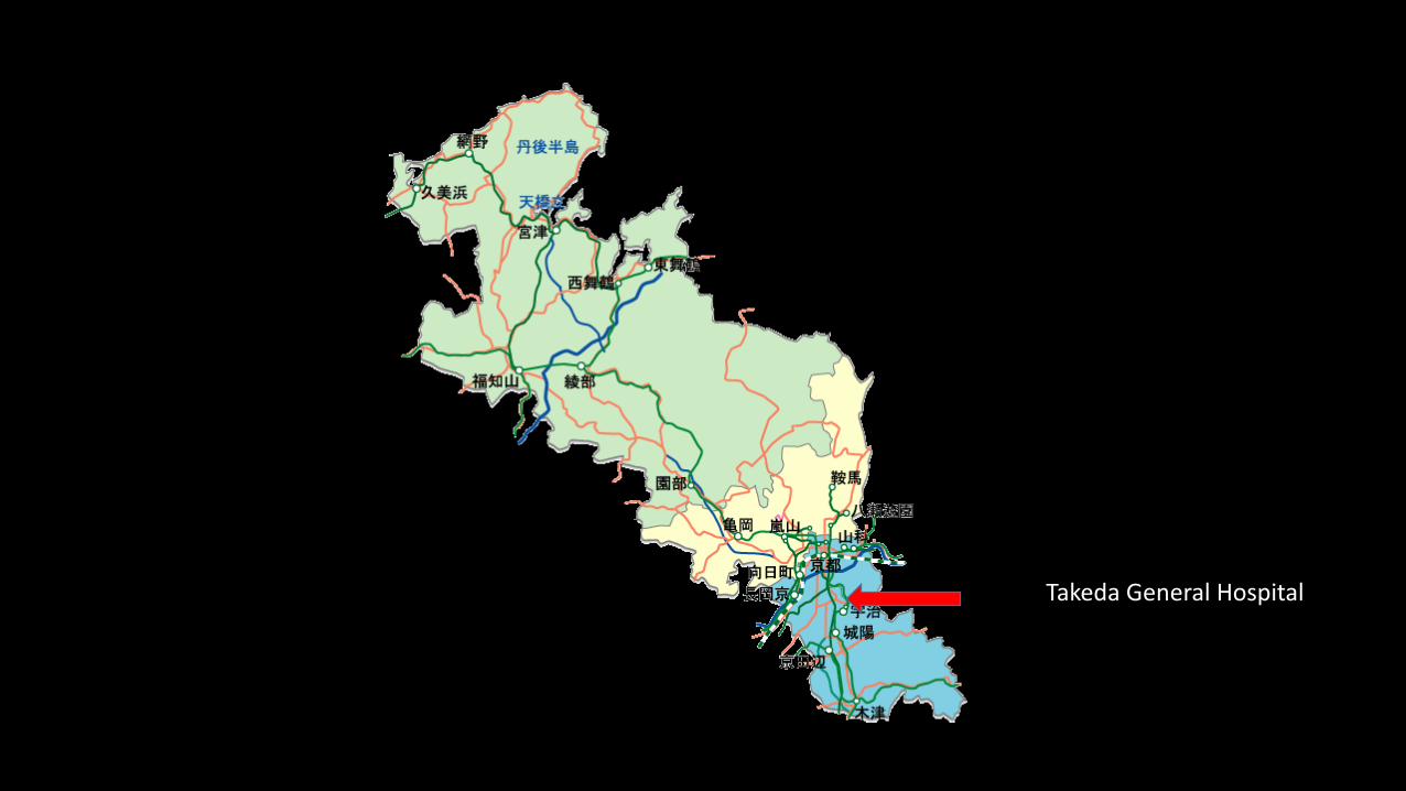
Department of Cardiology Practical performance
Year 2012 2013
Outpatient (daily) 23,713 (79.0) 22,432 (75.8)
In patient 13,391 12,306
Cardiac catheterization(PCI) 982(291) 887 (252)
Percutaneous peripheral intervention 89 67
Catheter ablation 94 93
ICD/CRT implantation 54 44
Coronary CT 550 446
Ultrasonic cardiography 7536 7551
Treadmill test 639 491
Holter ECG 388 412
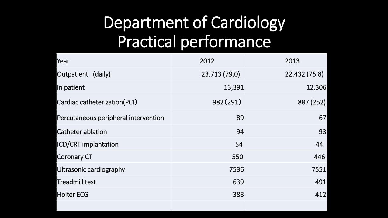
Heat illness: Epidemiology
0
200
400
600
800
1000
1200
1400
1600
1800
2000
2010 2011 2012 2013 2014
seriously ill death
1% 14%
39%
46%
0-7y.o
7-19y.o.
20-64y.o.
>65y.o.
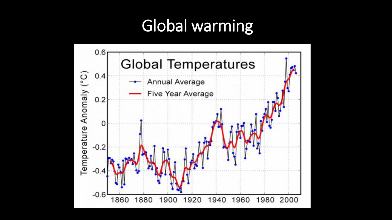
Global warming

Heat stroke <Definition>
Severe illness characterized by a core temperature >40℃
and central nervous system abnormalities such as delirium,
convulsions, or coma resulting from exposure to environmental
heat or strenuous physical exercise
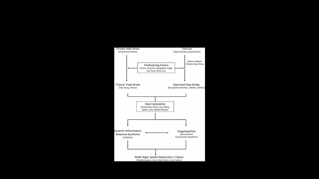
<Classification>
Classic: primary occurs in compromised individuals during annual heat waves
Exertional: in young fit individuals performing strenuous physical exercise

Thermoregulation
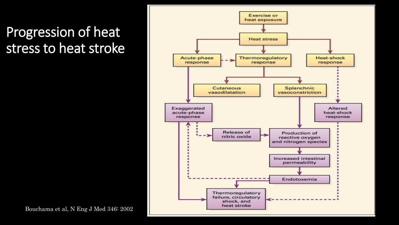
Progression of heat stress to heat stroke
Bouchama et al, N Eng J Med 346: 2002
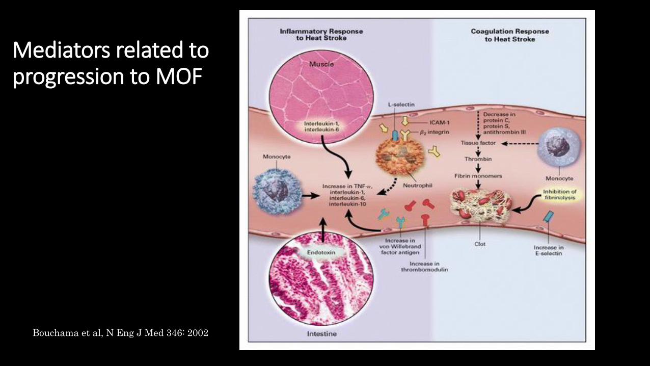
Mediators related to progression to MOF
Bouchama et al, N Eng J Med 346: 2002
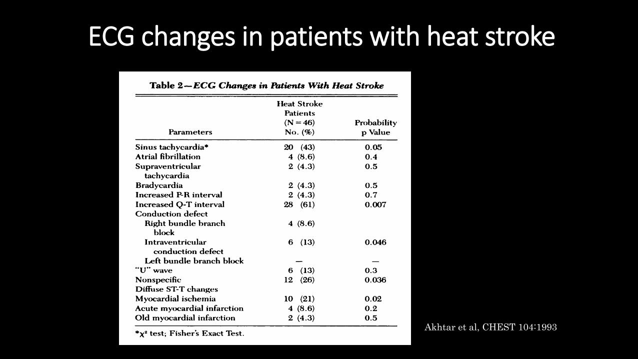
ECG changes in patients with heat stroke
Akhtar et al, CHEST 104:1993
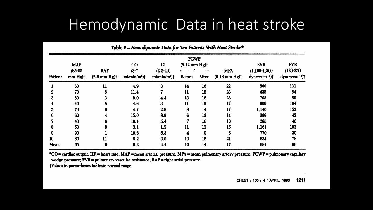
Hemodynamic Data in heat stroke

Case 1 ( TTC )
87 y.o. Japanese man
C.C.: consciousness disturbance, generalized convulsion
P.I.: He had a 30-year history of epilepsy and hypertension treated by a neurologist until 17 months previously. He was barely able to walk indoors, had not been eating properly recently. On a hot summer morning of admission, his son found him immobile in the bathroom. His son called an ambulance because the patient gradually became unresponsive and had a convulsion.

Physical Examination
Consciousness GCS 6PT, pulse 160-200bpm,
B.P. 110/43mmHg,
B.T. 41.2℃, SpO2 96%( O2 9L mask inhalation)
Skin & tongue: dry
Chest: unremarkable
Abdomen: unremarkable except operation scar
No peripheral edema
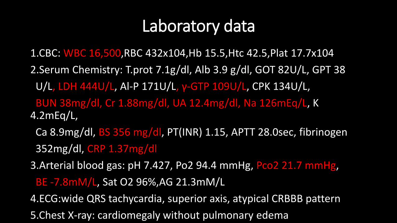
Laboratory data
1.CBC: WBC 16,500,RBC 432x104,Hb 15.5,Htc 42.5,Plat 17.7x10⁴
2.Serum Chemistry: T.prot 7.1g/dl, Alb 3.9 g/dl, GOT 82U/L, GPT 38
U/L, LDH 444U/L, Al-P 171U/L, γ-GTP 109U/L, CPK 134U/L,
BUN 38mg/dl, Cr 1.88mg/dl, UA 12.4mg/dl, Na 126mEq/L, K 4.2mEq/L,
Ca 8.9mg/dl, BS 356 mg/dl, PT(INR) 1.15, APTT 28.0sec, fibrinogen
352mg/dl, CRP 1.37mg/dl
3.Arterial blood gas: pH 7.427, Po2 94.4 mmHg, Pco2 21.7 mmHg,
BE -7.8mM/L, Sat O2 96%,AG 21.3mM/L
4.ECG:wide QRS tachycardia, superior axis, atypical CRBBB pattern
5.Chest X-ray: cardiomegaly without pulmonary edema

ECG on admission
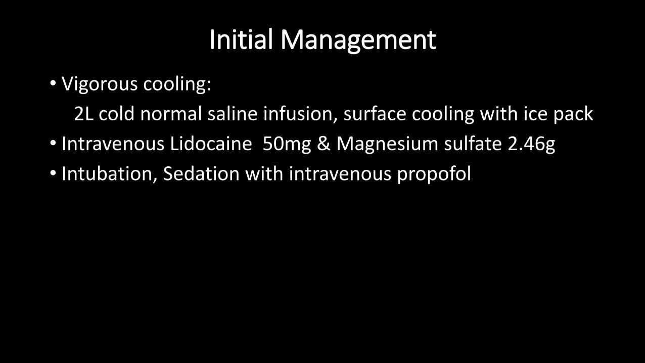
Initial Management
• Vigorous cooling:
2L cold normal saline infusion, surface cooling with ice pack
• Intravenous Lidocaine 50mg & Magnesium sulfate 2.46g
• Intubation, Sedation with intravenous propofol
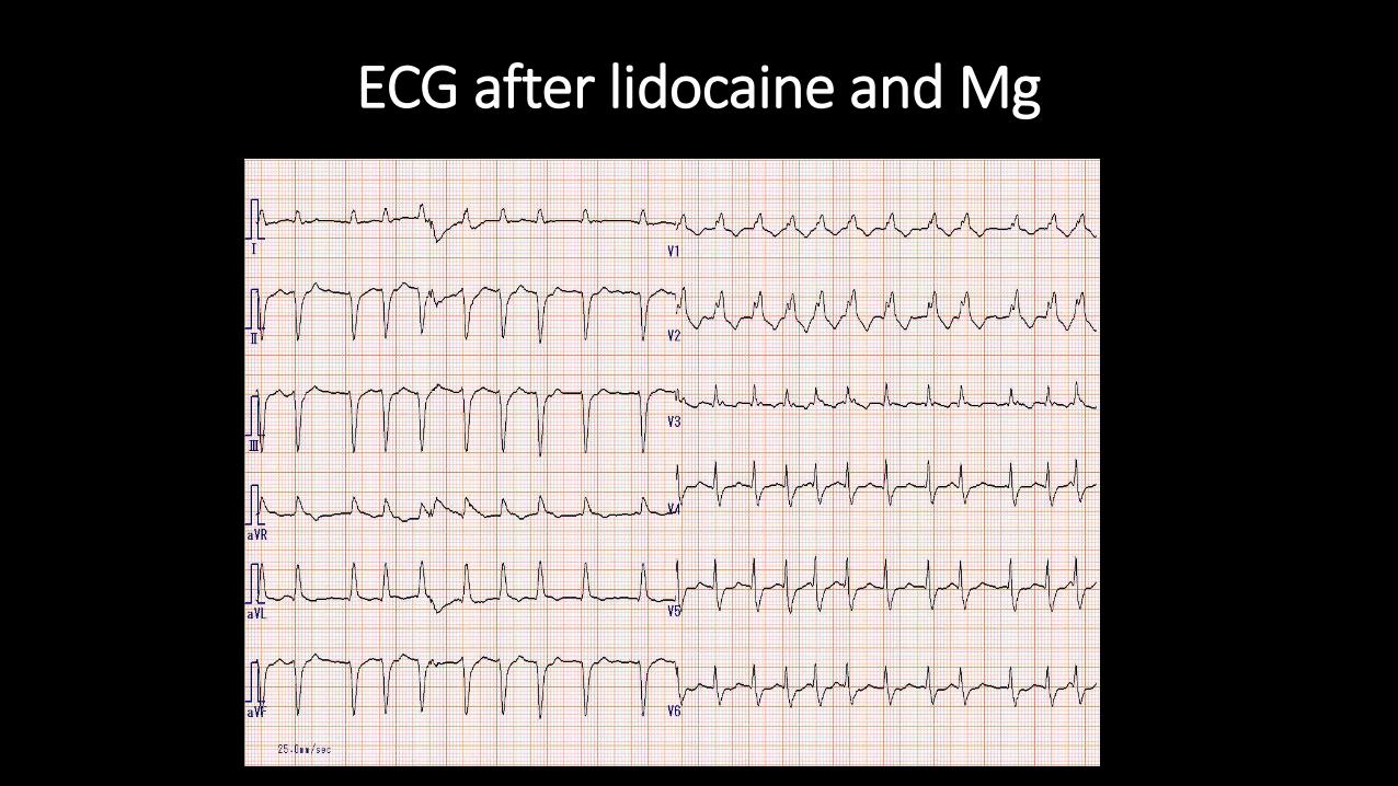
ECG after lidocaine and Mg

Management in ICU • Continue evaporative cooling techniques
• Body temperature 37.5℃ 4hours later
• Fell into shock state after conversion to af
➡drip infusion of NAd(0.3μg/kg/min) keeping BP >90mmHg
• Drip infusion of Heparin(500U/h) to prevent thrombus formation
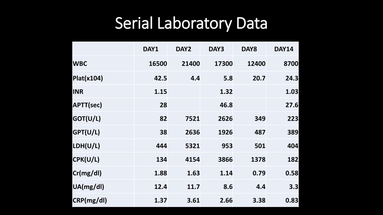
Serial Laboratory Data
DAY1 DAY2 DAY3 DAY8 DAY14
WBC 16500 21400 17300 12400 8700
Plat(x104) 42.5 4.4 5.8 20.7 24.3
INR 1.15 1.32 1.03
APTT(sec) 28 46.8 27.6
GOT(U/L) 82 7521 2626 349 223
GPT(U/L) 38 2636 1926 487 389
LDH(U/L) 444 5321 953 501 404
CPK(U/L) 134 4154 3866 1378 182
Cr(mg/dl) 1.88 1.63 1.14 0.79 0.58
UA(mg/dl) 12.4 11.7 8.6 4.4 3.3
CRP(mg/dl) 1.37 3.61 2.66 3.38 0.83
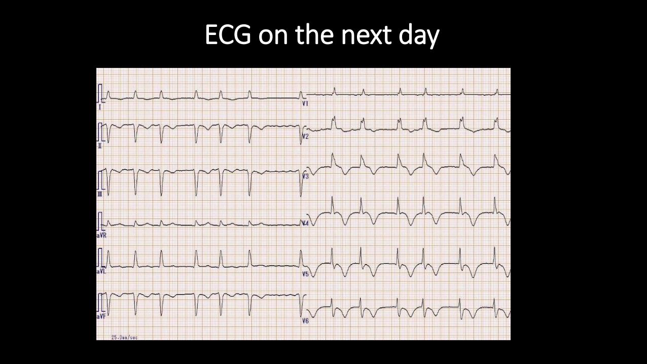
ECG on the next day

UCG day2

Coronary CT
RCA LCX LAD

Clinical course
• Stable hemodynamics after tapering NAd
➡given carvedilol(2.5mg/day) and enalapril(2.5mg/day)
• No recurrence of tachycardia
• Recovered consciousness without neurological deficit on the day 4
• Rhabdomyolysis, DIC: treated without complication
• Complete recovery of LV wall motion on the day 14

UCG after recovery
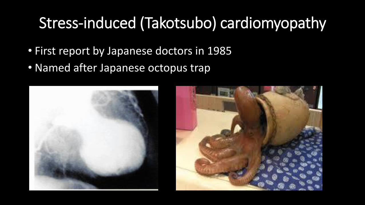
Stress-induced (Takotsubo) cardiomyopathy
• First report by Japanese doctors in 1985
• Named after Japanese octopus trap

Clinical features
Usually occurs in postmenopausal women
Trigger: Emotional stress mostly in women
Physical stress mostly in men
Common symptom: chest pain, chest discomfort, dyspnea
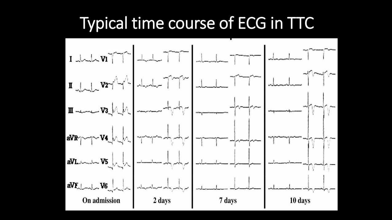
Typical time course of ECG in TTC

Diagnosis Mayo clinic criteria 1)Transient hypokinesis, akinesis, or dyskinesis of the left ventricular mid segments with or without apical involvement; the regional wall motion abnormalities extend beyond a single epicardial vascular distribution; a stressful trigger is often, but not always , present 2)Absence of obstructive coronary disease or angiographic evidence of acute plaque rupture 3)New electrocardiographic abnormalities(either ST-segment elevation and /or T-wave inversion) or modest elevation in cardiac troponin 4)Absence of pheochromocytoma, myocarditis

Pathophysiology
1) Vasospasm of coronary arteries
2) Disturbance of the microcirculation
3) Catecholamine toxicity
4) Obstruction of the LVOT
5) Estrogen deficiency
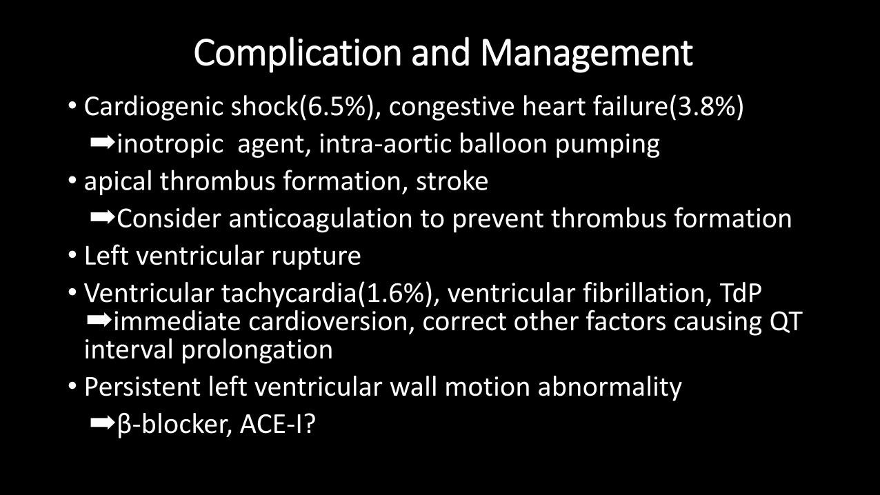
Complication and Management • Cardiogenic shock(6.5%), congestive heart failure(3.8%)
➡inotropic agent, intra-aortic balloon pumping
• apical thrombus formation, stroke
➡Consider anticoagulation to prevent thrombus formation
• Left ventricular rupture
• Ventricular tachycardia(1.6%), ventricular fibrillation, TdP ➡immediate cardioversion, correct other factors causing QT interval prolongation
• Persistent left ventricular wall motion abnormality
➡β-blocker, ACE-I?
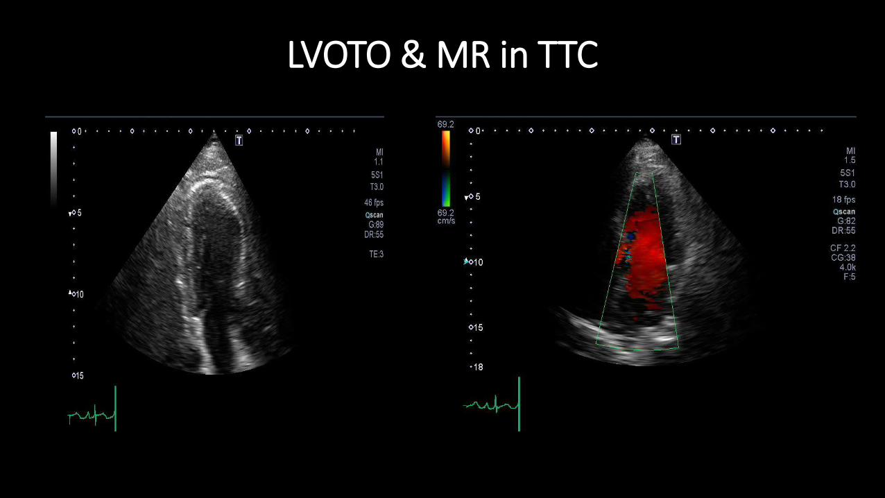
LVOTO & MR in TTC

Complication and Management • Cardiogenic shock(6.5%), congestive heart failure(3.8%)
➡inotropic agent, intra-aortic balloon pumping
• apical thrombus formation, stroke
➡Consider anticoagulation to prevent thrombus formation
• Left ventricular rupture
• Ventricular tachycardia(1.6%), ventricular fibrillation, TdP ➡immediate cardioversion, correct other factors causing QT interval prolongation
• Persistent left ventricular wall motion abnormality
➡β-blocker, ACE-I?

Prognosis
• Overall favorable outcome, almost complete recovery in 96%
• In hospital mortality 1.1-2%, recurrence rate11.4%
AA Elesber et al, JACC 50:2007

Case 2 ( AMI )
Case: 67 years old, Japanese female
C.C.: lethargy, vomiting, abdominal pain
P.I.: She has no medical or health check history. She had lost appetite and felt lethargy recently. On the day of admission in July, she had been working outside from the morning. In the afternoon, she was transferred to our hospital for fever, vomiting and abdominal pain.
P.H.: none
Physical exam.: consciousness , BT 38.8℃, pulse 48bpm reg, BP 124/88, chest & abdomen; unremarkable
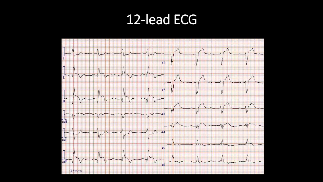
12-lead ECG
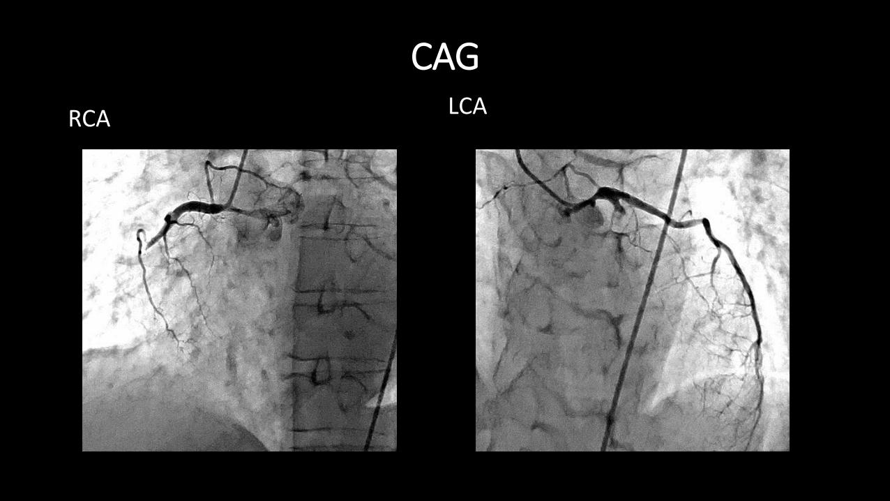
CAG
RCA LCA

CAG after PCI
RCA LCA

Pathology of plaque rupture
Conclusion
• Heat-stroke is a form of hyperthermia associated with a systemic inflammatory response leading to a syndrome of multi-organ dysfunction, accompanied by considerable increase in morbidity and mortality.
• Systemic inflammation, coagulopathy, and increased level of catecholamine in heat stroke may be related to development of cardiovascular abnormality.
• Cardiovascular events might contribute significantly to mortality in patients with heat stroke.
Take home message
• Heat stroke can cause multiple organ failure and the presentation of circulatory failure in heat stroke may be the sign of myocardial dysfunction.
• To distinguish acute coronary syndrome and stress-induced cardiomyopathy, both of which could be evoked by heat stroke, the evaluation of coronary artery is necessary.
• Stress-induced cardiomyopathy may cause lethal arrhythmia or circulatory collapse in acute phase.
• Invasive circulatory monitoring is recommended in the patients with severe heat stroke.


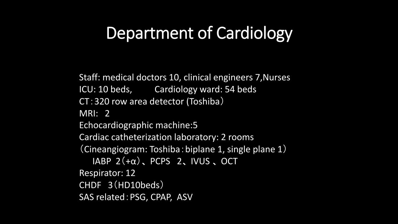
Department of Cardiology
Staff: medical doctors 10, clinical engineers 7,Nurses ICU: 10 beds, Cardiology ward: 54 beds CT:320 row area detector (Toshiba) MRI: 2 Echocardiographic machine:5 Cardiac catheterization laboratory: 2 rooms (Cineangiogram: Toshiba:biplane 1, single plane 1) IABP 2(+α)、 PCPS 2、 IVUS 、 OCT Respirator: 12 CHDF 3(HD10beds) SAS related:PSG, CPAP, ASV
Heat Illnesses
Heat related illnesses:
by exposure without alteration of hypothalamic thermoregulation
Fever:
by changes to the hypothalamic set point by pyrogenic condition
<Types of Heat illnesses>
Heat edema, Heat rash, Heat cramps
Heat tetany, Heat syncope, Heat exhaustion
Heat stroke
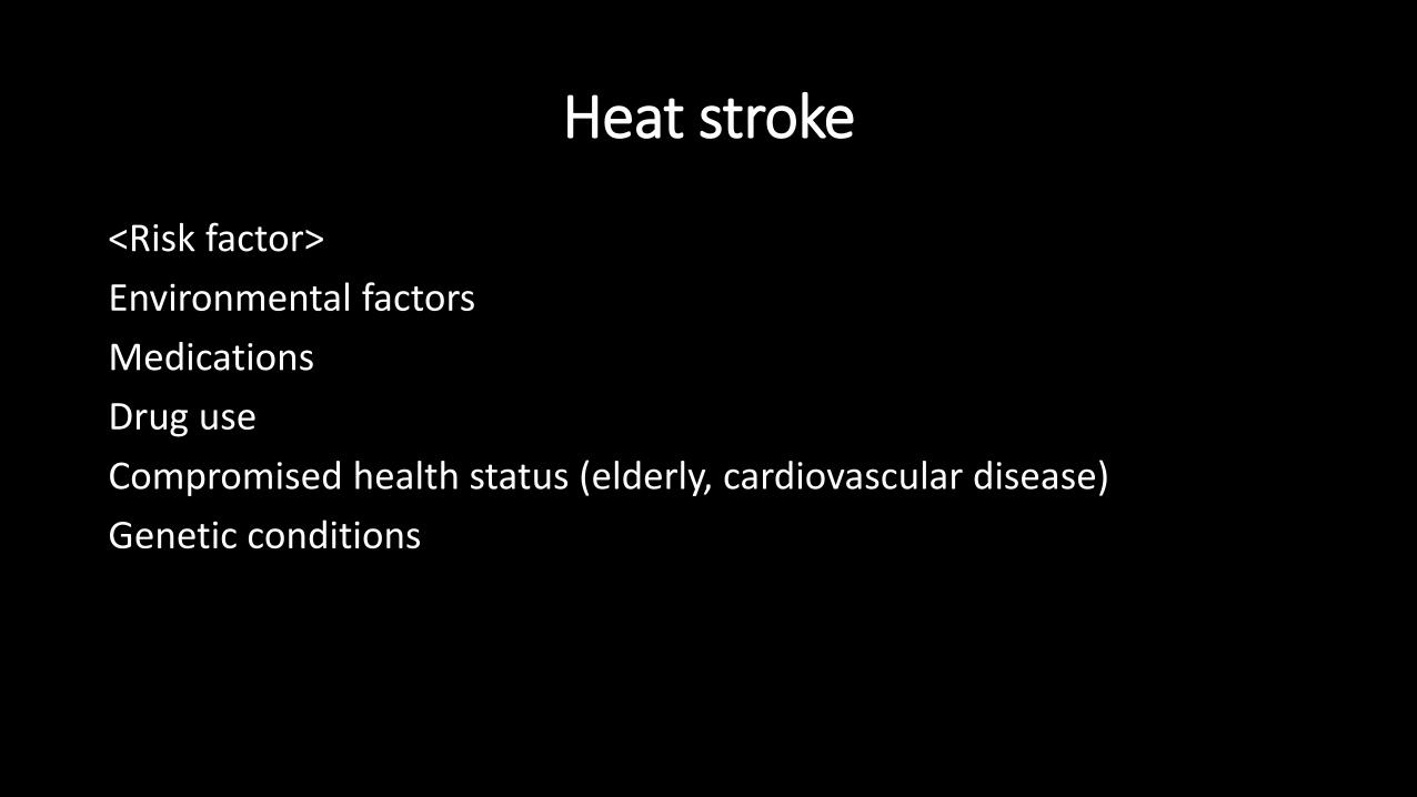
Heat stroke
<Risk factor>
Environmental factors
Medications
Drug use
Compromised health status (elderly, cardiovascular disease)
Genetic conditions

Differential Diagnosis in hyperthermia
Endocrine Pheochromocytoma, Thyroid storm
Infectious Brain absess, Encephalitis, Meningitis
Malaria, Sepsis, Tetanus, Typhoid fever
Neurologic CVA, Seizures
Toxicological Alcohol withdrawal, Anticholinergic toxidromes
Aspirin overdose, Malignant hyperthermia
MAO inhibitors, Malignant syndrome,
Serotonin syndrome

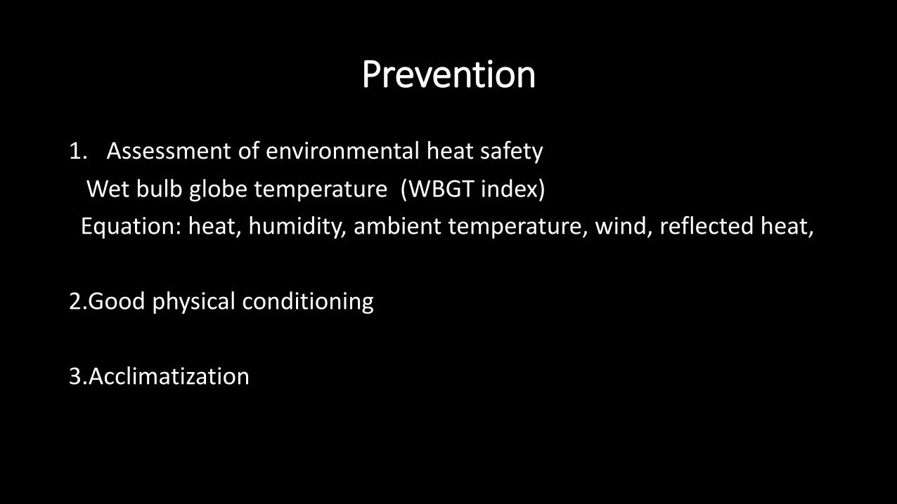
Prevention
1. Assessment of environmental heat safety
Wet bulb globe temperature (WBGT index)
Equation: heat, humidity, ambient temperature, wind, reflected heat,
2.Good physical conditioning
3.Acclimatization

Acclimatization
Takes several weeks
• Enhancement of cardiovascular performance
• Activation of the renin-angiotensin-aldosterone axis
• Increase in the capacity to secrete sweat
• Salt conservation by the sweat glands and kidneys
• Expansion of plasma volume
• Increase in the glomerular filtration rate
• Increase in the resistance to exertional rhabdomyolysis
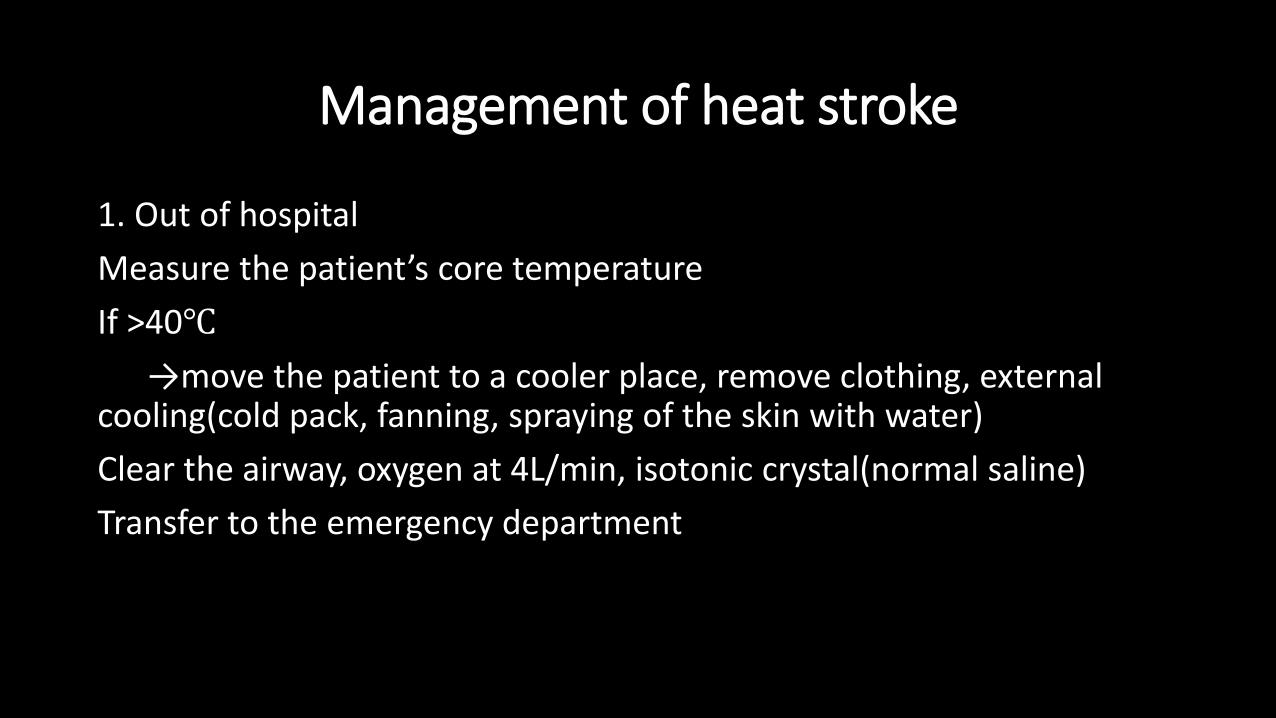
Management of heat stroke
1. Out of hospital
Measure the patient’s core temperature
If >40℃
→move the patient to a cooler place, remove clothing, external cooling(cold pack, fanning, spraying of the skin with water)
Clear the airway, oxygen at 4L/min, isotonic crystal(normal saline)
Transfer to the emergency department
Management of heat stroke
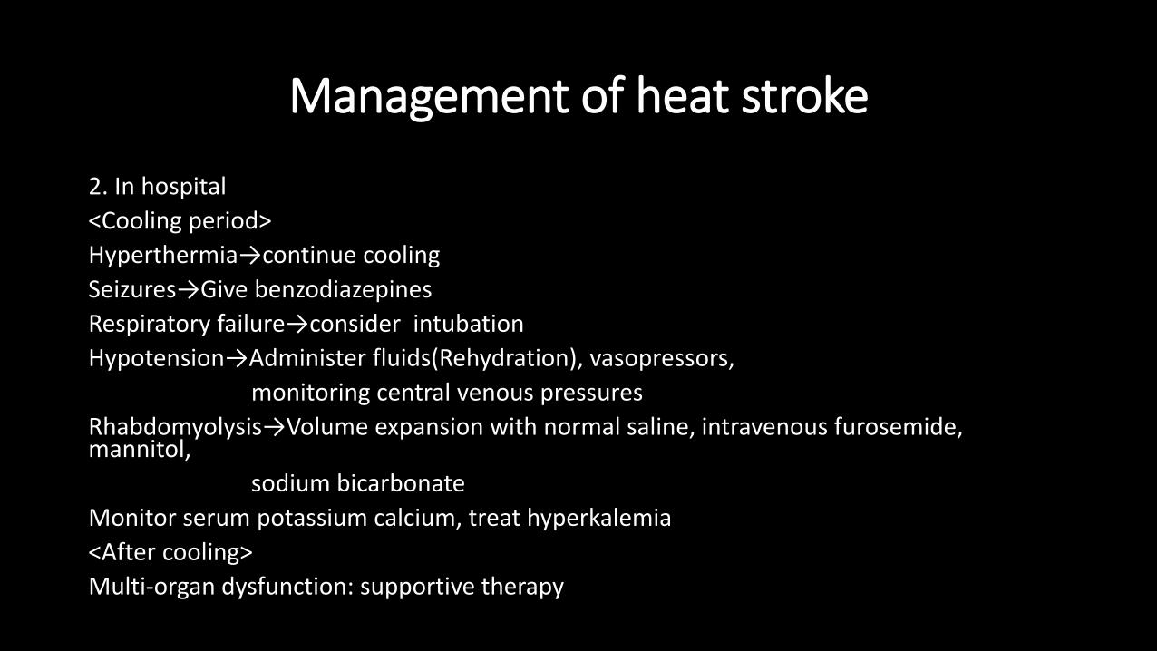
2. In hospital
<Cooling period>
Hyperthermia→continue cooling
Seizures→Give benzodiazepines
Respiratory failure→consider intubation
Hypotension→Administer fluids(Rehydration), vasopressors,
monitoring central venous pressures
Rhabdomyolysis→Volume expansion with normal saline, intravenous furosemide, mannitol,
sodium bicarbonate
Monitor serum potassium calcium, treat hyperkalemia
<After cooling>
Multi-organ dysfunction: supportive therapy
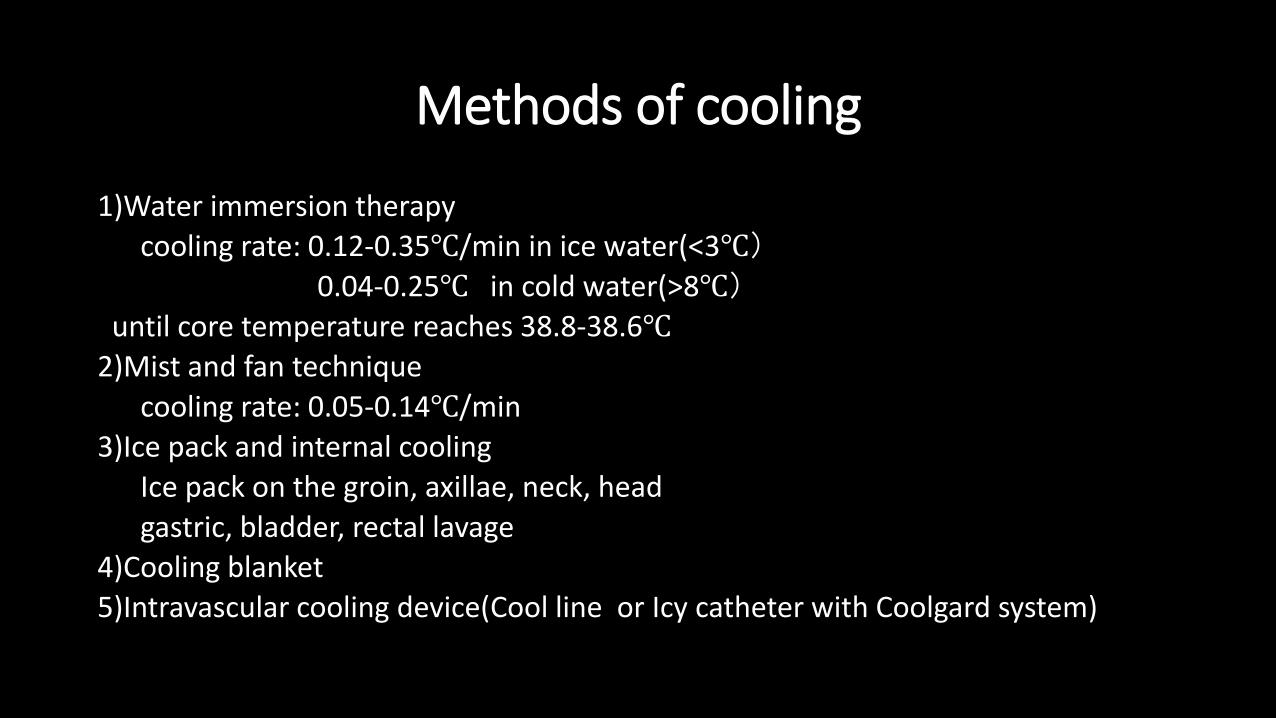
Methods of cooling
1)Water immersion therapy
cooling rate: 0.12-0.35℃/min in ice water(<3℃)
0.04-0.25℃ in cold water(>8℃)
until core temperature reaches 38.8-38.6℃
2)Mist and fan technique
cooling rate: 0.05-0.14℃/min
3)Ice pack and internal cooling
Ice pack on the groin, axillae, neck, head
gastric, bladder, rectal lavage
4)Cooling blanket
5)Intravascular cooling device(Cool line or Icy catheter with Coolgard system)

Case 1 ( TTC )
P.H.: 27y.o. colon volvulus, Epilepsy & HTN, leg phlegmon
Medication: none
L.H.: alcohol 50mg/day, non-smoker
Lived in a house without air-conditioning
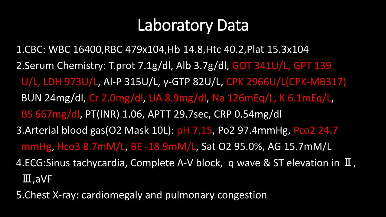
Laboratory Data
1.CBC: WBC 16400,RBC 479x104,Hb 14.8,Htc 40.2,Plat 15.3x10⁴
2.Serum Chemistry: T.prot 7.1g/dl, Alb 3.7g/dl, GOT 341U/L, GPT 139
U/L, LDH 973U/L, Al-P 315U/L, γ-GTP 82U/L, CPK 2966U/L(CPK-MB317)
BUN 24mg/dl, Cr 2.0mg/dl, UA 8.9mg/dl, Na 126mEq/L, K 6.1mEq/L,
BS 667mg/dl, PT(INR) 1.06, APTT 29.7sec, CRP 0.54mg/dl
3.Arterial blood gas(O2 Mask 10L): pH 7.15, Po2 97.4mmHg, Pco2 24.7
mmHg, Hco3 8.7mM/L, BE -18.9mM/L, Sat O2 95.0%, AG 15.7mM/L
4.ECG:Sinus tachycardia, Complete A-V block, q wave & ST elevation in Ⅱ,
Ⅲ,aVF
5.Chest X-ray: cardiomegaly and pulmonary congestion
Related Documents