Cardiopulmonary bypass in small baby JUNG EUN LEE THORACIC & CARDIOVASCULAR SURGERY GEONGSANG NATIONAL UNIVERSITY

Welcome message from author
This document is posted to help you gain knowledge. Please leave a comment to let me know what you think about it! Share it to your friends and learn new things together.
Transcript

Cardiopulmonary bypass
in small baby
JUNG EUN LEE
THORACIC & CARDIOVASCULAR SURGERY
GEONGSANG NATIONAL UNIVERSITY

Histories in CPB
In 1950 Bigelow :
the first application of hypothermia in cardiac surgery
In 1952 Lewis & Taufic :
the first application of hypothermia and inflow occlusion for
repair of ASD in humans
In 1953 Gibbon :
establish the feasibility of artificially supported circulation during
temporary occlusion of he pulmonary artery
successfully used extracorporeal circulation in a young woman

Histories in CPB
In 1954 Lillehei et al :
technique of controlled cross-circulation
In 1954 Cooley :
the application of heat exchangers
In 1960s :
emphasized the use of bubble oxygenators
In 1970s :
switching to membrane oxygenators
Next advances
miniaturization of elements of the CPB circuits
modulation of the systemic inflammatory response and injury from CPB

CPB for infants vs adults
• Immature organ systems
• Smaller circulation blood volumes
• Higher oxygen consumption rate
• Reactive pulmonary vascular bed
• Presence of intracardiac and extracardiac shunting
• Impaired temperature control
• Poor tolerance to microemboli

Immature organ systems
Liver :
decreased clotting factors
Lung :
fragile, potential for pulmonary edema & pulmonary hypertension
Kidney :
sodium reabsorption & excretion, concentration &
diluting mechanism are limited
Immune system :
complement generation is low
neonatal mononuclear cells are dysfunctional

Brain in neonates & infants
Low cerebral oxygen consumption rate :
low cerebral blood flow
low energy requirements (small number of active synapses)
high activity of glycolytic enzyme
Cerebral response to hypoxia :
circulatory adaptation
rapid induction of electrical silence
blood glucose tend to rise (by catecholamine release)
in adult : intracellular acidosis ↑, neural injury ↑
in neonate : neuroprotective ( mechanism is unclear)
Smaller circulating blood volume
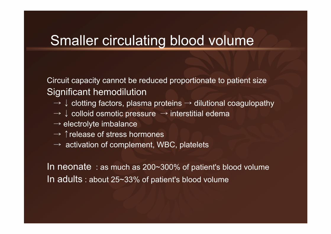
Circuit capacity cannot be reduced proportionate to patient size
Significant hemodilution→ ↓ clotting factors, plasma proteins → dilutional coagulopathy
→ ↓ colloid osmotic pressure → interstitial edema
→ electrolyte imbalance
→ ↑release of stress hormones
→ activation of complement, WBC, platelets
In neonate : as much as 200~300% of patient's blood volume
In adults : about 25~33% of patient's blood volume

Higher oxygen consumption rate
Higher flow rates per BSA to meet metabolic demands
(maintained both cooling & rewarming phase of CPB)
< 3 kg 150 ~ 200 ml/kg/min
3 ~ 10kg 125 ~ 175 ml/kg/min
10 ~ 15kg 120 ~ 150 ml/kg/min
15 ~ 30kg 100 ~ 120 ml/kg/min
30 ~ 50kg 75 ~ 100 ml/kg/min
> 50kg 50 ~ 75 ml/kg/min
Switch from a relatively anaerobic metabolism in a immature heart to
more aerobic metabolism.

Difference between adult and
immature myocardium
� Denser structure with a higher water & protein content per gram
� Less compliant, less preload reserve, narrower range of function
closer to the peak of the Frank-Starling curve
� Lower rate of maximum tension development
� Reduced inotropic reserve
� Operate under maximal adrenergic stimulation
Difference between adult and
immature myocardium
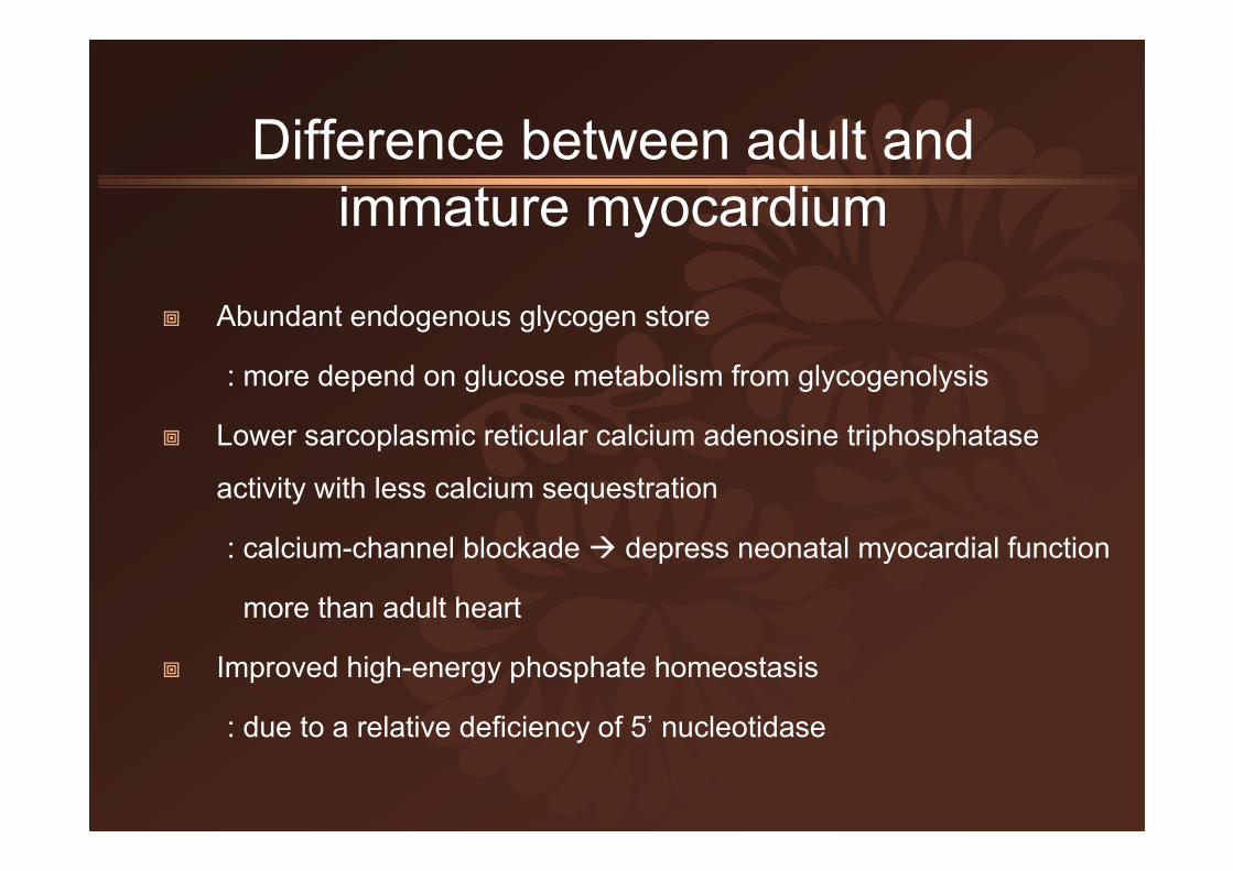
� Abundant endogenous glycogen store
: more depend on glucose metabolism from glycogenolysis
� Lower sarcoplasmic reticular calcium adenosine triphosphatase
activity with less calcium sequestration
: calcium-channel blockade � depress neonatal myocardial function
more than adult heart
� Improved high-energy phosphate homeostasis
: due to a relative deficiency of 5’ nucleotidase


Ischemic tolerance of
the immature heart
� Immature heart has a greater tolerance to hypoxia and
ischemia than the adult
: greater glycogen stores
: improved anaerobic metabolism
: better maintenance of ischemic calcium exchange
: higher levels of adenosine triphosphate
: increased amino acid substrate utilization

Tolerance of the immature heart to
hypoxia or ischemia
� Better tolerable
: increased glycolytic capacity
: better preservation intracellular, high-energy phosphates
: increased ability to utilize amino acid as substrate during
hypothermic ischemia
� Lower tolerable
: greater intracellular accumulation of lactic acid as a result of
anaerobic metabolism
: myocardial ischemic times (>85min) were associated with a
significant mortality risk in infants, despite the use of cardioplegia

Ischemic tolerance of the
immature heart
� Although laboratory models suggest an improved
tolerance to ischemia, most research has been
conducted in the normal heart
� Adverse preoperative conditions such as acidosis,
cyanosis, and hypertrophy may seriously compromise
myocardial protection in the immature heart

Special situations affecting myocardial
protection in neonates with CHD
� Severe hypoxia
� Chronic cyanosis
� Children with decreased pulmonary blood flow have
increased bronchial collateral flow to the left heart that
can markedly compromise intraoperative myocardial
protection
� noncoronary collateral flow
: wash out cardioplegia, rewarms the heart,
causes resumption of contractile activity

Principle of myocardial protection
� Reduction of metabolic activity by hypothermia
� Arrest of contractile apparatus and electrical activity of
the myocyte by administering cardioplegic solution
� Others
: buffering the cardioplegic solution,
: increasing osmolarity,
: decreasing calcium content,
: adding substrate to enhance recovery,
: incorporate leukocyte filters in the CPB circuit

Causes of post-op Low CO
� Residual volume or pressure load – most important
� Ventricular distention
� Retraction / stretch injury to the myocardium
� Coronary artery injury
� Ventriculotomy
� Edema – inappropriate degree of hemodilution of red
cells or colloid oncotic pressure
� Reperfusion condition, e.g. pressure, calcium,
oxygen, additives such as adenosine and free radical
scavengers
� Other perfusion factors, e.g. pH strategy
Strategies of CO₂management :
Alpha stat vs pH stat
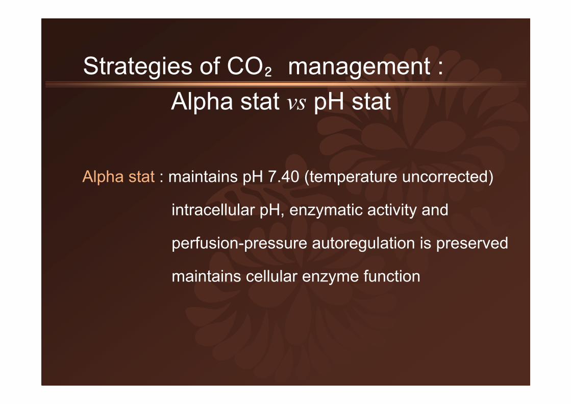
Alpha stat : maintains pH 7.40 (temperature uncorrected)
intracellular pH, enzymatic activity and
perfusion-pressure autoregulation is preserved
maintains cellular enzyme function
Strategies of CO₂management :
Alpha stat vs pH stat
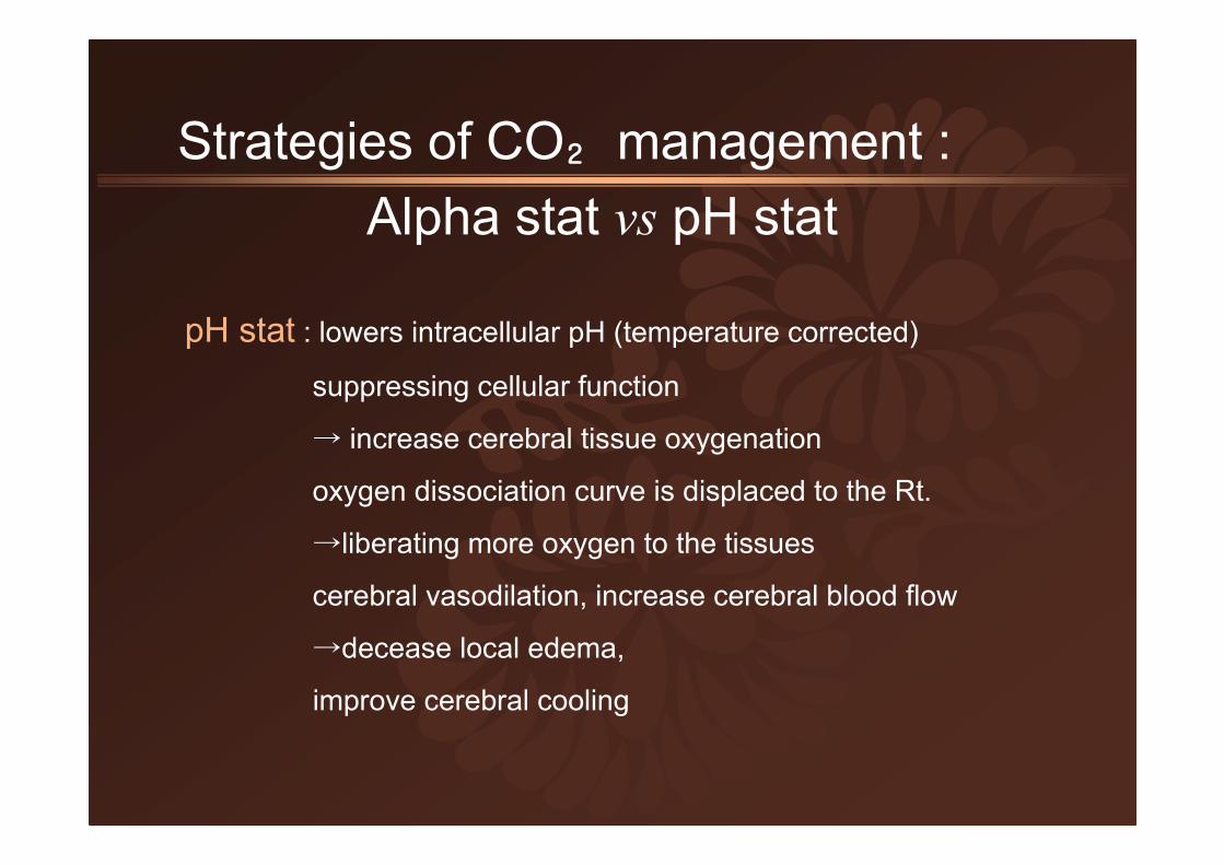
pH stat : lowers intracellular pH (temperature corrected)
suppressing cellular function
→ increase cerebral tissue oxygenation
oxygen dissociation curve is displaced to the Rt.
→liberating more oxygen to the tissues
cerebral vasodilation, increase cerebral blood flow
→decease local edema,
improve cerebral cooling

Alpha stat vs pH stat

Alpha stat vs pH stat

Hypothermia
• Reducing Oxygen requirements
- flow rates can be reduced
• Reducing the temperature difference between the heart and body
- enhances the safe duration of cardiac ischemia.
• Adds safety to the perfusion, since more time is available for
repairs if perfusion must be interrupted because of accidents in the
surgical field or failure of the perfusion apparatus.


Neurologic injury
� 10-25% of incidence
� preexisting risk (associated structural anomalies with the brain)
: esp. in Down sydnrome, CATCH 22
� injury induced by CPB
: microembolic event, esp. air embolism
low cerebral flow
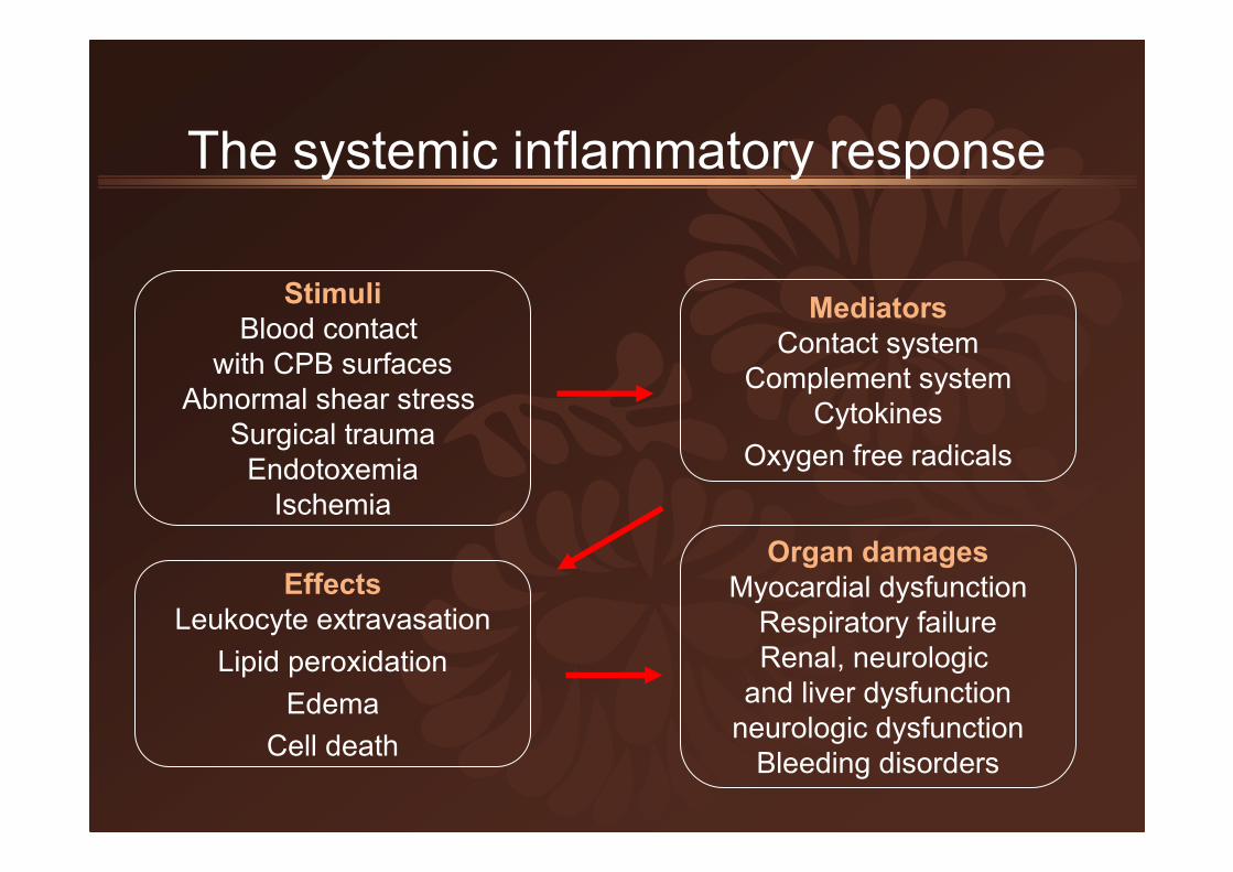
The systemic inflammatory response
Stimuli
Blood contact
with CPB surfaces
Abnormal shear stress
Surgical trauma
Endotoxemia
Ischemia
Effects
Leukocyte extravasation
Lipid peroxidation
Edema
Cell death
Organ damages
Myocardial dysfunction
Respiratory failure
Renal, neurologic
and liver dysfunction
neurologic dysfunction
Bleeding disorders
Mediators
Contact system
Complement system
Cytokines
Oxygen free radicals
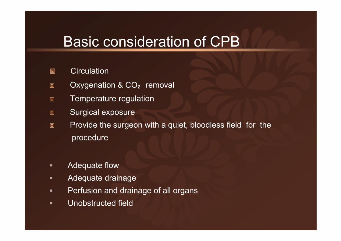
Basic consideration of CPB
� Circulation
� Oxygenation & CO₂removal
� Temperature regulation
� Surgical exposure
� Provide the surgeon with a quiet, bloodless field for the
procedure
� Adequate flow
� Adequate drainage
� Perfusion and drainage of all organs
� Unobstructed field

The pediatric CPB circuits
� Cannulation
� Perfusion pump
� Oxygenators
� Prime
� Initiation of cardiopulmonary bypass
� Delivery system of cardioplegic solution
� Weaning from cardiopulmonary bypass
� Ultrafiltration
� Anticoagulation
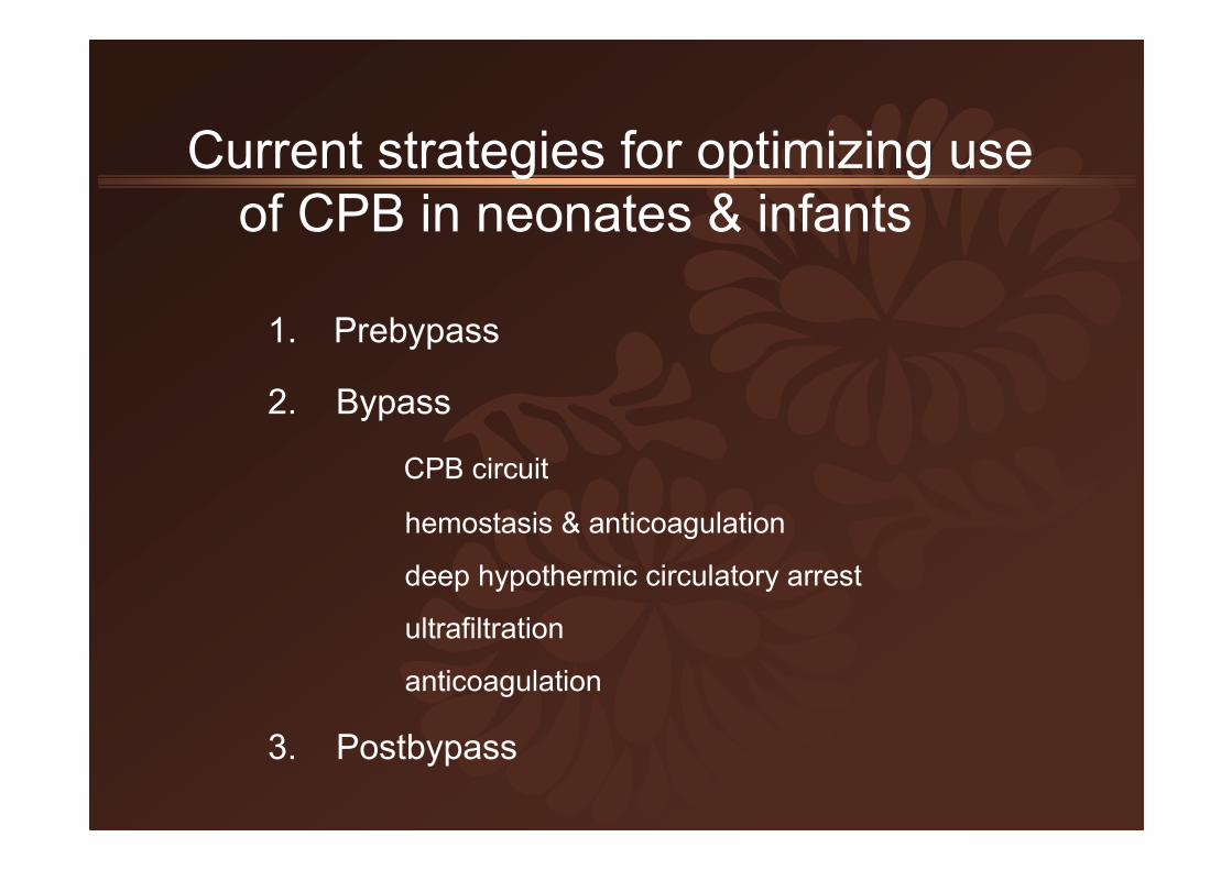
Current strategies for optimizing use
of CPB in neonates & infants
1. Prebypass
2. Bypass
CPB circuit
hemostasis & anticoagulation
deep hypothermic circulatory arrest
ultrafiltration
anticoagulation
3. Postbypass

Prebypass
One of potential complications as a result of exposure to CPB is a
systemic inflammatory response (leukocytes are partly response )
→ capillary leakage, soft tissue edema, end-organ dysfunction
=> 1. using leukocyte filter
2. high dose steroid before CPB

Prebypass
High dose steroid before CPB :
( IV methylprednisolone at 10mg/kg 8hr & 2hr before CPB )
decrease in post-CPB fluid gain
less postoperative edema
improvement in pulmonary compliance
& pulmonary vascular resistance
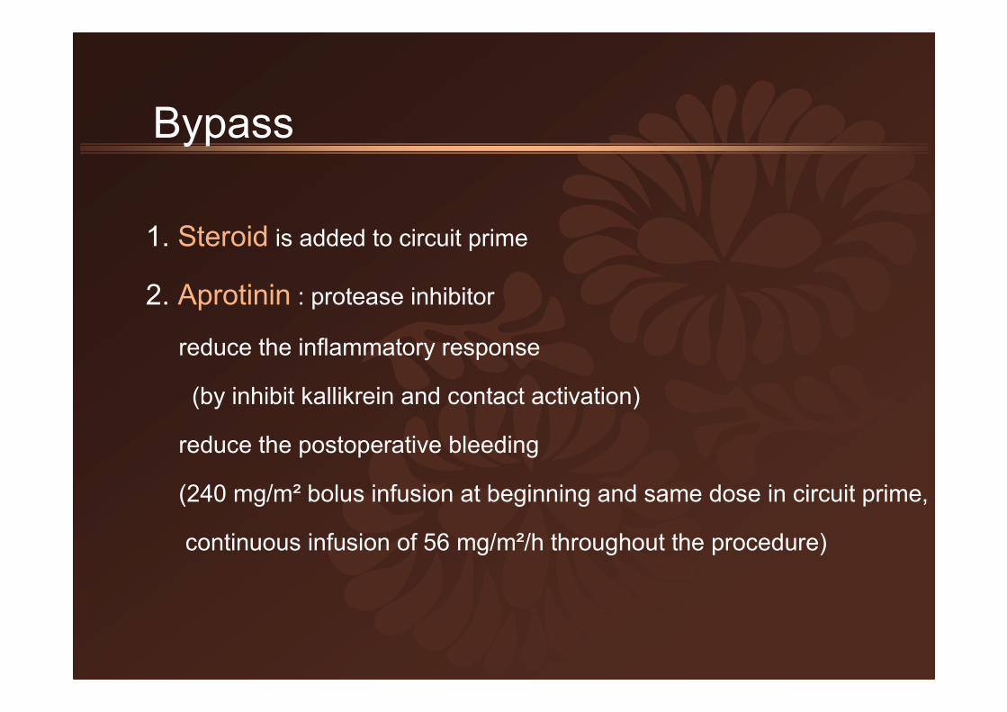
Bypass
1. Steroid is added to circuit prime
2. Aprotinin : protease inhibitor
reduce the inflammatory response
(by inhibit kallikrein and contact activation)
reduce the postoperative bleeding
(240 mg/m² bolus infusion at beginning and same dose in circuit prime,
continuous infusion of 56 mg/m²/h throughout the procedure)

Aprotinin

Aprotinin

Bypass
3. CPB circuitry
• Miniaturization of the CPB
• Using biocompatible-coated circuits (heparin-coated circuit) :
reduce the direct contact of blood cell with foreign materials
• Using vacuum-assisted venous drainage (VAVD)

Heparin-coated curcuit
Bypass
4. Deep hypothermic circulatory arrest and low flow CPB
continuous hypothermic low flow CPB :
more soft tissue edema
diminished pulmonary function
substantial cerebral edema
damage to neural golgi apparatus
There is some acute neurologic metabolic injury after prolonged
exposure to continuous hypothermic low flow CPB that is not
apparent if brain is exposed to short duration of DHCA

Bypass
4. Deep hypothermic circulatory arrest and low flow CPB
Modified DHCA :
1)prebypass with steroid & aprotinin
2)hyperoxygenation before the initiation of DHCA
3)adequate cooling duration(≥20 min)
4)maintenance of higher Hct during the cooling phase
5)using pH stat during cooling phase
6)limiting duration of DHCA
(by intermittent cerebral perfusion for 1-2 min at 15-20 min interval)
7)use of MUF
8)attension to postoperative cerebral energetics
-much cerebral injury can occur
Bypass
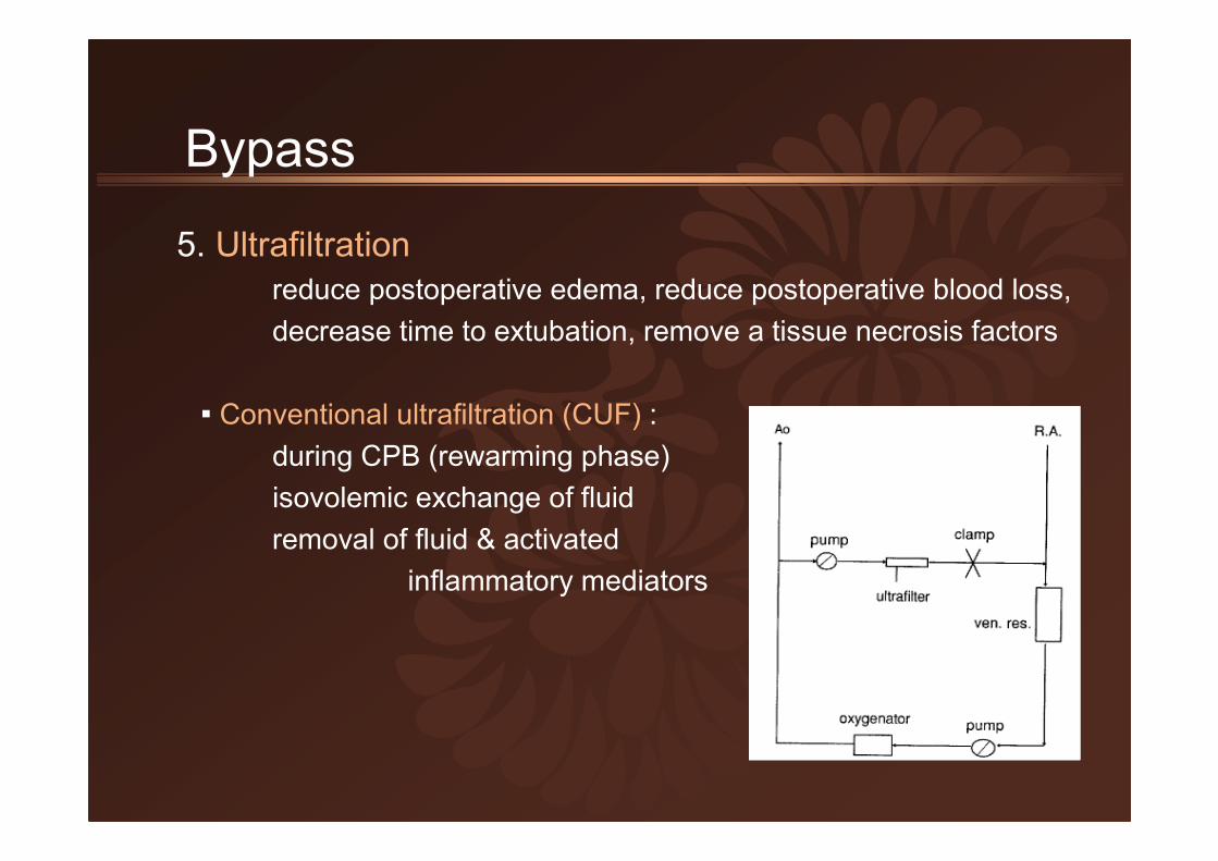
5. Ultrafiltration
reduce postoperative edema, reduce postoperative blood loss,
decrease time to extubation, remove a tissue necrosis factors
▪ Conventional ultrafiltration (CUF) :
during CPB (rewarming phase)
isovolemic exchange of fluid
removal of fluid & activated
inflammatory mediators

Bypass
5. Ultrafiltration
▪ Modified ultrafiltration (MUF) :
more effective in hemoconcentration &
improving ventricular functional recovery
after the completion of CPB
remove the patient 500 to 700ml of fluid
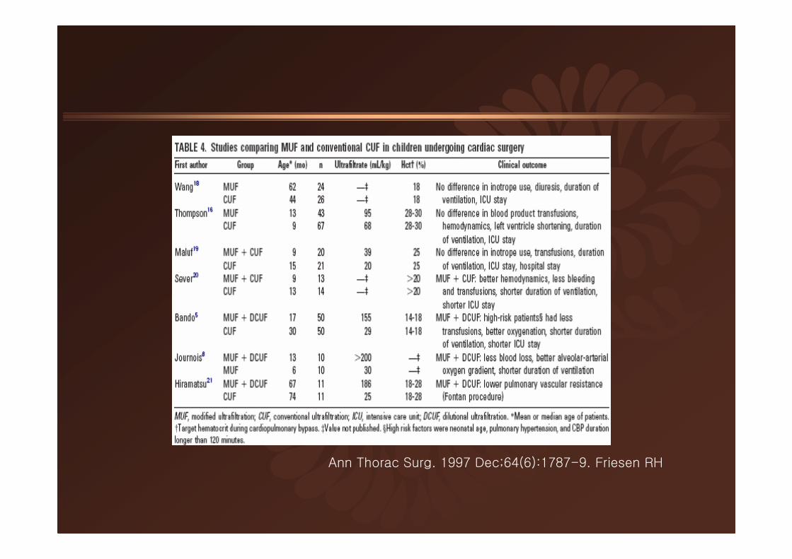
Ann Thorac Surg. 1997 Dec;64(6):1787-9. Friesen RH

Bypass
6. Anticoagulation
The amount of heparin to be delivered based on the patient's weight
(Dosage : adult 2 mg/kg, child 3 mg/kg)
do not based on the patient's blood volume
←effects of hypothermia, hemodilution, pre-existing heparin therapy
children require high doses of heparin to maintain ACT of 350-450 sec
> 200 sec : insertion cannula
> 400 sec : CPB start
> 480 sec : during CPB
> 750 sec : aprotinin is added

Bypass
6. Anticoagulation
after injection of initial heparin : ACT check q 30min
< 400 sec : 1mg/kg heparin
400-480 sec : 0.5mg/kg heparin
after CPB stop
Protamine dosage: 1.0 -1.5 mg for 100 unit (or mg) of heparin
> 480 sec : protamine 130% of initial doses of heparin
130-150 sec : 1/10 of initial doses of protamine
120-200 sec : 1/5 of initial doses of protamine

Postbypass
Once separated from CPB, the patient may continue to capillary
leakage and accumulate excessive soft tissue fluid for 24 to 36hr
• Leaving a foramen defect open
• Use of inotropic agents
• Leaving the sternum open
• Placement of peritoneal dialysis catheters
• Short period of ECMO

Thank you for your attention!
Related Documents