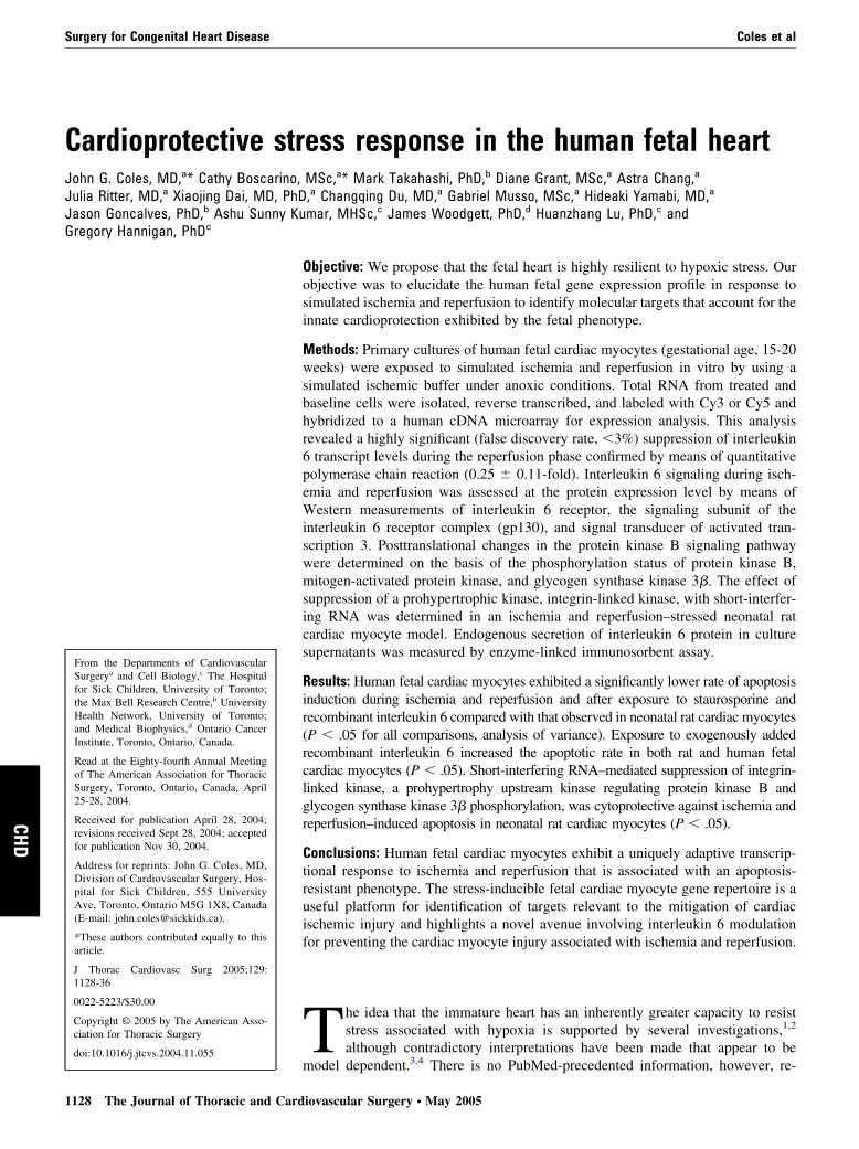
Cardioprotective stress response in the human fetal heart John G. Coles, MD, a * Cathy Boscarino, MSc, a * Mark Takahashi, PhD, b Diane Grant, MSc, a Astra Chang, a Julia Ritter, MD, a Xiaojing Dai, MD, PhD, a Changqing Du, MD, a Gabriel Musso, MSc, a Hideaki Yamabi, MD, a Jason Goncalves, PhD, b Ashu Sunny Kumar, MHSc, c James Woodgett, PhD, d Huanzhang Lu, PhD, c and Gregory Hannigan, PhD c Objective: We propose that the fetal heart is highly resilient to hypoxic stress. Our objective was to elucidate the human fetal gene expression profile in response to simulated ischemia and reperfusion to identify molecular targets that account for the innate cardioprotection exhibited by the fetal phenotype. Methods: Primary cultures of human fetal cardiac myocytes (gestational age, 15-20 weeks) were exposed to simulated ischemia and reperfusion in vitro by using a simulated ischemic buffer under anoxic conditions. Total RNA from treated and baseline cells were isolated, reverse transcribed, and labeled with Cy3 or Cy5 and hybridized to a human cDNA microarray for expression analysis. This analysis revealed a highly significant (false discovery rate, 3%) suppression of interleukin 6 transcript levels during the reperfusion phase confirmed by means of quantitative polymerase chain reaction (0.25 0.11-fold). Interleukin 6 signaling during isch- emia and reperfusion was assessed at the protein expression level by means of Western measurements of interleukin 6 receptor, the signaling subunit of the interleukin 6 receptor complex (gp130), and signal transducer of activated tran- scription 3. Posttranslational changes in the protein kinase B signaling pathway were determined on the basis of the phosphorylation status of protein kinase B, mitogen-activated protein kinase, and glycogen synthase kinase 3. The effect of suppression of a prohypertrophic kinase, integrin-linked kinase, with short-interfer- ing RNA was determined in an ischemia and reperfusion–stressed neonatal rat cardiac myocyte model. Endogenous secretion of interleukin 6 protein in culture supernatants was measured by enzyme-linked immunosorbent assay. Results: Human fetal cardiac myocytes exhibited a significantly lower rate of apoptosis induction during ischemia and reperfusion and after exposure to staurosporine and recombinant interleukin 6 compared with that observed in neonatal rat cardiac myocytes (P .05 for all comparisons, analysis of variance). Exposure to exogenously added recombinant interleukin 6 increased the apoptotic rate in both rat and human fetal cardiac myocytes (P .05). Short-interfering RNA–mediated suppression of integrin- linked kinase, a prohypertrophy upstream kinase regulating protein kinase B and glycogen synthase kinase 3 phosphorylation, was cytoprotective against ischemia and reperfusion–induced apoptosis in neonatal rat cardiac myocytes (P .05). Conclusions: Human fetal cardiac myocytes exhibit a uniquely adaptive transcrip- tional response to ischemia and reperfusion that is associated with an apoptosis- resistant phenotype. The stress-inducible fetal cardiac myocyte gene repertoire is a useful platform for identification of targets relevant to the mitigation of cardiac ischemic injury and highlights a novel avenue involving interleukin 6 modulation for preventing the cardiac myocyte injury associated with ischemia and reperfusion. T he idea that the immature heart has an inherently greater capacity to resist stress associated with hypoxia is supported by several investigations, 1,2 although contradictory interpretations have been made that appear to be model dependent. 3,4 There is no PubMed-precedented information, however, re- From the Departments of Cardiovascular Surgery a and Cell Biology, c The Hospital for Sick Children, University of Toronto; the Max Bell Research Centre, b University Health Network, University of Toronto; and Medical Biophysics, d Ontario Cancer Institute, Toronto, Ontario, Canada. Read at the Eighty-fourth Annual Meeting of The American Association for Thoracic Surgery, Toronto, Ontario, Canada, April 25-28, 2004. Received for publication April 28, 2004; revisions received Sept 28, 2004; accepted for publication Nov 30, 2004. Address for reprints: John G. Coles, MD, Division of Cardiovascular Surgery, Hos- pital for Sick Children, 555 University Ave, Toronto, Ontario M5G 1X8, Canada (E-mail: [email protected]). *These authors contributed equally to this article. J Thorac Cardiovasc Surg 2005;129: 1128-36 0022-5223/$30.00 Copyright © 2005 by The American Asso- ciation for Thoracic Surgery doi:10.1016/j.jtcvs.2004.11.055 Surgery for Congenital Heart Disease Coles et al 1128 The Journal of Thoracic and Cardiovascular Surgery ● May 2005 CHD

Welcome message from author
This document is posted to help you gain knowledge. Please leave a comment to let me know what you think about it! Share it to your friends and learn new things together.
Transcript
Surgery for Congenital Heart Disease Coles et al
CHD
Cardioprotective stress response in the human fetal heartJohn G. Coles, MD,a* Cathy Boscarino, MSc,a* Mark Takahashi, PhD,b Diane Grant, MSc,a Astra Chang,a
Julia Ritter, MD,a Xiaojing Dai, MD, PhD,a Changqing Du, MD,a Gabriel Musso, MSc,a Hideaki Yamabi, MD,a
Jason Goncalves, PhD,b Ashu Sunny Kumar, MHSc,c James Woodgett, PhD,d Huanzhang Lu, PhD,c and
Gregory Hannigan, PhDcFrom the Departments of CardiovascularSurgerya and Cell Biology,c The Hospitalfor Sick Children, University of Toronto;the Max Bell Research Centre,b UniversityHealth Network, University of Toronto;and Medical Biophysics,d Ontario CancerInstitute, Toronto, Ontario, Canada.
Read at the Eighty-fourth Annual Meetingof The American Association for ThoracicSurgery, Toronto, Ontario, Canada, April25-28, 2004.
Received for publication April 28, 2004;revisions received Sept 28, 2004; acceptedfor publication Nov 30, 2004.
Address for reprints: John G. Coles, MD,Division of Cardiovascular Surgery, Hos-pital for Sick Children, 555 UniversityAve, Toronto, Ontario M5G 1X8, Canada(E-mail: [email protected]).
*These authors contributed equally to thisarticle.
J Thorac Cardiovasc Surg 2005;129:1128-36
0022-5223/$30.00
Copyright © 2005 by The American Asso-ciation for Thoracic Surgery
doi:10.1016/j.jtcvs.2004.11.055
1128 The Journal of Thoracic and Card
Objective: We propose that the fetal heart is highly resilient to hypoxic stress. Ourobjective was to elucidate the human fetal gene expression profile in response tosimulated ischemia and reperfusion to identify molecular targets that account for theinnate cardioprotection exhibited by the fetal phenotype.
Methods: Primary cultures of human fetal cardiac myocytes (gestational age, 15-20weeks) were exposed to simulated ischemia and reperfusion in vitro by using asimulated ischemic buffer under anoxic conditions. Total RNA from treated andbaseline cells were isolated, reverse transcribed, and labeled with Cy3 or Cy5 andhybridized to a human cDNA microarray for expression analysis. This analysisrevealed a highly significant (false discovery rate, �3%) suppression of interleukin6 transcript levels during the reperfusion phase confirmed by means of quantitativepolymerase chain reaction (0.25 � 0.11-fold). Interleukin 6 signaling during isch-emia and reperfusion was assessed at the protein expression level by means ofWestern measurements of interleukin 6 receptor, the signaling subunit of theinterleukin 6 receptor complex (gp130), and signal transducer of activated tran-scription 3. Posttranslational changes in the protein kinase B signaling pathwaywere determined on the basis of the phosphorylation status of protein kinase B,mitogen-activated protein kinase, and glycogen synthase kinase 3�. The effect ofsuppression of a prohypertrophic kinase, integrin-linked kinase, with short-interfer-ing RNA was determined in an ischemia and reperfusion–stressed neonatal ratcardiac myocyte model. Endogenous secretion of interleukin 6 protein in culturesupernatants was measured by enzyme-linked immunosorbent assay.
Results: Human fetal cardiac myocytes exhibited a significantly lower rate of apoptosisinduction during ischemia and reperfusion and after exposure to staurosporine andrecombinant interleukin 6 compared with that observed in neonatal rat cardiac myocytes(P � .05 for all comparisons, analysis of variance). Exposure to exogenously addedrecombinant interleukin 6 increased the apoptotic rate in both rat and human fetalcardiac myocytes (P � .05). Short-interfering RNA–mediated suppression of integrin-linked kinase, a prohypertrophy upstream kinase regulating protein kinase B andglycogen synthase kinase 3� phosphorylation, was cytoprotective against ischemia andreperfusion–induced apoptosis in neonatal rat cardiac myocytes (P � .05).
Conclusions: Human fetal cardiac myocytes exhibit a uniquely adaptive transcrip-tional response to ischemia and reperfusion that is associated with an apoptosis-resistant phenotype. The stress-inducible fetal cardiac myocyte gene repertoire is auseful platform for identification of targets relevant to the mitigation of cardiacischemic injury and highlights a novel avenue involving interleukin 6 modulationfor preventing the cardiac myocyte injury associated with ischemia and reperfusion.
T he idea that the immature heart has an inherently greater capacity to resiststress associated with hypoxia is supported by several investigations,1,2
although contradictory interpretations have been made that appear to be
model dependent.3,4 There is no PubMed-precedented information, however, re-iovascular Surgery ● May 2005
h. SA
Coles et al Surgery for Congenital Heart Disease
CHD
garding potential developmental changes in cardiomyocytegene expression, which might reveal the molecular mecha-nisms accounting for the enhanced stress resistance in theimmature human cardiac myocyte.
The idea that interleukin (IL) 6 pathway activation ad-versely affects cardiac function is solidly supported byclinical studies indicating that IL-6 and its specific receptor(IL-6R�) and the 130-kd glycoprotein signaling subunit ofthe IL-6 receptor, gp130,5 are upregulated at the mRNA and
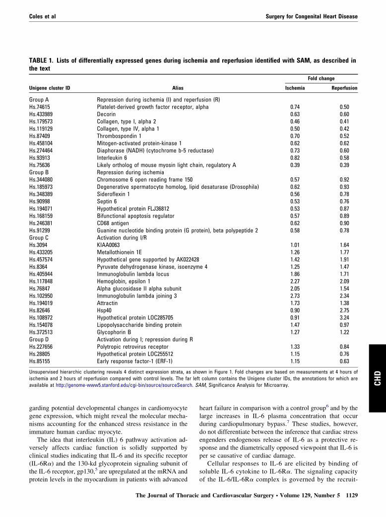
TABLE 1. Lists of differentially expressed genes during isthe text
Unigene cluster ID Alias
Group A Repression during ischemia (I) and reHs.74615 Platelet-derived growth factor receptoHs.433989 DecorinHs.179573 Collagen, type I, alpha 2Hs.119129 Collagen, type IV, alpha 1Hs.87409 Thrombospondin 1Hs.458104 Mitogen-activated protein-kinase 1Hs.274464 Diaphorase (NADH) (cytochrome b-5Hs.93913 Interleukin 6Hs.75636 Likely ortholog of mouse myosin lightGroup B Repression during ischemiaHs.344080 Chromosome 6 open reading frame 15Hs.185973 Degenerative spermatocyte homolog,Hs.348389 Sideroflexin 1Hs.90998 Septin 6Hs.194071 Hypothetical protein FLJ36812Hs.168159 Bifunctional apoptosis regulatorHs.246381 CD68 antigenHs.91299 Guanine nucleotide binding protein (GGroup C Activation during I/RHs.3094 KIAA0063Hs.433205 Metallothionein 1EHs.457574 Hypothetical gene supported by AK02Hs.8364 Pyruvate dehydrogenase kinase, isoeHs.405944 Immunoglobulin lambda locusHs.117848 Hemoglobin, epsilon 1Hs.76847 Alpha glucosidase II alpha subunitHs.102950 Immunoglobulin lambda joining 3Hs.194019 AttractinHs.82646 Hsp40Hs.108972 Hypothetical protein LOC285705Hs.154078 Lipopolysaccharide binding proteinHs.372513 Glycophorin BGroup D Activation during I; repression duringHs.227656 Polytropic retrovirus receptorHs.28805 Hypothetical protein LOC255512Hs.85155 Early response factor-1 (ERF-1)
Unsupervised hierarchic clustering reveals 4 distinct expression strata, aischemia and 2 hours of reperfusion compared with control levels. The faavailable at http://genome-www5.stanford.edu/cgi-bin/source/sourceSearc
protein levels in the myocardium in patients with advanced
The Journal of Thoracic
heart failure in comparison with a control group6 and by thelarge increases in IL-6 plasma concentration that occurduring cardiopulmonary bypass.7 These studies, however,do not differentiate between the inference that cardiac stressengenders endogenous release of IL-6 as a protective re-sponse and the diametrically opposed viewpoint that IL-6 isper se causative of cardiac damage.
Cellular responses to IL-6 are elicited by binding ofsoluble IL-6 cytokine to IL-6R�. The signaling capacity
ia and reperfusion identified with SAM, as described in
Fold change
Ischemia Reperfusion
sion (R)pha 0.74 0.50
0.63 0.600.46 0.410.50 0.420.70 0.520.62 0.62
tase) 0.73 0.600.82 0.58
n, regulatory A 0.39 0.39
0.57 0.92desaturase (Drosophila) 0.62 0.93
0.56 0.780.53 0.760.53 0.870.57 0.890.62 0.90
ein), beta polypeptide 2 0.58 0.78
1.01 1.641.26 1.771.42 1.91
e 4 1.25 1.471.86 1.712.27 2.092.05 1.542.73 2.341.73 1.380.90 2.750.91 3.241.47 0.971.27 1.22
1.33 0.841.15 0.761.15 0.63
wn in Figure 1. Fold changes are based on measurements at 4 hours ofcolumn contains the Unigene cluster IDs, the annotations for which areM, Significance Analysis for Microarray.
chem
perfur, al
reduc
chai
0lipid
prot
2428nzym
R
s shor left
of the IL-6/IL-6R� complex is governed by the recruit-
and Cardiovascular Surgery ● Volume 129, Number 5 1129
Surgery for Congenital Heart Disease Coles et al
CHD
ment of 2 gp130 subunits to the activated, multisubunitreceptor complex. Activation of the IL-6 receptor-ligandcomplex is dependent on the recruitment and dimeriza-tion of gp130,8 which triggers activation of several col-lateral signaling pathways, including the Janus kinase/signal transducer and activator of transcription (JAK/STAT), Ras/mitogen-activated protein kinase (MAPK),and phosphatidylinositol 3– kinase (PI3-K)– dependentpathway involving sequential phosphorylation of proteinkinase B (PKB/Akt) and glycogen synthase kinase 3�(GSK-3� ).9 Thus the IL-6 receptor complex consists of aligand-binding molecule (IL-6R�) and a signaling sub-unit, gp130, which provides a rapid membrane-to-nucleus signaling system regulating inflammatory geneexpression.
We show here in expression-profiling experiments thathuman fetal cardiac myocytes (HFCMs) exposed to simu-lated ischemia with reperfusion (I/R) or without reperfusionexhibit a uniquely adaptive transcriptional response. Thefetal response includes a limited number of functional clus-ters dominated by predicted anti-inflammatory properties,featuring repression of the IL-6 signaling evident at both themRNA and protein expression levels during reperfusion-mediated stress. These data provide a plausible and thera-peutically important explanation for the innately apoptosis-resistant HFCM phenotype.
MethodsCardiac Myocyte CulturesPrimary cultures of HFCMs were derived from structurally normalhearts after elective pregnancy termination (gestational age, 15-20weeks) under an institutional review board–approved protocol.Primary cultures of cardiac myocytes were prepared from humanfetal ventricles and from 2- to 3-day neonatal rat cardiac myocytes(NRCMs) by the following previously described protocol10 incompliance with Institutional Animal Care Guidelines. Cardiomy-ocytes were dissociated by means of repeated (3�) enzymaticdigestion with 0.05% trypsin-collagenase solution (Life Technol-ogies) at 37°C and the dissociated cells concentrated from thesupernatant by means of centrifugation (2600 rpm � 7 minutes)and plated at a density of 1 � 105 cells/cm2 on 35-mm culturedishes with 2 mL of culture medium (Dulbeccco’s modifiedEagle’s medium with 10% fetal bovine serum). Preplating ofseeded cells onto 100-mm culture dishes to remove noncardiom-yocytes yielded cultures containing approximately 80% �-myosinheavy chain–positive cardiac myocytes.
HFCMs and NRCMs were exposed to simulated I/R or isch-emia without reperfusion in vitro for the indicated time intervalsby using ischemic buffer and anoxic conditions, as previouslydescribed.11 To simulate ischemia, the cultures were washed twicewith phosphate-buffered saline and transferred to a pH 6.5 simu-lated ischemia solution containing, in 100 mL of deionized water,0.8 g of NaCl, 0.119 g of N-2-hydroxyethylpiperazine-N-2-ethanesulfonic acid, 0.164 g of 2-deoxyglucose, 0.89 mL of KCl,0.472 mL of CaCl , 0.238 mL of MgCl , and 0.135 mL of
2 2DL-lactic acid saturated in 100% N2 for 1 hour, followed by
1130 The Journal of Thoracic and Cardiovascular Surgery ● Ma
addition of 0.1 g of bovine serum albumin. Cultures were placed inan airtight container at 37°C with 100% N2 inflow for periodsranging from 2 to 24 hours and subsequently washed twice withphosphate-buffered saline before harvesting. Cardiomyocyte cul-tures were re-exposed to the control normoxic serum-free mediumto simulate reperfusion. Control cardiomyocyte cultures weremaintained under normoxic conditions at 37°C until analysis. Thusischemia is simulated by a combination of acidic pH, interferencewith glucose metabolism, and anoxia, and reperfusion is simulatedby means of re-exposure to normoxia, which has been shown toprovoke extensive apoptosis in similar models12 and is hereinreferred to as simulated I/R.
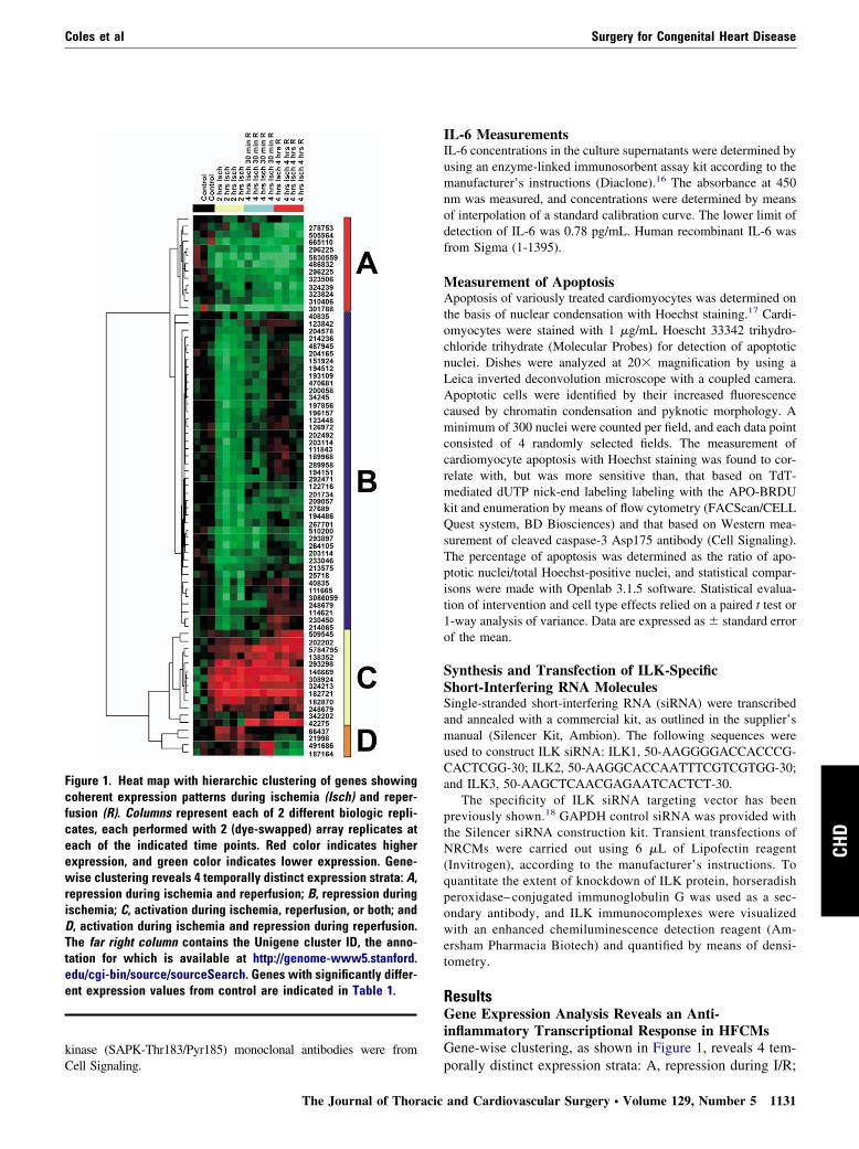
Microarray Gene Expression AnalysisRNA isolation, fluorescence labeling of cDNA, hybridization tospotted arrays containing 15,264 sequence-verified cDNA clones,and quantitative fluorescence scanning of gene expression inten-sity were performed at the University of Toronto Health NetworkMicroarray Centre (www.microarray.ca), as previously reportedby us13 and others (for a list of publications, see http://www.microarrays.ca/about/pub.html). Significance of changes in se-quential gene expression in HFCMs exposed to I/R (at control, 4hours of ischemia, and 4 hours of ischemia plus 4 hours ofreperfusion) were determined by means of repeated permutation ofMIAME-compliant (www.mged.org) data with Significance Anal-ysis for Microarray.14 Results from the Significance Analysis forMicroarray analysis were visualized as hierarchic clusters in GeneTraffic (www.iobion.com), and significant genes were classified bytheir differential response to ischemia, reperfusion, or both. Theresults shown in Table 1 and Figure 1 are based on 2 biologic and2 technical (array) replicates at each indicated time point, with afalse discovery rate indicative of the statistical risk of incorrectidentification of differentially expressed genes set to less than3%.15
Validation With Quantitative Polymerase ChainReactionIndependent confirmation of changes in IL-6 transcription levelswas performed by using real-time quantitative polymerase chainreaction (qPCR), as previously described by us.13 Primers wereconstructed against the 3= ends of IL-6, and amplicon abundancewas determined in real time with SYBR Green Dye (AppliedBiosystems) fluorescence measurement during the logarithmicphase and normalized to that of a control gene, cyclophilin.
Western Blot AnalysisFetal cardiomyocyte extracts containing 20 �g of protein were sub-jected to sodium dodecylsulfate–polyacrylamide gel electrophoresiswith 10% polyacrylamide gel and transferred onto Immobilon-Ptransfer membranes (Millipore). Analysis was performed with poly-clonal PKB antibody (Transduction Laboratories), polyclonal serine437 catalytically active, phosphorylation-specific PKB antibody (CellSignaling Technology), polyclonal integrin-linked kinase (ILK) anti-body (Upstate Biotechnology), and anti-IL-6 receptor (IL-6R�) andanti-gp130 antibodies (Santa Cruz Biotech). Monoclonal antibodiesused for the determination of total and phosphorylated GSK-3�protein levels were from Biosource; total and phosphorylated (Py705)
STAT-3, (Thr202/Tyr204) MAPK42/44, and stress-activated proteiny 2005
Coles et al Surgery for Congenital Heart Disease
CHD
kinase (SAPK-Thr183/Pyr185) monoclonal antibodies were from
Figure 1. Heat map with hierarchic clustering of genes showingcoherent expression patterns during ischemia (Isch) and reper-fusion (R). Columns represent each of 2 different biologic repli-cates, each performed with 2 (dye-swapped) array replicates ateach of the indicated time points. Red color indicates higherexpression, and green color indicates lower expression. Gene-wise clustering reveals 4 temporally distinct expression strata: A,repression during ischemia and reperfusion; B, repression duringischemia; C, activation during ischemia, reperfusion, or both; andD, activation during ischemia and repression during reperfusion.The far right column contains the Unigene cluster ID, the anno-tation for which is available at http://genome-www5.stanford.edu/cgi-bin/source/sourceSearch. Genes with significantly differ-ent expression values from control are indicated in Table 1.
Cell Signaling.
The Journal of Thoracic
IL-6 MeasurementsIL-6 concentrations in the culture supernatants were determined byusing an enzyme-linked immunosorbent assay kit according to themanufacturer’s instructions (Diaclone).16 The absorbance at 450nm was measured, and concentrations were determined by meansof interpolation of a standard calibration curve. The lower limit ofdetection of IL-6 was 0.78 pg/mL. Human recombinant IL-6 wasfrom Sigma (1-1395).
Measurement of ApoptosisApoptosis of variously treated cardiomyocytes was determined onthe basis of nuclear condensation with Hoechst staining.17 Cardi-omyocytes were stained with 1 �g/mL Hoescht 33342 trihydro-chloride trihydrate (Molecular Probes) for detection of apoptoticnuclei. Dishes were analyzed at 20� magnification by using aLeica inverted deconvolution microscope with a coupled camera.Apoptotic cells were identified by their increased fluorescencecaused by chromatin condensation and pyknotic morphology. Aminimum of 300 nuclei were counted per field, and each data pointconsisted of 4 randomly selected fields. The measurement ofcardiomyocyte apoptosis with Hoechst staining was found to cor-relate with, but was more sensitive than, that based on TdT-mediated dUTP nick-end labeling labeling with the APO-BRDUkit and enumeration by means of flow cytometry (FACScan/CELLQuest system, BD Biosciences) and that based on Western mea-surement of cleaved caspase-3 Asp175 antibody (Cell Signaling).The percentage of apoptosis was determined as the ratio of apo-ptotic nuclei/total Hoechst-positive nuclei, and statistical compar-isons were made with Openlab 3.1.5 software. Statistical evalua-tion of intervention and cell type effects relied on a paired t test or1-way analysis of variance. Data are expressed as � standard errorof the mean.
Synthesis and Transfection of ILK-SpecificShort-Interfering RNA MoleculesSingle-stranded short-interfering RNA (siRNA) were transcribedand annealed with a commercial kit, as outlined in the supplier’smanual (Silencer Kit, Ambion). The following sequences wereused to construct ILK siRNA: ILK1, 50-AAGGGGACCACCCG-CACTCGG-30; ILK2, 50-AAGGCACCAATTTCGTCGTGG-30;and ILK3, 50-AAGCTCAACGAGAATCACTCT-30.
The specificity of ILK siRNA targeting vector has beenpreviously shown.18 GAPDH control siRNA was provided withthe Silencer siRNA construction kit. Transient transfections ofNRCMs were carried out using 6 �L of Lipofectin reagent(Invitrogen), according to the manufacturer’s instructions. Toquantitate the extent of knockdown of ILK protein, horseradishperoxidase– conjugated immunoglobulin G was used as a sec-ondary antibody, and ILK immunocomplexes were visualizedwith an enhanced chemiluminescence detection reagent (Am-ersham Pharmacia Biotech) and quantified by means of densi-tometry.
ResultsGene Expression Analysis Reveals an Anti-inflammatory Transcriptional Response in HFCMsGene-wise clustering, as shown in Figure 1, reveals 4 tem-
porally distinct expression strata: A, repression during I/R;and Cardiovascular Surgery ● Volume 129, Number 5 1131
Surgery for Congenital Heart Disease Coles et al
CHD
B, repression during ischemia; C, activation during isch-emia, reperfusion, or both; and D, activation during isch-emia and repression during reperfusion. The annotation ofsignificant genes and corresponding expression values areindicated in Table 1. Noteworthy was the significant repres-sion of IL-6 transcription during ischemia and especiallyduring reperfusion to 58% of preischemic levels by meansof microarray analysis and to 25% � 11% by means ofqPCR measurement.
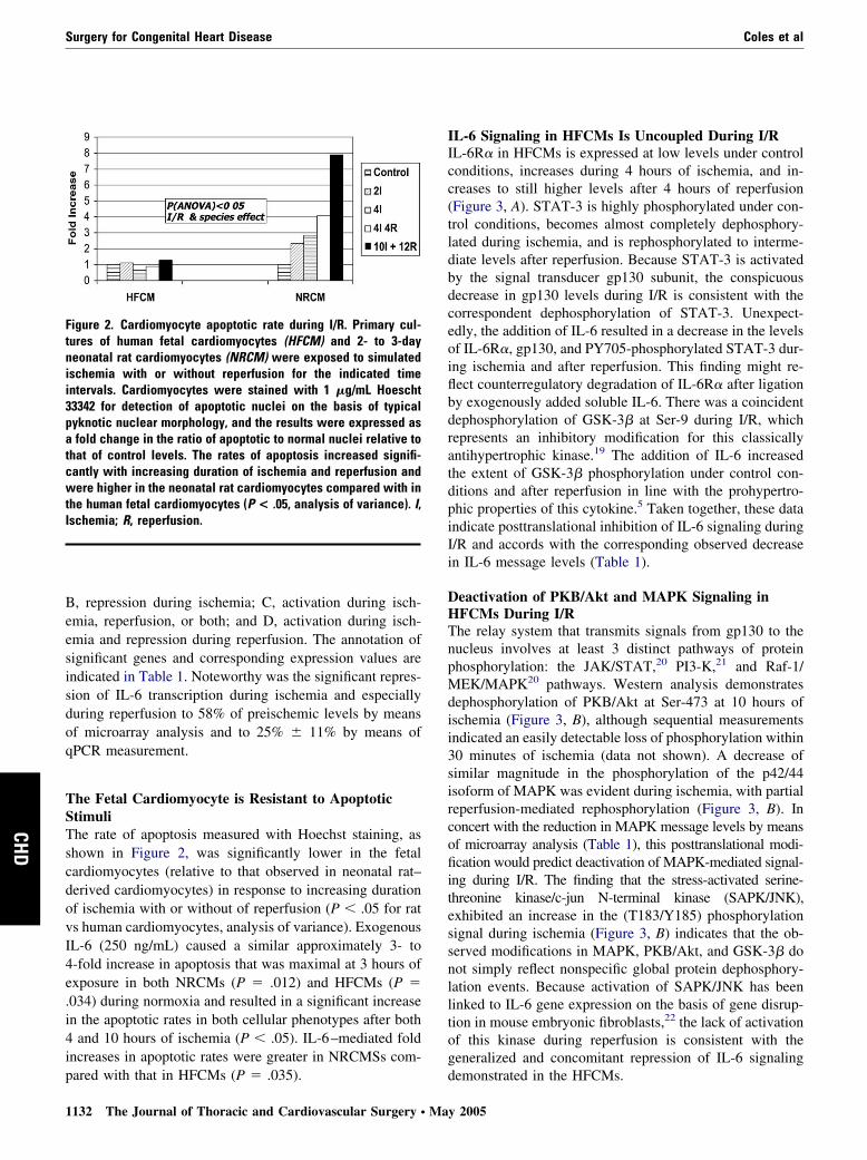
The Fetal Cardiomyocyte is Resistant to ApoptoticStimuliThe rate of apoptosis measured with Hoechst staining, asshown in Figure 2, was significantly lower in the fetalcardiomyocytes (relative to that observed in neonatal rat–derived cardiomyocytes) in response to increasing durationof ischemia with or without of reperfusion (P � .05 for ratvs human cardiomyocytes, analysis of variance). ExogenousIL-6 (250 ng/mL) caused a similar approximately 3- to4-fold increase in apoptosis that was maximal at 3 hours ofexposure in both NRCMs (P � .012) and HFCMs (P �.034) during normoxia and resulted in a significant increasein the apoptotic rates in both cellular phenotypes after both4 and 10 hours of ischemia (P � .05). IL-6–mediated foldincreases in apoptotic rates were greater in NRCMSs com-
Figure 2. Cardiomyocyte apoptotic rate during I/R. Primary cul-tures of human fetal cardiomyocytes (HFCM) and 2- to 3-dayneonatal rat cardiomyocytes (NRCM) were exposed to simulatedischemia with or without reperfusion for the indicated timeintervals. Cardiomyocytes were stained with 1 �g/mL Hoescht33342 for detection of apoptotic nuclei on the basis of typicalpyknotic nuclear morphology, and the results were expressed asa fold change in the ratio of apoptotic to normal nuclei relative tothat of control levels. The rates of apoptosis increased signifi-cantly with increasing duration of ischemia and reperfusion andwere higher in the neonatal rat cardiomyocytes compared with inthe human fetal cardiomyocytes (P < .05, analysis of variance). I,Ischemia; R, reperfusion.
pared with that in HFCMs (P � .035).
1132 The Journal of Thoracic and Cardiovascular Surgery ● Ma
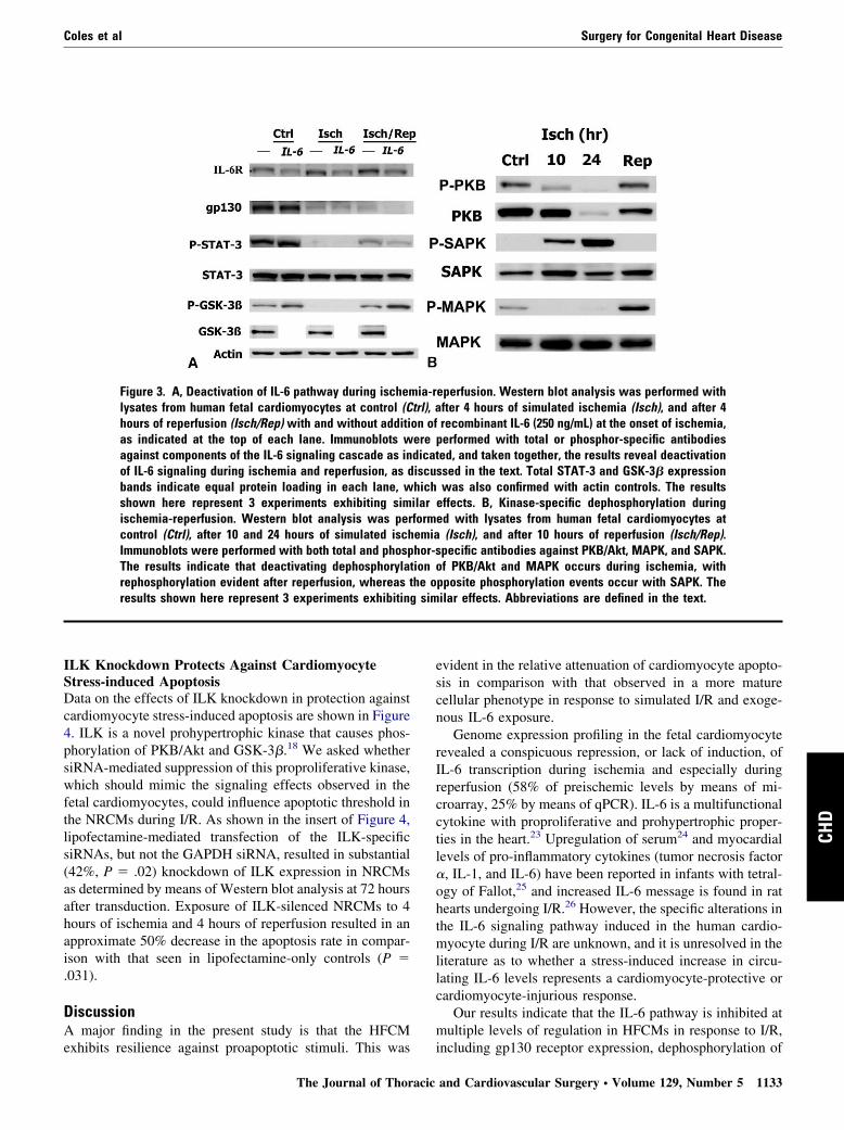
IL-6 Signaling in HFCMs Is Uncoupled During I/RIL-6R� in HFCMs is expressed at low levels under controlconditions, increases during 4 hours of ischemia, and in-creases to still higher levels after 4 hours of reperfusion(Figure 3, A). STAT-3 is highly phosphorylated under con-trol conditions, becomes almost completely dephosphory-lated during ischemia, and is rephosphorylated to interme-diate levels after reperfusion. Because STAT-3 is activatedby the signal transducer gp130 subunit, the conspicuousdecrease in gp130 levels during I/R is consistent with thecorrespondent dephosphorylation of STAT-3. Unexpect-edly, the addition of IL-6 resulted in a decrease in the levelsof IL-6R�, gp130, and PY705-phosphorylated STAT-3 dur-ing ischemia and after reperfusion. This finding might re-flect counterregulatory degradation of IL-6R� after ligationby exogenously added soluble IL-6. There was a coincidentdephosphorylation of GSK-3� at Ser-9 during I/R, whichrepresents an inhibitory modification for this classicallyantihypertrophic kinase.19 The addition of IL-6 increasedthe extent of GSK-3� phosphorylation under control con-ditions and after reperfusion in line with the prohypertro-phic properties of this cytokine.5 Taken together, these dataindicate posttranslational inhibition of IL-6 signaling duringI/R and accords with the corresponding observed decreasein IL-6 message levels (Table 1).
Deactivation of PKB/Akt and MAPK Signaling inHFCMs During I/RThe relay system that transmits signals from gp130 to thenucleus involves at least 3 distinct pathways of proteinphosphorylation: the JAK/STAT,20 PI3-K,21 and Raf-1/MEK/MAPK20 pathways. Western analysis demonstratesdephosphorylation of PKB/Akt at Ser-473 at 10 hours ofischemia (Figure 3, B), although sequential measurementsindicated an easily detectable loss of phosphorylation within30 minutes of ischemia (data not shown). A decrease ofsimilar magnitude in the phosphorylation of the p42/44isoform of MAPK was evident during ischemia, with partialreperfusion-mediated rephosphorylation (Figure 3, B). Inconcert with the reduction in MAPK message levels by meansof microarray analysis (Table 1), this posttranslational modi-fication would predict deactivation of MAPK-mediated signal-ing during I/R. The finding that the stress-activated serine-threonine kinase/c-jun N-terminal kinase (SAPK/JNK),exhibited an increase in the (T183/Y185) phosphorylationsignal during ischemia (Figure 3, B) indicates that the ob-served modifications in MAPK, PKB/Akt, and GSK-3� donot simply reflect nonspecific global protein dephosphory-lation events. Because activation of SAPK/JNK has beenlinked to IL-6 gene expression on the basis of gene disrup-tion in mouse embryonic fibroblasts,22 the lack of activationof this kinase during reperfusion is consistent with thegeneralized and concomitant repression of IL-6 signaling
demonstrated in the HFCMs.y 2005
Coles et al Surgery for Congenital Heart Disease
CHD
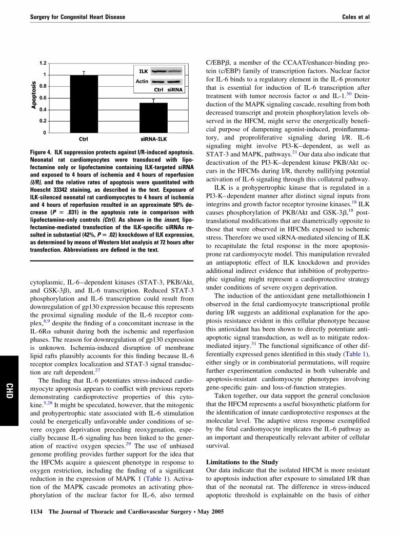
ILK Knockdown Protects Against CardiomyocyteStress-induced ApoptosisData on the effects of ILK knockdown in protection againstcardiomyocyte stress-induced apoptosis are shown in Figure4. ILK is a novel prohypertrophic kinase that causes phos-phorylation of PKB/Akt and GSK-3�.18 We asked whethersiRNA-mediated suppression of this proproliferative kinase,which should mimic the signaling effects observed in thefetal cardiomyocytes, could influence apoptotic threshold inthe NRCMs during I/R. As shown in the insert of Figure 4,lipofectamine-mediated transfection of the ILK-specificsiRNAs, but not the GAPDH siRNA, resulted in substantial(42%, P � .02) knockdown of ILK expression in NRCMsas determined by means of Western blot analysis at 72 hoursafter transduction. Exposure of ILK-silenced NRCMs to 4hours of ischemia and 4 hours of reperfusion resulted in anapproximate 50% decrease in the apoptosis rate in compar-ison with that seen in lipofectamine-only controls (P �.031).
DiscussionA major finding in the present study is that the HFCM
Figure 3. A, Deactivation of IL-6 pathway during ischemlysates from human fetal cardiomyocytes at control (Chours of reperfusion (Isch/Rep) with and without additias indicated at the top of each lane. Immunoblots wagainst components of the IL-6 signaling cascade as inof IL-6 signaling during ischemia and reperfusion, as dbands indicate equal protein loading in each lane, wshown here represent 3 experiments exhibiting simischemia-reperfusion. Western blot analysis was percontrol (Ctrl), after 10 and 24 hours of simulated ischImmunoblots were performed with both total and phospThe results indicate that deactivating dephosphorylarephosphorylation evident after reperfusion, whereas tresults shown here represent 3 experiments exhibitin
exhibits resilience against proapoptotic stimuli. This was
The Journal of Thoracic
evident in the relative attenuation of cardiomyocyte apopto-sis in comparison with that observed in a more maturecellular phenotype in response to simulated I/R and exoge-nous IL-6 exposure.
Genome expression profiling in the fetal cardiomyocyterevealed a conspicuous repression, or lack of induction, ofIL-6 transcription during ischemia and especially duringreperfusion (58% of preischemic levels by means of mi-croarray, 25% by means of qPCR). IL-6 is a multifunctionalcytokine with proproliferative and prohypertrophic proper-ties in the heart.23 Upregulation of serum24 and myocardiallevels of pro-inflammatory cytokines (tumor necrosis factor�, IL-1, and IL-6) have been reported in infants with tetral-ogy of Fallot,25 and increased IL-6 message is found in rathearts undergoing I/R.26 However, the specific alterations inthe IL-6 signaling pathway induced in the human cardio-myocyte during I/R are unknown, and it is unresolved in theliterature as to whether a stress-induced increase in circu-lating IL-6 levels represents a cardiomyocyte-protective orcardiomyocyte-injurious response.
Our results indicate that the IL-6 pathway is inhibited atmultiple levels of regulation in HFCMs in response to I/R,
eperfusion. Western blot analysis was performed withfter 4 hours of simulated ischemia (Isch), and after 4recombinant IL-6 (250 ng/mL) at the onset of ischemia,
performed with total or phosphor-specific antibodiesed, and taken together, the results reveal deactivationssed in the text. Total STAT-3 and GSK-3� expressionwas also confirmed with actin controls. The results
effects. B, Kinase-specific dephosphorylation duringed with lysates from human fetal cardiomyocytes at
(Isch), and after 10 hours of reperfusion (Isch/Rep).pecific antibodies against PKB/Akt, MAPK, and SAPK.
of PKB/Akt and MAPK occurs during ischemia, withpposite phosphorylation events occur with SAPK. Theilar effects. Abbreviations are defined in the text.
ia-rtrl), aon oferedicatiscuhichilarformemiahor-s
tionhe o
g sim
including gp130 receptor expression, dephosphorylation of
and Cardiovascular Surgery ● Volume 129, Number 5 1133
Surgery for Congenital Heart Disease Coles et al
CHD
cytoplasmic, IL-6–dependent kinases (STAT-3, PKB/Akt,and GSK-3�), and IL-6 transcription. Reduced STAT-3phosphorylation and IL-6 transcription could result fromdownregulation of gp130 expression because this representsthe proximal signaling module of the IL-6 receptor com-plex,8,9 despite the finding of a concomitant increase in theIL-6R� subunit during both the ischemic and reperfusionphases. The reason for downregulation of gp130 expressionis unknown. Ischemia-induced disruption of membranelipid rafts plausibly accounts for this finding because IL-6receptor complex localization and STAT-3 signal transduc-tion are raft dependent.27
The finding that IL-6 potentiates stress-induced cardio-myocyte apoptosis appears to conflict with previous reportsdemonstrating cardioprotective properties of this cyto-kine.5,28 It might be speculated, however, that the mitogenicand prohypertrophic state associated with IL-6 stimulationcould be energetically unfavorable under conditions of se-vere oxygen deprivation preceding reoxygenation, espe-cially because IL-6 signaling has been linked to the gener-ation of reactive oxygen species.29 The use of unbiasedgenome profiling provides further support for the idea thatthe HFCMs acquire a quiescent phenotype in response tooxygen restriction, including the finding of a significantreduction in the expression of MAPK 1 (Table 1). Activa-tion of the MAPK cascade promotes an activating phos-
Figure 4. ILK suppression protects against I/R-induced apoptosis.Neonatal rat cardiomyocytes were transduced with lipo-fectamine only or lipofectamine containing ILK-targeted siRNAand exposed to 4 hours of ischemia and 4 hours of reperfusion(I/R), and the relative rates of apoptosis were quantitated withHoescht 33342 staining, as described in the text. Exposure ofILK-silenced neonatal rat cardiomyocytes to 4 hours of ischemiaand 4 hours of reperfusion resulted in an approximate 50% de-crease (P � .031) in the apoptosis rate in comparison withlipofectamine-only controls (Ctrl). As shown in the insert, lipo-fectamine-mediated transfection of the ILK-specific siRNAs re-sulted in substantial (42%, P � .02) knockdown of ILK expression,as determined by means of Western blot analysis at 72 hours aftertransfection. Abbreviations are defined in the text.
phorylation of the nuclear factor for IL-6, also termed
1134 The Journal of Thoracic and Cardiovascular Surgery ● Ma
C/EBP�, a member of the CCAAT/enhancer-binding pro-tein (c/EBP) family of transcription factors. Nuclear factorfor IL-6 binds to a regulatory element in the IL-6 promoterthat is essential for induction of IL-6 transcription aftertreatment with tumor necrosis factor � and IL-1.30 Dein-duction of the MAPK signaling cascade, resulting from bothdecreased transcript and protein phosphorylation levels ob-served in the HFCM, might serve the energetically benefi-cial purpose of dampening agonist-induced, proinflamma-tory, and proproliferative signaling during I/R. IL-6signaling might involve PI3-K–dependent, as well asSTAT-3 and MAPK, pathways.21 Our data also indicate thatdeactivation of the PI3-K–dependent kinase PKB/Akt oc-curs in the HFCMs during I/R, thereby nullifying potentialactivation of IL-6 signaling through this collateral pathway.
ILK is a prohypertrophic kinase that is regulated in aPI3-K–dependent manner after distinct signal inputs fromintegrins and growth factor receptor tyrosine kinases.18 ILKcauses phosphorylation of PKB/Akt and GSK-3�,18 post-translational modifications that are diametrically opposite tothose that were observed in HFCMs exposed to ischemicstress. Therefore we used siRNA-mediated silencing of ILKto recapitulate the fetal response in the more apoptosis-prone rat cardiomyocyte model. This manipulation revealedan antiapoptotic effect of ILK knockdown and providesadditional indirect evidence that inhibition of prohypertro-phic signaling might represent a cardioprotective strategyunder conditions of severe oxygen deprivation.
The induction of the antioxidant gene metallothionein Iobserved in the fetal cardiomyocyte transcriptional profileduring I/R suggests an additional explanation for the apo-ptosis resistance evident in this cellular phenotype becausethis antioxidant has been shown to directly potentiate anti-apoptotic signal transduction, as well as to mitigate redox-mediated injury.31 The functional significance of other dif-ferentially expressed genes identified in this study (Table 1),either singly or in combinatorial permutations, will requirefurther experimentation conducted in both vulnerable andapoptosis-resistant cardiomyocyte phenotypes involvinggene-specific gain- and loss-of-function strategies.
Taken together, our data support the general conclusionthat the HFCM represents a useful biosynthetic platform forthe identification of innate cardioprotective responses at themolecular level. The adaptive stress response exemplifiedby the fetal cardiomyocyte implicates the IL-6 pathway asan important and therapeutically relevant arbiter of cellularsurvival.
Limitations to the StudyOur data indicate that the isolated HFCM is more resistantto apoptosis induction after exposure to simulated I/R thanthat of the neonatal rat. The difference in stress-induced
apoptotic threshold is explainable on the basis of eithery 2005
Coles et al Surgery for Congenital Heart Disease
CHD
species-related or development stage–related differences in-herent in these distinct cellular phenotypes. The elucidationof developmental stage-specific aspects of the human car-diomyocyte stress response would require a comparativeanalysis of human neonatal or adult cardiomyocytes withthat of the fetal phenotype. The fact that postnatal humancardiomyocytes have an inherently greater susceptibility toapoptosis during in vitro culture renders such studies diffi-cult and implies that cardiomyocyte stress resistance de-grades with increasing maturation. Nevertheless, our studyis directed to the investigation of IL-6 signaling duringredox-induced stress in the apoptosis-resistant HFCM phe-notype and does not specifically address the developmentalaspects of this response.
References
1. Shi Y, Baker JE, Zhang C, Tweddell JS, Su J, Pritchard KA Jr. Chronichypoxia increases endothelial nitric oxide synthase generation of nitricoxide by increasing heat shock protein 90 association and serinephosphorylation. Circ Res. 2002;91:300-6.
2. Madan A, Varma S, Cohen HJ. Developmental stage-specific expres-sion of the alpha and beta subunits of the HIF-1 protein in the mouseand human fetus. Mol Genet Metab. 2002;75:244-9.
3. Friehs I, Cao-Danh H, Stamm C, Cowan DB, McGowan FX, del NidoPJ. Postnatal increase in insulin-sensitive glucose transporter expres-sion is associated with improved recovery of postischemic myocardialfunction. J Thorac Cardiovasc Surg. 2003;126:263-71.
4. Karimi M, Wang LX, Hammel JM, Mascio CE, Abdulhamid M,Barner EW, et al. Neonatal vulnerability to ischemia and reperfusion:cardioplegic arrest causes greater myocardial apoptosis in neonatallambs than in mature lambs. J Thorac Cardiovasc Surg. 2004;127:490-7.
5. Ancey C, Menet E, Corbi P, Fredj S, Garcia M, Rucker-Martin C,et al. Human cardiomyocyte hypertrophy induced in vitro by gp130stimulation. Cardiovasc Res. 2003;59:78-85.
6. Zolk O, Ng LL, O’Brien RJ, Weyand M, Eschenhagen T. Augmentedexpression of cardiotrophin-1 in failing human hearts is accompaniedby diminished glycoprotein 130 receptor protein abundance. Circula-tion. 2002;106:1442-6.
7. Corbi P, Rahmati M, Delwail A, Potreau D, Menu P, Wijdenes J, et al.Circulating soluble gp130, soluble IL-6R, and IL-6 in patients under-going cardiac surgery, with or without extracorporeal circulation. EurJ Cardiothorac Surg. 2000;18:98-103.
8. Jostock T, Mullberg J, Ozbek S, Atreya R, Blinn G, Voltz N, et al.Soluble gp130 is the natural inhibitor of soluble interleukin-6 receptortranssignaling responses. Eur J Biochem. 2001;268:160-7.
9. Heinrich PC, Behrmann I, Muller-Newen G, Schaper F, Graeve L.Interleukin-6-type cytokine signalling through the gp130/Jak/STATpathway. Biochem J. 1998;334:297-314.
10. Komuro I, Kaida T, Shibazaki Y, Kurabayashi M, Katoh Y, Hoh E,et al. Stretching cardiac myocytes stimulates protooncogene expres-sion. J Biol Chem. 1990;265:3595-8.
11. Vitadello M, Penzo D, Petronilli V, Michieli G, Gomirato S, MenaboR, et al. Overexpression of the stress protein Grp94 reduces cardio-myocyte necrosis due to calcium overload and simulated ischemia.FASEB J. 2003;17:923-5.
12. Liu, H, McPherson BC, Yao Z. Preconditioning attenuates apoptosisand necrosis: role of protein kinase C� and -� isoforms. Am J PhysiolHeart Circ Physiol. 2001;281:H404-10.
13. Konstantinov IE, Coles JG, Boscarino C, Takahashi M, Goncalves J,Ritter J, et al. Gene expression profiles in children undergoing cardiacsurgery for right heart obstructive lesions. J Thorac Cardiovasc Surg.
2004;127:746-54.The Journal of Thoracic
14. Tusher VG, Tibshiran R, Chu G. Significance analysis of microarraysapplied to the ionizing radiation response. Proc Natl Acad Sci U S A.2001;98:5116-21.
15. Holloway AJ, vanLaar RK, Tothill RW, Bowtell DDL. Options avail-able—from start to finish—for obtaining data from DNA microarraysII. Nat Genet. 2002;32:481-9.
16. Ito T, Ikeda U, Shimpo M, Ohki R, Takahashi M, Yamamoto K, et al.HMG-CoA reductase inhibitors reduce interleukin-6 synthesis in hu-man vascular smooth muscle cells. Cardiovasc Drugs Ther. 2002;16:121-6.
17. Craig R, Wagner M, McCardle T, Craig AG, Glembotski CC. Thecytoprotective effects of the glycoprotein 130 receptor-coupled cyto-kine, cardiotrophin-1, require activation of NF-kappa B. J Biol Chem.2001;276:37621-9.
18. Kumar AS, Naruszewicz I, Wang P, Leung-Hagesteijn C, Hannigan GE.ILKAP regulates ILK signaling and inhibits anchorage-independentgrowth. Oncogene. 2004;23:3454-61.
19. Antos CL, McKinsey TA, Frey N, Kutschke W, McAnally J, SheltonJM, et al. Activated glycogen synthase-3 beta suppresses cardiachypertrophy in vivo. Proc Natl Acad Sci U S A. 2002;99:907-12.
20. Kunisada K, Hirota H, Fujio Y, Matsui H, Tani Y, Yamauchi-TakiharaK, et al. Activation of JAK-STAT and MAP kinases by leukemiainhibitory factor through gp130 in cardiac myocytes. Circulation.1996;94:2626-32.
21. Oh H, Fujio Y, Kunisada K, Hirota H, Matsui H, Kishimoto T, et al.Activation of phosphatidylinositol 3-kinase through glycoprotein 130induces protein kinase B and p70 S6 kinase phosphorylation in cardiacmyocytes. J Biol Chem. 1998;273:9703-10.
22. Nishitai G, Shimizu N, Negishi T, Kishimoto H, Nakagawa K, Kita-gawa D, et al. Stress induces mitochondria-mediated apoptosis inde-pendent of SAPK/JNK activation in embryonic stem cells. J BiolChem. 2004;279:1621-6.
23. Hunter JJ, Chien KR. Signaling pathway for cardiac hypertrophy andfailure. N Engl J Med. 1999;341:1276-83.
24. Hovels-Gurich HH, Schumacher K, Vazquez-Jimenez JF, Qing M,Huffmeier U, Buding B, et al. Cytokine balance in infants undergoingcardiac operation. Ann Thorac Surg. 2002;73:601-8.
25. Qing M, Schumacher K, Heise R, Woltje M, Vazquez-Jimenez JF, Rich-ter T, et al. Intramyocardial synthesis of pro- and anti-inflammatorycytokines in infants with congenital cardiac defects. J Am Coll Cardiol.2003;41:2266-74.
26. Onody A, Zvara A, Hackler L Jr, Vigh L, Ferdinandy P, Puskas LG.Effect of classic preconditioning on the gene expression pattern of rathearts: a DNA microarray study. FEBS Lett. 2003;536:35-40.
27. Kim J, Adam RM, Solomon KR, Freeman MR. Involvement of cho-lesterol-rich lipid rafts in interleukin-6-induced neuroendocrine differ-entiation of LNCaP prostate cancer cells. Endocrinology. 2004;145:613-9.
28. Pennica D, King KL, Shaw KJ, Luis E, Rullamas J, Luoh SM, et al.Expression cloning of cardiotrophin 1, a cytokine that induces cardiacmyocyte hypertrophy. Proc Natl Acad Sci U S A. 1995;92:1142-6.
29. Dong Y, Benveniste EN. Immune function of astrocytes. Glia. 2001;36:180-90.
30. Hattori T, Ohoka N, Hayashi H, Onozaki K. C/EBP homologousprotein (CHOP) up-regulates IL-6 transcription by trapping negativeregulating NF-IL6 isoform. FEBS Lett. 2003;541:33-9.
31. Hirota H, Chen J, Betz UA, Rajewsky K, Gu Y, Ross J Jr, et al. Loss ofa gp130 cardiac muscle cell survival pathway is a critical event in theonset of heart failure during biomechanical stress. Cell. 1999;97:189-98.
DiscussionDr W. Randolph Chitwood, Jr (Greenville, NC). It is a fascinat-ing study. Is there age dependency? If you let these cells go outlonger in time, is there a change?
Dr Coles. We have not successfully cultured them for morethan about 2 weeks.
Dr Chitwood. What would you think would happen at a
month?and Cardiovascular Surgery ● Volume 129, Number 5 1135
Surgery for Congenital Heart Disease Coles et al
CHD
Dr Coles. Well, I think in culture you can have unanticipatedeffects, and sometimes the cells will dedifferentiate.
Dr Chitwood. So they might differentiate from being a cardi-omyocyte and become something else.
Dr Coles. Correct. However, longer-term studies would beimportant and might reveal phenotypic differences unique to thefetal cell.
Dr Frank A. Pigula (Boston, Mass). John, that was fasci-nating. I think it would be particularly interesting to look atpostnatal myocardial expression to see whether there is anydifference between the two. And the only other question thatcomes to my mind is that these hearts, by virtue of how youobtain them, have been preconditioned to some degree, and Iwonder whether that has any influence on the ultimate genotype
and the expressions.1136 The Journal of Thoracic and Cardiovascular Surgery ● Ma
Dr Coles. Those are both fair points.What we want to do is look at more apoptosis- vulnerable
phenotypes, including, for example, cardiomyocytes derivedfrom patients with congenital heart disease, such as tetralogy ofFallot.
Those cells are difficult to grow, however, which, I think,speaks to the fact that they are less resilient than the fetalphenotype. But I agree that if we can look at gain- and loss-of-function manipulations affecting the IL-6 pathway in cellssuch as tetralogy cells, this will help reinforce our findingsregarding the importance of the IL-6 pathway. Preconditioningeffects, if any, inherent in the cell-isolation procedure, would bedifficult to measure but presumably would be factored out bycomparison with more mature cellular phenotypes using the
same protocol.y 2005
Related Documents