1691 H eart failure (HF) is a leading cause of death world- wide. Despite contemporary medical advances, about half of patients with HF die within 5 years of diagnosis. 1 Pharmacological approaches improve HF symptoms and de- lay mortality but do not reverse disease progression. Cardiac resynchronization therapy (CRT) is the only approach docu- mented to improve left ventricular (LV) function and reduce mortality, 2–4 albeit by mechanisms that are incompletely un- derstood. 5 Improving this understanding may help us improve on the response to CRT and identify new therapeutic targets that extend the benefits of CRT to a wider HF population. Editorial, see p 1639 CRT has salutary effects beyond restoration of electrome- chanical synchrony that involve remodeling of β-adrenergic signaling pathways and restoring sympathovagal balance. 6–8 Although mechanisms leading to depressed β-adrenergic sig- naling have been studied extensively, far less is known about concurrent functional alterations in cholinergic (parasympa- thetic/muscarinic) signaling or its role in the HF phenotype. 9 Evidence from animal models 10,11 and ongoing clinical tri- als 12–14 suggests modulating cholinergic activity and restor- ing sympathovagal balance has salutary effects in HF, but the underlying molecular mechanisms have not been established. 9 The effect of CRT on cholinergic signaling is unexplored. Integrative Physiology © 2015 American Heart Association, Inc. Circulation Research is available at http://circres.ahajournals.org DOI: 10.1161/CIRCRESAHA.116.305268 Rationale: Cardiac resynchronization therapy (CRT) is the only heart failure (HF) therapy documented to improve left ventricular function and reduce mortality. The underlying mechanisms are incompletely understood. Although β-adrenergic signaling has been studied extensively, the effect of CRT on cholinergic signaling is unexplored. Objective: We hypothesized that remodeling of cholinergic signaling plays an important role in the aberrant calcium signaling and depressed contractile and β-adrenergic responsiveness in dyssynchronous HF that are restored by CRT. Methods and Results: Canine tachypaced dyssynchronous HF and CRT models were generated to interrogate responses specific to dyssynchronous versus resynchronized ventricular contraction during hemodynamic decompensation. Echocardiographic, electrocardiographic, and invasive hemodynamic data were collected from normal controls, dyssynchronous HF and CRT models. Left ventricular tissue was used for biochemical analyses and functional measurements (calcium transient, sarcomere shortening) from isolated myocytes (n=42–104 myocytes per model; 6–9 hearts per model). Human left ventricular myocardium was obtained for biochemical analyses from explanted failing (n=18) and nonfailing (n=7) hearts. The M2 subtype of muscarinic acetylcholine receptors was upregulated in human and canine HF compared with nonfailing controls. CRT attenuated the increased M2 subtype of muscarinic acetylcholine receptor expression and Gαi coupling and enhanced M3 subtype of muscarinic acetylcholine receptor expression in association with enhanced calcium cycling, sarcomere shortening, and β-adrenergic responsiveness. Despite model-dependent remodeling, cholinergic stimulation completely abolished isoproterenol-induced triggered activity in both dyssynchronous HF and CRT myocytes. Conclusions: Remodeling of cholinergic signaling is a critical pathological component of human and canine HF. Differential remodeling of cholinergic signaling represents a novel mechanism for enhancing sympathovagal balance with CRT and may identify new targets for treatment of systolic HF. (Circ Res. 2015;116:1691-1699. DOI: 10.1161/CIRCRESAHA.116.305268.) Key Words: acetylcholine ■ autonomic nervous system ■ cardiac resynchronization therapy ■ heart failure ■ receptors, muscarinic ■ vagal nerve stimulation Original received September 14, 2014; revision received February 22, 2015; accepted March 2, 2015. In February 2015, the average time from submission to first decision for all original research papers submitted to Circulation Research was 13.9 days. From the Department of Medicine, Division of Cardiology, Johns Hopkins University School of Medicine, Baltimore, MD. The online-only Data Supplement is available with this article at http://circres.ahajournals.org/lookup/suppl/doi:10.1161/CIRCRESAHA. 116.305268/-/DC1. Correspondence to Gordon F. Tomaselli, MD, Division of Cardiology, Johns Hopkins University School of Medicine, 720 N Rutland Ave, Ross 844, Baltimore, MD 21205. E-mail [email protected] Cardiac Resynchronization Therapy Restores Sympathovagal Balance in the Failing Heart by Differential Remodeling of Cholinergic Signaling Deeptankar DeMazumder, David A. Kass, Brian O’Rourke, Gordon F. Tomaselli by guest on May 2, 2016 http://circres.ahajournals.org/ Downloaded from by guest on May 2, 2016 http://circres.ahajournals.org/ Downloaded from by guest on May 2, 2016 http://circres.ahajournals.org/ Downloaded from by guest on May 2, 2016 http://circres.ahajournals.org/ Downloaded from by guest on May 2, 2016 http://circres.ahajournals.org/ Downloaded from by guest on May 2, 2016 http://circres.ahajournals.org/ Downloaded from by guest on May 2, 2016 http://circres.ahajournals.org/ Downloaded from by guest on May 2, 2016 http://circres.ahajournals.org/ Downloaded from by guest on May 2, 2016 http://circres.ahajournals.org/ Downloaded from by guest on May 2, 2016 http://circres.ahajournals.org/ Downloaded from by guest on May 2, 2016 http://circres.ahajournals.org/ Downloaded from by guest on May 2, 2016 http://circres.ahajournals.org/ Downloaded from by guest on May 2, 2016 http://circres.ahajournals.org/ Downloaded from

Welcome message from author
This document is posted to help you gain knowledge. Please leave a comment to let me know what you think about it! Share it to your friends and learn new things together.
Transcript
1691
Heart failure (HF) is a leading cause of death world-wide. Despite contemporary medical advances, about
half of patients with HF die within 5 years of diagnosis.1 Pharmacological approaches improve HF symptoms and de-lay mortality but do not reverse disease progression. Cardiac resynchronization therapy (CRT) is the only approach docu-mented to improve left ventricular (LV) function and reduce mortality,2–4 albeit by mechanisms that are incompletely un-derstood.5 Improving this understanding may help us improve on the response to CRT and identify new therapeutic targets that extend the benefits of CRT to a wider HF population.
Editorial, see p 1639
CRT has salutary effects beyond restoration of electrome-chanical synchrony that involve remodeling of β-adrenergic signaling pathways and restoring sympathovagal balance.6–8 Although mechanisms leading to depressed β-adrenergic sig-naling have been studied extensively, far less is known about concurrent functional alterations in cholinergic (parasympa-thetic/muscarinic) signaling or its role in the HF phenotype.9 Evidence from animal models10,11 and ongoing clinical tri-als12–14 suggests modulating cholinergic activity and restor-ing sympathovagal balance has salutary effects in HF, but the underlying molecular mechanisms have not been established.9 The effect of CRT on cholinergic signaling is unexplored.
Integrative Physiology
© 2015 American Heart Association, Inc.
Circulation Research is available at http://circres.ahajournals.org DOI: 10.1161/CIRCRESAHA.116.305268
Rationale: Cardiac resynchronization therapy (CRT) is the only heart failure (HF) therapy documented to improve left ventricular function and reduce mortality. The underlying mechanisms are incompletely understood. Although β-adrenergic signaling has been studied extensively, the effect of CRT on cholinergic signaling is unexplored.
Objective: We hypothesized that remodeling of cholinergic signaling plays an important role in the aberrant calcium signaling and depressed contractile and β-adrenergic responsiveness in dyssynchronous HF that are restored by CRT.
Methods and Results: Canine tachypaced dyssynchronous HF and CRT models were generated to interrogate responses specific to dyssynchronous versus resynchronized ventricular contraction during hemodynamic decompensation. Echocardiographic, electrocardiographic, and invasive hemodynamic data were collected from normal controls, dyssynchronous HF and CRT models. Left ventricular tissue was used for biochemical analyses and functional measurements (calcium transient, sarcomere shortening) from isolated myocytes (n=42–104 myocytes per model; 6–9 hearts per model). Human left ventricular myocardium was obtained for biochemical analyses from explanted failing (n=18) and nonfailing (n=7) hearts. The M2 subtype of muscarinic acetylcholine receptors was upregulated in human and canine HF compared with nonfailing controls. CRT attenuated the increased M2 subtype of muscarinic acetylcholine receptor expression and Gαi coupling and enhanced M3 subtype of muscarinic acetylcholine receptor expression in association with enhanced calcium cycling, sarcomere shortening, and β-adrenergic responsiveness. Despite model-dependent remodeling, cholinergic stimulation completely abolished isoproterenol-induced triggered activity in both dyssynchronous HF and CRT myocytes.
Conclusions: Remodeling of cholinergic signaling is a critical pathological component of human and canine HF. Differential remodeling of cholinergic signaling represents a novel mechanism for enhancing sympathovagal balance with CRT and may identify new targets for treatment of systolic HF. (Circ Res. 2015;116:1691-1699. DOI: 10.1161/CIRCRESAHA.116.305268.)
Key Words: acetylcholine ■ autonomic nervous system ■ cardiac resynchronization therapy ■ heart failure ■ receptors, muscarinic ■ vagal nerve stimulation
Original received September 14, 2014; revision received February 22, 2015; accepted March 2, 2015. In February 2015, the average time from submission to first decision for all original research papers submitted to Circulation Research was 13.9 days.
From the Department of Medicine, Division of Cardiology, Johns Hopkins University School of Medicine, Baltimore, MD. The online-only Data Supplement is available with this article at http://circres.ahajournals.org/lookup/suppl/doi:10.1161/CIRCRESAHA.
116.305268/-/DC1. Correspondence to Gordon F. Tomaselli, MD, Division of Cardiology, Johns Hopkins University School of Medicine, 720 N Rutland Ave, Ross 844,
Baltimore, MD 21205. E-mail [email protected]
Cardiac Resynchronization Therapy Restores Sympathovagal Balance in the Failing Heart by Differential Remodeling
of Cholinergic SignalingDeeptankar DeMazumder, David A. Kass, Brian O’Rourke, Gordon F. Tomaselli
by guest on May 2, 2016http://circres.ahajournals.org/Downloaded from by guest on May 2, 2016http://circres.ahajournals.org/Downloaded from by guest on May 2, 2016http://circres.ahajournals.org/Downloaded from by guest on May 2, 2016http://circres.ahajournals.org/Downloaded from by guest on May 2, 2016http://circres.ahajournals.org/Downloaded from by guest on May 2, 2016http://circres.ahajournals.org/Downloaded from by guest on May 2, 2016http://circres.ahajournals.org/Downloaded from by guest on May 2, 2016http://circres.ahajournals.org/Downloaded from by guest on May 2, 2016http://circres.ahajournals.org/Downloaded from by guest on May 2, 2016http://circres.ahajournals.org/Downloaded from by guest on May 2, 2016http://circres.ahajournals.org/Downloaded from by guest on May 2, 2016http://circres.ahajournals.org/Downloaded from by guest on May 2, 2016http://circres.ahajournals.org/Downloaded from
1692 Circulation Research May 8, 2015
Because β-adrenergic and cholinergic signaling pathways are intimately coupled, we hypothesized that remodeling of cholinergic signaling plays an important role in the aberrant calcium signaling and depressed contractile responses to β-adrenergic stimulation in dyssynchronous HF (DHF) that are restored by CRT.
MethodsWe studied 3 canine models: (1) normal controls (n=8); (2) DHF (n=10), which were first subjected to ablation of the left bundle branch and then, 6 weeks of right atrial tachypacing at 200 bpm; (3) CRT (n=10), which was developed as DHF for the first 3 weeks followed by biventricular tachypacing (LV lateral and right ventricular antero-apical epicardium) for the next 3 weeks. Echocardiography, electro-cardiography, and tissue Doppler (longitudinal strain speckle tracking with 4-chamber views) were performed at 3 and 6 weeks to assess dyssynchrony (variance of peak systolic strain timing) as previously described.6 The electrocardiograms and accuracy of automated detec-tion of RR and QT intervals were manually reviewed with the aid of a graphical display using applications written in Matlab (MathWorks, Inc, Natick, MA). Heart rate variability in the time domain was calcu-lated as the SD of NN intervals as previously described.15 At terminal study, dogs were anesthetized with pentobarbital, pacing was suspend-ed, and a micromanometer (Millar Instruments Inc) was advanced into the LV to record pressure. The chest was opened and hearts were rap-idly removed under cold cardioplegia. The midmyocardial layer from
the LV lateral wall, that is, region between the left anterior descending and circumflex arteries, was frozen for tissue analysis or perfused for myocyte isolation as previously described.6,8
Freshly isolated myocytes were loaded with the ratiometric calcium indicator, Indo-1 AM (Life Technologies, Grand Island, NY) at room temperature. Myocyte sarcomere shortening (SS) and whole-cell cal-cium transients (CaT) were assessed with an inverted Olympus micro-scope equipped with fluorescence imaging (MyoCam, IonOptix) using different solution exposure protocols (Online Figure I). Functional studies were performed at 37°C with field stimulation at 1 Hz, as previ-ously described.6 Calcium recordings and SS were assessed at 0.5 and 2 Hz, and with 2 mmol/L extracellular calcium, and yielded concordant findings. Isoproterenol, carbamylcholine, pirenzapine (M1-mAChR antagonist), 1,1-dimethyl-4-diphenylacetoxypiperidinium iodide (4-DAMP; M3-mAChR antagonist), atropine, and pertussis toxin (PTX) were purchased from Sigma-Aldrich Inc (St. Louis, MO). J 104129 fumarate, an antagonist with higher specificity than 4-DAMP for M3-mAChR, was purchased from Tocris Bioscience (Bristol, United Kingdom) to confirm findings in some experiments with 4-DAMP.
Myocardium was homogenized in lysis buffer (Cell Signaling Technology), and 50 to 100 μg of total protein was loaded for gel electrophoresis as previously described.8 mRNA expres-sion was assessed by Taqman real-time polymerase chain re-action using the Path-ID Multiplex One Step RT-PCR kit (Applied Biosystems) as previously described.8 The canine-spe-cific primer and probe sequences for M2-mAChR were as fol-lows: cCHRM2-F (GGACAATTGGTTATTGGCTTTGTTA), cCHRM2-R (GTGGCGTTACAAAGTGCATAGC), cCHRM2-T (ATCAACAGCACCATCAATCCCGCC).
The antibodies used for Western blots were M2-mAChR (Sigma M9558 1:1500 for canine samples and Millipore Ab5166 1:1500 for human samples), M3-mAChR Santa Cruz sc-9108 1:1200), Gαq/11 (Santa Cruz sc-392 1:250 dilution), and connexin-43 (Cx43) (Chemicon mab3068 1:1000). The same antibodies for M2 and Cx43 were used in immunostaining at dilutions of 1:100 and 1:50, respectively. Western blot analysis from LV myocardial samples has been previously reported.6,8 LV tissue was obtained from ex-planted failing and nonfailing hearts deemed unsuitable for trans-plantation after approval from the Institutional Review Boards at the Johns Hopkins University and University of Munich, Germany. Immunohistochemical analysis was performed and representative sections were selected by a senior pathologist not involved in the study and blinded to the canine model groups.
Nonstandard Abbreviations and Acronyms
4-DAMP 1,1-dimethyl-4-diphenylacetoxypiperidinium iodide
CaT calcium transient
CRT cardiac resynchronization therapy
DHF dyssynchronous heart failure
HF heart failure
LV left ventricular
mAChR muscarinic acetylcholine receptor
PTX pertussis toxin
SS sarcomere shortening
A B C D
E F G H
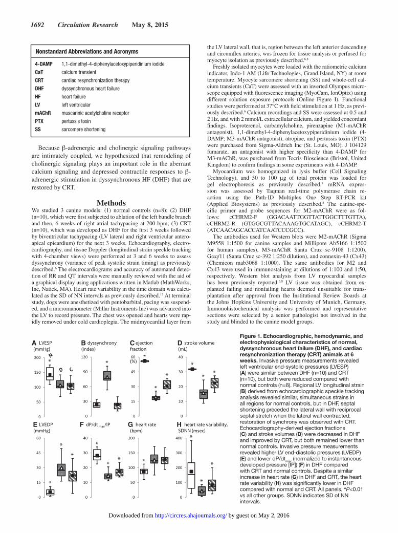
Figure 1. Echocardiographic, hemodynamic, and electrophysiological characteristics of normal, dyssynchronous heart failure (DHF), and cardiac resynchronization therapy (CRT) animals at 6 weeks. Invasive pressure measurements revealed left ventricular end-systolic pressures (LVESP) (A) were similar between DHF (n=10) and CRT (n=10), but both were reduced compared with normal controls (n=8). Regional LV longitudinal strain (B) derived from echocardiographic speckle tracking analysis revealed similar, simultaneous strains in all regions for normal controls, but in DHF, septal shortening preceded the lateral wall with reciprocal septal stretch when the lateral wall contracted; restoration of synchrony was observed with CRT. Echocardiography-derived ejection fractions (C) and stroke volumes (D) were decreased in DHF and improved by CRT, but both remained lower than normal controls. Invasive pressure measurements revealed higher LV end-diastolic pressures (LVEDP) (E) and lower dP/dtmax (normalized to instantaneous developed pressure [IP]) (F) in DHF compared with CRT and normal controls. Despite a similar increase in heart rate (G) in DHF and CRT, the heart rate variability (H) was significantly lower in DHF compared with normal and CRT. All panels, *P<0.01 vs all other groups. SDNN indicates SD of NN intervals.
by guest on May 2, 2016http://circres.ahajournals.org/Downloaded from

DeMazumder et al CRT Remodels Cholinergic Signaling in HF 1693
Fractional changes are presented as mean±SEM and compared us-ing a paired t test. Categorical comparisons were performed using a χ2 test. Absolute values are reported in box and whisker plots (mean, median, interquartile range, minimum, and maximum). Comparisons between multiple experimental groups were performed by ANOVA and a Tukey test. In vivo data at multiple time points were analyzed by repeated-measures ANOVA.
ResultsThe canine DHF model6–8 was generated by left bundle branch ablation to disrupt synchronous activation combined with rapid right atrial pacing for 6 weeks. CRT was produced by switch-ing to biventricular tachypacing from weeks 4 to 6. Because both models were tachypaced, they developed similar global
Figure 2. Response to cholinergic stimulation in the setting of tonic β-adrenergic stimulation. A, Representative calcium transient (CaT) and sarcomere shortening (SS) of left ventricular (LV) cardiomyocytes from normal control (black tracing), dyssynchronous heart failure (DHF; red), and cardiac resynchronization therapy (CRT; blue) hearts are shown for the following solution exchange protocol in sequence (indicated by the horizontal bars at top): β-adrenergic stimulation with isoproterenol ([I], left column); cholinergic stimulation with carbamylcholine (CCh) in the continued presence of isoproterenol ([I+C], middle column); atropine to assess reversal of muscarinic acetylcholine receptor (mAChR)–specific effects ([I+C+A], right column]. B, The ratio of the peak responses to CCh added to isoproterenol compared with isoproterenol alone (I+C:I) on CaT (top) and SS (bottom) in normal control (empty bar), DHF (red), and CRT (blue) myocytes is plotted on the left column (log2 scale; mean±SEM; n=30–52 myocytes from n=6–9 hearts for each bar). Similarly, the ratio of I+C+A to I+C (I+C+A:I+C) is plotted on the right column. The individual data points are plotted in Online Figure IIA. Cholinergic stimulation reduced the respective peak CaT and SS by 59±4% and 74±4% in DHF; by 41±3% and 55±3% in normal control; and by 23±3% and 36±2% in CRT myocytes. In 15% of DHF myocytes, contraction was arrested despite continued isoproterenol exposure, and subsequent exposure to atropine fully restored CaT and contraction (Online Movie I); these data were not included in this analysis. C, The ratio of the 80% duration of CaT and SS are plotted in a format similar to B. The individual data points are plotted in Online Figure IIB. Cholinergic stimulation markedly prolonged the respective CaT and SS by 81±18% and 74±14% in DHF; by 65±6% and 36±7% in normal control; and by 47±8% and 22±7% in CRT myocytes. D, Representative immunohistochemical staining sections of canine LV tissue (top) revealed increased M2-mAChR density in DHF myocytes at intercalated discs and sarcolemma. Quantitative analyses of real-time polymerase chain reaction, Western blot and immunohistochemical fluorescence data in canine LV tissue (bottom) revealed increased mRNA and protein expression of M2-mAChR in DHF but not in CRT myocytes (n=5 hearts per group). Similar results were obtained with Western blot analysis of cardiomyocytes isolated on a percoll gradient (data not shown), suggesting these M2-mAChR protein changes occur within cardiomyocytes. E, Representative immunohistochemistry staining sections of human LV tissue samples from failing and nonfailing hearts are shown on top. In failing human LV, M2-mAChR expression was upregulated and redistributed from the intercalated discs to the entire cell border similar to that observed in canines. Western blot analysis of tissue lysates from human LV (bottom) revealed 2-fold higher M2-mAChR protein expression in HF (n=18) compared with non-HF (n=7), regardless of ischemic (ICM; n=4) or non–ischemic cardiomyopathy (NICM; n=14). All panels, *P<0.01 vs all other groups. DAPI indicates 4′,6-diamidino-2-phenylindole.
by guest on May 2, 2016http://circres.ahajournals.org/Downloaded from
1694 Circulation Research May 8, 2015
LV dysfunction (Figure 1A) and were designed to interrogate responses specific to dyssynchronous versus resynchronized ventricular contraction during hemodynamic decompensation. Compared with non-HF controls, DHF animals demonstrated significant regional dyssynchrony of LV shortening and de-pressed ejection fraction, stroke volume, dP/dt
max, and heart
rate variability (Figure 1B–1H). These hemodynamic and elec-trophysiological changes were significantly improved by CRT.
We hypothesized that during tonic β-adrenergic stimula-tion, acute cholinergic stimulation suppresses CaT and SS in DHF more than in normal or CRT. In the continued presence of isoproterenol, cholinergic stimulation with carbamylcholine markedly depressed peak CaT and SS amplitudes in DHF my-ocytes by 59% and 74%, respectively. These responses were only modestly diminished in normal controls and even less so in CRT (Figure 2A and 2B; Online Figure IIA). Reversal of depression by atropine indicated an mAChR-specific effect.16,17 Some DHF cells were so sensitive to cholinergic stimulation that contraction was arrested despite continued isoproterenol exposure, and full restoration of the CaT and contraction re-quired atropine (Online Movie I). Cholinergic stimulation also prolonged the CaT and SS more in DHF than in normal or CRT myocytes (Figure 2A and 2C; Online Figure IIB).
To identify the molecular basis of the model-dependent cholinergic responses, we performed immunohistochemistry, Western blot, and real-time polymerase chain reaction analy-ses. We observed an increase in M2-muscarinic acetylcholine receptor (M2-mAChR18,19) mRNA and protein expression
with dyssynchrony that was reversed by resynchronization (Figure 2D). The canine DHF model exhibits similar changes in β-adrenergic signaling as human cardiomyopathy.6–8 Thus, we performed similar experiments on LV myocardium from human hearts failing of ischemic and nonischemic pathogen-eses. We observed similar increases in immunoreactive pro-tein expression and subcellular localization of M2-mAChR (Figure 1E), suggesting that M2-mAChR remodeling is a gen-eralizable pathophysiological feature of HF.
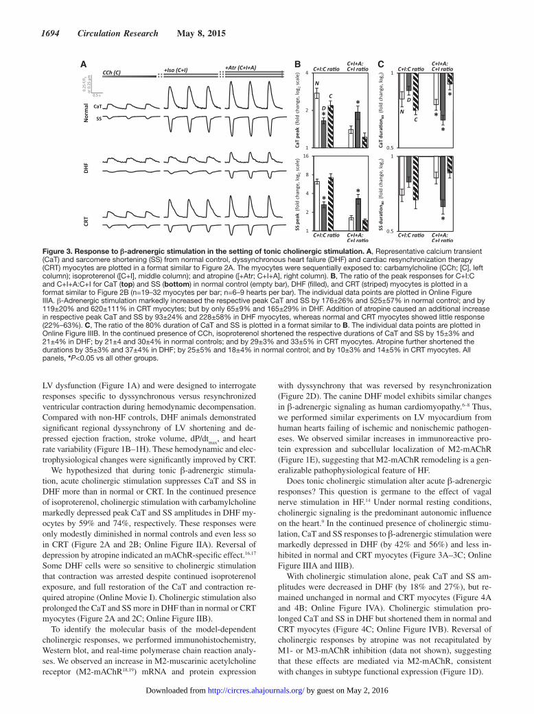
Does tonic cholinergic stimulation alter acute β-adrenergic responses? This question is germane to the effect of vagal nerve stimulation in HF.14 Under normal resting conditions, cholinergic signaling is the predominant autonomic influence on the heart.9 In the continued presence of cholinergic stimu-lation, CaT and SS responses to β-adrenergic stimulation were markedly depressed in DHF (by 42% and 56%) and less in-hibited in normal and CRT myocytes (Figure 3A–3C; Online Figure IIIA and IIIB).
With cholinergic stimulation alone, peak CaT and SS am-plitudes were decreased in DHF (by 18% and 27%), but re-mained unchanged in normal and CRT myocytes (Figure 4A and 4B; Online Figure IVA). Cholinergic stimulation pro-longed CaT and SS in DHF but shortened them in normal and CRT myocytes (Figure 4C; Online Figure IVB). Reversal of cholinergic responses by atropine was not recapitulated by M1- or M3-mAChR inhibition (data not shown), suggesting that these effects are mediated via M2-mAChR, consistent with changes in subtype functional expression (Figure 1D).
A B C
Figure 3. Response to β-adrenergic stimulation in the setting of tonic cholinergic stimulation. A, Representative calcium transient (CaT) and sarcomere shortening (SS) from normal control, dyssynchronous heart failure (DHF) and cardiac resynchronization therapy (CRT) myocytes are plotted in a format similar to Figure 2A. The myocytes were sequentially exposed to: carbamylcholine (CCh; [C], left column); isoproterenol ([C+I], middle column); and atropine ([+Atr; C+I+A], right column). B, The ratio of the peak responses for C+I:C and C+I+A:C+I for CaT (top) and SS (bottom) in normal control (empty bar), DHF (filled), and CRT (striped) myocytes is plotted in a format similar to Figure 2B (n=19–32 myocytes per bar; n=6–9 hearts per bar). The individual data points are plotted in Online Figure IIIA. β-Adrenergic stimulation markedly increased the respective peak CaT and SS by 176±26% and 525±57% in normal control; and by 119±20% and 620±111% in CRT myocytes; but by only 65±9% and 165±29% in DHF. Addition of atropine caused an additional increase in respective peak CaT and SS by 93±24% and 228±58% in DHF myocytes, whereas normal and CRT myocytes showed little response (22%–63%). C, The ratio of the 80% duration of CaT and SS is plotted in a format similar to B. The individual data points are plotted in Online Figure IIIB. In the continued presence of CCh, isoproterenol shortened the respective durations of CaT and SS by 15±3% and 21±4% in DHF; by 21±4 and 30±4% in normal controls; and by 29±3% and 33±5% in CRT myocytes. Atropine further shortened the durations by 35±3% and 37±4% in DHF; by 25±5% and 18±4% in normal control; and by 10±3% and 14±5% in CRT myocytes. All panels, *P<0.05 vs all other groups.
by guest on May 2, 2016http://circres.ahajournals.org/Downloaded from

DeMazumder et al CRT Remodels Cholinergic Signaling in HF 1695
Does M2-mAChR remodeling have functional effects inde-pendent of receptor activation? Exposure of DHF myocytes to atropine alone reversibly increased peak CaT and SS ampli-tudes (by 26% and 33%; Figure 4D). These atropine-induced effects were infrequently observed in CRT and normal cells, suggesting that DHF hearts are biased toward M2-mAChR-Gαi–coupled signaling both in the absence and in the pres-ence of cholinergic stimulation.
How does M2-mAChR-Gαi remodeling alter arrhythmic risk? In 30% to 50% of myocytes from all models, isoproterenol alone induced after-transients and after-contractions (Figure 5A), consistent with findings from a recent study on normal canine myocytes.20 Cholinergic stimulation completely abolished these disturbances in DHF and CRT, with little effect on peak CaT and SS in CRT myocytes. In the presence of tonic cholinergic stimula-tion, isoproterenol did not induce after-transients and after-contrac-tions until exposure to atropine (Figure 5B). Compared with DHF, early after-transients and after-contractions were more frequently seen in normal and CRT myocytes. The atropine effect was not recapitulated by M1- or M3-mAChR inhibition (data not shown). In myocytes from all models pretreated with PTX, isoproterenol promptly induced after-transients and after-contractions that were not affected by cholinergic stimulation (Figure 5C).
To characterize the role of M3-mAChR-Gαi signaling, we pretreated myocytes with PTX to inhibit Gαi signaling. With tonic β-adrenergic stimulation, PTX completely abolished the negative inotropic effects from cholinergic stimulation in all models (Figure 6A; Online Figure VA). In the presence of PTX
and pirenzapine, an M1-mAChR–specific inhibitor, cholinergic stimulation increased peak CaT and SS in CRT but not in DHF cells (Figure 6B); this effect was suppressed by atropine (Online Figure VB) or M3-mAChR inhibition (Figure 6B; Online Figure VC). Consistent with these findings, immunohisto-chemical analyses demonstrated increased M3-mAChR expres-sion, prominently at the intercalated discs in CRT (Figure 6C). Western blot analyses revealed increased M3-mAChR protein without any apparent effect on Gαq/11 expression (Figure 6D).
DiscussionWe have identified upregulated M2-mAChR18,19 expression and function in human and canine HF compared with non-failing controls. CRT attenuated M2-mAChR expression and Gαi17,21,22-coupling and enhanced M3-mAChR23,24 expression in association with enhanced calcium cycling and SS. These changes in cholinergic signaling represent a novel mechanism for enhancing sympathovagal balance in CRT and may iden-tify new targets for treatment of systolic HF.
Tonic β-adrenergic stimulation and increased Gαi expression are a hallmark of DHF. CRT reverses this phenotype by upregu-lating regulator of G-protein signal 2 (RGS2) and inhibiting Gαi signaling without decreasing Gαi expression, thereby improving calcium handling and sarcomere contraction.6,7 The M2-mAChR subtype18,19 in the heart is selectively coupled to Gαi17 and acts via well-characterized second messenger pathways. Coordinated increases in M2-mAChR and Gαi expression and coupling have been noted in the LV from synchronized failing hearts21,22 but
A B C
D
Figure 4. Response to cholinergic stimulation alone. A, Representative calcium transient (CaT) and sarcomere shortening (SS) are shown from normal controls, dyssynchronous heart failure (DHF), and cardiac resynchronization therapy (CRT) myocytes sequentially exposed to standard Tyrode’s extracellular solution ([ECS; E], thin gray line) followed by carbamylcholine (CCh [C], thick black line). B, The ratio of the peak responses for C:E and after the addition of atropine to CCh compared with CCh alone (C+A:C) for CaT (top) and SS (bottom) in normal control (empty bar), DHF (filled) and CRT (striped) myocytes is plotted in a format similar to Figure 3B (n=20–30 myocytes per bar; n=6–9 hearts per bar). The individual data points are plotted in Online Figure IVA. Cholinergic stimulation decreased the respective peak CaT and SS by 18±5% and 27±7% in DHF but had little or no effect in normal and CRT myocytes. These effects were reversed by subsequent addition of atropine. C, The ratio of the 80% duration of CaT and SS is plotted in a format similar to B. The individual data points are plotted in Online Figure IVB. Cholinergic stimulation prolonged the CaT and SS durations by 14±3% and 5±3% in DHF, whereas in normal and CRT myocytes, the CaT and SS durations were either shortened or unchanged. D, The ratio of the peak responses to atropine (A:E) and washout for CaT and SS in normal control (empty bar), DHF (filled), and CRT (striped) myocytes is plotted in a format similar to B (n=8 myocytes per bar; n=3 hearts per bar). Atropine increased the peak CaT (left) and SS (right) by 26±5% and 33±11% in DHF and by 9±4% and 10±3% in normal controls, but had no effect in CRT myocytes. These effects were reversed by washing off atropine. All panels, *P<0.05 vs all other groups.
by guest on May 2, 2016http://circres.ahajournals.org/Downloaded from

1696 Circulation Research May 8, 2015
the functional significance was not known. Whether this coordi-nated remodeling is also a feature of dyssynchronized and resyn-chronized HF had not been explored.
Our results indicate that DHF hearts are biased toward M2-mAChR-Gαi–coupled signaling, even in the absence of cho-linergic stimulation. Extensive in vitro and ex vivo evidence indicates that Gαi-coupled cardiac M2-mAChRs are activated in proportion to Gαi expression25 and in this setting, atropine may act as an inverse agonist.16 These chronically activated M2-mAChRs may be susceptible to agonist-induced desen-sitization,26,27 a well-characterized phenomenon that may be a
mechanism for the improved hemodynamics14 noted with ton-ic cholinergic stimulation in ongoing clinical HF trials.12,13,28
It is plausible that M2-mAChR remodeling occurs early on in HF as a compensatory mechanism to heightened sympathetic tone that, in the long-term, contributes to the pathology of HF, perhaps by depressing myocyte function, calcium handling, and β-adrenergic responsiveness. Increased cholinergic tone has been noted early in HF development29 and cholinergic transdif-ferentiation of cardiac sympathetic neurons has been observed in some HF models.30 By decreasing M2-mAChR-Gαi–mediated signaling, CRT improves β-adrenergic responsiveness. This,
A B
C
Figure 5. Cholinergic stimulation suppresses after-transients and after-contractions trigged by β-adrenergic stimulation. A, Representative calcium transient (CaT) and sarcomere shortening (SS) from normal control, dyssynchronous heart failure (DHF), and cardiac resynchronization therapy (CRT) myocytes are plotted (top) in a format similar to Figure 2A. Isoproterenol (I)-induced after-transients and after-contractions were suppressed by carbamylcholine (CCh; I+C) and recurred with atropine (I+C+A). The bar graph (bottom) shows the percent of normal, DHF, and CRT myocytes that demonstrated triggered activity (after-transients and after-contractions) in response to the corresponding solution exposures (n=42–104 myocytes per group; n=6–9 hearts per group). Myocytes that demonstrated isoproterenol-induced triggered activity were used only for analysis in this section and excluded from the analyses as shown in Figures 2 to 4 and 6. B, Representative CaT and SS from CRT myocytes are plotted (top) in a format similar to Figure 3A. In the presence of CCh (C), addition of isoproterenol (C+I) did not induce after-transients and after-contractions until addition of atropine (C+I+A). The percent of normal, DHF, and CRT myocytes demonstrating triggered activity are plotted (n=25–63 myocytes per group; n=6–9 hearts per group). Myocytes that demonstrated isoproterenol-induced triggered activity were used only for analysis in this section and excluded from the analyses as shown in Figures 2 to 4 and 6. C, Representative CaT and SS from normal control, DHF, and CRT myocytes pretreated with pertussis toxin (PTX) to inhibit Gαi are plotted in a format similar to A. In all myocytes from all models, sustained after-transients and after-contractions were noted with isoproterenol regardless of exposure to CCh. All panels, *P<0.05 vs all other groups.
by guest on May 2, 2016http://circres.ahajournals.org/Downloaded from

DeMazumder et al CRT Remodels Cholinergic Signaling in HF 1697
A
B
D
C
Figure 6. Cholinergic stimulation mediates positive and negative inotropic effects via distinct muscarinic receptor subtypes. A, The peak sarcomere shortening (SS) responses (mean±SEM) corresponding to the indicated solution exchange protocol are compared in the absence (empty bars) or presence (filled) of pertussis toxin (PTX) for normal control (Nor; gray), dyssynchronous heart failure (DHF; red bars) and cardiac resynchronization therapy (CRT; blue) myocytes (n=30–52 myocytes from n=6–9 hearts for each bar). The individual data points are plotted in Online Figure VA. PTX increased the peak SS response to isoproterenol (left column) in DHF myocytes, but had no effect in normal and CRT myocytes. This is consistent with enhanced baseline Gαi activity in DHF. In the continued presence of isoproterenol, pretreatment with PTX abolished the negative inotropic effects of cholinergic stimulation in all groups (middle column). The peak SS after addition of atropine was not significantly different with and without PTX for normal (P=0.43), DHF (P=0.13), and CRT (P=0.32) myocytes (right column). These data suggest that in the presence of saturating β-adrenergic stimulation, the negative inotropic effect from cholinergic stimulation is mediated via M2-muscarinic acetylcholine receptor (mAChR)-Gαi signaling. B, The ratio of the peak SS responses to carbamylcholine (CCh) alone compared with Tyrode’s extracellular solution (ECS) using the same protocol as in Figure 4B is plotted in the absence and presence of PTX and an M3-mAChR–specific inhibitor (M3i; n=8–30 myocytes per bar; n=3–9 hearts per bar). All myocytes were continuously perfused with pirenzapine to block M1-mAChR–specific effects. The individual data points are plotted in Online Figure VB and VC. Compared with the absence of PTX, cholinergic stimulation in the presence of PTX increased the peak SS by 45±8% in CRT myocytes, but this effect was abolished with M3i. In DHF myocytes, PTX abolished the negative inotropic effect from cholinergic stimulation, but M3i had no significant effect. These data suggest that CRT myocytes are biased toward M3-mAChR–mediated positive inotropic effect, whereas normal and DHF myocytes are not. C, Representative immunohistochemical staining sections of canine midmyocardial tissue from the left ventricular (LV) lateral wall (top) revealed increased M3-mAChR density in CRT myocytes at the intercalated discs. Western blots of tissue lysates (5 hearts per group) revealed CRT increased M3-mAChR protein expression without any change in Gαq/11 protein expression (bottom). D, Proposed mechanism for autonomic remodeling in DHF and with CRT. Cholinergic stimulation of LV myocytes by acetylcholine (ACh) released from parasympathetic vagus nerve branches can produce both inhibitory and stimulatory calcium and contractile responses in the heart via well-characterized M2-mAChR-Gαi and M3-mAChR-Gαq coupled signaling, respectively. DHF (red arrows and tracings) is associated with downregulation of β1-adrenergic receptors (β-AR) and inhibition of adenylate cyclase (AC) from direct interactions with the α-subunit of the PTX-sensitive inhibitory G protein (Gαi) selectively coupled to M2-mAChRs. Furthermore, coordinated increases in M2-mAChR-Gαi–coupled expression and signaling chronically inhibit basal AC-mediated downstream signaling and markedly impairs the efficiency of β-adrenergic responsiveness, resulting in smaller amplitudes and prolonged relaxation of CaT and SS. CRT (blue arrows and tracings) reverses this phenotype by differentially remodeling cholinergic mAChR signaling. By concurrently decreasing M2-mAChR and increasing RGS2 expression, CRT decreases the negative inotropic effects of Gαi signaling. In addition, CRT increases M3-mAChR-Gαq–mediated signaling associated with positive inotropic responses and putative cardioprotective effects. All panels, *P<0.05 vs all other groups.
by guest on May 2, 2016http://circres.ahajournals.org/Downloaded from
1698 Circulation Research May 8, 2015
along with functional inhibition of Gαi by RGS2,6,7 results in positive inotropic effects because of improved calcium handling and sarcomere response to β-adrenergic stimulation.
How does M2-mAChR-Gαi remodeling alter arrhythmic risk? Ventricular arrhythmias are a major cause of death in patients with HF.31,32 Since the first report in 1859,33 exten-sive evidence from animal and clinical studies indicates that β-adrenergic signaling increases arrhythmic risk31,32 and cho-linergic stimulation protects the heart from lethal arrhyth-mias.9,34 Despite model-dependent remodeling, there seems to be a large margin of safety at the cellular level for M2-AChR-Gαi–coupled signaling to protect and rescue normal, DHF, and CRT hearts from arrhythmias. These results provide a mechanistic basis for prior observations, that is, increased arrhythmias with β-adrenergic stimulation and PTX,35 antiar-rhythmic effects of cholinergic stimulation,9,33 and may have important implications for vagus nerve stimulation9–14,28 and development of new antiarrhythmic therapies.34
Whereas the highly prevalent M2-mAChR18,19 subtype is se-lectively coupled to Gαi,17 the relatively scarce M3-mAChR23,24 is highly specific for stimulatory Gαq with putative cardiopro-tective effects.36–38 Recent insights into the molecular structure, function, pharmacology, and fundamental physiological role of M3-mAChRs have identified them as a major target for drug development.36,39 Our results indicate that CRT may exert beneficial effects via M3-mAChR-Gαq signaling, including enhanced calcium handling, sarcomere responsiveness, and positive inotropy. Notably, CRT increased M3-mAChR expres-sion at the intercalated discs. In cardiomyocytes, M3-mAChR activation during ischemia preserves the phosphorylated levels of sarcolemmal connexin 43 to provide delayed cardioprotec-tion.40 Furthermore, M3-mAChR-Gαq signaling augments inositol 1,4,5-triphosphate (IP3)/diacylglycerol (DAG)-mediated calcium release, protein kinase C (PKC)-mediated phosphorylation, phsophoinositide-3-kinase/Akt-mediated re-duced apoptosis, and RGS2 expression.36–38,41 CRT has similar effects, including increasing RGS2 expression particularly in clinical responders7 that may be exerted via M3-mAChR-Gαq signaling. We could not specifically address this here because an in vitro model of CRT does not currently exist.
The notion that cholinergic signaling has a relatively limited effect on LV function belies much evidence.9 Our results indicate that remodeling of cholinergic signaling is a critical pathological component of human and canine HF, and differential remodel-ing of cholinergic signaling is paramount for restoration of au-tonomic balance by CRT (Figure 4E). The novel mechanisms identified herein offers an opportunity to apply targeted down-regulation of M2-mAChR and upregulation of M3-mAChR in patients with HF who are not CRT responders5 or candidates. Moreover, the beneficial effects of CRT might be enhanced by vagus nerve stimulation9–13,28 and remodeling of the key sig-naling components reported herein may represent mechanistic pathways engaged by vagus nerve stimulation and open new av-enues for pharmacological or pacing treatments for HF.
AcknowledgmentsWe gratefully acknowledge Dr Charles Steenbergen for blinded analysis of immunohistochemistry slides and selection of representa-tive samples, Deborah DiSilvestre and Dr Swati Dey for technical
assistance with some of the experimental protocols, Drs Federica Farinelli and Khalid Chakir for myocyte isolation and procurement, and Rick Tunin for preparation of the animal models.
Sources of FundingThis work was supported by P01 HL 77180 (D.A. Kass, B. O’Rourke, and G.F. Tomaselli).
DisclosuresNone.
References 1. Go AS, Mozaffarian D, Roger VL, et al; American Heart Association
Statistics Committee and Stroke Statistics Subcommittee. Heart dis-ease and stroke statistics–2013 update: a report from the American Heart Association. Circulation. 2013;127:e6–e245. doi: 10.1161/CIR.0b013e31828124ad.
2. Cleland JG, Daubert JC, Erdmann E, Freemantle N, Gras D, Kappenberger L, Tavazzi L. Longer-term effects of cardiac resynchronization therapy on mortality in heart failure [the CArdiac REsynchronization-Heart Failure (CARE-HF) trial extension phase]. Eur Heart J. 2006;27:1928–1932. doi: 10.1093/eurheartj/ehl099.
3. Bristow MR, Saxon LA, Boehmer J, Krueger S, Kass DA, De Marco T, Carson P, DiCarlo L, DeMets D, White BG, DeVries DW, Feldman AM; Comparison of Medical Therapy, Pacing, and Defibrillation in Heart Failure (COMPANION) Investigators. Cardiac-resynchronization therapy with or without an implantable defibrillator in advanced chronic heart fail-ure. N Engl J Med. 2004;350:2140–2150. doi: 10.1056/NEJMoa032423.
4. Cleland JG, Daubert JC, Erdmann E, Freemantle N, Gras D, Kappenberger L, Tavazzi L; Cardiac Resynchronization-Heart Failure (CARE-HF) Study Investigators. The effect of cardiac resynchronization on morbidity and mortality in heart failure. N Engl J Med. 2005;352:1539–1549. doi: 10.1056/NEJMoa050496.
5. Kirk JA, Kass DA. Electromechanical dyssynchrony and resynchroni-zation of the failing heart. Circ Res. 2013;113:765–776. doi: 10.1161/CIRCRESAHA.113.300270.
6. Chakir K, Daya SK, Aiba T, Tunin RS, Dimaano VL, Abraham TP, Jaques-Robinson KM, Jacques K, Lai EW, Pacak K, Zhu WZ, Xiao RP, Tomaselli GF, Kass DA. Mechanisms of enhanced beta-adrenergic reserve from car-diac resynchronization therapy. Circulation. 2009;119:1231–1240. doi: 10.1161/CIRCULATIONAHA.108.774752.
7. Chakir K, Depry C, Dimaano VL, et al. Galphas-biased beta2-ad-renergic receptor signaling from restoring synchronous contraction in the failing heart. Sci Transl Med. 2011;3:100ra88. doi: 10.1126/scitranslmed.3001909.
8. Aiba T, Hesketh GG, Barth AS, Liu T, Daya S, Chakir K, Dimaano VL, Abraham TP, O’Rourke B, Akar FG, Kass DA, Tomaselli GF. Electrophysiological consequences of dyssynchronous heart failure and its restoration by resynchronization therapy. Circulation. 2009;119: 1220–1230. doi: 10.1161/CIRCULATIONAHA.108.794834.
9. Coote JH. Myths and realities of the cardiac vagus. J Physiol. 2013;591:4073–4085. doi: 10.1113/jphysiol.2013.257758.
10. Zhang Y, Popovic ZB, Bibevski S, Fakhry I, Sica DA, Van Wagoner DR, Mazgalev TN. Chronic vagus nerve stimulation improves autonomic con-trol and attenuates systemic inflammation and heart failure progression in a canine high-rate pacing model. Circ Heart Fail. 2009;2:692–699. doi: 10.1161/CIRCHEARTFAILURE.109.873968.
11. Hamann JJ, Ruble SB, Stolen C, Wang M, Gupta RC, Rastogi S, Sabbah HN. Vagus nerve stimulation improves left ventricular function in a canine model of chronic heart failure. Eur J Heart Fail. 2013;15:1319–1326. doi: 10.1093/eurjhf/hft118.
12. Hauptman PJ, Schwartz PJ, Gold MR, Borggrefe M, Van Veldhuisen DJ, Starling RC, Mann DL. Rationale and study design of the increase of vagal tone in heart failure study: INOVATE-HF. Am Heart J. 2012;163:954–962.e1. doi: 10.1016/j.ahj.2012.03.021.
13. De Ferrari GM, Tuinenburg AE, Ruble S, Brugada J, Klein H, Butter C, Wright DJ, Schubert B, Solomon S, Meyer S, Stein K, Ramuzat A, Zannad F. Rationale and study design of the NEuroCardiac TherApy foR Heart Failure Study: NECTAR-HF. Eur J Heart Fail. 2014;16:692–699. doi: 10.1002/ejhf.80.
14. Premchand RK, Sharma K, Mittal S, Monteiro R, Dixit S, Libbus I, DiCarlo LA, Ardell JL, Rector TS, Amurthur B, KenKnight BH, Anand IS. auto-nomic regulation therapy via left or right cervical vagus nerve stimulation
by guest on May 2, 2016http://circres.ahajournals.org/Downloaded from
DeMazumder et al CRT Remodels Cholinergic Signaling in HF 1699
in patients with chronic heart failure: results of the ANTHEM-HF trial. J Card Fail. 2014;20:808–816. doi: 10.1016/j.cardfail.2014.08.009.
15. Heart rate variability: Standards of measurement, physiological interpreta-tion and clinical use. Task force of the european society of cardiology and the north american society of pacing and electrophysiology. Circulation. 1996;93:1043–1065
16. Eglen RM. Overview of muscarinic receptor subtypes. Handb Exp Pharmacol. 2012;208:3–28. doi: 10.1007/978-3-642-23274-9_1.
17. Haga K, Haga T, Ichiyama A, Katada T, Kurose H, Ui M. Functional reconstitution of purified muscarinic receptors and inhibitory guanine nucleotide regulatory protein. Nature. 1985;316:731–733.
18. Haga K, Kruse AC, Asada H, Yurugi-Kobayashi T, Shiroishi M, Zhang C, Weis WI, Okada T, Kobilka BK, Haga T, Kobayashi T. Structure of the human M2 muscarinic acetylcholine receptor bound to an antagonist. Nature. 2012;482:547–551. doi: 10.1038/nature10753.
19. Kruse AC, Ring AM, Manglik A, et al. Activation and allosteric modula-tion of a muscarinic acetylcholine receptor. Nature. 2013;504:101–106. doi: 10.1038/nature12735.
20. Johnson DM, Heijman J, Bode EF, Greensmith DJ, van der Linde H, Abi-Gerges N, Eisner DA, Trafford AW, Volders PG. Diastolic sponta-neous calcium release from the sarcoplasmic reticulum increases beat-to-beat variability of repolarization in canine ventricular myocytes after β-adrenergic stimulation. Circ Res. 2013;112:246–256. doi: 10.1161/CIRCRESAHA.112.275735.
21. Vatner DE, Sato N, Galper JB, Vatner SF. Physiological and biochemical evidence for coordinate increases in muscarinic receptors and Gi during pacing-induced heart failure. Circulation. 1996;94:102–107.
22. Wilkinson M, Giles A, Armour JA, Cardinal R. Ventricular, but not atrial, M2-muscarinic receptors increase in the canine pacing-overdrive model of heart failure. Can J Cardiol. 1996;12:71–76.
23. Kruse AC, Hu J, Pan AC, Arlow DH, Rosenbaum DM, Rosemond E, Green HF, Liu T, Chae PS, Dror RO, Shaw DE, Weis WI, Wess J, Kobilka BK. Structure and dynamics of the M3 muscarinic acetylcholine receptor. Nature. 2012;482:552–556. doi: 10.1038/nature10867.
24. Nash MS, Young KW, Challiss RA, Nahorski SR. Intracellular signalling. Receptor-specific messenger oscillations. Nature. 2001;413:381–382. doi: 10.1038/35096643.
25. Spalding TA, Burstein ES. Constitutive activity of muscarinic acetyl-choline receptors. J Recept Signal Transduct Res. 2006;26:61–85. doi: 10.1080/10799890600567349.
26. Murakami S, Inanobe A, Kurachi Y. Short-term desensitization of mus-carinic K+ current in the heart. Biophys J. 2013;105:1515–1525. doi: 10.1016/j.bpj.2013.08.009.
27. Haga T. Molecular properties of muscarinic acetylcholine receptors. Proc Jpn Acad Ser B Phys Biol Sci. 2013;89:226–256.
28. Dicarlo L, Libbus I, Amurthur B, Kenknight BH, Anand IS. Autonomic regulation therapy for the improvement of left ventricular function
and heart failure symptoms: the ANTHEM-HF study. J Card Fail. 2013;19:655–660. doi: 10.1016/j.cardfail.2013.07.002.
29. Kinugawa T, Dibner-Dunlap ME. Altered vagal and sympathetic control of heart rate in left ventricular dysfunction and heart failure. Am J Physiol. 1995;268:R310–R316.
30. Kanazawa H, Ieda M, Kimura K, et al. Heart failure causes cholinergic transdifferentiation of cardiac sympathetic nerves via gp130-signaling cytokines in rodents. J Clin Invest. 2010;120:408–421. doi: 10.1172/JCI39778.
31. DeMazumder D, Tomaselli GF. Molecular and cellular mechanisms of cardiac arrhythmias. In: Hill JA, Olson, EN, eds. Muscle 2-Volume Set: Fundamental Biology and Mechanisms of Disease. Philadelphia, PA: Elsevier; 2012.
32. Tomaselli GF, Zipes DP. What causes sudden death in heart failure? Circ Res. 2004;95:754–763. doi: 10.1161/01.RES.0000145047.14691.db.
33. Einbrodt PP. Ueber herzreizung und ihr verhaeltnis zum blutdruck. Akademie der Wissenschaften (Vienna) Sitzungsberichte. 1859;38:345–359
34. Shen MJ, Zipes DP. Role of the autonomic nervous system in modulat-ing cardiac arrhythmias. Circ Res. 2014;114:1004–1021. doi: 10.1161/CIRCRESAHA.113.302549.
35. Grimm M, Gsell S, Mittmann C, Nose M, Scholz H, Weil J, Eschenhagen T. Inactivation of (Gialpha) proteins increases arrhythmogenic ef-fects of beta-adrenergic stimulation in the heart. J Mol Cell Cardiol. 1998;30:1917–1928. doi: 10.1006/jmcc.1998.0769.
36. Dror RO, Green HF, Valant C, Borhani DW, Valcourt JR, Pan AC, Arlow DH, Canals M, Lane JR, Rahmani R, Baell JB, Sexton PM, Christopoulos A, Shaw DE. Structural basis for modulation of a G-protein-coupled receptor by allosteric drugs. Nature. 2013;503:295–299. doi: 10.1038/nature12595.
37. Hang P, Zhao J, Qi J, Wang Y, Wu J, Du Z. Novel insights into the per-vasive role of M(3) muscarinic receptor in cardiac diseases. Curr Drug Targets. 2013;14:372–377.
38. Pan Z, Guo Y, Qi H, Fan K, Wang S, Zhao H, Fan Y, Xie J, Guo F, Hou Y, Wang N, Huo R, Zhang Y, Liu Y, Du Z. M3 subtype of muscarinic acetyl-choline receptor promotes cardioprotection via the suppression of miR-376b-5p. PLoS One. 2012;7:e32571. doi: 10.1371/journal.pone.0032571.
39. Kruse AC, Kobilka BK, Gautam D, Sexton PM, Christopoulos A, Wess J. Muscarinic acetylcholine receptors: novel opportunities for drug develop-ment. Nat Rev Drug Discov. 2014;13:549–560. doi: 10.1038/nrd4295.
40. Zhao J, Su Y, Zhang Y, Pan Z, Yang L, Chen X, Liu Y, Lu Y, Du Z, Yang B. Activation of cardiac muscarinic M3 receptors induces delayed car-dioprotection by preserving phosphorylated connexin43 and up-regulating cyclooxygenase-2 expression. Br J Pharmacol. 2010;159:1217–1225. doi: 10.1111/j.1476-5381.2009.00606.x.
41. Hao J, Michalek C, Zhang W, Zhu M, Xu X, Mende U. Regulation of cardiomyocyte signaling by RGS proteins: differential selectivity towards G proteins and susceptibility to regulation. J Mol Cell Cardiol. 2006;41: 51–61. doi: 10.1016/j.yjmcc.2006.04.003.
What Is Known?
• In the healthy heart, parasympathetic activation tunes sympathetic β1-
adrenergic receptor (β-AR) signaling, via acetylcholine acting at mus-carinic acetylcholine receptors (mAChRs) to suppress Ca2+ transients and contraction.
• In the failing heart, compensatory increases in sympathetic β-AR sig-naling ultimately are maladaptive.
• Although sympathetic β-AR signaling has been extensively studied, the role of parasympathetic mAChR signaling in the failing heart is unknown, and the effect of cardiac resynchronization therapy (CRT) on mAChR signaling has been unexplored.
What New Information Does This Article Contribute?
• Compared with nonfailing controls, expression of the M2 subtype of parasympathetic mAChRs is markedly upregulated in left ventricular myocytes from failing canine and human hearts.
• In the left ventricular myocytes of failing hearts, hyperactive para-sympathetic M2-mAChR signaling (coupled to Gαi) protects against
electric instability (a substrate for lethal arrhythmias) caused by heightened sympathetic β-AR signaling, but this also reduces me-chanical function.
• CRT decreases M2- and increases M3-mAChR expression, resulting in improved β-AR responsiveness and mechanical function while maintaining electric stability.
The development of new and improved HF therapies remains a clinical, research and public health priority. CRT is the only HF therapy to decrease long-term mortality, restore autonomic bal-ance, and improve both acute and chronic left ventricular function. The underlying mechanisms are largely unknown. Autonomic im-balance is associated with worsening HF and increased mortality risk, independent of left ventricular function and ventricular ar-rhythmias. The present study demonstrates a critical role of para-sympathetic mAChRs in HF, arrhythmic risk, and CRT. It suggests that the beneficial effects of CRT involve differential remodeling of mAChRs. Further understanding these mechanisms can lead to the design and development of new, more effective HF therapies.
Novelty and Significance
by guest on May 2, 2016http://circres.ahajournals.org/Downloaded from

Deeptankar DeMazumder, David A. Kass, Brian O'Rourke and Gordon F. Tomaselliby Differential Remodeling of Cholinergic Signaling
Cardiac Resynchronization Therapy Restores Sympathovagal Balance in the Failing Heart
Print ISSN: 0009-7330. Online ISSN: 1524-4571 Copyright © 2015 American Heart Association, Inc. All rights reserved.is published by the American Heart Association, 7272 Greenville Avenue, Dallas, TX 75231Circulation Research
doi: 10.1161/CIRCRESAHA.116.3052682015;116:1691-1699; originally published online March 2, 2015;Circ Res.
http://circres.ahajournals.org/content/116/10/1691World Wide Web at:
The online version of this article, along with updated information and services, is located on the
http://circres.ahajournals.org/content/suppl/2015/03/02/CIRCRESAHA.116.305268.DC1.htmlData Supplement (unedited) at:
http://circres.ahajournals.org//subscriptions/
is online at: Circulation Research Information about subscribing to Subscriptions:
http://www.lww.com/reprints Information about reprints can be found online at: Reprints:
document. Permissions and Rights Question and Answer about this process is available in the
located, click Request Permissions in the middle column of the Web page under Services. Further informationEditorial Office. Once the online version of the published article for which permission is being requested is
can be obtained via RightsLink, a service of the Copyright Clearance Center, not theCirculation Researchin Requests for permissions to reproduce figures, tables, or portions of articles originally publishedPermissions:
by guest on May 2, 2016http://circres.ahajournals.org/Downloaded from
Supplemental MaterialsDeMazumder et al., Cardiac resynchronization therapy restores sympathovagal balance in the failing heart by differential remodeling of cholinergic signaling. Circulation Research (2015)
a. d.
b. e.
c. f.
ONLINE FIGURE I: Schematic index of protocols employed for functional experiments on myocytes isolated from the LV lateral wall
cytosolic Ca+2& contraction
M2
Gαi
β1AR
AC
Iso
cytosolic Ca+2& contraction
M2
Gαi
β1AR
AC
CChAtr Iso
cytosolic Ca+2& contraction
M2
Gαi
β1AR
AC
CChAtr Iso
cytosolic Ca+2& contraction
M2
Gαi
β1AR
AC
CCh
M2
Gαi
cytosolic Ca+2& contraction
CCh Atr
cytosolic Ca+2& contraction
M2
Gαi
β1AR
AC
CChAtr Iso
PTX
PTX
cytosolic Ca+2& contraction
M2
Gαi
M3
Gαq
CCh Atr
M1
Gαq
M1i
PTX
cytosolic Ca+2& contraction
M2
Gαi
M3
Gαq
CCh Atr
M1
Gαq
M1iM3i
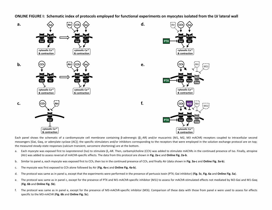
Each panel shows the schematic of a cardiomyocyte cell membrane containing β‐adrenergic (β1‐AR) and/or muscarinic (M1, M2, M3 mAChR) receptors coupled to intracellular secondmessengers [Gαi, Gαq, or adenylate cyclase (AC)]; the specific stimulators and/or inhibitors corresponding to the receptors that were employed in the solution exchange protocol are on top;the measured steady‐state responses (calcium transient, sarcomere shortening) are at the bottom.
a. Each myocyte was exposed first to isoproterenol (Iso) to stimulate β1‐AR. Then, carbamylcholine (CCh) was added to stimulate mAChRs in the continued presence of Iso. Finally, atropine(Atr) was added to assess reversal of mAChR‐specific effects. The data from this protocol are shown in Fig. 2a‐c and Online Fig. 2a‐b.
b. Similar to panel a, each myocyte was exposed first to CCh, then Iso in the continued presence of CCh, and finally Atr (data shown in Fig. 3a‐c and Online Fig. 3a‐b).
c. The myocyte was first exposed to CCh alone followed by Atr (Fig. 4a‐c and Online Fig. 4a‐b).
d. The protocol was same as in panel a, except that the experiments were performed in the presence of pertussis toxin (PTX; Gαi inhibitor) (Fig. 5c, Fig. 6a and Online Fig. 5a).
e. The protocol was same as in panel c, except for the presence of PTX and M1‐mAChR‐specific inhibitor (M1i) to assess for mAChR‐stimulated effects not mediated by M2‐Gαi and M1‐Gαq(Fig. 6b and Online Fig. 5b).
f. The protocol was same as in panel e, except for the presence of M3‐mAChR‐specific inhibitor (M3i). Comparison of these data with those from panel e were used to assess for effectsspecific to the M3‐mAChR (Fig. 6b and Online Fig. 5c).
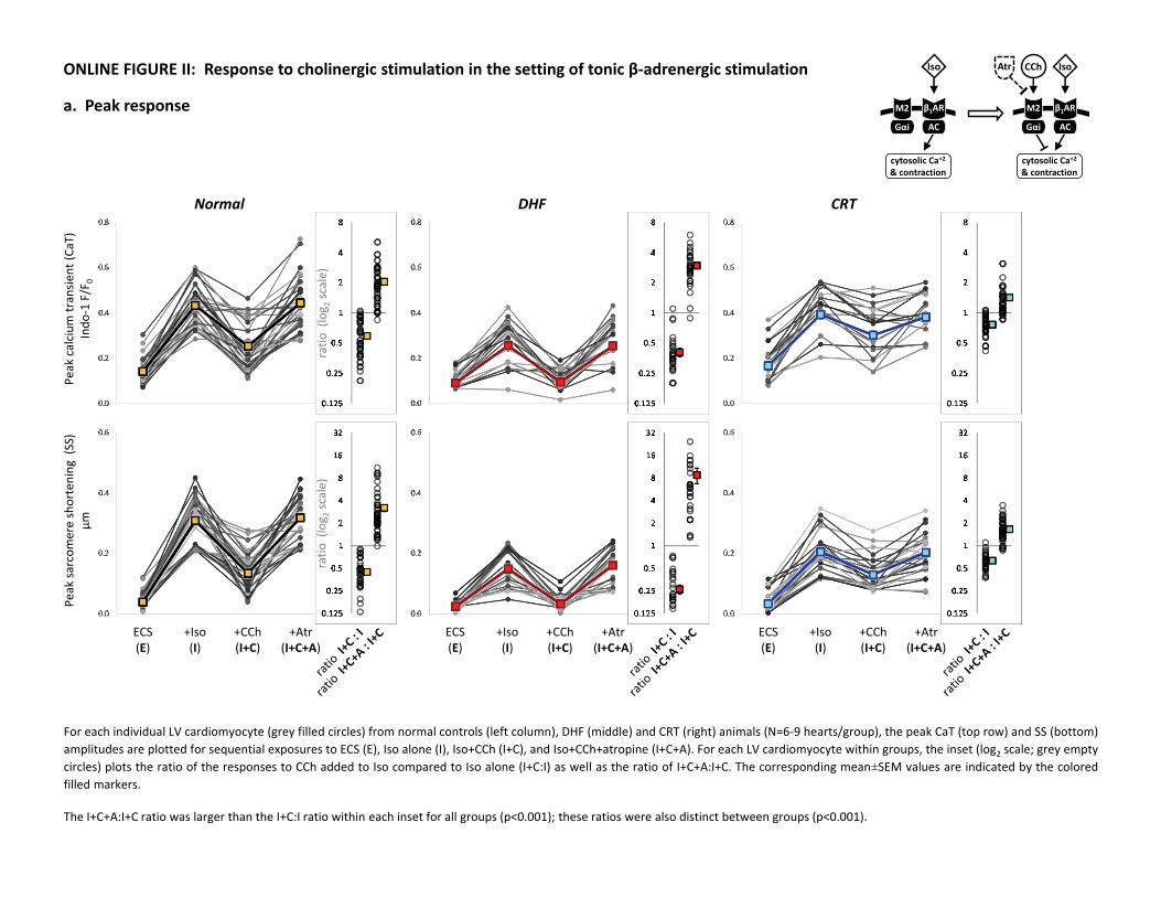
a. Peak response
ONLINE FIGURE II: Response to cholinergic stimulation in the setting of tonic β‐adrenergic stimulationPe
ak calcium
transie
nt (C
aT)
Indo
‐1 F/F
0
Peak sa
rcom
ere shortening
(SS)
µm
Normal DHF CRT
ratio
(log 2
scale)
ratio
(log 2
scale)
ECS(E)
+CCh(I+C)
+Iso(I)
+Atr(I+C+A)
ECS(E)
+CCh(I+C)
+Iso(I)
+Atr(I+C+A)
ECS(E)
+CCh(I+C)
+Iso(I)
+Atr(I+C+A)
cytosolic Ca+2& contraction
M2
Gαi
β1AR
AC
Iso
cytosolic Ca+2& contraction
M2
Gαi
β1AR
AC
CChAtr Iso
For each individual LV cardiomyocyte (grey filled circles) from normal controls (left column), DHF (middle) and CRT (right) animals (N=6‐9 hearts/group), the peak CaT (top row) and SS (bottom)amplitudes are plotted for sequential exposures to ECS (E), Iso alone (I), Iso+CCh (I+C), and Iso+CCh+atropine (I+C+A). For each LV cardiomyocyte within groups, the inset (log2 scale; grey emptycircles) plots the ratio of the responses to CCh added to Iso compared to Iso alone (I+C:I) as well as the ratio of I+C+A:I+C. The corresponding mean±SEM values are indicated by the coloredfilled markers.
The I+C+A:I+C ratio was larger than the I+C:I ratio within each inset for all groups (p<0.001); these ratios were also distinct between groups (p<0.001).
CaT80
% duration
(s)
SS 80%
duration
(s)
Normal DHF CRT
ratio
(log 2
scale)
ratio
(log 2
scale)
ECS(E)
+CCh(I+C)
+Iso(I)
+Atr(I+C+A)
ECS(E)
+CCh(I+C)
+Iso(I)
+Atr(I+C+A)
ECS(E)
+CCh(I+C)
+Iso(I)
+Atr(I+C+A)
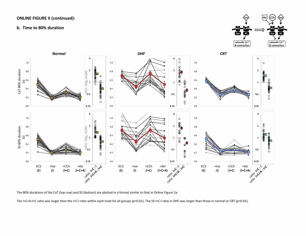
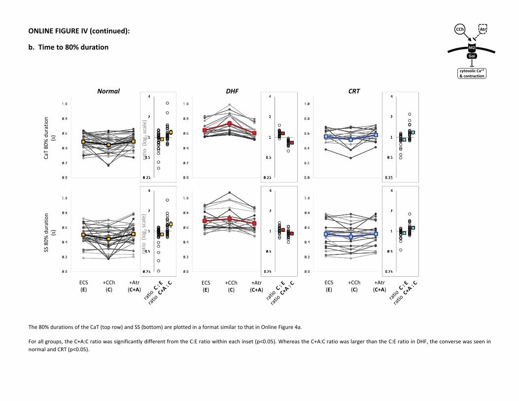
b. Time to 80% duration
ONLINE FIGURE II (continued):
cytosolic Ca+2& contraction
M2
Gαi
β1AR
AC
Iso
cytosolic Ca+2& contraction
M2
Gαi
β1AR
AC
CChAtr Iso
The 80% durations of the CaT (top row) and SS (bottom) are plotted in a format similar to that in Online Figure 2a.
The I+C+A:I+C ratio was larger than the I+C:I ratio within each inset for all groups (p<0.01). The SS I+C:I ratio in DHF was larger than those in normal or CRT (p<0.01).

Peak calcium
transie
nt (C
aT)
Indo
‐1 F/F
0
Peak sa
rcom
ere shortening
(SS)
µm
Normal DHF CRT
ratio
(log 2
scale)
ratio
(log 2
scale)
ECS(E)
+Iso(C+I)
+CCh(C)
+Atr(C+I+A)
a. Peak response
ONLINE FIGURE III: Response to β‐adrenergic stimulation in the presence of tonic cholinergic stimulation
cytosolic Ca+2& contraction
M2
Gαi
β1AR
AC
CChAtr Iso
cytosolic Ca+2& contraction
M2
Gαi
β1AR
AC
CCh
ECS(E)
+Iso(C+I)
+CCh(C)
+Atr(C+I+A)
ECS(E)
+Iso(C+I)
+CCh(C)
+Atr(C+I+A)
For each individual LV cardiomyocyte (grey filled circles) from normal controls (left column), DHF (middle) and CRT (right) animals (N=6‐9 hearts/group), the peak CaT (top row) and SS (bottom)amplitudes are plotted for sequential exposures to ECS (E), CCh alone (C), CCh+Iso (C+I), and CCh+Iso+atropine (C+I+A). For each LV cardiomyocyte within groups, the inset (log2 scale; greyempty circles) plots the ratio of the responses to Iso added to CCh compared to CCh alone (C+I:C) as well as the ratio of C+I+A:C+I. The corresponding mean±SEM values are indicated by thecolored filled markers.
The I+C+A:I+C ratio was larger than the I+C:I ratio within each inset in normal and CRT (p<0.001) but not in DHF. Whereas the C+I:C ratios for the CaT and SS in DHF were smaller than those innormal or CRT (p<0.01), the C+I+A:C+I ratios in DHF were larger (p<0.05).
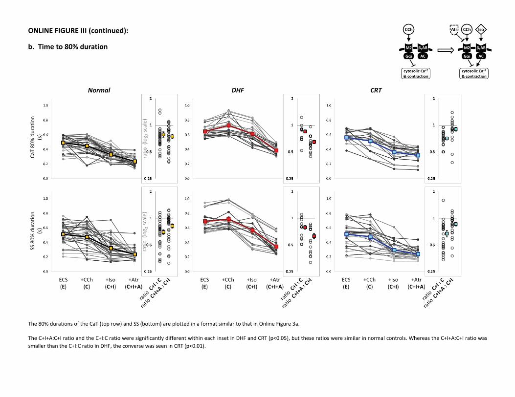
b. Time to 80% duration
ONLINE FIGURE III (continued):CaT80
% duration
(s)
SS 80%
duration
(s)
Normal DHF CRT
ratio
(log 2
scale)
ratio
(log 2
scale)
cytosolic Ca+2& contraction
M2
Gαi
β1AR
AC
CChAtr Iso
cytosolic Ca+2& contraction
M2
Gαi
β1AR
AC
CCh
ECS(E)
+Iso(C+I)
+CCh(C)
+Atr(C+I+A)
ECS(E)
+Iso(C+I)
+CCh(C)
+Atr(C+I+A)
ECS(E)
+Iso(C+I)
+CCh(C)
+Atr(C+I+A)
The 80% durations of the CaT (top row) and SS (bottom) are plotted in a format similar to that in Online Figure 3a.
The C+I+A:C+I ratio and the C+I:C ratio were significantly different within each inset in DHF and CRT (p<0.05), but these ratios were similar in normal controls. Whereas the C+I+A:C+I ratio wassmaller than the C+I:C ratio in DHF, the converse was seen in CRT (p<0.01).
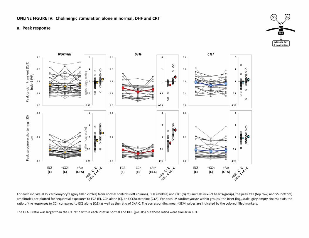
ONLINE FIGURE IV: Cholinergic stimulation alone in normal, DHF and CRT
a. Peak responsePe
ak calcium
transie
nt (C
aT)
Indo
‐1 F/F
0
Peak sa
rcom
ere shortening
(SS)
µm
Normal DHF CRT
ratio
(log 2
scale)
ratio
(log 2
scale)
+CCh(C)
ECS(E)
+Atr(C+A)
+CCh(C)
ECS(E)
+Atr(C+A)
+CCh(C)
ECS(E)
+Atr(C+A)
M2
Gαi
cytosolic Ca+2& contraction
CCh Atr
For each individual LV cardiomyocyte (grey filled circles) from normal controls (left column), DHF (middle) and CRT (right) animals (N=6‐9 hearts/group), the peak CaT (top row) and SS (bottom)amplitudes are plotted for sequential exposures to ECS (E), CCh alone (C), and CCh+atropine (C+A). For each LV cardiomyocyte within groups, the inset (log2 scale; grey empty circles) plots theratio of the responses to CCh compared to ECS alone (C:E) as well as the ratio of C+A:C. The corresponding mean±SEM values are indicated by the colored filled markers.
The C+A:C ratio was larger than the C:E ratio within each inset in normal and DHF (p<0.05) but these ratios were similar in CRT.
CaT80
% duration
(s)
SS 80%
duration
(s)
Normal DHF CRT
ratio
(log 2
scale)
ratio
(log 2
scale)
+CCh(C)
ECS(E)
+Atr(C+A)
+CCh(C)
ECS(E)
+Atr(C+A)
+CCh(C)
ECS(E)
+Atr(C+A)
ONLINE FIGURE IV (continued):
b. Time to 80% duration M2
Gαi
cytosolic Ca+2& contraction
CCh Atr
The 80% durations of the CaT (top row) and SS (bottom) are plotted in a format similar to that in Online Figure 4a.
For all groups, the C+A:C ratio was significantly different from the C:E ratio within each inset (p<0.05). Whereas the C+A:C ratio was larger than the C:E ratio in DHF, the converse was seen innormal and CRT (p<0.05).
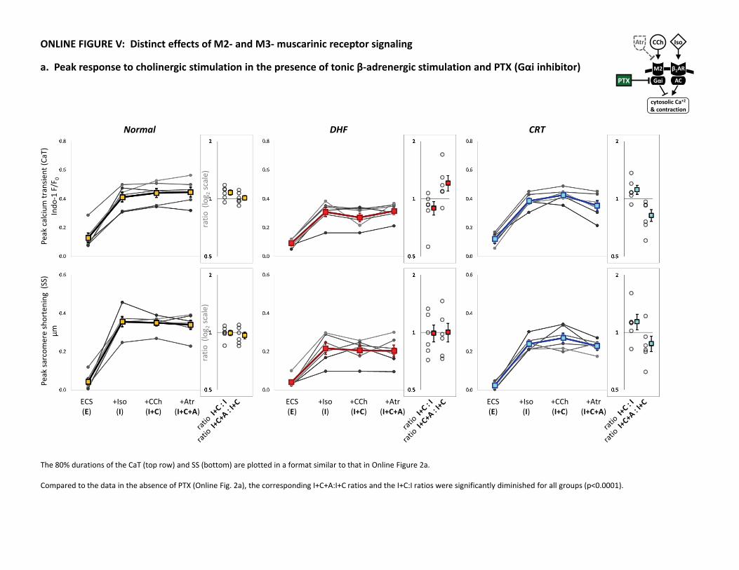
a. Peak response to cholinergic stimulation in the presence of tonic β‐adrenergic stimulation and PTX (Gαi inhibitor)
ONLINE FIGURE V: Distinct effects of M2‐ and M3‐muscarinic receptor signalingPe
ak calcium
transie
nt (C
aT)
Indo
‐1 F/F
0
Peak sa
rcom
ere shortening
(SS)
µm
Normal DHF CRT
ratio
(log 2
scale)
ratio
(log 2
scale)
ECS(E)
+CCh(I+C)
+Iso(I)
+Atr(I+C+A)
ECS(E)
+CCh(I+C)
+Iso(I)
+Atr(I+C+A)
ECS(E)
+CCh(I+C)
+Iso(I)
+Atr(I+C+A)
cytosolic Ca+2& contraction
M2
Gαi
β1AR
AC
CChAtr Iso
PTX
The 80% durations of the CaT (top row) and SS (bottom) are plotted in a format similar to that in Online Figure 2a.
Compared to the data in the absence of PTX (Online Fig. 2a), the corresponding I+C+A:I+C ratios and the I+C:I ratios were significantly diminished for all groups (p<0.0001).
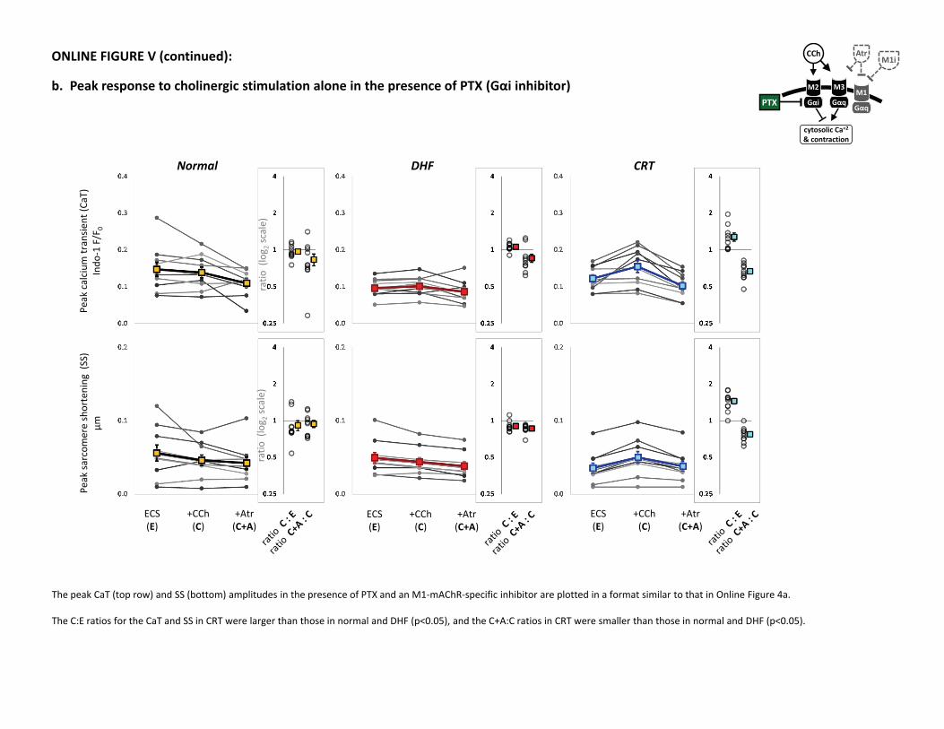
ONLINE FIGURE V (continued):
b. Peak response to cholinergic stimulation alone in the presence of PTX (Gαi inhibitor)Pe
ak calcium
transie
nt (C
aT)
Indo
‐1 F/F
0
Peak sa
rcom
ere shortening
(SS)
µm
Normal DHF CRT
ratio
(log 2
scale)
ratio
(log 2
scale)
+CCh(C)
ECS(E)
+Atr(C+A)
+CCh(C)
ECS(E)
+Atr(C+A)
+CCh(C)
ECS(E)
+Atr(C+A)
The peak CaT (top row) and SS (bottom) amplitudes in the presence of PTX and an M1‐mAChR‐specific inhibitor are plotted in a format similar to that in Online Figure 4a.
The C:E ratios for the CaT and SS in CRT were larger than those in normal and DHF (p<0.05), and the C+A:C ratios in CRT were smaller than those in normal and DHF (p<0.05).
PTX
cytosolic Ca+2& contraction
M2
Gαi
M3
Gαq
CCh Atr
M1
Gαq
M1i
Peak calcium
transie
nt (C
aT)
Indo
‐1 F/F
0
Peak sa
rcom
ere shortening
(SS)
µm
Normal DHF CRT
ratio
(log 2
scale)
ratio
(log 2
scale)
+CCh(C)
ECS(E)
+Atr(C+A)
+CCh(C)
ECS(E)
+Atr(C+A)
+CCh(C)
ECS(E)
+Atr(C+A)
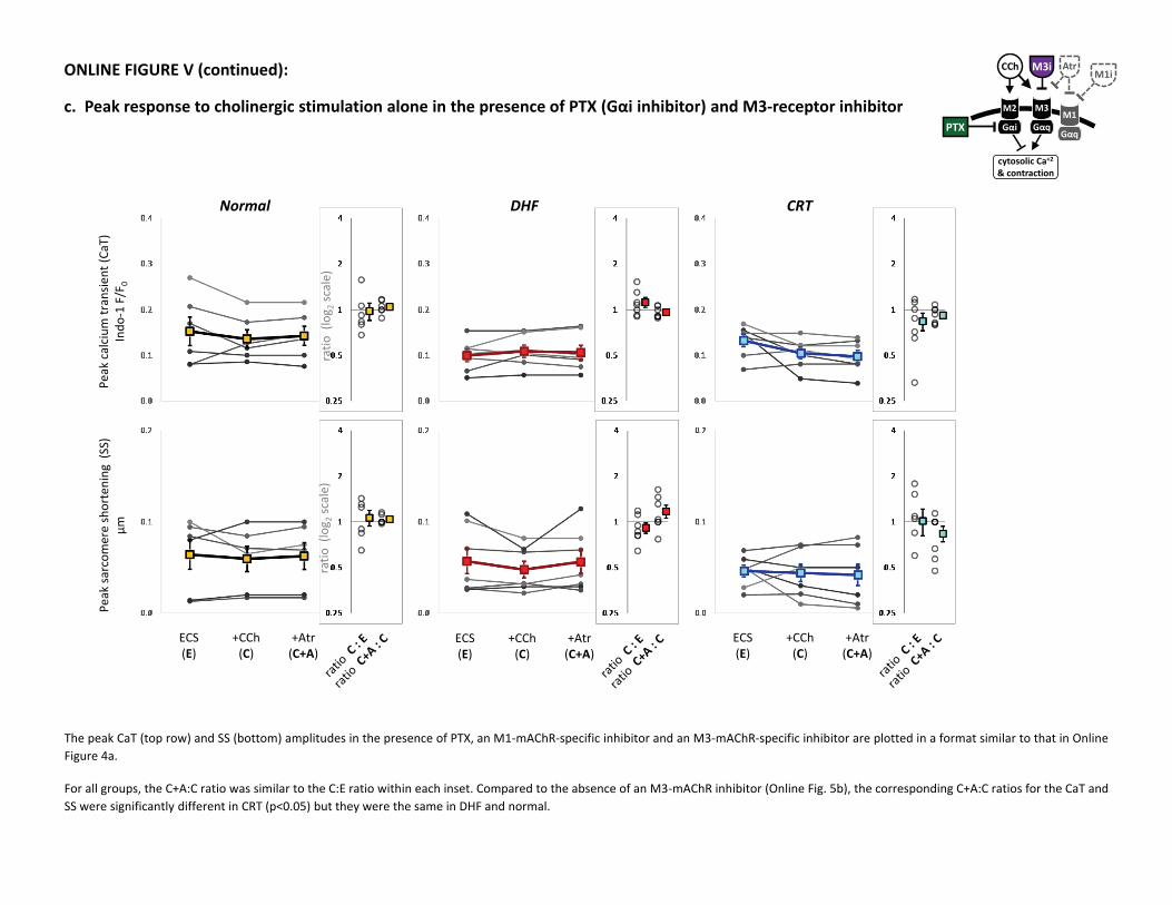
ONLINE FIGURE V (continued):
c. Peak response to cholinergic stimulation alone in the presence of PTX (Gαi inhibitor) and M3‐receptor inhibitorPTX
cytosolic Ca+2& contraction
M2
Gαi
M3
Gαq
CCh Atr
M1
Gαq
M1iM3i
The peak CaT (top row) and SS (bottom) amplitudes in the presence of PTX, an M1‐mAChR‐specific inhibitor and an M3‐mAChR‐specific inhibitor are plotted in a format similar to that in OnlineFigure 4a.
For all groups, the C+A:C ratio was similar to the C:E ratio within each inset. Compared to the absence of an M3‐mAChR inhibitor (Online Fig. 5b), the corresponding C+A:C ratios for the CaT andSS were significantly different in CRT (p<0.05) but they were the same in DHF and normal.
Related Documents